valvular heart diseases prof. mohammed arafah mb,bs facp frcpc facc
TRANSCRIPT

VALVULAR HEART Diseases
Prof. Mohammed Arafah
MB,BS FACP FRCPC FACC

ALL cardiac valves can be involved in pathological processes

EtiologyCongenital : - Bicuspid or unicuspid .
- Subvalvular or supravalvular .
Acquired : - Rheumatic .
- Degeneration .
- myxomatous
- calcification
- Ischaemic .
- Infective Endocarditis .
- Valve ring dilatation .
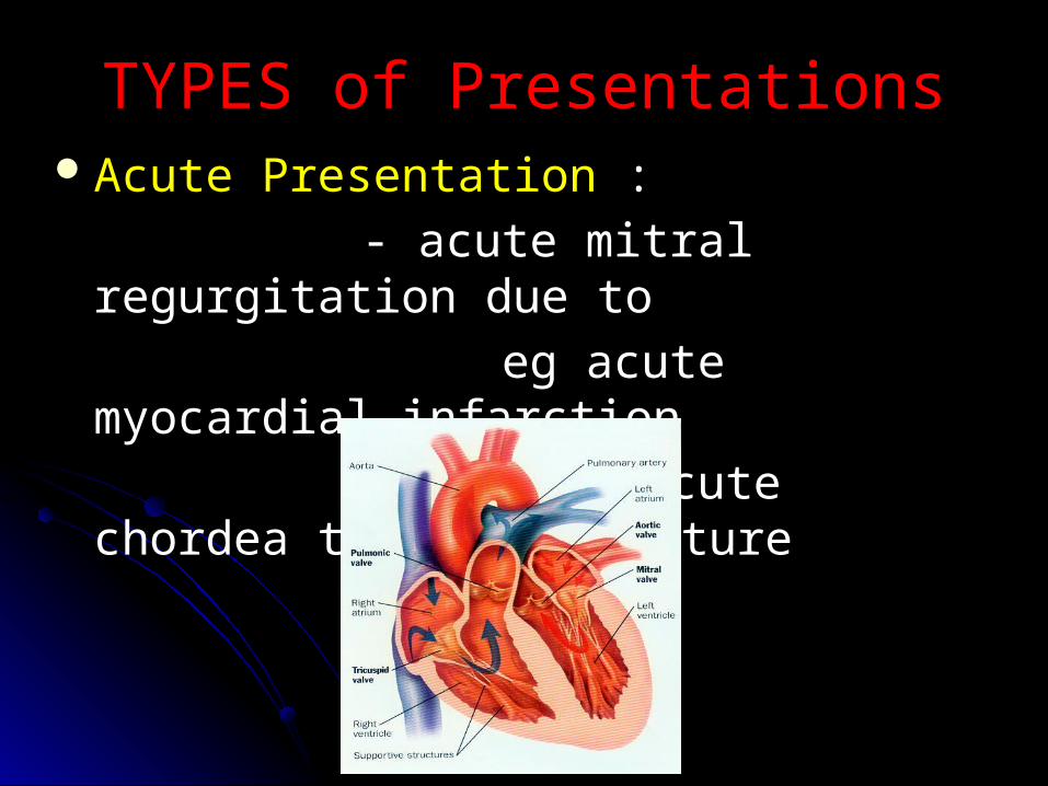
TYPES of PresentationsAcute Presentation :
- acute mitral regurgitation due to
eg acute myocardial infarction
acute chordea tendineae rupture
TYPES of Presentations
Chronic Presentation :
- chronic mitral regurgitation due to
eg RHRUMATIC fever .
Mitral valve Prolapse .
- chronic aortic regurgitation due to
eg Bicuspid Aortic valve .

HEAMODYNAMICS ConsequencesPressure Overload :
- Aortic stenosis
Left Ventricular hypertrophy
- Mitral stenosis
Left Atriarl hypertrophy & dilatation

HEAMODYNAMICS Consequences
Volume Overload :
- chronic mitral regurgitation
dilated left ventricle & left atria
- chronic tricuspid regurgitation
dilated right ventricle & right atria

SYMPTOMSDyspnea , paroxysmal nocturnal dyspnea
orthopnea .Palpitation .Chest pain .Dizziness , prefainting ,syncope .Oedema , Ascites Cough .FatigueHemoptysisSymptoms of thromboembolic complication .

SINGS of VALVULAR Diseases
Abnormal look ( mitral facies ) .Abnormal pulse ( Atrial fibrillation ) .Apex beat abnormality .Sternal or parasternal heave .Thrill .Abnormal heart sound .MURMURS .
Systolic or Diastolic .


INVESTIGATION
ECG .CXR .Echo cardiology .
M mode , 2D ,3D . 4 D . TEE .
Doppler .24 hours monitor for heart rhythm .MRI .Cardiac catheterization .

MITRAL STENOSIS

Rheumatic Fever which is related to streptococcus infections, causing damage to the mitral valve and leading to mitral stenosis later in life.
ETIOLOGY

OTHER LESS COMMON CAUSES OF MITRAL STENOSIS
Congenital Mitral Stenosis
Systemic Lupus Erythematosus
Rheumatoid Arthritis
Atrial Myxoma
Malignant Carcinoid
Bacterial Endocarditis
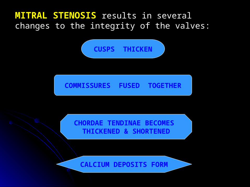
MITRAL STENOSIS results in several changes to the integrity of the valves:
CUSPS THICKEN
COMMISSURES FUSED TOGETHER
CHORDAE TENDINAE BECOMES THICKENED & SHORTENED
CALCIUM DEPOSITS FORM

PATHOPHYSIOLOGY
In normal adults the mitral valve orifice is 4-6cm².
When the orifice is reduced to approximately 2cm², which is considered mild mitral stenosis, blood can flow from the left atrium to the left ventricle only if propelled by an abnormal pressure gradient – the hemodynamic hallmark of Mitral Stenosis.
When the mitral valve opening is reduced to 1 cm² , which is considered critical mitral stenosis, a left atrioventricular pressure gradient of approximately 20mmHg is required to maintain normal cardiac output at rest.

HEAMODYNAMICS Consequences of Mitral stenosis

SIGNS & SYMPTOMS
Symptoms of mitral stenosis usually begin with the hallmark signs of DYSPNEA ON EXERTION!
The first bouts of dyspnea in patients with mitral stenosis are usually precipitated by exercise, emotional stress, infection, or atrial fibrillation, all of which increase the rate of blood flow across the mitral orifice & result in further elevation of Left

OTHER PRINCIPAL SIGNS AND SYMPTOMS INCLUDES:
FatigueOrthopneaParoxysmal nocturnal dyspneaPulmonary edema – develops when there’s a sudden ↑ in flow rate across a markedly narrowed mitral orifice.Palpitations – owing to presence of arrhythmiasHemoptysis – due to rupture of thin dilated bronchial veins. Peripheral edema .

DIAGNOSTIC EVALUATIONS Clinical evaluation of Mitral Stenosis
begins with an in-depth history and physical exam.
ON PHYSICAL EXAMINATION
a. a loud S1 – due to abrupt leaflet closure
b. a loud S2 – due to ↑ PAP; an opening snap due to tension on valve leaflet
c. Diastolic rumble – due to turbulent blood flow across the stenotic valve.


The Diagnostic testing used to evaluate the presence & severity of Mitral
Stenosis includes:
ECG
Chest Radiograph
2D Echocardiogram
Doppler Study
TransEsophageal Echocardiography


LEFT PARASTERNAL, LONG AXIS VIEW
STENOTIC MITRAL VALVE


MVA–2D ECHO = 0.9cm²

COMPLICATIONS OF MITRAL STENOSIS
ATRIAL FIBRILLATION
LUNG CONGESTION
BLOOD CLOTS with SYSTEMIC EMBOLIZATION
PULMOARY HYPERTENSION
CONGESTIVE HEART FAILURE

MEDICAL MANAGEMENT
DIURETICS
DIGITALIS
ANTI-ARRYHTHMICS
ANTICOAGULANTS
ANTIBIOTICS

InterventionPERCUTANEOUS
TRANSVENOUS MITRAL COMMISSUROTOMY (PTMC)
SURGICAL COMMISSUROTOMYMITRAL VALVE Replacement .


MVA = .982 cm²
PRE-PROCEDURE
MVA = 1.84 cm²
POST-PROCEDURE

MV MEAN GRADIENT=17mmHg
MV MEAN GRADIENT =2mmHg
PRE-INFLATION POST-INFLATION


MITRAL REGURGITATION
ETIOLOGYRHEUMATIC HEART disease .MITRAL Valve Prolapse .Others
- IHD
- Cardiomyopathy ( dilated , hypertrophic )
- Hypertensive heart disease
- infective endocarditis
- Myocarditis
- connective tissue disorders - (SLE)
- collagen abnormalities - Marfan's syndrome

SIGNS
Laterally displaced (forceful) diffuse apex beat and a systolic thrill .
Soft first heart sound . Pansystolic murmur .Prominent third heart sound .


Real time 4D color full volume imaging
Real time 4D, parasternal long axis, full volume imaging

Management of mitral regurgitation
Evidence of progressive cardiac enlargement generally warrants early surgical intervention by either mitral valve repair or replacement .
Treatment with ACE inhibitors, diuretics and possibly anticoagulants .

Mitral Valve Prolapse

Pathology
Large mitral valve leaflets, an enlarged mitral annulus, abnormally long chordae or disordered papillary muscle contraction .
Demonstrate myxomatous degeneration of the mitral valve leaflets .
Associated with Marfan's syndrome, thyrotoxicosis, rheumatic or ischaemic heart disease .

Symptoms
Atypical chest pain is the most common symptom .
Palpitations may be experienced because of the abnormal ventricular contraction or because of the atrial and ventricular arrhythmias .
Sudden cardiac death due to fatal ventricular arrhythmias is a very rare but recognized complication.

SIGNS
The most common sign is a mid-systolic click .
Produced by the sudden prolapse of the valve and the tensing of the chordae tendineae that occurs during systole .
A late systolic murmur owing to some regurgitation

Treatment
Beta-blockade is effective for the treatment of the atypical chest pain and palpitations .
Mitral valve prolapse associated with significant mitral regurgitation and atrial fibrillation, anticoagulation is advised to prevent thromboembolism .
Mitral valve prolapse associated with severe mitral regurgitation has a risk of sudden cardiac death.

AORTIC STENOSIS


Treatment
In patients with aortic stenosis, symptoms are a good index of severity and all symptomatic patients should have aortic valve replacement.
Asymptomatic patients should be under regular review for assessment of symptoms and echocardiography .

AORTIC REGURGITATION

Acute aortic regurgitation
Acute rheumatic fever Infective endocarditis Dissection of the aorta Ruptured sinus of Valsalva aneurysm Failure of prosthetic heart valve

Chronic aortic regurgitation
Rheumatic heart disease Syphilis Arthritides:
Reiter's syndrome Ankylosing spondylitis Rheumatoid arthritis
Hypertension (severe) Bicuspid aortic valve Aortic endocarditis Marfan's syndrome Osteogenesis imperfecta


Treatment : Aortic valve replacement
Because symptoms do not develop until the myocardium fails and because the myocardium does not recover fully after surgery, operation is performed before significant symptoms occur.
The timing of the operation is best determined according to haemodynamic, echocardiographic or angiographic criteria

PULMONIC Valve Diseases
PULMONIC Valve stenosisPULMONIC Valve Rergurgitation

TRICUSPID Valve Diseases
TRICUSPID Valve RegurgitationTRICUSPID Valve stenosis
