uwo nanofabrication facility and science studio. facility to be hooked into science studio: western...
TRANSCRIPT

cience Studio
UWO Nanofabrication Facility
andScience Studio

Facility to be hooked into Science Studio:
Western Nanofabrication Facility, University of Western Ontario
University Of Western Ontario
Canadian Light Source
Science Studio
Science StudioElectron Microscope
Beamline
Science Studio Directory Service
Science Studio Meta Data
Science Studio
CLS Active Data
Science Studio
Service Bus
Beamline 1
CLS Device Operator
UWO Active Data
Service Bus
EM Control & Data Processing into
Simulated Microscope
UWO Electron Microscope
Operator
External Facility
External User
internet
EM Control & Data Processing

Device: Zeiss 1540XB FIB/SEM
Technique: Scanning Electron Microscopy (SEM) combined with Energy Dispersive X-ray (EDX) Analysis
A highly focussed electron (primary) beam is scanned across a sample surface at an energy between 0.5-30 keV. Low energy secondary electrons are generated as a result. An image of the sample surface is constructed by measuring secondary electron intensity as a function of the primary electron beam position on the sample.
Back scattered electrons and x-rays are also emitted from the bombardment of the sample surface by primary electrons. The intensity of the backscattered electrons is related to the atomic number of the element present and provides element contrast.
X-rays emitted are characteristic of elements present in the sample. Quantitative elemental information can be obtained. Elemental maps can be generated.

◊ The Scanning Electron Microscope is controlled by a computer system. ◊ The system is also fitted with an Oxford Instruments x-ray system allowing for elemental mapping and analysis of the sample.
◊ The x-ray system is controlled by a separate computer. This system is connected to the microscope and detector, and takes control of the microscope and detector in order to collect the emitted x-rays during elemental mapping and analysis of the sample.
Incorporation into Science Studio
◊ The Oxford Instruments x-ray system is proprietary. Incorporating the microscope/x-ray system into Science Studio would be done by means of ‘wrapping’, i.e., creating an interface between Science Studio and the system.
A basic screen capture interface (terminal window) can be included in ScienceStudio to enable a user to operate the Oxford Instruments software. The terminalwindow can be provided within the main SS window.
Sample Analysis Data
► Scanning electron micrographs (secondary electron images)► Backscattered electron images► EDX spectra and elemental maps
Resulting data from sample analyses are incorporated into a Word document report. Data can also be exported in other formats.
Data will be accessible for download from Science Studio.

Data is organized under the context of project, similar to our scheme in Science Studio

Software is user friendly.
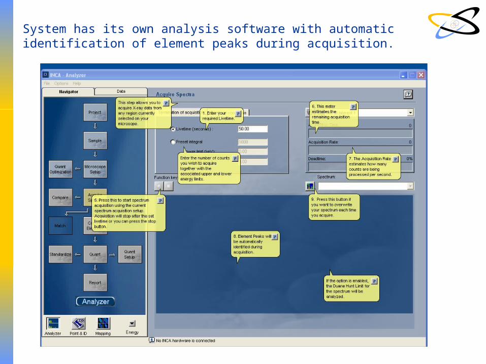
System has its own analysis software with automatic identification of element peaks during acquisition.