uva-dare (digital academic repository) insights into ... · cleaning efficacy of passive ultrasonic...
TRANSCRIPT
UvA-DARE is a service provided by the library of the University of Amsterdam (http://dare.uva.nl)
UvA-DARE (Digital Academic Repository)
Insights into passive ultrasonic irrigation
Jiang, L.M.
Link to publication
Citation for published version (APA):Jiang, L. (2012). Insights into passive ultrasonic irrigation
General rightsIt is not permitted to download or to forward/distribute the text or part of it without the consent of the author(s) and/or copyright holder(s),other than for strictly personal, individual use, unless the work is under an open content license (like Creative Commons).
Disclaimer/Complaints regulationsIf you believe that digital publication of certain material infringes any of your rights or (privacy) interests, please let the Library know, statingyour reasons. In case of a legitimate complaint, the Library will make the material inaccessible and/or remove it from the website. Please Askthe Library: http://uba.uva.nl/en/contact, or a letter to: Library of the University of Amsterdam, Secretariat, Singel 425, 1012 WP Amsterdam,The Netherlands. You will be contacted as soon as possible.
Download date: 06 Jul 2018
43
5
The influence of the ultrasonic
intensity on the cleaning efficacy of
passive ultrasonic irrigation
This chapter has been published as: Jiang LM, Verhaagen B, Versluis M, Langedijk J, Wesselink P, van der Sluis LW. The influence of the ultrasonic intensity on the cleaning efficacy of passive ultrasonic irrigation. J Endod. 2011 May;36(5):688-692
Chapter 5
44
Abstract
Introduction: It is not clear that whether increasing of the ultrasonic intensity would enhance the cleaning efficacy of Passive Ultrasonic Irrigation (PUI) inside a root canal. The aim of this study was to
evaluate the effect of the ultrasonic intensity on PUI to remove dentine debris and whether there is any
lateral effect beyond the ultrasonic tip. Methods: Each of fifteen in vitro root canal models with four
standard depressions in the apical part of one canal wall were filled with dentin debris and received
PUI repeatedly. The most apical depression was localized apically from the ultrasonic tip. The highest
intensity was applied in group 1, the lowest in group 3 and syringe irrigation was performed in group 4,
as a control. Before and after irrigation, images of the canal wall with depressions were taken and
compared. The removal of dentine debris in the depressions was categorized as clean or not clean. The
data were analyzed by means of the chi-square test. The oscillation amplitude of the ultrasonic file at
each intensity was recorded in vitro using time-resolved high-speed imaging. Results: Group 1
(highest intensity) exhibited significantly better cleaning than all the other groups (p<0.05); no
significant difference was found between the four levels of the depressions within any of the four
groups. High-speed imaging showed that the amplitude of the oscillating file increased as the intensity
went up, which leads to a higher velocity of the irrigant around the file. Conclusion: Higher ultrasonic
intensity resulted in a higher amplitude of the oscillating file, and consequently enhanced the cleaning
efficacy of PUI.

Ultrasonic intensity and PUI
45
Introduction
The improvement of currently available irrigation protocols to disinfect the inaccessible regions of the
root canal space and the uninstrumented canal surfaces is important (1) due to insufficient debridement by instrumentation alone (2, 3). An ultrasonically oscillating file transmits energy, causing acoustic
microstreaming, which results in mixing of the irrigant, enabling it to reach those inaccessible regions
and enhancing shear stress on the root canal surfaces at a distance from the file (4-7).
The apical third of the root canal system is particularly difficult to clean because of the
typically challenging complexity of the root canal morphology (8, 9), making irrigant delivery and
activation less effective (10). Lee et al. (11) studied the efficiency of ultrasonic irrigation using root
canal models with grooves and depressions in the apical portion of the root canal wall to simulate root
canal irregularities. These studies indicated that the depressions in the root canal wall were more
difficult to clean than the grooves (11, 12), and also revealed a lateral effect of the acoustic streaming
around the file. As an ultrasonic file does not always extend to the full length of the root canal and the
cleaning of the apical area close to the apex could be critical (13), information about the cleaning
efficacy in front of the tip could be valuable.
The intensity of ultrasonic activation, adjusted by the power setting on the ultrasonic device,
influences the energy transmission from the ultrasonically oscillating file to the irrigant. Previous
research showed that an increasing intensity did not always result in a linear increase of the
displacement amplitude of the oscillating file (14, 15). As those observations investigated the
oscillation of the file in free air, a direct relationship with acoustic microstreaming around and in front
of the file could not be established. A full understanding of the effect of intensity on the cleaning efficacy of Passive Ultrasonic Iirrigation (PUI) through microstreaming is still lacking.
The aims of this study were (1) to measure the displacement amplitude of the oscillating file
under different ultrasonic intensities by using high-speed imaging, (2) to evaluate the effect of
ultrasonic intensity on the cleaning efficacy of PUI, and (3) to investigate the lateral effect beyond the
file tip.
Materials and Methods
Dentin debris removal model Straight roots from 15 extracted human maxillary canines were decoronated to obtain uniform root
sections of 15 mm, following the protocol as described previously (16, 17). Briefly, the roots were
embedded in resin and bisected longitudinally. The surfaces of both halves were then ground to leave
only little of the original root canal lumen. Four depressions were drilled in the resin part and the two halves were reassembled by four self-tapping bolts through the depressions. All the models were
checked if there was any leakage of liquid or gas apically or laterally before experiments. If there was
any, rubber dam caulk would be applied to ensure that the root canal modeled a closed system.
Standardized root canals were established by K-flexofiles #15/.02 (Dentsply Maillefer,
Ballaigues, Switzerland) and GT (Dentsply, Maillefer) Ni-Ti rotary instruments to a working length
(WL) of 15 mm, ISO size 30 and taper 0.06. The final apical enlargement was done with the Mtwo
(VDW, Munich, Germany) Ni-Ti rotary instrument #35/.04. During instrumentation, the canals were
rinsed with 1 mL of 2% NaOCl after each file, delivered by a 10 mL syringe (Terumo, Leuven,
Belgium) and a 30-gauge needle (Navitip, Ultradent, South Jordan, UT, USA).
With the help of a microscope and a round bur H71.104.003 (Komet, Lemgo Germany)
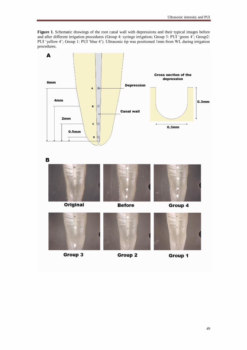
attached to a drilling machine, four standard depressions (Ø=0.3 mm), located at 0.5, 2, 4 and 6 mm
from WL were drilled in the wall of one half of each root canal (Fig.1).
Each depression was filled with dentin debris, which was mixed with 2% NaOCl for five
minutes, to simulate a situation in which dentin debris accumulates in uninstrumented canal extensions
(11). This model was introduced to standardize the root canal space and the amount of dentin debris
present in the root canal before the irrigation procedure, to increase the reliability of the dentin debris
removal evaluation. The methodology is sensitive and the data are reproducible (18). The 15 models were used repeatedly in the four experimental groups which are shown in Table 1.
Irrigation Procedure Specimens in all the experimental groups were rinsed with 2 mL irrigant (2% NaOCl) using 10 mL
syringes with 30-gauge needles (Navitip) placed 1 mm from WL, and the flow rate was approximately
5 mL/min. Then PUI was performed with a stainless steel #25/.00 file (IrriSafe, Acteon, Merignac,
France) driven by an ultrasonic device (Suprasson PMax; Satelec Acteon, Merignac, France) for 10

Chapter 5
46
seconds with the in-plane oscillation direction toward the depressions. The device has four ultrasound
intensity ranges, indicated by different colors, green, yellow, blue and red. These colors correspond to
an ultrasound intensity range, from the weakest to the strongest, each adjustable with 10 levels.
Different intensity settings were used in this study: „blue 4‟ (group 1), „yellow 4‟ (group 2) and „green
4‟ (group 3). Every attempt was made to keep the file centered in the canal to minimize contact with
the canal walls, as any contact with the canal wall could dampen the oscillatory motion of the file.
Group 4 acted as the control group, in which the ultrasonic file was inserted but not activated. All the
experimental specimens received 2 mL irrigant which was delivered again by syringe as final flush.
Image evaluation and statistical analyses Before and after each irrigation procedure, the root halves were separated and the depressions were
viewed through a stereomicroscope (Stemi®
SV6, Carl Zeiss, Göttingen, Germany) using a cold light
source (KL 2500 LCD, Carl Zeiss). Pictures were taken with a digital camera (Axio Cam, Carl Zeiss).
The sequence of all the pictures was randomized, and two calibrated examiners were blind to the group
assignment.
The debris removal from each depression after irrigation was scored independently by two
calibrated dentists. The samples were graded as ”clean” if the depression was without any debris left, or
“not clean” if the depression was not completely clean. The percentage of inter agreement should be
more than 95%; if this percentage was lower than 95%, a consensus had to be reached.
The differences in debris removal between the depressions within each group and between the
groups were analyzed by Chi-square analysis. The level of significance was set at α = 0.05.
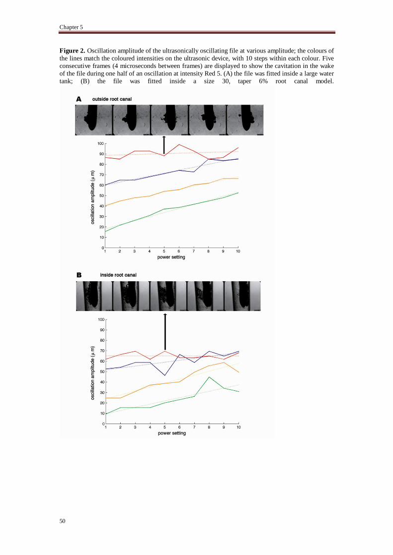
High speed imaging experiments High-speed imaging was used to record the oscillation amplitude of the ultrasonic file at each intensity,
both inside a large water-filled tank and inside a transparent, silicon model of a root canal of apical size
30 and taper 6%. The silicon model was made by casting Poly-DiMethyl-Siloxane (PDMS) (Sylgard 184, Silicone Elastomer Kit, Dow-Corning, Coventry, UK) around a D-size hand spreader (Dentsply).
Recordings were made with a high-speed camera (HPV-1, Shimadzu Corp., Kyoto, Japan) at a frame
rate of 250,000 fps. A microscope with 20x magnification (BX-FM, Olympus, Tokyo, Japan) was used
for magnification. The Irrisafe #25/.00 file was fitted in front of the microscope, illuminated in bright-
field mode by a continuous wave light source (ILP-1, Olympus, Japan). By tracking the tip of the file in
each of the recordings, the displacement amplitude was determined.
Results
At each depression level, dentin debris removal was significantly different between the experimental
groups. The number of completely clean samples in each group and the p values when compared
between groups are shown in Table 1. The value of kappa is statistically significantly different from 0,
and its value of 0.828 suggests that the evaluators' scorings are mostly similar, with some exceptions.
If the number of completely clean depressions at all four levels were combined, all the ultrasonically activated groups removed more dentin debris than the non-activated group (Group 4)
(p<.05); group 1 with the highest intensity, exhibited significantly better cleaning efficacy than all the
other groups; no significant difference was seen between the other two groups with lower intensities.
There was no significant difference between the four levels of the depressions within any of
the four groups.
From the high-speed recordings the displacement amplitude at each intensity was determined
(Fig 2). Increasing the intensity, resulted in a linear increase in oscillation amplitude, although there is
some overlap between color codes. The confinement of the root canal caused a reduction in the
oscillation amplitude of approximately 40%.
In the large water tank, cavitation was observed on the file tip for the intensities above Blue 5.
Inside the confinement of the root canal this threshold was Green 5. The bubble grows in the wake of
the file when it moves from one maximum of displacement to the other and collapses when the file
reaches the other maximum displacement.
Discussion
The model with depressions on the root canal wall used in this study was a modification of a previous
one (11). The modifications and advantages of the current model are threefold. Firstly, since the natural
root canal varies considerably, the removal of the original root canal space allowed for the
establishment of standardized root canals. Secondly, it is easy to assemble or disassemble the model,
Ultrasonic intensity and PUI
47
facilitating the root canal treatment procedure in vitro within the enclosed system and the evaluation of
the interested area before and after the irrigation. Finally, the cleaning of the extra depression located at
0.5 mm from the WL, could give an indication of the cleaning efficacy in front of the ultrasonically
oscillating file tip.
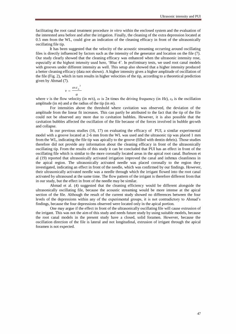
It has been suggested that the velocity of the acoustic streaming occurring around oscillating
files is directly influenced by factors such as the intensity of the generator and location on the file (7).
Our study clearly showed that the cleaning efficacy was enhanced when the ultrasonic intensity rose,
especially at the highest intensity used here, „Blue 4‟. In preliminary tests, we used root canal models
with grooves under different intensity as well. This setup also showed that a higher intensity produced a better cleaning efficacy (data not shown). A higher intensity gives a higher amplitude of oscillation of
the file (Fig. 2), which in turn results in higher velocities of the tip, according to a theoretical prediction
given by Ahmad (7). 2
0v
a
where v is the flow velocity (in m/s), ω is 2π times the driving frequency (in Hz), ε0 is the oscillation
amplitude (in m) and a the radius of the tip (in m).
For intensities above the threshold where cavitation was observed, the deviation of the
amplitude from the linear fit increases. This can partly be attributed to the fact that the tip of the file
could not be observed any more due to cavitation bubbles. However, it is also possible that the
cavitation bubbles affected the oscillation of the file because of the forces involved in bubble growth
and collapse. In our previous studies (16, 17) on evaluating the efficacy of PUI, a similar experimental
model with a groove located at 2-6 mm from the WL was used and the ultrasonic tip was placed 1 mm
from the WL, indicating the file tip was apically to the groove (filled with dentin debris). Those studies
therefore did not provide any information about the cleaning efficacy in front of the ultrasonically
oscillating tip. From the results of this study it can be concluded that PUI has an effect in front of the
oscillating file which is similar to the more coronally located areas in the apical root canal. Burleson et
al (19) reported that ultrasonically activated irrigation improved the canal and isthmus cleanliness in
the apical region. The ultrasonically activated needle was placed coronally to the region they
investigated, indicating an effect in front of the needle, which was confirmed by our findings. However,
their ultrasonically activated needle was a needle through which the irrigant flowed into the root canal
activated by ultrasound at the same time. The flow pattern of the irrigant is therefore different from that
in our study, but the effect in front of the needle may be similar.
Ahmad et al. (4) suggested that the cleaning efficiency would be different alongside the
ultrasonically oscillating file, because the acoustic streaming would be more intense at the apical
section of the file. Although the result of the current study showed no differences between the four
levels of the depressions within any of the experimental groups, it is not contradictory to Ahmad‟s
findings, because the four depressions observed were located only in the apical portion.
One may argue if the effect in front of the ultrasonically oscillating file will cause extrusion of the irrigant. This was not the aim of this study and needs future study by using suitable models, because
the root canal models in the present study have a closed, solid foramen. However, because the
oscillation direction of the file is lateral and not longitudinal, extrusion of irrigant through the apical
foramen is not expected.
Chapter 5
48
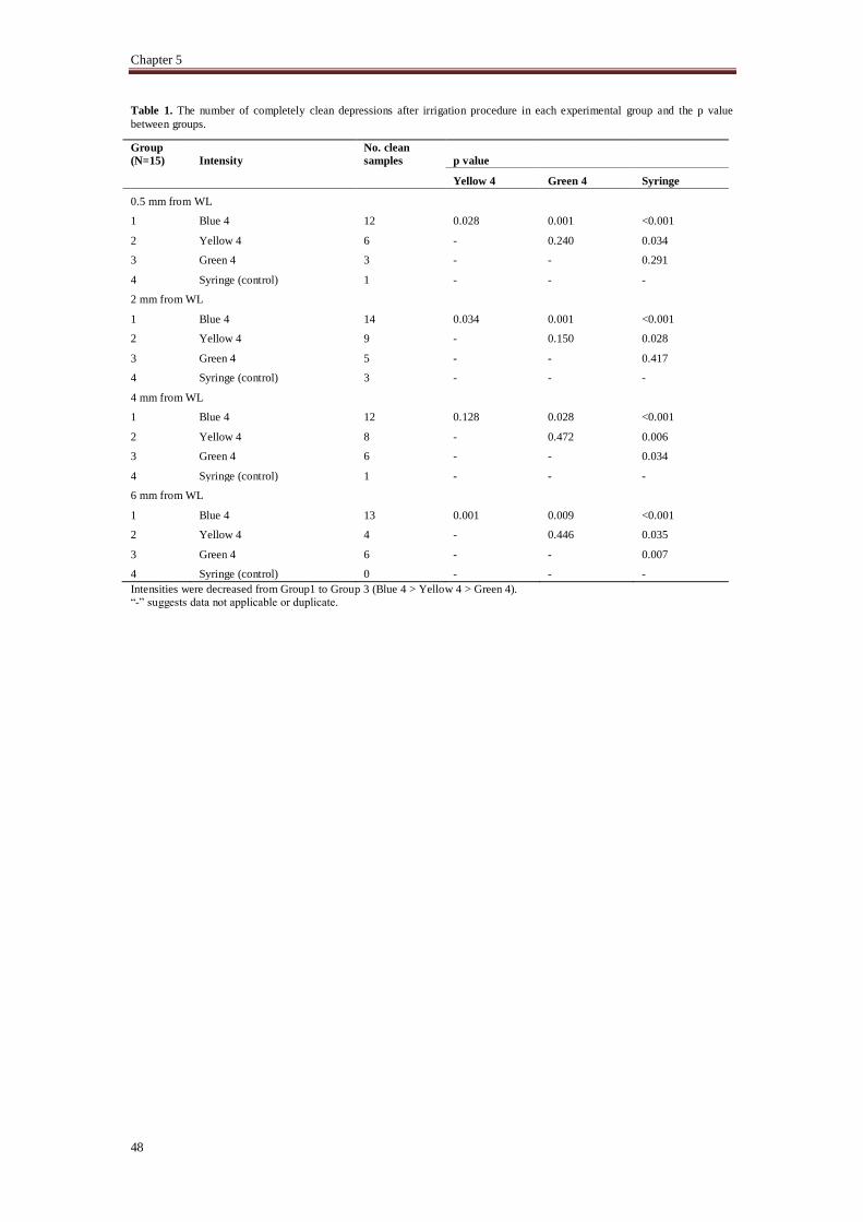
Table 1. The number of completely clean depressions after irrigation procedure in each experimental group and the p value
between groups.
Group
(N=15) Intensity
No. clean
samples p value
Yellow 4 Green 4 Syringe
0.5 mm from WL
1 Blue 4 12 0.028 0.001 <0.001
2 Yellow 4 6 - 0.240 0.034
3 Green 4 3 - - 0.291
4 Syringe (control)
1 - - -
2 mm from WL
1 Blue 4 14 0.034 0.001 <0.001
2 Yellow 4 9 - 0.150 0.028
3 Green 4 5 - - 0.417
4 Syringe (control)
3 - - -
4 mm from WL
1 Blue 4 12 0.128 0.028 <0.001
2 Yellow 4 8 - 0.472 0.006
3 Green 4 6 - - 0.034
4 Syringe (control)
1 - - -
6 mm from WL
1 Blue 4 13 0.001 0.009 <0.001
2 Yellow 4 4 - 0.446 0.035
3 Green 4 6 - - 0.007
4 Syringe (control)
0 - - -
Intensities were decreased from Group1 to Group 3 (Blue 4 > Yellow 4 > Green 4).
“-” suggests data not applicable or duplicate.
Ultrasonic intensity and PUI
49
Figure 1. Schematic drawings of the root canal wall with depressions and their typical images before
and after different irrigation procedures (Group 4: syringe irrigation; Group 3: PUI „green 4‟; Group2:
PUI „yellow 4‟; Group 1: PUI „blue 4‟). Ultrasonic tip was positioned 1mm from WL during irrigation
procedures.
Chapter 5
50
Figure 2. Oscillation amplitude of the ultrasonically oscillating file at various amplitude; the colours of
the lines match the coloured intensities on the ultrasonic device, with 10 steps within each colour. Five
consecutive frames (4 microseconds between frames) are displayed to show the cavitation in the wake
of the file during one half of an oscillation at intensity Red 5. (A) the file was fitted inside a large water
tank; (B) the file was fitted inside a size 30, taper 6% root canal model.
Ultrasonic intensity and PUI
51
Reference
1. Gomes-Filho JE, Aurelio KG, Costa MM, Bernabe PF. Comparison of the biocompatibility of
different root canal irrigants. J Appl Oral Sci 2008;16:137-44. 2. Peters OA, Schonenberger K, Laib A. Effects of four Ni-Ti preparation techniques on root canal
geometry assessed by micro computed tomography. Int Endod J 2001;34:221-30.
3. Wu MK, van der Sluis LW, Wesselink PR. The capability of two hand instrumentation techniques
to remove the inner layer of dentine in oval canals. Int Endod J 2003;36:218-24.
4. Al-Jadaa A, Paque F, Attin T, Zehnder M. Acoustic hypochlorite activation in simulated curved
canals. J Endod 2009;35:1408-11.
5. Al-Jadaa A, Paque F, Attin T, Zehnder M. Necrotic pulp tissue dissolution by passive ultrasonic
irrigation in simulated accessory canals: impact of canal location and angulation. Int Endod J
2009;42:59-65.
6. de Gregorio C, Estevez R, Cisneros R, Heilborn C, Cohenca N. Effect of EDTA, sonic, and
ultrasonic activation on the penetration of sodium hypochlorite into simulated lateral canals: an in
vitro study. J Endod 2009;35:891-5.
7. Ahmad M, Pitt Ford TR, Crum LA. Ultrasonic debridement of root canals: an insight into the
mechanisms involved. J Endod 1987;13:93-101.
8. Pineda F, Kuttler Y. Mesiodistal and buccolingual roentgenographic investigation of 7,275 root
canals. Oral surgery, oral medicine, and oral pathology 1972;33:101-10.
9. Vertucci FJ. Root canal anatomy of the human permanent teeth. Oral Surg Oral Med Oral Pathol
Oral Radiol Endod 1984;58:589-99. 10. Foschi F, Nucci C, Montebugnoli L, Marchionni S, Breschi L, Malagnino VA, et al. SEM
evaluation of canal wall dentine following use of Mtwo and ProTaper NiTi rotary instruments. Int
Endod J 2004;37:832-9.
11. Lee SJ, Wu MK, Wesselink PR. The effectiveness of syringe irrigation and ultrasonics to remove
debris from simulated irregularities within prepared root canal walls. Int Endod J 2004;37:672-8.
12. Lee SJ, Wu MK, Wesselink PR. The efficacy of ultrasonic irrigation to remove artificially placed
dentine debris from different-sized simulated plastic root canals. Int Endod J 2004;37:607-12.
13. Nair PN, Henry S, Cano V, Vera J. Microbial status of apical root canal system of human
mandibular first molars with primary apical periodontitis after "one-visit" endodontic treatment.
Oral Surg Oral Med Oral Pathol Oral Radiol Endod 2005;99:231-52.
14. Walmsley AD, Williams AR. Effects of constraint on the oscillatory pattern of endosonic files. J
Endod 1989;15:189-94.
15. Lea SC, Walmsley AD, Lumley PJ, Landini G. A new insight into the oscillation characteristics
of endosonic files used in dentistry. Phys Med Biol 2004;49:2095-102.
16. Jiang LM, Verhaagen B, Versluis M, van der Sluis LW. Influence of the oscillation direction of an
ultrasonic file on the cleaning efficacy of passive ultrasonic irrigation. J Endod 2010;36:1372-6.
17. Jiang LM, Verhaagen B, Versluis M, van der Sluis LW. Evaluation of a sonic device designed to activate irrigant in the root canal. J Endod 2010;36:143-6.
18. van der Sluis LW, Wu MK, Wesselink PR. The evaluation of removal of calcium hydroxide paste
from an artificial standardized groove in the apical root canal using different irrigation
methodologies. Int Endod J 2007;40:52-7.
19. Burleson A, Nusstein J, Reader A, Beck M. The in vivo evaluation of hand/rotary/ultrasound
instrumentation in necrotic, human mandibular molars. J Endod 2007;33:782-7.