uterine gland microstructure in the pregnant and the non … · uterine gland microstructure in the...
TRANSCRIPT
771
Int. J. Morphol.,31(2):771-776, 2013.
Uterine Gland Microstructure in the Pregnant and theNon-pregnant Lesser Galago (Galago senegalensis)
Microestructura de la Glándula Uterina en la Gálago Menor(Galago senegalensis) Preñada y No Preñada
Allan Njogu*; George Otiang’a Owiti*,** & Dominic Oduor-Okelo*
NJOGU, A.; OWITI, G. O. & ODUOR-OKELO, D. Uterine gland microstructure in the pregnant and the non- pregnant lesser galago(Galago senegalensis). Int. J. Morphol., 31(2):771-776, 2013.
SUMMARY: The histology and the ultrastructure of the uterine glands of the lesser bushbaby (Galago senegalensis) were studied insix specimens (5 pregnant and one non pregnant) which were fixed partly with bouin’s fixative and part with 2.5% glutaraldehyde in 0.1 Mcacodylate buffer. An overview of the main results revealed uterine glands in the non-pregnant uterus are rudimentary and scarce within themucosa. In early pregnancy (first trimester) the uterine glands profiles appear in clusters. In late stage pregnancy (third trimester) the uterinegland profiles appear opposite chorionic vesicles. In the later stages of gestation maternal glandular epithelium consisted mainly of simplecolumnar epithelium. The cells had abundant flattened cisternae of granular endoplasmic reticulum usually with an apical-basal orientation.Their nuclei had abundant euchromatin relative to the amount of heterochromatin. They also had a prominent Golgi apparatus quite characteristicof protein synthesizing cells. The basal plasmalemma was thrown into infoldings that have the effect of increasing the surface area acrosswhich nutrients could pass from the maternal circulation and are secreted by the cell as histiotrophe. Both physiologic hypertrophy andhyperplasia of the uterine glands are observed to occur with advancement of pregnancy.
KEY WORDS: Lesser bushbaby; Uterine gland; Microstructure.
INTRODUCTION
Gestation, in viviparous mammals, is the period ofembryo and fetus development from the fusion of the femaleand male gametes (conception) to birth.
Histiotrophe is the nutritional material in spacesbetween the maternal and fetal tissues and is derived from thematernal tubal secretions, endometrium and the uterine glands.This nutritional material is absorbed by phagocytosis initiallyby blastocyst trophectoderm (Latham et al., 1999) and thenby trophoblast of the placenta (Bevilacqua et al., 2010). Inlater placental development nutrition is by the exchange ofblood-borne materials between the maternal and fetalcirculations (hemotrophic nutrition) (Beck 1976). Histiotropherepresents an important source of nutrients for the developingembryo, as well as containing a variety of growth factors duringthe first trimester of pregnancy in the humans (Burton et al.,2002; Hempstock et al., 2004; Burton et al., 2007; Jones etal., 2010).
In the lower primates the trophoblast of the placentalvilli is engaged in both hemotrophic and histiotrophic
nourishment of the fetus. Both histiotrophic and hemotrophicnutrition morphologically appear side by side throughoutpregnancy. The uterine glands in the galago, open for the mostpart in groups, their openings being on depressed areas of themucosa opposite chorionic vesicles which are speciallymodified absorptive areas of the chorion termed the chorionicvesicles. Chorionic vesicles are invaginations of the chorionopposite the mouths of the uterine glands (King, 1984; Njoguet al., 2006; Wooding & Burton, 2008).
This report describes the ultrastructural detail of thestructures involved in the one type of fetal nourishment(histiotrophic) seen in this species of primates i.e the uterinegland epithelium.
MATERIAL AND METHOD
Bush babies belong to the order Primate and the sub order;Prosimii (lower primates). A total of six female lesser bush
* Department of Veterinary Anatomy and Physiology, University of Nairobi, Nairobi, Kenya.** Principal, Kenya Wildlife Service Training Institute, Naivasha, Kenya.

772
babies were used in this study. Fixation of tissues was doneboth by injection and immersion to achieve good andthorough fixation especially for the early conceptus andplacenta. Tissues from the later stage pregnancy were fixedfor transmission electron microscopy while the non-pregnantand the early pregnancy uteri were fixed for histology.Fixatives used were either Bouin's or glutaraldehyde fixative(depending on whether the tissues were to be processedfurther for light or electron microscopy). 2.5%glutaraldehyde mixture in 0.1M cacodylate buffer (pH=7.2)was used for electron microscopy whereas other tissuesamples were fixed in Bouin's for routine histology.
The Bouin's solution fixed tissues were processed forroutine histological sections. This involved dehydrationthrough ascending concentrations of ethanol (50%, 70%,90% and 100%), clearing using methyl benzoate andinfiltration and embedding in molten wax. The embeddedtissues were then mounted on wooden blocks and 5µm thicksections cut with a sliding microtome. The sections obtainedwere subsequently stained with haematoxylin and eosin andexamined for light microscopy studies.
Tissues fixed in 2.5% glutaraldehyde in 0.1 Mcacodylate buffer were subsequently diced, washed in 0.1Mcacodylate buffer, post fixed in 2% osmium tetroxide for 2hours at room temperature, dehydrated through ascendingconcentrations of ethanol (50% for 15 minutes, 2 changesof 70% for 10 minutes each, 2 changes of 80% each for 15minutes, 2 changes of 95% each for 15 minutes and finally2 changes of 100% each for 30 minutes) and embedded inepon resin. Semi-thin sections were cut with glass kniveson a Sorvall MT-1 ̀ Porter Blum' microtome and stained withmethylene blue or toluidine blue stain and examined withthe light microscope for the purpose of tissue selection andorientation. Ultra-thin sections were cut with diamond knife(Diatomeâ) using an Ultra Reichert microtome. These weresubsequently mounted on copper grids and double stainedwith uranyl acetate followed with lead citrate and examinedwith a Zeiss EM microscope.
RESULTS
The non-pregnant uterus is bicornuate with a smalluterine body, about 1 centimeter in length. This is unlikethat of higher primates which have a uterus simplex. Theuterus is suspended on either side by an extensive andtransparent mesometrium. The uterine wall is composed ofan endometrium with numerous longitudinal folds, amyometrium and a perimetrium (Fig. 1). The folds are linedby a pseudo-stratified type of epithelium (Fig. 2).
Histologically the uterus exhibits the typical 3 layeredstructure (i.e. the endometrium, the myometrium and theperimetrium that is characteristic of other mammals. Theendometrium in the non-pregnant uterus has scarce andatrophied uterine glands (Fig. 2), some of which havecollapsed lumen. The uterine gland cells in the non-pregnantuterus had scanty cytoplasm and there cell sizes were about10 micrometers in size (Fig. 2).
The earlier stage of development obtained isrepresented by figure 3. The pregnant uterine horn isrecognizable by a single locular swelling. The blastocyst iscentrally implanted and the depth of implantation is super-ficial with the uterine epithelium remaining intact. Thetrophoblasts form a simple cuboidal to columnar epithelium(Fig. 3). The trophoblast and uterine epithelium are apposedto each other forming an epithliochorial placenta.
Fig. 2 A photomicrograph of the uterine endometrium. Note thefolds with a pseudostratified epithelium and a uterine gland with acollapsed lumen (arrow). X 400. H-E stain.
Fig. 1. A photomicrograph of a cross section of the non-pregnantuterine wall. Note the endometrium (e) with longitudinal folds, amyometrium (m) and a perimetrium (arrow). X 100. H-E stain.
NJOGU, A.; OWITI, G. O. & ODUOR-OKELO, D. Uterine gland microstructure in the pregnant and the non- pregnant lesser galago (Galago senegalensis). Int. J. Morphol., 31(2):771-776, 2013.

773
In the later stage pregnancy (third trimester) specimen,the glands undergo hypertrophy and hyperplasia and are foundopposite foetal membrane structures known as chorionicvesicles (Fig. 4). Tubular uterine glands appear in cross sectionon semi thin sections (Fig. 5). Uterine glandular epitheliumconsists of cuboidal or columnar epithelial cells. These haveabundant flattened cisternae of granular endoplasmicreticulum. Their nuclei have abundant euchromatin relativeto the amount of heterochromatin. They also have a prominentGolgi apparatus, moderate numbers of secretory granules andmultivesicular bodies. Both elongate and circularmitochondria are observed interspersed between longcisternae of granular endoplasmic reticulum (Figs. 6, 7 and8). Their apical membranes are modified into numerous small
Fig. 3. A photomicrograph showing a section of the earliest stageof development. te – trophoblast epithelium, arrows – uterine glandprofiles. X 200. H-E stain.
Fig. 4. An illustration of the spatial organization of the uterine glandsopposite the chorionic vesicles. 1. Trophoblast epithelium, 2.Chorionic vesicle epithelium, 3. Contact membrane; formed by thetrophoblast epithelium and the Uterine epithelium (epitheliochorialplacenta), 4. Uterine gland epithelium, 5. Uterine epithelium.
Fig. 6. An electron micrograph of maternal glandular epithelial cells(ep). Note the abundant flattened cisternae of granular endoplasmicreticulum (rer) and the numerous apical villi (solid arrow). Note thetight junctions between neighboring cells (dashed arrow) X 2500.
Fig. 5. A photomicrograph of the profiles of the uterine glands(ug). Methylene blue stain.
Fig. 7 An electron micrograph showing the structural detailsof the nucleus (nu) and the apical and supranuclear cytoplasmof a maternal glandular epithelial cell. Note the abundantgranular (rer) and agranular endoplasmic reticulum (ser) anda well-developed Golgi complex (go). X 20.000.
NJOGU, A.; OWITI, G. O. & ODUOR-OKELO, D. Uterine gland microstructure in the pregnant and the non- pregnant lesser galago (Galago senegalensis). Int. J. Morphol., 31(2):771-776, 2013.
774
microvilli, tight junctions are present between the cells justbelow the surface and below the tight junctions, the lateralcell membranes are closely apposed (Fig. 6).
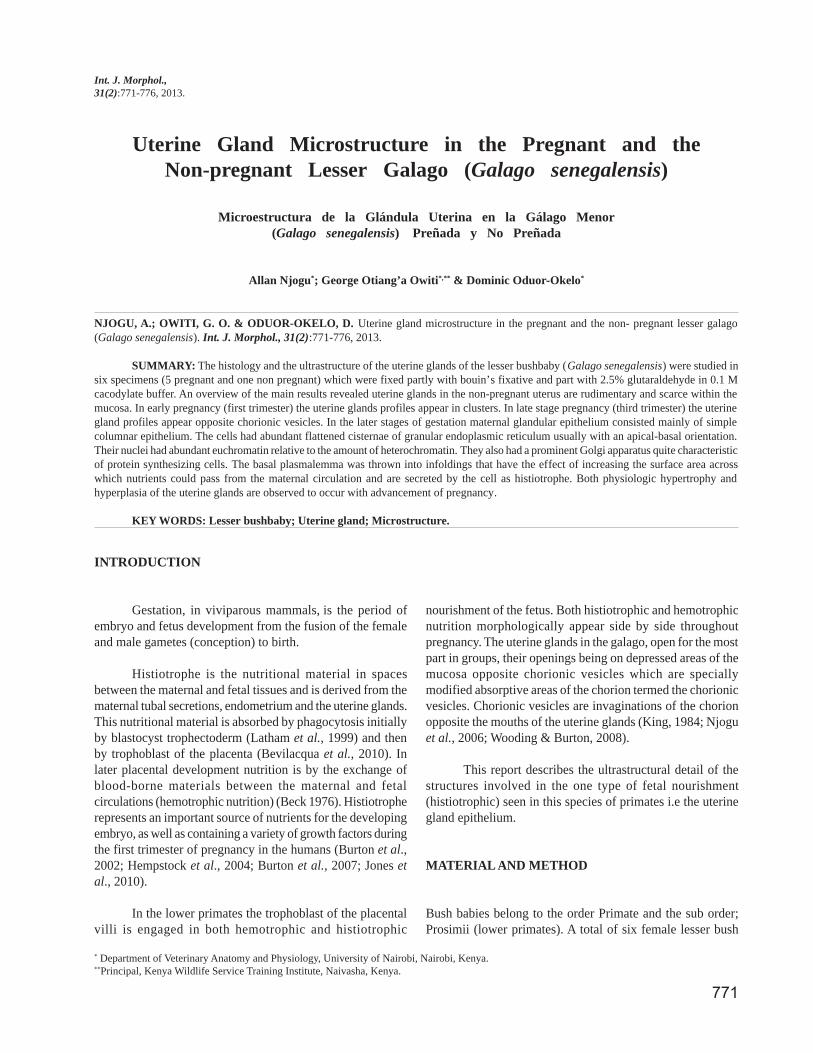
The epithelium rests on a basal lamina. Smooth musclecells are also part of this wall and are found interspersed withinthe connective tissue below the basal lamina. The basementmembranes of the uterine gland cells have numerous infoldingsthat in effect increase its surface area (Fig. 9).
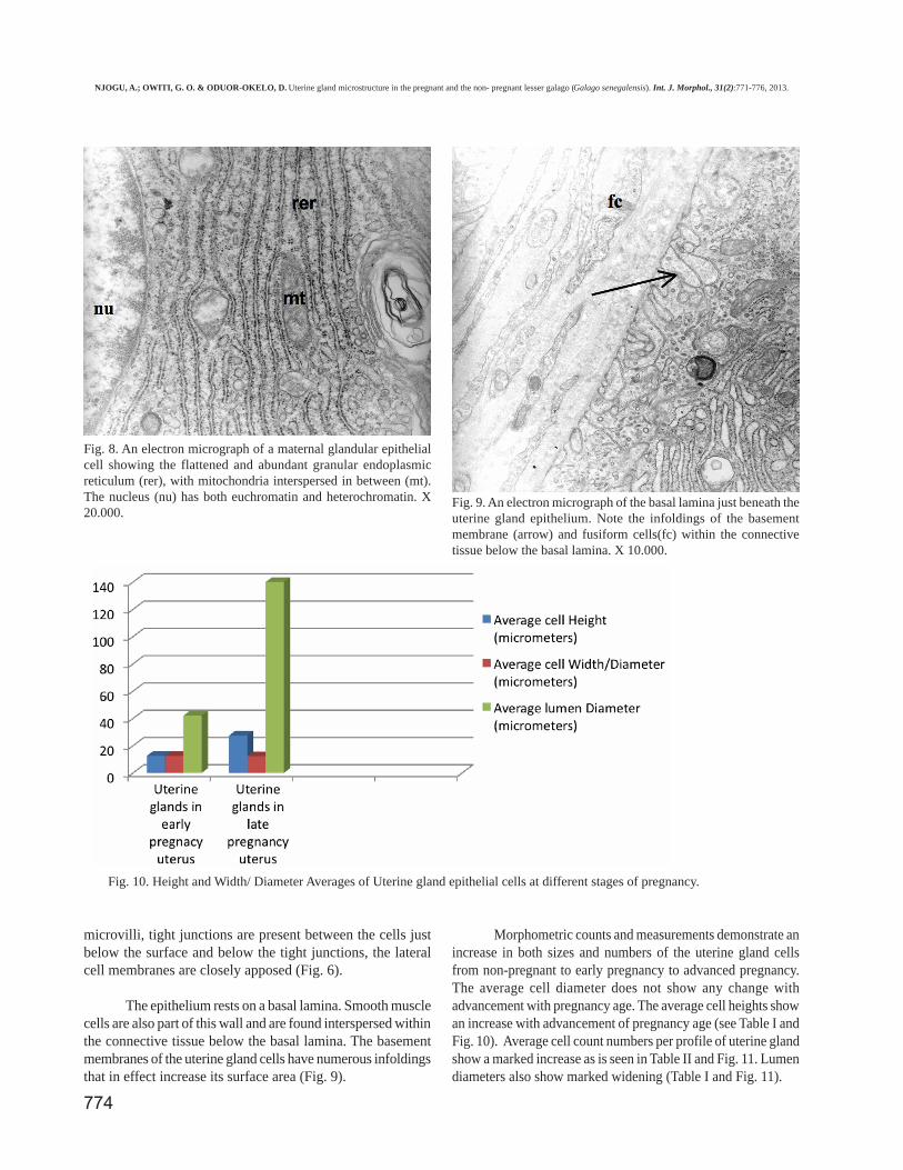
Morphometric counts and measurements demonstrate anincrease in both sizes and numbers of the uterine gland cellsfrom non-pregnant to early pregnancy to advanced pregnancy.The average cell diameter does not show any change withadvancement with pregnancy age. The average cell heights showan increase with advancement of pregnancy age (see Table I andFig. 10). Average cell count numbers per profile of uterine glandshow a marked increase as is seen in Table II and Fig. 11. Lumendiameters also show marked widening (Table I and Fig. 11).
Fig. 8. An electron micrograph of a maternal glandular epithelialcell showing the flattened and abundant granular endoplasmicreticulum (rer), with mitochondria interspersed in between (mt).The nucleus (nu) has both euchromatin and heterochromatin. X20.000.
Fig. 9. An electron micrograph of the basal lamina just beneath theuterine gland epithelium. Note the infoldings of the basementmembrane (arrow) and fusiform cells(fc) within the connectivetissue below the basal lamina. X 10.000.
Fig. 10. Height and Width/ Diameter Averages of Uterine gland epithelial cells at different stages of pregnancy.
NJOGU, A.; OWITI, G. O. & ODUOR-OKELO, D. Uterine gland microstructure in the pregnant and the non- pregnant lesser galago (Galago senegalensis). Int. J. Morphol., 31(2):771-776, 2013.

775
DISCUSSION
The uterine glands in the non-pregnant lesser galagoare scarce and where present are reduced in size and have acollapsed lumen. The ultrastructural characteristics of theuterine glands in the pregnant lesser galago suggest activesynthesis, packaging and secretory activity due to thepresence of extensive rough endoplasmic reticular systemand a well-developed Golgi apparatus (Okada et al., 1994).Presence of smooth muscle cells in the wall of the glandssuggests active expulsion of secretion from the lumen of theglands. The location of the uterine glands opposite thechorionic vesicles is strategic in that the two structures com-bine to form an efficient and direct route through which thedeveloping fetus gets adequate nutrients (histiotrophe) fromthe mother. Provision of adequate nutrition to a fetus is thekey to a successful pregnancy. The uterine glands are animportant source of nutrients to the human fetus duringorganogenesis, when metabolism is essentially anaerobic(Burton et al., 2002). Histiotrophe is the nutritional mate-rial accumulated in spaces between the maternal and fetaltissues, derived from the maternal endometrium and theuterine glands. The uterine glands synthesize and secretesubstances essential for survival and development of theembryo or fetus (Gray et al., 2001). These include nutrientsand growth factors that modulate the growth of the embryo(Hempstock et al., 2004).
Infoldings of the basement membrane, of the uterinegland cell as observed in this study, has been observed inthe salivary gland of the mouse (Pícoli et al., 2011). It isinferred that, coupled with the abundant mitochondria, theincrease in this surface area provides for effective uptake ofnecessary macromolecules that go to form the secretionreleased from the apical surface.
This study demonstrates a definite increase in thenumber of cells that surround each glandular profile. Thisphysiologic hyperplasia is necessary as the demand for nutrientsby the fetus increases. It has been demonstrated in the pig as aresult of stimulation by the hormone progesterone (Roberts &Bazer, 1988; Bailey et al., 2010). However an increase in uterinegland cell numbers may also be a sign of abnormal changes, asis the case in precancerous changes of endometrial carcinomasin women (Wang et al., 2005). The study also demonstrateshypertrophy of the uterine gland cells. Even though the avera-ge cell diameter does not show any change with advancementwith pregnancy age; average cell heights show an increase withadvancement in pregnancy age. The net effect is such that thereis a net increase in volume as volume is a function of both thediameter and the cell height. It is inferred that the increase inthe cell sizes is a result of an increase in cellular organelles thattake part in the secretory function of these cells.
Fig. 11. Average numbers of cellsper profile of the uterine gland.
Table I. Averages on Height and width/Diameter of uterine gland cells at different stages of pregnancy.
Table II. Average numbers of cells per profile of the uterine glands
Average cell Height(micrometers)
Average cellWidth/Diameter(micrometers)
Average lumen Diameter(micrometers)
Uterine glands in early pregnancy uterus 12.5 12.5 42Uterine glands in late pregnancy uterus 27.5 12.2 140
Average n of cells perprofile of the Uterine glands
Uterine glands in early pregnancy uterus 13Uterine glands in late pregnancy uterus 46
NJOGU, A.; OWITI, G. O. & ODUOR-OKELO, D. Uterine gland microstructure in the pregnant and the non- pregnant lesser galago (Galago senegalensis). Int. J. Morphol., 31(2):771-776, 2013.

776
NJOGU, A.; OWITI, G. O. & ODUOR-OKELO, D. Microestructura de la glándula uterina en la gálago menor (Galago senegalensis)preñada y no preñada. Int. J. Morphol., 31(2):771-776, 2013.
RESUMEN: La histología y ultraestructura de las glándulas uterinas de la gálago menor (Galago senegalensis) fueron estudiadas enseis ejemplares (5 preñadas y 1 no preñada). Una parte de las glándula se fijó con Bouin y otra con glutaraldehído al 2,5% en tampóncacodilato 0,1 M. Una visión general de los principales resultados reveló que las glándulas uterinas en el útero no gestante son rudimentariasy escasas dentro de la mucosa. Al principio de la preñez (primer trimestre) las glándulas uterinas aparecen en racimos. En la última etapa dela preñez (tercer trimestre) las glándulas uterinas aparecen opuestas a las vesículas coriónicas. En las últimas etapas de gestación el epitelioglandular materno consiste principalmente en epitelio cilíndrico simple. Las células tenían abundantes cisternas aplanadas en el retículoendoplásmico rugoso, por lo general con una orientación apico-basal. Sus núcleos tenían abundante eucromatina en relación con la cantidadde heterocromatina. También tenían un aparato de Golgi prominente bastante característico de células que sintetizan proteínas. El plasmalemabasal fue rechazado en repliegues que psoeen el efecto de aumentar el área de superficie a través del cual los nutrientes podrían pasar desdela circulación materna y son secretadas por las célula como histiotrofo. Hipertrofia fisiológica e hiperplasia de las glándulas uterinas seobservaron con el avance de la preñez.
PALABRAS CLAVE: Gálago menor; Glándulas uterinas; Microestructura.
REFERENCES
Bailey, D. W.; Dunlap, K. A.; Frank, J. W.; Erikson, D. W.; White, B.G.; Bazer, F. W.; et al. Effects of long-term progesterone ondevelopmental and functional aspects of porcine uterine epitheliaand vasculature: Progesterone alone does not supportdevelopment of uterine glands comparable to that of pregnancy.Reproduction, 140(4):583-94, 2010.
Beck, F. Comparative Placental morphology and function. Environ.Health Perspect., 18:5-12, 1976.
Bevilacqua, E.; Hoshida, M. S.; Amarante-Paffaro, A.; Albieri-Borges,A. & Zago Gomes, S. Trophoblast phagocytic program: roles indifferent placental systems. Int. J. Dev. Biol., 54(2-3):495-505,2010.
Burton, G. J.; Watson, A. L.; Hempstock, J.; Skepper, J. N. & Jauniaux,E. Uterine glands provide histiotrophic nutrition for the humanfetus during the first trimester of pregnancy. J. Clin. Endocrinol.Metab., 87(6):2954-9, 2002.
Burton, G. J.; Jauniaux, E. & Charnock-Jones, D. S. Human earlyplacental development: potential roles of the endometrial glands.Placenta, 28 Suppl. A:S64-9, 2007.
Gray, C. A.; Bartol, F. F.; Tarleton, B. J.; Wiley, A. A.; Johnson, G.A.; Bazer, F. W.; et al. Developmental biology of uterine glands.Biol. Reprod., 65(5):1311-23, 2001.
Hempstock, J.; Cindrova-Davies, T.; Jauniaux, E. & Burton, G. J.Endometrial glands as a source of nutrients, growth factors andcytokines during the first trimester of human pregnancy: Amorphological and immunihistochemical study. Reprod. Biol.Endocrinol., 2:58, 2004.
Jones, C. J.; Aplin J. D. & Burton, G. J. First trimester histiotropheshows altered sialylation compared with secretory phaseglycoconjugates in human endometrium. Placenta, 31(7):576-80, 2010.
King, B. F. The fine structure of the placenta and chorionic vesiclesof the Senegal bush-baby, Galago crassicaudata. Am. J. Anat.,169(1):101-16, 1984.
Latham, K. E.; Kutyna, K. & Wang, Q. Genetic variation introphectoderm function in parthenogenetic mouse embryos. Dev.Genet., 24(3-4):329-35, 1999.
Njogu, A.; Owiti, G. O.; Persson, E. & Oduor-Okelo, D. Ultrastructureof the Chorioallantoic placenta and chorionic vesicles of the lesserbush baby (Galago senegalensis). Placenta, 27(6-7):771-9, 2006.
Okada, H.; Merryman, J. I.; Rosol, T. J. & Capen, C. C. Effects ofhumoral hypercalcemia of malignancy and gallium nitrate onthyroid C cells in nude mice: immunohistochemical andultrastructural investigations. Vet. Pathol., 31(3):349-57, 1994.
Pícoli, L. C.; Dias, F. J.; Issa, J. P.; Ogawa, K.; Ciena, A.; Iyomasa,M. M.; et al. Ultrastructure of submandibular salivary glands ofmouse: TEM and HRSEM observations. Microsc. Res. Tech.,74(12):1154-60, 2011.
Roberts, R. M. & Bazer, F. W. The functions of uterine secretions. J.Reprod. Fertil., 82(2):875-92, 1988.
Wang, M. Q.; Zhang, Q. H.; Guo, L. L.; Cai, Y. R. & Wang, Y.Pathologic diagnosis of endometrial carcinoma in curettagespecimens in women under forty years of age. Zhonghua BingLi Xue Za Zhi., 34(5):262-5, 2005.
Wooding, F. B. P. & Burton, G. J. Comparative Placentation:Structures, Functions and Evolution. Berlin Heidelberg, Springer-Verlag, 2008. pp.17-20, Chapter 1.4.6.
Correspondence to:Allan NjoguDepartment of Veterinary Anatomy and PhysiologyUniversity of Nairobi. P.O Box 30197Nairobi - KENYA Email: [email protected]
Received: 13-09-2012Accepted: 25-02-2013
NJOGU, A.; OWITI, G. O. & ODUOR-OKELO, D. Uterine gland microstructure in the pregnant and the non- pregnant lesser galago (Galago senegalensis). Int. J. Morphol., 31(2):771-776, 2013.