use of vector diagnostics during military deployments ... · james c. mcavin, msf; adeline s. chan,...
TRANSCRIPT

MILITARY MEDICINE. 174, 9:904, 2009
Use of Vector Diagnostics During Military Deployments:Recent Experience in Iraq and Afghanistan
COL Russell E. Coleman, MS USA*; Lisa P. Hoch berg, MS*;Col John L Putnam, USAF BSCf; Katherine I. Swanson, PhD*; LTC John S. Lee, MS USARt;
James C. McAvin, MSf; Adeline S. Chan, PhD*; LTC Monica L O'Guinn, MS USAp,LTC Jeffry R. Ryan, MS USA (Ret.)*; COL Robert A. Wirtz, MS USAR (Ret.)§;
John K. Moulton, PhDH; Kirti Dave, PhDlJ; Michael K. Faulde, PhD**
ABSTRACT Vector-bome diseases such as malaria, dengue, and leishmaniasis are a threal to military forces deployedoutside of ihe United States. The availability of specific information on the vector-borne disease threat (e.g.. presenceor absence of a specific disease agenl, temporal and get)graphic dislributiim of competent vectors, and vector infecfionrales) allows lor effeclive implementation oí appropriate measures to prolccl our deployed military forces. Vector diag-nostics can provide critical, rcal-lime information crucial to esiablii.hing effective vector prevention/control programs.In this article we provide an overview of current vector diagnostic capabilities, evaluate the use of vector diagnostics inOperation Rndiiring Freedom and Operatit>n Iraqi Freedom, and discuss the ct>ncept of operations under which vectordiagnostics are employed.
INTRODUCTION
Military Threat of Arthropod-Borne DiseasesArthropod-borne diseases have historically posed a significantthreat to deployed military forces.'" The pathogens causingthese diseases are transtnitted by a variety of biting arthro-pod.s, in include iiiostjiiitocs. licks, chiggers. sand nies, lice,fleas, and biting midges. Arthropod-borne diseases consid-ered a significant threat to military forces include malaria/dengue,""'- leishmaniasis,^''"'*' scrub typhus,'-'^ epidemicand endemic typhus.' "̂ Crimean-Congo bemorrhagic fever(CCHF) virus, Rifi Valley fever (RVF) virus, Sindbis virus,sandfly fever viruses (SFV), Venezuelan equine encephalilis
*Dep;irlmciU of Entomology. Waller Reed Army Institute of Research,Silver Spring, MD.
tEpidemioIogical Surveillatice Division. Air Force Institute of Opera-tional Heiilth, San Antonio, TX.
tVirology Division, U.S. Army Medical Research Institute of InfectiousDiseases, Forl Dctritk, MD.
§Divisioii of Parasitic Diseases, National Center for Zoonotic, Vector-borne and Enteric Diseases, Centers for Disease Control and Prevention,Atlanta, GA.
IIVecTOR Test Systems, Inc., Thousand Oaks. CA.'IIDeparlnient of Entomology and Plant Pathology, University of
Tennessee, Knoxville, TN,**Departmenl of Medical Zoology, Central Instilulc of the Bundeswehr
Medical Service, Koblenz. Geniiiiny.Disclaimer: Material ha.s been reviewed by the Walter Reed Army Institute
of Research antl the U.S. Army Metlical Research and Material Command.There is no objection to its preseniation and/or publication. The opinions orassenions contained herein are the private views of the author, and are not tobe consinietl ¡is official, or as reHecting true views of the Department of theArmy or the Department of Defense. One of tbe coauthors (Dr. Kirti Dave)bas an intere.st in a commercial company that produces and sells some of theband-held assays described in this article.
This manu.script was received for review in January 2009. Tbe revisedmanuscript was accepted for publication in June 2(H)y.
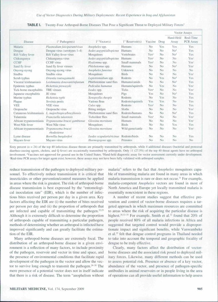
(VEE) virus, tick-borne encephalitis (TBE) viruses. West Nile(WN) virus and Japanese encephalitis (JE) virus. A variety ofadditional arthropod-borne diseases could potentially impactmilitary operations. A summary of some of tbe tnost signifi-cant arthropod-borne threat agents,' to include pathogen, pri-mary reservoiris), and vectors, is presented in Table I.
Altbough vaccines and/or prophylactic drugs are the pre-ferred method of protecting deployed military personnel frominfectious diseases,'"^ these protective measures are not avail-able tor many arthropod-borne diseases (Table I). Currently,FDA-licensed vaccines tor widespread use are available foryellow fever virus, JE virus, and plague, while limited-usevaccines (normally restricted to individuals at high risk ofinfection) are available for VEB, RVF, CCHF, TBE, and east-ern and western equine encephalitis (EEE and WEE) viruses.Prophylactic drugs are widely used for the prevention ofmalaria and less frequently for protection trom scrub lyphusand various rickettsial diseases, but provide no protectionagainst the majority of atthropod-borne diseases (Table I).
Requirement for Vector DiagnosticsIn the absence of a vaccine or prophylactic drug, the mosteffective means of protecting deployed military personnelfrom arthropod-borne diseases is to prevent infected artbro-pods frt)tii biting them. Prevention of bites from infectedarlhropods can be achieved through effective use of personalprotective measures (PPMs) or by reducing vector popula-tions. Effective PPMs include application of DEET-containinginsect repellents to exposed skiti, wearing a permethriti-treated uniform, and sleeping under an insecticide-treated bednet,"" '^'" while vector populations tnay be reduced by judi-ciously using insecticides or eliminating vector habitat.''"'' Akey tenet of military vector control operations is that the goalis not to merely decrease vector populations but to actually
'Í04 MILITARY MEDICINE. Vol. 174, September 2009
Report Documentation Page Form ApprovedOMB No. 0704-0188
Public reporting burden for the collection of information is estimated to average 1 hour per response, including the time for reviewing instructions, searching existing data sources, gathering andmaintaining the data needed, and completing and reviewing the collection of information. Send comments regarding this burden estimate or any other aspect of this collection of information,including suggestions for reducing this burden, to Washington Headquarters Services, Directorate for Information Operations and Reports, 1215 Jefferson Davis Highway, Suite 1204, ArlingtonVA 22202-4302. Respondents should be aware that notwithstanding any other provision of law, no person shall be subject to a penalty for failing to comply with a collection of information if itdoes not display a currently valid OMB control number.
1. REPORT DATE 2009 2. REPORT TYPE
3. DATES COVERED 00-00-2009 to 00-00-2009
4. TITLE AND SUBTITLE Use of Vector Diagnostics During Military Deployments: RecentExperience in Iraq and Afghanistan
5a. CONTRACT NUMBER
5b. GRANT NUMBER
5c. PROGRAM ELEMENT NUMBER
6. AUTHOR(S) 5d. PROJECT NUMBER
5e. TASK NUMBER
5f. WORK UNIT NUMBER
7. PERFORMING ORGANIZATION NAME(S) AND ADDRESS(ES) Walter Reed Army Institute of Research,Department ofEntomology,Silver Spring ,MD,20910-7500
8. PERFORMING ORGANIZATIONREPORT NUMBER
9. SPONSORING/MONITORING AGENCY NAME(S) AND ADDRESS(ES) 10. SPONSOR/MONITOR’S ACRONYM(S)
11. SPONSOR/MONITOR’S REPORT NUMBER(S)
12. DISTRIBUTION/AVAILABILITY STATEMENT Approved for public release; distribution unlimited
13. SUPPLEMENTARY NOTES
14. ABSTRACT Vector-bome diseases such as malaria, dengue, and leishmaniasis are a threal to military forces deployedoutside of ihe United States. The availability of specific information on the vector-borne disease threat (e.g..presence or absence of a specific disease agenl, temporal and get)graphic dislributiim of competent vectors,and vector infecfion rales) allows lor effeclive implementation o?ppropriate measures to prolccl ourdeployed military forces. Vector diagnostics can provide critical, rcal-lime information crucial toesiablii.hing effective vector prevention/control programs. In this article we provide an overview of currentvector diagnostic capabilities, evaluate the use of vector diagnostics in Operation Rndiiring Freedom andOperatit>n Iraqi Freedom, and discuss the ct>ncept of operations under which vector diagnostics are employed.
15. SUBJECT TERMS
16. SECURITY CLASSIFICATION OF: 17. LIMITATION OF ABSTRACT Same as
Report (SAR)
18. NUMBEROF PAGES
18
19a. NAME OFRESPONSIBLE PERSON
a. REPORT unclassified
b. ABSTRACT unclassified
c. THIS PAGE unclassified
Standard Form 298 (Rev. 8-98) Prescribed by ANSI Std Z39-18
Use of Vector Diagnostics During Military Deployments: Recent Experience in Iraq and Afghanistan
TABLE 1.
Disease
MalariaDengueRitt Valley feverCliikungunyaCCHh.Sane] tly feverOnyi)iig-ny(ingSindhisScrublyphusVisceral leishmaniasisEpidemic typhusTick-borne encephalitisJapanese eticephalitisMiirine lyphusPlagueVEEOropouL'heCutaneous leishmaniasisTiitaremiaAfricati IrypanosoiniasisWest Nile leverAfrican irypanosomiasis
Lyme diseaseMay aro fever
Twenty-Four Arthrüpod-Bome Diseases Thai Pose a Significant Threat to Deployed Mihtary Forces^
r Pathogen(s)
Pktsmoäium falcipa riiin/vivaxDengue virus (serotypes 1—4)Rift Valley fever virusChikutigunya virusCCHF virusSand Hy fever virusesOnyong-nyong vimsSindbis vimsOriemia r\utsi(i;uniusliiU'i.'iliinanitiilonovtim/infhnliiniHiiii'tl.siii prowazfkiTBE virusesJE virusRicken.sifi fypliiYeisiniii pestisVEE vimsOropouche viru.sL major/tropiia/l)raziliensisFraitcisella ¡ukirensisTrypiinosoma hrucei gambienxeWest Nile virusTrypatiosoma hnicf i
yhotk'sienxeBorreilia burgdorferiMayaro virus
i°Vector(s)
Anopheles spp.Aedes ae^ypti/atbopiaiisMosquitoesAedes aegypti/atbopicUisHyalommii spp.Phkhotomus spp.Anopheles funestusMosquitoesLeploiromidiuin spp.Phiebototiiine satid fliesPedicidus humamisIxodes spp.MosquiloesXenopsyUa cheopisVarious lleasCidex spp.Biting midgesPhlebolomine sand fliesTicks/deer fliesGlo.'i.sinii nuirsiltuisMosquitoesGUissina mornitam
Ixodes scapidah.s/riciti usHaemogogoiis spp.
r Reservoir(s)
HumansHumansVertebralesHumansSmall mammalsHumansUnknownBirdsRodentsHunians/caiiidsHuni;ms/squirreisTicksPigsRddenisRodcnis/squirrelsRodenlsSlothsRtxlenls/humatisSmall mammalsH u [Hans
BirdsWild game/cattle
Rodents/birdsPrimates
Vaccine
NoNo
Yes"Yes"Yes"NoNoNoNoNoNo
Yes"YesNoYesYes-NoNo
Yes-NoNnNo
NoNo
Drug
YesNoNoNoNoNoNoNoYesNoYesNoNoYesYesNoNoNoNoNoNoNo
NoNo
Vector Assays
Hand-HeicAssay
YesNo*No"NoNoNo"NoNoNo"No"NoNoNo"NoNoNoNoNo"NoNoYesNo
NoNo
Real-TimePCR Assay
YesYesYesYes'-Yes'Yes'No
Yes'YesYesYes'Yes'YesYesYesYesYes'YesYes'Yes^YesYes'
YesNo
Sixly percent {n = 24) ofthe top 40 infectious disease threats are primarily iranstiiitted by anhropods, while 4 additional diseases (bacterial atid prouv.oaldiarrhea-causing agents, cholera, and Q fever) are occasionally transmitted by anhnipods. Only 11 (27.5%) of the ttip 40 threat agents have no anhroptidinvolvement. "Vaccines tiot approved for general use in the United States. ''Hand-held diagnostic assay for vector assessment eurrently under development.' Real-time PCR assays fnr target ayen! exist: however, these assays may not have been fully validated wiih arthropod samples.
reduce transmission ofthe pathogen to deployed military per-sonnel. To effectively reduce transmission it is critical thatinsecticides or other prevention/control measures be appliedin areas where the risk is greatest. The riskof arlhropod-borncdisease transtnission is best expressed by the "entomologi-cal inoculation rale" (EIR). which is the number of infec-tious bites received per person per day in a given area. Keyfactors affecting the EIR are (i) the number of bites receivedper person per day and (ii) tbe proportion of arthropods thatare infected and capable of transmittitig the pathogen.--'Although it is extremely difficult to determine tbe proportionof arthropods capable of transmitting a particular pathogen,methods of detertTiining whether an arthropod is infected haveimproved significantly and can greatly facilitate the estima-tion ofthe EIR.
Many arthropod-borne diseases are extremely fiK'al. Thedistribution of an arthropod-borne disease in a given envi-ronment is a reflection of many factors, to include proximityof the pathogen, reservoir and vector in time and space, andthe presence of environmental conditions tbat facilitate rapiddevelopment of the pathogen in the vector and allow tbe vec-tor to survive long enough to transmit the pathogen.-' "' Themere presence of a potential vector does not in itself indicatethat there is a risk of disease. The term "anopbelistn without
malaria" refers to tbe fact that Anopheles mosquitoes capa-ble of transmitting tnalaria are found in many areas in whichmalatia transmission is rare or does not occur." For example,malaria-competent Anopheles vectors are found in most ofNorth America and Europe yet locally transmitted malaria isessentially nonexistent in these regions.
A number of recent sttidies suggest that effective pre-vention and control of vector-borne diseases requires a tar-geted approach in which maximum resources arc committedto areas where the risk of acqtiiring the particular di.sease isbighest.-**'̂ --̂ "* For example. Stnith et al." found that 20% ofpeople received 80% of ail malaria infections in Africa andsuggested tbat targeted control would prcnide a dispropor-tionate impact and significant benefits., while Vanwambekeet al.''' felt tbat dengue control programs in Thailand neededto take into account the temporal and geographic focality ofdengue to be truly effective.
Clearly, many factors affect tbc distribution of vector-borne diseases and the associated risk posed to deployed mil-itary forces. Likewi.se, many different methods can be usedto assess potential risk. Presence or ab.sence of a key vector,abundance of tbe vector, and presence of patbogcn-specificantibodies in animal reservoirs or in people living in the areaof operations can all provide useful information to help assess
MIIJTARY MEDICINE, Vol. 174. September 2009

V.ie of Vector Diagnostics During Military Deployments: Recent Experience in Iraq and Afghanistan
risk and facilitate the development of targeted control pro-grams. With the emergence of tield-deployable diagnosticassays, early detection of the pathogen in vector populationshas also emerged as an effective method of rapidly assess-ing risk.^'^" Ideally, detection of a vector-borne disease threatwould occur before the occurrence of cases in deployed mili-tary personnel.'' thereby allowing for the early implementa-tion of vector control measures and minimizing the impact ofthe disease on military operations.
Methods of Conducting Vector DiagnosticsVector diagnostics is (he detection of disease-causing patho-gens within the arthropod vector. One of the earliest methodsof determining if an arthropod was infected was to visuallyexamine appropriate organs (e.g.. the midgut or salivaryglands of AnopheU'.s' mosquitoes for malaria parasites) under amicroscope.'** This method is normally not appropriate for useduring military operations as it is time consuming and requiresa high level of training. The development of immunologicalmethods in general, and the enzyme-linked immunosorbentassay (ELISA) in particular, revolutionized the tield of vec-tor diagnostics."'''^' The circumsporozoite protein ELISA forthe detection of human malaria parasites in mosquitoes washrsl developed at the Walter Reed Army Institute of Research(WKAIR)and rapidly became the standard method for assess-ing mosquito infection rates.̂ """ ELISA assays are capable oftesting large numbers of arthropods very rapidly; however,they are not routinely u.sed during military operations becauseof the amounl of equipment required, complexity, and therequirement for a cold chain.
The need for vector surveillance during military deploy-ments led to the development of a series of hand-held immu-nochromatographic assays. These assays are simple, canhe used anywhere, and do not require a cold chain, therebyovercoming most of the challenges associated with the useof ELISA assays. The malaria VecTest assay was developedthrough a collahoralive effort between Navix. Inc. (subse-quently Medical Analysis Systems, Inc.) and the WRAIR.The assay detects Plasmodium falciparum and P. vivax cir-cumsporozoite protein in anopheline mosquitoes and is soldas a kit containing 20 assays, with each assay capable of test-ing up to 20 mosquitoes in 15 minutes (Fig. 1). Sensitivityranges from 91 to liW/c and speciticity from 94 to 99.7%, onthe basis of tield trials conducted in Africa. Asia, and SouthAmerica.''^^' The malaria VecTest kit requires no refrigera-tion or free/ing and is stable up to 24 monihs at temperaturesup to 32°C and for shorter periods at temperatures up to SO'̂ C.The malaria VecTest kit has been assigned a national stocknumber (NSN: 6íi5()-0I-íi51-5327) and is currently availablefrom VecTOR Test Systems. Inc., Thousand Oaks, CA.
Subsequent to the development of the malaria VecTestkit. the Centers for Disease Control and Prevention (CDC),the WRAIR, and Medical Analysis Systems. Inc. developeda series of five VecTest assay kits for the detection of arthro-pod-borne viru.ses. Each kit contains 50 assays, with each
FIGURE 1. The Malaria VecTEST Assay.
assay capable of testing up to 50 mosquitoes. Available kitsinclude (i) a WN virus assay (NSN; 6550-01-533-3943), (ii)a Saint Louis encephalitis (SLE) virus assay, (iii) a combinedWN/SLE virus assay, (iv) a combined WN/SLE/EF.E virusassay (NSN: 6550-01-533-1564), and (v) a combined WN/SLE/WEE virus assay (NSN: 6550-01-533-15721. Althoughthese assays were originally developed for the detection ofviruses in mosquitoes.''̂ ''*' the WN virus assay has also beenused to detect WN virus in birds.*'̂ '̂ Efforts currently focuson the development of hand-held immunochromatographicassays to detect lA'i.shmania parasites and dengue. JE, RVF.SFV. and Ross River viruses.
Although hand-held immunochromatographic assays areideal for ñeld use. specificity and sensitivity of the assayscan be lower than desired. Confirmatory assays, although notabsolutely essential, are therefore desirable. Polymerase chainreaction (PCR)-based methods bave revolutionized the lieldof vector diagnostics and can serve as stand-alone screeningassays or as confirmatory assays. Initially developed in the1980s. PCR was first used to detect dengue virus and malariaparasites in mosquitoes in the early 199()s."" Although tradi-tional PCR equipment has become cheaper, lighter, and easierto use. requirement for gels, multistep procedures, and riskof contamination still preclude routine use under tield condi-tions. However, the development of fluorogenic or real-timePCR assays has overcome many of the limitations of tradi-tional PCR and offers great potential for use during militarydeployments. Recently, real-time PCR assays were used dur-ing Operation Iraqi Freedom to detect Leishmania parasites insand Hies, rodents, and human patients. " '̂'•'̂
The Joint Biological Agent Identification and DiagnosticSystem (JBAIDS) is the U.S. military's field-deployable plat-form tor real-time PCR assays. JBAIDS is a military-specificversion of the Idaho Technology R.A.P.I.D. (RuggedizedAdvanced Pathogen Identification Device), The JBAIDS inte-grates Idaho Technology's LightCycier real-time PCR tech-nology into a portable, impact-resistant package ideal forfield use. Distinctive software allows simple "push-hutton"
906 MILITARY MEDICINE, Vol. 174. September 2009

Use of Vector Diagnostics During Military Deployments: Recent Experience in lra(¡ and Afghani.stan
use of the JBAIDS by field personnel with minimal training.The JBAIDS Is currently used by fietd-deployuble forces suchas Army Area Medical Laboratories, Army Combat vSupportHospitals, and Navy Forward Deployed Preventive MedicineUnits. Although the JBAIDS is primarily being developed forthe detection of biological threat agents in clinical samples/''it can be used for vector assessment as well. A limited num-ber of assays for the detection of arthropod-borne pathogensare currently available on the JBAIDS platform, to includeYersinia pestis (plague), Franci.sella !ularc'n.sis (tularemia),and Ricicettsia prowazeki (epidemic typhus); however, to datenone of these assays has been validated for use in the detec-tion of pathogens within the arthropod vector.
Rationale for This StudyUnlil recently, vector diagnostics had not been routinely usedduring military deployments. However, the development ofhand held assays and real-time PCR assays that can be usedill a field environment resulted in the employment of both .sys-tems during Operation Iraqi Freedom (OIF) and OperationEnduring Freedom (OEF) by U.S. and German military forces.The goal of this article is to assess the use of these assays dur-ing OIF and OEF, to identify issues related to their use, and tomake recommendations for future use.
MATERIALS AND METHODS
Collection of Mosquitoes and Sand fliesMosquitoes and sand flies were collected as part of a systematiceffort by U.S. and German preventive medicine (PVNTMED)assets to assess vector populations in areas where mili-tary forces were located throughout Iraq and Afghanistan.Standardized guideline on collection procedures were provideto each PVNTMED unit that participated in this study. Thiseffort was initiated in 2(X)3 and has continued to date (2009).Insect collections were primarily made using unbaited CDCminiature light traps; however. German PVNTMED person-nel also collected Anopheles mosquitoes from inside of tents.buildings, and latrines using a commercially available hand-held mouth aspirator with a hepa filter. Although a variety ofindividuals from a number of differenl unils were responsiblefor insect collections, the following procedures were gener-ally used. Light traps were normally placed shortly beforesunset and were retrieved soon after dawn. Traps were nor-mally placed within I meter ofthe ground. Fine mesh collect-ing cups suitable for the collection of sand flies, mosquitoes,and other small insects were used with the light traps. Uponreturn to the field laboratory, collection cups were placed ina freezer to kill the collected insects. Mosquitoes collectedusing aspirators were placed in 1-pint screened cartons andreturned to the field laboratory where they were killed byfreezing at -20"C or using ethanol. Within 1-2 hours the col-lection cups were removed from the freezer, contents placedin a Petri dish, and mosquitoes and sand fiies separated fromthe remaining insects using a di.ssecting microscope.
Collections in Iraq and Afghanistan began in 2003 and2004, respectively, and have continued since then. At the timethat Ihis article was prepared, U.S. Army data from 2(K)3-2005 ami German dala from 2006 were available and wereused for all analyses. Sand flies collected in 2003 and 2(X)4were identified to subfamily (Phlebotominae), while thosecollected in 2005 were identified to genus (Fhlehoiomus andSergenîomyia) and those in 2(X)6 to species. Sand flies werestored frozen at -VO^C or in 80-l(X)'/f ethanol until tested.Mosquitoes were identified to genus,'" with all anophelinemosquitoes subsequently identified to species using the key ofGlick et al.'̂ '̂ Female anopheline mosquitœs were either testedimmediately with the malaria VecTest assay or were storedfrozen at -70°C for testing at a later date.
Testing Anopheline Mosquitoes Using the MalariaVecTest AssayProcedures described in the insert for the malaria VecTestkit were used. Ali supplies required for running the assay areprovided with the kit (Fig. 1 ). In brief, from I-10 anophelinemosquitoes were placed into a conical grinding tube and 13drops of grinding solution dispen.sed into each tube. A grind-ing pestle was placed in each tube and rotated vigorously forapproximately 1 minute or until ihe mosquitoes were thor-oughly homogenized. A test strip was labeled and placed intothe grinding tube containing the mosquito suspension. After15 minutes the test strip was removed and results read imme-diately per the insert.
Testing Sand Flies Using Real-Time PolymeraseChain Reaction Assays
DNA E.\írmíiim
U.S. units used the Qiagen QlAamp DNA Mini kit to extractDNA from pools of sand flies per procedures de.scribed in theproduct insert. German units homogenized sand flies mechan-ically using a Roche Diagnostics MagNA Lyser device andextracted DNA using a Roche Diagnostics High Pure PCRTemplate Preparation kit. Tubes containing extracted DNAwere labeled and stored at 4 X or -2O'C if PCR was lo beperformed within 3 days or at -70°C if PCR was to be per-formed more than 3 days later,
Real-Time PCR Assays
Sand nies were initially tested using a ¿Í'/.V/Í/Í/Í/ÍÍ/VÍ-genus real-time (fluorogenic) PCR assay modified from an assay devel-oped by Worimann et al,'*'' for testing of clinical samples. Theassay was modified so that each reaction contained I puReTaqReady-to-Go PCR bead, 6mM MgCl,. 800 nM of each primer(LEISLKLEISUI), 120nMofprobê(LEISPI),and2,0Mloftemplate DNA. The assay was established and validated at theWRAIR in 2001. Assay validation consisted of an evaluationof the limit of detection of the assay as well as sensitivity andspecificity of the assay using cultured L. major, L. dottovani.
MILITARY' MEDICINE, Vol. 174, September 2009 907
Use of Vector Diagno.stics During Militar)' Deployments: Recent Experience in Iraq and Afghanistan
L. infantum. atid L. trapica amastigotes, uninfected sand fliesand sand flies infected with L major.^' The assay was used atthe WRAIR lor approximately 2 years before deploying theassay to OIF in 2003. A nmre detailed description ot the assayis contained in Coleman et al.*'̂
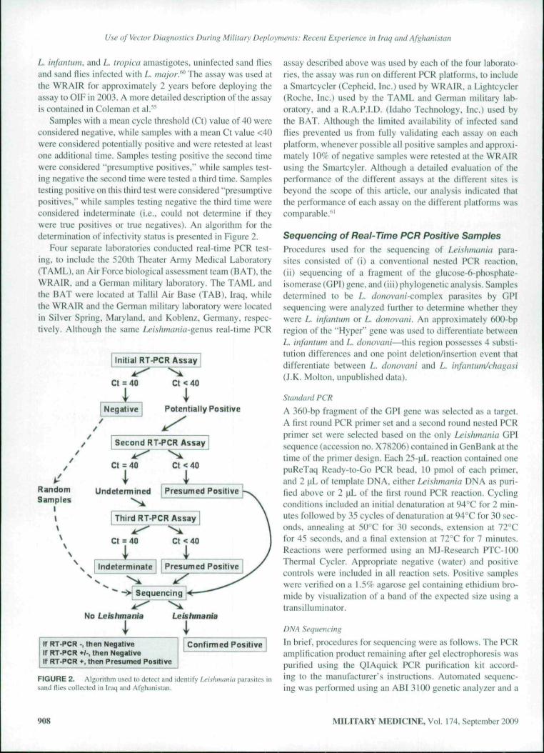
Samples with a mean cycle threshold (Ct) value of 40 wereconsidered negative, while samples with a tnean Ct value <4()were considered potentially positive and were retested at leastone additional time. Samples testing positive the second timewere considered "presumptive posilives," while samples test-ing negative the second time were tested a third time. Samplestesting positive on this third test were considered "presumptivepositives," while samples testing negative the third time wereconsidered indeterminate (i.e., could not determine if theywere trtte positives or true negatives). An algorithm for thedetermination of infectivity status is presented in Figute 2.
Four separate laboratories conducted real-time PCR test-ing, to include the 520th Theater Army Medical Laboratory(TAML). an Air Force biological as.sessment team (BAT), theWRAIR. and a German military laboratory. The TAML andthe BAT were located at Tallil Air Base (TAB). Iraq, whilethe WRAIR and the German military laboratory were locatedin Silver Spring, Maryland, and Koblenz. Germany, respec-tively. Although the same Leishmania-genus real-time PCR
I Initial RT-PCR Assay I
ct = 40 Ct < 40
Negative Potentially Positive
Second RT-PCR Assay
RandomSamples
I\
Ct = 40 Ct < 40
i , IUndetenmined I Presumed Positive r
Third RT-PCR Assay
Ct = 40 Ct < 40
Indeterminate
No Leishmania
WKT-PCR •, then NegativeI If RT-PCR */-, then Negative; If RT-PCR +, then Presumed Positive
FIGURE 2. Algorithm used to detect and ideniify Leishmania parasites insand flies collected in Iraq and Afghunistati.
assay described above was used by each of the four laborato-ries, the assay was run on different PCR platforms, to includea Smartcycler (Cepheid. Inc.) used by WRAIR. a Lightcycler(Roche, Inc.) used by the TAML and German tnilitary lab-oratory, and a R.A.P.I.D. (Idaho Technology, Inc.) used bythe BAT. Although lhe limited availability o\' infected sandflies prevented us from fully validating each assay on eachplatform, whenever possible all positive samples and approxi-mately 10*̂ of negative samples were retested at the WRAIRusing the Smartcyler. Although a detailed evaluation of theperformance of the different assays at the different sites isbeyond the scope of this article, our analysis indicated thatthe perfonnance of each assay on the different platforms wascomparable.'''
Sequencing of Real-Time PCR Positive SamplesProcedures used for the sequencing of Leishmunia para-sites consisted of (i) a conventional nested PCR reaction,(ii) sequencing of a fragment of the glucose-6-phosphate-isomerase (GPI) gene, and ( iii ) phylogenetic analysis. Samplesdetermined to be L. donovani-conycAex parasites by GPIsequencing were analyzed further to determine whether theywere L. infantuni or L. donovuni. An approximately 600-bpregion of the "Hyper" gene was used to differentiate betweenL. infaiitiim and L. donov¿ini^lh\s region possesses 4 substi-tution differences and one point deletion/insertion event Ihaidifferentiate between L. donovani and L. infantum/chagasi(J,K, Molton, unpublished data).
Standard PCR
A ."iftO-bp fragment of the GPI gene was selected as a target.A tirst rt)und PCR primer set and a second round nested PCRprimer set were selected based on the only Leishmania GPIsequence (accession no. X78206) contained in GenBank at thetime of the primer design. Each 25-|iL teaction contained onepuReTaq Ready-to-Go PCR bead, 10 pmol of each primer,and 2 ¡lL of témplale DNA. either Leishimmia DNA as puri-fied above or 2 |iL of the first round PCR reaction. Cyclingconditions included an initial denaturation at 94°C for 2 min-utes foilowed by 35 cycles of denaturation at 94"C for 30 sec-onds, annealing at 50°C for 30 seconds, extension at 72"Cfor 45 seconds, and a final extension at 72°C for 7 minutes.Reactions were petfortned using an MJ-Research PTC-100Thermal Cycler. Apptopriate negative (water) and positivecontrols were included in all reaction sets. Positive sampleswere verified on a \.59c agarose gel containing ethidiuni bro-mide by visualization of a band of the expected size using atransilluminator.
DNA Sec¡uencinfí
In brief, procedures for sequencing were as follows. The PCRamplilicalion product remaining after gel electrophoresis waspurified using the QIAquick PCR purification kit accord-ing to the manufacturer's instructions. Automated sequenc-ing was performed using an ABI 3100 genetic analyzer and a
908 MILITARY MEDICINE. Vol, 174. September 2009
Use of Vector Diagnostics During Military Deployment's: Recent Experience in Iraq and Afghanistan
Big-Dye vl.l or v3,l sequencing kit according to the manu-facturer's instructions. Primer., excess nucleotides, and bufferwere removed from tbe Big-Dye sequencing reaction by elut-ing the tiiatcriul from a Sepbadex G-30 colutnn equilibratedwith water. Sequencing of the approximately 600-bp regiono{ the "Hyper" gene was conducted using a BaseStation-U)0Automated DNA Sequencer with accompanying BCS andCartographer software,
Phyiogenetic Analysis
Sequences were aligned using the MegAlign program andsequence ends were trimmed to a uniform length, Phyiogeneticanalyses of aligned sequences were performed using theCtustalW method''' witb a gap penalty of 15 and a gap lengthof 6.66. The phyiogenetic tree generated by MegAlign is arooted tree with the number of substitution events indicated atihe bottom of the tree. Bootstrap replication was used to eval-uate the strength of the clustering analysis. Unknown samplesequences were compared to sequences determined for knownculture isolates and to other sequences present in GenBank.
RESULTS
Evaluation ofAnopheline Mosquitoes forPlasmodium Parasites
¡niq
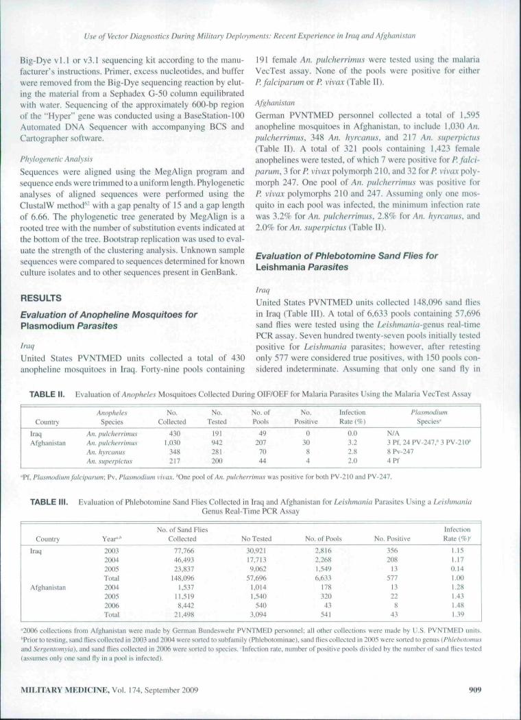
United States PVNTMED units collected a total of 430ant)pheline mosquitoes in Iraq, Forty-nine pools containing
191 female An. piilclicnimus were tested using the malariaVecTest assay. None of the pools were positive for eitherP. falcipartini or P. viva.x (Table 11).
Afghani.stan
German PVNTMED personnel collected a total of 1,595anopheline tiiosquitoes in Afghanistan, to include 1,()3() An.pidcherrimus, 348 An. hyixanus. and 217 An. superpictus(Table II), A total of 321 pools containing 1,423 femaleanophelines were tested, of which 7 were positive for P. falci-parum, 3 for P. vivax polymorph 210. and 32 for P. VÍVÍLX poly-morph 247. One pool of An. pulcherrimus was positive forP. vivax polymorphs 210 and 247. Assuming only one mos-quito in each pool was infected, the minimum infection ratewas 3.2% for An. puklwrrimus. 2,8% for An. hyrcamts, and2.0% for A/!, superpictus (Table II).
Evaluation of Phiebotomine Sand Flies forLeishmania Parasites
Iraq
United States PVNTMED units collected 148,096 sand fliesin Iraq (Table III). A total of 6,633 pools containing 57,696sand nies were tested using the Lei.shniania-gsfím real-timePCR assay. Seven hundred twenty-seven pools initially testedpositive for Leishmania parasites; however, after tctestingonly 577 were considered true pt)sitives, with 150 pools con-sidered indeterminate. Assuming that only one sand fly in
TABLE IL Evaluation oí Anopheles Mosquitoes Collected During OIF/OEF for Malaria Parasites Using the Malaria VecTest Assay
Country
IraqAfgbanistan
AnophelesSpecies
An. pulcherrimusAll. ¡mlcherrimu.sAn. h\n:iiiiusAn. superpictus
No.
Collecied
4301,030
348217
No,Tested
19194228120(1
No, ofPools
49207
70
44
No.
Positive
0
3084
InfectionRateC'/f')
0.03.22.82.0
PlasmntliwnSpecies'
N/A3Pf,24PV-247,''3PV-2IO*8 Pv-2474Pf
"Pf. Pltismihiuirn fakipanim: Pv, Pkismadiiim vivax. ''One pool of/\ii. puUiicrnmus was positive for botb PV-210 and PV-247.
TABLE III. Evaluation of Phiebotomine Sand Ries Collected in Iraq and Afghanistan for Leishmania Parasites Using a LeishmaniaGenus Real-Time PCR Assay
Couniry
Iraq
Afghanistan
Year' '
200320042IX)5Total200420052006Total
No. of Sand FliesCollected
77,76646,49323,837
148,0961,537
11.5198,442
21.498
No Tested
30.92117,7139,062
57,6961.0141.540
5403,094
No. of Pool.s
2,816
1,5496^33
17832043
541
No. Positive
356208
13577
13228
43
[tifecttonRale {%)'
I.I51.170.14I.(KI1.281.431,481.39
2006 collections frotn Afghanistan were tnade by German Bundeswehr PVNTMED personnel; all other collections were made by U.S. PVNTMED units.''Prior to testing, sand flies collecleil in 2003 and 2004 were sorted lo subfamily (Pbleboiominae), sand flies collected in 2005 were sorted to i.cm\s{Phlfhoiomu.sand Serfic'iiiomyiii). and sand (lies collected in 2006 were sorted to species. Itifection rate, number of positive pools divided hy the number of sand tlics tested(assumes only one sand lly in a pool is infected).
MILITARY MEDICINE, Vol. 174, Septetiiber 2009 909

Use of Vector Diagnostics During Military Deployments: Recent Experience in Iraq and Afghanistan
eaeh pool was infected, tbe minimum field infection rate was1.00% (Table HI). Overall infection rates were almost identi-cal for 2003 (1.15%) and 2004 (1.17%); however, infectionrates in 2(X)5 (0.14%) were signilicantly lower (Pearson's X'test, p < 0.05) (Table III). A summary of results for a varietyof separate areas in Iraq is presented in Table IV. Over 55% ofthe sand Hies were collected from the vicinity of TAB; bow-ever, large (>I ,000) numbers were collected from 9 other sitesin Iraq. Infection rates ranged frotn a low of 0% in severalareas to a high ot 5.6% in Ashraf.
Afghani.Stan
United States and German PVNTMED units collected a totalof 21,498 sand flies in Afghanistan, with 541 pools containing3,094 sand dies tested for the presence of Leishnumia para-sites using the Leishmiinia-gemis real-time PCR assay (TableIII). Sixty pools initially tested positive for Le I.s h mania para-sites; however, after extensive retesting only 43 pools were
determined to be true positives, with 17 considered indeter-minate (Table III). The tninimum field infection rate was1.39%. Eight PCR-positive pools collected by the GermanBundeswehr were positive when subsequently tested using aL /íiíí7í;r-specific assay. A summary of results for a variety ofareas in Afghanistan is presented in Table IV. Large f>l.(K>())numbers of sand flies were only collected from Kandahar andMazar-e Sharif. Infection rates ranged from a low of 0% inseveral areas to a bigh of 1.7% in Kandahar.
Sequencing of LeishmaniaWe sequenccd a 36()-bp region of the GPI gene from 731pools of sand flies collected in Iraq and Afghanistan byU.S. PVNTMED units (Table V) and S .samples collected inAfghanistan by tbe German Bundeswehr. DNA from each ofthese pools had been previously extracted and assessed usingthe Lei.sluminiii-gcnus real-time PCR assay. These 739 poolsincluded 570 of tbe 620 PCR-positive samples, 158 of the 167
TABLE IV. .SiimtiKiry (if Sand Kly Collections Made in a Variety of Locations in Iraq (2003-2005) and Afghanistan (2004-2006), toInclude Real-Time PCR and Sequencing Results
Location
IraqTallil Air BaselîaglidadBaladTikritDiwaniyahBabylonMuqdadiyahTaj iBaquabaAl-AsadMosulAshrafKirk Ilk
HabbaniyahAlKmRumadiTal AfarTu/.BayjiOther SitesTotal
AfghanistanKandaharMa/ai-e SharifBagramSalernoJalaiabadKabulOther SitesTotal
Sand Fly Collections"
No. Collected
82.0S413.52310.010<).5I67.9935,8614,8894,4883.0292,491
915685558532417272198Ifí4174247
148.09ft
12.1128,442
26323523019125
21,498
No. of Traps
1.836955
1.3311,986
Unknown2,082
Unknown301
63513161604215
UnknownUnknown
598
35053
Unknown
316157244
504
I(X)2
873
X/Trap
44.714.27.54.8
N/A2.8
N/A
14.948.1
4.96.1
11.413.335.5N/A
N/A
0.3
61.33.54.7
N/A
38.357.8
I.I4.7
57.51.9
12.524.6
No. of Pools
2.525629637541325562231336195256116685129202434
101331
6.633
33543744617233
541
Real-Timc PCR Assay Results"
No. Flies Tested
26.8515.4714.7543.0943.2623,9132.1642.5381.5831,445
6354822912542982406986
102164
57.696
1.88854020415115314018
3.094
No. Positive Infection Rate
456LO1418787
15333
2702210010
577
32802100
43
1.700.180.290.580.210.200.320.590.190.210.475.600.000,790.670.420.000.000.980.001.00
1.691.480.001.320.650.000.001.39
Ld
18
1
I
20
0
Sequencing Results'
Lt
31
4
0
Lm
1
1
3
8
8
Ltar
2622
5132153112
12
I1I
1
313
25
1I
27
Neg
23816199
18M3
176662
12
354
9
I
10
Total
52319
242221138
217
78
1402310
010
694
34802I00
45
Ld, L donovanI-camphx: Lt, L trópica, Lm. L major; Ltar. L tarentolae: Neg. samples thai did not yield a sequence determined to he Lrishmaniti. "All col-lections were made by U.S. military PVNTMED unites except for collections from Mazar-e Sharif. Afghanistan, which were made by German BundeswehrPVNTMED units, ''includes only contirnied PCR-positive samples. A lotal of 739 samples were sequenced. to include 731 sequenced tiy the WRAIR. and8 by the German Bundeswehr llhese 739 samples include 571) (hat were PCR-posilive. 158 that were PCR-indetemiinate, and 11 that were PCR-negative).•These two samples are similar tti boih L mujar and L impiia (L majar/impica "like").
910 MILITARY MEDICINE, Vol. 174, September 2009

UseofVectorDiagno.stiis During; Military Deployments: Recent Experience in Iraq and Afghanistan
TABLE V. Determination of Species of Leishmania ParasitesBused Upon Sequencing of a Portion of the Glucose 6-Phosphate
I.somerase Gene
SequencingResult
L donovani complexL. major"L. trópicaL. major/tropica "like"/... larentolaeUon-Lei.'ihmania GPITotal
Number(% of Total)
20(2.7)1(0.1)4(0.5)2 (0.3)
340(46.5)364 (49.9)731 (100.0)
Mean Real-Time PCRCt Value (STD DEVI
29.4 (4.S2)31.3 (-)38.0(1.13)28.4 (3.68)28.1 (3.74)36.6(2.88)32.7 (5.29)
"Eight (8) additional L major sequences were detected by the GermanBundeswehr.
PCR-indetermJnate samples, and 1 I PCR-negative samples.Sequencing indicated that 35 (4.6%) of these 739 samplesconliiined human-pathogenic Leishmania. to include 20 sam-ples containing L donovani-complex parasites, 9 containingL major, 4 containing L trópica, and 2 containing a parasitethat appeared similar to both L major ünúL fm/?í(Y/(Table V).Three hundred forty (46.5%) samples contained L iairmo-lae DNA. while no Leishmania DNA was detected in 365samples (49.8%). A 600-bp region of the "Hyper" gene wassequenced for 14 of the samples that contained L. donovani-complex DNA. All 14 samples contained L. infantum DNAwhile none contained L donovani DNA.
Pathogenic Leishmania detected from sand flies fromIraq included L. í/o/ií)Víií)/-complex parasites, L. iropica. andL major. The majority (23/27) of the paihogenic samples fromIraq came from TAB in southern Iraq (Table IV). Eighteen oíthese samples wereZ,. £/fjnov«/i/-complex parasites (with 12 ofthese identifled as L. infantum). 3 were L trópica, and 2 weresimilar to both L. trópica and L. major. Additional pathogenicsamples included a L rrapica-positive sample from CampVictory in Baghdad, a single L. infamum-posuiwe sample eachfrom Babylon and from Diwaniyah. and a L major-pos\X\\e
sample from Taji (Table IV). Eight L. major-positive samplesfrom Mazar-e Sharif were the only pathogenic Leishmaniadetected in sand flies collected in Afghanistan.
Sorting of Sand Flies Before TestingPathogenic Old-World Leishmania are transmitted byPhleholomiis spp. sand flies whereas the nonpathogcnic sau-rian Lei.shmania are Iransmitted hy Ser^icntomyia spp. sandflies.*"' The high proportion of the saurian L. tarentolaedetected in 2003 and 2004 led to a decision by the WRAIR toseparate sand flies by genus in 2005 and lo focus on the test-ing of only Phlebotomus spp. sand flies. This was an attemptto minimize the detection of saurian Ix'ishmania. In an effortto further retine the testing strategy, German PVNTMHD per-sonnel identified all sand flies collected in 2(X)6 to speciesbefore testing.
This change in protocol resulted in several immediateobservations. First, infection rates in Ser^entomyia .spp. sandflies were five times higher than in Phtehotomus spp. sand flies(Table VI). Secondly. L tarentolae was commonly detected inboth Phleholomiis and Seri^entomyia spp. sand flies, account-ing for 11 of 12 of the Leishmania species identified fromPhlehotonnts spp. sand flies and 13 of 13 of those identifiedfrom Sergentomyia spp, sand flies (Table VI). No pathogenicLeishmania were detected in Sergentomyia spp. sand flies;however. L Í/ÍJ/JÍHÍ/ÍÍ/-complex parasites were detected in onep(K)l of Phlehotomu.s spp. sand flies. Although the numberof samples collected and tested by German PVNTMED per-sonnel was far lower than those tested by U.S. laboratories,the high proportion (H/32) o\ P. papatasi pools infected withL major was noteworthy, as was the 2.5% infection rate.
Reiationship Between Reai-Time PCR Ct Value andSequencing ResultsBecause many samples were positive for Leishmania DNAby real-time PCR but were negative when sequenced, we
TABLE VI. Impact That Sorting Sand Flies to Subfamily (Phleboloniinae). Genus (Phlebotomus or Sergentomyia) or Species(P. papatasi. P. cauca.sicus, or S. sintoni) Had on Lei.shmania Infection Rates
Level Sorted to"
2003 and 2004Mixed femalesMixed males
2(K)5PlileboiomusSergentomyia
2006P. papatasiP. caucasicusS, sinltmi
Total
No. ofSand Ries
49,122575
8.3722.181
32040
18060.790
No. ofPools
5.23241
1.567291
3229
7.174
No.Positive
.'Î770
15
20
800
620
-A.
Infected
1.170.00
0.180.92
2.5().(X)0.001.02
Ld
19(5.6)
1 (8.3)0(0)
0(0)
20(5.3)
Leisbmaniii Species by Sequencing {% of Total)*
Li
4(1.2)
0 (0)0 (0)
0(0)
4(1.1)
Lm
3 (0.9 V
0 (0)0 (0)
8(1(K))
11 (2.9)
Ltar
3lfi(92.4i
II (91.7)13(100)
0(0)
340 (90.7)
Total
.342
1213
8
375
"Sand flies cnllccted in 2(X)3 and 2004 were snrled lo subfamily (mixed pmils). those collected in 2(H)5 were soned to genus, and those in 2006 to species.''Ld. L JoíKii'í/íii'-complex; Ll. L tropicu; Lm. /.. riiiijur: Liar. L. tinfiitoUie: Neg. samples that did not yield a sequence determined to bc Lfishmaiiia. ' Includestwo samples that were similar to boih L. majar and L. rrnpifti (¿. majorltmpiva "like").
MILITARY MEDICINE, Vol. 174, September 2009 911

Use of Vector Diagnostics During Military Deployments: Recent Experience in Iraq and Afghanistan
compured real-titne PCR Ct values with sequeticitig resultsto determine whether there were any relationships between.strength of the PCR reaction and the proportion of samplesdetermined to be Leishmania by sequencing (Table VII). Asthe mean Ct values decreased (i,e., reaction became stronger),the proportion ol" samples that tested positive for Leishmaniaparasite GPI sequences increased while the proportion ol otherGPI sequences decrea,sed. For example, 99% of the sampleswith a mean Ct value <26 matched known Leishmania spp.sequences, whereas only 3% of the samples with a mean Ctvalue between 38 and 39,99 matched any known Leishmaniaspp. sequence (Table VII).
DISCUSSION
Evaluation of the Malaria VecTest AssayIn spite of an intensive surveillance effort, only 430 anophelinemosquitoes were ctillected in Iraq and all 191 that were testedusing the mahu-ia VecTest assay were negative. In contrast.1,595 anopheline mosquitoes were collected in Afghanistan,with a total of 35 pools infected with Phisnwdiitm vivax and7 with /Í falcipartim. Although we were not able to deter-mine whether any of the infected mosquitoes were capableof transmitting malaria. An. pulcherrimus. An. hrycanus, andAn. superpictus are all known vectors of malaria in SouthwestA.sia,''̂ '''̂ These data suggest that malaria rates should bc muchhigher in service members deployed to Afghanistan comparedto those deployed to Iraq, and that the majority of cases shouldhe the result of infection with P. vivax. Unfortunately, the longincubation period of temperate-strain P. vivax,^^ combinedwith the mobility of U.S. military personnel makes it diffi-cult to determine exactly where vjvax malaria infections wereacquired," Although 60 soldiers who deployed to Iraq since2003 have been diagnosed with malaria, only 7 individuals InIraq during the transmission season had no other documented
exposure, suggesting that most of these soldiers acquiredtheir infections elsewhere."^ In contrast, significant numbersof military personnel appear to have been infected while inAfghanistan,'" Cimera and Brundage'" rept)rted that 74 mili-tary members with malaria had served in Afghanistan duringthe transmission season, with 41 (55%) having no other docu-mented exposure risk, while Koiwal et al.''** reported 38 activeduty soldiers from a 725-man Ranger Task Force contractedmalaria while operating in eastern Afghanistan in 2002.
Our data suggest that hand-held assays are a remarkablypowerful tool with which to assess the threat of vector-bornediseases to deployed mililary forces. These assays can be usedanywhere and can provide real-time feedback. These assayswill be most valuable when used to assess the vector-bornedisease threat immediately before or soon after moving mili-tary forces into a given area—the goal sbould be the detec-tion of pathogens before the onset of disease in our deployedmilitary forces. Early detection of a pathogen will allow forthe implementation of pathogen/vector-specitic protectivemeasures that can minimize casualties to our military forces.For example, useof the malaria VecTest assay in Afghanistanclearly demonstrated that infected anopheline mosquitoeswere present—these positive assay results were extremelyuseful in obtaining command support for mandatory use ofPPM. In contrast, all of the mosquitoes tested with this assayat TAB. Iraq were negative, suggesting that malaria was notpresent or was exceedingly rare at tbis site. Information onthe scarcity of anopheline mosquitoes in most areas in Iraqcombined wilh the negative malaria VecTest assay resultshelped medical authorities implement a policy in which man-datory malaria prophylaxis was discontinued. We believethat hand held vector assays are a valuable force multiplierand should play a key role in the Deployment EnvironmentalSurveillance Program directed by the U.S. Army Center forHealth Promotion and Preventive Medicine (USACHPPM),
TABLE VII. Relationship Between Threshi)ld Cycle (Cl) Values of a Real Time ¿f/.v/)»íí(/i(íf Genus PCR As.say and the Proportionof Samples Determined to Be Positive Upon Retesting With the Same PCR Assay and by Sequencing of a Portion of ihe Glucose-6-
Phosphate-Isomerase Gene
InitialCt Vülue"
40
38-39.9936-37.9934-35.9932-33.9930-31.9928-29.9926-27.99
<26Total
No. ofSamples
6,352147
113S372897871
1267.131
Real-Time PCR Assay
No. {,%)
Negative''
6,352(100)0(0)0(0)0(0)0(0)0(0)0(0)0(0)0(0)
6,352 (89)
Results
No. (%)Positive''
0(0)7(5)
88(78)82¡99)71 (99)89(l()0)78(100)71(100)
126(100)612(9)
No. {%)Indelerminate''
0(0)140(95)25 (22)
t ( l )1(1)0(0)0(0)0(0)0 (0)
167(2)
No.
Sequenced
11
131
no7368867162
119731
Sequencing Results
No. (%)Leishitumia
0(0)4(3)8(7)
20 (27)32(47)61(71)63(89)61(98)
US (99)367(50)
No. {%)Other
I I (100)127(97)102 (93)53 (73)36(53)25 (29)8(11)1 (2)1 (I)
364(50)
"The Ct value is ihe numher i)t*cycles ai which (he sample wa.s considered positive. The lower the value the .stronger the reaction. A Ct value of 40 is considerednegative. 'The dctcniiinaiion as to whether Uie sample was considered positive, negative, or indeterminate was based on reiesiing of the .sample as outlined inFigure 2.
912 MILITARY MEDICINE. Vol. 174. September 2009

Use of Vector Diagnostics During Military Deployments: Recent Experience in Iraq and Afghanistan
Hand-held vector assays should also be integrated intoPVNTMED units of all services.
The malaria VectorTest (irademark of VecTOR TestSystems,, Inc.) assay and the various other VecTest (trade-mark of products from Medical Analysis System, Inc., now apart of ThermoFisher) assays for the detection ttf EEE. WEE.SLE, and WN viruses are currently the only hand-held assaysavailable for vector surveillance during military deployments.A variety of additional VectorTest assays, to include assaystor the detection oí Leishnuinia parasites and dengue. RVE.JE. SFV. and Ross River viruses, are currently being devel-oped and could potentially be fielded within the next severalyears. The ultimate goal is the Helding of hand-held vectorassays for al! of the top threat agents identified in Table I,
Evaluation of Real-Time PCR Assays forLeishmania SurveillanceIn contrast to hand-held vector assays that can be used almostanywhere.̂ *^ real-time PCR assays normally require a powersupply and a cold chain. In spite of these limitations, real-timePCR assays can be used under a variety of field conditions, toinclude tents and other portable structures. Although not asrapid as the hand-held assays, under optimal conditions DNAcan be extracted and assay results obtained within severalhours. We found that the Lei.shmania-genus real time PCRassay was a usetul tool that allowed us to screen hundredsof sand flies each day for Leishmania parasites. However, aswith alt other tools, the limitations of the assay must be under-stood when interpreting results.
The high infection rates in sand flies collected in the vicin-ity of TAB and several other areas in Iraq suggested that ser-vice members stationed in these areas were at high risk ofbecoming infected with Leishmania (Table IV). It was onlyonce sequencing of the 360-bp region of the GPI gene hadbeen completed that we were able to determine that the actualthreat was much lower than originally suggested, since thenonpathogenic L. tarentolae accounted for the majority ofihc PCR-positive samples. Before our deployment to Iraq in2003. there was little information available on the abundanceof nonpathogenic Leishmania in sand flics in the Middle East.Although we realized that nonpathogenic species such asL. tarentolae, L. turanica. and L gerbilli were potentiallypresent in Iraq, there was no published data that suggestednonpathogenic Leishmania were present or that they wouldaccount for over 90% of the PCR-positive samples from sandHies. Unfortunately, lhe Leishmania-g^enus real-time PCRassay is not capable of differentiating pathogenic from non-pathogenic species.
Clearly, appropriate targets must be developed forassays that will be used in vector surveillance. Although aLeishmania-genus real-time PCR assay is acceptable for usewith human samples, as any Leishmania detected in a humansample is intuitively pathogenic, our data demonstrate thata Z í̂í/im£íHíV/-genus assay cannot be used independently toassess the medical threat posed by sand flies. Sequencing of
appropriate targets (e.g., the GPI and "Hyper" genes a.s donein this study) can be used to identify the Leishmania spe-cies; however, sequencing is currently not possible in a fieldenvironment and can take several weeks. Assays that can dif-ferentiate pathogenic from nonpathogenic Leishmania areclearly needed, as are species-specilic assays. We have devel-oped two assays specific for L. d<>novani-comp\cx parasitesand one for L. tnajor and are currently developing assays forL. trapica and L tarcntolac. We have also developed an assaythai is specilic tor pathogenic Leishmaniu parasites of theOld World, to include L. trapica. L. majar, L aethiopica, andL. (/í)/jíMí//í/-coniplex parasites. This assay does not detectnonpathogenic species. Once fully validated, these assays willallow for a more rapid assessment of the medical threat posedby sand flies in the Middle East,
In addition to requiring additional species-specific assays,it is critical thai the performance of each assay be well estab-lished before use in an operational setting. Procedures used tovalidate initial test results should be established, as should arubric for identifying the causative agent. At the onset of OIF.we had completed initial validation of the Leishmania-gcnu'sassay using laboratory-infected sand flies and had calculatedthe limit of detection for L. majar. We had also developedassays specific for L major, L ¡mpica, and L. donovani-complex parasites; however, we had not yet fully validatedthese assays. Although we knew that these species-speciticassays were approximately 10 times less sensitive than theLeislvnania-genus assay, we had not determined the limit ofdetection for each assay nor had we evaluated cross-reactivityof the assays. Initially, all samples that tested positive with theLeishmania-gem\s assay were subsequently tested using lheL. major, L. trópica, and L. dana\ani'<¿om\')\e\ assays. Over90% of the samples testing positive using the Leishmania-genus assay were negative with the species-specific assays.Because of our incomplete understanding of llie performanceof each assay, we were not able to determine whether the spe-cies-speciflc assays were negative because (i) they were lesssensitive than the Leishmania-^cnus assay, (ii) the Leishmania-genus assay was yielding false positive results, or (iii) theU'ishmania-genus assay was detecting a species of parasite notrecognized by the species-specific assays, The development ofa testing algorithm (Fig. 2) that included multiple retestingusing real-time PCR and sequencing of a poiiion of the GPIgene was an attempt to overcome these limitations.
When using real-time PCR assays it is also important toestablish realistic cut-off values that can be used to deter-mine whether samples arc positive, negative, or indetermi-nate. When establishing the Leishmania-genxxs PCR assayat WRAIR. our laboratory data suggested that all Ct values<40 should bc considered positive. However, when our fielddata were carefully analyzed (Table VII) to include the useof sequencing in our testing paradigm, it became clear thatit was necessary to establish an indeterminate range (i.e.. notpossible to determine if the result was a true positive or a truenegative). Retesting of samples that initially tested positive
MILITARY MEDICINE. Vol. 174. September 2009 913

Use of Vector Diagnostics During Military Deployments: Recent Experience in Iraq and Afghanistan
with the real-time PCR assay suggested that an appropriatecut-off might be at a Ct of 38. as only 5% of samples with a Ctbetween 38 and 39.99 were positive uptin retesting, whereas78% of samples with a Ct between 36 and 37.99 tested posi-tive upon retesting (Table VII), Although sequencing provedto be a useful tool for conñrming the identify of parasites,it was difhcull lo determine if a negative sequencing resultmeant that the original PCR assay result was a false positive orwhether the large number of PCR-posilive samples that werenegative by sequencing reflected limitations ol our sequenc-ing procedures. Our results clearly indicate that coniirmatoryassays are a key component of any testing program. Ideally,any conrtrmatory assay should have a limit of detection simi-lar to or below that of the screening assay and should target aseparate genomic region of the target pathogen.
In addition to retining our test procedures as a means ofconclusively identifying Leishmania parasites in sand Mies,we also attempted to determine whether sorting sand fliesto genus and/or species could eliminate the nonpathogenicLeishmania. Since Sergentomyia spp. sand flies are believedto be the vectors of Sauroleishmania (e,g., L. tarentolae),^^ wehoped that by focusing testing on Phlehotomus spp. sand flieswe would reduce or eliminate positive .samples resuliing fromdetection of nonpathogenic species of Leishmania. AlthoughLeishmania parasites were detected in Sergentomyia spp, sandHies much more frequently than in the Phlehotomus spp. sandflies, L tarentolae nevertheless accounted for 92% (11/12} ofthe Leislimania samples detected in Phlehotomus spp. sandflies. We were not able lo determine whether the Phlebotomusspp, sand flies were capable of transmitting L. tarentolae,however, these data clearly demonstrated thai sorting of sandHies to genus will not eliminate the detection of nonpatho-genic Leishmania.
To dale, only three military personnel deployed to Iraqsince 2003 have developed visceral leishmaniasis. However,our data demonstrate that parasites that cause visceral leish-maniasis posed a threat to military personnel in Iraq in 2(K)3and 2(M)4, The fact that more symptomatic cases have not yetoccurred in deployed military personnel is not unexpected asvisceral leishmaniasis caused by L infantum has historicallybeen considered a disease of young chiidren who are mal-nourished and/or immunocompromised.'''' Among residents inthe Mediierranean basin, symptomatic visceral leishmaniasisin adults is almost exclusively a result of concomitant infec-tion with L. infantum and human immunodeficiency virus,̂ ^™The fact that deployed military personnel are presumablyhealthy, well-fed, immune-competent adults suggests that therisk of developing symptomatic visceral leishmaniasis result-ing from infection with L. infantum is low; however, theremay be potential long-term concerns involving the disease.Leishmania parasites can persist In the body for life, even fol-lowing successful treatment of symptomatic individuals.^'and asymptomatic carriers can become symptomatic follow-ing suppression of their immune system.^" Although the exacttiimiber of military personnel actually exposed to parasites
that cause visceral leishmaniasis may never be detennined, asthere are currently no FDA-licensed tests that can be used toassess exposure, our data clearly demonstrate that sand Hiesinfected with L. infantum were present in areas where U.S,military personnel were stationed.
Concept of Operations for Vector DiagnosticsVector diagnostics provides deployed military forces with apowerful tool to assess the threat from vector-borne diseases.Vector diagnostics is not a stand-alone process, but rather ispart of an integrated prevention effort consisting of (i) vectorsurveillance, (ii) vector identification, (iii) vector diagnostics.(iv) individual protective measures, and (v) collective pro-tective measures. When this integrated prevention process iseffectively implemented it can result in a rapid assessment ofthe vector-borne disease threat and assist in the establishmentof disease prevention programs, such as the "LeishmaniasisControl Program" established at TAB, Iraq in 2003." Thedevelopment of diagnostic procedures for identifying patho-gens in arthropods has lagged far behind the other four stepsin this process. However, technological advances since theearly 1990s have led to the creation of diagnostic tools thatallow trained personnel to identify pathogens in arthrt)podsin a rapid and efficient manner. The following is our pro-posed concept of operations (CONOPS) for the use of vectordiagnostics:
As.say s Used
We propose a two-tiered system for use by deployed mili-tary forces, with more extensive capabilities available in fixedmedical facilities such as the WRAIR or USACHPPM, In thistwo-tiered system, hand-held immunochromatographic assaysare used as screening tools and real-time PCR assays are usedas confirmatory tools. The hand-held assays will ideally targeta broad range of pathogens (e.g., genus-level assays), whileeach real-time PCR assay would ideally target a single patho-gen (e,g,, species-level assays). For example, a Leishmania-genus hand-held screening assay could be complemented byreal-time PCR assays specific for L. major, L. trópica, andL i/i?niiV£ï«/-complex parasites. Confirmatory assays sht)uldconsist of two real-time PCR assays that recognize two sepa-rate targets on different genes for each pathogen of interest.Currently, very few hand-held or real-time PCR assays areavailable for vector diagnostics (Table I). The developmentand validation of additional assays and employment prac-tices should be a priority of the Military Infectious DiseaseResearch Program administered by the U.S, Army MedicalResearch and Material Command.
Fixed facilities such as the WRAIR and the U.S.Army Medical Research Institute of Infectious Diseases(USAMRllD) possess a variety of more sophisticated diag-nostic capabilities that would not normally be available todeployed forces, to include procedures such as sequencingof genomic material and culturing and subsequent identifica-tion of pathogenic agents. These fixed facilities are a valuable
914 MTIJTARY MEDICINE, Vol. 174. September 200*)
Use of Vector Diagnostics During Military Deployments: Recent Experience in Iraq and Afghanistan
resource that can facilitate the iderttitícation of vector-bornepathogens.
The Armed Forces Pest Management Board (www.afpmb.org) maintains a database that Msts a variety of vector assaysthat have been developed by various military laboratories andthe CDC. The intent of this spreadsheet is to provide potentialusers with a list of assays that may be used to support theirindividual requirements. Inclusion of a specific assay in thisspreadsheet does not imply that the assay has been endorsedby the AFPMB except where noted.
Assay Validation
If possible, all assays used during military deploymentsshould be fully validated for all target organisms. Standardperformance criteria should be calculated for each assay, toinclude litnit-of-detection, sensitivity, specificity, and posi-tive- and negative-predictive value, ideally, performanceshould be evaluated using infected arthropods. Unfortunately.the challenges associated with obtaining infected arthropodswill frequently preclude comprehensive evaluation of assayperformance. In these Instances, it is critical that the limita-tions ofthe assay be fully understood and that these limita-tions be considered when interpreting assay results.
Usiti^ Vector Diagnostics
Hand-held assays are primarily used by units actually conduct-ing vector surveillance operations, such as the PVNTMEDSection of a Brigade Combat Team or an Army PVNTMEDDetachment. These units will normally not be equipped withreal-titiie PCR assays and will therefore need to ship specimensto a medical laboratory for confirmatory testing. Real-titnePCR assays are primarily used by units with sophisticated lab-oratory capabilities beyond those found in a Brigade CombatTeam or Army PVNTMED Detachtnent, Units with real-timePCR capabilities include Army Area Medical Laboratories,Navy Eorward Deployed PVNTMED Units, and Air EorceBiological Assesstnent Teams, among others. Although thesesophisticated laboratories may on occasion conduct vectorsurveillance operations, their primary mission is testing envi-ronmental samples to assess the medical threat to deployedforces. Therefore, in most instances samples will be shippedto these laboratories by the units actually conducting vectorsurveillance operations.
Collection of Arthropods
A variety of procedures may be used to collect arthropods thatwill subsequently be tested for the presence of pathogens. Thecollection pnx'edure .should not interfere with the diagnosticassay to be performed. Although the tiiajority of collectionprocedures will not interfere with diagnostic procedures, someprocedures (e.g.. sticky traps) could potentially interfere andshould not be used unless proven to be compatible with diag-nostic procedures. Standardized sampling procedures shouldbe used whenever possible, with guidance on procedures pro-vided before the onset of sampling. The type of trap used.
height of trap, period of trap placement, and numbers and loca-tions of traps should all be standardized, as should the use ofattractants such as dry ice or compres.sed carbon dioxide. Datacollection begins at this step and the collectors tnust enter andmaintain the data in electronic or written form so that this infor-mation can be forwarded with and linked to the samples. Basicdata should include location, date/time, collector's name, col-lection method, and habitat information. Other parameters canbe added to the data set as needed by the study design.
Killing, Preserving, and Storing ArthropodsThe procedure used to kill, preserve., and store collected anhro-pods should not interfere with the diagnostic assay that willsubsequently be perfortiied. Acceptable methods of killingarthropods include freezitig or immersion in ethanol. Whilea variety of addititinal methods (e.g.. heat or use t)f chemi-cals such as ethyl acetate or potassium cyanide) are frequentlyused to kill collected arthropods, further studies are needed toensure that these procedures are ccMnpatible with each diag-nostic procedure to be used. Each diagnostic assay should pro-vide detailed infonnation on how specimens should be storedbefore testing. In general, specimens should be tested immedi-ately or stored frozen at -2Ü'C or colder {-l(fC is preferableif samples will be stored for months or years before testing) orin 9()-IOOÇ'r ethanol. A variety of additional storage methodsmay be compatible with test procedures: however, these stor-age procedures should not be used unless empirical data detii-onstrate that they will not adversely affect the petfortnanceofthe diagnostic procedure. Unnecessary free/.e-thaw cyclesshould be minimized as this can affect assay results.
Pooling Specimens
In an unconstrained environtnent, each anhropod collectedwould be tested individually; however, because of cost andtime this is rarely feasible. Instead, groups of "like" speci-mens are normally combined into pools of 5-50 individualsand the pools tested. Whenever possible, anhropods in a sin-gle pool should come from a single collection made at a par-ticular site and at a panicular time. For example, arthropodsin a given pool would come from a single light trap collec-tion made on a single night, in many cases, the specimens areplaced into pools during the sorting process to facilitate futurespecimen testing.
Labeling Samples
Each sample (e.g.. vial containing whole arthropods, homog-enized arthropods, or DNA/RNA extracts) should receive aunique identifier that allows that particular sample to be linkedto the collection data. Identifiers should be written directlyon vials using a permanent, water/ethanol resistant marker (trusing preprinted labels intended for use with cryovials.
Hcmogenizcition/Extraciion Procedures
A variety of procedures may be used for the homogenizationof samples and/or the extraction of DNA or RNA. Anhropods
MILITARY MEDICINE, Vo], 174. September 2(K)9 M15

Use of Vector Diagnostics During Military Deployments: Recent Experience in ¡raq and Afghanistan
tested using hand-held VecTest assays are homogenized in agrinding buffer using a small mortar and pestle provided withIhc kii. wiih the dipstick placed directly into the honiogcnate.Samples lo be tested using real-time PCR assays are homog-enized and then DNA or RNA are extracted using a varietyof different protocols. Safety issues related to homogeniza-tion procedures are discussed further in the biological safetysection.
Assay Protocols
Each assay used for vector diagnostics should include adetailed protocol specifying procedures to he used and limita-tions of the assay. A point-of-contact (telephone numher ande-mail address) should be provided so that questions regard-ing the assay can he rapidly addressed. Whenever possihle.il given surveillance program should be directed hy a centralcoordinating agency (e.g.. USACHPPM) so that protocolscan be implemented consistently and data from different sitescompared to one another.
Use of Multiple Test Procedures
Although most samples will he tested using only a single pro-cedure, in some instances additional diagnostic testing maybe required. Reasons for conducting additional testing includecontirmalion of the identity of a positive sample or testing foradditional pathogens. In all instances where muiliple test pro-cedures may be used, a protocol should be established thai willallow for subsequent testing of samples. For example, samplesthat are stored in cthanol cannot subsequently be used to cul-ture viable patliogcns. while the grinding buffer used in cer-tain assays may be incompatible with other test procedures.
Interpretation of Results
All test results, whether positive or negative, should herecorded. Negative test results have limited value, particu-larly when a small number of samples have been evaluated.Negative results do not indicate that the specific pathogen isnot present, but rather that the pathogen was not detected inthe satiiple population. Although a comprehensive surveil-lance program would ideally collect periodic samples froma variety of sites over the course of a transmission season, inmany instances samples are collected from a limited numberof sites over a short period of time. In these instances negativetest results may he the result of surveillance that misses keysites or surveillance conducted at a time when infected arthro-pods were not present. Although the value of negative testresults increase as the sample si/e increases, it is difficult toestimate a minimum nitmber of negative samples required toassure a high degree of confidence that a particular pathogenhas a low or nonexistent threat hecause many factors affectthe efficiency of pathogen transmission.
Positive samples are much more valuable than negativesamples, as they indicate that a particular pathogen was pres-ent at a certain location on a certain date. Positive samplescan be used to calculate minimum field infection rates that
can provide a rough estimate of the magnitude of the threat.However, as described in this article, care must be used wheninterpreting positive assay results, as the organism detectedmay not he pathogenic to humans, the vector may not beinfectious, or the vector may not feed on humans.
Rcportini; of Test Results
Standardized form.s should be used for recording and report-ing diagnostic assay results. These forms will ideally he pre-pared hy a single organization such as the USACHPPM andreviewed and approved by the AFPMB. Copies of all data andreports should be provided to the organization that collectedthe samples and to a single organization that will archive thedata. Summaries of data should be prt)vided to all organisa-tions with an inherent interest in vector diagnostic test results,to include the National Center for Medical Intelligence (for-merly the Armed Ft)rces Medical Intelligence Center), theAFPMB, the USACHPPM. and through the medical chain-of-command to the Combatant Commands Surgeon's office.
Biological Safety Considerations
The use of vector diagnostics poses a potential risk to person-nel conducting the testing. The primaiy risk is exposure toaerosolized pathogens during homogeni/ation of the arthro-pods. Although to our knowledge no disease transmission hasever occurred during processing of field-collected arthropodsfor vector diagnostics, the CDC publication "Biosafcty inmicrobiological and biomédical laboratories" has documented151 instances in which a laboratory worker was infected withan arthropod borne virus—the majority of these infections arebelieved to have resulted from aerosol i zation of the virus.'-Although no specific guidelines for the handling of field-collected arthropods are available, the CDC recommendsthat Biosafety Level 2 practices be used for processing field-collected mosquito pools for West Nile virus.̂ ^ In the abseneeof specific Department of Defense (DoD) or Department ofHealth and Human Services (HHS) guidelines on the handlingof field-collected arthropods, whenever possible arthropodsshould be hotnogenized in a sealed tube containing a grind-ing medium or diluent that will inactivate arboviruses andother easily aerosolized pathogens.̂ "* Unfortunately, the useof diluents that inactivate pathogens may preclude the use ofthese samples with some diagnostic assays.
Transport of Specimens
The U.S. Code of Federal Regulations (CFR) Title 49 is thedocument that governs the transportation of hazardous mate-rials, to include infectious substances. Detailed informationon the shipment of diagnostic specimens can be found in theUSACHPPM fact sheet "Packaging hospital samples andspecimens for transport" (http://chppm-www.apgea.army.mil/hmwp/Factsheets/Transport Summary.htm). Arthropods,to include homogenized samples and/or DNA/RNA extracts,are considered general diagnostic specimens unless a spe-cific pathogen has been identified or is suspected. Regulations
916 MILITARY MEDICINE, Vol. 174. Septetnber 2009

Use of Vector Diagnostics During Military Deployments: Recent Experience in Iraq and Afghanistan
pertaining to the air shipment of general diagnostic specimensinclude 9 CFR (Animal and Plant Health Inspection Service),21 CFR (Food and Drug Administration), and 42 CFR (Centersfor Disease Control and Prevention), To ensure that properprotection is in place to contain any undetected pathogenicmicro-organisms, general diagnostic specimens should bepackaged in accordance with International Air TransportationAssociation (lATA) Dangerous Goods regulations. In gen-eral, procedures for packing general diagnostic specimens forair shipment include (i) sample placed in a primary watertightreceptacle with a leakproof seal, (ii) watertight receptaclewrapped in absorbent material, (iii) wrapped container placedin a secondary watertight receptacle, (iv) the entire packageis placed in a strong outer packaging approved by the U.S,Department of Transportation (DOT) for transport ofthe haz-ardous material, (v) itemized list describing content.s placedin the unsealed shipping box. and (vi) designated certifyingoflicial approves package for transport. If needed, ice, dry ice.or prefrozen packs should be placed between the secondarywatertight receptacle and the outer packaging. Packages con-taining dry ice must permit the release of carbon dioxide.
Diagnostic samples in which an infectious agent has beendetected are no longer considered general diagnostic samplesbut rather are considered infectious substances. Infectioussubstances are defined as "a viable micro-organism, or itstoxin, that causes or may cause disease in humans or animals,and includes those agents listed in 42 CFR 72.3 ofthe regula-tions of the Department of Health and Human Services or anyother agent that causes or may cau.se severe., disabling or fata!disease." Very stringent regulations pertain to the shipment ofinfectious substances, with requirements described in detail in49 CFR Part 173,196, The shipment of infectious substancesrequires coordinated action by the shipper, the transporter,and the receiver to ensure safe transport and arrival on time.Based on the detinition of infectious substances as a "viablemicro-organism," diagnostic samples containing a pathogeninactivated using appropriate grinding media or diluent areconsidered genera! diagnostic samples and not infectious sub-stances. As such, the less stringent procedures pertaining tothe shipment of general diagnostic samples apply. Dependingon the type of samples (e.g.. dead arthropods, general diag-nostic sample, or infectious substances), appropriate CDCor U.S, Department of Agriculture (USDA) permits may berequired to ship samples to the United States,
Biological Select Agents and Toxins
Certain microbial pathogens and toxins that potentiallypose a severe threat to human, animal, or plant health arereferred (o as biological select agents and toxins (BSATs).The U,S. Department of Health and Human Services and theUSDA have regulatory authority over BSAT that can affecthuman and animal/plant health, re.spectively. A current listof USDA and HHS regulated BSATs can be found at http://www,aphis,usda.gov/programs/ag_selectagent/ag_bioterr_toxinslist.btml and http://www.selectagents.gov/. respec-
tively. Laws regulating BSATs include Section 511 of theAntiterrorism and Effective Death Penalty Act of 1996, theUniting and Strengthening America hy Providing AppropriateTools Required la intercept and Oixstruct Terrorism Act of200Î (USA PATRIOT Act), and the Public Health Securityand Bioterrorism Preparedness and Response Act of 2002(Bioterrorism Act), On March 18. 2005, HHS and USDApublished the final Select Agent Regulations (42 CFR Part 73,7 CFR Part 331. 9 CFR Piui 121) in the Federal Register.Subsequent to the publication of these regulations, the DoDand each of the military services have published specificinstructions pertaining to BSATs. to include DoD Instructions5210.88 (Safeguarding Biological .Select Agents and Toxins)and 5210.89 (Minimum Security Standards for SafeguardingBiological Select Agents and Toxins), Army Regulations 50-1(Biological Surety), and 190-17 (Biological Select Agent andToxins Security Program), and Air Force Policy Directive10-39 (Safeguarding Biological Select Agents and Toxins).
A number of BSATs are transmitted by arthropods, toinclude African swine fever, bluetongue. CCHF, EEE, JE.RVF, TBE, and VEE viruses, as well as Rickettsiaprowazekii,R. rickettsii, FranciscUa lidarensis, and Yersinia pestis. Eachof these pathogens could potentially be detected in arthropodsduring military deployments. Because of the severe threatposed by BSATs. the handling, transport, security, destruc-tion, and reporting of Ihese agents are highly regulated. Whilethe various regulations provide clear guidance on proceduresused within the United States, tbere is little guidance on thedetection and identification of BSATs in vectors during mili-tary deployments. Accepted practice during military deploy-ments is to implement procedures that best meet the intent ofrelevant U.S, BSAT laws and regulations.
A key issue during military deployments is to determinewhether a diagnostic sample should be considered a BSAT,as determination that a sample is a BSAT triggers a varietyof specific actions/responses. In general, diagnostic samplesare only considered to contain BSATs once a viable patho-gen has been identified using a confirmatory assay (42 CFR73), As described previously, a true confirmatory assay shouldconsist of two separate, validated tests thai detect differenttargets on different genes, Tbere are currently no field-deploy-able vector assays that meet this requirement; therefore, vec-tor samples are normally considered "presumptive positives"and would not be considered BSATs. However, the futuredevelopment and fielding of true confirmatory assays couldpotentially result in vector .samples being classified as BSAT.An additional issue is the fad tbat procedures u.sed to pre-pare samples for both screening and confirmatory diagnosticassays frequently inactivate any pathogens that are present,so that even tbough a confirmatory assay identified a partic-ular pathogen, tbat sample would not be considered BSATas no viable pathogen is present. However, laboratory per-sonnel should use caution when making a determination thata positive sample is not a BSAT. as it is extremely ditficultin a field setting to determine whether a viable pathogen is
MILITARY MEDICINE, Vol. 174, September 2009 917

Use of Vector Diagnostics During Military Deployments: Recent Experience in Iraq and Afghanistan
present. Additionally, any portion of the diagnostic samplethat did not undergo nucleic acid extraction or other steril-izing procedures that would have inactivated the infectiousvirus may still contain viable BSATs (if identified by a confir-matory assay) and should be destroyed within 7 days as speci-fied in 42 CFR 73. To further complicate matters, nucleic acidfrom positive-stranded RNA viruses can be used to produceinfectious virus—this materiai would be considered a BSATif detected using a confirmatory assay.
Clearly, personnel conducting diagnostic testing forBSATs should understand the rules and regulations pertain-ing to BSATs. Criteria for determining whether a sample con-tains a BSAT should be established, as should proceduresfor securing, transporting, and destroying these samples perArmy. DoD. and U.S. laws and regulations. Personnel con-ducting diagnostic testing must also understand thai samplesthat do nol meet lhe strict definition of a BSAT may still posea considerable threat to anyone exposed to these samples. Forexample, a sample that tests positive for RVF vims using ahand-held screening assay is not considered a BSAT; how-ever, a potentially lethal virus may still be present in the sam-ple if the grinding diluent does not completely inactivate thepathogen.
SUMMARYVector diagnostics arc a powerful tool thai can greatly facil-itate the assessment of the threat posed by arthropod-bornediseases. In the absence of appropriate vaccines or prophy-lactic drugs, appropriately used vector diagnostics can pro-vide an early warning system that can be used to mitigatethe threat of vector-borne diseases. The goal of the diagnos-tics effort described in this article was to provide the vari-ous PVNTMED units stationed in Iraq and Afghanistan withinformation on the vector-borne disease threat in their respec-tive areas of operation. These PVNTMED units were carry-ing out vector control operations on a routine basis, and webelieved that lhe rapid evaluation of mosquitoes and sand fliesfor the presence of Plasmodium and Leishmania parasites,respectively, and the dissemination of results would allowthe PVNTMED units to more effectively develop control pro-grams that focused on areas that were at greatest risk for dis-ease transmission. During OIF. implementation of a vectordiagnostics program during the weeks following the invasionof Iraq resulted in the determination that leishmaniasis poseda significant threat to coalition military forces.''' This deter-mination was made before the detection of any human casesand resulted in the aggressive implementation of preventionand control measures. Unfortunately, the diagnostic assaysthat were used did not adequately differentiate pathogenicfrom nonpathogenic Leishmania, resulting in an overestimateof the risk posed to deployed military torces. Nevertheless,we believe that the sand fly surveillance program providedvaluable information that raised the awareness of the leish-maniasis threat to deployed military forces in bolh Iraq andAfghanistan. Currently, the USACHPPM continues to test
sand flies collected in iraq and Afghanistan for Leishmaniaparasites, and the WRAIR and other organization such as theAir Force In.stitute for Operational Health continue to developimproved diagnostic assays for vector surveillance.
ACKNOWLEDGMENTSFunding fctr this project was provided by the Mililary Infeciious DiseasesResearch Program, lhe Armed Ft)rce,s MeJical Inielligencc Center, and theDeployed War-Fighter Protection Pnigram. Many thanks lo I.TC Bill Samesfor his review of this manuscript, and lo LTC (Ret.) Richard Lotts. COLDavid Craft, and Dr. Michael Turell for their guidance on BSATs. This studywould not have been possible without the suppon of the many entomologistsand environmental science offic-ers conducting vector surveillance througlioulIraq and Afghanistan, In addition, the superb .support of our laboratory tech-nicians ninning the PCR assays is greatly appreciated-—ihanks to Wayne andLara Gilmore. Monzia Moodie, Cathy Westhrook. and SGT Ronald Hadley.
REFERENCES!, Buvaro MF. Kelly DJ. Dasch GA. et al: History "f V.S. military contri-
butions to the study of rickettsial diseases. Mil Meci2(X)5; 170(4. SuppI):49-60,
2, Corwin A. Sodcrquisi R. Suwanabun N. et al: Scrub typhus and militaryoperations in Indochina, Clin infect Dl.s 1999: 29: 94()-l.
.1, Bumette WN, Hoke CH. Scovill J. et al: Infectious diseases investmentdecision evaluation algorithm: a quantitative algorithm for prioriliza-tion of naturally occurring infectious disease threats to the U.S, military.Mil Med 2008: 173(2): 174-KL
4. Elston DM, Miller SD: Leishmaniasis acquired in the Iraqi Theater ofOperations: lessons learned. Cutis 2004: 74: 253-5,
5, Kelly DJ. Richards AL. Tetnenak J. Strickman D. Dasch GA: The pasiand present threat of rickettsial diseases to military medicine and interna-tional public health. Clin Infect Dis 2002: 34(Suppl 4): S145-69,
6, Kitchener S. Leggat PA. Brennan L. McCall B: Importation of dengueby soldiers reluming from Ea,st Timor to north Queensland, Australia.J Travel Med 2002: 9: IS()-3,
7, Martin S. Ganibel J. Jackson J. et al: Leishmaniasis in the United Statesmilitary. Mil Med 199«; 163: 801-7.
8. Ockenhouse CF. Magill A. Smith D. Milhous W: History of U,S, mili-tary contributions to the study of malaria. Mil Med 2005; 170(4. SuppI ):12-6,
9. Sanders JW. Putnam SD. Frankart C, et ai: Impact ot" illness and non-combat injury during Operations Iraqi Freedom and iinduring Freedom(AfghanistanI, Am J Trop Med Hyg 2005: 73: 713-9,
10, Zapor MJ. Muran KA: Infectious diseases during wartime, Curr OpinInfect Dis 2(XI5: 18:395-9,
11. Endy TP. Thomas SJ.Lawler JV: History of U.S. military contributionsto the study of viral hemorrhagic fevers. Mil Med 2005: 170(4, SuppI):77-91.
12. Innis BL. Eckels KH: Progress in development of a live-attenuated.tetravalent dengue virus vaccine by the United States Army MedicalResearch and Materiel Command, Am J Trop Med Hyg 2(K)3: 69(6, SuppI): 1-4,
13, Culpepper RC, Pratt WD: Advances in medical biological defense tech-nology. Clin Lab Med 2(HH: 21: 679-89,
14. Takafuji ET. Russell PK: Military immunizations. Past, present, andfuture prospects. Infect Dis Clin North Am 1990; 4: 143-58,
15, Touze JE. Paule P. Baudon D. Boutin JP: Malaria prophylaxis in theFrench armed forces: evolution of concepts. Med Trop (Mars) 2(X)I: 61:79-82,
i 6, Roben LL: Malaria prevention and control in the United States military.Med Trop (Mars) 2001: 61: 67-76,
17, Ganibel JM, Brundage JF. Bürge RJ. et al: Survey of U,S, Army sol-diers' knowledge, attitudes, and practices regarding personal protection
918 MILITARY MEDICINE. Vol. 174. September 20{}9

Use of Vector Diagnostics During Military Deployments: Recent Experience in ¡ruq and Afghanistan
mea.sures to prevent arthropod-related diseases and nuisance bites. 42.Mil Med 1998; 16?: 695-701.
IH. Younj! ÜD, Evans S: Safety and efficacy of DEET and permethrin in theprevention of arthropod attack. Mil Med IWS; 163: 324-30. 43.
19. Mehr ZA. Rutledge LC, Echano NM, Gupta RK: U.S. Army soldiers'perceptions of arthropixl pests and their effects on military missions.Mil Med 1997; 162;S(H-7.
20. Crofi AM. Baker D, von Bertele MJ: An evidence-based vecinr control 44.strategy for mi I itary deployments: the British Anny experience. Med Trop(Mars) 2001: 61: 91-8.
21. Horosko S 3rd, Robert LL: U.S. Amiy vector control (preventive med-icine) operations during Operation Restore Hope. Somalia. Mil Med 45.1996: 161:577-8!.
22. Onori E, Grah B: Indicators for the forecasting of malaria epidemics.Biiil World Health Organ 1980: 58: 91-8. 4f).
23. Smith DL, McKen/.ie FE: Statics and dynamics of malaria infection inAnopheles mosquitoes. Malar J 2004; 3: 13,
24. Keesirig F. Holt RD, Ostfeld RS: Effects of species diversity oti disease 47.risk. Ecol Leii 2006: 9: 485-98.
25. Ogurt.sov AA. Rlazantseva GA, Levkov PA, et al: Efficiency of controloi ¡xodes ticks, vectors of natural and f(X:al infections n Ihe south of the 4S.Tyiitnen Region. Med Pani/.itol (Mosk) 2<X)5; 3: 15-9 (In Russian).
26. Botto C, Escalona E, Vivas-Martinez. S. ct al: Geographical patterns ofonchocerciasis in southern Venezuela: relationships between environ- 49.mein and infection prevalence. Para.ssitologia 2(X)5: 47: 145-50.
27. Schulze TL, Jordan RA: Influence of meso- and microscale habitat struc- 50.ture on focal distribution of synipatric ¡xodes scapuluris and Amhiyommaamerh-amim (Acari: Ixodidae). J Med Entoniol 2005: 42: 285-94. 51,
28. Carter R. Mendis KN, Roberts D: Spaiiai targeting of interventionsagainsi malaria. Bull World Health Organ 2000; 78: 1401-11.
29. Randolph SE, Green KM, Peacey MF, Rogers DJ: Seasonal synchrony: 52.the key lo tick-borne encephalitis foci identified by satellite data.Parasitology 2(H)0; l2í(Pt I): 15-23.
30. Claboni D: Abundance of three mosquito vectors in Okinawa with rel- 53.evance to di.sease risk. Mil Med 1995: 160: 172-4.
31. Kuhn KG, Campbell-Lendrum DH, Davies CR: A continental risk mapfor malaria mosquito (Diptera: Cuiicidae) vectors in Europe. J Med 54,Eniomol 2002:39:621-30.
32. Smith DL, Dushoff J, Snow RW, Hay SI: The entomological inoculationrale and Plu.smodium fiikipurum infection in African children. Nature 55.2005:438:492-3.
33. Tachiiri K, Ktinkenberg B, Mak S, Kazmi J: Predicting outbreaks: a spa-tial risk assessment of West Nile virus in British Coiumbia. Int J HealthGeogr 2006:5:21. 56.
34. Werneck GL, Costa CH, Walker AM. et al: The urban spread of vis-cera! leishmaniasis: clues from spatial analysis. Epidemiology 2002; 13:364-7.
35. Vanwambeke SO, Van Benthem BH, Khantikul N, et al: Multi-level 57.analyses of spatial and temporal determinants for dengue infection. Int JHealihGeogr2()06:5:5.
36. Gu W, Novak RJ: Short report: detection probability of arbovirus infec-tion in mosquito populations. AmJ Trop Med Hyg 2004: 71: 636-8. 58.
37. Coletiiaii RE. Burkett DA, Putnam JL, el al: Impact of phiebotom-ine sand dies on U.S. military operations al Tallil Air Base. Iraq: L 59.Background, military situation, and development of a "LeishmaniasisControl Program."" J Med Entomol 2006: 43: 647-62.
38. Beier JC: Malaria parasite development in mosquitoes. Annu Rev 60.Entorno! 1998:43: 519-43.
39. Beier JC: Vector incrimination and entomological inoculation rates.Methods Mol Med 2002: 72: 3-11.
40. Wirtz RA, Burkot TR, Andre RG, et al: Identification of Plasmodium 61.vivax sporo/oites in mosquitoes using an enzyme-linked immunosorbentassay. Am J Trop Med Hyg 1985: 34: HWS-.M.
41. Wirtz RA, Duncan JF. Njelesani EK, et al: ELISA method for detectingPlasmodium fakiparwn circumsporozoite antibody. Bull World Health 62,Organ 1989; 67: 535^2.
Philip Samuel P, Tyagi BK: Diagnostic methods for detection and isola-tion of dengue viruses from vector mosquitoes. Indian J Med Res 2006:Í23:6!5-28.Sattabongkot J. Kiattibut C, Kumpitak C, et al: Evaluation of the VecTestmalaria antigen panel assay for the deteclion of Phsmodium fah-iparumand P. vivo.x circumsporo/.oitc protein in anop!ieline nu)squit(.)es inThailand. J Med Enlomoi 2(K)4: 41: 209-14.Appawu MA, BosoETipem KM, Dad/.ie S. et al: Detection of malariasporozoites hy standard ELISA and VecTest dipstick assay in field-col-lected anophcline mosquitoes from a malaria endemic site in Ghana.Trop Med Int Health 2003: 8: 1012-7,Ryan JR, Dave K, Collins KM, el al: Extensive multiple test centre eval-uation of the VecTest malaria antigen panel a.ssay. Med Vel Entomol2(K)2: l6:,í2l-7.Ryan JR, Dave K, Emmerich E, et al: Dipsticks for rapid detection ofplasmodium in vectoring anopheles mosquitoes, Med Vet Entomol 2001 ;15:225-30.Nasci RS. Gottfried KL, Burkhalter KL. et al: Sensitivity of the VecTestantigen assay for eastern equine encephalitis and western equine enceph-alitis viruses. J Am Mt>sq Control Assoc 2(K)3: 19: 440-4.Ryan J, Dave K. Emmerich E. el al: Wicking assays for the rapid detec-tion of Wesi Nile and St. Louis i-ncephalitls viral antigens in mosquitoes(Diptera: Cuiicidae). J Med Enlomo! 2003: 40: 95-9.Panelia NA, Burkhaher KL, Langevin SA. et al: Rapid West Nile vimsantigen detection. Emerg Infect Dis 2005; II: 1633-5.Nemeth NM, Beckett S. Edwards E. et al: Avian mortality surveillancefor West Nile virus in Colorado. Am J Trop Med Hyg 2(K)7; 76: 431 -7.Stone WB, Okonicwski JC. Therrien JE. et al: VecTest as diagnostic andsurveillance tool for West Nile virus in dead birds. Emerg Infecí Dis2(H)4: 10:2175-81.
Schriefer ME. Sacci JB Jr. Wirt/ RA. ct ai: Detection of iK)lynierasechain rcaction-ampliücd malarial DNA in infected blood and individualmosquiioes. Exp Parásito! 199t; 73: 311-6,Tardieux I, Poupel O: Use of DNA amplification for rapid detection ofdengue viruses in midgut cells of individual mosquitoes. Res Virol 1990:141:455-7.Weina PJ, Neutle RC, Worlniann G, et al: Old world leishmaniasis: anemerging infection among deployed US military and civilian workers.Clin infect Dis 2004; 39: 1674-80.Coleman RE, HiK'hberg L, Swanson K, et al: Impact of phiebotominesand flies on U.S, military operations at Tallil Air Base, Iraq: 4. Detectionand Identification o\ Leishmaniu parasites in sand Hies. J Med Entomol2009:46:(>49-63,
Christensen DR, Hartman LJ, Loveless BM, et al: Detection of biologicalihreat agents by real-Iinie PCR: comparison of assay performance on theR A P I D . . Ihe LightCycler, and the Smart Cycler platforms. Clin Chem2(H)6:52: ¡41-5.
Harbach R: Pictorial keys lo the genera of mosquiioes, subgenera ofCule.\ and the species of Culex (CH/C.VJ occtirring in Southwestern Asiaand Egypt, with a note on the subgeneric placement of Culex deserticola(Diptera: Cuiicidae). Mosq Syst 1985: 17: 83-107.Glick J: Illustrated key to the female Anophèle."! of Southwe.stem Asiaand Egypt (Diplera: Culicidael. Mosq Syst 1992: 24: 12.5-53.Wortmann G, Sweeney C, Houng HS. et al: Rapid diagnosis of Icishma-nia.sis by fluorogenic polymerase chain reaction. Am J Trop Med Hyg2(H)1: 65: 583-7.
Hochberg LP, Moodie M. Liebig C, et al: The validation and use ofa real-time PCR assay to detect leishmania in phiebotomine sand llyvectors in South We.st Asia. 2005, PriKeedings of the 5'*" InternationalSymposium on Phiebotomine sand flies, Tunis, Tunisia,HiKhberg LP, Gilmore L, Coleman RE: A compari.son of two real timePCR platforms for the detectioti of l^ishnumia in sand lly vectors fromIraq. 2004. Proceedings of the Annual meeting of the American Societyof Tropical Medicine and Hygiene, Miami. FL.
Higgins DG, Thompson JD, Gibson TJ: Using CLUSTAL for multiplesequence alignments. Methods Enzymol 1996; 266: 383-402.
MILITARY MEDICINE, Vo!. 174, September 2009 919

Use of Vector Diagnostics During Military Deployments: Recent Experience in Iraq and Afghanistan
63. Lane RP: Sandt1ie.s (Phleboiominitel. In: Medical Insects and Arachnids,pp 78-119. Edilcd by Lane RP. Crosskey RW. London. Chaptiian &Haii. !993.
64. Severini C, Menegon M. Di LULU M. et al: Risk of Plasmodium vivaxmalaria reintriiduction iti U/bckisian: genciic characterization of para-siles and status of potential malaria vectors in the .Surkliandarya region.Trans R Soc Trop Med Hyg 2<HU: 48: 5^f-*-)l.
65. Rowland M. Moiiamtned N. Relitnaii H. et al: Anopheline vectors andnmlaria transmission in eastern Afghanistan. Trans R ScK Trop Med Hyg2(X)2: 96: 62Ü-6.
66. Schwartz E, Parise M, Kozarsky P. Cetron M: Delayed onset of malaria-implications for chemopropli>Iaxis in travelers. N Eniil J Med 20().'î; 349:1510-6.
67. Ciminera P, Brunda^c J: Malaria in tJ.S. military forces: a descriptionof deploytnent exposures from 2003 through 21H)5. Am J Trop Med Hyg2007; 76: 27Í-9.
68. Kotwal RS. Wenzel RB, Sterling RA, et al: An outbreak of malaria iti USArmy Rangers returning from Afghanistan. JAMA 2005; 29."̂ : 212-6,
69. Fenech FF: Leishmaniasis in Malta and lhe Mediterranean basin. AnnTrop Med Parásito! 1997; 91: 747-53.
70. Dedet JP. Pratlong F: Leishmania. Trypanosomu and monoxcnoiis try-panosomatids as emerging opportunistic agents. J Eukaryot Microbiol2000; 47; .•Î7-9.
71. Bogdan C. Riillinghoff M: The immune respon.se to Leishmania:mechanisms of parasite ctmirol and evasion. Int J Parasitol 1998; 28:121-34.
72. Richmond JY. McKinney RW: Biosatety in microbiological and binmed-ical laboratories, Ed 4. Washington. D . C U.S. Department of Healthand Human Services. Centers for Disease Control and Prevention andNational Institutes of Heallh, 1999.
73. Centers for Disease Controi and Prevention: Epidemic/epizootic WestNile virus in Ihe United Slates: guidelines for surveillance, prevention,and controi. Atlanta. GA. Centers for Disease Conirol and Prevention.2(H)3.
74. Blow JA, Dohm DJ. Negiey DL: Virus inactivation by nucleic acidextraction reagents. J Virol Methods 2004; 119; 195-8.
920 MILITARY MEDICINE, Vol. 174. September 2009
