use of dual-energy x-ray absorptiometry (dxa) with non-human
TRANSCRIPT

6
Use of Dual-Energy X-Ray Absorptiometry (DXA) with Non-Human Vertebrates:
Application, Challenges, and Practical Considerations for Research
and Clinical Practice
Matthew D. Stone and Alec J. Turner Kutztown University,
USA
1. Introduction
The applications of Dual-energy X-ray Absorptiometry (DXA) to vertebrate research and veterinary practice are many. DXA has been used successfully to rapidly and non-invasively quantify bone density and body composition in a variety of animals. The use of DXA has been limited, primarily, to basic and applied research, but DXA technology has great promise for clinical practice involving animals. Despite this potential, a number of limitations hinder its use in veterinary practice. These issues must be resolved before DXA can be widely used in traditional veterinary practice and a goal of this chapter is to discuss these limitations. This chapter reviews the past and current uses of DXA in basic and applied research involving non-human vertebrates.
2. DXA theory
DXA is a non-invasive technique for the determination of body composition. Users of DXA are able to rapidly quantify lean tissue mass, fat mass, total body mass, bone mineral mass, and bone mineral density. A comprehensive review of how DXA quantifies body composition can be found in (Adams, 1997; Jebb, 1997; Peppler & Mazess, 1981; Pietrobelli et al., 1996). The method by which DXA estimates body composition is based on the principle that the intensity of X-rays as they pass through tissues is attenuated in proportion to tissue mass (Figure 1).
The attenuation of a single intensity X-ray beam, as it passes through a single-tissue model (e.g. bone) of unknown mass, can be calculated based on the equation below (modified from Jebb, 1997):
迎 = 警喋岫迎喋岻 (1)
where R is the degree of attenuation, MB is the mass of the tissue (e.g. bone), and RB is the tissue-specific attenuation coefficient. In this scenario, bone mass is the unknown of interest
www.intechopen.com

A Bird's-Eye View of Veterinary Medicine 100
Fig. 1. Depiction of X-ray intensity attenuation as it passes through a tissue. X-ray attenuation is proportional to tissue mass.
while the degree of attenuation is determined by measuring the difference in intensity of X-rays between the source and detector. The attenuation coefficient is a known constant for a particular tissue that has been derived theoretically and empirically (Jebb, 1997). If more than one tissue type is present, the attenuation of the X-ray is a function of each of the individual tissue components contribution to the total beam attenuation (Figure 2).
Fig. 2. Depiction of X-ray intensity as it passes through two tissue types. The total beam attenuation is a combination of the individual contributions of the two tissues, each at a different rate.
The relationship between the individual tissue contributions to total beam attenuation in a two tissue model (e.g. fat and lean tissue) can be summarized in the following equation, which is an extension of equation 1 (modified from Jebb, 1997).
迎 = 警庁岫迎庁岻 + 警挑岫迎挑岻 (2)
Subscripts F and L denote fat and lean tissue contributions to total beam attenuation. In this equation there are two tissues of unknown mass contributing to beam attenuation. Directly solving for both of these unknowns is impossible, so the use of single energy X-ray absorptiometry is limited by its ability to distinguish various tissue components. DXA technology was developed to overcome this limitation. DXA utilizes X-rays of two different peak energies (high and low) and the attenuation of these beams can be used to calculate both unknowns. Each of these beams is attenuated differently when they pass through specific tissues, as indicated in equation 3.
迎張沈直朕 = 警庁岫迎庁岻 + 警挑岫迎挑岻 (3) 迎挑墜栂 = 警庁岫迎庁岻 + 警挑岫迎挑岻
www.intechopen.com

Use of Dual-Energy X-Ray Absorptiomtetry (DXA) with Non-Human Vertebrates: Application, Challenges, and Practical Considerations for Research and Clinical Practice 101
When more than two tissue components exist (e.g. bone, fat, and lean tissue) DXA cannot directly estimate the relative proportion of all three components. DXA indirectly estimates these three components by first distinguishing areas of the scan that contain soft tissue (lean & fat mass combined) from areas containing bone and soft tissue (Figure 3).
Fig. 3. X-ray image depicting the method by which DXA estimates body composition from a scan. DXA distinguishes regions (pixels) containing bone (red squares) from regions without bone (blue pixels).
In areas lacking bone, DXA can directly estimate the proportion of fat and lean tissue. For areas that contain bone, DXA determines the proportion of bone and soft tissue. Since DXA cannot distinguish between lean and fat tissue in these areas, DXA applies the proportion of fat and lean tissue, determined at neighboring non-bone areas, to the soft-tissue component of bone containing regions (Pietrobelli et al., 1996).
3. Basic applications of DXA
DXA is a non-invasive technique that was originally designed for the purpose of predicting current and future risk of bone fracture in humans by measuring bone mineral density (Grier et al., 1996). DXA can rapidly quantify total body mass, fat mass, lean tissue mass, and bone content and density, so it has potential applications in a variety of research and clinical fields. DXA has been used in and/or has practical applications to the fields of nutrition, sports and exercise science, physical therapy, animal science and nutrition, food science, pharmacology, pathology, metabolism, endocrinology, dentistry, and veterinary medicine. Because DXA is a non-invasive technique it is well suited for longitudinal studies. This advantage allows for reduced sampling error and potentially reduces required sample sizes and reduces those associated costs.
4. DXA use with animals
To date DXA has been applied to a diversity of small and large animal species, representing most major taxonomic groups. Of the major taxonomic groups, mammals have received the most attention, in part, due to their role as human models of osteoporosis or their role in the food industry. Even though mammals have received the most attention, other taxa are becoming increasingly well represented.
www.intechopen.com

A Bird's-Eye View of Veterinary Medicine 102
In mammals, DXA has been used most extensively with rodents. Due to the extent of
published studies covering the use of DXA with rodents we elected to avoid their inclusion
in this chapter except in a few instances for comparison. DXA has been used with species of
agricultural concern including sheep (Mercier et al., 2006; Ponnampalam et al., 2007; Pouilles
et al., 2000; Turner et al., 1995a), pigs (Clarys et al., 2010; Elowsson et al., 1998; Lee et al.,
2011; Losel et al., 2010; Lukaski et al., 1999; Mitchell et al., 1998; Nielson et al., 2004), horses
(McClure et al., 2001; Secombe et al., 2002), goats (Corten et al., 1997), and cattle (Zotti et al.,
2010). Additionally, DXA has been applied to wildlife such as the grizzly bear (Felicetti, 2003
as cited in Stevenson & van Tets, 2008). DXA has been used to analyze excised bones of
several species of marine mammals including Blainville’s beaked whale (Zotti et al., 2009),
Mediterranean monk seals (Mo et al., 2009), and bottlenose dolphins (Lucic et al., 2010).
Finally, DXA has been applied to domestic pets including cats (Buelund et al., 2011; Turner
et al., 1995b), rabbits (Castaneda et al., 2006; Hanafusa et al., 1995), guinea pigs (Fink et al.,
2002), and dogs (Lorinson, 2009; Markel & Bogdanske, 1994a, 1994b; Mawby et al., 2004;
Schneider et al., 2004; Toll et al., 1994; Zotti et al., 2004a). The use of DXA with ferrets has
been noted (Grier et al., 1996); however few published studies have been cited.
In birds, DXA has been used primarily with species in the food science industry such as
domestic poultry, including the red junglefowl (Jensen et al., 2005), white leghorns (Kim et
al., 2006; Jensen et al., 2005), and turkey (Zotti et al., 2003). It has also been applied to
wildlife including wild turkey, ruffed grouse, bobwhite quail (Dirrigl et al., 2004) and small
passerines (Korine et al., 2004).
In reptiles, fewer species have been used with DXA, but most major taxonomic groups are
represented, including snakes (Secor & Nagy, 2003); turtles (Fledelius et al., 2005; Stone et
al., 2010), and lizards (Zotti et al., 2004b). The focus of these studies was validating the use of
DXA with these species; however, Zotti et al., (2004b) used DXA to study metabolic bone
disease in the green iguana (Iguana iguana) and Fledelius et al, (2005) used DXA to
investigate how supplementing calcium in the diet of tortoises impacts bone density.
DXA has been used successfully and/or validated in a diverse number of animal species; however, there are a number of common household pets and research model species to which DXA has never been applied. This is especially true for exotic animals. Further research is needed to validate the use of DXA with these species before DXA can be put into practical use in animal research or medicine. To our knowledge, DXA has not been used or validated with amphibians or fish, despite the importance of these groups to animal research. In addition to filling in these “species gaps,” further research is needed with species to which DXA has already been applied. Establishing a series of reference intervals for body composition in these species will provide important baseline data for future studies and will provide a frame of reference from which clinicians can use to diagnose pathology (Jebb, 1997).
5. Applications to veterinary medicine
5.1 Patient monitoring of bone density and body composition
As a non-invasive methodology, DXA is effective for longitudinal quantification of bone health and nutritional status of patients. Thus, DXA has potential to be a powerful tool for
www.intechopen.com
Use of Dual-Energy X-Ray Absorptiomtetry (DXA) with Non-Human Vertebrates: Application, Challenges, and Practical Considerations for Research and Clinical Practice 103
preventative and post-operative health care because it allows veterinary practitioners to quantify and systematically monitor patients’ body condition and bone health over time.
DXA has been previously used to monitor bone density changes during fracture healing in rodents (Millett et al., 1998). Significant increases in femur bone density were measured during fracture healing of Sprague-Dawley rats. The region of the bone closest to the callus showed the largest difference in bone mineral density between the experimental and control groups (non-fractured). A similar study was conducted in dogs, where DXA was used to quantify changes in bone mineral density at the site of fracture following ostectomies of various widths (Markel & Bogdanske, 1994b). The results of these and other studies suggest that DXA can be used to effectively monitor changes in bone density after fracture. The ability to conduct analyses at particular regions of interest (ROIs), in addition to whole body analyses, makes this an extremely effective tool for post-operative monitoring.
In addition to post-operative monitoring of bone healing, DXA shows great promise in preventative medicine, such as early identification/diagnosis of metabolic bone diseases. This would serve an important service to exotic animal practice, because metabolic bone disease is the most common disease of some captive reptiles (Raiti & Haramati, 1997). Furthermore, DXA has the potential for diagnosing increases in bone density found in third carpal bone disease of horses; however, its utility was not deemed practical due to the logistical issues of scanning time with large animals (Secombe et al., 2002).
Monitoring of changes in fat mass in patients is another effective use of DXA. In the domestic dog and cat, obesity is the most common nutritional disorder (Mawby et al., 2004; German, 2006). Therefore, DXA is potentially a useful tool to monitor the efficacy of therapies, nutrition, and weight reduction regimes for obese or overweight pets. Effective monitoring programs have been shown to be critical for successful weight loss (Yaissle et al., 2004 as cited in German, 2006) and long-term maintenance of a healthy body mass (Laflamme & Kuhlman, 1995 as cited in German, 2006).
5.2 Visual diagnosis of gravidity & foreign objects
Because DXA scanners provide a digital image of the patient, DXA can be used for basic
radiographic applications. The resolution of images produced by DXA is relatively poor
compared to traditional X-ray radiography; therefore, DXA scanning is unlikely to replace
traditional radiography for most applications requiring high resolution imaging (e.g.
diagnosing of minor stress fractures). The poor resolution of images produced by DXA is a
software issue, rather than a technological limitation of the technique per se. For instance,
certain DXA imaging procedures provide sufficient resolution for visual diagnosis for
minute quantities of calcification such as with aortic atherosclerosis (Wilson, 2006). Despite
the limitations in resolution of routinely produced DXA images, image quality is sufficient
to diagnose a variety of conditions. For example, in our research we have used DXA to
determine if female turtles are gravid (Figure 4).
Additionally, we have used DXA to visually detect the presence of foreign objects in
wildlife. Specifically, we have identified the ingestion of fish hooks in turtles (Figure 5).
Fishhook injuries are common in aquatic wildlife such as turtles. Swallowing of fishhooks in
household pets is also relatively common (Michels et al., 1995).
www.intechopen.com

A Bird's-Eye View of Veterinary Medicine 104
Fig. 4. X-ray image of a gravid female Eastern Box Turtle (Terrapene carolina). Arrow identifies a single egg.
Fig. 5. X-ray image produced from a DXA scan of a wild-caught red-eared slider turtle (Trachemys scripta). Arrow indicates the presence of a fish hook lodged in the gastrointestinal tract.
Finally, we have used DXA to diagnose a gunshot wound in an adult wild-caught red-eared slider turtle (Trachemys scripta; Figure 6). In this case the subject showed no obvious signs of
www.intechopen.com

Use of Dual-Energy X-Ray Absorptiomtetry (DXA) with Non-Human Vertebrates: Application, Challenges, and Practical Considerations for Research and Clinical Practice 105
trauma and the external wound had completely healed. Upon our initial external inspection of the subject we failed to recognize the injury. It wasn’t until we performed a DXA scan that we discovered a bullet located in the forelimb. After the diagnosis from the X-ray image, we visually identified the object as a hard palpable mass. The 0.22 caliber bullet was encysted in connective tissue surrounding the humerus, which was fractured and had not healed from the gunshot wound.
Fig. 6. X-ray image of a red-eared slider turtle (Trachemys scripta) that had been shot by a 0.22 caliber bullet (yellow arrow).
Even though DXA scans typically result in lower image quality, there are benefits to using it over traditional X-ray radiography. For instance, DXA offers important health benefits over traditional radiography for both the radiographer and the patient. X-ray exposure using DXA is a fraction of that produced by other means. Patient X-ray exposure during a typical whole-body DXA scan is a fraction of that during a typical chest X-ray. Furthermore, the technician’s exposure to X-rays is generally negligible if the unit is operated several meters away from the source, where scatter radiation is minimal.
6. DXA precision in non-human vertebrates
Critical to the successful use of DXA in clinical and research settings is its ability to make accurate and, more importantly, precise estimates of body composition. Accuracy is of lesser importance because estimates can be corrected when systematic biases exist with a particular technique (Stone et al, 2010). On the other hand, precise estimates of body composition are critical to be able to detect longitudinal changes. We reviewed the precision of DXA in estimating body composition among a variety of taxa (Table 1). The precision among taxa was relatively low in most cases. Fat mass tended to be the least precisely estimated parameter of body composition; however, in most cases was within ranges seen in humans. A notable
www.intechopen.com

A Bird's-Eye View of Veterinary Medicine 106
exception is with turtles. DXA’s poor ability to estimate fat content is a result of the relatively high proportion of bone in turtles (Stone et al., 2010, discussed below).
Source Study
Animal Location
Subject Treatment
Subject moved between scans? (#scans)
CV%
Lean Tissue Mass
Fat MassBone
Mineral Content
Bone Mineral Density
Secor & Nagy, 2003
Snake Whole body Euthanasia No (2) 0.6 9.2 1.0 NR
Zotti et al., 2004b
Iguana Femur1 Anesthesia Yes (5) NR NR NR 1.7
Zotti et al., 2004b
Iguana Lumbar
Spine Anesthesia Yes (5) NR NR NR 1.6
Zotti et al., 2004b
Iguana Head Anesthesia Yes (5) NR NR NR 1.3
Stone et al., 2010
Turtle Whole body Anesthesia No (2) 1.05 28.54 1.00 0.97
Nagy & Clair, 2000
Mouse Whole body
excluding head
Anesthesia Yes (3) 0.86 2.20 1.60 0.84
Rose et al., 1998
Zucker Rat
Whole body Anesthesia Yes (3) 2.88 12.16 6.34 NR
Kastl et al., 2002
Lewis Rat
Humerus Excised bone No (6) NR NR 0.90 0.76
Kastl et al., 2002
Lewis Rat
Humerus Excised bone Yes (6) NR NR 1.32 0.86
Stevenson & van Tets,
2008 Vole Whole body Euthanized Yes (>5) 1.6 6.8 2.3 3.6
Elowsson et al., 1998
Pig Processed
carcass Euthanized No (3) 0.94 13.51 1.91 NR
Lukaski et al.,1999
Pig Whole body Anesthesia (3) 0.72 2.37 1.12 NR
Korine et al., 2004
Bird Whole bodyPhysical restraint
? (2-3) 1.28 4.92 NR NR
Korine et al., 2004
Bird Whole body Euthanized ? (2-3) 0.47 1.71 NR NR
Korine et al., 2004
Bird Whole body
(feathers removed)
Euthanized ? (2-3) 0.16 2.06 NR NR
Castaneda et al., 2006
Rabbit Lumbar
spine Anesthesia Yes (3) NR NR NR 7.8
Black et al., 2001
Rhesus Monkey
Whole body Anesthesia Yes (5) 2.3 10.3 1.2 NR
Toll et al., 1994
Dog Whole body Anesthesia No (6) 0.51 1.55 1.40 0.79
Table 1. Literature review of the precision, as determined by mean intraindividual coefficients of variation, of DXA estimates of lean tissue mass, fat mass, bone mineral content, and bone mineral density in various non-human vertebrates. NR = not reported. Table adapted from Stone (2009).
www.intechopen.com

Use of Dual-Energy X-Ray Absorptiomtetry (DXA) with Non-Human Vertebrates: Application, Challenges, and Practical Considerations for Research and Clinical Practice 107
7. Limitations/impediments for use of DXA in clinical practice
Even though there are a variety of important applications of DXA to veterinary research and clinical practice, there are a number of logistical issues that preclude its widespread use. These limitations include the expense to purchase, operate, and maintain DXA equipment, the space required to house a unit, the time to scan a subject, the need to restrain the test subjects during scanning, and the potential for certain confounding variables to influence accurate/precise estimates of body composition as a result of technological limitations of DXA. In this section we discuss these limitations and offer some potential solutions.
7.1 Expense
In most veterinary practices the purchase of DXA equipment is likely to be cost prohibitive. The cost of a new unit averages $35,000 USD (Walpert, 2000). Additionally, there are a number of hidden costs such as software upgrades, equipment repair and maintenance, technician training, and remodeling costs associated with installation (e.g. electrical, space, etc.) Also, considering that the majority of diagnosis in a veterinary clinic would utilize traditional X-ray units, it is unlikely that clinics will purchase both traditional X-ray equipment and a dual-energy X-ray absorptiometer. Even though the costs of purchasing a DXA unit might be prohibitive, alternatives might exist. Potential users might be able to contract DXA services from local clinical or academic institutions.
7.2 Size
Space limitations are potentially major impediments for use of DXA in small animal practice. Since most DXA scanners are designed to be large enough to perform a whole body scan of an adult human, the housing of DXA equipment necessitates a dedicated room, which will likely deter or preclude its use in most small animal veterinary businesses. Some manufacturers, after recognizing this limitation, have designed DXA models that double as an exam table when not in use. Smaller models exist (e.g. PIXImus, GE Medical Systems), but are limited to small rodent-sized species, and not likely useful for most veterinary applications. Despite the space demands that a full-size DXA scanner necessitates, the size offers potential for its use with large-animal practices and has already proven useful for food-industry research. Even though there are benefits of the large scanner size for these applications, there are upper limits in body size that DXA can handle. Although DXA has been used previously with horses and cattle, its uses have been limited to analyses of bone density on excised bones (Secombe et al., 2002; Zotti et al., 2010). Currently, scanners are not large enough to allow for full-body scanning of larger animals, with the exception of carcass analysis.
7.3 Time
The use of DXA incurs a variable, and in some cases a significant, time component; however, the total time involved might not vary much more than it takes to produce a traditional X-ray radiograph. The total time it takes to scan an individual will vary depending on the type of equipment, its application, and the species under investigation. The time involved in producing a completed DXA scan of a patient involves four separate phases, with the vast majority of time allocated to machine and subject preparation. Phase 1 includes unit powering, calibration, and quality control. Phase 2 includes subject
www.intechopen.com
A Bird's-Eye View of Veterinary Medicine 108
preparation, handling, orientation, and restraint. Phase 3 includes subject scanning time. Phase 4 is computational analyses. Subject scanning time can vary depending on a variety of factors including scanner brand, beam type (pencil vs. fan-beam), scanner resolution, and subject size/region of interest. Phase 1 is typically performed once each day that scanning takes place. The total time to power the unit and perform calibrations and quality control will likely vary depending on the unit manufacturer. In our experience this takes approximately 20-30 minutes. Phase 2 is likely to vary drastically with the patient under investigation due to the nuances on each organism’s physiology and their body size. For instance, larger organisms pose more of a challenge to manipulate and orient on the scanner bed and may require more advanced planning. Phase 3 will vary primarily with patient size. For small patients such as rodents, where high-resolution small-animal software is necessary, scan time will take up to a few minutes. Scan time will increase with body size of the subject. The completion of DXA computational analyses (Phase 4) exemplifies the power of this tool. DXA nearly instantaneously calculates body composition at the completion of scanning. The relatively rapid scanning time combined with quick analyses makes DXA a potentially powerful tool in a clinical setting.
7.4 Need for chemical/physical restraint
Despite the relatively rapid scanning time of DXA, the accuracy and precision of DXA
estimates of body composition are sensitive to the movements of subjects on the scanner bed
(Cawkwell, 1998; Engelke et al., 1995). Due to this caveat, chemical or physical restraint of
subjects is typically required with the use of DXA. The choice and efficacy of a particular
method of restraint will vary among taxonomic groups. Use of restraint for full - body
scanning of mammals and birds will invariably require general anesthesia to prevent
movement artifacts. In birds, other methods of restraint have been used. Korine et al. (2004)
covered the heads of small birds to immobilize them during scanning, but they found this
method decreased precision in estimating lean tissue and fat mass compared to estimates
determined after euthanasia (Table 1). In other exotic animals, reptiles in particular,
additional methods are available. Cooling of body temperature is an effective method to
immobilize subjects during scanning (Stone et al., 2010). This method requires planning on
the part of the practitioner because safe cooling of core body temperature can take several
hours for large reptiles. Despite its potential as a viable source of restraint, cooling of body
temperature in reptiles tended to result in less precise estimates of body composition
compared to anesthesia and euthanasia. (Stone et al., 2010)
7.5 Intestinal artifacts: A case study on the ingestion of foreign particles and their associated impacts on DXA estimates of body composition
DXA estimates of body composition from whole body scans are likely to be influenced by superimposing the contents of the gastrointestinal tract with the composition of the subject’s tissues. Even though the effects of intestinal contents are thought to have a negligible impact on DXA estimates of body composition in humans, the consumption of certain items in animals might impact DXA estimates. For instance, the consumption of calcified particles such as sand, stones, or bone might influence estimates of bone mineral content because they will result in similar beam attenuation as bone. In reptiles and birds the consumption of calcified particles is common as these items might aid in digestion or provide particular
www.intechopen.com

Use of Dual-Energy X-Ray Absorptiomtetry (DXA) with Non-Human Vertebrates: Application, Challenges, and Practical Considerations for Research and Clinical Practice 109
nutrients (Walde et al., 2007). In this study we investigated whether ingestion of intestinal artifacts (sand in this case) impacts DXA estimates of body composition in turtles.
To determine the effects of intestinal artifacts on DXA estimates of body composition, we
performed DXA analysis on 55 captive box turtles, Terrapene carolina. This captive
population of turtles was housed outdoors in a pen with a sand substrate. Prior to scanning,
straight carapace length (SCL) and mass were recorded for each individual. All specimens,
who were not fasted prior to analysis, were then cooled for a minimum of eight hours at 4oC
to prevent movement artifacts while scanning. Following the same procedure for turtles as
Stone et al. (2010), scanning was performed using a Hologic QDR-4500A fan-beam scanner
equipped with a small-animal software program on three occasions: 13 May 2004, 15
October 2005, and 10 May 2005. During each scanning day, duplicate scans, without
movement between scans, were performed on each individual. If the same individual was
scanned on multiple dates, only scans from the first day were used in analyses. Prior to
statistical analyses, duplicate DXA estimates of body composition (bone mineral content
(BMC), fat mass (FM), lean tissue mass (LTM), and body mass (BM)) were averaged for each
individual. All turtles were qualitatively scored based on the amount of sand in the
gastrointestinal tract. Turtles were scored as having zero (n=27), moderate (n=20), or heavy
levels (n=8) of intestinal artifacts (Figure 7). Following DXA analyses, fecal samples were
collected to confirm the presence of sand in the intestine.
Fig. 7. DXA scans of box turtles (Terrapene carolina) showing, from left to right, (a.) zero, (b.) moderate, and (c.) heavy levels of intestinal artifacts (yellow circles).
To examine the effects of intestinal artifacts on DXA estimates of bone mineral content and
lean tissue mass, we performed analysis of covariance (ANCOVA). Prior to analyses, we
log10 transformed bone mineral content and lean tissue mass (dependent variables) to
linearize the relationship between covariate (straight carapace length) and the response
variable. Prior to analysis, all assumptions of ANCOVA were tested and not violated,
including the assumption of parallelism (BMC, F2,48=0.24, p>0.05; LTM, F2,48=0.11, p=0.89).
To compare fat mass among turtles with different artifact scores we used ANOVA, because
body size did not significantly influence fat mass in this study. Data were left
untransformed. For all analyses, when significant differences among intestinal artifact levels
were found, Tukey multiple-comparisons were used to determine where differences
occurred. Statistical analyses were performed using the general linear model procedure on
Minitab version 13.2.
www.intechopen.com

A Bird's-Eye View of Veterinary Medicine 110
DXA estimates for bone mineral content were significantly different among turtles with intestinal artifact scores (ANCOVA; F2,50=3.12, p=0.05). Bone mineral content was not different between turtles with zero and moderate levels of intestinal sand (T=1.12, p=0.52), as well as between turtles with moderate and heavy levels (T=1.62, p=0.25). A significant difference was found in mean BMC between turtles with zero and heavy levels of intestinal artifacts (T=2.48, p=0.04; Figure 8).
Fig. 8. Least square means of log-transformed DXA estimates of bone mineral content for turtles with no sand, moderate sand, and heavy sand in the gastrointestinal tract. Significant differences (P<0.05) are indicated by different letters. Data are presented as mean ± 1SE.
DXA estimates for lean tissue mass were not significantly different among turtles with different intestinal artifact scores (F2,50=1.73, p=0.19; Figure 9).
Fig. 9. Least square means of log-transformed DXA estimates of lean tissue mass for turtles with no sand, moderate sand, and heavy sand in the gastrointestinal tract. Data are presented as mean ± 1SE.
The effects of intestinal artifacts on estimates of fat mass followed similar trends to lean tissue mass. We observed a no significant effect of intestinal contents on DXA estimates of fat mass (F = 1.76, P=0.18; Figure 10).
1.0
1.1
1.2
1.3
1.4
No Sand Moderate Sand Heavy Sand
Log
BM
C
a
ab
b
www.intechopen.com
Use of Dual-Energy X-Ray Absorptiomtetry (DXA) with Non-Human Vertebrates: Application, Challenges, and Practical Considerations for Research and Clinical Practice 111
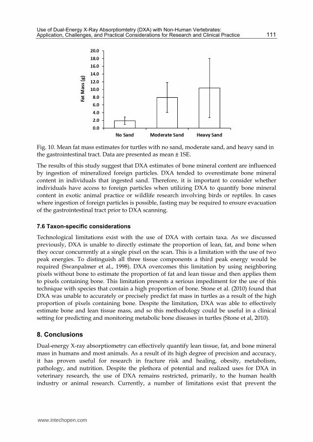
Fig. 10. Mean fat mass estimates for turtles with no sand, moderate sand, and heavy sand in the gastrointestinal tract. Data are presented as mean ± 1SE.
The results of this study suggest that DXA estimates of bone mineral content are influenced by ingestion of mineralized foreign particles. DXA tended to overestimate bone mineral content in individuals that ingested sand. Therefore, it is important to consider whether individuals have access to foreign particles when utilizing DXA to quantify bone mineral content in exotic animal practice or wildlife research involving birds or reptiles. In cases where ingestion of foreign particles is possible, fasting may be required to ensure evacuation of the gastrointestinal tract prior to DXA scanning.
7.6 Taxon-specific considerations
Technological limitations exist with the use of DXA with certain taxa. As we discussed previously, DXA is unable to directly estimate the proportion of lean, fat, and bone when they occur concurrently at a single pixel on the scan. This is a limitation with the use of two peak energies. To distinguish all three tissue components a third peak energy would be required (Swanpalmer et al., 1998). DXA overcomes this limitation by using neighboring pixels without bone to estimate the proportion of fat and lean tissue and then applies them to pixels containing bone. This limitation presents a serious impediment for the use of this technique with species that contain a high proportion of bone. Stone et al. (2010) found that DXA was unable to accurately or precisely predict fat mass in turtles as a result of the high proportion of pixels containing bone. Despite the limitation, DXA was able to effectively estimate bone and lean tissue mass, and so this methodology could be useful in a clinical setting for predicting and monitoring metabolic bone diseases in turtles (Stone et al, 2010).
8. Conclusions
Dual-energy X-ray absorptiometry can effectively quantify lean tissue, fat, and bone mineral mass in humans and most animals. As a result of its high degree of precision and accuracy, it has proven useful for research in fracture risk and healing, obesity, metabolism, pathology, and nutrition. Despite the plethora of potential and realized uses for DXA in veterinary research, the use of DXA remains restricted, primarily, to the human health industry or animal research. Currently, a number of limitations exist that prevent the
www.intechopen.com

A Bird's-Eye View of Veterinary Medicine 112
widespread use of DXA in veterinary medicine. Most notably, the high purchase and maintenance costs and relatively short life-span of DXA units, combined with their size, are major limitations for DXA and hinder its widespread use in traditional veterinary practice. Until these limitations are resolved, DXA will continue to be restricted to human-based clinical settings or with animal research institutions.
9. Acknowledgment
We thank the Oklahoma State University Department of Nutritional Sciences for providing logistical support and access to their DXA equipment.
10. References
Adams, J.E. (1997). Single and dual energy X-ray absorptiometry. European Radiology, Vol. 7, Suppl. 2, pp. (S20-S31), ISSN: 0938-7994
Buelund, L., Nielsen, D., Mcevoy, F., Svalastoga, E., & Bjornvad, C. (2011). Measurement of body composition in cats using computed tomography and dual energy X-ray absorptiometry. Veterinary Radiology and Ultrasound, Vol. 52, No. 2, (March-April 2011), pp. (179-184), ISSN: 1058-8183
Black, A., Tilmont, E.M., Baer, D.J., Rumpler, W.V., Ingram, D.K. Roth, G.S., & Lane, M.A. (2001). Accuracy and precision of dual-energy X-ray absorptiometry for body composition measurements in Rhesus monkeys. Journal of Medical Primatology, Vol. 30, pp. (94-99), ISSN:0047-2565
Castaneda, S., Largo, R., Calvo, E., Rodriguez-Salvanes, F., Marcos, M.E., az-Curiel, M., & Herrero-Beaumont, G. (2006). Bone mineral measurements of subchondral and trabecular bone in healthy and osteoporotic rabbits. Skeletal Radiology, Vol. 35, No. 1, (January 2006), pp. (34-41), ISSN: 0364-2348
Cawkwell, G.D. (1998). Movement artifact and dual X-ray absorptiometry. Journal of Clinical Densitometry, Vol. 1, No. 2, pp. 141-147, ISSN 1094-6950
Clarys, J.P., Scafoglieri, A., Provyn, S., Louis, O., Wallace, J.A., & De Mey, J. (2010). A macro-quality evaluation of DXA variables using whole dissection, ashing, and computer tomography in pigs. Obesity, Vol. 18, No. 8, (August 2010), pp. (1477-1485), ISSN: 1930-7381
Corten, F.G.A., Caulier, H., van der Waerden, J.P.C.M., Kalk, W., Corstens, F.H.M., & Jansen, J.A. (1997). Assessment of bone surrounding implants in goats: Ex vivo measurements by dual X-ray absorptiometry. Biomaterials, Vol. 18, No. 6, (March 1997), pp. (495-501), ISSN: 0142-9612
Dirrigl, F.J., Dalsky, G.P., & Warner, S.E. (2004). Dual-energy X-ray absorptiometry of birds: An examination of excised skeletal specimens. Journal of Veterinary Medicine: Series A, Vol. 51, No. 6, (August 2004), pp. (313-319), ISSN: 0931-184X
Elowsson, P., Forslund, A.H., Mallmin, H., Feuk, U., Hansson, I., & Carlsten, J. (1998). An evaluation of dual-energy X-ray absorptiometry and underwater weighing to estimate body composition by means of carcass analysis in piglets. The Journal of Nutrition, Vol. 128, No. 9, (September 1998), pp. (1543-1549), ISSN: 0022-3166
Engelke, K., Gluer, C.C., & Genant, H.K. (1995). Factors influencing short-term precision of dual X-ray bone absorptiometry (DXA) of spine and femur. Calcified Tissue International, Vol. 56, No. 1, (January 1995), pp. (19-25), ISSN: 0171-967X
www.intechopen.com

Use of Dual-Energy X-Ray Absorptiomtetry (DXA) with Non-Human Vertebrates: Application, Challenges, and Practical Considerations for Research and Clinical Practice 113
Felicetti, L.A., Robbins, C.T., & Shipley, L.A. (2003). Dietary protein content alters energy expenditure and composition of the mass gain in grizzly bears (Ursus arctos horribilis). Physiological and Biochemical Zoology, Vol. 76, No. 2, (March-April 2003), pp. (256-261), ISSN: 1522-2152
Fink, C., Cooper, H.J., Huebner, J.L., Guilak, F., & Kraus, V.B. (2002). Precision and accuracy of a transportable dual-energy X-ray absorptiometry unit for bone mineral measurements in guinea pigs. Calcified Tissue International, Vol. 70, No. 3, (March 2002), pp. (164-169), ISSN: 0171-967X
Fledelius, B., Jorgensen, G.W., Jensen, H.E., & Brimer, L. (2005). Influence of the calcium content of the diet offered to leopard tortoises (Geochelone pardalis). Veterinary Record, Vol. 56, No. 26, (June 2005), pp. (831-835), ISSN 0042-4900
German, A.J. (2006). The growing problem of obesity in dogs and cats. The Journal of Nutrition, Vol. 136, No. 7, (July 2006), pp. (1940S-1946S), ISSN: 0022-3166
Grier, S.J., Turner, A.S., & Alvis, M.R. (1996). The use of dual-energy x-ray absorptiometry in animals. Investigative Radiology, Vol. 31, No. 1, (January 1996), pp. (50-62), ISSN: 0020-9996
Hanafusa, S., Matsusue, Y., Yasunaga, T., Yamamuro, T., Oka, M., Shikinami, Y., & Ikada, Y. (1995). Biodegradable plate fixation of rabbit femoral-shaft osteotomies: A comparative study. Clinical Orthopaedics and Related Research, Vol. 315, (June 1995), pp. (262-271), ISSN: 0009-921X
Jebb, S.A. (1997). Measurement of soft tissue composition by dual energy X-ray absorptiometry. British Journal of Nutrition, Vol. 77, No. 2, (February 1997), pp. (151-163), ISSN: 0007-1145
Jensen, P., Keeling, L., Schutz, K., Andersson, L., Mormede, P., Brandstrom, H., Forkman, B., Kerje, S., Fredriksson, R., Ohlsson, C., Larsson, S., Mallmin, H., & Kindmark, A. (2005). Feather pecking in chickens is genetically related to behavioural and developmental traits. Physiology and Behavior, Vol. 86, (September 2005), pp. (52-60), ISSN: 0031-9384
Kastl, S., Sommer, T., Klein, P., Hohenberger, W., & Engelke, K. (2002). Accuracy and precision of bone mineral density and bone mineral content in excised rat humeri using fan beam dual-energy X-ray absorptiometry. Bone, Vol. 30, No. 1, (January 2002), p. (243-246), ISSN 8756-3282
Kim, W.K., Donalson, L.M., Mitchell, A.D., Kubena, L.F., Nisbet, D.J., & Ricke, S.C. (2006). Effects of alfalfa and fructooligosaccharide on molting parameters and bone qualities using dual energy X-ray absorptiometry and conventional bone assays. Poultry Science, Vol. 85, No. 1, (January 2006), pp. (15-20), ISSN: 0032-5791
Korine, C., Daniel, S., van Tets, I.G., Yosef, R., & Pinshow, B. (2004). Measuring fat mass in small birds by dual-energy X-ray absorptiometry. Physiological and Biochemical Zoology, Vol. 77, No. 3, (May-June 2004), pp. (522-529), ISSN: 1522-2152
Laflamme, D.P., & Kuhlman, G. (1995). The effect of weight loss regimen on subsequent weight maintenance in dogs. Nutrition Research, Vol. 15, No. 7, (July 1995), pp. (1019–1028), ISSN: 0271-5317
Lee, C.Y., Chan, S.H., Lai, H.Y., & Lee, S.T. (2011). A method to develop an in vitro osteoporosis model of porcine vertebrae: Histological and biomechanical study Laboratory investigation. Journal of Neurosurgery: Spine, Vol. 14, No. 6, (June 2011), pp. (789-798), ISSN: 1547-5654
Lorinson, K., Lobcke, S., Skalicky, M., & Lorinson, D. (2009). Densitometric properties of canine appendicular bones based on dual-energy X-ray absorptiometry
www.intechopen.com
A Bird's-Eye View of Veterinary Medicine 114
measurements. Wiener Tierarztliche Monatsschrift, Vol. 96, pp. (72-77), ISSN 0043-535X
Losel, D., Kremer, P., Albrecht, E., & Scholz, A.M. (2010). Comparison of a GE Lunar DPX-IQ and a Norland XR-26 dual energy X-ray absorptiometry scanner for body composition measurements in pigs - in vivo. Archiv Tierzucht, Vol. 53, No. 2, pp. (162-175), ISSN 0003-9438
Lucic, H., Vukovic, S., Posavac, V., Gomercic, M.D., Gomercic, T., Galov, A., Skrtic, D., Curkovic, S., & Gomercic, H. (2010). Application of dual energy X-ray absorptiometry method for small animals in measuring bone mineral density of the humerus of bottlenose dolphins (Tursiops truncatus) from the Adriatic Sea. Veterinarski Arhiv, Vol. 80, No. 2, pp. (299-310), ISSN 0372-5480
Lukaski, H.C., Marchello, M.J., Hall, C.B., Schafer, D.M., & Siders, W.A. (1999). Soft tissue composition of pigs measured with dual X-ray absorptiometry. Nutrition, Vol. 15, No. 9, (September 1999), pp. (697-703), ISSN 0899-9007
Markel, M.D., & Bogdanske, J.J. (1994a). Dual-energy X-ray absorptiometry of canine femurs with and without fracture fixation devices. American Journal of Veterinary Research, Vol. 55, No. 6, (June 1994), pp. (862-866), ISSN 0002-9645
Markel, M.D., & Bogdanske, J.J. (1994b). The effect of increasing gap width on localized densitometric changes within tibial ostectomies in a canine model. Calcified Tissue International, Vol. 54, No. 2, (February 1994), pp. (155-159), ISSN 0171-967X
Mawby, D.I., Bartges, J.W., d'Avignon, A., Laflamme, D.P., Moyers, T.D., & Cottrell, T. (2004). Comparison of various methods for estimating body fat in dogs. Journal of the American Animal Hospital Association, Vol. 40, No. 2, (March-April 2004), pp. (109-114), ISSN 0587-2871
McClure, S.R., Glickman, L.T., Glickman, N.W., & Weaver, C.M. (2001). Evaluation of dual energy X-ray absorptiometry for in situ measurement of bone mineral density of equine metacarpi. American Journal of Veterinary Research, Vol. 62, No. 5, (May 2001), pp. (752-756), ISSN 0002-9645
Mercier, J., Pomar, C., Marcoux, M., Goulet, F., Theriault, M., & Castonguay, F.W. (2006). The use of dual-energy X-ray absorptiometry to estimate the dissected composition of lamb carcasses. Meat Science, Vol. 73, No. 2, (June 2006), pp. (249-257), ISSN 0309-1740
Michels, G.M., Jones, B.D., Huss, B.T., & Wagner-Mann, C. (1995). Endoscopic and surgical retrieval of fishhooks from the stomach and esophagus in dogs and cats: 75 cases (1977-1993). Journal of the American Veterinary Medical Association, Vol. 207, No. 9, (November 1995), pp. (1194-1197), ISSN 0003-1488
Millett, P.J., Cohen, B., Allen, M.J., & Rushton, N. (1998). Bone mineral density changes during fracture healing: a densitometric study in rats, In: Internet Journal of Orthopedic Surgery and related Subjects, 18.8.2011, Available from: http://www.rz.uni-duesseldorf.de/WWW/MedFak/Orthopaedie/journal/, ISSN 0949-2607
Mitchell, A.D., Scholz, A.M., & Conway, J.M. (1998). Body composition analysis of small pigs by dual-energy X-ray absorptiometry. Journal of Animal Science, Vol. 76, No. 9, (September 1998), pp. (2392-2398), ISSN 0021-8812
Mo, G., Zotti, A., Agnesi, S., Finola, M.G., Bernardini, D., & Cozzi, B. (2009). Age classes and sex differences in the skull of the Mediterranean monk seal, Monachus monachus (Hermann, 1779): A Study Based on Bone Shape and Density. Anatomical Record: Advances in Integrative Anatomy and Evolutionary Biology, Vol. 292, No. 4, (April 2009), pp. (544-556), ISSN 1932-8486
www.intechopen.com

Use of Dual-Energy X-Ray Absorptiomtetry (DXA) with Non-Human Vertebrates: Application, Challenges, and Practical Considerations for Research and Clinical Practice 115
Nagy, T.R., & Clair, A.L. (2000). Precision and accuracy of dual-energy X-ray absorptiometry for determining in vivo body composition of mice. Obesity Research, Vol. 8, No. 5, (August 2000), p. (392-398), ISSN 1071-7323
Nielson, D.H., McEvoy, F.J., Poulsen, H.L., Madsen, M.T., Buelund, L.E., & Svalastoga, E. (2004). Dual-energy X-ray absorptiometry of the pig: Protocol development and evaluation. Meat Science, Vol. 68, No. 2, (October 2004), pp. (235-241), ISSN 0309-1740
Peppler, W.W., & Mazess, R.B. (1981). Total body mineral and lean body mass by dual-photon absorptiometry: theory and measurement procedure. Calcified Tissue International, Vol. 33, No. 1, (December 1981), pp. (353-359), ISSN 0171-967X
Pietrobelli, A., Formica, C., Wang, Z.M., & Heymsfield, S.B. (1996). Dual-energy X-ray absorptiometry body composition model: Review of physical concepts. American Journal of Physiology: Endocrinology and Metabolism, Vol. 271, No. 6, (December 1996), pp. (E941-E951), ISSN 0193-1849
Ponnampalam, E.N., Hopkins, D.L., Dunshea, F.R., Pethick, D.W., Butler, K.L., & Warner, R.D. (2007). Genotype and age effects on sheep meat production: Carcass composition predicted by dual energy X-ray absorptiometry. Australian Journal of Experimental Agriculture, Vol. 47, No. 10, pp. (1172-1179), ISSN 0816-1089
Pouilles, J.M., Collard, P., Tremollieres, F., Frayssinet, P., Railhac, J.J., Cahuzac, J.P., Autefage, A., & Ribot, C. (2000). Accuracy and precision of in vivo bone mineral measurements in sheep using dual-energy X-ray absorptiometry. Calcified Tissue International, Vol. 66, No. 1, (January 2000), pp. (70-73), ISSN 0171-967X
Raiti, P., & Haramati, N. (1997). Magnetic resonance imaging and computerized tomography of a gravid leopard tortoise (Geochelone pardalis pardalis) with metabolic bone disease. Journal of Zoo and Wildlife Medicine, Vol. 28, No. 2, (June 1997), p. (189-197), ISSN 1042-7260
Rose, B.S., Flatt, W.P., Martin, R.J., & Lewis, R.D. (1998). Whole body composition of rats determined by dual energy X-ray absorptiometry is correlated with chemical analysis. Journal of Nutrition, Vol. 128, No. 2, (February 1998), pp. (246-250), ISSN 0022-3166
Schneider, S., Breit, S.M., Grampp, S., Kunzel, W.W.F., Liesegang, A., Mayrhofer, E., & Zentek, F. (2004). Comparative assessment of bone mineral measurements obtained by use of dual-energy X-ray absorptiometry, peripheral quantitative computed tomography, and chemical-physical analyses in femurs of juvenile and adult dogs. American Journal of Veterinary Research, Vol. 65, No. 7, (July 2004), pp. (891-900), ISSN 0002-9645
Secombe, C.J., Firth, E.C., Perkins, N.R., & Anderson, B.H. (2002). Pathophysiology and diagnosis of third carpal bone disease in horses: A review. New Zealand Veterinary Journal, Vol. 50, No. 1, (February 2002), pp. (2-8), ISSN 0048-0169
Secor, S.M., & Nagy, T.R. (2003). Non-invasive measure of body composition of snakes using dual-energy X-ray absorptiometry. Comparative Biochemistry and Physiology A: Molecular and Integrative Physiology, Vol. 136, No. 2, (October 2003), pp. (379-389), ISSN 1095-6433
Stevenson, K.T., & van Tets, I.G. (2008). Dual-energy X-ray absorptiometry (DXA) can accurately and nondestructively measure the body composition of small, free-living rodents. Physiological and Biochemical Zoology, Vol. 81, No. 3, (May-June 2008), pp. (373-382), ISSN 1522-2152
Stone, M.D. (2009). Effects of season, sex, and age on the calcium physiology and bone dynamics of turtles. Oklahoma State University, Ph.D. dissertation, Stillwater, OK
www.intechopen.com

A Bird's-Eye View of Veterinary Medicine 116
Stone, M.D., Arjmandi, B.H., & Lovern, M.B. (2010). Dual-energy X-ray absorptiometry (DXA) as a non-invasive tool for the prediction of bone density and body composition of turtles. Herpetological Review, Vol. 41, No. 1, (March 2010), pp. (36-42), ISSN 0018-084X
Swanpalmer, J., Kullenberg, R., & Hansson, T. ( 1998). Measurement of bone mineral using multiple-energy X-ray absorptiometry. Physics in Medicine and Biology, Vol. 43, pp. (379-387), ISSN 0031-9155
Toll, P.W., Gross, K.L., Berryhill, S.A., & Jewell, D.E. (1994). Usefulness of dual energy X-ray absorptiometry for body composition measurement in adult dogs. The Journal of Nutrition, Vol. 124, Suppl. 12, (December 1994), pp. (2601S-2603S), ISSN 0022-3166
Turner, A.S., Mallinckrodt, C.H., Alvis, M.R., & Bryant, H.U. (1995a). Dual-energy X-ray absorptiometry in sheep: Experiences with in-vivo and ex-vivo studies. Bone, Vol. 17, No. 4, (October 1995), pp. (S381-S387), ISSN 8756-3282
Turner, A.S., Norrdin, R.W., Gaarde, S., Connally, H.E., & Thrall, M.A. (1995b). Bone mineral density in feline mucopolysaccharidosis VI measured using dual-energy X-ray absorptiometry. Calcified Tissue International, Vol. 57, No. 3, (September 1995), pp. (191-195), ISSN 0171-967X
Walde, A.D., Delaney, D.K., Harless, M.L., & Pater, L.L. (2007). Osteophagy by the desert tortoise (Gopherus agassizii). Southwestern Naturalist, Vol. 52, No. 1, (March 2007), pp. (147-149), ISSN 0038-4909
Walpert, B. (2000). With ancillary services, you can do well by doing good. In: American College of Physicians Observer, October, 8.2.2011, Available from:
http://www.acpinternist.org Wilson, K.E. (2006). Detection of abdominal aortic calcification with IVA. 8.1.2011, Available
from: http://www.hologic.com/data/W_158_AAC_05-06.pdf Yaissle, J.E., Holloway, C., & Buffington, C.A.T. (2004). Evaluation of owner education as a
component of obesity treatment programs for dogs. Journal of the American Veterinary Medical Association, Vol. 224, No. 12, (June 2004), pp. (1932–1935), ISSN 0003-1488
Zotti, A., Gianesella, M., Ceccato, C., & Morgante, M. (2010). Physiological values and factors affecting the metacarpal bone density of healthy feedlot beef cattle as measured by dual-energy X-ray absorptiometry. Journal of Animal Physiology and Animal Nutrition, Vol. 94, No. 5, (October 2010), pp. (615-622), ISSN 0931-2439
Zotti, A., Isola, M., Sturaro, E., Menegazzo, L., Piccinini, P., & Bernardini, D. (2004a). Vertebral mineral density measured by dual-energy X-ray absorptiometry (DEXA) in a group of healthy Italian boxer dogs. Journal of Veterinary Medicine: Series A, Vol. 51, No. 5, (June 2004), pp. (254-258), ISSN 0931-184X
Zotti, A., Poggi, R., & Cozzi, B. (2009). Exceptional bone density DXA values of the rostrum of a deep-diving marine mammal: A new technical insight in the adaptation of bone to aquatic life. Skeletal Radiology, Vol. 38, No. 12, (December 2009), pp. (1123-1125), ISSN 0364-2348
Zotti, A., Rizzi, C., Chiericato, G., & Bernardini, D. (2003). Accuracy and precision of dual-energy X-ray absorptiometry for ex vivo determination of mineral content in turkey poult bones. Veterinary Radiology and Ultrasound, Vol. 44, No. 1, (January-February 2003), pp. (49-52), ISSN 1058-8183
Zotti, A., Selleri, P., Carnier, P., Morgante, M., & Bernardini, D. (2004b). Relationship between metabolic bone disease and bone mineral density measured by dual-energy X-ray absorptiometry in the green iguana (Iguana iguana). Veterinary Radiology and Ultrasound, Vol. 45, No. 1, (January 2004), pp. (10-16), ISSN 1058-8183
www.intechopen.com
A Bird's-Eye View of Veterinary MedicineEdited by Dr. Carlos C. Perez-Marin
ISBN 978-953-51-0031-7Hard cover, 626 pagesPublisher InTechPublished online 22, February, 2012Published in print edition February, 2012
InTech EuropeUniversity Campus STeP Ri Slavka Krautzeka 83/A 51000 Rijeka, Croatia Phone: +385 (51) 770 447 Fax: +385 (51) 686 166www.intechopen.com
InTech ChinaUnit 405, Office Block, Hotel Equatorial Shanghai No.65, Yan An Road (West), Shanghai, 200040, China
Phone: +86-21-62489820 Fax: +86-21-62489821
Veterinary medicine is advancing at a very rapid pace, particularly given the breadth of the discipline. Thisbook examines new developments covering a wide range of issues from health and welfare in livestock, pets,and wild animals to public health supervision and biomedical research. As well as containing reviews offeringfresh insight into specific issues, this book includes a selection of scientific articles which help to chart theadvance of this science. The book is divided into several sections. The opening chapters cover the veterinaryprofession and veterinary science in general, while later chapters look at specific aspects of applied veterinarymedicine in pets and in livestock. Finally, research papers are grouped by specialisms with a view to exploringprogress in areas such as organ transplantation, therapeutic use of natural substances, and the use of newdiagnostic techniques for disease control. This book was produced during World Veterinary Year 2011, whichmarked the 250th anniversary of the veterinary profession. It provides a fittingly concise and enjoyableoverview of the whole science of veterinary medicine.
How to referenceIn order to correctly reference this scholarly work, feel free to copy and paste the following:
Matthew D. Stone and Alec J. Turner (2012). Use of Dual-Energy X-Ray Absorptiometry..., A Bird's-Eye Viewof Veterinary Medicine, Dr. Carlos C. Perez-Marin (Ed.), ISBN: 978-953-51-0031-7, InTech, Available from:http://www.intechopen.com/books/a-bird-s-eye-view-of-veterinary-medicine/use-of-dxa-with-non-human-vertebrates-applications-limitations-and-practical-considerations-for-rese

© 2012 The Author(s). Licensee IntechOpen. This is an open access articledistributed under the terms of the Creative Commons Attribution 3.0License, which permits unrestricted use, distribution, and reproduction inany medium, provided the original work is properly cited.