urinary and endocrine problems. choose the correct statement(s) about microscopic hematuria. a) it...
TRANSCRIPT

Urinary and Endocrine problems

Choose the correct statement(s) about microscopic hematuria.
A) It is defined as defined as >10 red blood cells per high-power field B) It occurs at a prevalence of ≤2%
C) Referral to a nephrologist is indicated if dysmorphic red blood
cells are present D) All the above

Answer
• C) Referral to a nephrologist is indicated if dysmorphic red blood cells are present
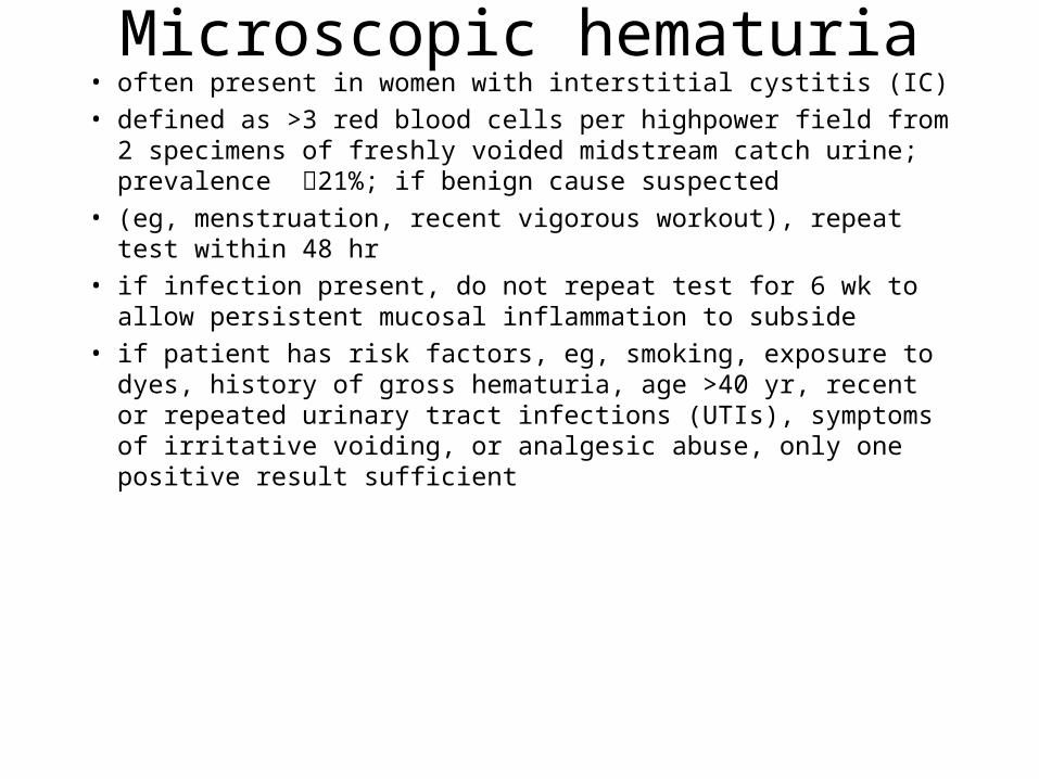
Microscopic hematuria• often present in women with interstitial cystitis (IC)
• defined as >3 red blood cells per highpower field from 2 specimens of freshly voided midstream catch urine; prevalence 21%; if benign cause suspected
• (eg, menstruation, recent vigorous workout), repeat test within 48 hr
• if infection present, do not repeat test for 6 wk to allow persistent mucosal inflammation to subside
• if patient has risk factors, eg, smoking, exposure to dyes, history of gross hematuria, age >40 yr, recent or repeated urinary tract infections (UTIs), symptoms of irritative voiding, or analgesic abuse, only one positive result sufficient

Choose the correct statements about interstitial cystitis (IC).It is usually associated with urinary
urgency, frequency, and nocturiaPrevalence is 38% to 85% in women with chronic
pelvic painIt is rarely associated with other disorders
It is thought to involve activation of pain C-fibers that cause release of substance P
The glycosaminoglycan layer is considered an important component of its etiology
A) 2,3,4,5 B) 1,2,4,5 C) 1,2,3,4 D) 1,3,4,5

It is usually associated with urinary urgency, frequency, and nocturia
Prevalence is 38% to 85% in women with chronic pelvic pain
It is thought to involve activation of pain C-fibers that cause release of substance P
The glycosaminoglycan layer is considered an important component of
its etiology B) 1,2,4,5
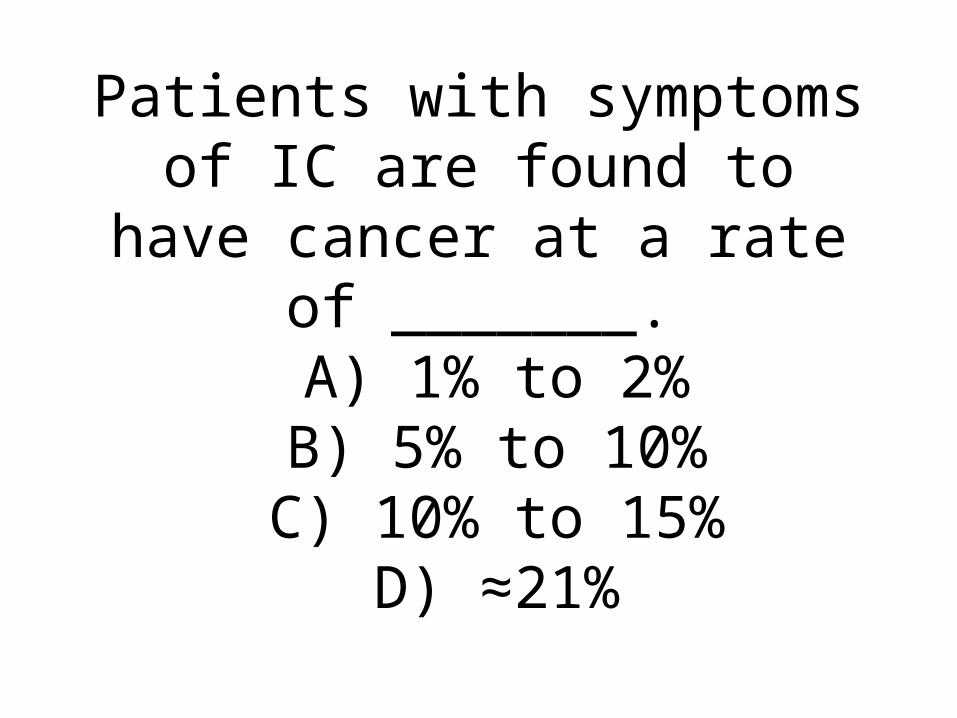
Patients with symptoms of IC are found to have cancer at a rate of
_______. A) 1% to 2% B) 5% to 10%
C) 10% to 15% D) ≈21%

Answer
• A) 1% to 2%

Which of the following statements about the treatment of IC is incorrect?
A) Pentosan polysulfate is the only oral agent approved for IC
B) Hydroxyzine is helpful in women with IC who suffer from allergies
C) Agents that promote alkalinity of urine may help
D) Kegel exercises are beneficial

Answer
• D) Kegel exercises are beneficial

First-line treatment of IC
• avoid foods that exacerbate symptoms;
• agents that promote alkalinity of urine may help, eg, calcium glycerophosphate (Prelief) or calcium carbonate
• stress management important; exercise (eg, yoga, Pilates

IC• Oral medications: amitriptyline—also used for vulvar pain;• speaker starts at 10 mg and tapers dose upward (improvement seen at 25-50 mg);
also helps with nocturia• hydroxyzine —causes sedation; start with 25 mg at bed time and may increase to
50 mg• cimetidine speaker uses infrequently• Pentosan polysulfate: only oral agent approved for IC• Heparin analogue; may help renew GAG lining of bladder• Therapy may take 6 mo (discontinue if no improvement after 1 yr)• side effects—well tolerated• upper gastrointestinal symptoms seen in some patients; discontinue if diarrhea,
reversible loss of hair (in <3%), or increase in liver enzymes develops (check every 6 mo and discontinue until normalized, then try lower dose) may cause easy bruising (decrease dose)
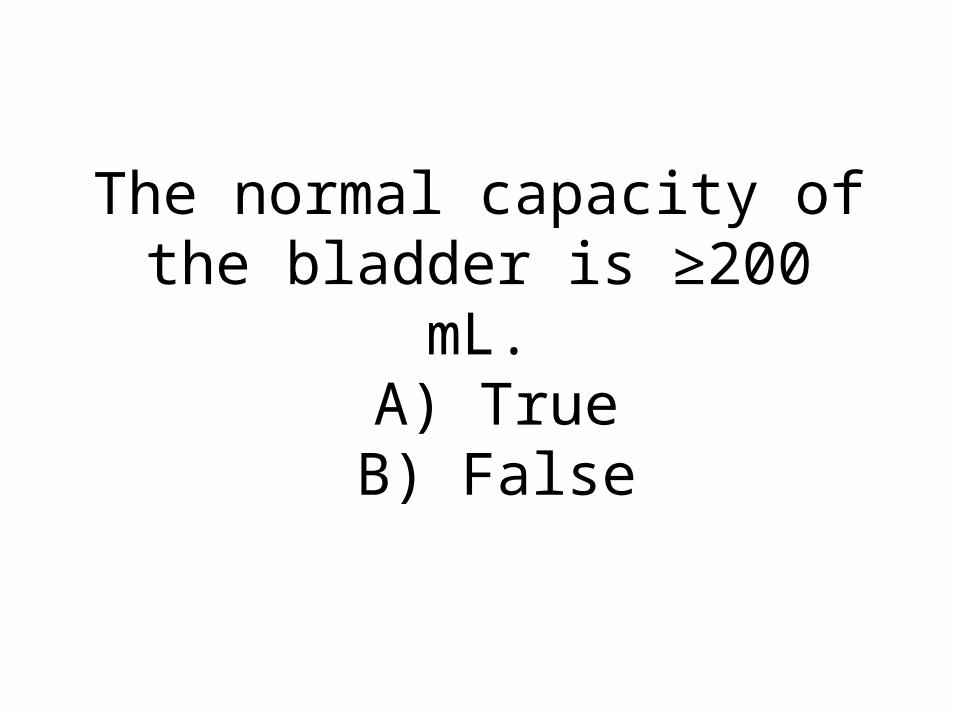
The normal capacity of the bladder is ≥200 mL.
A) True B) False
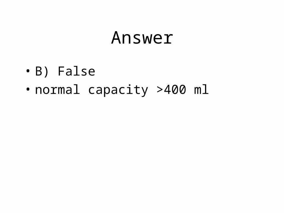
Answer
• B) False
• normal capacity >400 ml

Choose the correct statements about bladder function and urinary incontinence .Stress incontinence is associated with
exertionUrinary frequency is usually due to urge incontinence
Urinary frequency is defined as needing to urinate >8 times daily (>10 for older patients)
The normal rate of urine production 1 mL/minNocturia is defined as needing to urinate >3 times per night
A) 1,3,4,5 B) 2,3,4,5 C) 1,2,3,4 D) 1,2,4,5
Answer
• Stress incontinence is associated with exertion• Urinary frequency is usually due to urge
incontinence• Urinary frequency is defined as needing to
urinate >8 times daily (>10 for older patients)• The normal rate of urine production 1 mL/min• C) 1,2,3,4

normal values
• frequency considered as >8 times daily (>10 for older patients)
• nocturia defined as >1 per night
• normal intake of fluid varies from 50 to 100 oz per day
• normal urine production 1 mL/min
• postvoid residual volume <100 mL for patients >65 yr of age (<50 mL if younger
Which of the following should be evaluated during physical examination of a woman with
urinary incontinence? A) Gait and reflexes
B) Contraction of anal sphincter after tapping the clitoris or stroking along the
bulbocavernosus C) The strength of a Kegel contraction using
only the pelvic floor muscles D) All the above

Answer
• D) All the above

In the Knack procedure, the patient performs a Kegel
exercise, then walks to the bathroom with the pelvic floor
muscles contracted. A) True B) False

Answer
• A) True

Which of the following options has (have) been shown to be
effective for treatment of urge incontinence?
A) Posterior tibial nerve stimulation
B) OnobotulinumtoxinA C) Sacral nerve stimulation
D) All the above

Answer
• D) All the above

Medication
• anticholinergic agents— consider trial for overactive bladder symptoms; allow 4 to 6 wk before increasing dose or trying other medication
• newer oral agents have greater selectivity for receptors in bladder and cause fewer visual changes, but all may cause some cognitive impairment
• May interact with coumadin and digoxin, and prolong QT interval in older patient

Double-blind randomized controlled trials have proven that nonsteroidal anti-inflammatory drugs and antioxidants reduce
risk for Alzheimer disease (AD). A) True B) False

Answer
• B) False

Personality and behavior are often preserved in patients with
_______, and patients may appear normal on mental status
testing. A) AD
B) Vascular dementia C) Lewy body dementia
D) Frontotemporal dementia
Answer
• B) Vascular dementia

Cholinesterase inhibitors: A) Are typically recommended
for severe AD B) Modify the course of AD
C) May slow progression of AD D) Should never be used
indefinitely

Answer
• C) May slow progression of AD

Double-blind randomized controlled trials have proven that nonsteroidal anti-inflammatory drugs and antioxidants reduce
risk for Alzheimer disease (AD). A) True B) False

Answer
• B) False

Personality and behavior are often preserved in patients with
_______, and patients may appear normal on mental status
testing. A) AD
B) Vascular dementia C) Lewy body dementia
D) Frontotemporal dementia

Answer
• B) Vascular dementia
Cholinesterase inhibitors: A) Are typically recommended
for severe AD B) Modify the course of AD
C) May slow progression of AD D) Should never be used
indefinitely

Answer
• C) May slow progression of AD
Memantine: A) Is typically indicated for mild
AD B) Is not associated with
improved behavioral symptoms C) Is useful in stabilizing nerve
cells D) Should not be combined with
cholinesterase inhibitors

Answer
• C) Is useful in stabilizing nerve cells

Which of the following is the least reliable indicator of
driving risk? A) Patient's self-report
B) Patient's self-restriction C) Score of ≥1 on Clinical
Dementia Rating scale D) Adverse rating by caregiver

Answer
• A) Patient's self-report

During the early stage of AD, which of the following should be
considered? A) Advance directives and goals
of care B) Establishment of surrogate
decision maker C) Financial planning
D) All the above

Answer
• D) All the above

Choose the correct statement about attention-deficit/hyperactivity
disorder (ADHD). A) Present at birth
B) Symptoms do not present until after age 7 yr
C) In adult ADHD, symptoms typically present after age 25 yr
D) Symptoms resolve in ≈60% of patients after age 25 yr

Answer
• A) Present at birth

All the following are criteria for ADHD in children, except:
A) ≥6 hyperactive or inattentive symptoms
B) Impairment before age of 7 yr C) Impairment in ≥1 setting
D) Symptoms occur often and are clinically significant

Answer
• C) Impairment in ≥1 setting
A patient with symptoms of ADHD that occur exclusively during manic, depressive, or
psychotic episodes is more likely to have ADHD than another
psychiatric disorder. A) True B) False

Answer
• B) False

Which of the following can cause hyperactive or inattentive symptoms that can lead to
impairment? A) Concussion B) Sleep apnea C) Medications
D) All the above

Answer
• D) All the above

A 34-year-old woman is evaluated for amenorrhea and infertility. Her last menses occurred 4 months ago, before which her menses were regular. The patient has two children, age 3 and 6 years. She had a spontaneous abortion 6 months ago and underwent dilation and curettage 1 month later. There was no withdrawal bleeding while she was on an oral contraceptive pill
for 2 months after the procedure. A recent progestin withdrawal challenge with medroxyprogesterone acetate did not produce withdrawal bleeding. There is no relevant
family history. She takes no other medications. On physical examination, vital signs are normal, and BMI is 30. No acne or hirsutism is
detected. All other examination findings are normal. Laboratory studies: Follicle-stimulating hormone
6 mU/mL (6 U/L)Prolactin
15 ng/mL (15 µg/L)Thyroid-stimulating hormone
3 µU/mL (3 mU/L)Thyroxine (T4), free
1.3 ng/dL (16.8 pmol/L)Pregnancy test
NegativeWhich of the following is the most appropriate next diagnostic test?
AHysteroscopyBKaryotype
CMeasurement of serum 17-hydroxyprogesterone levelDTransvaginal pelvic ultrasonography

D Transvaginal pelvic ultrasonography
• Objective:Evaluate secondary amenorrhea.
• Key Point
• In secondary amenorrhea, absence of menstrual flow after a progestin withdrawal challenge with medroxyprogesterone acetate indicates estrogen absence and/or an anatomic defect; when such absence occurs after dilation and curettage, the possibility of Asherman syndrome must be considered.
• This patient should undergo transvaginal pelvic ultrasonography. If results of the initial laboratory assessment are normal, the cornerstone of the evaluation for secondary amenorrhea rests on the results of a progestin withdrawal challenge (medroxyprogesterone acetate, 10 mg orally for 10 days). Menstrual flow on progestin withdrawal indicates relatively normal estrogen production and a patent outflow tract, which limits diagnostic evaluation to chronic anovulation. Absence of flow indicates estrogen absence and/or an anatomic defect. In patients with no flow, pelvic anatomy is assessed with ultrasonography and/or MRI. The absence of menses for several months after dilation and curettage in this patient suggests severe endometrial damage or formation of scar tissue (Asherman syndrome). Therefore, an ultrasound is appropriate to assess the pelvic anatomy.
• A hysteroscope is a fiberoptic device inserted into the uterus via the vagina and cervix that enables direct visualization of the endometrial cavity. It is typically not the first diagnostic test to assess for the presence of anatomic disorders that may be associated with amenorrhea because of its cost and invasiveness.
• Approximately 50% of primary amenorrhea is caused by chromosomal disorders that result in gonadal dysgenesis and depletion of ovarian follicles. Turner syndrome, the most common disorder in this category, is classically associated with a 45,XO genotype and is characterized by a lack of secondary sexual characteristics, growth retardation, a webbed neck, and frequent skeletal abnormalities. Because this patient has secondary, not primary, amenorrhea, a karyotype is not needed.
• The patient previously had normal menses and fertility and has no stigmata of hyperandrogenism. Therefore, 21-hydroxylase deficiency is unlikely, and measurement of the serum 17-hydroxyprogesterone level is unlikely to provide useful information.
• Bibliography
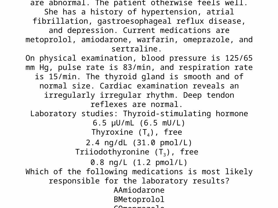
A 76-year-old woman is reevaluated after results of thyroid function tests performed 2 weeks ago are abnormal. The patient otherwise feels well. She has a history of hypertension, atrial fibrillation, gastroesophageal reflux disease, and depression. Current medications are metoprolol,
amiodarone, warfarin, omeprazole, and sertraline. On physical examination, blood pressure is 125/65 mm Hg, pulse rate is
83/min, and respiration rate is 15/min. The thyroid gland is smooth and of normal size. Cardiac examination reveals an irregularly irregular rhythm.
Deep tendon reflexes are normal. Laboratory studies: Thyroid-stimulating hormone
6.5 µU/mL (6.5 mU/L)Thyroxine (T4), free
2.4 ng/dL (31.0 pmol/L)Triiodothyronine (T3), free
0.8 ng/L (1.2 pmol/L)Which of the following medications is most likely responsible for the
laboratory results?AAmiodaroneBMetoprololCOmeprazoleDSertraline
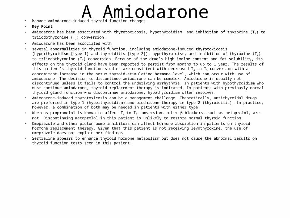
A Amiodarone• Manage amiodarone-induced thyroid function changes. • Key Point
• Amiodarone has been associated with thyrotoxicosis, hypothyroidism, and inhibition of thyroxine (T4) to triiodothyronine (T3) conversion.
• Amiodarone has been associated with• several abnormalities in thyroid function, including amiodarone-induced thyrotoxicosis (hyperthyroidism [type 1] and thyroiditis
[type 2]), hypothyroidism, and inhibition of thyroxine (T4) to triiodothyronine (T3) conversion. Because of the drug’s high iodine content and fat solubility, its effects on the thyroid gland have been reported to persist from months to up to 1 year. The results of this patient’s thyroid function studies are consistent with decreased T4 to T3 conversion with a concomitant increase in the serum thyroid-stimulating hormone level, which can occur with use of amiodarone. The decision to discontinue amiodarone can be complex. Amiodarone is usually not discontinued unless it fails to control the underlying arrhythmia. In patients with hypothyroidism who must continue amiodarone, thyroid replacement therapy is indicated. In patients with previously normal thyroid gland function who discontinue amiodarone, hypothyroidism often resolves.
• Amiodarone-induced thyrotoxicosis can be a management challenge. Theoretically, antithyroidal drugs are preferred in type 1 (hyperthyroidism) and prednisone therapy in type 2 (thyroiditis). In practice, however, a combination of both may be needed in patients with either type.
• Whereas propranolol is known to affect T4 to T3 conversion, other β-blockers, such as metoprolol, are not. Discontinuing metoprolol in this patient is unlikely to restore normal thyroid function.
• Omeprazole and other proton pump inhibitors can affect hormone absorption in patients on thyroid hormone replacement therapy. Given that this patient is not receiving levothyroxine, the use of omeprazole does not explain her findings.
• Sertraline appears to enhance thyroid hormone metabolism but does not cause the abnormal results on thyroid function tests seen in this patient.

A 30-year-old woman is evaluated in the hospital for paresthesias of the fingers and mouth that started 2 days after she underwent a difficult total
thyroidectomy for Graves disease. Her only medication is atenolol. On physical examination, vital signs are normal except for a respiration
rate of 24/min. Trousseau and Chvostek signs are present. Laboratory studies: Albumin
4.1 g/dL (41 g/L)Calcium
7.5 mg/dL (1.88 mmol/L)Phosphorus
3.8 mg/dL (1.2 mmol/L)Parathyroid hormone
6 pg/mL (6 ng/L)Which of the following is the most likely cause of the hypocalcemia?
AHyperventilationBHypoparathyroidism
CPseudohypoparathyroidismDPseudo-pseudohypoparathyroidism

B Hypoparathyroidism• Diagnose hypoparathyroidism after thyroidectomy.
• Key Point
• Hypocalcemia following neck surgery is likely due to hypoparathyroidism.
• The most likely explanation for this
• patient’s hypocalcemia is hypoparathyroidism. She has hypocalcemia because of inadvertent removal of or damage to the parathyroid glands during thyroidectomy, which has led to hypoparathyroidism. Absence of parathyroid hormone (PTH) action causes a lack of stimulation of osteoclasts with lack of mobilization of calcium from bone, increased urine calcium loss, and resultant hypocalcemia. Additionally, 1α-hydroxylase is downregulated, with a resultant decreased production of 1,25-dihydroxy vitamin D. This decreased production impairs the absorption of calcium and phosphorus in the gut.
• Anxiety-induced hyperventilation can induce a decrease in the ionized calcium level. As the partial pressure of carbon dioxide falls, there is a dissociation of hydrogen ions from albumin to compensate for the respiratory alkalosis. This dissociation leads to increased binding of calcium ions to albumin, which causes the ionized calcium level to decrease. This decrease can be sufficient to induce clinical features of hypocalcemia. However, the total calcium level does not decrease, as it has in this patient.
• Tissue resistance to the action of PTH occurs in the rare congenital condition of pseudohypoparathyroidism. Despite increased PTH levels (not decreased, as in this patient), patients with this condition have hypocalcemia and hyperphosphatemia. Phenotypically, patients with pseudohypoparathyroidism have a short round face, short neck, and short fourth metacarpal bone.
• Pseudo-pseudohypoparathyroidism refers to the condition in which patients have the phenotypic appearance of pseudohypoparathyroidism but normal calcium and phosphorus levels because of normal PTH secretion, function, and action.

A 55-year-old man is evaluated for new-onset type 2 diabetes mellitus. The patient also reports a chronic productive cough and poor exercise tolerance. Six months ago, he had normal results on physical
examination and normal laboratory values, including glucose, lipids, and electrolytes. He has a 40-pack-year smoking history. There is no pertinent family history. The patient takes no medications.
On physical examination, temperature is 36.5 °C (97.7 °F), blood pressure is 172/92 mm Hg, pulse rate is 90/min, respiration rate is 21/min, and BMI is 24.5. Mucous membranes and nail beds are
hyperpigmented. There is temporal muscle wasting and proximal muscle weakness in the upper and lower extremities. He does not have a dorsal fat pad, moon facies, or purple striae.
Laboratory studies: ElectrolytesSodium
146 meq/L (146 mmol/L)Potassium
2.4 meq/L (2.4 mmol/L)Chloride
101 meq/L (101 mmol/L)Bicarbonate
33 meq/L (33 mmol/L)Adrenocorticotropic hormone (ACTH)
365 pg/mL (80.3 pmol/L)Cortisol (8 AM)
58 µg/dL (1601 nmol/L) (normal range, 5-25 µg/dL [138-690 nmol/L])Which of the following is the most likely cause of this patient’s findings?
AAdrenal adenomaBAdrenal carcinoma
CCushing diseaseDEctopic ACTH secretion

D Ectopic ACTH secretion• dentify secondary causes of diabetes mellitus. • Key Point• A secondary cause should be strongly considered in patients with rapid onset of diabetes mellitus in the
absence of risk factors.• This patient’s findings are most likely caused by ectopic secretion ofVadrenocorticotropic hormone (ACTH).
He had rapid onset of diabetes mellitus associated with features of excess glucocorticoid and mineralocorticoid activity (metabolic alkalosis, unprovoked hypokalemia, and hypertension). Cortisol binds to the glucocorticoid receptors to exert its effects in various tissues. However, at high concentrations, such as in this patient, cortisol also binds to the mineralocorticoid receptors in the kidney; hence the observed increase in mineralocorticoid activity can occur. Examination findings include features consistent with excessive ACTH secretion: hyperpigmented mucous membranes, proximal myopathy, and elevated levels of cortisol and ACTH. Notably, the patient had a rapid onset of disease that is more typical of a malignant process than an ACTH-secreting pituitary tumor (Cushing disease). A chest radiograph to detect a small cell lung cancer is a reasonable next diagnostic step for this patient who is a cigarette smoker and has findings of excess cortisol and ACTH.
• Both adrenal adenoma and carcinoma are unlikely diagnoses. These disorders will suppress, not elevate, the ACTH level because of the autonomous production of cortisol.
• Bibliography• Arnaldi G, Angeli A, Atkinson AB, et al. Diagnosis and complications of Cushing’s syndrome: a consensus
statement. J Clin Endocrinol Metab. 2003;88(12):5593-5602. [PMID:14671138] - See PubMed

A 54-year-old woman undergoes transsphenoidal surgery to remove a nonfunctioning pituitary macroadenoma. The patient develops polyuria and
polydipsia postoperatively, and urine output is 8 L/d; central diabetes insipidus is diagnosed. After she is treated with desmopressin, urine output decreases. Four
days after her operation, she is discharged with instructions to take desmopressin nasal spray, one puff (10 µg) twice daily, and hydrocortisone, 20 mg on arising in
the morning. After a few days at home, her appetite becomes normal again and she begins eating her normal amount of food and resumes her years-long habit of drinking 8 glasses (64 oz) of water daily. One month after discharge, her husband
brings her to the emergency department after she becomes progressively more somnolent and incoherent over a 3-hour period. She has been taking no
medications except for the desmopressin and hydrocortisone. Physical examination reveals an awake but confused woman. Blood pressure is 132/80 mm Hg, pulse rate is 88/min, respiration rate is 16/min, and BMI is 24.
Skin turgor is normal, mucous membranes are moist, and there is no edema. Other than confusion, findings from the remainder of the general physical and
neurologic examinations are normal. Laboratory results reveal a serum sodium level of 116 meq/L (116 mmol/L).
Which of the following is the most likely cause of the hyponatremia?ACerebral salt-wasting syndrome
BCortisol deficiencyCExcessive water ingestion
DSyndrome of inappropriate antidiuretic hormone secretion
C C Excessive water ingestion• Evaluate hyponatremia caused by desmopressin treatment in a patient with diabetes insipidus. • Key Point• Continued drinking without fluid restriction while taking a fixed dose of desmopressin can cause hyponatremia.• This patient’s hyponatremia is most likely• the result of excessive water ingestion. Patients with central diabetes insipidus cannot concentrate their urine and
respond to subcutaneous desmopressin administration by decreased urine output and increased urine osmolality. Desmopressin can cause hyponatremia if a person continues to drink without any fluid restriction, particularly if their fluid intake is excessive, although progressive nausea usually limits the intake. For patients with chronic central diabetes insipidus, allowing breakthrough polyuria to occur by temporarily reducing or stopping desmopressin can prevent hyponatremia and enable recognition of the patients in whom the disease remits. Patients who develop diabetes insipidus after trauma or neurosurgery have been noted to recover normal urinary concentrating ability and normal urine output as late as 10 years after the initial insult.
• Cerebral salt wasting, a syndrome characterized by hypovolemia and hyponatremia, usually occurs within 10 days of a neurosurgical procedure or disease, particularly subarachnoid hemorrhage. Cerebral salt wasting is an unlikely diagnosis in this patient in the absence of hypotension or other signs of hypovolemia.
• In patients receiving desmopressin who develop severe hyponatremia and a low urine output, cortisol deficiency should be part of the differential diagnosis. Cortisol deficiency is unlikely in this patient because she is on an adequate dosage of replacement cortisol.
• The syndrome of inappropriate antidiuretic hormone secretion seems an unlikely cause of hyponatremia in a patient who has a recent diagnosis of diabetes insipidus and responded to an appropriate dosage of desmopressin.

A 35-year-old woman is evaluated for diaphoresis, a 3.6-kg (8.0-lb) weight loss, and elevated blood glucose levels 6 weeks after delivering her second child. She has an 18-year history of type 1 diabetes mellitus, which has been successfully treated with an insulin pump for the past 8
years. Her glycemic control during this period has been outstanding, with an average hemoglobin A1c value of 6.2%. Although her diabetes was
well controlled during most of the gestation, the insulin dosage needed to be increased by almost 50% to maintain glycemic control. Postpartum, the
patient reduced the insulin dosage to her prepregnancy baseline requirements, but this step has been inadequate to control her blood
glucose level. On physical examination, vital signs are normal. Tachycardia, tremor,
hyperreflexia, and a wide pulse pressure are noted.Results of a complete blood count and metabolic panel are normal.
Measurement of which of the following is most likely to diagnose the cause of her deteriorating control of her blood glucose level?
AAntitransglutaminase antibody titersBHemoglobin A1c value
CPostprandial glucose levelDThyroid-stimulating hormone level
EUrine free cortisol level
D Thyroid-stimulating hormone level
• Evaluate hyperthyroidism as a cause of loss of glycemic control in a patient with diabetes mellitus. • Key Point• Hyperthyroidism can contribute to poor glycemic control in patients with diabetes mellitus.• Measurement of this patient’sthyroid-stimulating hormone (TSH) level is most likely to diagnose the cause of her
deteriorating glycemic control. The deterioration in glycemic control in patients with previously stable diabetes mellitus should always raise the suspicion of an underlying illness that may increase insulin resistance or diminish insulin production. This patient’s symptoms of diaphoresis and weight loss and her deteriorating glycemic control suggest thyrotoxicosis; in the postpartum setting, women with type 1 diabetes have an increased risk of postpartum thyroiditis. An excess of thyroid hormone increases hepatic glucose production and can contribute to new deterioration in glycemic control in diabetes. Therefore, her TSH level should be checked.
• The levels of antitransglutaminase antibodies are elevated in patients with celiac disease, which does occur with increased frequency in patients with type 1 diabetes. Diarrhea is a clinically significant finding in approximately 50% of patients. There are no suggestive gastrointestinal symptoms of this condition other than weight loss, which is seen in this patient. However, weight loss by itself is insufficient to suggest the disease, and celiac disease cannot explain her other symptoms.
• Hemoglobin A1c values and postprandial glucose levels are measurements of glycemic control but do not provide any useful information about the nature of this patient’s recently deteriorated glycemic control.
• Cushing syndrome can be diagnosed with a urine free cortisol level. Cushing syndrome is caused by excessive amounts of endogenous or exogenous glucocorticoids causing central adiposity and is marked by weight gain, supraclavicular fat pads, round or “moon” facies, and a “buffalo hump.” Other findings include violaceous striae and incidental bruising, hirsutism, amenorrhea, and abnormal libido. Antagonism of insulin action causes hyperglycemia. However, there is no evidence suggestive of Cushing syndrome in this patient, so obtaining a urine free cortisol level is not the best option.
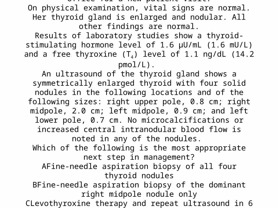
An asymptomatic 65-year old woman comes to the office for a new patient visit.
On physical examination, vital signs are normal. Her thyroid gland is enlarged and nodular. All other findings are normal.
Results of laboratory studies show a thyroid-stimulating hormone level of 1.6 µU/mL (1.6 mU/L) and a free thyroxine (T4) level of 1.1 ng/dL (14.2
pmol/L). An ultrasound of the thyroid gland shows a symmetrically enlarged thyroid with four solid nodules in the following locations and of the following sizes: right upper pole, 0.8 cm; right midpole, 2.0 cm; left
midpole, 0.9 cm; and left lower pole, 0.7 cm. No microcalcifications or increased central intranodular blood flow is noted in any of the nodules. Which of the following is the most appropriate next step in management?
AFine-needle aspiration biopsy of all four thyroid nodulesBFine-needle aspiration biopsy of the dominant right midpole nodule only
CLevothyroxine therapy and repeat ultrasound in 6 monthsDThyroid scan
BFine-needle aspiration biopsy of the dominant right midpole
nodule only• Manage a multinodular goiter. • Key Point• The risk of cancer is the same in solitary thyroid nodules and multinodular goiters; a fine-needle aspiration biopsy is
recommended for nonfunctioning nodules greater than 1.0 to 1.5 cm in diameter or for nodules that have concerning ultrasound characteristics.
• The most appropriate management for this\• patient is fine-needle aspiration biopsy of the dominant nodule. Several studies have shown that multinodular goiters harbor
the same risk of thyroid cancer as solitary thyroid nodules. Most experts perform fine-needle aspiration biopsy on individual nodules greater than 1.0 to 1.5 cm in diameter or on nodules that have concerning ultrasound characteristics, such as microcalcifications or prominent central intranodular blood flow. In this patient, only the right midpole nodule meets these criteria; the other three nodules do not require fine-needle aspiration biopsy.
• This patient who is currently euthyroid has a dominant thyroid nodule that is associated with a significantly increased risk of cancer. Repeating the ultrasound in 6 months could delay the diagnosis of cancer. Additionally, the routine use of levothyroxine for nodule shrinkage is not recommended because the drug is generally ineffective in reducing nodule size, cannot be used to determine whether a nodule is benign or malignant, and exposes patients to the undesirable side effects of thyroid hormone replacement, such as tachycardia and osteoporosis.
• Because the thyroid-stimulating hormone (TSH) and free thyroxine (T4) levels are normal, there is little clinical utility in obtaining a thyroid scan. If the TSH level had been suppressed, a thyroid scan and radioactive iodine uptake would have been warranted to look for a toxic nodule or toxic multinodular goiter.
• Bibliography• Hegedüs L. Clinical practice. The thyroid nodule. N Engl J Med. 2004;351(17):1764-1771. [PMID:15496625] - See PubMed • Related Syllabus
A 54-year-old woman with a 6-year history of type 2 diabetes mellitus is evaluated for suboptimal glycemic control. Her
current hemoglobin A1c value is 8.1%. Her diabetes regimen consists of metformin, 1000 mg twice daily, and glimepiride, 4 mg/d; the patient declines all injection therapy at this time.
She has no other medical problems. There are no abnormalities in cardiovascular, kidney, or liver
function.In addition to reinforcing lifestyle modifications, which of the following is most likely to maximally improve her glycemic
control? AAdd acarboseBAdd exenatide
CAdd pioglitazoneDIncrease the glimepiride dosageEIncrease the metformin dosage

C Add pioglitazone• Treat a patient whose type 2 diabetes mellitus is inadequately controlled on dual oral therapy. • Key Point• A recent American Diabetes Association/European Association for the Study of Diabetes Consensus Statement on the
management of type 2 diabetes mellitus endorses the addition of pioglitazone to the regimen of a patient with suboptimal glycemic control on dual oral therapy.
• In addition to her current therapy, this• patient should also take pioglitazone to improve her glycemic control. According to the American Diabetes
Association, most patients with diabetes mellitus should have a hemoglobin A1c value of less than 7%. This patient is substantially above that target despite dual therapy with metformin and a sulfonylurea. Several options are available, including increasing lifestyle modifications, adding a third oral agent (such as a thiazolidinedione, an α-glucosidase inhibitor, or a dipeptidyl peptidase-IV inhibitor), or adding an injectable agent, such as exenatide or insulin. This patient has expressed the desire to avoid injections, at least for the time being. As a result, her options are more limited. Of the choices provided, the only one that has been shown to reduce hemoglobin A1c values by approximately 1% is pioglitazone. This treatment has been endorsed by the recent American Diabetes Association/European Association for the Study of Diabetes Consensus Statement on the management of type 2 diabetes.
• The glucose-lowering power of acarbose ranges between a 0.5% and 0.8% reduction in hemoglobin A1c values and is thus inferior to that of pioglitazone. Furthermore, acarbose may have substantial gastrointestinal side effects, including bloating, abdominal cramps, diarrhea, and flatulence.
• Increasing the dosage of a sulfonylurea, such as glimepiride, above half the maximum recommended dosage provides little additional therapeutic benefit. Similarly, patients taking metformin, 2000 mg/d, are unlikely to get much additional benefit from increasing the dosage to 2550 mg/d (the maximum recommended dosage).

A 56-year-old woman is evaluated for a 2-year history of chronic low back pain. She also has had a 2-cm (0.8-in) height loss during this period. Medical history is remarkable for chronic obstructive pulmonary disease requiring intermittent high doses of prednisone.
Current medications are albuterol and ipratropium bromide inhalers; prednisone, 20 mg/d; vitamin D, 800 U/d; and calcium, 1500 mg/d.
On physical examination, temperature is normal, blood pressure is 135/80 mm Hg, pulse rate is 100/min, respiration rate is 24/min, and BMI is 28. Breath sounds are distant with an
occasional wheeze. There is back tenderness. Neurologic examination findings are unremarkable.
Laboratory studies show normal serum calcium, phosphorus, parathyroid hormone, and vitamin D levels.
A radiograph of the spine shows a compression fracture of the T8 vertebra. A dual-energy x-ray absorptiometry scan reveals a T-score of –2.2 in the lumbosacral spine and –2.5 in the
left hip. Which of the following is the best treatment for this patient?
ACalcitoninBIncreased dosage of vitamin D (to 1000 U/d)
CRaloxifeneDRisedronate
D Risedronate• Treat corticosteroid-induced osteoporosis. • Key Point• Oral bisphosphonates are the therapy of choice for corticosteroid-induced osteoporosis; all patients with corticosteroid-induced
osteoporosis should also receive adequate calcium and vitamin D supplementation. • This patient with corticosteroid-induced osteoporosis should be treated with risedronate. Bone loss induced by exogenous
corticosteroids is the most common form of secondary osteoporosis. The extent is determined by the dose and duration of therapy. Both risedronate and alendronate have been shown to increase bone mineral density (BMD) in patients treated with corticosteroids. In addition, both agents decrease the risk of new vertebral fractures by up to 70%. A dual energy x-ray absorptiometry scan to assess BMD should be performed at the initiation of corticosteroid therapy. An oral bisphosphonate, such as risedronate or alendronate, which are specifically approved as therapy of corticosteroid-induced osteoporosis by the U.S. Food and Drug Administration (FDA), should be started in patients in whom the BMD is already low. Recently, an annual intravenous infusion of zoledronate was also approved by the FDA as therapy of corticosteroid-induced osteoporosis. All patients also should receive appropriate calcium and vitamin D therapy.
• Calcitonin decreases bone resorption by attenuating osteoclast activity. Its use may be beneficial in decreasing pain associated with acute or subacute fracture, but because of the availability of other medications that have better efficacy in fracture reduction, calcitonin is not considered a first-line treatment for osteoporosis and is not FDA approved for the treatment of corticosteroid-induced osteoporosis.
• The prevention and treatment of corticosteroid-induced osteoporosis includes oral calcium supplementation (1500 mg/d) and oral vitamin D (800 U/d). The patient is on sufficient dosages of both vitamin D and calcium.
• Raloxifene is a selective estrogen receptor modulator with suppressive effects on osteoclast and bone resorption and is associated with an increase in bone mass and decreased vertebral fractures. It is not recommended for use in premenopausal women or in women taking estrogen replacement therapy. Adverse effects include an increased risk of thromboembolism, fatal stroke, and increased vasomotor symptoms. It is not FDA approved for the treatment of corticosteroid-induced osteoporosis and would also be inappropriate for this patient because of its adverse effect profile.

A 56-year-old woman is evaluated for a 6-month history of bilateral carpal tunnel syndrome and a 3-year history of dull
aches in her knees and hips. She says that she has had to increase the size of her gloves and shoes twice over the past 2
years. She takes no medications. On physical examination, blood pressure is 146/88 mm Hg, pulse rate is 74/min, respiration rate is 16/min, and BMI is 27. Other findings include frontal bossing, a broad nose,
accentuated nasolabial folds, a large tongue, and large, thick hands and feet.
Results of laboratory studies, including a complete blood count and a standard serum chemistry panel, are normal.
Which of the following is the best initial step in diagnosis?AMeasurement of the growth hormone level
BMeasurement of the insulin-like growth factor 1 levelCMRI of the pituitary gland
DOctreotide scan

B Measurement of the insulin-like growth factor 1 level• Evaluate suspected acromegaly.
• Key Point
• Acromegaly can be diagnosed by demonstrating an elevated insulin-like growth factor 1 level in a patient in whom there is clinical suspicion of the disorder.
• The best initial step in diagnosis is measurement of the insulin-like growth factor 1 (IGF-1) level. Acromegaly is usually caused by excess growth hormone (GH) secretion by a tumor of the GH-secreting cells of the pituitary gland. If this tumor occurs in childhood before the closure of the epiphyses, pituitary gigantism results. Patients with acromegaly develop organomegaly and tissue hypertrophy. High GH levels stimulate the liver and other tissues to increase synthesis of IGF-1, also known as somatomedin C, which exerts its actions on multiple tissues in the body and results in organomegaly and soft-tissue and bony hypertrophy. Diagnostic laboratory abnormalities include elevated GH and IGF-1 levels. This patient should have her IGF-1 level measured because a single IGF-1 level reflects integrated GH secretion, and an elevated level is highly reliable in indicating GH hypersecretion and a diagnosis of acromegaly.
• GH is secreted episodically with high spike and low trough levels, which makes a single GH measurement (or even several measurements) potentially misleading. For GH measurements to be used to diagnose acromegaly, GH secretion must first be shown to be autonomous with a glucose tolerance test, which can show that GH levels are not suppressible by hyperglycemia, as they normally would be.
• An MRI of the pituitary gland should only be performed after the biochemical diagnosis has been established.
• Somatostatin receptors are present on GH-secreting adenomas but often not in sufficient amounts to make a tumor visible on an octreotide scan. Such testing is not part of the routine evaluation of patients with acromegaly.