upper gi bleeding. reca conference 2016€¦ · · 2016-10-01upper gi bleeding - definitions ......
TRANSCRIPT

Upper GI Bleeding.RECA CONFERENCE
2016
DUSABEJAMBO Vincent, MD.
CMHS / UR.

Upper GI Bleeding - Definitions
• Upper GI bleeding – Proximal to Ligament of Treitz
• Hematemesis – Vomiting blood (red blood or hematin “coffee-ground” emesis)
• Melena – Dark BLACK tarry stools
• Hematochezia – Bloody stools (bright red OR maroon)

Definitions (cont)• Obscure bleeding – No clear source despite standard
upper and lower endoscopy
• Occult bleeding – No typical SYMPTOMS of bleeding (no melena, hematemesis, hematochezia) despite SIGNS of bleeding (iron-deficiency anemia, hemoccult positive stool)
• Obscure, occult bleeding – No clear source, no typical symptoms

UGT BLEEDING: CAUSES.
USA CHUK
Gatric ulcers7% Others
7%
Gatritis8%
Tumors13%
Normal findings14%
Esophageal varices17%
Duodenal ulcers34%
Others:Gastroduodenal
anastomosis.
Esophagitis.
Mallory Weiss tear.
Gastric submucosal
hemorrhage.
Congestive gastric mucosa.

UGI HEMORRHAGE -CHUK
CHUK 2015: N=150 CHUK JAN_ JULY 2016: N=98
Gatric ulcers7% Others
7%
Gatritis8%
Tumors13%
Normal findings14%
Esophageal varices17%
Duodenal ulcers34%
Others:Gastroduodenal
anastomosis.
Esophagitis.
Mallory Weiss tear.
Gastric submucosal
hemorrhage.
Congestive gastric mucosa.
Others5% Gastric ulcers
7%
Tumors9%
Gastritis9%
Esophageal varices18%
Normal findings20%
Duodenal ulcers32%
Upper GI bleeding N=98Others:
Gastroduodenal anastomosis
Esophagitis
Mallory Weiss tear
Gastric submucosal hemorrhage
Congestive gastric mucosa

UGI HEMORRHAGE - USA
OVERALL RARE CASES

Resuscitation: Initial Evaluation
• Evaluate hemodynamics and intravascular volume- Monitor vitals (BP, HR, RR)- Check orthostatic vital signs
• TWO large bore IV access (18 g or larger)
• Laboratory investigations• FBC, coagulation profile (aPTT & PT), renal function, LFTs,Hepatitis sceening,
BLOOD GROUP/Rh, type and cross 2-4 units of packed red cells,

Resuscitation: Goals
• TWO 16-18g IV in SEPARATE limbs
• IV Crystalloid (0.9% Na)
• Target SBP >100, HR <100
• Hgb >7 g/dl (10 g/dl for signs of CAD)
*should not delay treatment

Resuscitation :Correction of Coagulopathy
Stop NSAIDs and Anti-coagulants
Platelets >50K
INR >1.6 =>FFP 2-4 units.
Vit K SC or IM - 10mg OD X 3/7 for patients elevatedINR or prolonged PT
Protamine IV : 1mg reverses 100 U heparin

Indications for Intubation
• Massive hematemesis
• Shock
• Suspected portal hypertension
• Altered mental status

Diagnosis
• ANAMNESE & PHYSICAL EXAMINATION: HEMATEMESIS, MELENA (PR exam)
• EGD : AS SOON AS THE PATIENT IS HEMODYNAMICALLY STABLE
• TAGGED RED BLOOD CELL [TRBC] SCANING.
• ARTERIOGRAPHY
• ENTEROSCOPY AND CAPSULE ENDOSCOPY

UGI HEMORRHAGE MANAGEMENT:CURRENT CONCEPTS:
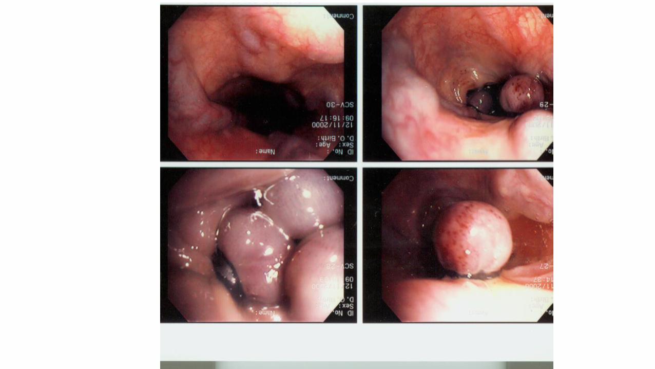
VARICES AND VARICEAL HEMORRHAGE
1. Primary Prophylaxis against Variceal Hemorrhage.
2. First-Line Management of Acute Esophageal Variceal Hemorrhage.
3. First-Line Prevention of Recurrent Variceal Hemorrhage.
NON-VARICEAL HEMORRHAGE :
A. Resuscitation, risk assessment, and preendoscopy management.
B. Endoscopic management.
C. Pharmacologic management.
D. Nonendoscopic and nonpharmacologic in-hospital management.
E. Postdischarge, ASA, and NSAIDs
NEJM 362;9 nejm.org march 4, 2010:Current Concepts Management of Varices and Variceal
Clinical Guidelines: International Consensus on Non-variceal Upper
Gastrointestinal Bleeding : 5 STATEMENTS: A,B,C,D,E.

1. VARICES AND VARICEAL HEMORRHAGE:Primary Prophylaxis against Variceal Hemorrhage.
NEJM 362;9 nejm.org march 4, 2010:Current Concepts Management of Varices and Variceal Hemorrhage in Cirrhosis
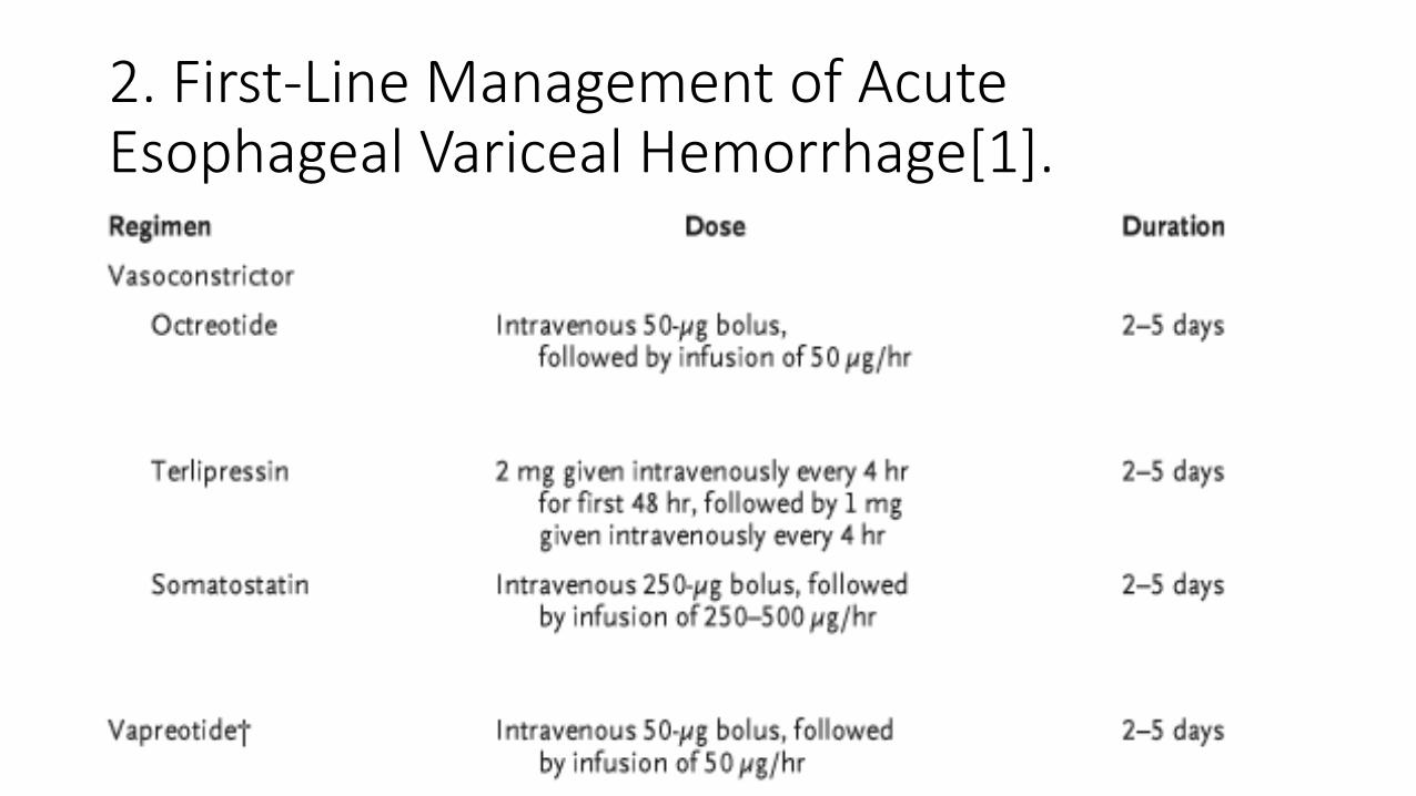
2. First-Line Management of Acute Esophageal Variceal Hemorrhage[1].

2..First-Line Management of Acute Esophageal Variceal Hemorrhage[2]….

3. First-Line Prevention of Recurrent Variceal Hemorrhage.

Nonvariceal UGI-Bleeding:A. Resuscitation, risk assessment, preendoscopymanagement.A1. Immediately evaluate and initiate appropriate resuscitation.
A2. Prognostic scales are recommended for early stratification of patients into low- and high-risk categories for rebleeding and mortality.
A3. Consider placement of a nasogastric tube in selected patients because the findings may have prognostic value.
A4. Blood transfusions should be administered to a patient with a hemoglobin level >70 g/L.
A5. In patients receiving anticoagulants, correction of coagulopathy is recommended but should not delay endoscopy.
A6. Promotility agents should not be used routinely before endoscopy to increase the diagnostic yield.
A7. Selected patients with acute ulcer bleeding who are at low risk for rebleeding on the basis of clinical and endoscopic criteria may be discharged promptly after endoscopy.
A8. Preendoscopic PPI therapy may be considered to downstage the endoscopic lesion and decrease the need for endoscopic intervention but should not delay endoscopy.

B. Endoscopic management [1]
B1. Develop institution-specific protocols for multidisciplinary management.* Include access to an endoscopist trained in endoscopic hemostasis.
B2. Have available on an urgent basis support staff trained to assist in endoscopy.
B3. Early endoscopy (within 24 hours of presentation) is recommended for most patients with acute upper gastrointestinal bleeding.
B4. Endoscopic hemostatic therapy is not indicated for patients with low-risk stigmata (a clean- based ulcer or a nonprotuberant pigmented dot in an ulcer bed).
B5. A finding of a clot in an ulcer bed warrants targeted irrigation in an attempt at dislodgement, with appropriate treatment of the underlying lesion.
B6. The role of endoscopic therapy for ulcers with adherent clots is controversial. Endoscopic therapy may be considered, although intensive PPI therapy alone may be sufficient.

B. Endoscopic management [2]
B7. Endoscopic hemostatic therapy is indicated for patients with high-risk stigmata (active bleeding or a visible vessel in an ulcer bed).
B8. Epinephrine injection alone provides suboptimal efficacy and should be used in combination with another method.
B9. No single method of endoscopic thermal coaptive therapy is superior to another.
B10. Clips, thermocoagulation, or sclerosant injection should be used in patients with high-risk lesions, alone or in combination with epinephrine injection.
B11. Routine second-look endoscopy is not recommended.B12. A second attempt at endoscopic therapy is generally recommended in
cases of rebleeding.

C. Pharmacologic management
C1. Histamine-2 receptor antagonists are not recommended for patients with acute ulcer bleeding.
C2. Somatostatin and octreotide are not routinely recommended for patients with acute ulcer bleeding.
C3. An intravenous bolus followed by continuous-infusion PPI therapy should be used to decrease rebleeding and mortality in patients with high-risk stigmata who have undergone successful endoscopic therapy.
C4. Patients should be discharged with a prescription for a single daily-dose oral PPI for a duration as dictated by the underlying etiology.

D. Nonendoscopic and nonpharmacologicin-hospital management
D1. Patients at low risk after endoscopy can be fed within 24 hours.
D2. Most patients who have undergone endoscopic hemostasis for high-risk stigmata should be hospitalized for at least 72 hours thereafter.
D3. Seek surgical consultation for patients for whom endoscopic therapy has failed.
D4. Where available, percutaneous embolization can be considered as an alternative to surgery for patients for whom endoscopic therapy has failed.
D5. Patients with bleeding peptic ulcers should be tested for H. pylori and receive eradication therapy if it is present, with confirmation of eradication.
D6. Negative H. pylori diagnostic tests obtained in the acute setting should be repeated.

E. Postdischarge, ASA and NSAIDs
E1. In patients with previous ulcer bleeding who require an NSAID, it should be recognized that treatment with a traditional NSAID plus PPI or a COX-2 inhibitor alone is still associated with a clinically important risk for recurrent ulcer bleeding.
E2. In patients with previous ulcer bleeding who require an NSAID, the combination of a PPI and a COX-2 inhibitor is recommended to reduce the risk for recurrent bleeding from that of COX-2 inhibitors alone.
E3. In patients who receive low-dose ASA and develop acute ulcer bleeding, ASA therapy should be restarted as soon as the risk for cardiovascular complication is thought to outweigh the risk for bleeding.
E4. In patients with previous ulcer bleeding who require cardiovascular prophylaxis, it should be recognized that clopidogrel alone has a higher risk for rebleeding than ASA combined with a PPI.

Endoscopic Therapeutic options
• Injection
• Thermal for PUD, AVM, or Mallory Weiss• Heater Probe• Bipolar Probe• Argon Plasma Coagulator
• Mechanical• Hemoclips for PUD or Mallory-Weiss• Banding for varices
• Combination

Thermal Probes
• Monopolar
• Bipolar
• Bipolar + Inj

Clips


Initial Pharmacologic Therapy
• Erythromycin improves visibility• Decreases need for 2nd EGD
• PPI• Decreases need for endoscopic therapy in PUD
• More effective than H2 blockers
• Octreotide for suspected cirrhotics• Increase Splanchnic vasoconstriction => Decreases Portal flow
S/ 50 mcg bolus, 50 mcg/hr gtt
• Antibiotics in cirrhotics• Reduce 30-d mortality, infection, rebleeding
Gastro 2002:123:17; AJG 2006:101:1211; GIE 2001:56:174; NEJM 2007;356:1631; Mayo Clin Proc 2007;82:286; Lancet 1995;346:865; Lancet 1997;350:1495

Surgical Options
• Splenectomy for gastric varices due to splenic vein thrombosis
• Ongoing bleeding despite any therapy modality with transfusion of >4-6 units/24Hrs or >10 units overall
• 2-3 recurrent bleeds from the same source

SummaryApproach to Acute Upper Gastrointestinal Bleeding:
Triage and resuscitation.
Correction of coagulopathy.
Pharmacologic treatments:PPI, Erythromycin, Octreotide, antibiotics
Endoscopy (Diagnosis, Prognosis, Therapy).
Post-endoscopy (PPI, antibiotics, medical management of portal HTN, anti-coagulation/aspirin management, etc).
