lower gi bleeding: interventional radiology or colonoscopy? bleeding - nasreddine.pdf · lower gi...
TRANSCRIPT

Lower GI Bleeding: Interventional
Radiology or Colonoscopy?
Walid Nasreddine, MD
Gastroenterologist
Chairman,
Department of Internal Medicine
Makassed General Hospital

Goals
•General approach to LGIB
•To define early predictors of Severe LGIB
•Timing of Colonoscopy and its Impacts
•Endoscopic Hemostasis techniques.

A 68 year old man presented to the ER with several episodes of hematochezia and passing clots per rectum.
PMHX: CAD Med:Aspirin
On Exam, the patient was pale. The blood pressure was90/60mmHg and the pulse 110/min.
DRE revealed fresh blood and clots.
HCT: 33%

A 68 year old man presented to the ER with several episodes of hematochezia and passing clots per rectum.
PMHX: CAD Med:Aspirin
On Exam, the patient was pale. The blood pressure was90/60mmHg and the pulse 110/min.
DRE revealed fresh blood and clots.
HCT: 33%

Estimation of blood loss based on vital signs
barnert et al, Nat.Rev. Gastroenterol. Hepatol. 6, 637-646(2009)
• ≤ 200ml :No effect on blood pressure or heart rate.
• ≥ 800ml :A drop of blood pressure by 10mmHg and/or an increase of pulse by 10bpm
• Orthostasis : Loss of at least 15% of blood volume
• ˃ 1500ml; Shock

Correction Fluid Losses-Restore Hemodynamic
Stability• Venous access
- 2 “large-bore” peripheral Ivs
- Central line if Necessary
• Initiate volume replacement-Saline, PRBCs
• Correct coagulopathy-FFP, platelets.

Correction Fluid Losses-Restore Hemodynamic
Stability• Venous access
- 2 “large-bore” peripheral Ivs
- Central line if Necessary
• Initiate volume replacement-Saline, PRBCs
• Correct coagulopathy-FFP, platelets.
Villaneuva et al. NEJM 2013; 368
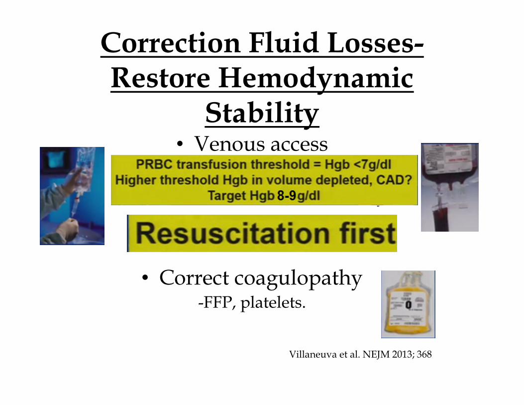
Correction Fluid Losses-Restore Hemodynamic
Stability• Venous access
- 2 “large-bore” peripheral Ivs
- Central line if Necessary
• Correct coagulopathy-FFP, platelets.
Villaneuva et al. NEJM 2013; 368
Sources of Hematochezia in the literature
Zukerman et al: GIE, 1999, 49: 228-238
• Diverticulosis 17-40%
• Angiodysplasia: 09-21%
• Cotitis(ischemic, infect, IBD,rad..) 02-30%
• Neoplasia: 11-14%
• Anorectal disease(including rectal varices) 04-10%
• UGIB 0-11%
• Small bowel bleeding 2-9%

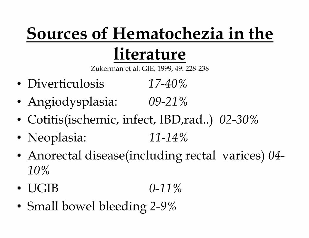
Sources of Hematochezia in the literature
Zukerman et al: GIE, 1999, 49: 228-238
• Diverticulosis 17-40%
• Angiodysplasia: 09-21%
• Cotitis(ischemic, infect, IBD,rad..) 02-30%
• Neoplasia: 11-14%
• Anorectal disease(including rectal varices) 04-10%
• UGIB 0-11%• Small bowel bleeding 2-9%

A 68 year old man presented to the ER with several episodes of hemahoclezia and passing clots per rectum.
PMHX: CAD Med:Aspirin
On Exam, the patient was pale.The blood pressure was90/60mmHg and the pulse 110/min.
DRE revealed fresh blood and clots.
HCT: 33%

Early predictors of Severe LGIBStrate etal: Arch. Int. Med. 2003: 163(7): 838-843
• Heart rate ≥100/min
• Systolic blood pressure ≤ 115mmHg
• Syncope
• Non tender abdominal examination
• Bleeding in the 1st 4 hrs of evaluation
• Aspirin
• ˃2 active comorbid conditions

Early Predictions of Severe LGIB
velayos el al. CGH: 2004, 2(6): 485-490
• Initial hematocrit ≤ 35%
• Abnormal Vital signs SBP˂100mmHg
H.R˃100/min
• Gross Blood on initial rectal examination .

Risk for Ongoing or Recurrent Bleed
strate et al, CGH, 2010, 8: 333-343
• 3 risk factors ⇒ 79%
• 2 risk factors ⇒ 54%
• 1 risk factor ⇒ 17%
• No risk factor ⇒ 0%

A 68 year old man presented to the ER with several episodes of hemahoclezia and passing clots per rectum.
PMHX: CAD Med:Aspirin
On Exam, the patient was pale. The blood pressure was90/60mmHg and the pulse 110/min.
DRE revealed fresh blood and clots.
HCT: 33%

Timing of Colonoscopy in LGIB
Jensen et al. Urgent Colonoscopy for the diagnosis and treatment of severe diverticular hemorrhage NEJM 2000; 342: 78-82
Two-arm Study
Urgent Colonoscopywith Endoscopic
Therapy
Urgent Colonoscopy
without Therapy P-Value
Rebleeding 0% 53% 0.005
Need of Surgery 0% 35% 0.03
Complication 0%

Timing of Colonoscopy in LGIB (2)
Green et al. Urgent Colonoscopy for evaluation and management of acute LGI hemorrhage. AJG 2005; 100: 2395-2402
Randomized Controlled Trial
Colonoscopy within 8 hrs
Elective Colonoscopy
P-Value
Definitive source of bleeding
42% 22% 0.03
Rebleeding 22% 30%No
statistical differenc
e
Surgery 14% 12%
Mortality 2% 4%
Blood Trasfusion 4.2 U 5 U
NB Complications < 2%
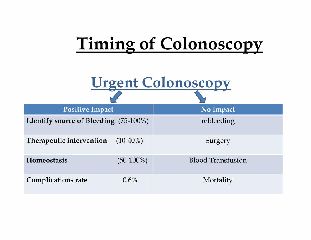
Timing of Colonoscopy
Urgent Colonoscopy
Positive Impact No Impact
Identify source of Bleeding (75-100%) rebleeding
Therapeutic intervention (10-40%) Surgery
Homeostasis (50-100%) Blood Transfusion
Complications rate 0.6% Mortality

Colon Preparation
• Is a must for completion of procedure
• Unprepared colon completion 55 ⇒ 70%
• Most patients need NG Tube- 5-6 liters of PEG
- 3-4 hours
-Monitor risk of aspiration and congestion
• No risk on clot dislodge or bleed activation
• Can’t be done in actively bleeding patient

Urgent Colonoscopy Comparing to Angiography
• High diagnostic yield (SRH) (75-100%)
Angiography (25-70%)
• High therapeutic capabilities (50-100%)
Angiography (80%)
• Low complication rate 0.6%
Angiography (17%)

Colonoscopy is the preferred initial test of choice in patient
who can be stabilized and can be prepared.

Endoscopic HemostasisTechniques

Diverticular Bleeding Jensen et al, NEJM, 2000; 342: 78-82
• Definitive: SRH in a diverticulum
• Presumptive: Diverticulosis in a patient with LGIB and negative work up for other sources
• Incidental: Diverticulosis in a patient with LGIB and work up reveal other source.

Importance of the Locations of the SRH in a Diverticular
Jensen DM, GIE =, 2012 Feb; 75(2): 388-391
• SRH was in the neck 50%
• The rest 50% in the base
• Active bleeding more common in the base
• Non Bleeding visible vessel in the neck
Large impact on type of Treatment
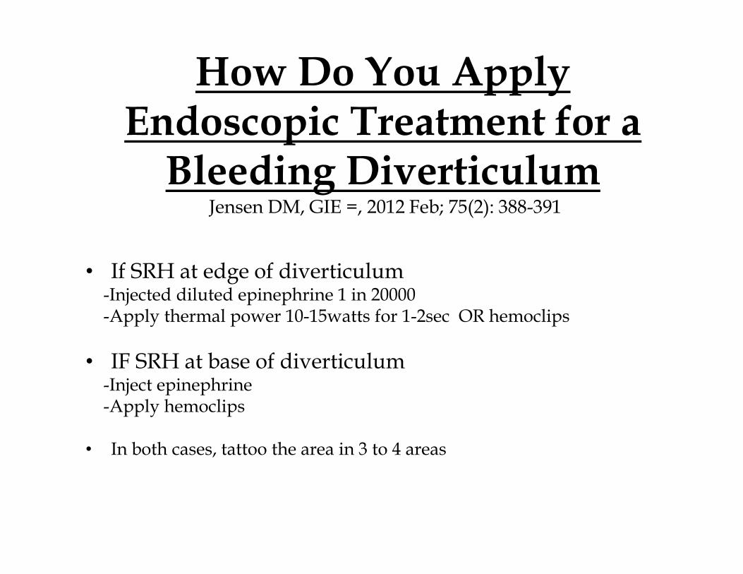
How Do You Apply Endoscopic Treatment for a
Bleeding DiverticulumJensen DM, GIE =, 2012 Feb; 75(2): 388-391
• If SRH at edge of diverticulum-Injected diluted epinephrine 1 in 20000-Apply thermal power 10-15watts for 1-2sec OR hemoclips
• IF SRH at base of diverticulum-Inject epinephrine -Apply hemoclips
• In both cases, tattoo the area in 3 to 4 areas

Endoscopic Band ligation for Colonic Diverticular
Hemorrhage(2): Ishii Net al, GIE, 2012
• Do colonoscopy
• Mark the diverticulum with SRH with a clip
• Withdraw, apply a cap and re scope
• Suction and band the diverticulum with SRH
-Success 87%
-Failure for initial hemostasis 24%
-Surgery in 1 of 29 patients.

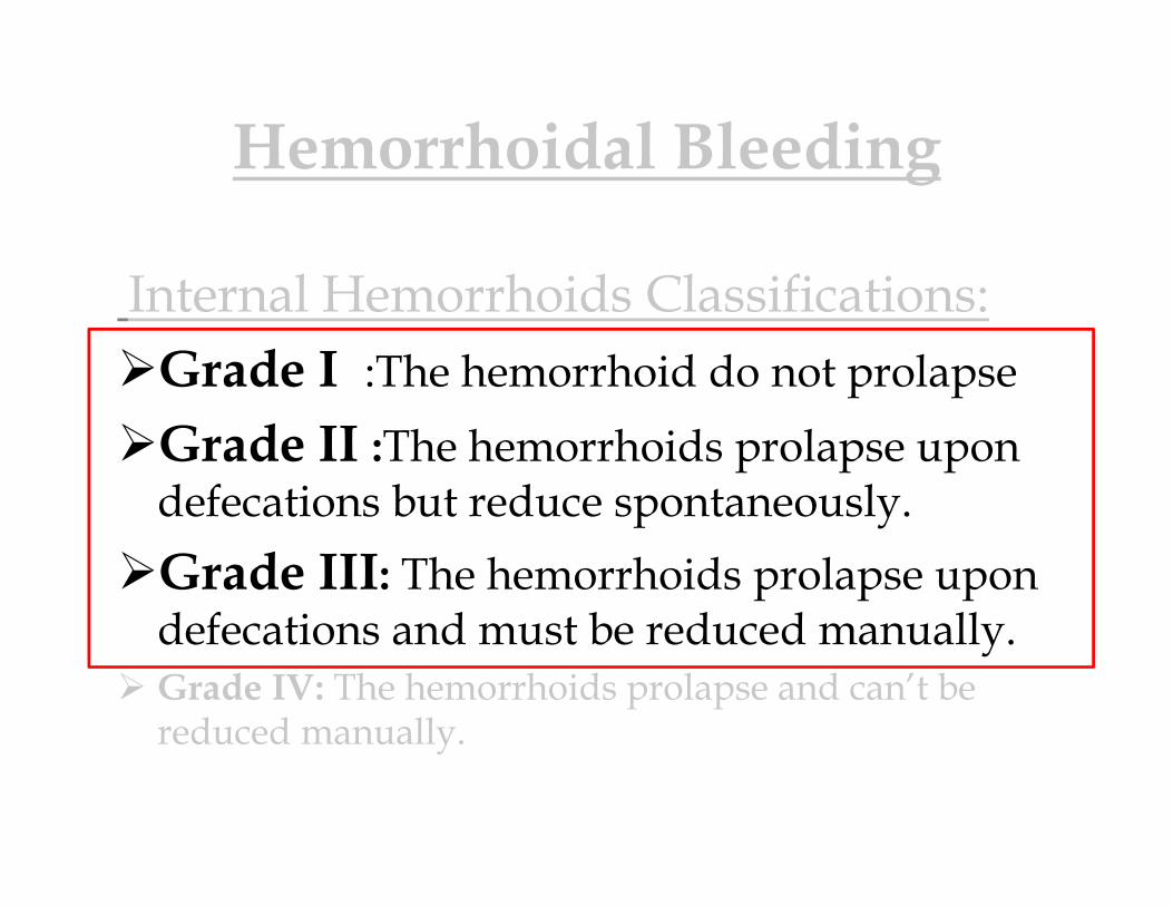
Hemorrhoidal Bleeding
Internal Hemorrhoids Classifications:� Grade I :The hemorrhoid do not prolapse
� Grade II :The hemorrhoids prolapse upon defecations but reduce spontaneously.
� Grade III: The hemorrhoids prolapse upon defecations and must be reduced manually.
� Grade IV: The hemorrhoids prolapse and can’t be reduced manually.
Hemorrhoidal Bleeding
Internal Hemorrhoids Classifications:
�Grade I :The hemorrhoid do not prolapse
�Grade II :The hemorrhoids prolapse upon defecations but reduce spontaneously.
�Grade III: The hemorrhoids prolapse upon defecations and must be reduced manually.
� Grade IV: The hemorrhoids prolapse and can’t be reduced manually.

Treatment of Bleeding Hemorrhoids
• Conservative ⇒ Fiber Supplement⇒Hydroxyethylrutoside(Daflo)
• Office based procedures:
-Banding ⇒endoscopic suction ligator⇒wall suction ligator or forceps
- Sclerotherapy⇒ endoscopic injections of hemorrhoids appex
- Infrared coagulations

Angiodysplasia
• 80% located in RT colon + cecum
• Two third of patients seen are over 70 years.
• 90% of hemorrhagic AD cease spontaneously
• Risk of overt rebleeding is 26%.

Endoscopic Treatment of GI Angio Dysplasia
• Argon plasma coagulations (APC)
-Complication rate 1.7%
-1year recurrence free survival 98%
K wom v et al, AJG 2006; 101: 58-63
• To decrease risk of perforation in proximal colon sub mucosal injection with saline solution is recommended.
Suzuki N etal GIE 2006; 64 424-7

GIAD(2)
• Laser applications
-High efficacy
-High risk of perforation
• Cryotherapy-Need further evaluations
• Electrocoagulations
-Monopolar coagulations
-Bipolar coagulations
• Elastic band ligation

How Do We Manage Post Polypectomy Bleeding
Management of immediate bleeding( 1.5-2.8%):• Application of pressure by regrasping the pedicle with
a snare
• Injection with epinephrine (preferably combined with other hemostatic techniques
• Cautery with thermal probe, bipolar, or the tip of a polypectomy snare
• Hemoclips
-Safe and effective
-If position is difficult cap device help to apply clips.
• Loops or band ligation.

Post Polypectomy Bleeding
Management of delayed bleeding (2%) occurs few hours up to 30 days.
• Epinehrine injections
• Thermal therapy
• Hemoclips
• Loops or band ligation

Few hours after stabilization our patient
developed 4 episodes of fresh blood per rectum + clots.
BP:95/60 H/R: 105

Few hours after stabilization our patient
developed 4 episodes of fresh blood per rectum + clots.
BP:95/60 H/R: 105

Thank You