update - kawasaki disease - fidssa · kawasaki disease - dx fever >5 days with no other...
TRANSCRIPT

Update - Kawasaki
disease
Children’s Infectious Diseases Clinical Trial Unit
Tygerberg Children’s
Hospital
Mark Cotton
& Simone Nicol
Stellenbosch University
(SU), South Africa
ID Workshop, SAPA Conference CTICC 9 Sept 2014

Tomisaku Kawasaki - 1st description of Kawasaki syndrome (disease)
Kawasaki T. Infantile acute febrile mucocutaneous lymph node syndrome with specific desquamation of the fingers and
toes. Clinical observation of 50 cases. Jpn J Allerg. 1967;178–222 (English abstract)

1st case – 4y boy
1st & only case of Coombs +ve
anaemia

Tomisaku Kawasaki’s timeline -
• Saw 1st case - Jan 1961 – 2nd case 1 year later – Suspected a syndrome
– 1st 7 cases reported as “non-scarlet fever with desquamation” at a meeting in 1962
– 50 cases reported in 1967 – Published in an Allergy Journal
• “New syndrome” disputed by colleagues
• Self-limiting and benign or serious? • Infantile polyarteritis nodosa = KD? • !970 – 1st nationwide KD survey
– 10 autopsy cases of sudden death after KD
• 1st English language publication - 1974

Dr Takajiro Yamamoto
Independently collected cases from 1950’s
1968 – patient with KD developed gallop
Yamamoto T, Kimura J. Acute febrile mucocutaneous lymph node syndrome (Kawasaki): subtype of mucocutaneous ocular syndrome of erythema multiforme complicated
with carditis
Shonika Rinsho (Jpn J Pediatr). 1968;21:336 –339
1963 – visiting professor at Cornell, USA – shown a case at Prof. Eichenwald’s grand rounds

In retrospect, 1st cases documented in 1950’s
• Sakurai K. Mucocutaneous ocular syndrome: report of a case [in Japanese]. Shonika Rinsho (Jpn J Pediatr). 1954;7:787–790
• Hanawa K. Two cases of pericardial hematoma in infancy [in Japanese]. Shonika Shinryo (J Pediatr Pract). 1959;22:820
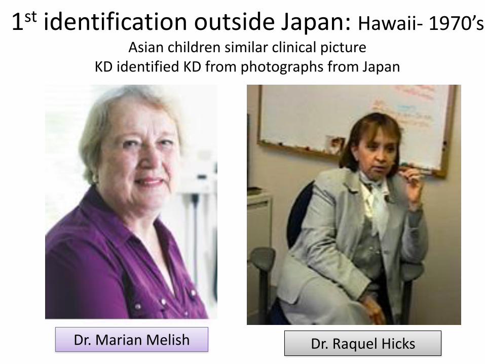
1st identification outside Japan: Hawaii- 1970’s Asian children similar clinical picture
KD identified KD from photographs from Japan
Dr. Marian Melish Dr. Raquel Hicks

Vd Merwe PL, Gie RP et al Mucocutaneous lymph node syndrome
(Kawasaki disease). A report of 2 cases
S Afr Med J 1980; 581014-6
• Pt 1: ECG inferior myocardial infarction
• Pt 2: aneurysm left coronary artery at
postmortem

Epidemiology
• US – Epidemics late winter and spring with 3-year intervals
– Children from the middle and upper-middle classes
– ~ 3000 children hospitalized annually
– More common in Japanese-American
• Japan – 5000-6000 per year
– Epidemics 1979, 1982, and 1985
• Sex: male-to-female 1.5:1
• Genetic: Japan 10x increase risk if affected sibling, 2x increase if parent, – similar in US (unpublished)

Cases collected 1970-2012
Burns JC et al Seasonality of KD PLOSOne 2013; 8: e75429

Cases in top 4 countries
Jane C Burns

Incidence per 100,000 <5y of age

Northern Hemisphere – transmissable agent in winter

Age
• USA – 80-90% of admissions < 5 years,
median age 3.14 years (1mo-21yrs),
10% under 6 months
• Japanese – Incidence boys 0-4 years
old 240 per 100 000 (2007-2008) peak
in children 6-11 months
• England - Incidence in children <5 yrs.
8 in 100 000 (1998-2003)
• Also < 6 months or > 5 years

Stanley TV, Grimwood K.
Classical Kawasaki disease in a neonate Arch Dis Child Fetal Neonatal 2002
Mar;86(2):F135-6
• Infant <2 weeks of age
• Echo: coronary artery
aneurysm day 5
• IVIG - rapid improvement

KD = leading cause acquired
heart disease in the USA ≤ 5y
ECHO
• asymptomatic coronary artery ectasis
• aneurysm incl giant coronary artery aneurysms with thrombosis
Coronary aneurysms in 25% untreated patients
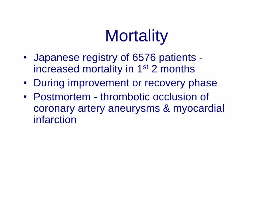
Mortality
• Japanese registry of 6576 patients - increased mortality in 1st 2 months
• During improvement or recovery phase
• Postmortem - thrombotic occlusion of coronary artery aneurysms & myocardial infarction

Coronary artery lesions
Dilatation
• Internal diameter
– <5y >3mm
– >5y >4mm
Persistence abnormalities
Months post acute
disease

Morbidity
• Giant CA >8mm greatest risk for
myocardial infarction
left ventricular function in ~ 50%
• Despite healing in smaller CA, vascular reactivity doesn’t return to baseline: follow up indefinitely
• Arthritis may persist
CA = coronary aneurysm
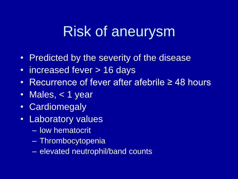
Risk of aneurysm
• Predicted by the severity of the disease
• increased fever > 16 days
• Recurrence of fever after afebrile ≥ 48 hours
• Males, < 1 year
• Cardiomegaly
• Laboratory values – low hematocrit
– Thrombocytopenia
– elevated neutrophil/band counts

Coron Artery Dis. 1995 6:857-64
Thrombocytopenia: a risk factor for acute MI - acute phase KD Niwa K et al
• 10 patients
• Coronary aneurysms & acute MI
• Platelet count: 4-12 x 104/mm3
• low ESR with high CRP in 7


Kawasaki Disease - Dx
Fever >5 days with no other
explanation & ≥4 of the 5
following criteria:
Bilateral bulbar conjuctival injection
Oral mucous membrane changes,
injected or fissured lips, injected
pharynx or strawberry tongue
Peripheral extremity changes:
erythema palms or soles, edema of
hands & feet (acute phase) &
periungual desquamation
(convalescent stage)
Polymorphous rash
Cervical lymphadenopathy (at least
one LN >1.5cm in diameter)
>5 of 6 with no other explanation
Fever
Bilateral bulbar conjuctival injection
Oral mucous membrane changes,
injected or fissured lips, injected
pharynx or strawberry tongue
Peripheral extremity changes, incl
erythema palms or soles, edema of
hands & feet (acute phase) &
periungual desquamation
(convalescent stage)
Polymorphous rash
Cervical lymphadenopathy (at least
one LN >1.5cm in diameter)
American Heart Association 2004 Japanese Circulation Society 2008

Incomplete KD <5/6 criteria supplementary lab criteria

Atypical vs Typical
Features of Typical KD Features of Atypical KD
Cervical LAD 60% Cervical LAD 10%
Rash 90% Rash 10%
Peripheral extremity changes 85% Peripheral extremity changes 60%
>90% mucous membrane changes >90% mucous membrane changes
LAD = lymphadenopathy

Tygerberg Children’s Hospital (TCH) Racial breakdown
N = 21
2006-2009

Longer fever in younger
patients (days)

What causes KD?
• Unusual response to infectious agent?
• Seasonality & age
– Novel retrovirus
– EBV
– Parvovirus
– Coronavirus
• Staphylococcal toxin mediated ?
Autoimmune rather than infectious agent?

Immune dysregulation
• DNA microarrays – upregulation neutrophil
response genes (adrenomedullin,
grancalcin and granulin)
• Evolving disease - upregulation of CD8
and NK responses & decreased neutrophil
response
• Oligoclonal IgA in respiratory tract arteries:
respiratory agent?

Polymorphisms with KD
• Inositol 1,4,5- triphosphate 3-kinase – Negative regulator of T cell activation
• Angiopoetin up-regulation & vascular endothelial growth factor down-regulation – disrupting vascular homeostasis
• Adenosine triphosphate binding cassette – Cellular efflux of prostaglandins
• CCR5 gene Chemokine receptor
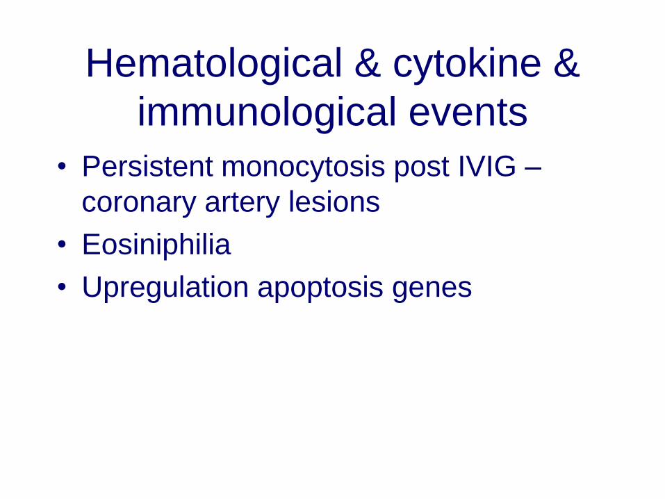
Hematological & cytokine &
immunological events
• Persistent monocytosis post IVIG –
coronary artery lesions
• Eosiniphilia
• Upregulation apoptosis genes

Animal models
• Intraperitoneal
extract of
Lactobacillus casei
• Coronary disease
• IVIG-responsive
Circulation 2012; 125: 1480

The Dr. who drank infectious
broth, gave himself an ulcer, &
solved a medical mystery
J Robin Warren & Barry J Marshall
Noble prize - 2005
http://discovermagazine.com/2
010

Clinical syndrome
Can be sequential
Always ask for features on history


Clinical Course
• Prodrome - respiratory or
gastrointestinal illness
• Abrupt onset fever
• Usually receive abx - no response
• Irritable +++


Hands
• Warm inflammatory
oedema
• Periungual
desquamation
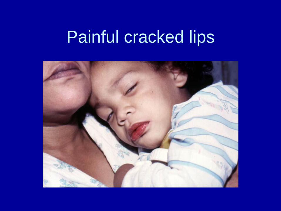
Painful cracked lips

Desquamation
Perineal
Urethritis Cervical

Strawberry tongue

22 months of age
• Fever for 5 days
• Red eyes
• Lt cervical lymph
node enlarged

3 days post polygam

BCG scar reactivation

Unusual features
• Hepatitis
• Hydrops gall bladder
• Meningism
• Interstitial pneumonitis
• Urethritis
• Diarrhoea
• Arthritis

Phases
• Acute febrile (days 1-11) – myocarditis and pericarditis
• Subacute (days 11-21) sudden death – Persistent irritability, anorexia, conjunctival
injection
– Fever usually resolves by this stage • If persists, greater risk of cardiac complications
– Thrombocytosis 1 million range
– Desquamation of the fingertips and toes
– Aneurysms
• Convalescence (days 21-60) - labs normalize
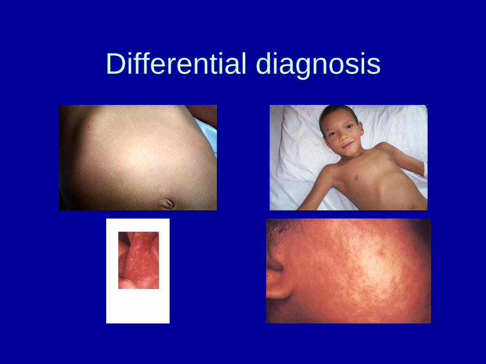
Differential diagnosis

Differential

Differential
• Drug reaction
– incl Stevens-Johnson syndrome
• Strep pharyngitis & Scarlet fever
• Toxic shock syndrome
• Measles
• Adenovirus
• Periodic syndrome
• Polyarteritis nodosa
• Hg poisoning

Treatment - effective
• IVIG 2g/kg IVI 12hrs
• Aspirin
– 80 - 100mg/kg’day - 4 doses X 2w
– 3 to 5mg/kg 6-8w or longer if coronary
arteries
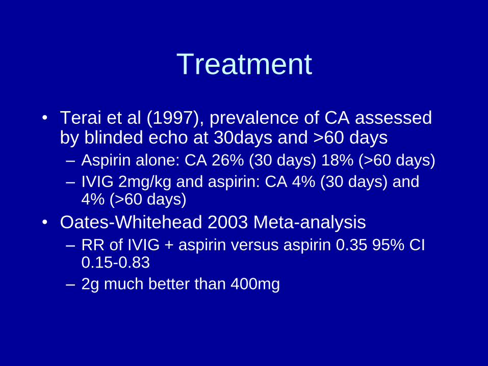
Treatment
• Terai et al (1997), prevalence of CA assessed by blinded echo at 30days and >60 days
– Aspirin alone: CA 26% (30 days) 18% (>60 days)
– IVIG 2mg/kg and aspirin: CA 4% (30 days) and 4% (>60 days)
• Oates-Whitehead 2003 Meta-analysis
– RR of IVIG + aspirin versus aspirin 0.35 95% CI 0.15-0.83
– 2g much better than 400mg

Timing of Treatment
• IVIG most effective early
• <5 days = 5 – 9 days
• <5 days - relapse?
• Expert consensus – treat within 4 days
• After day 10 – still treat if any
inflammation

J Pediatr June 2003
S Shulman

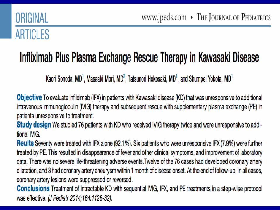
No response / Relapse
• IVIG resistance: 9 – 34%
• T >38oC post 48 hours
risk CA abnormalities
• Modalities
–Pulsed steroids
–TNFa blockade
– Immunosupression
• Cyclophosphamide
• Cyclosporin A

Kuo et al
Pediatrics &
Neonatology
2012; 53: 4-11

TNF inhibition
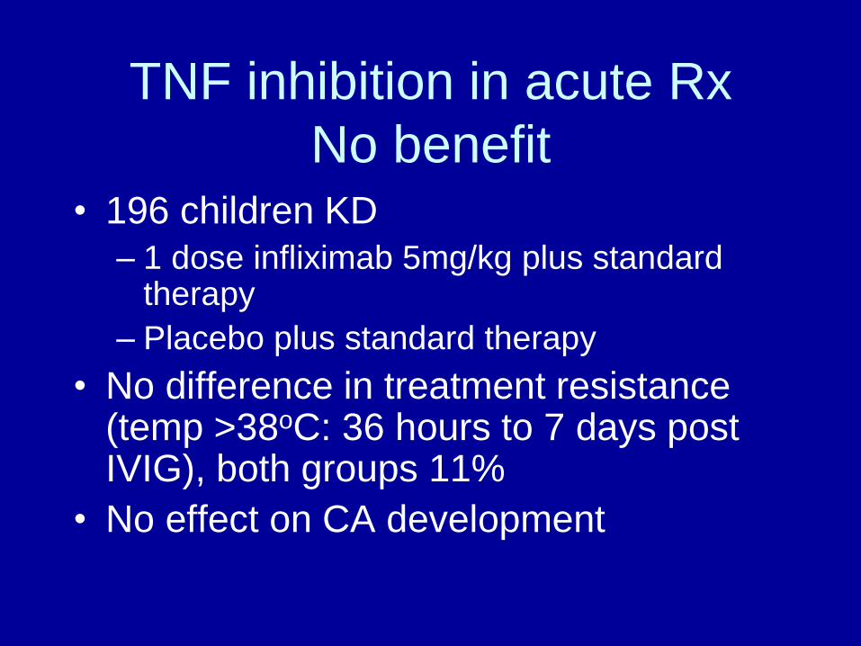
TNF inhibition in acute Rx
No benefit • 196 children KD
– 1 dose infliximab 5mg/kg plus standard therapy
– Placebo plus standard therapy
• No difference in treatment resistance (temp >38oC: 36 hours to 7 days post IVIG), both groups 11%
• No effect on CA development

Experimental
• Ulinastatin- urinary trypsin inhibitor with
anti-inflammatory properties (neutrophil
targeted)
– Not effective as monotherapy

KD shock
• In acute phase
• Hypotension & shock
• LV systolic dysfunction
• mitral regurgitation
• CA
• Resistant to Rx
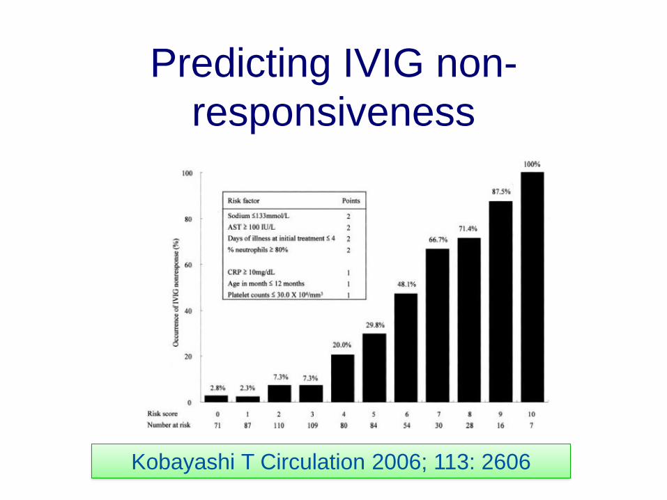
Predicting IVIG non-
responsiveness
Kobayashi T Circulation 2006; 113: 2606

IVIG (+ aspirin) ± prednisolone in
severe KD: randomised, open-
label, blinded-endpoints trial • IVIG 2g/kg + Aspirin (30mg/kg/day)
– N = 125
• + Methylprednisolone 2mg/kg/day X15
days post normal CRP
– N = 123
• Risk score ≥5
• Fever ≤9 days
• No CA abnormalities pre enrolment
Raise study: Kobayashi et al Lancet 2012; 379: 1613
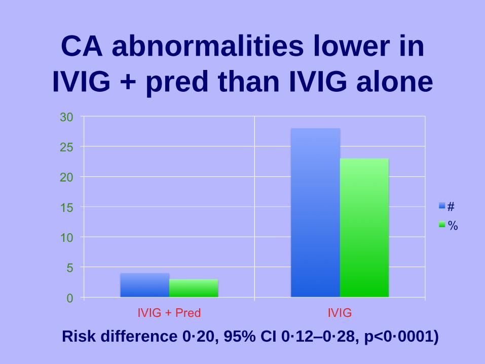
CA abnormalities lower in
IVIG + pred than IVIG alone
Risk difference 0·20, 95% CI 0·12–0·28, p<0·0001)
Long term issues
• Continued inflammation in CA’s
• Role for statins?

KS in adults
• 57 cases 18 – 30 years of age
• 2 cases & literature review
• Seve et al Sem Arthritis Rheumatol
2005: 34: 785-92

KS & HIV
J Infection 2007; 55: 48

Adult KD
• 20 cases
• High viral loads
• Low CD4
• Hepatitis virus co-
infection

Acknowledgements
• Helena Rabie - TCH
• Lisa Frigati – TCH
• Kate Carkeek - TCH