unusual acute encephalitis involving the thalamus: imaging ... · 68 korean j radiol 2(2), june...
TRANSCRIPT
68 Korean J Radiol 2(2), June 2001
Unusual Acute Encephalitis Involving the Thalamus: Imaging Features
Objective: To describe the brain CT and MR imaging findings of unusual acuteencephalitis involving the thalamus.
Materials and Methods: We retrospectively reviewed the medical records andCT and/or MR imaging findings of six patients with acute encephalitis involvingthe thalamus. CT (n=6) and MR imaging (n=6) were performed during the acuteand/or convalescent stage of the illness.
Results: Brain CT showed brain swelling (n=2), low attenuation of both thalami(n=1) or normal findings (n=3). Initial MR imaging indicated that in all patients thethalamus was involved either bilaterally (n=5) or unilaterally (n=1). Lesions werealso present in the midbrain (n=5), medial temporal lobe (n=4), pons (n=3), bothhippocampi (n=3) the insular cortex (n=2), medulla (n=2), lateral temporal lobecortex (n=1), both cingulate gyri (n=1), both basal ganglia (n=1), and the lefthemispheric cortex (n=1).
Conclusion: These CT or MR imaging findings of acute encephalitis ofunknown etiology were similar to a combination of those of Japanese encephalitisand herpes simplex encephalitis. In order to document the specific causativeagents which lead to the appearance of these imaging features, further investiga-tion is required.
n clinical practice, acute viral encephalitis is usually diagnosed on the ba-sis of clinical features and laboratory examinations, especially CSF (cere-brospinal fluid) findings. A specific viral etiology can usually be con-
firmed by a combination of serologic tests of acute and convalescent phase sera and bythe inoculation of blood or CSF into susceptible animals or tissue. In fatal cases, thevirus can often be isolated from the brain by the inoculation of susceptible animal andtissue culture (1). Brain imaging studies may help the diagnosis of some cases of acuteviral encephalitis; herpes simplex encephalitis (HSE), for example, characteristically in-volves the insula, temporal lobe and limbic system (2). Japanese encephalitis (JE), en-demic in some regions of Asia, also characteristically involves, in most patients, a par-ticular site, namely the thalamus (3 8). It is not uncommon, however, to encounterpatients who present with clinical and CSF features consistent with acute viral en-cephalitis but neither specific serologic nor characteristic imaging findings suggestive ofa specific viral etiology.
We recently encountered six cases of acute encephalitis of unknown etiology. Thebrain CT or MR imaging findings of these patients were similar, but serologic studieswere negative for JE and/or HSE. To our knowledge, no study has described the imag-ing findings of this unusual type of acute encephalitis. In an attempt to make radiolo-gists more aware of the diverse imaging findings of acute encephalitis, we present the
Sam Soo Kim, MD1, 2
Kee-Hyun Chang, MD3
Kyung Won Kim, MD3
Moon Hee Han, MD3
Sung Ho Park, MD4
Hyun Woo Nam, MD4
Kyu Ho Choi, MD5
Woo Ho Cho, MD6
Index terms:Brain, CT Brain, infectionBrain, MREncephalitisViruses
Korean J Radiol 2001;2:68-74Received September 19, 2000; accepted after revision January 22, 2001.
Department of 1Radiology, KangwonNational University Hospital; Departmentof 2Radiology and 4Neurology, Seoul CityBoramae Hospital; Department of3Radiology, Seoul National UniversityCollege of Medicine; Department of5Radiology, Kangnam St. Mary’s Hospital,The Catholic University of Korea;Department of 6Radiology, Sanggye PaikHospital, Inje University.
Address reprint requests to:Kee-Hyun Chang, MD, Department ofRadiology, Seoul National UniversityCollege of Medicine, 28 Yongon-dong,Chongno-gu, Seoul 110-744, Korea.Telephone: (822) 760-2584Fax: (822) 743-6385e-mail: [email protected]
I

brain CT and MR imaging findings of these six patients.
MATERIALS AND METHODS
Between 1997 and 1999, we retrospectively reviewedthe medical records and CT and/or MR imaging findings ofsix patients [four men and two women aged 25 to 42(mean, 37) years] with acute encephalitis involving thethalamus. The condition was diagnosed on the basis of clin-ical features and CSF examinations. The clinical featuresincluded fever, headache, disorientation, and confusion ormental alteration. In all patients, CSF examinations re-vealed lymphocyte-dominant pleocytosis in the range of32 to 442 cells per microliter, elevated protein, and normalglucose. The clinical and laboratory findings are summa-rized in Table 1. No patient had been recently immunizedor immunocompromised. In all patients, initial serologictests for JE and HSE viruses were performed during theacute and convalescent stages (7-40 days after the onset ofillness). In three, the serologic test for JE virus was per-formed twice, at an interval of 7 to 30 days, and in two,the serologic test for HSE virus was also performed twice,at an interval of 5 to 7 days. Serologic diagnosis of JE wasbased on the criteria developed by the research group forJE in Japan (i.e. a fourfold or greater rise in positive resultsin the haemagglutination inhibition test for the JE virus inpaired sera) (9). For serological diagnosis of HSE, poly-merase chain reaction (PCR) was used for the detection ofherpes simplex virus DNA in CSF (10). In addition, sero-logic tests for a variety of pathogens were performed: cy-tomegalovirus in four patients, Epstein-Barr virus in four,mycoplasma in one, enterovirus in one, rubella in one, andhuman immunodeficiency virus in one. In all patients,
serum and CSF were also examined for ordinary bacteria,fungus and acid-fast bacili (AFB) by direct staining and cul-ture. All the serologic examinations mentioned above werenegative in all patients. Despite the fact that all patientswere treated with acyclovir, clinical outcomes were verypoor: death (n=3) or a vegetative state (n=3).
Brain imaging studies were performed during the acuteand/or convalescent stage of the illness. CT (n=6) and MRimaging (n=6) were performed 3 to 18 days and 5 to 40days, respectively, after the onset of the condition. Fourpatients underwent follow-up MR imaging 11 to 45 daysafter onset, and one of these underwent further follow-upimaging 48 days after onset. For MR imaging, a 1.5 T im-ager (Magnetom; Siemens, Erlangen, Germany) was usedin four cases, and a 1.0 T imager (from the same source) intwo. All patients underwent spin-echo T1-weighted (repeti-tion time [msec]/echo time [msec]: 500 800/20 25), fastspin-echo T2-weighted (2,200 2,500/60-90), and fluid-at-tenuated inversion recovery MRI sequences. Contrast-en-hanced (IV injection of 0.1 mmol/kg of gadopentetatedimeglumine) T1-weighted spin-echo images were ob-tained in all patients except one (case 2).
Two radiologists evaluated the imaging features, focusingon the involved area, attenuation at CT and signal intensi-ty at MR imaging of the lesion, and whether or not en-hancement was observed.
RESULTS
The brain CT and MR imaging findings are summarizedin Table 2. Brain CT performed 3-18 days after the onsetof illness revealed diffuse brain swelling (n=2), low attenu-ation in both thalami (n=1), or no abnormal findings (n=3).
Imaging Features of Unusual Acute Encephalitis
Korean J Radiol 2(2), June 2001 69
Table 1. Clinical and Laboratory Findings of Six Patients
Case No. Age/Gender Clinical findings at admission Initial CSF profiles Results of serologic studies Outcome
1 40/F Fever, chill, altered mentality, Lymphocytosis, JEV ( ), HSV ( ), Rubella ( ), Deathstupor for 5 days increased protein CMV ( ), EBV ( )
2 39/M Fever, irritability, altered Lymphocytosis, JEV ( ), HSV ( ), EBV ( ), Vegetative mentality for 6 days increased protein Mycoplasma ( ), CMV ( ) state
3 42/M Mild febrile sense, sore throat Lymphocytosis, JEV ( ), HSV ( ), Deathdysarthria, dysphagia for 4 days increased protein Enterovirus ( )
4 25/F Fever, headache, neck stiffness Lymphocytosis JEV ( ), HSV ( ), CMV ( ), Deathfor 5 days EBV ( )
5 32/M Fever, disorientation for 3 days Lymphocytosis, JEV ( ), HSV ( ), Vegetative increased protein CMV ( ), EBV ( ), HIV ( ) state
6 42/M Fever, mental confusion for 2 days Lymphocytosis, JEV ( ), HSV ( ) Hopelessincreased protein discharge
Note. (-) = negative, CMV = cytomegalovirus, EBV = Epstein-Barr virus, HIV = human immunodeficiency virus, HSV = herpes simplex virus, JEV = Japanese encephalitis virus
Kim et al.
70 Korean J Radiol 2(2), June 2001
In all patients, initial MR images of the brain obtained 5-40days after onset revealed multiple patchy areas of focal ab-normality. The thalamus was involved either bilaterally(n=5) or unilaterally (n=1), thalamic lesions being homoge-neously hypointense on T1-weighted and hyperintense onT2-weighted images. In no patient did the signal intensitiesobserved on initial and follow-up MR images suggest hem-orrhage. Lesions were also seen in the midbrain (n=5), themedial area of the temporal lobe (n=4; 3 bilateral and 1unilateral), the pons (n=3), both hippocampi (n=3), the in-sular cortex (n=2; 1 bilateral and 1 unilateral), the medulla(n=2), the lateral temporal lobe cortex (n=1), both cingu-late gyri (n=1), both basal ganglia (n=1), and the cortex ofthe left hemisphere (n=1). Follow-up MR images obtainedin four patients showed that in two (cases 1 and 5), the ex-tent of the lesion had increased markedly. In one of thesetwo (case 5) the lesion subsequently became smaller, andthis was associated with diffuse brain atrophy and periven-tricular white matter change, suggesting microcystic cere-bromalacia or gliosis. In the remaining two patients (cases2 and 4), follow-up MR imaging of the lesion revealed noapparent change. Contrast-enhanced T1-weighted MR im-ages showed no parenchymal enhancement, though diffusecorticosulcal vascular or leptomeningeal enhancement wasseen in two patients (cases 3 and 5).
DISCUSSION
A wide range of pathogenic organisms, the most com-mon of which are viruses which include herpes simplextypes 1 and 2, herpes zoster, arboviruses and enterovirus-es, cause acute encephalitis (1, 11). In general, acute viralencephalitis causes diffuse parenchymal infiltration of in-flammatory cells, and this leads to chromatolysis and py-knosis of neurons and at times extensive necrosis (1, 11).These pathologic findings are reflected by areas of low at-tenuation on CT, low signal intensity on T1-weighted MRimages, and high signal intensity on T2-weighted MR im-ages, depending on the degree and severity of inflamma-tion.
In our cases, although no specific viral etiology wasproved in any patient, the CT and MR imaging findings ap-pear to be similar to those of JE in terms of the frequencyof involvement of the thalamus and brain stem, and -inpar-ticular-, of the substantia nigra and pons. In JE, pathologicchanges occur mainly in the gray matter and predominant-ly affect the diencephalon, mesencephalon, brain stem andcerebellar Purkinje cells, involving both thalami and thesubstantia nigra the most severely (3, 12 13). It has beenreported that the CT and MR imaging findings of JE areconsistent with the distribution of pathologic change.Previous articles describing these findings stated that thethalamus was usually symmetrically involved and other ar-
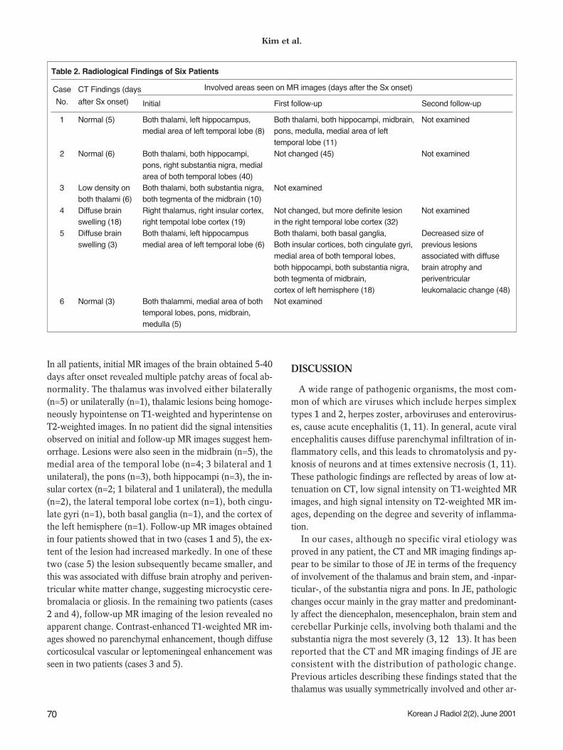
Table 2. Radiological Findings of Six Patients
Case CT Findings (days Involved areas seen on MR images (days after the Sx onset)
No. after Sx onset) Initial First follow-up Second follow-up
1 Normal (5) Both thalami, left hippocampus, Both thalami, both hippocampi, midbrain, Not examinedmedial area of left temporal lobe (8) pons, medulla, medial area of left
temporal lobe (11)2 Normal (6) Both thalami, both hippocampi, Not changed (45) Not examined
pons, right substantia nigra, medial area of both temporal lobes (40)
3 Low density on Both thalami, both substantia nigra, Not examinedboth thalami (6) both tegmenta of the midbrain (10)
4 Diffuse brain Right thalamus, right insular cortex, Not changed, but more definite lesion Not examinedswelling (18) right tempotal lobe cortex (19) in the right temporal lobe cortex (32)
5 Diffuse brain Both thalami, left hippocampus Both thalami, both basal ganglia, Decreased size of swelling (3) medial area of left temporal lobe (6) Both insular cortices, both cingulate gyri, previous lesions
medial area of both temporal lobes, associated with diffuse both hippocampi, both substantia nigra, brain atrophy and both tegmenta of midbrain, periventricular cortex of left hemisphere (18) leukomalacic change (48)
6 Normal (3) Both thalammi, medial area of both Not examinedtemporal lobes, pons, midbrain, medulla (5)

eas including the basal ganglia, midbrain, pons, cerebellum,cerebral cortex and spinal cord also showed frequent in-volvement (3 8). Hemorrhagic changes were very oftenseen in the primary lesion of JE, especially in the thalamus.Kumar et al. (5) reported that in all JE patients, follow-upimages obtained 10-60 days after the onset of illnessshowed hemorrhagic lesions in the thalamus. Kimura et al.(8) reported that HMPAO uptake in the bilateral thalamiand putamina, as seen on single-photon emission CT(SPECT), increased markedly in all the four patients inwhom JE was confirmed, a finding which might be usefulin differentiating JE from HSE and other types of en-cephalitis. In our cases, the hippocampus and medial area
of the temporal lobe were also frequently involved, thoughthese sites were very rarely involved in JE (3 8).Therefore, in our cases, imaging findings of no hemorrhag-ic foci, a low rate of involvement of the basal ganglia andcerebellum, frequent involvement of the hippocampus andmedial area of the temporal lobe, and the negative resultof serologic tests militate against a diagnosis of JE.
In our cases, lesions were also seen in the medial area ofthe temporal lobe, hippocampus and insular cortex, regionswhich are more frequently involved in HSE than in JE.HSE is known to lead to abnormal lesions in characteristiclocations of the brain; the medial temporal lobe, subfrontalarea, insular cortex and cingulate gyrus are preferentially
Imaging Features of Unusual Acute Encephalitis
Korean J Radiol 2(2), June 2001 71
A B C
Fig. 1. Case 1. A 40-year-old woman who presented with fever, chill and altered mentality.A-D. T2-weighted (A, B) and T1-weighted (C, D) images obtained 8 days after the onset of illness show abnormal hyperintensity and hy-pointensity in both thalami (large black arrows), the left hippocampus (small black arrow), and the medial area of the left temporal lobe(open white arrow).E-F. Follow-up T2-weighted images obtained 11 days after onset show marked enlargement of both thalamic lesions and the develop-ment of new lesions in the right hippocampus (white arrow) and brain stem.
D E F
Kim et al.
72 Korean J Radiol 2(2), June 2001
A B C
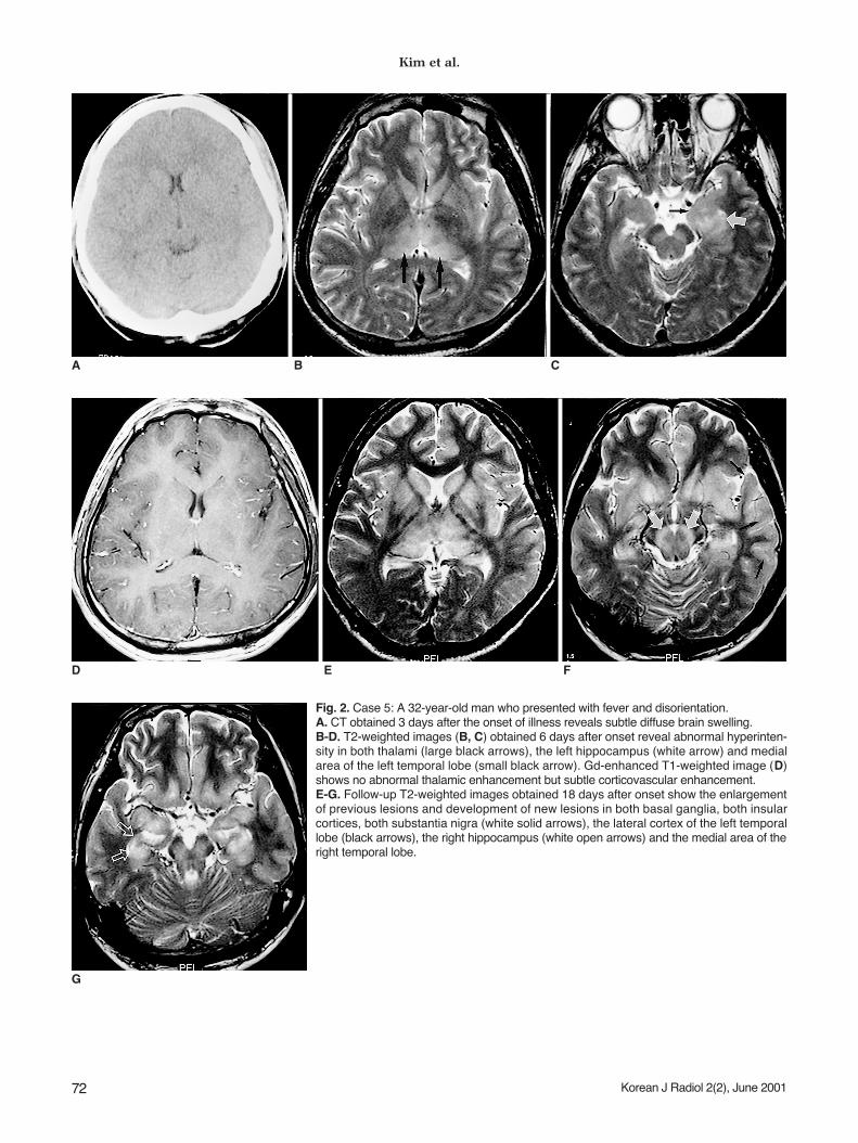
Fig. 2. Case 5: A 32-year-old man who presented with fever and disorientation.A. CT obtained 3 days after the onset of illness reveals subtle diffuse brain swelling.B-D. T2-weighted images (B, C) obtained 6 days after onset reveal abnormal hyperinten-sity in both thalami (large black arrows), the left hippocampus (white arrow) and medialarea of the left temporal lobe (small black arrow). Gd-enhanced T1-weighted image (D)shows no abnormal thalamic enhancement but subtle corticovascular enhancement.E-G. Follow-up T2-weighted images obtained 18 days after onset show the enlargementof previous lesions and development of new lesions in both basal ganglia, both insularcortices, both substantia nigra (white solid arrows), the lateral cortex of the left temporallobe (black arrows), the right hippocampus (white open arrows) and the medial area of theright temporal lobe.
D E F
G

involved, and rhombencephalitis involving the midbrainhas sometimes been reported (2, 14 15). In these pre-ferred areas, HSE lesions are seen as focal areas of low at-tenuation on CT, and as areas of low and high signal inten-sity, respectively, on T1-weighted and T2-weighted MRimages. In addition, a parenchymal or gyral pattern of en-hancement and foci of subacute hemorrhage may be ob-served slightly later. These imaging findings can help makea diagnosis of HSE fairly certain and prompt, though in-volvement of the thalamus, as in our cases, is very unusualin HSE.
In our cases, dual infection with the JE and the HSE viruswas a possibility. In analogous experiments in mice, JE vi-ral antigen was localized in herpes virus-infected areas ofthe brain, suggesting that the JE virus gains access to thecentral nervous system (CNS) at sites of blood-brain barri-er disruption caused by the HSE virus (12, 16), and imag-ing findings suggested a combination of JE and HSE.However, repetitive serologic tests failed to identify eitherthe JE or HSE virus. Unilateral thalamic involvement wasalso reported in a CNS lesion associated with Epstein-Barrvirus infection (17), though the good prognosis and fleetingimaging abnormality seen in this infection are differentfrom the observed findings in our cases.
The possible involvement, in our cases, of a new or dif-ferent viral agent, for which serologic tests were not per-formed, could not be excluded. In our series, repeatedserologic testing for JE involved the hemagglutination inhi-bition test, which in an appraisal of a recent diagnostic as-say for JE showed a sensitivity of about sixty percent (18).The use of polymerase chain reaction for detecting HSEvirus DNA in CSF has been reported as the most sensitivenoninvasive method for early diagnosis of HSE (19), but inour cases the technique failed to demonstrate that either
the JE or HSE virus was an etiologic agent. The mortalityrate for JE has been reported as 20-50% (20), and in casesof herpes encephalitis, acyclovir effectively reduces mortal-ity and morbidity if administered early (19). Comparedwith the prognosis of JE and HSE reported previously, thatof our patients was very poor despite the fact that in allcases, acyclovir was injected intravenously since the onsetof the condition.
Yagishita et al. (21) reported acute encephalopathy withbilateral thalamotegmental involvement in infants and chil-dren, which can be a postviral or postinfectious brain disor-der. There was, however, no clinical or laboratory evi-dence of encephalitis (no pleocytosis in the CSF). Althoughthe involved sites in our patients were similar to those inYagishita’s, the CSF findings and age range are completelydifferent.
The acute disseminated and immune-mediated forms ofencephalomyelitis may have clinical and CSF features simi-lar to those of our cases. The pathological findings of thefirst of these are, however, diffuse bilateral perivenular in-flammation and demyelination, mainly involving the cere-bral white matter, and for this reason - unlike in our cases,in which the gray matter was involved - MR imaging usual-ly demonstrates bilateral abnormalities in the cerebralwhite matter (22). Bilateral increased signal intensity onT2-weighted images of the basal ganglia and thalamus hasbeen reported in patients with sporadic and variantCreutzfeldt-Jakob disease (CJD) (23, 24). CJD, however,shows no leukocyte response in CSF and has clinical fea-tures different from those of our patients (25).
In conclusion, the CT and MR imaging findings of acuteencephalitis involving the thalamus were similar to a com-bination of those of JE and HSE. Further investigation,aimed at documenting the specific causative agent showing
Imaging Features of Unusual Acute Encephalitis
Korean J Radiol 2(2), June 2001 73
Fig. 2. H, I. Follow-up T2-weighted imagesobtained 48 days after onset show de-creased size of previous lesions.Periventricular white matter abnormalityaround both frontal horns and diffuse brainatrophy are noticed.
H I

Kim et al.
74 Korean J Radiol 2(2), June 2001
these imaging features, as well as histopathologic study, isneeded.
References1. Jubelt B, Miller JR. Viral infections. In Rowland LP ed. Merritt’s
Textbook of Neurology, 9th ed. Philadelphia: Williams &Wilkins, 1995:142-179
2. Tien RD, Felsberg GJ, Osumi AK. Herpes virus infection of theCNS: MR findings. AJR 1993;161:167-176
3. Abe T, Kojima K, Shoji H, et al. Japanese encephalitis. J MagnReson Imaging 1998;8:755-761
4. Shoji H, Hiraki Y, Kuwasaki N, et al. Japanese encephalitis inthe Kurume region of Japan: CT and MRI findings. J Neurol1989;236:255-259
5. Kumar S, Misra UK, Kalita J, et al. MRI in Japanese encephali-tis. Neuroradiology 1997;39:180-184
6. Misra UK, Kalita J, Jain SK, Mathur A. Radiological and neuro-physiological changes in Japanese encephalitis. J NeurolNeurosurg Psychiatry 1994;57:1484-1487
7. Shoji H, Murakami T, Murai I, et al. A follow-up study by CTand MRI in 3 cases of Japanese encephalitis. Neuroradiology1990;32:215-219
8. Kimura K, Dosaka A, Hashimoto Y, et al. Single-photon emis-sion CT findings in acute Japanese encephalitis. AJNR1997;18:465-469
9. Ishii K. Virological and serological diagnosis of Japanese en-cephalitis. Adv Neurol Sci 1967;11:300-311
10. Rowley AH, Whitley RJ, Lakeman FD, Wolinsky SM. Rapid de-tection of herpes-simplex virus DNA in cerebrospinal fluid ofpatients with herpes simplex encephalitis. Lancet 1990;335:440-441
11. Adams RD, Victor M, Ropper AH. Principles of Neurology, 6thed. New York: McGraw-Hill, 1997:749-759
12. Fields BN, Knipe DM, Chanock RM, eds. Virology. New York:Raven Press, 1985:967
13. Johnson RT, Burke DS, Elwell M, et al. Japanese encephalitis:immunocytochemical studies of viral antigen and inflammatory
cells in fatal cases. Ann Neurol 1985;18:567-57314. Soo MS, Tien RD, Gray L, Andrews PI, Friedman H.
Mesenrhombencephalitis: MR findings in nine patients. AJR1993;160:1089-1093
15. Tien RD, Dillon WP. Herpes trigeminal neuritis and rhomben-cephalitis on Gd-DTPA-enhanced MR imaging. AJNR1990;11:413-414
16. Hayashi K, Arita T. Experimental double infection of Japaneseencephalitis virus and herpes simplex virus in mouse brain.Japan J Exp Med 1977;47:9-13
17. Tolly TL, Wells RG, Sty JR. MR features of fleeting CNS lesionsassociated with Epstein-Barr virus infection. J Comput AssistTomogr 1989;13:665-668
18. Gajanana A, Samuel PP, Thenmozhi V, Rajendran R. An ap-praisal of some recent diagnostic assays for Japanese encephali-tis. Southeast Asian J Trop Med Public Health 1996;27:673-679
19. Corey L. Herpes simplex virus. In: Fauci AS, Braunwald E,Isselbacher KJ, eds. Harrison’s Principles of Internal Medicine,14th ed. New York: McGraw-Hill, 1998:1080-1086
20. Fauci AS, Braunwald E, Isselbacher KJ, eds. Harrison’sPrinciples of Internal Medicine, 14th ed. New York: McGraw-Hill, 1998:1137
21. Yagishita A, Nakano I, Ushioda T, Otsuki N, Hasegawa A.Acute encephalopathy with bilateral thalamotegmental involve-ment in infants and children: imaging and pathology findings.AJNR 1995;16:439-447
22. Osborn AG. Diagnostic Neuroradiology. St. Louis: Mosby,1994:704-706
23. Finkenstaedt M, Szudra A, Zerr I, et al. MR imaging ofCreutzfeldt-Jakob disease. Radiology 1996;199:793-798
24. Zeidler M, Sellar RJ, Collie DA, et al. The pulvinar sign on mag-netic resonance imaging in variant Creutzfeldt-Jakob disease.Lancet 2000;355:1412-1418
25. Zeidler M, Stewart GE, Barraclough CR, et al. New variantCreutzfeldt-Jakob disease: neurological features and diagnostictests. Lancet 1997;350:903-907