unlocking the in vitro anti-inflammatory and antidiabetic...
TRANSCRIPT

Full Terms & Conditions of access and use can be found athttp://www.tandfonline.com/action/journalInformation?journalCode=iphb20
Download by: [b-on: Biblioteca do conhecimento online UAlgarve] Date: 22 December 2017, At: 08:29
Pharmaceutical Biology
ISSN: 1388-0209 (Print) 1744-5116 (Online) Journal homepage: http://www.tandfonline.com/loi/iphb20
Unlocking the in vitro anti-inflammatory andantidiabetic potential of Polygonum maritimum
Maria João Rodrigues, Luísa Custódio, Andreia Lopes, Marta Oliveira, NunoR. Neng, José M. F. Nogueira, Alice Martins, Amélia P. Rauter, João Varela &Luísa Barreira
To cite this article: Maria João Rodrigues, Luísa Custódio, Andreia Lopes, Marta Oliveira, NunoR. Neng, José M. F. Nogueira, Alice Martins, Amélia P. Rauter, João Varela & Luísa Barreira(2017) Unlocking the in vitro anti-inflammatory and antidiabetic potential of Polygonum maritimum,Pharmaceutical Biology, 55:1, 1348-1357, DOI: 10.1080/13880209.2017.1301493
To link to this article: https://doi.org/10.1080/13880209.2017.1301493
© 2017 The Author(s). Published by InformaUK Limited, trading as Taylor & FrancisGroup.
Published online: 17 Mar 2017.
Submit your article to this journal Article views: 511
View related articles View Crossmark data
Citing articles: 2 View citing articles

RESEARCH ARTICLE
Unlocking the in vitro anti-inflammatory and antidiabetic potential ofPolygonum maritimum
Maria Jo~ao Rodriguesa , Lu�ısa Cust�odioa , Andreia Lopesa, Marta Oliveiraa , Nuno R. Nengb ,Jos�e M. F. Nogueirab , Alice Martinsb , Am�elia P. Rauterb , Jo~ao Varelaa and Lu�ısa Barreiraa
aCCMAR, Centro de Ciencias do Mar, Universidade do Algarve, Faro, Portugal; bCentro de Qu�ımica e Bioqu�ımica, Departamento de Qu�ımica eBioqu�ımica, Faculdade de Ciencias, Universidade de Lisboa, Lisboa, Portugal
ABSTRACTContext: Several Polygonum species (Polygonaceae) are used in traditional medicine in Asia, Europe andAfrica to treat inflammation and diabetes.Objective: Evaluate the in vitro antioxidant, anti-inflammatory and antidiabetic potential of methanol anddichloromethane extracts of leaves and roots of the halophyte Polygonum maritimum L.Material and methods: Antioxidant activity was determined (up to 1mg/mL) as radical-scavenging activ-ity (RSA) of 2,2-diphenyl-1-picrylhydrazyl (DPPH), 2,20-azino-bis(3-ethylbenzothiazoline-6-sulphonic acid)(ABTS), copper (CCA) and iron (ICA) chelating activities and iron reducing power (FRAP). NO productionwas measured in lipopolysaccharide (LPS)-stimulated macrophages for 24h at concentrations up to100lg/mL and antidiabetic potential was assessed by a-amylase and a-glucosidase inhibition (up to10mg/mL) assays. The phytochemical composition of the extracts was determined by gas chromatog-raphy-mass spectrometry (GC-MS).Results: The methanol leaf extract had the highest activity against DPPH· (IC50 ¼ 26lg/mL) and ABTS1·(IC50 ¼ 140lg/mL), FRAP (IC50 ¼ 48lg/mL) and CCA (IC50 ¼ 770lg/mL). Only the dichloromethane leafextract (LDCM) showed anti-inflammatory activity (IC50 ¼ 48lg/mL). The methanol root (IC50 ¼ 19lg/mL)and leaf (IC50 ¼ 29lg/mL) extracts strongly inhibited baker’s yeast a-glucosidase, but LDCM had higherrat’s a-glucosidase inhibition (IC50 ¼ 2527lg/mL) than acarbose (IC50 ¼ 4638lg/mL). GC-MS analysis iden-tified b-sitosterol, stigmasterol, 1-octacosanol and linolenic acid as possible molecules responsible for theobserved bioactivities.Conclusions: Our findings suggest P. maritimum as a source of high-value health promoting commoditiesfor alleviating symptoms associated with oxidative and inflammatory diseases, including diabetes.
ARTICLE HISTORYReceived 1 August 2016Revised 2 February 2017Accepted 28 February 2017
KEYWORDSHalophytes; antioxidantactivity; oxidative stress;macrophages; nitric oxide;a-amylase; a-glucosidase
Introduction
The genus Polygonum (Polygonaceae) includes more than 200 spe-cies worldwide, mainly in areas of temperate climate. SeveralPolygonum species are used in traditional medicine in China andJapan to treat health disorders such as dysentery, articular pain andinflammation (Takasaki et al. 2001; Kawai et al. 2006; Fan et al.2011). Some species are also used in traditional medicine inEurope, Africa and Asia to treat diabetes (Soumyanath 2005;Bothon et al. 2013). In Europe, approximately 36 species ofPolygonum can be found, including P. maritimum L., commonlyknown as sea knotgrass. Sea knotgrass is a perennial halophyteherb native from the sandy coasts of Europe, Mediterranean andBlack Sea, Channel Islands, England and Belgium, occurring fre-quently throughout the Portuguese coast (Kilinc & Karaer 1995;Cacador et al. 2013). Polygonum maritimum has described antioxi-dant and antimicrobial activities (El-Haci et al. 2013), and containsbioactive molecules such as polygonocinol, (þ)-8-hydroxycala-mene, octacosyl, triacontyl ferulate, arylpropane, quercetin, querci-trin, (þ)-catechin, and sitosterol (Kazantzoglou et al. 2009).
Diabetes is an emerging health problem in western societiesaffecting more than 300 million people worldwide and is
expected to be the 7th cause of death by 2030 (Mathers & Loncar2006; Danaei et al. 2011). Type 2 diabetes mellitus (T2DM) ismainly associated with genetics and lifestyle and encompassesmore than 90% of all diabetes cases globally (Mozaffarian et al.2009). The major characteristic of T2DM is high blood glucoselevel, which is caused by congenital or acquired deficiency insecretion of insulin combined with decreased responsiveness tothis hormone (WHO 1999; Yarchoan & Arnold 2014). The inhib-ition of carbohydrate-hydrolyzing enzymes, namely a-amylase anda-glucosidase, is thus an important strategy to manage hypergly-caemia linked to T2DM by decreasing the postprandial raise inblood glucose levels (Kwon et al. 2007). Acarbose, miglitol andvoglibose are clinically used compounds that target a-amylase anda-glucosidase; however, they present several side effects such asabdominal distension, flatulence and meteorism (Bischoff &Flower 1985). In this sense, there has been a growing effort tosearch for novel natural compounds with antidiabetic propertiesand reduced side effects (Kwon et al. 2007).
Hyperglycaemia found in T2DM patients may also inducemetabolic disturbances leading to the development ofoxidative stress and chronic inflammation states that contributeto diabetes-associated complications, namely, cardiovascular,
CONTACT Lu�ısa Barreira [email protected] CCMAR Centro de Ciencias do Mar, Universidade do Algarve, Campus de Gambelas, 8005-139 Faro, Portugal� 2017 The Author(s). Published by Informa UK Limited, trading as Taylor & Francis Group.This is an Open Access article distributed under the terms of the Creative Commons Attribution License (http://creativecommons.org/licenses/by/4.0/), which permits unrestricted use, distri-bution, and reproduction in any medium, provided the original work is properly cited.
PHARMACEUTICAL BIOLOGY, 2017VOL. 55, NO. 1, 1348–1357http://dx.doi.org/10.1080/13880209.2017.1301493
Dow
nloa
ded
by [
b-on
: Bib
liote
ca d
o co
nhec
imen
to o
nlin
e U
Alg
arve
] at
08:
29 2
2 D
ecem
ber
2017

urological, neurological, kidney and eyes disorders (AmericanDiabetes Association 2010; Vikram et al. 2014). Oxidative stresscoupled with reduced antioxidant defences enhances damagecaused by free radicals, such as reactive oxygen species (ROS),and contributes to disease progression (Sabu & Kuttan 2002;Maritim et al. 2003). In this context, natural antioxidants can beuseful in the prevention and/or management of oxidative stress-related disorders, including diabetes (Ruhe & McDonald 2001;Fardoun 2007). ROS also contributes to the production of pro-inflammatory cytokines and chemokines and to insulin resistance(Akash et al. 2013; Muriach et al. 2014). The role of oxidativestress and chronic inflammation in the progression of T2DMthus opens new avenues in the search for novel and combinedtherapies comprising the prevention of oxidative and inflamma-tory states (Akash et al. 2013).
As stated before, several Polygonum species are used in trad-itional medicine to treat inflammation and diabetes. However, tothe best of our knowledge, there is no information regarding theanti-inflammatory and/or antidiabetic potential of the sea knot-grass. In this context, we report for the first time a comparativeevaluation of the antioxidant and anti-inflammatory potentialand inhibitory activity on key enzymes relevant for hypergly-caemia (a-amylase and a-glucosidase) of extracts of sea knotgrassleaves and roots. The phytochemical characterization of theextracts is also presented.
Material and methods
Chemicals, culture media and supplements
Sigma-Aldrich (Germany) supplied the 1,1-diphenyl-2-picrylhy-drazyl (DPPH), 2,20-azino-bis(3-ethylbenzothiazoline-6-sulphonicacid (ABTS) radicals, sodium nitrite, lipopolysaccharide (LPS)from Escherichia coli, sulfanilamide, N-(1-Naphthyl) ethylenedi-amine dihydrochloride (NED) and 3-(4,5-dimethylthiazol-2-yl)2,5-diphenyl tetrazolium bromide (MTT). Folin-Ciocalteau(F-C) phenol reagent and phosphoric acid were purchased fromMerck (Germany). Lonza (Belgium) provided Roswell ParkMemorial Institute (RMPI) 1640 medium, fetal bovine serum(FBS), L-glutamine and penicillin/streptomycin. Additional chem-icals were acquired from VWR International (Belgium).
Sample collection
Whole plants of P. maritimum were hand collected in Ludo,South of Portugal, in June 2013. The taxonomical classificationwas performed by the botanist Dr. Manuel J. Pinto from theNational Museum of Natural History (University of Lisbon,Botanical Garden, Portugal) and a voucher specimen is kept inthe herbarium of MarBiotech laboratory (MBH22). Plants weredivided in roots and leaves, washed, oven dried for 3 days at50 �C, powdered and stored at �20 �C.
Preparation of the extracts
Dried samples were separately extracted with methanol anddichloromethane (1:40, w/v), overnight at room temperature(RT), under stirring. The extracts were filtered (Whatman no. 4),and evaporated to dryness at 40 �C in a rotary evaporator underreduced pressure (BUCHI R-210, Flawil, Switzerland). The driedextracts were dissolved in the corresponding solvent at the con-centration of 10mg/mL to be used in the chemical characteriza-tion assays, or in dimethyl sulfoxide (DMSO) to be used in the
bioactivity assays. All samples were stored at �20 �C untilneeded.
Gas chromatography and mass spectrometry (GC-MS)phytochemical analysis
The extracts (100 lL) were filtered (0.2 lm polytetrafluoroethyl-ene membrane syringe filters), transferred to a glass vial and thesolvent evaporated under a nitrogen stream. When dried, 50 lLof the derivatization reagent [N-methyl-N-(trimethylsilyl) tri-fluoroacetamide; MSTFA] was added. With the vial capped, theextracts were vortexed and heated for 20min in a dry blockheater at 40 �C (Pereira et al. 2012).
The GC/MS analyses were performed on an Agilent 6890 ser-ies gas chromatograph equipped with an Agilent 7683 automaticliquid sampler coupled to an Agilent 5973N mass selectivedetector (Agilent Technologies, Little Falls, DE). A programedtemperature vaporization injector with a septumless samplinghead (Gerstel, Mullheim a/d Ruhr, Germany) and a baffled linerwas used, operating in the solvent vent mode with compressedair for inlet cooling. Large volume injection was performed(vent time, 0.30min; flow, 50mL/min; pressure, 0 psi; purge,60mL/min at 2min), for which the inlet temperature was pro-gramed from 60 �C (0.4min) to 300 �C (3min isothermal) at arate of 60 �C/min and subsequently decreased to 200 �C (helduntil end) at a rate of 50 �C/min. The injection volume and speedwere set at 10 lL and 100lL/min, respectively. GC analysis wasperformed on a Zebron ZB-5 (30m� 0.25mm I.D., 0.25lm df;Phenomenex, USA) capillary column (5% phenyl, 95% polydime-thylsiloxane), using helium as carrier gas maintained in a con-stant inlet pressure mode of 7.81 psi. The oven temperature wasprogramed from 100 �C (1min) at 20 �C/min to 250 �C, then at10 �C/min to 300 �C and hold for 20min. The transfer line, ionsource and quadrupole analyzer temperatures were maintained at280 �C, 230 �C and 150 �C, respectively and a solvent delay of4min was selected. In the full-scan mode, electron ionizationmass spectra in the range 35-550Da were recorded at 70 eV withan ionization current of 34.6 lA. Data recording and instrumentcontrol were performed by MSD ChemStation software(G1701CA; version C.00.00; Agilent Technologies).
Radical-scavenging activity (RSA) on DPPH�
The RSA against DPPH was determined according to the methoddescribed by Cust�odio et al. (2015). Extracts (22 lL at concentra-tions ranging from 60 to 1000lg/mL) were mixed with 200lL ofDPPH solution (120 lM, in methanol) in 96-well microplatesand incubated in the dark for 30min (RT). The absorbance wasmeasured at 517 nm (Biotek Synergy 4) and results presented ashalf maximal inhibitory concentration (IC50, lg/mL). Butylatedhydroxytoluene (BHT) was used as a positive control.
RSA on ABTS�1
The RSA against ABTS radical was evaluated by the methoddescribed previously (Rodrigues et al. 2015). A stock solution ofABTS�þ (7.4mM) was generated by reacting equal amounts ofABTS with potassium persulfate (2.6mM) for 16 h in the dark atRT. The ABTS�þ solution was diluted with ethanol to obtain anabsorbance of at least 0.7 at 734 nm (Biotek Synergy 4). Extracts(10 lL at concentrations from 60 to 1000lg/mL) were mixed in96-well microplates with 190lL of ABTS�þ solution. After 6min
PHARMACEUTICAL BIOLOGY 1349
Dow
nloa
ded
by [
b-on
: Bib
liote
ca d
o co
nhec
imen
to o
nlin
e U
Alg
arve
] at
08:
29 2
2 D
ecem
ber
2017

of incubation the absorbance was measured at 734 nm (BiotekSynergy 4). Results were expressed IC50 values (lg/mL). BHTwas used as positive control.
RSA on nitric oxide (NO�)
The NO� scavenging activity was evaluated according toRodrigues et al. (2015) on extracts at concentrations between 60and 1000lg/mL. Samples (50 lL) were mixed with 50 lL of10mM sodium nitroprusside in phosphate buffer (PBS) andincubated for 90min at RT. After, 50 lL of Griess reagent (1% ofsulfanilamide and 0.1% of naphthylethylenediamine in 2.5%HPO3) were added. The absorbance was read at 546 nm, andresults were expressed as IC50 values (lg/mL). Nx-Nitro-L-argin-ine methyl ester hydrochloride (L-NAME) was used as standard.
Copper (Cu21) chelating activity (CCA)
The CCA was assessed using pyrocatechol violet as describedpreviously (Rodrigues et al. 2015). The extracts (30 lL) wereapplied at concentrations from 60 to 1000lg/mL and mixed with200lL of 50mM Na acetate buffer (pH 6), 6 lL of pyrocatecholviolet (4mM) in the above buffer and 100 lL of CuSO4�5H2O(50 lg/mL, in water). The change in the colour of the solutionwas measured at 632 nm using a microplate reader (BiotekSynergy 4). Results were expressed as IC50 values (lg/mL).Ethylenediamine tetraacetic acid (EDTA) was used as a positivecontrol.
Iron (Fe21) chelating activity (ICA)
The ICA chelating activity was determined by measuring the for-mation of the Fe2þ ferrozine complex (Meg�ıas et al. 2009),according to Rodrigues et al. (2015). Extracts (30 lL at concen-trations between 60 and 1000lg/mL) were mixed in 96-wellmicroplates with 200 lL of dH2O and 30 lL of a FeCl2 solution(0.1mg/mL in water). After 30min, 12.5 lL of ferrozine solution(40mM in water) was added. The change in colour was meas-ured in a microplate reader (Biotek Synergy 4) at 562 nm, andresults were expressed as IC50 values (lg/mL). EDTA was usedas standard.
Ferric reducing antioxidant power (FRAP) assay
The ability of the extracts to reduce Fe3þ was assayed by themethod described by Meg�ıas et al. (2009). Extracts (50 lL) weretested at concentrations ranging from 60 to 1000lg/mL andmixed with distilled water (50 lL) and 1% potassium ferricyanide(50 lL). After an incubation of 20min at 50 �C, 50 lL of 10% tri-chloroacetic acid (w/v) and ferric chloride solution (0.1%, w/v)were added and absorbance was measured at 700 nm. Increasedabsorbance of the reaction mixture indicates increased reducingpower. Antioxidant activity was calculated relatively to the posi-tive control (BHT; 1000lg/mL), and expressed as IC50 values(lg/mL).
Cell culture and cell viability
RAW 264.7 macrophages were maintained in RPMI culturemedium supplemented with 10% heat-inactivated FBS, 1% L-glu-tamine (2mM), and 1% penicillin (50U/mL)/streptomycin(50 lg/mL), and were maintained at 37 �C in humidified
atmosphere with 5% CO2. Exponentially growing cells wereplated in 96-well tissue plates at a concentration of 1� 104 cells/well and incubated for 24 h. Extracts were then applied at differ-ent concentrations (3 to 100 lg/mL) for 24 h. Control cells weretreated with DMSO at the highest concentration used in testwells (0.5%), and cell viability was determined by the MTT col-orimetric assay (Mosmann 1983). Briefly, 2 h prior to the end ofthe incubation period 20 lL of MTT (5mg/mL in PBS) wereadded to each well and further incubated at 37 �C. Then, 150lLof DMSO was added to each well in order to dissolve the forma-zan crystals and absorbance was measured at 590 nm (BiotekSynergy 4).
Measurement of NO production
The NO production was evaluated using RAW 264.7 macrophagesas described by Rodrigues et al. (2014). Cells were plated at2.5� 105 cells/mL in 96-well tissue plates and allowed to adhereovernight. Afterwards, they were treated with nontoxic concentra-tions of the extracts, i.e., those that allowed cellular viability higherthan 80%, in serum- and phenol-free culture medium, containing100 ng/mL of LPS, for 24 h (Nishishiro et al. 2005). NO produc-tion was assessed using the Griess assay (Miranda et al. 2001). Acalibration curve was prepared with different concentrations ofsodium nitrite (1.5–100 lM). Results were expressed as percentage(%) of NO production, relative to a control containing DMSO(0.5%, v/v), and as IC50 values (lg/mL).
a-Amylase inhibitory activity
The a-amylase inhibitory activity was determined by the methoddescribed by Xiao et al. (2006). Samples (40 lL at concentrationsranging from 1000 to 10,000lg/mL) were mixed in 96-wellmicroplates with 40 lL of amylase solution (100U/mL in 0.1Msodium phosphate buffer, pH 7.0) and 40 lL of 0.1% starch solu-tion (diluted in the previous buffer). After 10min at 37 �C, 20 lLof 1M hydrochloric acid (HCl) and 100lL of iodide solution(5mM iodine (I2)þ 5mM potassium iodide (KI), in distilledwater) were added and the absorbance was measured at 580 nm.Results were expressed as IC50 values (lg/mL). Acarbose wasused as the standard at concentrations between 250 and10,000 lg/mL.
Baker’s yeast a-glucosidase inhibitory activity
Microbial (Saccharomyces cerevisiae) a-glucosidase inhibitoryactivity was determined according to the method described byCust�odio et al. (2015). Samples (50 lL at concentrations rangingfrom 20 and 1000lg/mL) were mixed with 100lL of enzymesolution (1.0U/mL, in 0.1M sodium phosphate buffer, pH 7.0),and incubated for 10min at 25 �C. Then, 50 lL of 5mM p-nitro-phenyl-a-D-glucopyranoside (NGP; diluted in the previous buf-fer) were added and incubated more 5min at 25 �C. Theabsorbance was recorded at 405 nm using a microplate reader(Biotek Synergy 4) and results were expressed as IC50 values(lg/mL). Acarbose was used as positive control at concentrationsfrom 250 to 10,000 lg/mL.
Rat’s intestinal a-glucosidase inhibitory activity
Rat’s intestinal acetone powder was used as a crude enzymeextract as an example of enzyme of mammalian origin
1350 M. J. RODRIGUES ET AL.
Dow
nloa
ded
by [
b-on
: Bib
liote
ca d
o co
nhec
imen
to o
nlin
e U
Alg
arve
] at
08:
29 2
2 D
ecem
ber
2017

(Kwon et al. 2007). Rat’s intestinal acetone powder (250mg)were mixed with 10mL of 0.1M sodium phosphate buffer (pH7.0) and centrifuged at 5000 � g for 20min at 4 �C. The super-natant (100 lL) was mixed with the extracts (50 lL at concentra-tions between 500 and 10,000 lg/mL), and incubated for 10minat 25 �C. Then, 50 lL of 5mM p-nitrophenyl-a-D-glucopyrano-side (NGP; diluted in the previous buffer) was added and themixture was incubated for 30min at 37 �C. The absorbance wasread at 405 nm using a microplate reader (Biotek Synergy 4), andresults were expressed as IC50 values (lg/mL). Acarbose was usedas positive control at concentrations from 250 to 10,000 lg/mL.
Statistical analysis
Results were expressed as mean ± standard error of the mean(SEM), and experiments were carried out at least in triplicate.Significant differences were assessed by analysis of variance(ANOVA) followed by Duncan’s New Multiple Range Test, or byKruskal–Wallis test when parametricity of data did not prevail.SPSS statistical package for Windows (release 15.0, SPSS Inc.)was used. The IC50 values were calculated by sigmoidal fitting ofthe data in the GraphPad Prism v. 5.0 software.
Results
Phytochemical analysis
In order to determine their chemical composition, extracts wereanalyzed by GC-MS (Figure 1 and Table 1). This analysis wasable to identify a large number of compounds detected in themethanol extracts (70–75%). However, a large percentage ofcompounds detected in the dichloromethane extracts could notbe identified (45–48%). In total, 51 compounds were identifiedbelonging to different classes of biochemicals: alkanes andalkenes (AA), fatty acids (FA), phenolic compounds (PC), acyl-glycerols (GLY), saccharides (SAC), alcohols (ALC), phytosterols(PS) and minor groups (MG).
With similar contents, fatty acids (24.3%) and acylglycerols(22.8%) were the most represented categories in the methanol leafextract (LM), in which 2-monostearin (35) was the major com-pound (15.4%). Palmitic acid (18), linolenic acid (23), 1-monopal-mitin (34), glycerol (43), and b-sitosterol (47) were alsodetected at abundances higher than 5%. Acylglycerols were themost abundant constituents of the LM extract (34.4%), with 1-monostearin (31) being the most representative compound(24.0%), while 1-monopalmitin (34) and oleamide (51)
Figure 1. Main chemical compound classes identified by GC/MS in the dichloromethane and methanol extracts of roots and leaves of P. maritimum. (A) Methanol leafextract; (B) Methanol root extract; (C) Dichloromethane leaf extract; and (D) Dichloromethane root extract. AA: alkanes and alkenes; FA: fatty acids; PC: phenolic com-pounds; GLY: acylglycerols; SAC: saccharides; ALC: alcohols; PS: phytosterols; MG: minor groups; NI: non-identified compounds.
PHARMACEUTICAL BIOLOGY 1351
Dow
nloa
ded
by [
b-on
: Bib
liote
ca d
o co
nhec
imen
to o
nlin
e U
Alg
arve
] at
08:
29 2
2 D
ecem
ber
2017
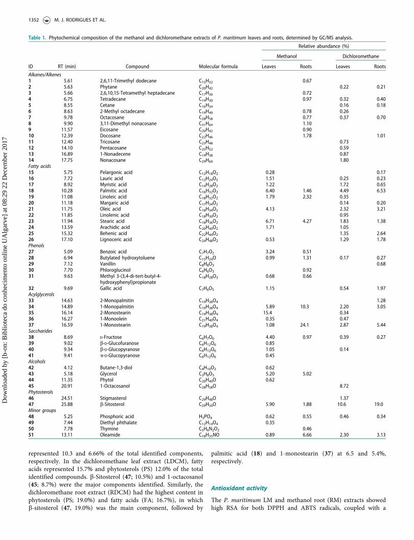
represented 10.3 and 6.66% of the total identified components,respectively. In the dichloromethane leaf extract (LDCM), fattyacids represented 15.7% and phytosterols (PS) 12.0% of the totalidentified compounds. b-Sitosterol (47; 10.5%) and 1-octacosanol(45; 8.7%) were the major components identified. Similarly, thedichloromethane root extract (RDCM) had the highest content inphytosterols (PS; 19.0%) and fatty acids (FA; 16.7%), in whichb-sitosterol (47, 19.0%) was the main component, followed by
palmitic acid (18) and 1-monostearin (37) at 6.5 and 5.4%,respectively.
Antioxidant activity
The P. maritimum LM and methanol root (RM) extracts showedhigh RSA for both DPPH and ABTS radicals, coupled with a
Table 1. Phytochemical composition of the methanol and dichloromethane extracts of P. maritimum leaves and roots, determined by GC/MS analysis.
Relative abundance (%)
Methanol Dichloromethane
ID RT (min) Compound Molecular formula Leaves Roots Leaves Roots
Alkanes/Alkenes1 5.61 2,6,11-Trimethyl dodecane C15H32 0.672 5.63 Phytane C20H42 0.22 0.213 5.66 2,6,10,15-Tetramethyl heptadecane C17H36 0.724 6.75 Tetradecane C14H30 0.97 0.32 0.405 8.55 Cetane C16H34 0.16 0.186 8.63 2-Methyl octadecane C19H40 0.78 0.267 9.78 Octacosane C28H58 0.77 0.37 0.708 9.90 3,11-Dimethyl nonacosane C31H64 1.109 11.57 Eicosane C20H42 0.9010 12.39 Docosane C22H46 1.78 1.0111 12.40 Tricosane C23H48 0.7312 14.10 Pentacosane C25H52 0.5913 16.89 1-Nonadecene C19H38 0.8714 17.75 Nonacosane C29H60 1.80Fatty acids15 5.75 Pelargonic acid C12H18O2 0.28 0.1716 7.72 Lauric acid C12H24O2 1.51 0.25 0.2317 8.92 Myristic acid C14H28O2 1.22 1.72 0.6518 10.28 Palmitic acid C16H32O2 6.40 1.46 4.49 6.5319 11.08 Linoleic acid C18H32O2 1.79 2.32 0.3520 11.18 Margaric acid C17H34O2 0.14 0.2021 11.75 Oleic acid C18H34O2 4.13 2.32 3.2122 11.85 Linolenic acid C18H30O2 0.9523 11.94 Stearic acid C18H36O2 6.71 4.27 1.83 1.3824 13.59 Arachidic acid C20H40O2 1.71 1.0525 15.32 Behenic acid C22H44O2 1.35 2.6426 17.10 Lignoceric acid C24H48O2 0.53 1.29 1.78Phenols27 5.09 Benzoic acid C7H7O2 3.24 0.5128 6.94 Butylated hydroxytoluene C15H24O 0.99 1.31 0.17 0.2729 7.12 Vanillin C8H8O3 0.6830 7.70 Phloroglucinol C6H6O3 0.9231 9.63 Methyl 3-(3,4-di-tert-butyl-4-
hydroxyphenyl)propionateC18H28O3 0.68 0.66
32 9.69 Gallic acid C7H6O5 1.15 0.54 1.97Acylglycerols33 14.63 2-Monopalmitin C19H38O4 1.2834 14.89 1-Monopalmitin C19H38O4 5.89 10.3 2.20 3.0535 16.14 2-Monostearin C19H38O4 15.4 0.3436 16.27 1-Monoolein C21H40O4 0.35 0.4737 16.59 1-Monostearin C19H38O4 1.08 24.1 2.87 5.44Saccharides38 8.69 D-Fructose C6H7O6 4.40 0.97 0.39 0.2739 9.02 b-D-Glucofuranose C6H12O6 0.8540 9.34 b-D-Glucopyranose C6H12O6 1.05 0.1441 9.41 a-D-Glucopyranose C6H12O6 0.45Alcohols42 4.12 Butane-1,3-diol C4H10O2 0.6243 5.18 Glycerol C3H8O3 5.20 5.0244 11.35 Phytol C20H40O 0.6245 20.91 1-Octacosanol C28H58O 8.72Phytosterols46 24.51 Stigmasterol C29H48O 1.3747 25.88 b-Sitosterol C29H50O 5.90 1.88 10.6 19.0Minor groups48 5.25 Phosphoric acid H3PO4 0.62 0.55 0.46 0.3449 7.44 Diethyl phthalate C12H14O4 0.3550 7.78 Thymine C5H6N2O2 0.4651 13.11 Oleamide C18H35NO 0.89 6.66 2.30 3.13
1352 M. J. RODRIGUES ET AL.
Dow
nloa
ded
by [
b-on
: Bib
liote
ca d
o co
nhec
imen
to o
nlin
e U
Alg
arve
] at
08:
29 2
2 D
ecem
ber
2017

strong capacity for reducing iron and chelating copper (Table 2).In the DPPH assay, the methanol extracts of both organs hadsimilar IC50 values (roots: 27 lg/mL; leaves: 26 lg/mL), whichwere significantly lower than the one obtained with the positivecontrol (BHT, 110lg/mL; Table 2). Regarding the ABTS radical,both the RM and LM extracts had IC50 of 192 and 140lg/mL,respectively, which were similar to that of BHT (140 lg/mL;Table 2). The RM and LM had also high ability to reduce ironwith IC50 values of 64 and 48 lg/mL, respectively (Table 2). Amoderate iron reduction was obtained upon addition of theRDCM (IC50¼ 770 lg/mL) and LDCM (IC50¼ 798 lg/mL)extracts. However, none of the extracts had the capacity for scav-enging the NO radical or for chelating iron (Table 2).
Anti-inflammatory activity
To assess the in vitro anti-inflammatory activity, the effect ofapplying nontoxic concentrations of the extracts (i.e. yielding cellviability >80%) on the NO production by LPS-stimulated RAW264.7 macrophage cells was determined. A significant reductionin cell viability was observed upon applying the LM extract at aconcentration of 100lg/mL to the cells. Loss of viability was alsoobserved with the RDCM extract at 50 and 100lg/mL (data notshown). Therefore, these concentrations were not used in theanti-inflammatory activity assessment.
Exposure of RAW 264.7 cells to LPS at 100 ng/mL increasedthe nitrite concentration in the culture medium from a basallevel of approximately 0.3 lM to around 13 lM (data not shown).This increase was significantly reduced in a dose-dependent man-ner by the treatment with LDCM, at concentrations rangingfrom 25 to 100 lg/mL, showing that the latter had an activitysimilar to the positive control (L-NAME) at the same concentra-tion (Figure 2). However, this extract had an IC50 value of48 lg/mL, higher than that of L-NAME (29.1 lg/mL; data notshown). Interestingly, incubating this cell line with the remainingextracts resulted in an increase in NO production when com-pared to the control (Figure 2). In particular, incubation withLM and RM extracts resulted in the most significant increases inNO production: 144% at the concentration of 50 lg/mL and139% at 25 lg/mL, respectively.
Antidiabetic activity
The inhibitory potential of extracts from P. maritimum was eval-uated against a-amylase and baker’s yeast and rat’s a-glucosi-dases (Table 3). The methanol extracts had the highest capacityto inhibit the baker’s yeast a-glucosidase with IC50 values of
19 and 29 lg/mL for roots and leaves, respectively, which weresignificantly lower than that of acarbose (IC50¼ 3144lg/mL).Despite the fact that methanol extracts had no capacity to inhibitrat’s a-glucosidase, LDCM had the highest rat’s a-glucosidaseinhibitory activity (IC50¼ 2527lg/mL), which was a significantlylower IC50 value than that of acarbose (4638 lg/mL; p< 0.05).
Discussion
In order to confirm the use of Polygonum species in traditionalmedicine in the treatment of inflammation and diabetes, weassessed for the first time the phytochemical composition of P.maritimum and its in vitro anti-inflammatory and antidiabeticpotential.
GC-MS analysis detected alkanes, alkenes and fatty acids asthe major categories of compounds in P. maritimum extracts.These are lipophilic compounds widely distributed in plantsas constituents of plant waxes and have been described inthe halophilic Mediterranean Suaeda vera Forssk. ex J.F.Gmel.(Amaranthaceae), Sarcocornia fruticosa (L.) A.J.Scott(Amaranthaceae), and Halimione portulacoides (L.) Aellen(Amaranthaceae) (Grossi & Raphel 2003). Waxes usually have aprotective role, for example, against microbial infections andavoiding excessive water losses and acute osmotic stress thathalophilic species suffer while growing in drenched soils (M€uller& Riederer 2005; Huang et al. 2011). Moreover, they are com-mon constituents of essential oils of halophyte species, such asSuaeda fructicosa and Limonium echioides (L.) Mill.(Plumbaginaceae) (Saïdana et al. 2008). From the compoundsdetected in this study, only b-sitosterol (47) had been previouslyidentified in P. maritimum also in dichloromethane extracts.Phytol (44), palmitic (18) and lauric (16) acids were also previ-ously detected as components of essential oils of Polygonum spe-cies, namely P. hydropiper L. and P. minus Huds. (Miyazawa &Tamura 2007; Baharum et al. 2010).
Our findings showed that P. maritimum has a strong antioxi-dant activity comparable to that reported by other authors forsimilar extracts made from aerial parts of the same species(El-Haci et al. 2013) and P. sachalinensis F.Schmidt and P. cuspi-datum Siebold & Zucc. (Pan et al. 2007; Fan et al. 2011).Moreover, IC50 values obtained with the RM and LM were sig-nificantly lower than or similar to the one obtained with thepositive control (BHT) for DPPH and ABTS assays, respectively.The significantly higher antioxidant activity of these extracts canbe related to the presence of some of the compounds identifiedin these extracts, namely, the phenolic compounds benzoic acid,BHT, vanillin and phloroglucinol, since these compounds are
Table 2. Radical scavenging activity (RSA) on DPPH, ABTS and NO radicals, metal chelating activity on iron (ICA) and copper (CCA)and ferric reducing activity (FRAP) activity of methanol (MeOH) and dichloromethane (DCM) extracts of roots and leaves of P.maritimum.
RSA Metal chelation/reduction
Extract/Standard Plant organ DPPH ABTS NO CCA ICA FRAP
MeOH Leaves 26 ± 0.7a 140 ± 6a n.a. 290 ± 10b n.a. 48 ± 1.4a
Roots 27 ± 0.0a 192 ± 5b n.a. 446 ± 8c n.a. 64 ± 3.8a
DCM Leaves n.a. n.a. n.a. n.a. n.a. 798 ± 36b
Roots n.a. n.a. n.a. n.a. n.a. 770 ± 53b
BHT� – 110 ± 10b 140 ± 10a – – – –L-NAME� – – – 2500 ± 10 – – –EDTA� – – – – 170 ± 10a 55.9 ± 3.7 –
Results are expressed as IC50 values (lg/mL). �Positive control; n.a.: not active. Values are means ± SEM of three separate experimentsperformed in triplicate (n¼ 9). In the same column, means labelled with different letters are significantly different by Duncan’s mul-tiple range test (p< 0.05).
PHARMACEUTICAL BIOLOGY 1353
Dow
nloa
ded
by [
b-on
: Bib
liote
ca d
o co
nhec
imen
to o
nlin
e U
Alg
arve
] at
08:
29 2
2 D
ecem
ber
2017

well described as strong antioxidants in vitro (Foti 2007; Dai &Mumper 2010). Phytol and linolenic acid have also beendescribed as potent in vitro and in vivo antioxidants due to theirhydroxyl group (Richard et al. 2008; Santos et al. 2013). Thus,their presence in the LM could also contribute to the high RSAand ICA of this extract. Interestingly, no significant differenceswere observed between the antioxidant activity of leaves androots as opposed to that reported for other halophytes species,such as Mesembryanthemum edule L. (Aizoaceae), Limoniastrummonopetalum (L.) Boiss. (Plumbaginaceae), Salsola kali L.(Chenopodiaceae), Tamarix gallica L. (Tamaricaceae) andLimonium algarvense Erben (Plumbaginaceae) (Ksouri et al.2008; Falleh et al. 2012; Trabelsi et al. 2012; Rodrigues et al.2015). This can be explained by the similar phytochemical profileof the methanol extracts of both organs (Figure 1 and Table 1),suggesting that the bioactive compound(s) are not organ-specific.
LPS is a cell wall endotoxin produced by Gram-negative bac-teria that activates macrophages to produce inflammatory media-tors such as NO (Martich et al. 1993), a radical associated withchronic inflammation (Kubes 2000; Joo et al. 2014). In this con-text, a decrease in NO production is used as an indicator of thepotential for the extracts to reduce an inflammatory response(Joo et al. 2014; Rodrigues et al. 2014). On the other hand,
an increase in NO production can indicate an immunostimula-tory effect of the extracts, which is important in macrophagedefence and protection against infection (Wink et al. 2011).
The anti-inflammatory effect of P. maritimum leaves may beattributed to the presence of compound(s) with potential anti-inflammatory properties, most likely b-sitosterol, stigmasterol,1-octacosanol, oleamide as well as linolenic and oleic acids(Table 1). For instance, b-sitosterol, one of the major constitu-ents of this extract, has anti-inflammatory properties throughTNF-a inhibition (Loizou et al. 2010), and so do 1-octacosanoland oleic acid (Vassiliou et al. 2009; de Oliveira et al. 2012). Inturn, linolenic acid and oleamide are able to reduce NO produc-tion and inducible nitric oxide synthase (iNOS) gene expression,through the inhibition of the NF-jB pathway (Ren & Chung2007; Oh et al. 2010). Stigmasterol was also reported to inhibitthe production of pro-inflammatory mediators associated withthe same pathway (Gabay et al. 2010). The combination of allthese compounds in the LDCM of P. maritimum are probablycontributing to its NO inhibitory capacity, since they were solelyidentified in this extract, or in higher quantities (Figure 2). Thesefindings are in accordance with several reports on the anti-inflammatory properties of Polygonum species (P. lapathifoliumL., P. cuspidatum and P. perfoliatum L. (Takasaki et al. 2001;Kim et al. 2007; Fan et al. 2011; Lei et al. 2015).
NO has an important role in the immune system modulation,being one of the macrophage-mediated primary responses againstpathogens such as fungi, helminthes, protozoa and bacteria(Wink et al. 2011). In this sense, the increased NO productionsuggests that both the LM and RM from P. maritimum may haveimmunostimulatory properties (Wink et al. 2011) most likely dueto the presence of specific molecules in these extracts, namely,saturated fatty acids (stearic and palmitic acids), which areknown inducers of the production of pro-inflammatory cytokinesin macrophages (Valdearcos et al. 2012; Miao et al. 2015).In fact, immunostimulatory properties are reported in
Treatments
Nitr
ite p
rodu
ctio
n (%
)
0
50
100
150
200
250
300
3.125 µg/mL6.25 µg/mL12.5 µg/mL25 µg/mL50 µg/mL100 µg/mLControl
Roots Leaves Roots L-NAME
Methanol
ab
def
abc
a
bcd
defg
bcd
j
efghi
defgh
a
j
i
g hi
fghi
cde
i
hi
g hi
fghi
jk
kk
Leaves
Dichloromethane
Figure 2. Effect of the application of dichloromethane and methanol extracts of roots and leaves of P. maritimum on NO production (%) by LPS-stimulated macro-phages. Control cells were treated with culture medium supplemented with 0.5% DMSO and 100 ng/mL of LPS. L-NAME (positive control) was applied at the concentra-tion of 100lg/mL. Solid and errors bars represent the average and SEM, respectively (n¼ 9). Bars followed by different letters are significantly different according tothe Duncan’s multiple ranges test (p< 0.05).
Table 3. Inhibitory activity of dichloromethane and methanol extracts of rootsand leaves of P. maritimum on a-amylase, baker’s yeast a -glucosidase and rat’sintestinal a-glucosidase.
Extract/Standard Organ a-Amylase Yeast a-glucosidase Rat a-glucosidase
Methanol Leaves n.a. 29 ± 0.7a n.a.Roots n.a. 19 ± 0.5a n.a.
Dichloromethane Leaves n.a. 585 ± 27b 2527 ± 37Roots n.a. 626 ± 14b >2500
Acarbose� 7797 ± 98 3144 ± 132c 4638 ± 438
Results are expressed as IC50 values (lg/mL). �Positive control; n.a.: not active.Values are means ± SEM of three separate experiments performed in triplicate(n¼ 9). In the same column, means labelled with different letters are signifi-cantly different by Duncan’s multiple range test (p< 0.05).
1354 M. J. RODRIGUES ET AL.
Dow
nloa
ded
by [
b-on
: Bib
liote
ca d
o co
nhec
imen
to o
nlin
e U
Alg
arve
] at
08:
29 2
2 D
ecem
ber
2017

P. multiflorum, P. minus and P. cuspidatum (Chen et al. 2012;Veerasamy et al. 2014; Chueh et al. 2015).
Compounds with the capacity to inhibit carbohydrate-hydrolyzing enzymes like a-amylase and a-glucosidase can delaythe digestion of carbohydrates, decreasing the postprandialincrease of blood glucose level after a mixed carbohydrate meal,and therefore can be an important strategy in managing hyper-glycaemia linked to T2DM (Krentz & Bailey 2005; Kwon et al.2007; Bhandari & Ansari 2008).
It is noteworthy that methanol extracts were approximately100-fold more active towards baker’s yeast a-glucosidase thanacarbose and that the LDCM had 2-times more ability to inhibitrat’s a-glucosidase. High IC50 values for acarbose against thisenzyme have also been reported by other authors (4823 lg/mL)(Gao et al. 2013). However, the methanol extracts had no cap-acity to inhibit rat�s a-glucosidase, which is a common feature ofmolecules with inhibitory capacity on a-glucosidase from micro-bial origin (Oki et al. 1999; Shai et al. 2011). Furthermore, a fewin vitro studies have discussed the low capacity of acarbose toinhibit mammalian a-glucosidase compared to crude extracts,including aqueous ethanol extracts from P. senegalensis (Meisn.)Soj�ak (Polygonaceae), which is used in folk medicine to treatT2DM (Shinde et al. 2008; Bothon et al. 2013). Those differencesare usually attributed to additive or synergistic interactions of thecompounds present in the extracts, resulting in a higher capacityto inhibit the mammalian a-glucosidase (Adisakwattana et al.2012). The higher activity of the LDCM can be related with thepresence of some particular compounds. For example, b-sitos-terol was the main compound identified in this extract (Table 1)and was previously reported to possess strong hypoglycaemicactivity through a-glucosidase inhibition (Ortiz-Andrade et al.2007). The same properties were described for stigmasterol andlinolenic acid, present only in this extract, as well as for oleicacid (Ortiz-Andrade et al. 2007; Lean Teik et al. 2013; Su et al.2013). In fact, Su et al. (2013) reported that linolenic and oleicacids were more active than acarbose. Although none of theextracts achieved 50% of inhibitory activity in the a-amylaseassay, our data suggest that P. maritimum may have potential asa source of antidiabetic molecules. This is in accordance with theantidiabetic activity found in several Polygonum species, such asP. aviculare L. (Polygonaceae), P. cuspidatum, P. multiflorum andP. senegalensis (Soumyanath 2005; Bothon et al. 2013).
Conclusions
The present study highlights for the first time the potential ofthe halophyte P. maritimum as a source of compounds with anti-oxidant, anti-inflammatory and antidiabetic activities. The metha-nol extracts had the highest antioxidant capacity, possibly due tothe presence of benzoic acid, BHT, phloroglucinol, phytol andlinolenic acid. The dichloromethane extracts from P. maritimumleaves had significant anti-inflammatory activity, most likelyrelated to its main constituents identified as b-sitosterol, stigmas-terol, 1-octacosanol, oleamide, linolenic and oleic acids.Moreover, its high a-glucosidase inhibitory activity may berelated to the presence of b-sitosterol, stigmasterol, linolenic andoleic acid. Overall, our results indicate that P. maritimumextracts are endowed with compounds with potential to be usedas a combined strategy to manage T2DM due to its anti-inflam-matory, antioxidant and a-glucosidase inhibitory properties.These results could be the starting points to further exploreP. maritimum, especially its leaves, as a source of value-added bioactive natural products. Nonetheless, isolation and
identification of the molecules(s) responsible for the detected bio-logical activities is already being pursued.
Acknowledgments
The authors thank the Faculty of Pharmacy and Center forNeurosciences and Cell Biology (University of Coimbra, Portugal)for kindly provide the murine leukemic monocyte-macrophage cellline (RAW 264.7).
Disclosure statement
The authors report that they have no conflicts of interest.
Funding
This work was supported by the XtremeBio project (PTDC/MAR-EST/4346/2012) funded by Foundation for Science and Technology(FCT) and the Portuguese National Budget. This work benefited alsofrom national funding through FCT project CCMAR/Multi/04326/2013. Lu�ısa Cust�odio was supported by the FCT InvestigatorProgram (IF/00049/2012). Nuno R. Neng and Jos�e M. F. Nogueiraalso acknowledge a FCT Post-Doc grant (SFRH/BPD/86071/2012)and funding (UID/Multi/00612/2013).
ORCID
Maria Jo~ao Rodrigues http://orcid.org/0000-0001-8732-710XLu�ısa Cust�odio http://orcid.org/0000-0003-4338-7703Marta Oliveira http://orcid.org/0000-0001-6793-8026Nuno R. Neng http://orcid.org/0000-0001-9879-1565Jos�e M. F. Nogueira http://orcid.org/0000-0001-5785-6780Alice Martins http://orcid.org/0000-0002-0542-2662Am�elia P. Rauter http://orcid.org/0000-0003-3790-7952Jo~ao Varela http://orcid.org/0000-0003-3101-693XLu�ısa Barreira http://orcid.org/0000-0002-4077-855X
References
Adisakwattana S, Ruengsamran T, Kampa P, Sompong W. 2012. In vitroinhibitory effects of plant-based foods and their combinations on intestinala-glucosidase and pancreatic a-amylase. BMC Complement Altern Med.12:110.
Akash MS, Rehman K, Chen S. 2013. Role of inflammatory mechanisms inpathogenesis of type 2 diabetes mellitus. J Cell Biochem. 114:525–531.
American Diabetes Association. 2010. Standards of Medical Care in Diabetes.Diabetes Care 33:S11–S61.
Baharum SN, Bunawan H, Ghani MA, Mustapha WAW, Noor NM. 2010.Analysis of the chemical composition of the essential oil of Polygonumminus Huds. using two-dimensional gas chromatography-time-of-flightmass spectrometry (GC-TOF MS). Molecules. 15:7006–7015.
Bhandari U, Ansari MN. 2008. Antihyperglycaemic activity of aqueous extractof Embelia ribes Burm in streptozotocin-induced diabetic rats. Indian JExp Biol. 46:607–613.
Bischoff PM, Flower RW. 1985. Ten years experience with choroidal angiog-raphy using indocyanine green dye: a new routine examination or an epi-logue? Doc Ophthalmol. 60:235–291.
Bothon FTD, Debiton E, Avlessi F, Forestier C, Teulade JC, SohounhloueDKC. 2013. In vitro biological effects of two anti-diabetic medicinal plantsused in Benin as folk medicine. BMC Complement Altern Med. 13:51.
Cacador I, Neto JM, Duarte B, Barroso DV, Pinto M, Marques JC. 2013.Development of an Angiosperm Quality Assessment Index (AQuA-Index)for ecological quality evaluation of Portuguese water bodies—A multi-met-ric approach. Ecol Indic. 25:141–148.
PHARMACEUTICAL BIOLOGY 1355
Dow
nloa
ded
by [
b-on
: Bib
liote
ca d
o co
nhec
imen
to o
nlin
e U
Alg
arve
] at
08:
29 2
2 D
ecem
ber
2017

Chen Q, Zhang SZ, Ying HZ, Dai XY, Li XX, Yu CH, Ye HC. 2012.Chemical characterization and immunostimulatory effects of a polysac-charide from Polygoni multiflori Radix Praeparata in cyclophosphamide-induced anemic mice. Carbohydr Polym. 88:1476–1482.
Chueh FS, Lin JJ, Lin JH, Weng SW, Huang YP, Chung JG. 2015. Crudeextract of Polygonum cuspidatum stimulates immune responses in normalmice by increasing the percentage of Mac-3-positive cells and enhancingmacrophage phagocytic activity and natural killer cell cytotoxicity. MolMed Rep. 11:127–132.
Cust�odio L, Patarra J, Alber�ıcio F, Neng NR, Nogueira JMF, Romano A.2015. Phenolic composition, antioxidant potential and in vitro inhibitoryactivity of leaves and acorns of Quercus suber on key enzymes relevant forhyperglycemia and Alzheimer's disease. Ind Crops Prod. 64:45–51.
Dai J, Mumper RJ. 2010. Plant phenolics: extraction, analysis and their anti-oxidant and anticancer properties. Molecules. 15:7313–7352.
Danaei G, Finucane MM, Lu Y, Singh GM, Cowan MJ, Paciorek CJ, Lin JK,Farzadfar F, Khang YH, Stevens GA, Global burden of metabolic risk fac-tors of chronic diseases collaborating group (blood glucose), et al. 2011.National, regional, and global trends in fasting plasma glucose and dia-betes prevalence since 1980: systematic analysis of health examination sur-veys and epidemiological studies with 370 country-years and 2�7 millionparticipants. Lancet. 378:31–40.
de Oliveira AM, Conserva LM, de Souza Ferro JN, de Almeida Brito F, LyraLemos RP, Barreto E. 2012. Antinociceptive and anti-inflammatory effectsof octacosanol from the leaves of Sabicea grisea var. grisea in mice. Int JMol Sci. 13:1598–1611.
El-Haci IA, Bekkara FA, Mazari W, Hassani F, Didi MA. 2013. Screening ofbiological activities of Polygonum maritimum L. from Algerian coast.Asian Pac J Trop Biomed. 3:611–616.
Falleh H, Jalleli I, Ksouri R, Boulaaba M, Guyot S, Magn�e C, Abdelly C.2012. Effect of salt treatment on phenolic compounds and antioxidantactivity of two Mesembryanthemum edule provenances. Plant PhysiolBiochem. 52:1–8.
Fan D, Zhou X, Zhao C, Chen H, Zhao Y, Gong X. 2011. Anti-inflammatory,antiviral and quantitative study of quercetin-3-O-b-D-glucuronide inPolygonum perfoliatum L. Fitoterapia. 82:805–810.
Fardoun RZ. 2007. The use of vitamin E in type 2 diabetes mellitus. Clin ExpHypertens. 29:135–148.
Foti MC. 2007. Antioxidant properties of phenols. J Pharm Pharmacol.59:1673–1685.
Gabay O, Sanchez C, Salvat C, Chevy F, Breton M, Nourissat G, Wolf C,Jacques C, Berenbaum F. 2010. Stigmasterol: a phytosterol with potentialanti-osteoarthritic properties. Osteoarthritis Cartilage. 18:106–116.
Gao J, Xu P, Wang Y, Wang Y, Hochstetter D. 2013. Combined effects ofgreen tea extracts, green tea polyphenols or epigallocatechin gallate withacarbose on inhibition against a-amylase and a-glucosidase in vitro.Molecules. 18:11614–11623.
Grossi V, Raphel D. 2003. Long-chain (C19-C29) 1-chloro-n-alkanes in leafwaxes of halophytes of the Chenopodiaceae. Phytochemistry. 63:693–698.
Huang X, Wang C, Zhang J, Wiesenberg GLB, Zhang Z, Xie S. 2011.Comparison of free lipid compositions between roots and leaves of plantsin the Dajiuhu Peatland, central China. Biochem J. 45:365–373.
Joo T, Sowndhararajan K, Hong S, Lee J, Park SY, Kim S, Jhoo JW. 2014.Inhibition of nitric oxide production in LPS-stimulated RAW 264.7 cellsby stem bark of Ulmus pumila L. Saudi J Biol Sci. 21:427–435.
Kawai Y, Kumagai H, Kurihara H, Yamazaki K, Sawano R, Inoue N. 2006.beta-Glucosidase inhibitory activities of phenylpropanoid glycosides, vani-coside A and B from Polygonum sachalinense rhizome. Fitoterapia.77:456–459.
Kazantzoglou G, Magiatis P, Kalpoutzakis E, Skaltsounis AL. 2009.Polygonophenone, the first MEM-substituted natural product, fromPolygonum maritimum. J Nat Prod. 72:187–189.
Kilinc M, Karaer F. 1995. The vegetation of Sinop Peninsula. Turk J Bot.19:107–124.
Kim KW, Ha KT, Park CS, Jin UH, Chang HW, Lee IS, Kim CH. 2007.Polygonum cuspidatum, compared with baicalin and berberine, inhibitsinducible nitric oxide synthase and cyclooxygenase-2 gene expressions inRAW 264.7 macrophages. Vascul Pharmacol. 47:99–107.
Krentz AJ, Bailey CJ. 2005. Oral antidiabetic agents: current role in type 2diabetes mellitus. Drugs. 65:385–411.
Ksouri R, Megdiche W, Falleh H, Trabelsi N, Boulaaba M, Smaoui A,Abdelly C. 2008. Influence of biological, environmental and technical fac-tors on phenolic content and antioxidant activities of Tunisian halophytes.C R Biol. 331:865–873.
Kubes P. 2000. Inducible nitric oxide synthase: a little bit of good in all of us.Gut 47:6–9.
Kwon YI, Apostolidis E, Shetty K. 2007. In vitro studies of eggplant (Solanummelongena) phenolics as inhibitors of key enzymes relevant for type 2 dia-betes and hypertension. Bioresour Technol. 99:2981–2988.
Lean Teik NG, Chun-Han S, Min-Nan L. inventors; Kang Jian Biotech Corp.,Ltd, assignee. 2013. alpha-Glucosidase inhibitor. US Patent 20130165679.
Lei X, Chen J, Ren J, Li Y, Zhai J, Mu W, Zhang L, Zheng W, Tian G, ShangH. 2015. Liver damage associated with Polygonum multiflorum Thunb: asystematic review of case reports and case series. Ev Based ComplementAlternat Med. 2015:9.
Loizou S, Lekakis I, Chrousos GP, Moutsatsou P. 2010. beta-Sitosterol exhib-its anti-inflammatory activity in human aortic endothelial cells. Mol NutrFood Res. 54:551–558.
Maritim AC, Sanders RA, Watkins JB. 2003. Diabetes, oxidative stress, andantioxidants: a review. J Biochem Mol Toxicol. 17:24–38.
Martich GD, Boujoukos AJ, Suffredini AF. 1993. Response of man to endo-toxin. Immunobiology. 187:403–416.
Mathers CD, Loncar D. 2006. Projections of global mortality and burden ofdisease from 2002 to 2030. PLoS Med. 3:e442.
Meg�ıas C, Pastor-Cavada E, Torres-Fuentes C, Gir�on-Calle J, Alaiz M, JuanR, Pastor J, Vioque J. 2009. Chelating, antioxidant and antiproliferativeactivity of Vicia sativa polyphenol extracts. Eur Food Res Technol.230:353–359.
Miao H, Chen L, Hao L, Zhang X, Chen Y, Ruan Z, Liang H. 2015. Stearicacid induces proinflammatory cytokine production partly through activa-tion of lactate-HIF1a pathway in chondrocytes. Sci Rep. 5:13092
Miranda KM, Espey MG, Wink DA. 2001. A rapid, simple spectrophotomet-ric method for simultaneous detection of nitrate and nitrite. Nitric Oxide.5:62–71.
Miyazawa M, Tamura N. 2007. Components of the essential oil from sproutsof Polygonum hydropiper L. (‘Benitade’). Flavour Frag J. 22:188–190.
Mosmann T. 1983. Rapid colorimetric assay for cellular growth and survival:application to proliferation and cytotoxicity assays. J Immunol Methods.65:55–63.
Mozaffarian D, Kamineni A, Carnethon M, Djouss�e L, Mukamal KJ,Siscovick D. 2009. Lifestyle risk factors and new-onset diabetes mellitus inolder adults: the cardiovascular health study. Arch Intern Med.169:798–807.
M€uller C, Riederer M. 2005. Plant surface properties in chemical ecology.J Chem Ecol. 1:2621–2651.
Muriach M, Flores-Bellver M, Romero FJ, Barcia JM. 2014. Diabetes and thebrain: oxidative stress, inflammation, and autophagy. Oxid Med CellLongev. 102:158.
Nishishiro M, Arikawa S, Wakabayashi H, Hashimoto K, Saton K, YokoyamaK, Unten S, Kakuta H, Kurihara T, Motohashi N, et al. 2005. Inhibition ofLPS-stimulated NO production in mouse macrophage-like cells by azule-nequinones. Anticancer Res. 25:4157–4163.
Oh YT, Lee JY, Lee J, Lee JH, Kim JE, Ha J. Kang 2010. Oleamide suppresseslipopolysaccharide-induced expression of iNOS and COX-2 through inhib-ition of NF-kappaB activation in BV2 murine microglial cells. NeurosciLett. 3:148–153.
Oki T, Matsui T, Osajima Y. 1999. Inhibitory effect of alpha-glucosidaseinhibitors varies according to its origin. J Agric Food Chem. 47:550–553.
Ortiz-Andrade RR, Garc�ıa-Jim�enez S, Castillo-Espa~na P, Ram�ırez-Avila G,Villalobos-Molina R, Estrada-Soto S. 2007. Alpha-glucosidase inhibitoryactivity of the methanolic extract from Tournefortia hartwegiana: an anti-hyperglycemic agent. J Ethnopharmacol. 109:48–53.
Pan Y, Zhang X, Wang H, Liang Y, Zhu J, Li H, Zhang Z, Wu Q. 2007.Antioxidant potential of ethanolic extract of Polygonum cuspidatum andapplication in peanut oil. Food Chem. 105:1518–1524.
Pereira DM, Vinholes J, Pinho PG, Valent~ao P, Mouga T, Teixeira N,Andrade PB. 2012. A gas chromatography-mass spectrometry multi-targetmethod for the simultaneous analysis of three classes of metabolites inmarine organisms. Talanta. 100:391–400.
Ren J, Chung SH. 2007. Anti-inflammatory effect of alpha-linolenic acid andits mode of action through the inhibition of nitric oxide production andinducible nitric oxide synthase gene expression via NF-kappaB and mito-gen-activated protein kinase pathways. J Agric Food Chem. 55:5073–5080.
Richard D, Kef K, Barbe U, Bausero P, Visioli F. 2008. Polyunsaturated fattyacids as antioxidants. Pharmacol Res. 57:451–455.
Rodrigues MJ, Gangadhar KN, Vizetto-Duarte C, Wubshet SG, Nyberg NT,Barreira L, Varela J, Cust�odio L. 2014. Maritime Halophyte species fromsouthern Portugal as sources of bioactive molecules. Mar Drugs.12:2228–2244.
Rodrigues MJ, Soszynski A, Martins A, Rauter AP, Neng NR, Nogueira JMF,Varela J, Barreira L, Cust�odio L. 2015. Unravelling the antioxidant poten-tial and the phenolic composition of different anatomical organs of themarine halophyte Limonium algarvense. Ind Crops Prod. 77:315–322.
1356 M. J. RODRIGUES ET AL.
Dow
nloa
ded
by [
b-on
: Bib
liote
ca d
o co
nhec
imen
to o
nlin
e U
Alg
arve
] at
08:
29 2
2 D
ecem
ber
2017
Ruhe RC, McDonald RB. 2001. Use of antioxidant nutrients in the preventionand treatment of type 2 diabetes. J Am Coll Nutr. 20:363S–369S.
Sabu MC, Kuttan R. 2002. Anti-diabetic activity of medicinal plants and itsrelationship with their antioxidant property. J Ethnopharmacol.81:155–160.
Saïdana D, Mahjoub S, Boussaada O, Chriaa J, Mahjoub MA, Ch�eraif I,Daami M, Mighri Z, Helal AN. 2008. Antibacterial and antifungal activ-ities of the essential oils of two saltcedar species from Tunisia. J Am OilChem Soc. 85:817–826.
Santos CCMP, Salvadori MS, Mota VG, Costa LM, Almeida AAC, OliveiraGAL, Costa JP, Sousa DP, Freitas RM, Almeira RN. 2013. Antinociceptiveand antioxidant activities of phytol in vivo and in vitro models. NeurosciJ. 2013:9.
Shai LJ, Magano SR, Lebelo SL, Mogale AM. 2011. Inhibitory effects of fivemedicinal plants on rat alpha-glucosidase: comparison with their effectson yeast alpha-glucosidase. J Med Plants Res. 5:2863–2867.
Shinde J, Taldone T, Barletta M, Kunaparaju N, Hu B, Kumar S, Placido J,Zito SW. 2008. alpha-Glucosidase inhibitory activity of Syzygium cumini(Linn.) Skeels seed kernel in vitro and in Goto-Kakizaki (GK) rats.Carbohydr Res. 343:1278–1281.
Soumyanath A. 2005. Traditional Medicines for Modern Times: Antidiabeticplants. New York: CRC Press, Taylor and Francis Group.
Su CH, Hsu CH, Ng LT. 2013. Inhibitory potential of fatty acids on keyenzymes related to type 2 diabetes. Biofactors. 39:415–421.
Takasaki M, Konoshima T, Kuroki S, Tokuda H, Nishino H. 2001. Cancerchemopreventive activity of phenylpropanoid esters of sucrose, vanicosideB and lapathoside A, from Polygonum lapathifolium. Cancer Lett.173:133–138.
Trabelsi N, Falleh H, Jallali I, Daly AB, Hajlaoui H. 2012. Variation of phen-olic composition and biological activities in Limoniastrum monopetalumL. organs. Acta Physiol Plant. 34:87–96.
Valdearcos M, Esquinas E, Meana C, Pe~na L, Gil-de-G�omez L, BalsindeJ, Balboa MA. 2012. Lipin-2 reduces proinflammatory signalinginduced by saturated fatty acids in macrophages. J Biol Chem.287:10894–10904.
Vassiliou EK, Gonzalez A, Garcia C, Tadros JH, Chakraborty G, Toney JH.2009. Oleic acid and peanut oil high in oleic acid reverse the inhibitoryeffect of insulin production of the inflammatory cytokine TNF-alpha bothin vitro and in vivo systems. Lipids Health Dis. 8:25.
Veerasamy R, Min LS, Pauine R, Sivadasan S, Varghese C, Rajak H,Marimuthu K. 2014. Effect of aqueous extract of Polygonum minus leaf onthe immunity and survival of African catfish (Clarias gariepinus). J CoastLife Med. 2:209–213.
Vikram A, Tripathi DN, Kumar A, Singh S. 2014. Oxidative stress andinflammation in diabetic complications. Int J Endocrinol. 2014:2.
Wink DA, Hines HB, Cheng RY, Switzer CH, Flores-Santana W, Vitek MP,Ridnour LA, Colton CA. 2011. Nitric oxide and redox mechanisms in theimmune response. J Leukoc Biol. 89:873–891.
World Health Organization (WHO). 1999. Definition, diagnosis and classifi-cation of diabetes mellitus and its complications. Part 1: diagnosis andclassification of diabetes mellitus. Geneva (WHO/NCD/NCS/99.2).
Xiao Z, Storms R, Tsang A. 2006. A quantitative starch-iodine method formeasuring alpha-amylase and glucoamylase activities. Anal Biochem.351:146–148.
Yarchoan M, Arnold SE. 2014. Repurposing diabetes drugs for brain insulinresistance in Alzheimer disease. Diabetes. 63:2253–2261.
PHARMACEUTICAL BIOLOGY 1357
Dow
nloa
ded
by [
b-on
: Bib
liote
ca d
o co
nhec
imen
to o
nlin
e U
Alg
arve
] at
08:
29 2
2 D
ecem
ber
2017