university of palestine college of dentistry human dentition i tooth structure dr. mustafa i....
TRANSCRIPT
University of Palestine College of Dentistry
Human Dentition ITooth structure
DR. MUSTAFA I. ELGHOULMaster of Orthodontic

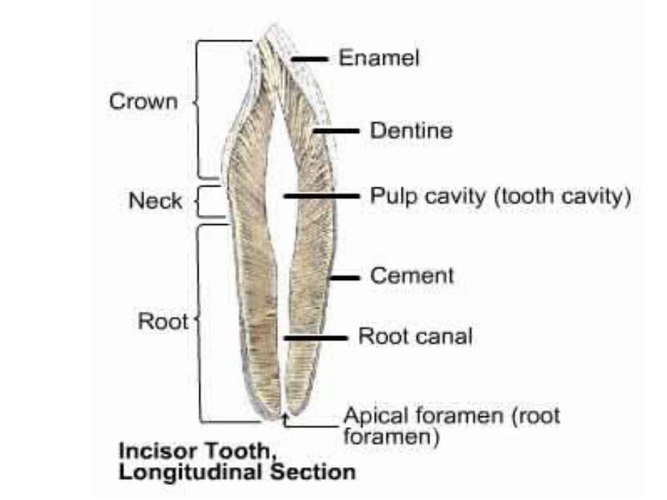
The Crown and Root
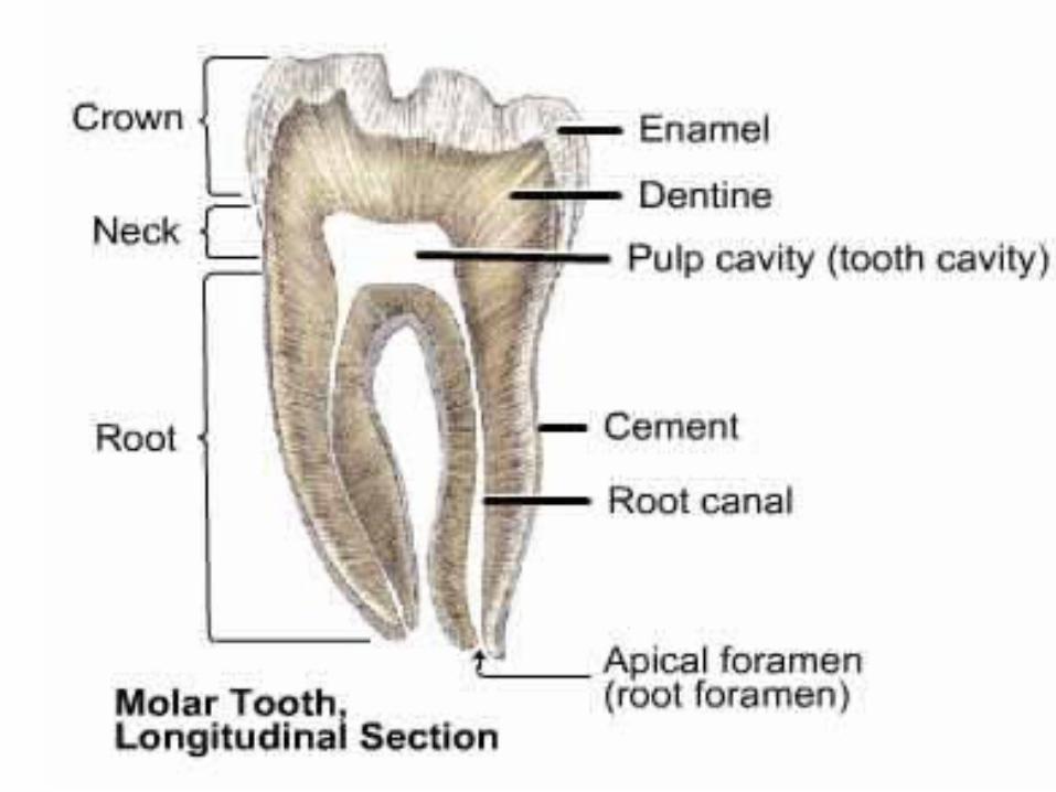
Each tooth has a crown and root portion. The crown is covered with enamel, and the root portion is covered with cementum. The root is embedded in the jaw bone.
The crown and root join at the cementoenamel junction. This junction also called the cervical line.
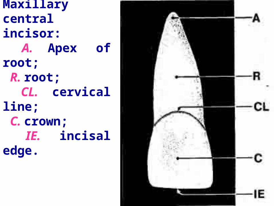
Maxillary central incisor: A. Apex of root; R. root; CL. cervical line; C. crown; IE. incisal edge.

The root portion of the tooth is firmly fixed in the bony process of the jaw, so that each tooth is held in its position relative to the others in the dental arch. That portion of the jaw which serves as a support for the tooth is called the alveolar process.
The crown portion is never covered by bone tissue after it is fully erupted, but it is partly covered at the cervical third in young adults by soft tissue of the mouth known as the gingiva or gingival tissue, or gum tissue. In older persons, all of the enamel and frequently some cervical cementum may be exposed in the oral cavity.
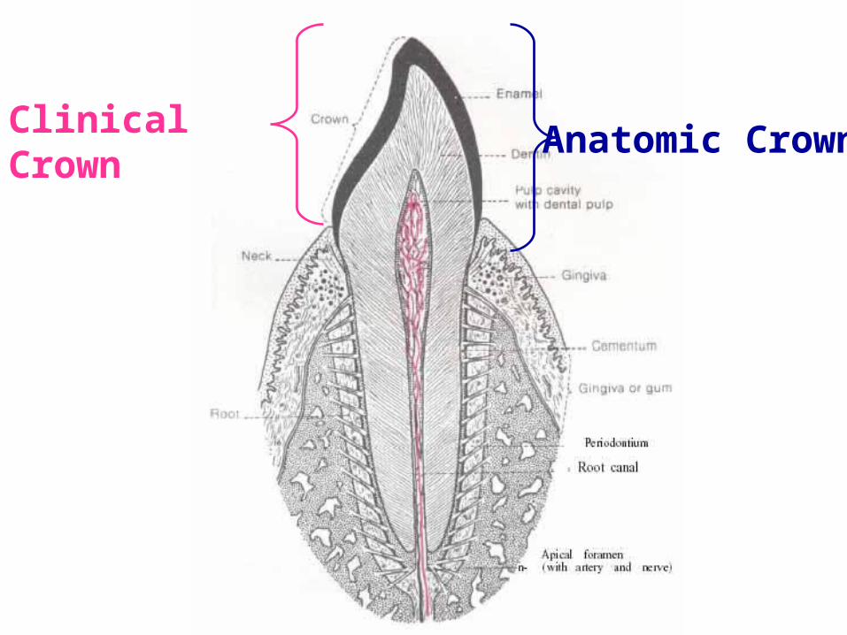
The Crown
• The Anatomic Crown is that portion of the tooth which is covered by enamel.
• The Clinical Crown is that portion of the tooth which is visible in the mouth regardless of whether it corresponds to the anatomical crown is constant in length whereas the clinical crown may change in length throughout life.
Clinical Crown Anatomic Crown

The crown of an incisor tooth may have an incisal ridge or edge, as in the central and lateral incisors; a single cusp, as in the canines; or two or more cusps, as on premolars and molars. Incisal ridges and cusps form the cutting surfaces on tooth crowns.

The Root
• The Anatomical Root is that portion of the tooth which is covered by cementum.
• The Clinical Root is that portion of the tooth which is not visible in the mouth. As with crown, the clinical root may change throughout life.

Clinical Root Anatomic Root

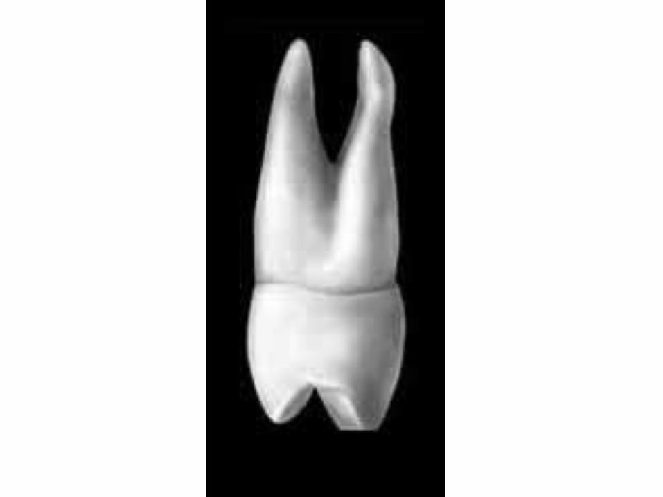
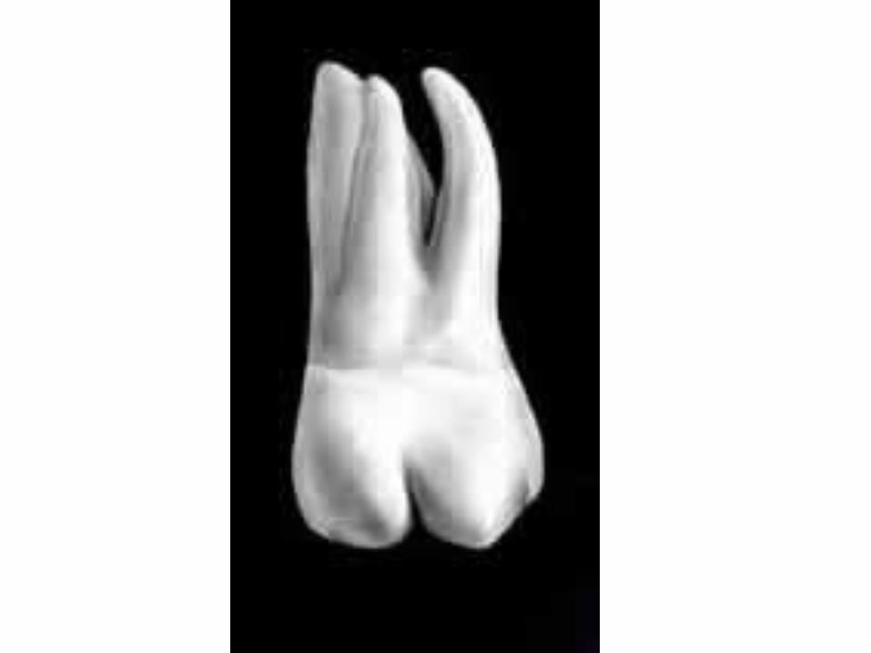
The root portion of the tooth may be single, with one apex or terminal end, as usually found in anterior teeth and some of the premolars; or multiple, with a bifurcation or trifurcation dividing the root portion into two or more extensions or roots with their apices or terminal ends, as found on all molars and in some premolars.

Single Root

Double Root, with a bifurcation

Triple Root, with a trifurcation
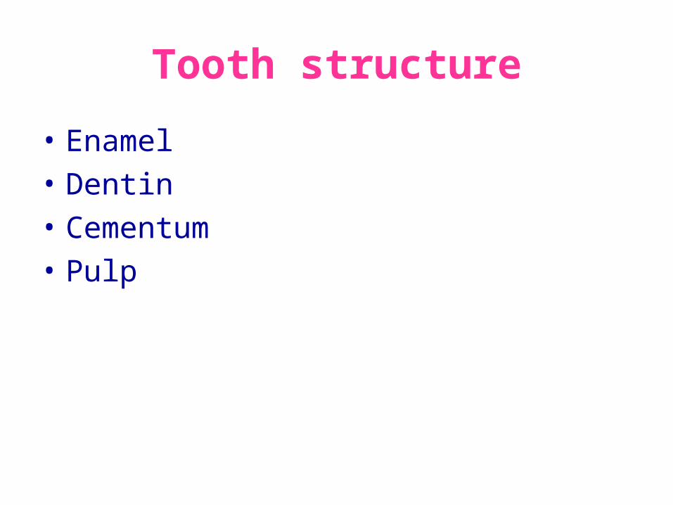
Tooth structure
• Enamel
• Dentin
• Cementum
• Pulp
The four tooth tissues are, Enamel, Dentin, Cementum, and Pulp. The first three are hard mineralized tissues composed of an organic matrix embedded by crystalline forms of calcium phosphate salt. The pulp is soft connective tissue.


Enamel
Enamel (mostly inorganic, calcified ) is the hard, white shiny surface of the anatomic crown. It is the outer surface of the crown. It is thickest over the tip of the crown and becomes thinner until ends at the cervical line. Enamel is the most mineralized and hardest tissue in the human body. It’s the protective outer surface of the anatomic crown, and is composed of: 95% calcium hydroxyapatite, 4% water and 1% enamel matrix.

This dense mineralization gives enamel thus the ability to resist the wear that the crown of a tooth is subject to. Enamel is smooth which gives the crown a self-cleaning ability.

Dentin
Dentin ( mostly inorganic, calcified) is the hard yellowish tissue underlying the enamel and cementum, making up the major bulk (main portion) of the tooth. Found in the crown and the root and surrounding the pulp cavity, it is composed of 70% calcium hydroxyapatite, 18% organic matter( collagen fibers) and 12% water.

It is softer than enamel, but harder than cementum or bone. The union of enamel and dentin is called Dentino-Enamel Junction, and the union of the dentin and cementum is called Dentino-Cemental Junction.

Dentino-Enamel Junction
Dentino-Cemental Junction

Cementum
Cementum (mostly inorganic, calcified) is a dull yellow external surface of the anatomic root (covering the dentin). It is a bony-like substance and its main functions is to provide a medium for the attachment of the tooth to the alveolar bone.
It is very thin layer next to the cervical line but is increases slightly in thickness at the apex of the root.

It is not as dense as hard as enamel or dentin, but is denser than bone, it is composed of 65% calcium hydroxyapatite, 23% organic matter and 12% water.
Pulp
Pulp is the soft (not calcified) tissue, it is found in the center part of the tooth, the pulp cavity is surrounded by dentine except at apical foramen . It has a coronal and a root portions
The functions of the pulp are: formative, sensory, nutritive and defensive. Pulp is composed of loose connective tissue, fibroblasts, blood vessels, nerves.

Anatomically the pulp cavity consists of two pares, the pulp chamber housed within the crown portion of the tooth, and the pulp canal which is located within the root of the tooth.
The constricted opening of the pulp canal is called the apical foramen. It is possible for the pulp canal to have two or more branches which make their exits at or near the apical end of the root, these are called multiple foramina or supplementary canals, on the other hand, many roots may be formed with more than one canal, which may end in a common foramen.
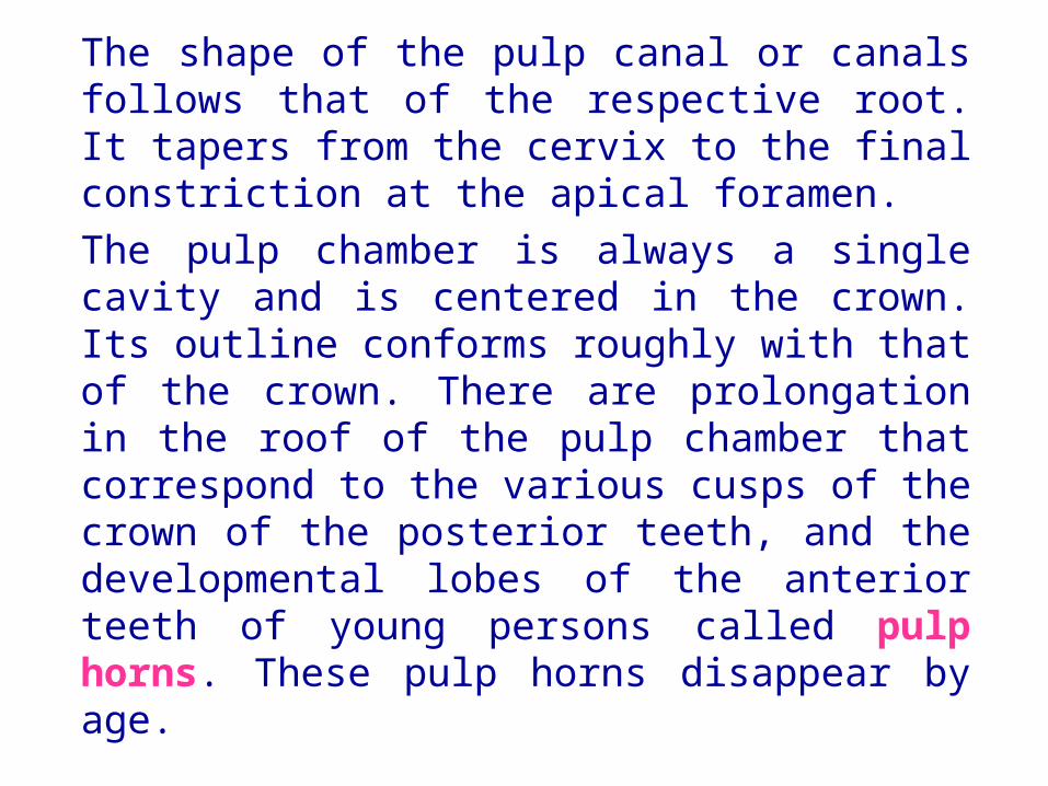
The shape of the pulp canal or canals follows that of the respective root. It tapers from the cervix to the final constriction at the apical foramen.
The pulp chamber is always a single cavity and is centered in the crown. Its outline conforms roughly with that of the crown. There are prolongation in the roof of the pulp chamber that correspond to the various cusps of the crown of the posterior teeth, and the developmental lobes of the anterior teeth of young persons called pulp horns. These pulp horns disappear by age.

Surfaces and Ridges

The crowns of the incisors and canines have four surfaces and a ridge, and the crowns of the premolars and molars have five surfaces. The surfaces are named according to their positions and uses. In the incisors and canines, the surfaces toward the lips are called labial surfaces; in the premolars and molars, those facing the cheek are the buccal surfaces. When labial and buccal surfaces are spoken of collectively, they are called facial surfaces. The inner surface of any maxillary tooth is called Palatal since it faces the palate of the mouth, and that of the mandibular teeth is called Lingual since it faces the tongue.

The surfaces of the premolars and molars which come in contact with those in the opposite jaw during the act of closure (called occlusion) are called occlusal surfaces. In incisors and canines, those surfaces are called incisal surfaces.
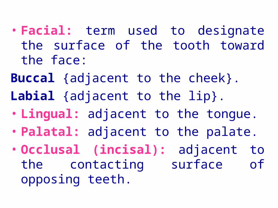
• Facial: term used to designate the surface of the tooth toward the face:
Buccal {adjacent to the cheek}.
Labial {adjacent to the lip}.
• Lingual: adjacent to the tongue.
• Palatal: adjacent to the palate.
• Occlusal (incisal): adjacent to the contacting surface of opposing teeth.
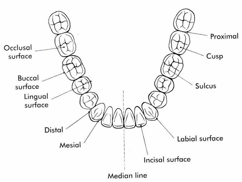
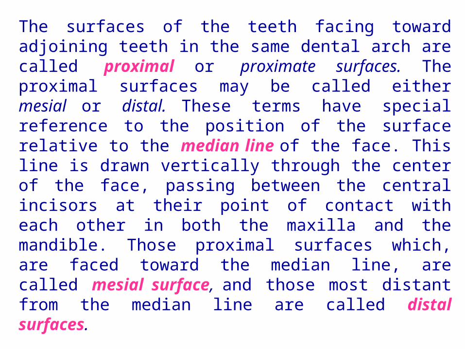
The surfaces of the teeth facing toward adjoining teeth in the same dental arch are called proximal or proximate surfaces. The proximal surfaces may be called either mesial or distal. These terms have special reference to the position of the surface relative to the median line of the face. This line is drawn vertically through the center of the face, passing between the central incisors at their point of contact with each other in both the maxilla and the mandible. Those proximal surfaces which, are faced toward the median line, are called mesial surface, and those most distant from the median line are called distal surfaces.

Four teeth have mesial surfaces that contact each other: the maxillary and mandibular central incisors. In all other instances, the mesial surface of one tooth contacts the distal surface of its neighbor, except for the distal surfaces of third molars of permanent teeth and distal surfaces of second molars in deciduous teeth, which have no teeth distal to them. The area of the mesial or distal surface of a tooth which touches its neighbor in the arch is called the contact area.
Central and lateral incisors and canines as a group are called anterior teeth; premolars and molars as a group, posterior teeth.
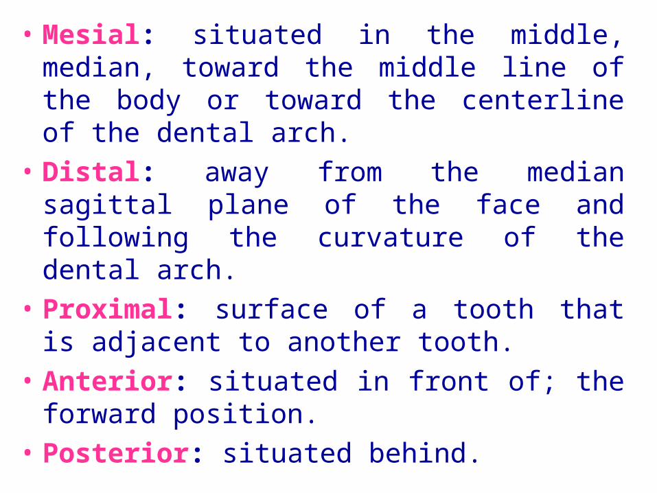
• Mesial: situated in the middle, median, toward the middle line of the body or toward the centerline of the dental arch.
• Distal: away from the median sagittal plane of the face and following the curvature of the dental arch.
• Proximal: surface of a tooth that is adjacent to another tooth.
• Anterior: situated in front of; the forward position.
• Posterior: situated behind.


Division of Surfaces,
Line Angles, and Point Angles
For purposes of description, the crowns and roots of teeth have been divided into thirds and junctions of the crown surfaces are described as line angles and point angles. Actually, there are no angles or points or plane surfaces on the teeth anywhere except those that appear from wear (abrasion) or from accidental fracture. Line angle and point angle are used only as descriptive terms to indicate a location.
When the surfaces of the crown and root portions are divided into thirds, these thirds are named according to their location. Looking at the tooth from the labial or buccal aspect, we see that the crown and root may be divided into thirds from the incisal or occlusal surface of the crown to the apex of the root. The crown is divided into an incisal or occlusal third, a middle third, and a cervical third. The root is divided into a cervical third, a middle third, and an apical third.

The crown may be divided into thirds in three directions: inciso- or occlusocervically. mesiodistally, or labio- or buccolingually. Mesiodistally, it is divided into the mesial, middle, and distal thirds. Labio- or buccolingually it is divided into labial or buccal, middle, and lingual thirds. Each of the five surfaces of a crown may be so divided. There will be one middle third and two other thirds, which are named according to their location, e.g., cervical, occlusal, mesial, lingual.

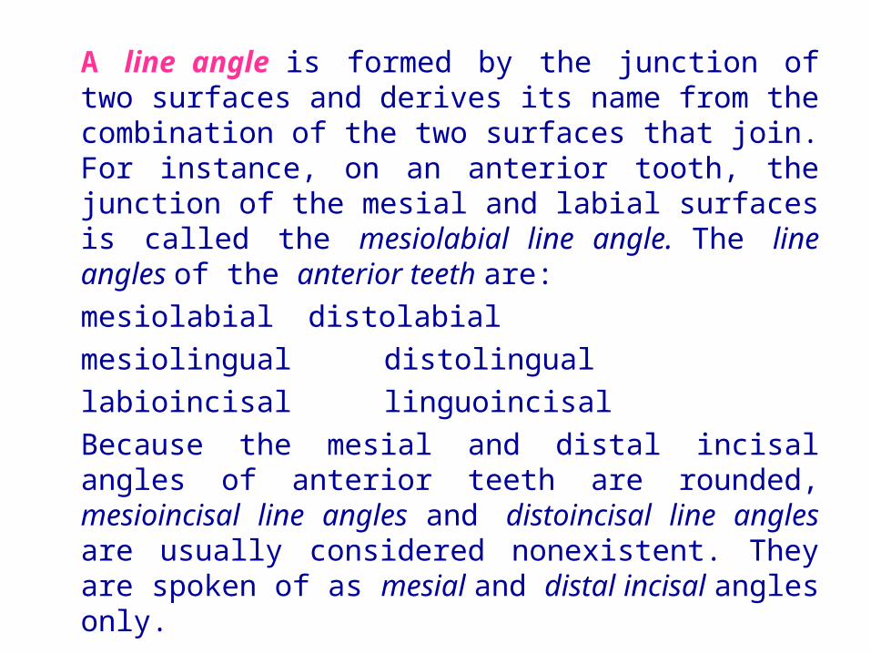
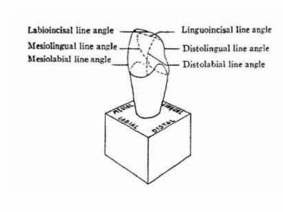
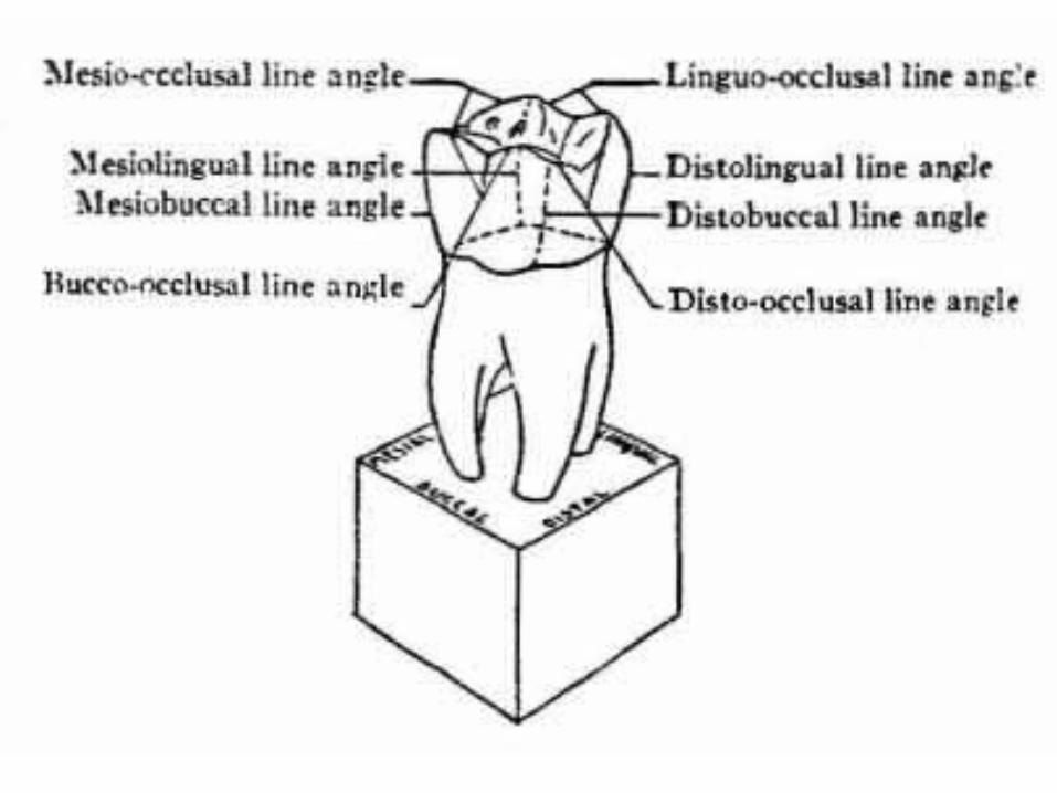
A line angle is formed by the junction of two surfaces and derives its name from the combination of the two surfaces that join. For instance, on an anterior tooth, the junction of the mesial and labial surfaces is called the mesiolabial line angle. The line angles of the anterior teeth are:
mesiolabial distolabial
mesiolingual distolingual
labioincisal linguoincisal
Because the mesial and distal incisal angles of anterior teeth are rounded, mesioincisal line angles and distoincisal line angles are usually considered nonexistent. They are spoken of as mesial and distal incisal angles only.

The line angles of the posterior teeth are:
mesiobuccal distobuccal
mesiolingual distolingual
mesio-occlusal disto-occlusal
bucco-occlusal linguo-occlusal

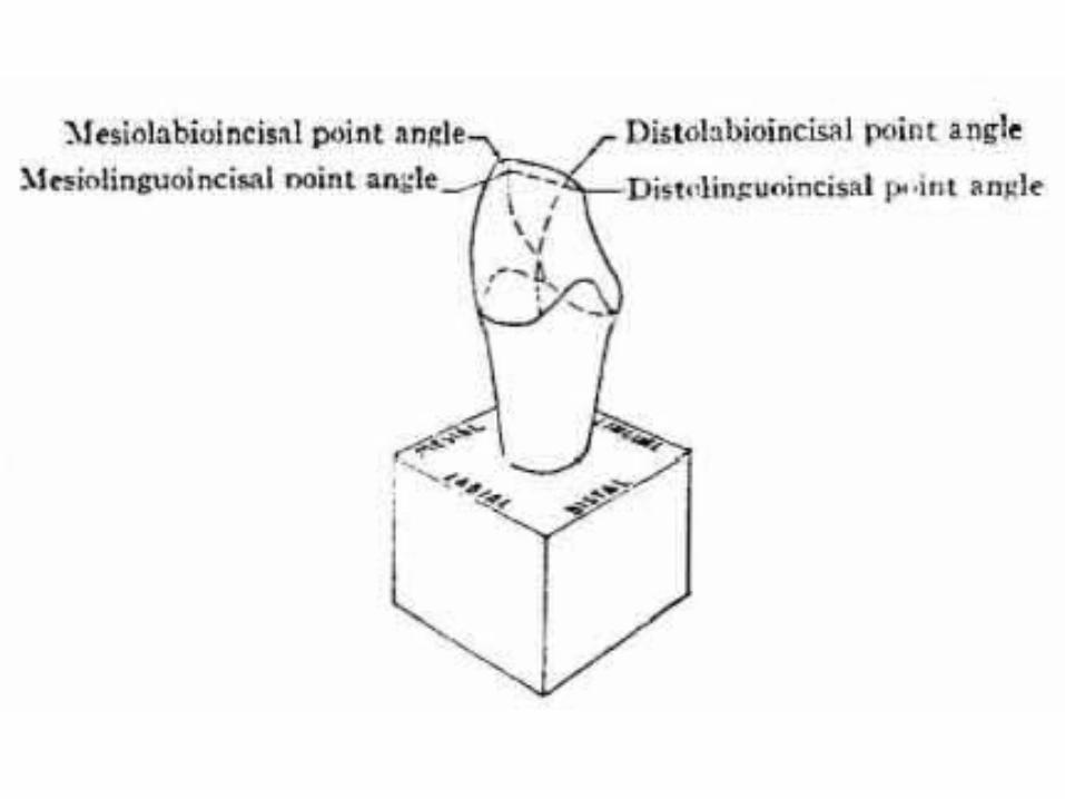
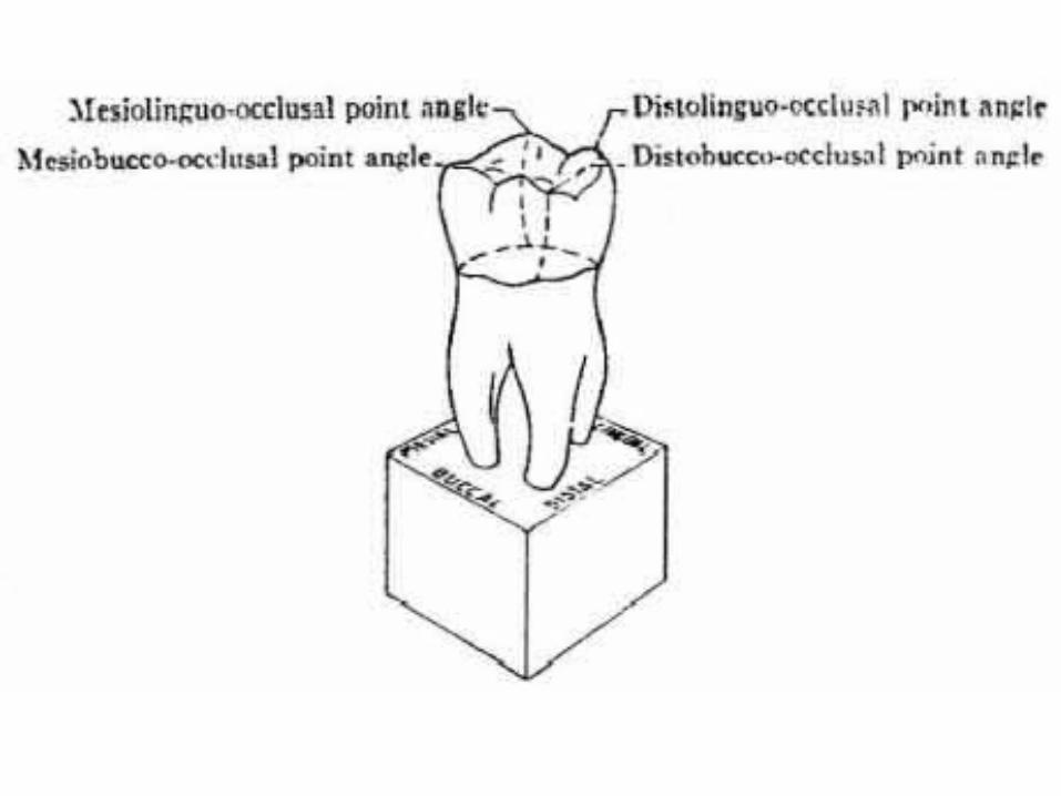
A point angle is formed by the junction of three surfaces. The point angle also derives its name from the combination of the names of the surfaces forming it. For example, the junction of the mesial, buccal, and occlusal surfaces of a molar is called the mesiobucco-occlusal point angle.
The point angles of the anterior teeth are:
mesiolabioincisal distolabioincisal mesiolinguoincisal distolinguoincisal
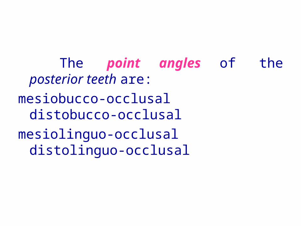
The point angles of the posterior teeth are:
mesiobucco-occlusal distobucco-occlusal
mesiolinguo-occlusal distolinguo-occlusal

The roots of the teeth may be single or multiple. Both maxillary and mandibular anterior teeth have only one root each. Mandibular first and second premolars and the maxillary second premolar are single-rooted, but the maxillary first premolar has two roots in most cases, one buccal and one lingual. Maxillary molars have three roots, one mesiobuccal, one distobuccal, and one lingual. Mandibular molars have two roots, one mesial and one distal. It must be understood that description in anatomy can never follow a hard-and-fast rule. Variations frequently occur. This is especially true regarding tooth roots, e.g., facial and lingual roots of mandibular canine.


Thank You