university of copenhagen and the future use of neutron and
TRANSCRIPT

ESS white paper · Spring 2013 · European Spallation Source · 1
University of Copenhagen and the future use of neutron and X-ray sources
May 2013 · A white paper on ESS and MAX IV

2 · ESS white paper · Spring 2013 · European Spallation SourcePhotos: University of Copenhagen · Graphic Design: Kliborg Design · Print: Nofo Print
CONTENTS 1. INTRODUCTION ….................................................................................... 52. SCIENTIFIC APPLICATIONS OF X-RAYS AND NEUTRONS .............................. 63. NEW POSSIBILITIES FOR RESEARCH AT ESS AND MAX IV ......................... 194. PERSPECTIVES FOR PRIVATE AND PUBLIC SECTOR COLLABORATION ........ 285. INITIATIVES IN TRAINING AND EDUCATION ............................................. 296. THE ESS DATA MANAGEMENT CENTRE… ................................................ 307. COMPLIMENTARY INFRASTRUCTURES ..................................................... 318. THE PRESENT SITUATION AND VISIONS FOR THE FUTURE .......................... 33 APPENDICES .......................................................................................... 35

ESS white paper · Spring 2013 · European Spallation Source · 3
The development of large neutron and X-ray research infrastructures (ESS and MAX IV) in Lund takes place with a significant engagement of researchers from University of Copenhagen. The opportunities offered by the new powerful neutron and X-ray sources open up new possibilities for studying all kinds of materials at an atomic level – from harder materials to soft biological materials. It will have a great impact, not only on the future development of science and education at the university, but also on the society. Already today, neutron and X-ray facilities have contributed to the development of a broad range of products from better computer chips to new medicine.
It is in the preparation for UCPHs future engagement in ESS and MAX that this white paper has been prepared. To set the stage, the background is provided for the scientific use of X-rays and neutron sources accompanied by a description of the unique opportunities offered by ESS and MAX IV. It is described how these may be exploited in innovative research of benefit to the private and public sector, and in new initiatives in education and training.
The experiments at the large facilities are supported by research infrastructure localized at or close to UCPH foremost the ESS Data Management Centre, but also by local X-ray equipment and studies by other experimental techniques.
Analysis of the present situation and visions for the future concludes this document that has been prepared by a working group, headed by Vice-Dean Sven Frokjaer, comprised of researchers from HEALTH and SCIENCE. We thank them for their work.
FOREWORD
Dean John Renner HansenFaculty of Science
Dean Ulla Wewer Faculty of Health and Medical Sciences

4 · ESS white paper · Spring 2013 · European Spallation Source
HOW DOES ESS WORK
ESS Data Management and Software Centre, North Campus, University of Copenhagen

ESS white paper · Spring 2013 · European Spallation Source · 5
In a few years, some of the world’s largest neutron and X-ray synchrotron research infrastructures will be operational in Lund, Sweden: European Spallation Source (ESS) operational in 2019 and MAX IV operational 2016. The ESS data management and software centre will be placed at North Campus, Copenhagen. Together with two other large research X-ray infrastructures located in Hamburg, European XFEL (operational in 2016) and Petra III (in operation since 2009), University of Copenhagen (UCPH) will have access to the most brilliant and smallest X-ray synchrotron source beams and the most intense neutron source that cannot be matched any other place in the world.
MAX IV, ESS, XFEL and Petra III are often referred to as super-microscopes, which are able to unravel the finest structural details of all types of matter from advanced solid state materials for IT devices to the proteins that govern the cellular processes fundamental for all living organisms. Access to the four facilities enables structural studies and studies of changes caused by the variation in the physical environment at a spatial resolution in the sub-nanometer range. Their capabilities may be exploited in a vast range of cutting-edge research in both materials and life sciences ranging from the development of new drugs (both delivery and their action in the biological environment), over the study and investigations of soft matter that plays an important role in our daily life, to the development of new materials with beneficial properties in a broad range of topics like energy conversion, data storage, and catalysis.
At present, there are several strong, highly recognized research groups at University of Copenhagen demonstrating their excellence and competences in producing world-leading results in biology, soft matter, and materials science based on experiments with neutrons and synchrotron radiation performed at large scale facilities. However, the current major challenge is to disseminate knowledge of the many applications that these infrastructures offer to a broader group of scientists and external stakeholders. Successful research groups at the university will not only be able to initiate state-of-the-art research but also be attractive partners for leading research groups, scientists and for public and private sector collaborations worldwide. University of Copenhagen has already taken a very important initiative by the funding of the UCPHs 2016 Funds for interdisciplinary research project “CoNEXT – Fertilizing the ground and harvesting the full potential of the new neutron and X-ray research infrastructures close to Copenhagen” by DKK 28 million in 2013-2016. This project serves to stimulate interdisciplinary research projects that depend on the use of the large research infrastructures and will be realized through fruitful collaborations between scientists with different expertise.
The aim of this report is to contribute to an understanding of the unique possibilities that these infrastructures represent for basic sciences and industrial applications. It is important to ensure that researchers at University of Copenhagen, in collaboration with scientists worldwide, are ready to harvest the full potential of the new X-ray and neutron sources, and that University of Copenhagen is well-prepared for acting as a portal for potential users of the facilities, such as Danish and international industries along with academic research groups.
1. INTRODUCTION

6 · ESS white paper · Spring 2013 · European Spallation Source
The use of X-rays to unravel structural details at atomic resolution for all types of matter from the most complex biological molecules to all types of materials can celebrate its 100th anniversary in 2013. In seminal experiments it was shown that X-rays are scattered by crystals and the scattering diffraction pattern may reveal the positions of the atoms in the crystal to the highest precision. These results mark the birth of modern crystallography.
Structure is intimately linked to properties and function of a molecule or a material, and the powerful combination of X-rays and crystals has since provided a wealth of information on crystalline materials that is available in data bases containing about 160,000 inorganic and mineral structures, 600,000 organic compounds, and almost 80,000 protein structures. The power of this method and its results are well illustrated by the nearly 30 Nobel Prizes in chemistry and physics that are based on the crystallographic measurements.
The scattering of X-rays is caused by interactions between X-rays and the electron distribution within the matter investigated. Since the discovery of X-rays in 1895, the variation in the absorption of X-rays between bone and soft tissue has been exploited in medical imaging. Also non-crystalline samples scatter X-rays, and the scattering recorded is used to provide valuable spatially averaged structural information on the sample. Finally, electronic transitions may occur through the interaction of X-rays, and spectroscopic measurements are able to reveal the chemical
composition of the sample. X-rays are a fantastic tool for revealing atomic composition and the detailed structural facets of all materials whatever state they are in, from crystalline over non-crystalline to solutions. However, since the signal from the scattering of X-rays is proportional to the electron density, which is again roughly proportional to the atomic number, X-rays are not ideal for investigating soft biological tissue, which is typically rich in hydrogen. Furthermore, they are damaging soft and biological materials, which cannot sustain long exposures of X-rays. Until about 20 years ago, X-rays were almost exclusively provided by in-house equipment comprised of high-voltage generators equipped with X-ray tubes. This setup was later improved by the technological developments of rotating anodes, which increased the intensity of the X-rays by an order of magnitude.
When electrons are accelerated to velocities close to the velocity of light they emit synchrotron radiation. This phenomenon in agreement with the relativity theory of Einstein was discovered in 1947. Initially, synchtron radiation was considered a nuisance by the particle physicists. Forty years later is was fully exploited in facilities dedicated to the production of synchrotron radiation by injecting the high-energy and high-velocity electrons into a storage ring and take out the radiation in the tangential directions of the ring.
2. SCIENTIFIC APPLICATIONS OF X-RAYS AND NEUTRONS

ESS white paper · Spring 2013 · European Spallation Source · 7
The advent of storage ring based synchrotron radiation has revolutionized X-ray science, as the intensity of synchrotron radiation obtained is more than 1010 larger than the radiation obtained from the in-house equipment. This has opened new avenues for the study of very small and dilute samples by all experimental X-ray techniques (see chapter 3). The worldwide growth of available synchrotron facilities now close to 100, convincingly demonstrates the need and success of these powerful X-ray sources.
The radiation produced this way in the whole energy range from infrared to gamma rays, is more intense and coherent than radiation produced by conventional sources. These unique characteristics of synchrotron radiation may be exploited as follows:
• The intensity of synchrotron radiation is more than 10 orders of magnitude larger than conventional sources. This enables studies of dilute and very small samples.
• The tunability of synchrotron radiation, e.g. selection of specific wavelengths for an experiment, is a powerful tool to identify not only the nature of the atoms in a sample but also their valence state. Furthermore, it allows for studies of vibrational and electronic transitions in a broad energy range
• The coherence of the synchrotron radiation, combined with the different interactions of X-rays with matter, has opened new use of X-rays for imaging.
The neutron was discovered in 1932 by James Chadwick, but it took some decades before it was realized that neutrons in many ways were complementary to X-rays and a very powerful tool to study the structure and properties of matter. The neutron interacts with the nucleus, and the absorption depends on the nature of nucleus. Their penetration of matter is very different from X-rays, and neutrons turn out to be a powerful tool for studying hydrogen atoms, and in contrast to X-rays they are non-destructive towards soft and biological materials.
Neutron sources with flux densities adequate for neutron scattering investigations of materials are based on one of two principles:• Fission: A high continuous flux of neutrons is produced in the
core of a conventional fission-based nuclear reactor. • Spallation: A pulsed production of neutrons is obtained by
bombarding a target of heavy elements with high-energy particles, typically accelerated protons.
Both neutron and synchroton sources are built as dedicated facilities hosting tens of instruments. All major sources are user facilities, meaning that they serve a research community much larger than the staff affiliated with the facilities.
More than 20 neutron facilities are in operation worldwide, the largest European facility being the reactor source ILL, Grenoble, France, and the spallation source ISIS, Oxfordshire, UK. However,

8 · ESS white paper · Spring 2013 · European Spallation Source
the European dominance is challenged by the powerful, newly commissioned spallation sources: Spallation Neutron Source (SNS), Oak Ridge, USA, and Japan Proton Accelerator Research Complex (J-PARC), Tokai, Japan. For these and other reasons, it has long been proposed to build a European Spallation Source (ESS). In 2009, it was decided to initiate the construction of this source in Lund, Sweden.
The neutron scattering facilities are used by about 8,000 researchers worldwide, the X-ray scientific community is significantly larger as it comprises about 20,000 scientists, which includes most of the neutron scattering community due to the complementarity of X-rays and neutrons .
Neutron and X-ray scattering represent the two most powerful and versatile experimental methods to study the structure and dynamics of materials at the atomic and nanometer scale. The scope of the application of X-ray and neutron scattering is continuously increasing as illustrated by the increasing demand for access to the neutron and synchrotron facilities. They have grown from being a tool in solid state physics and chemical crystallography to serve communities as diverse as biology, earth sciences, planetary science, engineering, polymer chemistry, and cultural heritage.
Although both techniques are very powerful, X-rays facilities are the brightest sources. Therefore, neutron scattering is used only
where X-ray scattering is inadequate. The rule of thumb goes: ‘If an experiment can be performed with X-rays, use X-rays’.
However, neutrons have a number of properties that make them extremely useful for purposes where X-rays are not suited or where complementary information is needed.• Energy and wavelength. Thermal neutrons have a
wavelength (around 1.8 Å) similar to inter-atomic distances, and an energy (around 25 meV) similar to elementary excitations in solids. In this way, one may obtain simultaneous information on the structure and dynamics of materials and e.g. measure dispersion relations (energy-wavelength dependence) of excitations in crystalline solids. With the words of the Nobel Prize committee in 1994: “… Neutrons tell you where the atoms are and what the atoms do”.
• Isotopes and light elements. The neutron scattering cross section varies in an almost random fashion between elements and even between different isotopes of the same element. One can thus use neutrons to study light isotopes. In particular, this is important for hydrogen, which is almost invisible to X-rays. With neutrons, hydrogen, H, and deuterium, D, change the contract in scattering. This is heavily exploited in biological and soft matter sciences to change the contrast in the scattering and also “highlight” selected groups within large molecules or aggregates.
• Quantitative measurements of bulk properties. The interaction between neutrons and matter is in general rather

ESS white paper · Spring 2013 · European Spallation Source · 9
weak, implying that neutrons probe the bulk of the sample, and not only its surface. The weak interaction also diminishes unwanted effects like multiple scattering. Therefore, quantitative comparisons between neutron scattering data and theoretical models can be performed to a high precision.
• Transparency. Since neutrons penetrate matter easily, neutron scattering can be performed with samples stored in all sorts of sample environment: Cryostats, magnets, furnaces, pressure cells, etc. Furthermore, very bulky samples can be studied, up to tens of cm thickness, depending on their elemental composition, and the sample is generally left unharmed by the neutron experiment.
• Magnetism. The neutron magnetic moment makes neutrons scatter from magnetic structures or magnetic field gradients. Unpolarized neutrons are used to learn about the periodicity and magnitude of the magnetic order, while scattering of spin-polarized neutrons can reveal the direction of the atomic magnetic moments.
In many cases, neutron scattering is performed in combination with other experimental techniques; often with neutron scattering as one of the final techniques to be applied before conclusions may be drawn.
PRESENT USE OF X-RAYS AND NEUTRONS AT UCPHScientists from University of Copenhagen are well-represented and frequent users at most of the international large-scale facilities for X-ray and neutron scattering, both at the largest European facilities, ESRF and ILL in Grenoble, but also at most of the many national synchrotron radiation and neutron scattering facilities in Europe.
At the University of Copenhagen, numerous large research projects funded primarily by external sources are more or less directly based on the access to experimental data obtained through large neutron and X-ray facilities.
DanScatt is an instrument center financed by the Danish Natural Science Foundation that enables the access to international large-scale infrastructure for synchrotron X-ray and neutron scattering by supported travelling and network meeting. It comprises all Danish users (about 350 including PhD students) and makes it possible that Danish user groups efficiently are able to exploit the opportunities at the facilities. Through its activities over the last decade, DanScatt has organized the Danish user communities across all universities and disciplines including structural biology, life science, physics, chemistry and materials research together with some of the larger industrial user groups.
The DanScatt initiative is an absolutely crucial instrument for Danish scientists in these areas and is internationally recognised at

10 · ESS white paper · Spring 2013 · European Spallation Source
the highest levels. Furthermore, DanScatt provides the background for the great cohesiveness that exist in the Danish scientific community of users of synchrotrons and neutrons sources. Equally important is that the DanScatt grant provides excellent contacts and collaborations between researchers from different institution, which have materialized in joint projects and applications.
A. EXAMPLES OF UCPH RESEARCH PROJECTSMagnetism in high-temperature superconductorsSuperconductivity is a fascinating state of matter, where the electrical conductivity vanish below a certain temperature. For the “high-temperature” superconductors, this temperature is of the order -100°C to -170°C, roughly half way between room temperature and the lowest temperature possible (the absolute zero), -273.15°C. For 25 years, scientists have been puzzled about the cause of superconductivity in these ceramic materials, which are bad conductors in their normal (warm) state. Many scientists believe that the intrinsic magnetic properties of these materials play an important, if not crucial, role. The group of Niels Bohr Institute is undertaking a huge effort to study the magnetic properties of high-temperature superconductors, a field where neutron scattering plays a decisive role, but also synchrotron X-rays and theoretical calculations are very important. Recent progress has shown that the mobile charges in some superconductors are most likely not evenly distributed, but agglomerate into charged regions and uncharged, magnetic regions. The ultimate goal of this research is to find the
mechanism behind high temperature superconductivity and, with this knowledge in hand, produce the first room-temperature superconductor – a discovery that would change the world.

ESS white paper · Spring 2013 · European Spallation Source · 11
a. Cube-shape magnet levitating over a superconductor. Room-temperature superconductivity could enable, e.g., levitating trains.
c. Recent neutron diffraction data reveal a highly complex crystal structure in a high-temperature superconductor. Details are still unexplained.
b. Atomic-scale picture of the high-temperature superconducting state. The current flows in the charged red channels of atoms, while the other atoms have a particular magnetic structure, revealed by neutron scattering.

12 · ESS white paper · Spring 2013 · European Spallation Source
Polymorphic materialsMost pharmaceuticals are manufactured as solids, in the form of tablets or capsules. It is crucial to achieve a form where the drug retains its optimized functionality, i.e. the crystals must dissolve in the body at the right position and at the right rate in order to be useful. However, it is often found that the drug exhibits polymorphism, i.e. several crystal forms may form and be stable at room temperature. Controlling polymorphism is of major industrial concern, not only for the production of pharmaceuticals, but also for production of many other solid materials, e.g. pigments and explosives: Different crystal forms can show very different properties, e.g. differences in dissolution rate and mechanical stability.It is not easy to understand and control the formation of polymorphic crystal forms.Elastic as well as inelastic neutron scattering can provide crucial information about the dynamics of molecules and atoms in crystals, which are crucial in order to understand their stability.
ESS white paper · Spring 2013 · European Spallation Source · 13
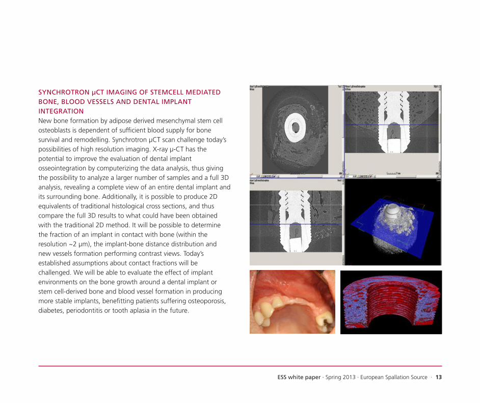
SYNCHROTRON μCT IMAGING OF STEMCELL MEDIATED BONE, BLOOD VESSELS AND DENTAL IMPLANT INTEGRATIONNew bone formation by adipose derived mesenchymal stem cell osteoblasts is dependent of sufficient blood supply for bone survival and remodelling. Synchrotron μCT scan challenge today’s possibilities of high resolution imaging. X-ray μ-CT has the potential to improve the evaluation of dental implant osseointegration by computerizing the data analysis, thus giving the possibility to analyze a larger number of samples and a full 3D analysis, revealing a complete view of an entire dental implant and its surrounding bone. Additionally, it is possible to produce 2D equivalents of traditional histological cross sections, and thus compare the full 3D results to what could have been obtained with the traditional 2D method. It will be possible to determine the fraction of an implant in contact with bone (within the resolution ~2 μm), the implant-bone distance distribution and new vessels formation performing contrast views. Today’s established assumptions about contact fractions will be challenged. We will be able to evaluate the effect of implant environments on the bone growth around a dental implant or stem cell-derived bone and blood vessel formation in producing more stable implants, benefitting patients suffering osteoporosis, diabetes, periodontitis or tooth aplasia in the future.

14 · ESS white paper · Spring 2013 · European Spallation Source
SPIN ARCHITECTURE IN SINGLE-MOLECULE MAGNETISM, MAGNETIC COOLING AND TOWARDS SPINTRONICSBased on coordination chemistry, it is possible to achieve control over the spatial arrangement of electron spins and their interactions. Such spin-architecture allows for design of magnetic properties of molecular and higher dimensional systems. New types of single-molecule magnets and magnetically cooling molecules are central goals, but also approaches to information processing via manipulation of spins, so-called spin-tronics, are being pursued. In unravelling the electronic and magnetic structure of these new materials, neutron scattering is a central technique, which provides information on magnetic interactions (inelastic scattering) and magnetic structure (polarized diffraction). Also X-ray techniques such as X-MCD are invaluable for studying magnetic systems organized on surfaces.
Dy
Dy Cr Dy
DyCr

ESS white paper · Spring 2013 · European Spallation Source · 15
WHAT HAPPENS WHEN MEAT IS BEING COOKED?Raw muscle tissue is a complicated system consisting of several types of globular and fibrous proteins that exhibit an hierarchical structure from the individual protein molecules that form the sarcomeres and collagen fibrils that constitute the individual muscle and connective tissue. From previous studies, it has been observed that many transitions take place during heating in the temperature interval from 40-80°C but so far these have only given indirect measure of the changes taking place on the surface using methods that destroy the food product in the process. Using X-ray phase-contrast tomography, it is possible to visualize the effect of heating on the same piece of muscle tissue giving for the first time a direct measurement of the transitions in full 3D. With this, it may be possible to directly study the influence of different cooking conditions while the food is being heated giving a fundamental knowledge that can be used for increasing the quality of food products.

16 · ESS white paper · Spring 2013 · European Spallation Source
UNDERSTANDING CHALK BEHAVIOURChalk is abundant in the Danish subsurface, both onshore, as aquifers for Danish drinking water, and offshore, in the North Sea, where it serves as the main reservoir for the Danish oil reserves. Chalk can be as much as 99% calcite, CaCO3, that was produced as coccoliths, the tiny platelets that some species of algae make to cover their one cell. Coccolithophorids still live in modern oceans but the biogenic calcite crystals that they produced more than 60 million years ago is what we see in Møns Klint, Dover and the vast chalk formations of Denmark and the North Sea.
The calcite crystals produced by the algae are very small (< 1 µm) and have remained small throughout geologic time, so the pores are also tiny. This makes it difficult for fluids to move through the material, even though the porosity is often high, as much as 40% in some cases. Characterization of the micro and nanostructure of chalk has been difficult up to now, but with synchrotron X-ray micro- and nanotomography, we can make pores visible and investigate their interconnectivity, also as a function of time.
Better understanding of the 3D microstructure of chalk, over a range of length scales can show us how fluids pass through pores and how flow channels can change as minerals dissolve and precipitate. This has important implications for oil production and also for cleaning contamination by hydrocarbons, pesticides and other toxic compounds in soil and groundwater. Also, 3D and 4D micro and nanostructural information provides new insight into the behaviour of a range of other porous media, such as catalysts, filtration membranes, tubing for hospital equipment, etc.
b) A series of images from the same chalk, where pore structure becomes increasingly visible as resolution increases.
c) A 3D reconstruction of a chalk sample showing the porosity and small pore throats.

ESS white paper · Spring 2013 · European Spallation Source · 17
a) A single coccolith from chalk. Each element is a single crystal of calcite, linked to form a disk.
18 · ESS white paper · Spring 2013 · European Spallation Source
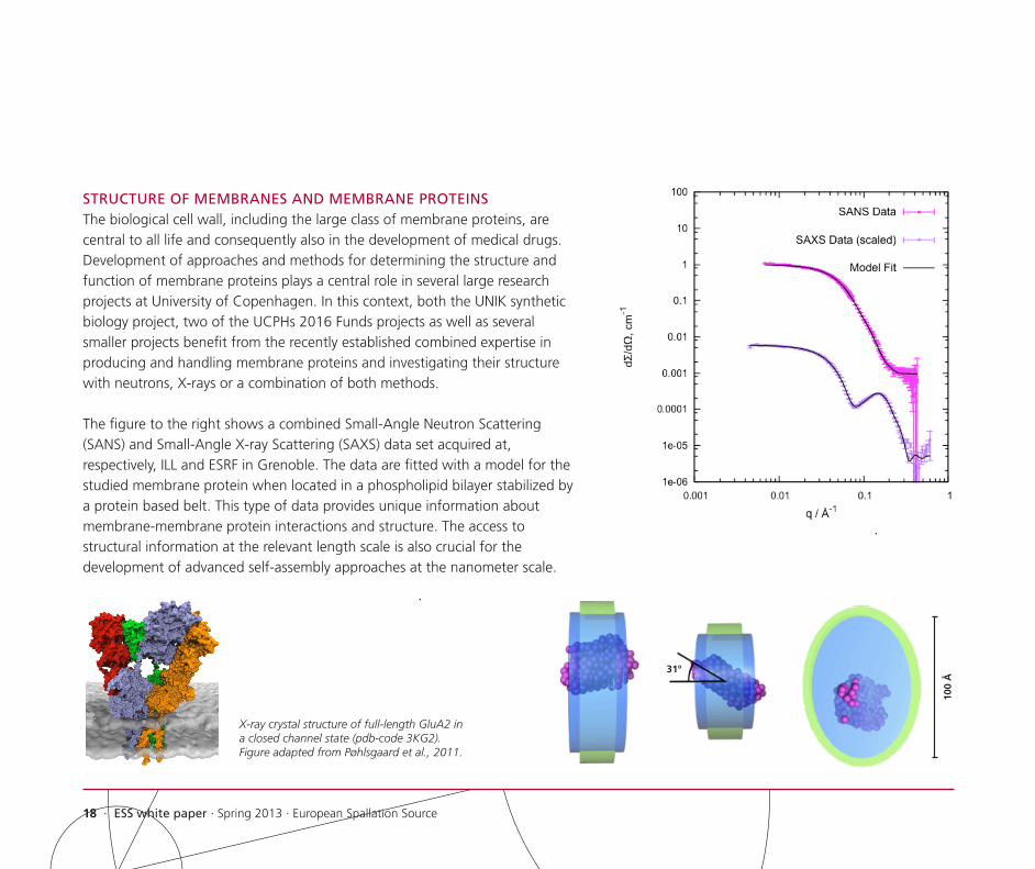
STRUCTURE OF MEMBRANES AND MEMBRANE PROTEINSThe biological cell wall, including the large class of membrane proteins, are central to all life and consequently also in the development of medical drugs. Development of approaches and methods for determining the structure and function of membrane proteins plays a central role in several large research projects at University of Copenhagen. In this context, both the UNIK synthetic biology project, two of the UCPHs 2016 Funds projects as well as several smaller projects benefit from the recently established combined expertise in producing and handling membrane proteins and investigating their structure with neutrons, X-rays or a combination of both methods.
The figure to the right shows a combined Small-Angle Neutron Scattering (SANS) and Small-Angle X-ray Scattering (SAXS) data set acquired at, respectively, ILL and ESRF in Grenoble. The data are fitted with a model for the studied membrane protein when located in a phospholipid bilayer stabilized by a protein based belt. This type of data provides unique information about membrane-membrane protein interactions and structure. The access to structural information at the relevant length scale is also crucial for the development of advanced self-assembly approaches at the nanometer scale.
31°
100
Å
X-ray crystal structure of full-length GluA2 in a closed channel state (pdb-code 3KG2). Figure adapted from Pøhlsgaard et al., 2011.

ESS white paper · Spring 2013 · European Spallation Source · 19
The third generation synchrotron sources dedicated exclusively to the scientific use of the X-rays generated have been in operation for more than twenty years, and it was believed that the brilliance and performance of these facilities had reached an optimum. However, the novel design that is used for MAX IV has opened a new era for synchrotron radiation. The MAX IV design gives the 3 GeV ring of MAX IV the highest brilliance and the smallest vertical emittance worldwide. These characteristics makes MAX IV a unique instrument in synchrotron radiation based science. It is highly suited for studying very small and dilute samples and superior for imaging and nanoscience. The first seven beamlines are presently under construction at MAX IV. In particular, the FemtoMAX beamline at the linear accelerator will enable time resolved studies in the femtosecond range, and the NANOMAX beamline will exploit the low emittance and high brilliance in imaging of nanosize objects.
The multiple bend magnet design used for MAX IV is used and developed further at other new facilities, e.g. the Brasilian synchrotron in Campinas and the second phase of the ESRF Upgrade in Grenoble, France.
Similarily, through the exploitation of all the technological advances ESS will be a factor of at least 10 more powerful than the best present available neutron sources, those in USA and Japan, and around a factor 100 more powerful than the current European neutron sources.
At MAX IV and ESS, X-rays and neutrons are going to be used for the same type of experiments but due their different properties, their interactions with matter are quite different. As a consequence, different but also complementary information is obtained by performing experiments on the same sample at MAX IV and ESS. The combination of the results from MAX IV and ESS creates unique scientific synergy, enabling studies of all types of matter, as described in chapter 2. As a basis for these investigations, an overview is given of the different types of experiments that can be performed with neutrons and X-rays listed in the order of increasing resolution.• Imaging Immediately after the discovery of X-rays by Röntgen
in 1895, it was recognized that they possess unique properties for medical imaging. With synchtrotron radiation, several new imaging tomographic techniques have been developed that provide unprecedented spatial resolution. Due to absorption, it is not always possible to get sufficient penetration of the sample, and here neutrons come to the rescue. Neutron tomography detects the inside of materials. This in particular highlights hydrogen, while some heavy materials (e.g. lead) are almost transparent.
• Reflectivity is the method used to investigate simple and complex surfaces and determine their layer thickness, composition, and structure by reflecting X-rays and neutrons at small angles.
• Diffraction provides detailed structural information at atomic resolution on crystalline materials with 2D or 3D symmetry.
3. NEW POSSIBILITIES FOR RESEARCH AT ESS AND MAX IV

20 · ESS white paper · Spring 2013 · European Spallation Source
X-ray diffraction spans widely and can provide accurate information on the atomic positions from minerals to protein crystals, except for the hydrogen atoms. The power of neutron diffraction is the ability to provide accurate information on the position of hydrogen, to distinguish elements of almost the same atomic number, and to study magnetic structures.
• Small-angle scattering is used to study nanometer-sized particles, inclusions and domains. It is often used to study macromolecules and soft matter. In Small Angle Neutron Scattering, the difference in scattering length of hydrogen and deuterium is exploited in the study of particularly deuterated samples.
• Spectroscopy X-ray absorption and emission are widely used techniques to provide information on the content and spatial location and neighbours of the heavier atoms in a sample. By neutron spectroscopy, one can detect the motion of atoms, molecules and magnetic moments over a frequency range that span 9 orders of magnitude.
In addition, a few novel concepts concern all experiment types:
• Time-resolved experiments that enable studies on structural changes with time can be performed to a resolution in the ps range with synchtrotron radiation. With the new free electron laser facilities, this time regime is further decreased to the femtosecond range. Time resolved experiments in the microsecond resolution will be feasible at ESS.
• In-situ experiments with X-rays and neutrons can be used to study “real” materials under “real” conditions, e.g. change of crystal grains during processing, growth and composition of material grains under chemical synthesis, or phase change of materials under high pressures.
A. PERSPECTIVES IN THE SCIENCE OF HARD AND SOFT MATERIALSOur society increasingly depends upon breakthroughs in materials science. From the magnetic read heads in the hard drives in our laptops, over the light but robust wind mill wings, to the plethora of plastic products around us, novel materials ease our lives every day. Materials science is also key to the solution of some of the present great challenges: the dilemma on energy supply vs. climate change, and the sustainability challenge. Moreover, materials science has facilitated the IT revolution that has changed, and is still changing, the way we process and use large amounts of information. In addition, improved understanding of materials will improve our knowledge of the world surrounding us, even when it has no direct engineering or commercial applications.
ESS and MAX IV will have a great impact on materials science in the broadest sense. This is based upon the ability of neutrons and X-rays to extract information on atomic position and motion, on the size and shape of nano-sized objects, on magnetism, and on their ability to penetrate bulk matter.

ESS white paper · Spring 2013 · European Spallation Source · 21
The potential of ESS and MAX IV for material studies, where one makes use of the methods described earlier in this chapter, is cross-disciplinary, covering topics in physics, chemistry, engineering, geology, and arts/archaeology. This will be exemplified in different areas in both the harder materials (containing heavier atoms) and softer materials (all light atoms). Many of these topics are already relevant for the present research at UCPH, or could be of future relevance.
HARDER MATERIALSGeo-sciencesThe geo-sciences will benefit from virtually all the techniques available at ESS and MAX IV: imaging, diffraction, spectroscopy and in-situ studies. With the capabilities at MAX IV, the mineral distribution in a rock determined by X-ray diffraction can be a standard technique, while ESS can support these investigations by locating the position of light elements, both on the atomic and larger scales, e.g. water content in pores in rock and soil. Both at MAX IV and ESS, diffraction and inelastic scattering under extremely high temperatures and pressure can reveal the structure of minerals and their phase transitions under conditions similar to those of the Earth’s mantle and address issues like metamorphism and multi-component melts. This information, in turn, is helpful for e.g. understanding and predicting vulcanic activity and earthquakes. Moreover, the 3D arrangement of minerals in strongly inhomogeneous rocks can be studied in details by X-ray and neutron tomography without the need of destroying rare
samples. In the case of meteorites, their composition and structure can reveal details about the conditions in the young solar system. The same technique can be used for detailed investigations of fossils and other paleontological evidence of life in the past.
In addition, Max IV and ESS have a large potential to reveal details on the crystal structure and hydrogen dynamics of ice cores obtained, e.g. in Greenland and Antarctica, adding important pieces of information about the past climate.
Energy materialsA most important task for ESS and MAX IV is the development of materials that may reduce the consumption of energy resources and improve energy storage. This strategic goal covers a number of important topics, including: lighter and stronger construction materials, battery materials with improved capacity and durability, lighter and more efficient hydrogen storage materials, solar cells with improved efficiency, improved thermoelectric materials for conversion of waste heat into electricity, and novel materials for fuel cells. Often, knowledge of the materials’ atomic or mesoscopic properties are essential to obtain a detailed understanding of how to improve the materials. In this connection, X-rays are used to obtain the general atomic and nano-scale structure of the materials, while neutrons are used to specifically probe the position of lighter elements (in particular hydrogen), and their vibrations and diffusive motion.

22 · ESS white paper · Spring 2013 · European Spallation Source
Functional materialsA large class of materials have physical properties that have a large potential for applications in society. These properties comprise magnetism, electrical conductivity and polarization and elasticity. Examples of research in this direction cover magnetism in molecules and other nanosized objects to optimize data storage, new strongly magnetic materials, e.g. generators, and superconductivity where the aim is to create materials that superconduct at room temperature with great benefits e.g. generation and transport of electrical power.
A new class of materials, the multiferroics, simultaneously possess electric, magnetic, and elastic properties and are predicted to have great potential, e.g. as sensor materials. MAX IV will be used to study the structural and magnetic properties of these materials, also on the nano-scale, while ESS will be particularly strong in the study of the lattice vibrations and magnetic structure and dynamics of the materials. Pump-probe experiments to study the time-dependent response to an impulse are particularly promising in the multiferroic materials and will be performed at the facilities in Lund as well as on the XFEL facility in Hamburg.
Engineering materialsIn combination, ESS and MAX IV will be extremely well suited for the study of materials that are today in common use for construction purposes like steel, aluminum, alloys, concrete, wood, and composite materials. The purpose of these studies are
to understand the mechanisms of current shortcomings like fatigue, welding faults, and dehydration. The X-ray and neutron methods will be able to reveal fractures, grain boundaries, stress and strain, as well as chemical changes.
Nanostructured materialsThe tremendous increase in brilliance has enabled the modern x-ray synchrotron radiation facilities to develop a completely new suite of techniques for visualizing the structure of soft and hard materials on the nanometre scale. This is done by exploiting the small source size and new optical devices to focus the X-ray beam to less than 50 nanometre and image the 3D structure of individual nano-scale objects. This new analytical device has great potential in the development of new functional materials and in life science for medical applications.
New states of matterOver the last decade, a series of materials have appeared, which at low temperatures reveal new states of matter, where electrons are correlated in a way that can only be described by quantum mechanics. These discoveries are fundamental for our understanding of nature in the same sense as cosmology and particle physics, and the behaviour of the materials is, in some cases, a direct manifestation of terms known from nuclear and particle physics, like the Yukawa force and the Higgs mechanism. These correlated materials may in the long term provide new paradigms for information processing, also known as quantum

ESS white paper · Spring 2013 · European Spallation Source · 23
computing. Present discoveries with these materials cover quantum phase transitions, spin ice, magnetic monopoles, skyrmions, and topological order. Pump-prope experiments on these systems with X-rays and neutrons can reveal the behaviour of these quantum states under external perturbation; a topic of strong interest in fundamental quantum mechanics.
Related to this topic, the ESS will also be used to study the properties of the neutron itself, e.g. its electrical dipole moment, its quantum mechanical wave properties, and its decay and lifetime.
SOFTER MATERIALSPolymer systemsPolymers and polymer/inorganic nanocomposite materials have wide applications in a large variety of daily-life systems, and potential for even more advanced applications. Examples are the use of self-assembly amphiphilic molecular systems into transparent gels that may act as drug carrier or drug delivery systems, or to replace parts of the human body. An example is a novel polymer/inorganic nanocomposite material to replace the vitreous body of the eye, where the inorganic nanoparticles serve to control the refractive index as well as the mechanical properties. A related material, but with complete different applications, is polymer/nanoparticle composites to be used for membranes with catalytic function in fuel cells. Both X-ray and neutron scattering are key techniques in studying such materials,
not least in combination securing different contrasts, and thereby making different parts of the material visible. Neutron scattering has a special advance in polymer science, since the large number of hydrogen atoms makes deuterium labelling a possibility to study ‘single’ molecular properties, both static structure and dynamic properties.
Drug developmentsOne of the major obstacles in the drug development process is the development of the optimum final dosage form, e.g. solid dosage forms (such as tablets, capsules) and the manufacturing process of these.
Traditionally, the suitability of a given dosage form has been investigated in a trial-and-error process, where the mechanical properties and the kinetic and thermodynamic stability have been tested experimentally. In other words, the traditional tools have been able to examine the bulk properties in posteriori characterization of the dosage forms. The intensity of the synchrotron light and the potent flux of neutrons from the spallation sources allow the design of hitherto impossible experiments, such as time resolved studies and studies of sub-micron species.
The new tools based on large-scale facilities opens the possibilities of studying changes as they happen, in situ, and of characterizing the inhomogeneity of crystalline materials at the nano-scale using

24 · ESS white paper · Spring 2013 · European Spallation Source
a large variety of experimental techniques, e.g. combined in-situ micro-indentation and X-ray microscopy studies of the mechanical behaviour of crystals; in-situ pressure studies using synchrotron powder diffraction; in-situ solubility studies using Powder Diffraction (PXRD) and Small-Angle Scattering (SAXS); Polymorph stability studied by multi-temperature X-ray and neutron experiments, and finally studies of crystal defects, disorder and mixed phases using X-ray microscopy and diffuse scattering measurements.
Food science A growing demand towards healthier food and food products with increased functionality requires a much deeper knowledge of the fundamental structures and interactions in food systems. So far, most of the methods used in food science have been indirect macroscopic measurements such as the water loss in sausages, the falling number of wheat or the tenderness of a beef that can be related to some overall quality parameter of the product. With the methods available at MAX IV and ESS, it will be possible to perform direct measurements of the microscopic structures and dynamics of food systems giving a new fundamental understanding of the processes taking place on the micro- and nanoscale. Using X-ray and neutron scattering techniques, the molecular building blocks of food, e.g. proteins, carbonhydrates and lipids, may be investigated, whereas novel, fast X-ray tomography methods allow for in-situ 3D imaging of the changes taking place in a food product during preparation, e.g. to directly
see what happens in the baking of bread or cooking of meat.
Art and cultural heritageIn a number of humanistic disciplines, the investigated objects are unique and can only be studied by truly non-destructive techniques. This comprises rare pieces of art and historical or archaeological artefacts. Here, X-rays and neutrons can be of great importance, as combined tomographic techniques can be used to image the internal structure and indicate the element distribution in the rare objects, and e.g. tell early drafts hiding beneath famous paintings. Moreover, diffraction techniques may be used to study crystalline grains and texture, which can reveal, e.g. the processes used to manufacture the objects.
B. PERSPECTIVES FOR LIFE SCIENCESIn recent years, our understanding of molecular and cellular processes in living organisms has improved dramatically thanks to stunning technological developments in molecular biology, cell biology, chemistry and physics. For example, both human genomes and genomes of multiple other organisms are now being almost routinely sequenced, giving rise to unprecedented insights into a plethora of genes encoding an impressive variety of different proteins. Through multifaceted interactions, these proteins play key roles in governing the functional complexity of biological systems from single cell organisms to humans. We are still, however, only beginning to understand the complexity of these systems, although such insights represent an evident

ESS white paper · Spring 2013 · European Spallation Source · 25
prerequisite for discovery of new treatments against major disorders such as cancer, diabetes, obesity, infectious diseases and neurodegenerative illnesses. Such insights also represent a prerequisite for developing strategies for prevention of disease and accordingly for increasing the overall quality of life. In light of a rapidly aging population, in particular in developed countries, the challenge is even greater and demands for large investments in health care science and access to the best possible instrumentation.
Indeed, to address the fundamental challenges in current biomedical research, access to advanced instrumentation and infrastructure has become progressively important together with cross-disciplinary interaction with other scientific disciplines such as physics and chemistry. Of pivotal and increasing importance is the ability to dissect and visualize the atomic and molecular structure of biomolecules, including not only individual proteins and protein complexes but also biomolecular structures involving DNA, lipid membranes and carbohydrates. This requires easy access to a broad array of different methodologies that all require large-scale instrumentation facilities such as X-ray and neutron scattering, nuclear magnetic resonance (NMR) or electron microscopy. Accordingly, the establishment MAX IV and the ESS will provide important novel possibilities for researchers in the Öresund Region by offering superb and world leading X-ray and neutron sources.
X-ray crystallography is playing a steadily increasing role in life sciences by allowing determination at atomic resolution the precise structure of e.g. protein and protein complexes. Notably, such information is essential for understanding the function of biological systems at the atomic level, and access to international state-of-the-art X-ray beamlines, as at the MAX IV, is vital for achieving optimal resolution and detailed insights. Competitive X-ray beamlines are also highly critical for optimal results in Small-Angle X-ray Scattering (SAXS) experiments that permit determination of e.g. the structure, morphology, shape and nanoscale dynamics of protein and protein complexes directly in solution, although with lower resolution than that achieved in crystallography experiments.
Scattering of neutrons may be used as an important complementary strategy to reveal structures and shapes of molecular complexes. The method depends on access to neutron sources, as those of the ESS, and opens up for completely novel approaches in biomedical research. Neutrons can ‘see’ carbon, nitrogen and oxygen equally well, while neutrons are highly sensitive to isotopes of hydrogen. Whereas the scattering power of hydrogen is very different from that of oxygen, deuterium scatters to the same extent as oxygen. Thus, substitution in selected molecules of hydrogen with deuterium makes it possible to detect the conformation or dynamics of specific parts of a biomolecular structure. Neutrons can also probe water in biological macromolecules, and it is therefore possible by altering

26 · ESS white paper · Spring 2013 · European Spallation Source
the ratio between hydrogen and deuterium to vary the scattering contrast between the molecules and the solution and thereby detect them separately within the complex. Importantly, neutrons easily penetrate most materials without causing any radiation damage. This makes neutrons suitable both for the study of delicate biological systems and for more robust materials used, e.g. in medical implants.
Altogether, access to MAX IV and ESS will open up for numerous applications in biomedical research/life sciences to the great benefit of researchers in the Öresund area. Major applications in areas, where strong expertise already exists at University of Copenhagen, include:
Pharmacology and drug discoveryRecent developments in crystallography techniques have dramatically facilitated crystallization of both existing and potential drug targets including in particular membrane receptors, transporters and ion channels. Crystallization of these proteins and atomic structure determination by use of forceful X-ray beams like MAX IV are expected to be a major focus area in the coming years. We cannot only expect substantial new insights into the molecular function of these proteins but also unprecedented insights into drug-receptor interactions and mechanisms of drug action. This might revolutionize drug discovery by facilitating rational drug design and development of novel types of pharmaceuticals with tailor-made functions and targeted delivery
mechanisms. Employment of a broader range of X-ray and neutron scattering techniques will strongly complement high-resolution X-ray crystallography by permitting dynamic analysis of e.g. drug-target interaction directly in aqueous environments.
Drug deliveryOf the many potential drug types that are identified every year, only a few are applicable due to the challenges associated with transporting the drug to the target while maintaining the drug activity. Depending on the molecular structure of the drug and on the type of the target, many different approaches have been developed to circumvent this problem. However, as most modern drug delivery systems rely on nanoscale particles, either formed by the drug particles themselves or as transport vehicles for the drugs, it is necessary to use a combination of neutron and X-ray scattering and electron microscopy to obtain the nanoscale structural information required for establishing an understanding and control of the systems. Application of the world-leading facilities at the nearby facilities ESS and MAX IV will give the research groups based at UCPH in this field a significant scientific edge compared to groups that mainly rely on standard and more broadly available small-laboratory techniques.
The potential of ESS and MAX IV for life sciences may be further exemplified by the following topics:• Cell biology and disease: Application of X-ray
crystallography together with X-ray and neutron scattering

ESS white paper · Spring 2013 · European Spallation Source · 27
represent key technologies when attempting to dissect mechanisms responsible for fine-tuned regulation of intracellular signalling networks and how alterations in these might change cell function and lead to disease. Application of X-ray and neutron scattering techniques should prove particularly useful for dissecting the molecular structure and dynamics of protein-protein and protein-lipid complexes critical for maintaining cell homeostasis. Specifically, when using deuterium exchange in neutron scattering experiment, it will be possible to examine the precise role and nanoscale dynamics of individual components in larger biomolecular complexes.
• Infectious diseases: Infectious diseases represent a major global challenge as a consequence of the increasing appearance of multiresistant bacteria. Development of new antibiotics with new mechanisms of action and delivery methods are therefore of utmost importance. Application of X-ray and neutron scattering techniques is of fundamental importance in these efforts permitting structural determination of new potential targets and delineation of molecular mechanisms underlying the infectious process. Also, scattering techniques should demonstrate their strength when attempting to reveal and describe infectious processes mediated by e.g. virus.
• Synthetic biology: Synthetic biology represents an emerging area that combines science and engineering to design and build novel biological functions and systems. By learning to
handle intact biological molecules or modules, in particular via self-assembly, it is the aim to develop insights, tools and technologies for preparing and characterizing systems with new, tailor-made biological functions. Synthetic biology is based on the “share your parts principle” enabling controlled construction of new biologically based devices for use in health care science e.g. nanoscale biosensors and diagnostic tools as well as in projects aimed at developing solar energy driven synthesis of a multitude of different high-valuable compounds with competitive edge and market potential for the future globalised bio-based society. Access to top class X-ray and neutron sources is essential for these efforts in order to obtain structural insight and control of the self-assembled nanoscale structures.
• Environmental science and toxicology: Pollution from e.g. nanoparticles represents a growing problem in today’s society and little is known about the consequences for human health. Application of both X-ray and neutron scattering should prove highly valuable in delineating the toxicological impact of these particles on human cells.
• Medical implants and materials: Both X-ray and neutron scattering methods are highly valuable for studying the properties and structure of biomaterials and, hence, has great potential in developing and improving medical implants. This includes e.g. improved dental implants and strategies for regeneration rather than replacing bone tissue.

28 · ESS white paper · Spring 2013 · European Spallation Source
Neutron and X-ray sources may be used in a broad range of areas in research and technology. The varieties of applications make ESS and MAX IV interesting not only to university researchers but also to private businesses, which will have the opportunity for analyz- ing and characterizing materials and processes on a far more detailed level than today.
Neutron and X-ray facilities have already contributed to the development of new and better computer chips, textiles, batter- ies, plastic, fuels, transportation technologies, food technologies, pharmaceuticals and medical devices. ESS and MAX IV will provide groundbreaking knowledge in all these areas facilitating the development of new and better products and materials to the benefit of society and the environment.
However, many challenges must be addressed in order to fulfill the industrial potential of ESS and MAX IV. Communicating the techniques and their potentials to the industry in general is crucial, and the industry must have easy access to experts in the field. Thus, UCPH wishes to support the industry in order to get full benefit of ESS and MAX IV. Dissemination to the public and contact to companies are already important elements of the CoNEXT project, and we will do our utmost to facilitate and strengthen the interaction between industry and scientists in the field.
For small and medium-sized enterprises there are significant barriers to overcome for them to use the large facilities, and they could benefit from collaboration with university researchers. While many small companies operate on a time scale of months with relatively small and very focused development projects, universities typically work on much longer time scales, and many industrial collaborations take place through industrial PhD and postdoc projects. This also relates to the typical time it takes to allocate the beam time at large-scale facilities which is typically at the order of months.
In an attempt to circumvent this problem, a pilot project is presently being initiated in collaboration between the Niels Bohr Institute and the Capital Region of Denmark (Region Hovedstaden). The idea is that specific short and problem-oriented projects where X-ray or neutron scattering may contribute significantly will be defined and carried out in collaboration between small companies and experienced postdocs at the scattering group of University of Copenhagen. As a part of the project, different organizational models for such an industry portal will be investigated.
4. PERSPECTIVES FOR EXTERNAL COLLABORATION

ESS white paper · Spring 2013 · European Spallation Source · 29
University of Copenhagen offers a range of courses relevant to the use of large research facilities such as ESS and MAX IV. Several of these courses offer the opportunity for students to perform actual experiments at a facility. Many of the courses are aimed at students in physics or chemistry. As can be seen in appendix 1, particularly synchrotron radiation is used by researchers at UCPH in many other fields such as plant and soil science, geology and pharmacy.
A list of courses related to neutron or X-ray scattering may be found in appendix 2. Of these, five master courses and one PhD course are dedicated to the subject of scattering. Most of the focused courses are given by the Niels Bohr Institute and the Department of Chemistry.
A further 14 courses with a more general focus discuss scattering as one of several topics. This group of courses come from a wider range of departments at both Faculty of Science and Faculty of Health and Medical Sciences, covering many, but not all, of the departments doing research in the field. The courses cover bachelor, master and PhD levels. In addition to the courses, many student projects, e.g. bachelor projects or MSc theses, are carried out in these fields.
As mentioned, several of the courses are aimed at PhD students. This reflects the fact that UCPH enrols many PhD students every year in projects directly or indirectly related to X-ray or neutron
scattering. Similarly, a substantial amount of postdoc projects are carried out in the field.
The Ministry of Research has made a proposal for a Nordic Graduate School in X-ray and Neutron Scattering to the Nordic Council of Ministers. The aim of this is to educate outstanding researchers to take advantage of the new large research facilities such as ESS and MAX IV. One possibility could be this centre being administered from UCPH. Although we already produce a large number of PhDs in the fields of X-ray and neutron science, this would be an opportunity to boost the education in order to accommodate the need for highly skilled researchers familiar with the possibilities offered, both at universities and in industry.
UCPH plays a leading role in e-learning for neutron scattering sciences. We are the main partner in a current EU work package on this topic. Here, we develop a portal for Virtual Neutron Teaching, including a textbook, interactive problems, and simulated experiments. This portal and framework could be adopted for X-ray sciences as well, making UCPH a natural centre for education related to ESS and MAX-IV.
The Niels Bohr International Academy has since 2011 held an annual graduate Copenhagen Summer School on ESS science, with shifting content. It is the aim to repeat this in the future.
5. INITIATIVES IN TRAINING AND EDUCATION

30 · ESS white paper · Spring 2013 · European Spallation Source
The Data Management and Software Centre (DMSC) will be located in Copenhagen, at UCPH North Campus, and it will have a work force of around 60 full-time equivalents. Through its location, it will create links with Danish universities.
DMSC activities cover data acquisition, transport, reduction, archiving, retrieval, analysis, general aspects of data reduction and visualization, modelling, instrument control software, simulation, and virtual experiments. It has been suggested to add a theory group as a user-service for data interpretation.DMSC activities fall in three categories according to activity time scales: • Short-time activities (e.g. day-to-day support of the
instruments and control systems, user support of standard data analysis) must be present in Lund.
• Medium-time activities (e.g. update of control systems and computer clusters, commissioning of new data analysis procedures, and data analysis service for non-experts), is a task for DMSC in Copenhagen.
• Long-time activities (e.g. modelling, simulation of new instruments, developing virtual experiment software, research in novel data analysis and modelling procedures) will be located in Copenhagen and benefit from close contact with university environments.
For the long-time projects, we envisage a close collaboration between DMSC and different UCPH sciences ranging from physics and chemistry, over mathematics, statistics, and computer science
to life science. Different methods of collaboration may be envisioned, including common PhDs and postdocs, joint applications, and co-financing and secondment of staff.
One of the rationales behind the decision to host DMSC in Denmark was competences in the strong, surrounding Danish research environment. During build-up and operations, continuous communication is essential to ensure that the activity is on the right track. Here, we benefit from the co-location between DMSC and Faculty of Science at UCPH.
It is of common interest to both UCPH and ESS to create as much synergy as possible. Long-time activities in data analysis, modelling, instrument simulation and virtual experiments (and theory if so decided) will clearly benefit from strong links to the surrounding UCPH (and other) groups with competence in these areas. From UCPH, we can identify a number of potential contributors to DMSC:• Niels Bohr Institute: Neutron and X-ray scattering, e-science,
particle physics• Department of Chemistry: Crystallography, Inorganic
Chemistry, Theoretical Chemistry• Department of Computer Science: e-science
In addition, we see a clear benefit for UCPH related to education. UCPH will serve as degree-granting institutions in ESS PhD projects, and DMSC will provide student projects with challenging problems to solve.
6. THE ESS DATA MANAGEMENT CENTRE

ESS white paper · Spring 2013 · European Spallation Source · 31
Experiments at the large facilities with neutrons and X-rays are frequently complemented by other analytical techniques, e.g. NMR, PET, SPECT, CT, and advanced light microscopy.
At UCPH, NMR is used extensively as an analytical infrastructure at several departments including;• Department of Biology focusing on protein structure and
dynamics. • Copenhagen Plant Science Centre focusing on metabolomics
and chemometry. • Department of Drug Design and Pharmacology and
Department of Pharmacy covering a number of research topics from drug discovery, metabolism/metabolomics, advanced drug analysis and formulation design.
• Core Facility for NMR-scanning with a focus on metabolism, mitochondrial function and metabolomics.
The Core Facility for Integrated Microscopy (CFIM) offers a wide range of state-of-the-art light and electron microscopes for users of all levels of experience and from any discipline. The scientists and the students coming to CFIM finds not only light and electron microscopes ready to use for their research but also the necessary technical assistance and support. At CFIM, light and electron microscopy are combined, which increase the possibility of inter-disciplinary, microscopic approach to the scientific questions.
The Cluster for Molecular Imaging, headed from Department of
Biomedical Sciences, runs a core facility for molecular imaging, which focuses on techniques in nuclear medicine (SPECT and PET) for both small and large animals. These techniques are supplemented with morphological imaging, such as CT scanning. Current research areas include: non-invasive molecular imaging of cancer as a basis for tailored therapy, in-vivo monitoring of gene expression, gene therapy research, research in trafficking and differentiation of stem cells and neuro-endocrine receptor ligands. Promising research results will be transferred to allow for human testing through collaboration with the clinical PET-centre, located at the nearby hospital, Rigshospitalet.
The Copenhagen Muscle Research Centre (CMRC) is an intensively collaborating research network, based on laboratories at the Copenhagen Hospital Cooperation (Rigshospitalet and Bispebjerg Hospital) and at the Faculty of Health and Medical Sciences and the Faculty of Science (Department of Nutrition, Exercise and Sports). The aim of the centre is to expand the knowledge about the biology of muscle and its role for the rest of the body. Studies are carried out on all experimental levels from subcellular structures to healthy or diseased human beings.
Niels Bohr Institute and Department of Chemistry have a number of instruments for measurement of materials’ magnetic and electric properties at low temperatures. The equipment consists of Electron Spin Resonance (ESR) for measuring local magnetic fields, a Superconducting Quantum Interference Device (SQUID) for
7. COMPLEMENTARY INFRASTRUCTURES

32 · ESS white paper · Spring 2013 · European Spallation Source
magnetisation measurements, an ac-susceptometer for measuring magnetic susceptibility, and equipment for measuring electrical resistivity. These measurements can take place down to temperatures of 25 mK and under magnetic fields up to 16 T. Together with in-house X-ray diffraction set-ups, this equipment is essential for selecting samples of magnetic and superconducting materials before neutron and synchroton X-ray experiments.
Grants from Villum Foundation and Carlsberg Foundation made it possible for the Department of Chemistry in 2013 to acquire two state-of-the-art X-ray diffractometers, one dedicated to the study of powder samples in different geometries and another dual source for the study of single crystals. The capacity of these instruments together with the already existing single crystal diffractometer gives completely new opportunities for the characterization of crystalline samples at different wavelengths and at a wide temperature range. The investigations provide the structural basis for a majority of the research at Department of Chemistry like the studies of magnetism described above and in the cases where neutrons or a stronger synchrotron source are required, the background for the experiments at these large facilities.
The Niels Bohr Institute has equipment for light scattering, which is able to provide knowledge of sizes of aggregates in solution. In addition, there is rheology equipment for the study of elastical properties of soft materials like polymers. In combination with the
in-house Small-Angle X-ray Scattering (SAXS) set-up, this is essential for preparing samples for neutron and X-ray scattering studies of soft and biological materials.

ESS white paper · Spring 2013 · European Spallation Source · 33
The previous chapters document the strong interests and scientific engagement of researchers at UCPH in the new research infrastructures ESS and MAX IV and in the new facilities in Hamburg, Petra III and XFEL. The research activities at UCPH that are based on these advanced neutron and X-ray sources are presently supported by several grants.
The CoNEXT project granted by UCPHs 2016 Funds will strongly intensify the neutron and X-ray based research at the university through several interfaculty collaborations that will lead to a great increase in the number of UCPH users of large facilities. Furthermore, it will enhance the interactions with industry, which should lead to an increase in the industrial use of neutron and X-ray sources through fruitful collaborations between academia and industry.
The grant from UCPHs 2016 Funds should, however, be complemented with other sources of funding, since the activities across the whole of UCPH will hopefully go beyond the scope and limits of any one project. In particular, there is a need to support research in also the hard/soft materials groups like geology and inorganic chemistry that could not be covered by the grant from UCPHs 2016 Funds.
The impact that MAX IV and ESS will have on the surrounding society is realized by the Capital Region of Denmark through the grant to the project “ESS and MAX IV som vækstmotorer i
Hovedstadsregionen” is supporting the industrial use and knowledge of the large facilities.
The present situation with growing interest in the use of neutron and X-ray sources in a great variety of research projects gives a rather positive picture of the development at UCPH, which has the potential to be in a very strong position to exploit the MAX IV and ESS when they become operational in 2016 and 2020, respectively. The majority of the CoNEXT project will be used to finance the education of young scientists (PhD students and postdocs) in the use of neutron and X-ray sources, increasing the number of Danish scientists with competences to perform measurements with X-ray and neutrons.
The present activities at UCPH in neutron and X-ray based research are in the hands of around 10 scientists with permanent positions, who are responsible for both the scientific development and the education of young scientists. If the present strong position of the UCPH in the use of X-ray and neutron should be maintained, it is essential that UCPH integrates the use of MAX IV and ESS in the overall scientific strategy and ensures that the competences are maintained and developed through the creation of new positions at all levels.
Therefore, it seems appropriate to finish this white paper with a wish list that, if fulfilled, may ensure that UCPH is able to exploit
8. THE PRESENT SITUATION AND VISIONS FOR THE FUTURE

34 · ESS white paper · Spring 2013 · European Spallation Source
the full potential of ESS and MAX IV: • Formation of a coordination committee that is responsible
for the coordination of activities (research, education, dissemination, industrial contacts) at UCPH related to ESS and MAX IV and be a mean of contact to the faculties. This committee could take over the role as the ESS reference group established by the deans for the faculties of science, life, pharma, health in 2009 and have deans/vice-deans as members. It would be valuable if a representative from the Ministry of Research and Education took part in some of these meetings as an observer.
• Development of a staffing strategy plan at faculty level that ensures that UCPH has the necessary scientific and educational competences in the use of large research infrastructures based on neutron and X-ray sources.
• Development of a strategy for education in the fields of X-ray and neutron science. There will be a large need for education of future users of X-rays and neutrons. This need covers a wide range from instrument and software specialists to casual users. We are already well prepared to take up this challenge with many existing courses at different levels. However, a coherent strategy could make us take the European lead. A first step could be for UCPH to be (co-)organiser of the proposed Nordic PhD school in X-ray and neutron science.
• Support the continuation of the DanScatt grant from the Danish Natural Science Foundation. Without this grant, it will
not be possible to maintain the great cohesiveness and scientific strengths that exist in the Danish scientific community of users of synchrotrons and neutrons sources.
• Strengthening the collaboration with industry making UCPH an innovative melting pot for industrial use of ESS of MAX IV.
Rendered image of the MAX IV facility

ESS white paper · Spring 2013 · European Spallation Source · 35
This appendix contains the results of a survey of neutron and synchrotron radiation related publications with authors from UCPH in the period 2007-2012 (November).
DEFINITIONSNeutron related publications were selected from a collection of Danish neutron papers compiled by Kim Lefmann. Papers where at least one of the authors is presently affiliated with the University of Copenhagen have been included.
Synchrotron radiation related papers were found through multiple searches on the Web of Knowledge;• A search of all publications by a selected list of professors/
associate professors known to be active in the field• A search of all publications by UCPH participants in DanScatt
activities in 2011 of postdoc level or above• A general search with the keywords “synchrotron” in Topic
and “Copenhagen” in Address• A general search with the keywords “synchrotron” in Address
and “Copenhagen” in Address• A search of all publications by other UCPH authors found to
occur more than once as co-authors in the other searchesThe full text of these publications was then searched for key words “synchrotron”, “beamline” and “x-ray” to determine if they reported on experiments using synchrotron radiation. Only papers mentioning synchrotrons outside of Denmark (i.e. not
Astrid in Aarhus) were included. Some papers were discarded based on title or journal (e.g. astronomy papers). Papers where at least one of the authors was listed as affiliated with the University of Copenhagen have been included.
RESULTSA total of 96 neutron-related papers and 191 synchrotron radiation related papers were found.
Many of the authors have multiple affiliations or have changed their affiliation during the period. Table 1 shows a list of all the affiliations represented.
Of these, only the Niels Bohr Institute and the Department of Chemistry had more than one author producing papers within neutron science, while a much broader range of researchers used synchrotron light (authors from the former Faculty of Life Sciences using neutrons are all affiliated with the Niels Bohr institute now).
The 96 neutron related papers had 36 unique authors affiliated with UCPH.
The 190 synchrotron radiation related papers had 232 unique authors affiliated with UCPH. This includes professors, associate professors, postdocs, PhD students etc.
APPENDIX 1: SCATTERING PUBLICATIONS AT UCPH

36 · ESS white paper · Spring 2013 · European Spallation Source
Figures 1 and 2 show the distribution of these authors on departments. Many of the authors were affiliated with several departments. To accommodate this, these have typically been assigned as ½ at each department. E.g. researchers listed as affiliated with the Centre for Molecular Movies or The Nano Science Centre have been assigned as ½ Niels Bohr Institute and ½ Department of Chemistry unless they were also specifically listed as belonging to one of these departments.
Some of the departments, particularly from the former Faculty of Pharmacy had a large overlap of staff and have been plotted together.
Particularly the Department of Medicinal Chemistry has a very high number of unique authors of synchrotron radiation related papers, but with a relatively high percentage of these co-authoring only a single paper. This may reflect the fact that this department has a high number of students co-authoring papers during their studies.
It is seen that synchrotrons are much more widely used throughout UCPH than neutrons.
NBI Niels Bohr Institute
KI Department of Chemistry
IGG Department of Geology and Geography
SNM Natural History Museum
BIO Department of Biology
IMB Department of Molecular Biology
IMK Department of Medicinal Chemistry
ILF Department of Drug Design and Pharmacology
IFA Department of Pharmaceutics and Analytical Chemistry
IF Department of Pharmacy
IFF Department of Pharmacology and Pharmacotherapy
INF Department of Neuroscience and Pharmacology
IGM Department of Basic Science and the Environment
IJØ Department of Agriculture and Ecology
ISIM Department of International Health, Immunology and Microbiology
LIFE Faculty of Life Science (unspecified department)
Table 1: Affiliations. After the restructuring of the UCPH faculties, some of these departments may have changed.

ESS white paper · Spring 2013 · European Spallation Source · 37
0
5
10
15
20
25
30
No longer at UCPH
BIOIGGKINBI0
10
20
30
40
50
60
70
LIFE
ISIM
IGG+SN
MLIØIGM
BIO+IM
B
IFA+IF+
IFF+IN
FNBIKI
IMK+ILF
Figure 1: Distribution of UCPH authors of neutron related publications. Note that these authors have been sorted based on their present affiliation.
Unique authors producing neutron papers Unique authors producing synchrotron papers
Figure 2: Distribution of UCPH authors of synchrotron radiation related papers.Note that these authors have been sorted according to their affiliation at the time of writing the paper (if the affiliation has changed during the period, the most recent affiliation has been used to the extent that it was possible to determine it).

38 · ESS white paper · Spring 2013 · European Spallation Source
APPENDIX 2: SCATTERING COURSES AT UCPH
Dedicated courses Level Institute
Biophysical techniques MSc KI
Neutron scattering in theory, simulation and practice MSc NBI, KI
Experimental X-ray physics MSc NBI
Applications of X-ray and neutron scattering in biology, chemistry and physics MSc NBI, KI, IMK, ext.
Grand challenges with neutrons PhD NBI, external
General courses
Experimental physics BSc NBI
Biophysics of membranes BSc NBI
Protein science A BSc Bio
Protein science C BSc Bio
Condensed matter physics 1 BSc NBI
Structural tools in nano science MSc NBI
Advanced protein science 1 – protein interactions and sequences MSc Bio
Magnetism and magnetic materials MSc NBI, external
Crystallography MSc KI
Structure and function of proteins MSc KI
Condensed matter physics 2 MSc NBI
Medicinal and biostructural chemistry MSc IMK
Protein crystallography – refinement and validation PhD IMK
Receptor structure and function PhD IMK
This appendix contains a list of all courses at UCPH related to neutron and X-ray scattering. The course list is taken from a mapping finished in 2012 listing all educational activities in the field at the universities in the Øresund region [1]. One course which is no longer offered has been removed from the list.
DEFINITIONSThe courses are divided into those dedicated to neutron or X-ray science and those of a more general subject where the topic is touched upon.
RESULTSThe following table lists the courses found in the mapping. Course descriptions can be found at www.sis.ku.dk
REFERENCES[1] Klas Risveden, Jessica Bolinsson, Kim Lefmann and Stine Stenfatt West: ØMIC educational mapping report 2012

ESS white paper · Spring 2013 · European Spallation Source · 39
CONTRIBUTORS
Faculty of Science Faculty of Health and Medical Sciences
Lise ArlethProfessorNiels Bohr [email protected]
Sven FrokjaerProfessor, Vice-DeanFaculty of Health and Medical [email protected]
Robert Krarup Feidenhans’lProfessor, Head of DepartmentNiels Bohr InstituteDirector of [email protected]
Ulrik GetherProfessorDepartment of Neuroscience and [email protected]
Sine LarsenProfessorDepartment of ChemistryPrincipal Investigator of [email protected]
Tine Buskjær NielsenAcademic OfficerFaculty of Health and Medical [email protected]
Kim LefmannAssociate professorNiels Bohr [email protected]
Stine Stenfatt WestPhD, Academic OfficerNiels Bohr [email protected]

PUBLISHED BYUNIVERSITY OF COPENHAGENFACULTY OF SCIENCEFACULTY OF HEALTH AND MEDICAL SCIENCES
KU.DK