universite montpellier ii sciences et techniques du ... · universite montpellier ii sciences et...
TRANSCRIPT

UNIVERSITE MONTPELLIER II
SCIENCES ET TECHNIQUES DU LANGUEDOC
T H E S E
pour obtenir le grade de
DOCTEUR DE L'UNIVERSITE MONTPELLIER II
Discipline : Virologie
Spécialité : Biologie-Santé
Ecole Doctorale : Sciences chimiques et biologiques pour la santé
présentée et soutenue publiquement
par
Zaheer Ahmed NIZAMANI
Le 03 décembre 2010
Délivrance in vivo de siRNA et évaluation de leur effet antiviral contre le virus de la peste des petits ruminants (PPRV)
(In vivo delivery of siRNA and evaluation of its antiviral effect against peste des petits ruminants virus)
JURY
Professeur Bernard LEBLEU, Université de Montpellier II Directeur de thèse Professeur Ülo LANGEL, Stockholm University, Sweden Rapporteur Docteur Stéphane ZIENTARA, ANSES/INRA/ENVA, Maisons-Alfort Rapporteur Docteur Gilles DIVITA, CNRS-Université de Montpellier II Examinateur Docteur Geneviève LIBEAU, CIRAD, Montpellier Membre invité Docteur Renata SERVAN DE ALMEIDA, CIRAD, Montpellier Membre invité

1
This work was realized at French Agricultural Research Centre for International Development
- CIRAD -
In the Joint Research Unit CIRAD-INRA UMR15 CMAEE
“Control of Exotic and Emerging Animal Diseases” and particularly in the virology team.
The work was financed by Higher Education Commission (HEC), Pakistan,
EPIZONE Network of Excellence for Epizootic Disease Diagnosis and Control and the CIRAD
2
ACKNOWLEDGEMENTS I am also highly thankful to Professor Bernard Lebleu for very kindly accepting to be
supervisor for PhD. I am also highly indebted to Dr. Emmanuel Albina, my co-supervisor, for
his interest, guidance and leadership through out the work and also for providing me with the
opportunity to join his excellent group where I have learned so much over the past few years.
My greatest acknowledgements belong to Renata Servan de Almeida for her availability,
patience, best advice and endless support throughout the laboratory work and the writing
process. My deep thanks also go to Dr. Geneviève LIBEAU for rectifying my manuscripts,
giving constructive criticism, and constant encouragement throughout my study.
I’m very grateful Dr. Gilles Divita for accepting to be part of the jury. I want to thank Pr. Ülo
Langel and Dr. Stéphan Zientara for their dedication and commitment to accept to be
“rapporteurs” in the Jury.
I would like to forward my special thanks Cécile Minet, for introducing me to molecular
biology and all the help provided for construction of reporter plasmids and also Carine HOLZ
for sparing her valuable time and all her help for experiments of in vivo imagery. I would like
also to appreciate for the help of Michael MOCKEY for preparation of liposomes,
Mathieu.Epardaud, Philippe Totté, and Valérie Rodrigues for all their help in flow cytometry.
My special thanks go to, Olivier Kwiatek, Christian Legoff, Djénéba Keita, Colette Grillet,
Patricia Gil, Catherine Cetre-Sossah, Vincent Michaud, and Emna Fakfakh for their
unreserved help and moral support provided to me. I also want to thank Saliha Hammoumi for
sharing her office. I want to express my heartfelt thanks and appreciation to the whole staff of
the UMR 15 joint research unit CIRAD/INRA CMAEE for their unforgettable hospitality and
support.
I would like to thank Dr. Ülo Langel (Department of Neurochemistry, Stockholm University,
Sweden) for providing us with PepFect6 and PepFect14 and Dr. Günther M. Keil (Friedrich-
Loeffler-Institut, Germany) for construction of rBac_NPPRV1shRNA and rBac_EGFPshRNA.
The most invaluable contribution has been from my family, especially my mother and my
brothers and sister, who continuously have encouraged me through out the work. And finally I
want to thank my wife, for all her support, her sharing with me all the good and bad times and
making the more than four years stay in France all the more memorable.

3
TABLE OF CONTENTS ACKNOWLEDGEMENTS .................................................................................................................................. 2 LIST OF ABBREVIATIONS ............................................................................................................................... 5 LIST OF FIGURES .............................................................................................................................................. 8 LIST OF TABLES .............................................................................................................................................. 10 RESUME EN FRANCAIS ................................................................................................................................. 11 INTRODUCTION ............................................................................................................................................... 25 REVIEW OF THE LITERATURE ................................................................................................................... 28
1. Peste de Petites Ruminants (PPR): A Morbillivirus infection ............................................................. 29 1.1 Classification .................................................................................................................................... 29 1.2 Morbillivirus infections: Geographical distribution, epidemiology and economic impact ....... 30 1.3 Morbillivirus genome, structure and replication .......................................................................... 35
1.3.1 Viral Proteins ............................................................................................................................ 36 1.3.1.1 Nucleoprotein (N) ............................................................................................................. 36 1.3.1.2 Phosphoprotein (P) .......................................................................................................... 37 1.3.1.3 V & C Proteins ................................................................................................................. 37 1.3.1.4 Matrix (M) protein ........................................................................................................... 37 1.3.1.5 Fusion (F) Protein ............................................................................................................ 37 1.3.1.6 Haemeagglutinin (H) Protein .......................................................................................... 38 1.3.1.7 Large (L) protein .............................................................................................................. 39
1.3.2 Viral mRNA synthesis and replication of genome ................................................................. 39 1.3.3 Replication of viral genome, assembly and release of the viral particles ............................. 41 1.3.4 Host immune response .............................................................................................................. 41
2. Peste de Petites Ruminants : The disease, its diagnosis and control ................................................... 42 2.1 Host range ........................................................................................................................................ 42 2.2 Transmission .................................................................................................................................... 42 2.3 Pathogenesis ..................................................................................................................................... 43 2.4 Gross and microscopic pathology .................................................................................................. 45 2.5 Clinical signs .................................................................................................................................... 45 2.6 Diagnosis .......................................................................................................................................... 47
2.6.1 Differential diagnosis ................................................................................................................ 47 2.7 Prophylaxis and treatment ............................................................................................................. 48
3. Interfering RNAs as antivirals and their delivery vectors .................................................................. 49 3.1 RNAi ................................................................................................................................................. 49
3.1.1 Discovery ................................................................................................................................... 50 3.1.2 Mechanism ................................................................................................................................. 50 3.1.3 Interfering RNAs as an antiviral therapeutics ....................................................................... 52 3.1.4 Difficulties in use of RNAi as an antiviral approach .............................................................. 52
3.1.4.1 Off-target effects: ............................................................................................................. 52 3.1.4.3 Viral encoded suppressors of RNAi ................................................................................ 54 3.1.4.4 Viral escape ....................................................................................................................... 54
3.2 In vivo delivery of interfering RNAs .............................................................................................. 55 3.2.1 Physical methods of siRNA delivery ........................................................................................ 56 3.2.2 Chemical vectors for delivery .................................................................................................. 57
3.2.2.1 Lipids as nucleic acid delivery vectors ........................................................................... 58 3.2.2.2 Cell penetrating peptides (CPPs) .................................................................................... 62 3.2.2.3 Other chemical vectors .................................................................................................... 65
3.2.3 Viral vectors .............................................................................................................................. 65 3.2.3.1 Adenovirus vectors ........................................................................................................... 67 3.2.3.2 Baculovirus vectors .......................................................................................................... 69
4. Control of Morbillivirus Replication by RNAi – State of the art ........................................................ 71 AIMS AND OBJECTIVES ................................................................................................................................ 74

4
PART. 1: .............................................................................................................................................................. 75 In vivo delivery of siRNA/shRNA against PPRV infection by adenoviral and cationic liposome vectors ... 75
1.2 Material and Methods ......................................................................................................................... 76 1.2.1 Cell culture ................................................................................................................................ 76 1.2.2 Production of PPRV ................................................................................................................. 76 1.2.3 Preparation of liposomes for in vitro validation of siRNA delivery ...................................... 77
1.2.3.1 Transfection in vitro of siRNA with cationic liposomes and challenge with PPRV ........ 77 1.2.3.2 Measurement of PPRV N protein expression and cytopathic effects (CPE) of PPRV in
siRNA transfected cells ........................................................................................................... 78 1.2.4 Construction of recombinant adenovirus rAd_NPPRV1shRNA .............................................. 79 1.2.5 In vivo delivery of siRNA/shRNA by “M2b” liposome and rAd_NPPRV1shRNA vectors .... 80
1.2.5.1 Preparation of liposomes and lipoplexes for in vivo application ...................................... 80 1.2.5.2 Experimental design ............................................................................................................. 80
1.2.5.2.1. Assignment of clinical scores .......................................................................................... 82 1.3 Results .................................................................................................................................................. 83
1.3.1 In vitro inhibition of PPRV N protein expression by siRNA NPPRV1 transfected with liposome formulation using various siRNA/lipid mass ratios ............................................... 83
1.3.2 In vivo delivery of siRNA by M2b liposome formulation and recombinant adenoviruses . 85 1.3.2.1 Clinical scores of animals treated with siRNA+M2b, rAd_NPPRV1shRNA and
rAd_SCRshRNA. ........................................................................................................................ 85 PART 2: ............................................................................................................................................................... 88 Potential of adenovirus and baculovirus vectors and cell penetrating peptides (CPPs) for the delivery of shRNA/siRNA against peste des petits ruminants virus. ................................................................................. 88
2.1 Introduction ......................................................................................................................................... 88 2.2 Material and Methods ......................................................................................................................... 90
2.2.1 Cell culture ................................................................................................................................ 90 2.2.2 Preparation of recombinant viruses ........................................................................................ 90
2.2.2.1 Construction of adenovirus rAd_NPPRV1shRNA ................................................................ 90 2.2.2.2 Construction of the baculovirus rBac_NPPRV1shRNA ........................................................ 91 2.2.3 Adenoviral and baculoviral transductions and PPRV challenge: ........................................ 92 2.2.4 Transfection of siRNA NPPRV1 with PF6 and PF14 ............................................................ 93
2.3 Results .................................................................................................................................................. 94 2.3.1 rBac_NPPRV1shRNA and rAd_NPPRV1shRNA challenged with PPRV MOI of 0.01 inhibit
PPRV progeny production 96 h post challenge ...................................................................... 94 2.3.2 Effect of higher transduction doses of rBac_NPPRV1shRNA and rAd_NPPRV1shRNA and
challenge doses of PPRV, upon inhibition of PPRV progeny production after over time .. 94 2.3.4 siRNA NPPRV1 delivered by PepFect6 and PepFect14 inhibits nucleoprotein expression
by PPRV..................................................................................................................................... 97 2.4 Discussion ........................................................................................................................................... 100
PART. 3: ............................................................................................................................................................ 103 First contributions towards the development of a small animal model for the assessment of siRNA activity in vivo ................................................................................................................................................................. 103
3.1 Introduction ....................................................................................................................................... 104 3.2 Development of a non-infectious model to test in vivo the delivery of siRNA .............................. 105
3.2.1 Material and methods ............................................................................................................. 106 3.2.1.1 Production and in vitro validation of a siRNA-NPPR1-Firefly_luciferase-2 reporter
system ..................................................................................................................................... 106 3.2.1.2 In vivo validation of Firefly luciferase reporter systems ................................................ 106 3.2.1.3 In vivo assessment of siRNA delivery against siRNA-Fluc ............................................. 107 3.2.1.4 Inclusion of a second reporter gene to normalize the in vivo psiRNA-Fluc .................. 107
3.2.2 Results ...................................................................................................................................... 109 GENERAL DISCUSSION................................................................................................................................ 115 CONSCLUSION AND PERSPECTIVES ....................................................................................................... 122 REFERENCES .................................................................................................................................................. 123

5
LIST OF ABBREVIATIONS
Ad5 Adenovirus type 5
AGP Anti-Genomic Promoter
BLI Bioluminescent Imaging
DNA Deoxy ribonucleic acid
CCD camera Cooled Charged Couple Detector camera
CCID50 Cell Culture Infectious Dose 50
CD150 Cluster of Differentiation (Cluster of Designation) 150, also
called SLAM
CD46 Cluster of Differentiation (Cluster of Designation) 46, a
complement regulatory protein
CDV Canine Distemper Virus
CPE Cytopathic Effect
CPPs Cell Penetrating Peptides
DMV Dolphin Morbillivirus
DOPE 1,2-dioleoyl-sn-glycero-3-phosphoethanolamine, a neutral lipid
DOTAP 1,2-dioleoyl-3-trimethylammonium-propane-chloride salt, a
cationic lipid
ECTAD Emergency Centre for Trans-boundary Animal Diseases (FAO)
EMEM Eagle’s Minimum Essential Medium
EMPRES Emergency Preventative System (FAO)
FAO Food and Agriculture Organization
FBS Foetal Bovine Serum
FITC Fluorescein isothiocyanate
FMDV Foot and Mouth Disease Virus
GFP Green Fluorescent Protein
GP Genomic Promoter
GREP Global Rinderpest Eradication Program
IC/89 Wild type strain of peste des petits ruminants virus isolated from
1989 outbreak in Côte d'Ivoire (Ivory Coast)
IFN Interferon
IFNAR-/- Double knock-out mouse for Interferon type I receptor
Ig G Immunoglobulin G

6
LOG Logarithm base 10
MAB Monoclonal Antibody
miRNA micro RNA
MOI Multiplicity of Infection
mRNA messenger RNA
MV Measles Virus
NPPRV Nucleoprotein gene of peste des petits ruminants virus
OIE Office International des Epizooties (The World Organisation for
Animal Health)
PBMCs Peripheral Blood Mononuclear Cells
PBS Phosphate buffered saline
pCMV Plasmid containing the expression cassette driven by the human
Cytomegalovirus Promoter
PCR Polymerase Chain Reaction
PDV Phocine Distemper Virus
PF6 PepFect6 (a cell penetrating peptide)
PF14 PepFect14 (a cell penetrating peptide)
PPR Peste des Petits Ruminants
PPRV Peste des Petits Ruminants Virus
PPRV 75/1 Vaccine strain of Peste des Petits Ruminants Virus obtained after
attenuation of wildtype Nigeria 1975/1 strain
PTDs Protein Transduction Domains
rAd_NPPRV1shRNA Recombinant replication deficient human Adenovirus type 5
expressing short hairpin RNAs against Nucleoprotein gene of
Peste des Petits Ruminants Virus
rAd_SCRshRNA Recombinant replication deficient human Adenovirus type 5
expressing Scrambled short hairpin RNAs
rBac_EGFPshRNA Recombinant Baculovirus expressing an irrelevant short hairpin
RNAs (against eGFP gene)
rBac_NPPRV1shRNA Recombinant Baculovirus expressing short hairpin RNAs
against Nucleoprotein gene of Peste des Petits Ruminants Virus
RdRp RNA-dependent RNA polymerase
RES Reticulo-Endothelial System
RISC RNA Induced Silencing Complex
RNA Ribonucleic Acid

7
RNase Ribonuclease; a nuclease that catalyzes the degradation of RNA
RNAi RNA interference
RNP Ribonucleoprotein
ROIs Regions Of Interest
shRNA short hairpin RNA
siRNA small interfering RNA
SLAM Signalling Lymphocyte Activation Molecule, found on activated
T cells, B cells, thymocytes, macrophages and dendritic cells.
SRS Suppressors of RNAi Silencing
TA tibialis anterior muscle (of mouse)
8
LIST OF FIGURES
Figure Title
Page
1 Phylogenetic tree showing the relationships between the different morbilliviruses based on partial sequence of the phosphoprotein (P) gene……………………………
29
2 The Global Rinderpest Eradication Programme. Status report on progress made to May 2010 in the eradication of rinderpest…………………………………………….
31
3 Number of reported measles cases with onset date from Feb 2010 to Sept 2010 ……
32
4 Geographical distribution of the phylogenetic lineages of PPRV.……………………
33
5 Morbillivirus structure and genome organization…………………………………….
35
6 Measles virus replication cycle……………………………………………………….
40
7 Possible strategy used by measles virus to cross epithelium of lung (A) and endothelial barriers (B). Antigen presentation and transmission of measles virus by the infected DCs to lymphocytes in the lymph glands (C) …………………………..
44
8 Oral lesions and nasal discharge caused by PPRV infection in goat ………………...
46
9 Mechanism of RNA interference ……………………………………………………..
51
10 Seed dependant off-target effect of siRNA …………………………………………..
53
11 Main physiological barriers to in vivo delivery of siRNA……………………………
58
12 Structure of a cationic liposome. (A) formed by phospholipids in an aqueous medium (B) Classification of liposomes according to size and number of bilayers (C) Representative structure of cationic lipid DOTMA ……………………………...
60
13 Structure of Adenovirus ……………………………………………………………...
67
14 Structure of a Budded baculovirus …………………………………………………...
69
15 Inhibition of cytopathic effects (CPE) in Vero cells, and PPRV progeny by rAd_NPPRVshRNA, at various time intervals after challenge with PPRV MOI 0.1...
73
16 Percentage scale of CPE produced by PPRV on Vero cells ………………………….
78
17 Schematic representation of (a) siRNA NPPRV1 sequence (b) shRNA sequence, (c) plasmid shuttle vector, and (d) expression plasmids for rAd_NPPRV1shRNA…..
79
18 Experimental design for in vivo delivery of shRNA/siRNA …………………………
81
19 Inhibition of PPRV N protein expression by siRNA NPPRV1 after transfection with liposome formulation M2b …………………………………………………………...
83
20 Effect of M2b liposome mediated transfection of siRNA NPPRV1, using siRNA/lipid ratio of 1/6.2 (m/m), upon CPE production by PPRV 96 hours after challenge with MOI 0.1 ………………………………………………………………
84

9
21 Clinical scores for occulo-nasal discharges, stomatitis, temperature and diarrhea for the group of animals treated with M2b liposome+siRNA NPPRV, rAd_NPPRV1shRNA and rAd_SCRshRNA ………………………………..
85
22 Sequences of Pepfect6 and PepFect14 peptides ……………………………………
90
23 Schematic representation of (a) siRNA NPPRV1 sequence (b) shRNA sequence, (c) plasmid shuttle vector, and (d) expression plasmid for rBac_shRNA_NPPRV1…
91
24 Inhibition of PPRV progeny production by rAd_NPPRV1shRNA and rBac_NPPRV1shRNA 96h post-challenge with PPRV MOI of 0.01 …………………..
94
25 Inhibition of PPRV progeny by rAd_NPPRV1shRNA 48h, 72h and 96h post-infection with PPRV MOIs of 0.1 ……………………………………………………
95
26 Inhibition of PPRV progeny virus production by rBac_NPPRV1shRNA 48h, 72h and 96h post-infection with PPRV MOIs of (a) 0.01 (b) 0.1 ………………………..
96
27 Inhibition of PPRV N protein expression by rBac_NPPRV1shRNA measured by flow cytometry ………………………………………………………………………
97
28 Inhibition of PPRV N protein expression by various siRNA NPPRV1 doses transfected by PF6, measured by flow cytometry ……………………………………
98
29 Inhibition of PPRV N protein expression by siRNA NPPRV1 transfected by PF6 upon transfection in the absence or presence of various FBS percentages, measured by flow cytometry ……………………………………………………………………
98
30 Inhibition of PPRV N protein expression by siRNA NPPRV1 transfected by PF14 upon transfection in the absence or presence of various FBS percentages, measured by flow cytometry ……………………………………………………………………
99
31 Inhibition of PPRV N protein expression by siRNA NPPRV1 transfected by PF14 in absence or presence of 30% FBS, measured by flow cytometry …………………….
99
32 Schematic presentation of siRNA-NPPR1-Firefly luciferase-2 reporter gene “psiRNA-Fluc” construct …………………………………………………………….
108
33 Determination of the optimal dose for psiRNA-Fluc in vivo ………………………
110
34 In vivo imagery of mice treated with siRNA-NPPRV1 or with an irrelevant siRNA ..
112
35 In vivo measurement of the co-expression of Renilla and Firefly luciferase genes ….
114

10
LIST OF TABLES Table Title
Page
1 Inhibition of PPRV N gene expression and PPRV titers by three effective siRNAs ...
73
2 Inhibition of CPE produced by PPRV in Vero cells transfected with siRNA NPPRV1 transfected by M2b liposome formulation using various siRNA/lipid (m/m) ratios …………………………………………………………………………..
84
3 Effect of rBac_NPPRV1shRNA and rAd_NPPRV1shRNA upon percentage of CPE induced by PPRV at 72h and 96h post infection ……………………………………..
97
4 In vitro inhibition of luciferase expression by psiRNA-Fluc after co-transfection with siRNA NPPRV1…………………………………………………………………
109
5 Current development stage for siRNA therapeutics …………………….…………… 117

11
Délivrance in vivo de siRNA et évaluation de leur effet antiviral contre le virus de la peste des petits ruminants
(PPRV)
RESUME EN FRANCAIS
par
NIZAMANI Zaheer Ahmed La peste des petits ruminants (PPR) est une maladie virale contagieuse des chèvres, des
moutons et de certains ruminants sauvages. Elle induit une maladie systémique sévère avec
fièvre, dégradation de l’état général, troubles respiratoires et digestifs et aboutit très souvent à
la mort de l’animal. La maladie est due à un morbillivirus de la famille des Paramyxoviridae.
Les morbillivirus infectent de nombreuses espèces, l’homme par le virus de la rougeole les
ruminants par les virus de la peste bovine et de la peste de petits ruminants, les carnivores par
le virus de la maladie de Carré et les mammifères marins. Il s’agit de virus enveloppés
pléiomorphes à ARN simple brin de polarité négative. Le virus est constitué de six protéines
structurales, la nucléoprotéine étant la plus représentée.
Figure 1 : Représentation schématique du Morbillivirus (d’après Moss et al., 2006 [1]).
Après pénétration dans l’organisme, le virus PPR infecte en premier lieu les cellules du
système lymphoïde, puis une seconde phase de réplication au niveau des cellules épithéliales
permet au virus d’être ré-excrété. Bien que peu résistant dans le milieu extérieur, le virus se
transmet toutefois de façon efficace à la faveur de contacts directs entre animaux infectés et
animaux sensibles.
Nucléoprotéine
Protéine de Matrice
Polymerase L
Phosphoprotéine
Protéine de Fusion ARN
Hémagglutinine Protéine

12
Décrite pour la première fois en 1942 en Afrique de l’ouest, la maladie est désormais
reconnue en Afrique, au Moyen-Orient et en Asie (Fig. 2). En Afrique, elle a longtemps été
cantonnée à l’Afrique subsaharienne. Cependant, dans les cinq dernières années, elle a eu
tendance à s’étendre vers le Maghreb (Maroc, 2008) et vers l’Afrique du Sud (Tanzanie,
2009).
Figure 2: Distribution géographique des lignées phylogénétiques du virus de la PPR.
Parmi les moyens de lutte disponible, il y a un vaccin très efficace, permettant en une seule
injection d’immuniser l’animal sur au moins 3 ans. Toutefois, ce vaccin est en pratique peu
utilisé dans les zones où la maladie sévit de façon enzootique. La vaccination est
pratiquement toujours mise en œuvre en situation d’urgence, lorsque l’incidence clinique est
déjà très marquée. Le contrôle de la maladie est alors plus complexe, plus long et plus
couteux. La possibilité de combiner une thérapie antivirale avec la vaccination pourrait, le cas
échéant, permettre d’accélérer le contrôle de la maladie.
Parmi les différentes stratégies thérapeutiques antivirales, il en est une qui suscite
actuellement et depuis dix ans, des recherches actives. Il s’agit de l’interférence ARN.
L’interférence ARN est un mécanisme naturel des cellules eucaryotes qui permet la régulation
de l’expression de gènes, qu’ils soient du soi ou du non soi (d’origine virale). Elle est basée
sur l’interaction d’un simple brin d’ARN d’une vingtaine de nucléotides (small interfering
RNA, siRNA) avec un ARN messager présentant la séquence complémentaire du siRNA.
Cette interaction médiée par un complexe protéique appelé RISC pour RNA « induced
silencing complex » permet la dégradation spécifique de l’ARNm cible. Cette régulation post-
13
transcriptionnelle est parfois si spécifique de la séquence cible qu’une seule mutation dans le
siRNA peut annuler l’effet. Toutefois, toute mutation n’implique pas forcément une perte
d’activité. Cela dépend pour l’essentiel de la position de la mutation dans le siRNA, certaines
positions étant critiques pour l’effet interférent. Par ailleurs, une interaction partielle entre un
siRNA et un autre ARNm distinct de sa cible est toujours possible et peut aboutir à une
dérégulation de l’expression d’une protéine importante avec des conséquences négatives.
Tous ces éléments permettent d’expliquer les contraintes liées à l’utilisation de l’interférence
ARN en thérapie antivirale :
- le risque d’échappement du virus aux siRNA par simple mutation, soit dans le site de
reconnaissance, soit à distance de ce site mais à un endroit qui entraîne un changement
de conformation de l’ARNm rendant le site inaccessible ;
- le risque d’effets indésirables par interférence ARN sur des ARNm non ciblés ;
- le risque d’effets secondaires liés à la compétition des siRNA thérapeutiques avec les
siRNA endogènes ayant un rôle dans la régulation du métabolisme cellulaire
A ces contraintes, s’ajoute la difficulté de délivrer efficacement les siRNA dans le cytoplasme
cellulaire, près du noyau où se localise le complexe protéique responsable de l’interférence
ARN. In vitro, la délivrance est assez aisée avec des agents de transfection basés sur des
liposomes, des peptides ou des vecteurs viraux. In vivo, la mise en œuvre des mêmes systèmes
de délivrance aboutit le plus souvent à une perte d’efficacité. Différentes raisons peuvent
expliquer cet écart. L’exposition des siRNA aux enzymes circulantes dont les RNases, est une
première cause de dégradation rapide des molécules actives, avant même qu’elles n’aient la
capacité d’entrer dans le cytoplasme des cellules qu’elles sont censées traiter. Les vecteurs
viraux peuvent avoir des limites dans le ciblage des cellules et par ailleurs, ils posent des
questions sur le plan de leur innocuité par rapport à leur capacité réplicative le cas échéant ou
parce qu’ils ont une phase nucléaire dans l’expression des siRNA.
Tout ceci illustre le challenge auquel sont confrontés les chercheurs pour que les ARN
interférents parviennent jusqu’à exploitation thérapeutique. Seuls quelques uns d’entre eux,
délivrés localement au niveau des muqueuses, ont été jusqu’à des essais en phase clinique.
Le CIRAD a engagé des travaux sur l’interférence ARN en 2004. Le premier objectif
consistait à identifier des siRNA actifs in vitro sur plusieurs morbillivirus. Le gène codant la

14
nucléoprotéine (N) virale a été initialement choisie car les outils de détection de ce gène ou de
son produit étaient disponibles (sondes, anticorps monoclonaux, etc.). Des régions conservées
de ce gène ont été identifiées et soumises à sélection de séquences siRNA actives par un
logiciel commercial ou selon des critères extraits de la littérature. Aucun siRNA ciblant une
région suffisamment conservée du gène N pour être actif sur plusieurs morbillivirus n’a pu
être identifié. En revanche, trois sites ont été identifiés qui peuvent être efficacement ciblés in
vitro par des siRNA spécifiques de trois morbillivirus différents, délivrés soit par transfection
soit par un adénovirus recombinant exprimant des ARN interférents. La stratégie actuelle du
laboratoire consiste à explorer la capacité d’échappement des morbillivirus au contrôle des
siRNA et à traiter la question de la délivrance in vivo des siRNA. Le premier volet est l’objet
d’un autre travail de thèse alors que le second volet est au cœur de ce mémoire.
Pour délivrer un siRNA actif contre le virus de la PPR, notre travail a commencé par la
production et l’évaluation in vivo de deux systèmes de délivrance, d’une part, des liposomes
et d’autre part, un adénovirus recombinant exprimant des siRNA. L’adénovirus que nous
avons choisi était le même que ceux utilisés par des groupes chinois ayant réussi à interférer
avec la réplication de virus porcins dans l’espèce cible. Nous avons donc décidé d’adopter un
protocole très similaire, adapté à la chèvre. Toutefois, ni les liposomes, ni l’adénovirus ne se
sont révélés suffisamment efficaces. Face à ce résultat plutôt décevant, nous avons décidé
d’explorer d’autres systèmes de délivrance et en parallèle de développer un modèle petit
animal permettant d’évaluer in vivo et comparativement, les différents systèmes de délivrance
de siRNA. Deux systèmes alternatifs de délivrance ont été développés puis évalués in vitro.
Le premier d’entre eux est un peptide capable de pénétrer la membrane cellulaire. Le second
est un vecteur baculovirus adapté à l’expression en cellules de mammifères. Ces deux
systèmes ont été évalués in vitro en comparaison avec l’adénovirus précédemment développé,
dans l’attente de les tester in vivo. Dans le but de comparer quantitativement des effets
interférents chez l’animal tout en limitant le nombre d’animaux utilisés pour des raisons
éthiques et en réduisant les coûts, nous avons opté pour le développement d’un modèle souris
avec évaluation de l’interférence ARN par imagerie in vivo.
1. Evaluation de deux systèmes de délivrance de siRNA chez la chèvre
Différentes formulations de liposomes basées sur un mélange de liposome cationique 1,2-
dioleoyl-3-trimethylammonium-propane-chloride (DOTAP), de lipide neutre 1,2-dioleoyl-sn-
glycero-3-phosphoethanolamine (DOPE) et de la cardiolipine anionique ont été préparées.
Ces préparations ont ensuite été intimement mixées avec le siRNA en phase acqueuse pour

15
former des complexes incorporant le siRNA. Un adénovirus recombinant exprimant un court
ARN en forme de tête d’épingle à cheveux (short hairpin RNA ou shRNA) avait été produit
en utilisant un kit commercial, juste avant que ne commence ce travail de thèse.
Les lipocomplexes ont été mis au contact des cellules pendant cinq heures, avec une
concentration finale de siRNA équivalente à 100 nM. D’autres cellules ont été transduites
avec l’adénovirus recombinant à une multiplicité d’infection de 80. Vingt quatre heures après
contact avec les lipocomplexes ou 72 heures après transduction par l’adénovirus, les cellules
ont été infectées avec le virus PPR à une multiplicité d’infection de 0,1 dose cytopathique
infectieuse 50% (DCI50) par cellule.
L’interférence virale a été mesurée sur le développement de l’effet cytopathique produit par le
virus PPR ou par quantification de la nucléoprotéine virale par cytométrie en flux. Le contrôle
positif correspondait à un ARN interférent transfecté par la Lipofectamine™ 2000
(Invitrogen) et le contrôle négatif était un ARN interférant sans rapport avec le virus PPR.
Une formulation lipidique utilisée avec un rapport de concentrations de 5 pour 1 siRNA s’est
révélée efficace pour neutraliser 80% de l’effet cytopathique dû au virus PPR. Cet effet était
comparable à celui obtenu avec la Lipofectamine™ 2000. En revanche, la formulation que
nous avons sélectionnée conservait un effet inhibiteur à 45% en présence de 60% de sérum de
chèvre, ce qui se rapproche de l’environnement normal dans lequel le siRNA devra évoluer
une fois administré à l’animal. Le bas coût de production de cette formulation permet aussi
son application à grande échelle. La Lipofectamine™ 2000 ou d’autres liposomes disponibles
dans le commerce seraient efficaces en présence de fortes concentrations de sérum comme
indiqués par leurs fournisseurs. Cependant, le coût astronomique de leur administration chez
la chèvre ne permettait pas d’envisager cette solution. L’adénovirus pour sa part, a inhibé
l’effet cytopathique viral à environ 95% et il n’est pas sensible à la présence de sérum dans le
milieu. La meilleure formulation lipidique et l’adénovirus ont donc été utilisés chez la chèvre
pour tenter d’inhiber la réplication du virus PPR.
Trente chèvres ont été réparties en trois groupes. Un premier groupe a reçu l’adénovirus
recombinant exprimant le siRNA d’intérêt (rAd_NPPRV1shRNA) à la dose de 0,5x10¹⁰ DCI50
par voie intraveineuse. Un second groupe a reçu la même dose d’un adénovirus du commerce
exprimant un siRNA sans rapport avec le virus PPR (rAd_SCRshRNA). Enfin, le troisième
groupe a reçu trois administrations consécutives à 24 heures d’intervalle de 12 mg de siRNA
incorporés dans des lipoplexes. Tous les animaux ont été éprouvés avec une souche virulente

16
Stomatite
0
1
2
3
4
5
6
7
J0- J6 J7 - J13 J14 - J20 J21 - J23
Semaines
Scor
es
rAd_ SCRshRNA rAd_ NPPRV1shRNA Liposomes+siRNA
Diarrhée
0
1
2
3
4
5
6
7
J0- J6 J7 - J13 J14 - J20 J21 - J23
Semaines
Scor
es
rAd_ SCRshRNA rAd_ NPPRV1shRNA Liposomes+siRNA
Température
0
1
2
3
4
5
6
7
J0- J6 J7 - J13 J14 - J20 J21 - J23
Semaines
Scor
es
rAd_ SCRshRNA rAd_ NPPRV1shRNA Liposomes+siRNA
Jetages nasal et oculaire
0
1
2
3
4
5
6
7
J0- D6 J7 - J13 J14 - J20 J21 - J23
Semaines
Scor
es
rAd_ SCRshRNA rAd_ NPPRV1shRNA Liposomes+siRNA
de virus PPR, 48 heures après la première administration des antiviraux. L’examen quotidien
des animaux selon une grille de scores cliniques a permis de quantifier le développement de la
maladie lorsqu’est présente. Les différences observées, en faveur de la formulation lipidique,
n’ont pas été significatives au plan statistique. Ceci souligne la difficulté de délivrer
correctement des siRNA dans les cellules cibles chez l’animal.
Figure 3 : Evolution des écoulements oculaires et nasaux, stomatite, diarrhée et températures après infection PPR dans les groupes de chèvres traitées avec liposome+siRNA NPPRV, rAd_NPPRV1shRNA et rAd_SCRshRNA
.
Ces premiers résultats insuffisants nous ont amenés à travailler dans deux directions parallèles
mais complémentaires. Nous avons cherché d’une part, à développer d’autres systèmes de
délivrances plus performantes et d’autre part, à mettre au point un modèle d’évaluation
comparative des différents systèmes de délivrance chez la souris, plus compatible avec les
exigences éthiques en matière d’expérimentation animale.

17
2. Evaluation in vitro de deux nouveaux systèmes de délivrance
Au cours de la thèse, une collaboration nouée avec un groupe allemand (Institut Friedrich
Loeffer) et un autre groupe suédois (Université de Stockholm) a permis d’obtenir deux
nouveaux systèmes de délivrance de siRNA à potentiel in vivo. Le premier est un baculovirus
BacMam adapté à l’expression en cellules de mammifères et dans lequel le groupe allemand a
inséré la même cassette d’expression de shRNA que nous avions clonée dans l’adénovirus
(rBac_NPPRV1shRNA). La représentation schématique de ce nouveau vecteur en comparaison
avec l’adénovirus est reproduite en Figure 4. Nous avons également obtenu du même groupe
un baculovirus exprimant un autre shRNA sans rapport avec la PPR (shRNA contre la
protéine eGFP).
Figure 4 : Représentation schématique des constructions de vecteurs viraux.
Deux peptides nommés PF6 et PF14 nous ont été fournis par le groupe suédois pour une
évaluation in vitro. Ces molécules, définies comme CPPs (pour cell penetrating peptides)
modifiées chimiquement, sont capables de former des complexes stables non-covalents avec
le siRNA et de délivrer efficacement dans l’intérieur de la cellule. Ces peptides avaient les
séquences suivantes:
Pol III terminatorshRNA NPPRV1u6 Promoter
Sense: 5'-GGAUCAACUGGUUUGAGAAtt-3'; Antisense: 3'-ttCCUAGUUGACCAAACUCUU-5'
Antisense SequenceLoop SequenceSense Sequence Linker
Top Strand 5'-CACCGGATCAACTGGTTTGAGAACGAATTCTCAAACCAGTTGATCC-3' Bottom Strand 5'-AAAAGGATCAACTGGTTTGAGAATTCGTTCTCAAACCAGTTGATCC-3'

18
Sequences peptidiques
PepFect 6 Stearyl-AGYLLGK[KK2sa4qn4]INLKALAALAKKIL-NH2
PepFect 14 Stearyl-LLOOLAAAALOOLL -NH2
Les peptides sont complexés avec le siRNA à température ambiante pendant une heure, des
ratio siRNA/peptide de 1/5 à 1/15 ont été évalués.
Les deux vecteurs viraux et les peptides ont été comparés in vitro à différentes doses avec
l’agent de transfection classique Lipofectamine™ 2000 pour délivrer le même siRNA contre
le virus PPR. Une première comparaison entre les deux vecteurs viraux a montré une
meilleure efficacité in vitro du baculovirus aux fortes concentrations (Fig. 5). L’adénovirus a
au maximum entraîné une réduction de la progénie virale de 0,7 Log DCI50, alors que le
baculovirus atteignait 2,2 Log DCI50 et la Lipofectamine™ 2000 3 à 4 Log DCI50.
Figure 5 : Inhibition de la production du virus PPR par l’adénovirus ou le baculovirus recombinant exprimant le shRNA NPPRV1 (rBac_NPPRV1shRNA). Le titrage du virus PPR a été effectué 96 heures après infection à MOI 0,01.
Les deux peptides se sont avérés efficaces dans la délivrance de siRNA in vitro. Le PF6 a été
le plus efficace, permettant d’inhiber l’expression de la nucléoprotéine virale jusqu’à 99%
comparativement à 74% avec la lipofectamine (Fig. 6). Cependant, le PF14 a été le plus
résistant à l’effet délétère de sérum de fœtus de bovin jusqu’à concentration de 30%.
L’addition de ce sérum en forte concentration avait pour objectif de tester la résistance des
peptides en milieu riche en sérum comme ce sera le cas lorsqu’ils seront délivrés in vivo.
1,0E+051,6E+05
3,2E+055,0E+056,3E+05
3,2E+03
4,0E+04
1,0E+051,6E+05
4,0E+05
1,0E+02
1,0E+03
1,0E+04
1,0E+05
1,0E+06
PPRV 25 50 100 200
MOIs
CC
ID50
/ml
rAd_NPPRV1_shRNA rBac_NPPRV1_shRNA

19
Toutefois, même si le PF6 a une activité fortement réduite par le sérum, à la concentration de
30% dans le milieu de culture, il reste encore plus efficace que le PF14. Les résultats de cette
étude ont montré que le baculovirus recombinant et les peptides PF6 et PF14 pouvait
présenter un intérêt dans la délivrance siRNA. Il serait alors intéressant de les évaluer in vivo,
sur un modèle souris.
Figure 6 : Effet de différentes concentrations de siRNA complexées avec le PF6 (graphe du
haut) et impact de la présence de sérum de fœtus de bovin dans les cultures sur l’efficacité du
PF6 (graphe du bas).
74,00
13,96
43,10
86,50
99,30
0
10
20
30
40
50
60
70
80
90
100
Inhi
bitio
n de
la N
pro
téin
vira
le (%
)
Lipofect+NPPRV1
100nM
PF6+ NPPRV112,5 nM
PF6+ NPPRV125nM
PF6+ NPPRV150nM
PF6+ NPPRV1100nM
68,57
92,60
68,59
53,79
12,21
0,050
10
20
30
40
50
60
70
80
90
100
Inhi
bitio
n de
la N
pro
téin
vira
le (%
)
Lipofect+NPPRV1
PF6+NPPRV1+0%SVF
PF6+NPPRV1+10%VF
PF6+NPPRV1
+30%SVF
PF6+NPPRV1
+60%SVF
PF6 +100%SVF

20
3. Contribution au développement d’un modèle petit animal pour l’évaluation in
vivo de systèmes de délivrance de siRNA
Face à la difficulté de passer des études in vitro à l’évaluation in vivo des siRNA dans
l’espèce cible qui concerne ce travail (les petits ruminants, voir section 1), nous avons
envisagé la possibilité de développer un modèle souris pour comparer différents systèmes de
délivrance. Toutefois, les souris classiques de laboratoire ne sont pas sensibles au virus de la
PPR. Des souris délétées du gène exprimant le récepteur aux interférons de type I se sont
révélées sensibles à l’infection par le virus de la fièvre catarrhale ovine due à un orbivirus.
Cependant, quand nous avons testés ces souris avec le virus PPR, aucune infection n’a pu être
mise en évidence par voie intra-péritonéale. Avec un autre morbillivirus, le virus de la
rougeole, une équipe INSERM de Lyon a pu développer un modèle d’infection sur des souris
exprimant le récepteur du virus de la rougeole, croisées avec des souris délétées du gène
exprimant le récepteur aux interférons de type I. Le laboratoire a donc programmé de tester
ces souris avec le virus de la PPR et le cas échéant de préparer une lignée de souris
transgénique exprimant le récepteur du virus PPR. Toutefois, les délais pour aboutir n’étaient
pas compatibles avec la durée de cette thèse. Aussi, un modèle intermédiaire a été défini. Il
s’agissait de préparer un système d’expression basé sur un ARNm comprenant la séquence
cible du siRNA_NPPRV1 et un gène rapporteur. Ce système d’expression doit être inhibé par
le siRNA in vitro. Puis il sera injecté chez l’animal et on tentera alors d’inhiber ce système
par la délivrance in vivo du siRNA. Afin de réduire le nombre d’animaux utilisé pour ce
travail, nous avons choisi d’utiliser l’imagerie in vivo qui permet de suivre de façon cinétique
chez le même animal l’effet interférent. Les gènes rapporteurs utilisables en imagerie in vivo
sont les gènes codant pour des enzymes de luminescence, car elles permettent de mesurer un
signal dans la profondeur des tissus.
Une première construction a été faite avec la séquence siRNA_NPPRV1 placée en amont du
gène de la Firefly luciférase (Fig. 7). Cette construction a été validée in vitro, dans le sens où
le siRNA_NPPRV1 a été capable d’éteindre la luminescence de la Firefly (Tab. 1). Pour
passer cette construction in vivo, il nous a fallu identifier un site d’expression périphérique,
circonscrit mais accessible aux siRNA par le sang périphérique. Notre choix s’est porté sur
une injection intramusculaire dans le muscle tibial antérieur. Après avoir validé le principe
dans un essai réduit sur souris, nous avons engagé un essai pour tester la délivrance de nos
siRNA avec une préparation à base de liposomes fournie par une entreprise privée avec
laquelle nous avons noué un partenariat. Cette entreprise avait démontré précédemment dans
un autre modèle l’efficacité de son système de délivrance de siRNA chez la souris.

21
Cependant, cet essai n’a pas permis de mettre en évidence un effet interférent, principalement
à cause d’une hétérogénéité d’expression importante d’un membre à l’autre d’un même
animal et entre animaux. Jusqu’à 30-40% des souris peuvent ne pas répondre à l’injection
dans le muscle tibialis. Et pour celles qui répondent, le niveau d’expression peut varier dans
des proportions importantes. Cette hétérogénéité est propre à la qualité de l’injection dans le
muscle tibialis. Pour parer à cette variabilité, nous avons décidé de combiner au plasmide
rapporteur cible du siRNA_NPPRV1, un autre rapporteur qui lui est insensible. Ainsi, il nous
sera possible de normaliser le signal d’une souris à l’autre.
Figure 7 : Schéma de la construction préparée pour l’imagerie in vivo. La séquence cible du siRNA_NPPRV1 est placée directement en amont de la séquence ARNm du gène de la Firefly luciférase. L’expression est contrôlée par le promoteur du cytomégalovirus humain (pCMV).
Tableau 1 : Validation de la construction du gène rapporteur placé en aval de la séquence cible du siRNA. Les résultats sont exprimés en unités de luminescence relatives, le contrôle positif étant le plasmide d’origine du gène de la luciférine (pGL4.51) et le contrôle négatif étant les cellules non transfectées.
pGL4.51
(100 ng)
psiRNA-Fluc
(100 ng)
Contrôle
négatif
24h 48h 24h 48h 24h 48h
20 pmole siRNA NPPRV1 16924 3183 13672 2584 134 174
20 pmole -siRNA IR* 26527 7440 634592 92659
40 pmole siRNA NPPRV1 17954 6166 9592 2344
40 pmole-siRNA IR 21335 3047 257299 72207
*IR: irrelevant
Séquence cible du siRNA NPPRV1
Gène Luciferase-2

22
Après une revue exhaustive de la littérature, notre choix pour le deuxième gène rapporteur,
tenant compte de nos contraintes (deuxième marqueur luminescence, n’interférant pas avec la
mesure de la Firefly luciférase), s’est porté sur la Rénilla luciférase. Un plasmide commercial
a été acquis et évalué in vitro puis in vivo. Nous avons pu alors montrer que la normalisation
pouvait être effectuée (Fig. 8).
Figure 8 : Mesure de la co-expression des gènes Renilla (en haut) et siRNA_NPPRV1-Firefly luciférase (en bas). Les plasmides d’expression ont été mélangés puis injectés dans le muscle tibialis. On constate une bonne corrélation des deux signaux lorsqu’il y a expression (partie droite du graphe représentant en abscisse le niveau d’expression Renilla et en ordonnée, le niveau d’expression Firefly).
R2 = 0.8109
1.00E+04
1.00E+05
1.00E+06
1.00E+07
1.00E+00 1.00E+01 1.00E+02 1.00E+03 1.00E+04

23
Cet essai nous incite désormais à faire une construction plasmidique unique contenant les
deux cassettes d’expression de sorte à éliminer tout risque d’expression différentielle entre les
deux plasmides mélangés. Une fois cette construction faite et validée in vivo, nous serons en
mesure de mettre en œuvre un nouvel essai de délivrance de siRNA in vivo.
4. Discussion générale et conclusion
La délivrance in vivo des siRNA est le point critique pour franchir l’espace qui sépare
l’identification d’une séquence active à un produit thérapeutique. Les quelques publications
décrivant des essais réussis sur gros animaux sont rares. Chez l’homme, il y a moins de 6
essais cliniques en phase II pour une délivrance systémique de siRNA qui sont actuellement
engagées. Lorsque nous avons tentés de délivrer un siRNA in vivo chez la chèvre, en se
basant sur les données de la littérature, nous n’avons pas obtenu d’effet significatif. Cela est
du probablement à l’incapacité de nos systèmes de délivrance à protéger le siRNA de sa
dégradation ou de sa dispersion dans l’organisme, ou son efficacité insuffisante dans
l’adressage du siRNA dans le cytoplasme des cellules cibles. Toutes ces hypothèses
soulignent la nécessité de continuer à travailler sur la qualité de systèmes de délivrance et de
développer un outil d’évaluation in vivo de ces systèmes de délivrance.
Dans cette thèse, nous avons développé deux nouveaux systèmes de délivrance qui se sont
avérés in vitro plus efficaces que ceux que nous avions testés précédemment sur la chèvre. Par
ailleurs, un partenariat noué avec le secteur privé nous a permis d’avoir accès un troisième
système de délivrance, également prometteur dans notre approche puisqu’ayant déjà fait ses
preuves chez la souris dans un autre modèle. Les trois systèmes de délivrance sont des
candidats jugés très intéressants pour une approche in vivo.
Plusieurs raisons nous ont amené à penser à un modèle souris pour tester ces systèmes de
délivrance. En premier lieu, l’administration de siRNA sur l’espèce cible, le petit ruminant est
très couteux lorsqu’il s’agit d’administrer du siRNA synthétisé chimiquement (de l’ordre de
1.700 euros les 3 milligrammes de siRNA). Sur la souris, les quantités sont bien évidemment
considérablement réduites (40 µg/souris). Par ailleurs, l’évaluation de l’efficacité de la
délivrance chez l’espèce cible ne peut se faire que par épreuve virulente avec le virus PPR ce
qui pose des questions d’éthique lorsqu’il s’agit de sacrifier un grand nombre d’animaux. En
outre, la souris est un animal facile à imager, ce qui permettait d’envisager un modèle
dynamique de suivi de l’activité interférente chez le même animal, permettant à nouveau de
réduire le nombre d’animaux nécessaires à l’expérience. Enfin, nous avons opté pour le

24
développement d’un modèle non infectieux chez la souris permettant d’éviter à l’animal la
souffrance liée au développement d’une maladie systémique dans le but de comparer
différents systèmes de délivrance. Cette première étape sera cependant suivie d’une seconde
toujours chez la souris afin d’évaluer le meilleur système de délivrance dans le contexte d’une
épreuve infectieuse. Toutefois, les essais d’épreuve virulente sur souris seront limités à
quelques systèmes de délivrance, les moins bons ayant été écartés en phase 1. Ce modèle
d’épreuve sur souris nécessite la production d’animaux transgéniques sensibles au virus PPR.
Il est en cours d’acquisition au laboratoire. Il sera un intermédiaire indispensable avant
passage sur chèvres.
Nos travaux ont permis de montrer que le modèle non infectieux d’évaluation des systèmes de
délivrance était à portée de main. Il ne reste plus qu’à établir la reproductibilité de ce modèle
pour ensuite commencer l’évaluation de nos trois systèmes de délivrance.
En conclusion, après avoir été confronté en direct à des difficultés de mise en œuvre de la
délivrance de siRNA anti PPR chez la chèvre, nous avons engagé un travail de fond pour
identifier de nouveaux systèmes de délivrance qui s’avèrent très prometteurs, basé soit sur un
vecteur viral, soit sur un peptide, soit sur une préparation à base de liposomes, couvrant ainsi
assez largement les systèmes déjà testés avec succès dans d’autres modèles d’atténuation de
l’expression de gènes in vivo. Par ailleurs, nos efforts pour le développement d’un modèle in
vivo ont été fructueux, dans un domaine où les compétences du laboratoire étaient inexistantes
au début de cette thèse. L’état d’avancement de ce projet, même s’il n’a pas permis d’arriver à
montrer une efficacité in vivo de notre siRNA, offre désormais des perspectives de résultats à
court terme. C’est la contribution majeure de ce travail.
25
INTRODUCTION
Peste des petits ruminants (PPR) is an infectious and highly contagious viral disease of
domestic and wild small ruminants, clinically characterized by pyrexia, pneumonia, oral

26
erosive lesions, occulo-nasal discharges, and diarrhoea. Morbidity rate of 90% and mortality
rate of 50–80% may occur in susceptible populations. The disease transmission occurs
primarily through the aerosols from infected animals in close contact with healthy animals. It
is caused by the virus of Peste des Petits Ruminants (PPRV), which belongs to the genus
Morbillivirus and family Paramyxoviridae. Goats and sheep are the primary targets of the
virus, however, gazelles, ibex and deer are also susceptible to the disease [2]. Furthermore,
unapparent infections in cattle, buffalo, and camels have been reported [3-5].
PPR is an emerging trans-boundary disease, which has apparently spread from its original
place of discovery in West Africa (Côte d'Ivoire) to East Africa, Middle East, and South Asia
[6-10]. PPR is presently extending its traditionally boundaries and has reached Tibet and
Tajikistan on one hand and on the other hand has recently emerged in Turkey, Morocco and
Tunis, thus threatening Europe [11-15]. It is on the list of animal diseases that must be
declared, in case of an outbreak, to the World Organization for Animal Health (Office
international des epizooties, OIE). The economic losses are due to mortality, loss of
productivity in sick animals, treatment costs and ban on the international trade once the
disease is declared.
Morbilliviruses are important pathogens of humans, ruminants, carnivores and marine
mammals characterized by their high contagiosity and severity of the disease. Good vaccines
inducing long-term immunity are available against measles, rinderpest (RP), peste des petits
ruminants (PPR) and canine distemper (CD). However, the vaccination coverage is only
partial and recurrent outbreaks of measles, CD and PPR are observed. No specific treatment is
available when the disease appears and serious and definitive after-effects can develop
particularly of the nervous system [16, 17]. Therefore, an antiviral therapy could be useful not
only in the treatment, control and eradication strategies against PPR, but it will also have
implications for diseases caused by other morbilliviruses.
Since its first discovery in 1998, the phenomenon of RNA interference (RNAi) has been
successfully used against many viruses in vitro, both through chemical and viral vectors.
RNAi can be used as an effective mean for antiviral therapy if well delivered into the cells.
However, efforts for in vivo delivery have been facing similar obstacles as gene therapy since
1980s, i.e., low bioavailability, non-specificity, toxicity, low transfection efficiency of
chemical vectors in presence of serum, and immunogenicity of viral vectors.
With the aim to develop an antiviral strategy against these diseases, various siRNA sequences
were designed at CIRAD, to target the nucleoprotein (N) gene of peste des petits ruminants
27
virus (PPRV), rinderpest virus (RPV) and measles virus (MV) which could effectively inhibit
viral replication, N protein expression and copies of viral genomes in vitro. However,
therapeutic application of siRNA requires correct delivery of these molecules to the cell
cytoplasm which poses significant problems in vivo. To overcome these problems several
delivery strategies have been developed, like those based on chemical vectors transfecting
siRNAs (small interfering RNAs) as well as those based on viral vectors expressing nucleic
acids [18]. Although siRNAs have been successfully delivered against viral infections by
local administration [19], the systemic delivery of siRNAs is still a major stumbling block for
the use of siRNAs to therapeutic use. The siRNAs have been successfully delivered
systemically through chemical vectors against hepatitis B virus (HBV) [20], hepatitis C virus
(HCV) [21], and HIV [22], but these involved the murine models. Whereas viral vectors have
been used to inhibit infections of farm animals with some success. Adenoviral vector
expressing shRNA against FMDV has been used in swine [23], while recombinant
herpesvirus of turkey (HVT) [24] and avian leukosis virus-based retroviral vectors [25]
targeting the Marek’s disease virus (MDV) have been able to moderately reduce viremia in
chicken. Thus far, no work has been reported on in vivo delivery of siRNAs against
morbilliviruses.
The present work is divided in two parts. The first part concerns the review of literature about
PPR, RNAi, and vectors used for siRNA delivery. The second part concerns the results
obtained in the course of the research project. This second part is divided in three sections and
ended by a general discussion.

28
REVIEW OF THE LITERATURE

29
1. Peste de Petites Ruminants (PPR): A Morbillivirus infection
Peste des petits ruminants virus (PPRV) shares many similarities with the diseases caused by
other viruses in the Morbillivirus genus, not only in terms of structure but also in its
pathogenesis and epidemiology. Therefore, first of all, there will be a general review about
morbillivirus classification, economic importance, epidemiology, and structure while in the
later part will be reviewed the PPR itself.
1.1 Classification
The morbilliviruses make up an antigenically homogenous group in the family
Paramyxoviridae. The viruses belonging to the Morbillivirus genus are negative sense, single
stranded, non segmented RNA viruses which are classified into the order Mononegavirales,
family Paramyxoviridae and sub-family Paramyxovirinae. In addition to PPRV, the
Morbillivirus genus includes measles virus (MV), dolphin and porpoise morbilliviruses
(DMV & PMV), canine distemper virus (CDV), phocine distemper virus (PDV), and
rinderpest virus (RPV). Morbilliviruses can infect a wide variety of hosts like primates,
ruminants, carnivores and marine mammals with devastating ecological, demographic and
economic consequences. In general, each morbillivirus infects only one order of mammals to
cause serious disease (Fig. 1), however, these viruses can extend host range by jumping the
species barrier. Thus RPV infects artiodactyls, PPRV infects sheep and goats, MV infects
primates, CDV infects young canines, felines, and phoques, and two new members of
Morbillivirus genus, the DMV and PMV affect dolphins and porpoises, respectively.
Moreover, each of the morbilliviruses can be also further classified into several distinct
phylogenetic lineages.
Figure 1: Phylogenetic tree showing the relationships between the different morbilliviruses based on partial sequence of the phosphoprotein (P) gene, according to Barrett et al., 2006 [26].

30
1.2 Morbillivirus infections: Geographical distribution, epidemiology and economic
impact
Morbilliviruses share many similarities. They are primarily lymphotrophic and secondarily
epitheliotropic. They use CD150 or signaling lymphocyte activation molecule (SLAM), found
on lymphocytes, monocytes, and dendritic cells, as a primary receptor [27]. Each virus in the
Morbillivirus group has a single serotype, and the individuals surviving infection generally
develop long lived immunity involving cell mediated immune response [28]. However,
despite being antigenically conserved, they are genetically variable and have mutation rates
similar to other RNA viruses [29]. There is no carrier state; therefore all morbilliviral
infections require a constant supply of susceptible hosts for maintenance continuity of chain
of transmission for the infection. Through mathematical models it has been estimated that at
least a susceptible population of 250,000-500,000 is needed to MV and RPV maintain MV
and RPV infections [30-32].
Morbilliviral infections have plagued human and animal populations since centuries. Among
the group, rinderpest (RP), also called cattle plague, is the oldest, best documented, and most
dreaded disease of farm animals, with descriptions of the disease in Europe dating back to late
Roman times [33]. The disease originating in Asia spread to Europe, Africa towards the end
of nineteenth century, Brazil in 1920 and Australia in 1923 [34]. Being highly infectious and
with about 90% mortality rate, it caused terrible destruction of cattle, affecting agriculture and
rural livelihoods bringing famine and starvation. Outbreaks of RP in Europe in 1920s lead to
creation of OIE in 1932 [35]. The disease in Europe was controlled by early twentieth century
through zoo-sanitary measures except for few introductions due to importation of infected
cattle and zoo animals [36]. However efforts were made to develop a vaccine, and it was not
until 1962 that a safe tissue culture derived attenuated vaccine was developed [37]. Despite
various attempts at controlling the disease with vaccinations, the disease resurged many times
[36]. In 1994, Food and Agriculture Organization (FAO) launched “Global Rinderpest
Eradication Programme (GREP)” in association with World Organization for Animal Health
(OIE), and donor agencies, for control and eradication of rinderpest through vaccination
campaigns. The programs of vaccination followed by sero-monitoring were very successful
and last outbreak of RP was reported in 2001. The vaccination has been stopped since 2007
and FAO is in the process of ending the field operations. An international declaration of
Global Rinderpest Freedom is expected to be made by 2011. After smallpox in humans, this
would only be the second time in history that a disease has been eradicated worldwide [38].
31
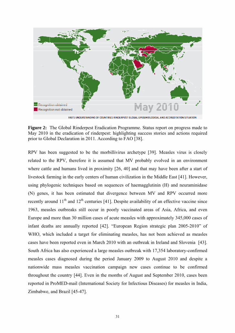
Figure 2: The Global Rinderpest Eradication Programme. Status report on progress made to May 2010 in the eradication of rinderpest: highlighting success stories and actions required prior to Global Declaration in 2011. According to FAO [38].
RPV has been suggested to be the morbillivirus archetype [39]. Measles virus is closely
related to the RPV, therefore it is assumed that MV probably evolved in an environment
where cattle and humans lived in proximity [26, 40] and that may have been after a start of
livestock farming in the early centers of human civilization in the Middle East [41]. However,
using phylogenic techniques based on sequences of haemagglutinin (H) and neuraminidase
(N) genes, it has been estimated that divergence between MV and RPV occurred more
recently around 11th and 12th centuries [41]. Despite availability of an effective vaccine since
1963, measles outbreaks still occur in poorly vaccinated areas of Asia, Africa, and even
Europe and more than 30 million cases of acute measles with approximately 345,000 cases of
infant deaths are annually reported [42]. “European Region strategic plan 2005-2010” of
WHO, which included a target for eliminating measles, has not been achieved as measles
cases have been reported even in March 2010 with an outbreak in Ireland and Slovenia [43].
South Africa has also experienced a large measles outbreak with 17,354 laboratory-confirmed
measles cases diagnosed during the period January 2009 to August 2010 and despite a
nationwide mass measles vaccination campaign new cases continue to be confirmed
throughout the country [44]. Even in the months of August and September 2010, cases been
reported in ProMED-mail (International Society for Infectious Diseases) for measles in India,
Zimbabwe, and Brazil [45-47].

32
Figure 3: Number of reported measles cases with onset date from Feb 2010 to Sept 2010 [44]. The number of reported cases of measles reflects a small proportion of the true number of cases occurring in the community. Many measles cases do not seek health care or, if diagnosed, are not reported thus the data shown in the map under-represents the true number of cases, particularly those occurring in the last one to two months.
Canine distemper (CD) is a fatal disease for many domestic as well as wild carnivores with an
ability to cross species barrier. It has a global distribution with seven phylogenetic lineages
found in Arctic, America, Europe, Asia, and Africa [48]. CD shares some antigenic and
taxonomic similarities with measles virus, but it is assumed to have emerged much more
recently [29]. Effective vaccines are available but resurgences of canine distemper have been
reported even in vaccinated dogs [49, 50]. The haemagglutinin (H) protein of the virus
interacts with host cell receptors for morbillivirus attachment and this protein of CD has a
higher mutation rate as compared to mumps and measles [29]. This partly explains a broader
host range for CDV as compared to other morbilliviruses. Furthermore, after mapping of the
distribution of mutations sites on H protein of CDV isolates from new host species, it has
been found these are not only to be under influence of positive selection but also that the
majority of amino acid substitutions fall into the SLAM-binding domains of the H protein,
which are responsible for host specificity [51, 52].
PPR was first described by Gargadennec & Lalanne in 1942, as an outbreak in Côte d'Ivoire,
they reported it to be a highly fatal disease and resembling rinderpest but affecting only sheep
and goats while cattle remained unaffected [6]. PPR was later found in Senegal in 1962 [53].
WHO
33
Although PPR is a recently recognized disease, the disease probably already existed but
diagnosis was perhaps confused with rinderpest and pasteurellosis [54]. Since, small
ruminants are less susceptible to even more virulent strains of RPV and the infection tends to
be either sub-clinical or mild thus perhaps even some of the reported RP outbreaks in small
ruminants in past may have been due to PPRV [55]. Mornet et al., had reported in 1956 that
virulent strains of PPRV was not pathogenic for cattle but conferred immunity against RP
[56]. Based on these results the authors suggested that PPRV can be considered a variant of
RPV, better adapted to the small ruminants. Serologic cross-reactions with RPV also confused
early recognition of the disease as a distinct entity of separate etiology, however by late 1970,
experimental infections, virological studies, and serologic studies established that PPR was a
disease distinct from RP [57-59]. The two viruses were definitively differentiated by
biochemical tests as well in 1987 [60]. Until 1979 this disease was declared to be limited to
West and central Africa [61], however it was reported in 1984 that apparent RP outbreaks in
1972 and 1973 in Sudan were later serologically confirmed as PPR [7]. By 1990s, it was also
reported in East Africa in Kenya [8] and Uganda and Ethiopia [62]. Outside Africa, the PPR
was first discovered in southern India in 1987 [9] and later on it spread across Arabian
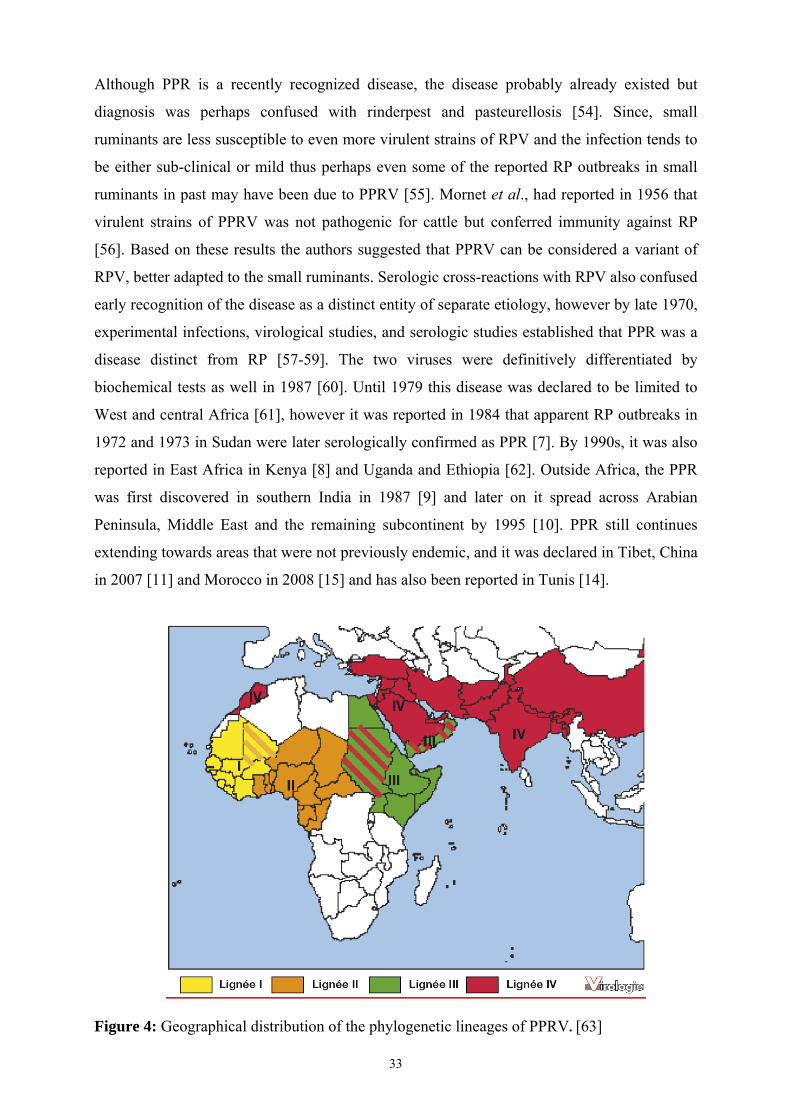
Peninsula, Middle East and the remaining subcontinent by 1995 [10]. PPR still continues
extending towards areas that were not previously endemic, and it was declared in Tibet, China
in 2007 [11] and Morocco in 2008 [15] and has also been reported in Tunis [14].
Figure 4: Geographical distribution of the phylogenetic lineages of PPRV. [63]

34
PPRV found in different geographic areas has been differentiated into four (1–4) lineages
based upon nucleic acid sequencing (Fig. 4). The apparent rapid spread of PPR during the last
three decades is perhaps due to a combination of factors like greater trade of live animals,
wider availability better diagnostic tools, vigilance and more awareness after eradication of
RP [64]. Furthermore, it has also been postulated that the chronological spread of PPR from
West Africa to Bangladesh may give a false impression that the disease spread has occurred
from West to East and that perhaps PPRV like RPV also has its origin in Eurasia and has
spread towards the West [37].
The economical impact of PPR is greatest on the poorest populations. Since large ruminants
are costly, a large proportion of rural populations breed sheep and goats for subsistence. The
small ruminants are therefore considered to be the “cattle of the poor”. Not only are the goats
and sheep raised as a source of meat and milk for family consumption, but they are also an
important source of income. In an international study aimed at prioritizing research on animal
diseases in order to have maximum impact on improving conditions in developing countries,
highlighted the economic importance of goats and sheep for the poor and ranked them at first
or second position for Asia and Africa [65]. Furthermore, the PPR was ranked among the top
ten diseases in these animals and thus affecting livelihoods of the poor in Asia and Africa. It
thus showed a need for the control of PPR to be taken into consideration for any poverty
alleviation policies [65]. PPR is one of the priorities of FAO Emergency Preventative System
(EMPRES) program which considers more than a billion small ruminants to be at risk of
PPRV infection and the rate of global domestic small ruminant population to be at risk was
62.5% [66, 67]. A socioeconomic study by Emergency Centre for Trans-boundary Animal
Diseases (ECTAD) of the Food and Agriculture Organization (FAO) of the United Nations,
conducted to understand the livelihood impacts of PPR in the arid and semi-arid lands of
Northern Kenya, found that PPR outbreaks resulted in better-off farmers slipping into poverty
and the poor and very poor became destitute [68]. The livestock-derived income losses due to
PPR ranged between 21% and 99 % and it was found that most households were unable to
maintain sustainable flock size owing to high mortality. Therefore, they had to leave
pastoralist livelihoods resulting in a long term dependency on food aid and a drain on national
resources [68]. Eradication of rinderpest has increased the relative economic importance, thus
highlighting the need for PPR control.

35
5’ “Trailer” or
(Anti-genomic Promoter)
3’ “Leader” or
(Genomic Promoter)
1688 1657 1473 2377 1949 6639b
1.3 Morbillivirus genome, structure and replication
The PPRV shares structural, biological, antigenic and molecular features in common with
other morbilliviruses. Morbilliviruses are enveloped, pleomorphic, single stranded, negative
sense RNA viruses. When viewed by electron microscopy, the members of Paramyxoviridae
family are indistinguishable [69]. The virion of RPV attains a maximum diameter of about
300 nm while PPRV tends to be larger, having a mean diameter of about 400-500 nm [58,
70]. The viral particle consists of an outer lipid envelop and an inner ribonucleoprotein (RNP)
core. The RNP core comprises of the viral genome encapsidated by nucleocapsid protein. The
RNP complex, but not the naked RNA genome, acts as a template for both transcription and
replication [71]. Typically, morbillivirus genome is made up of about 16,000 nucleotides, the
genome of PPRV being 15,948 bases in size, and is organized into six contiguous
transcriptional units encoding six structural and two non-structural viral proteins [72]. The
structural proteins are nucleocapsid
(N), phosphoprotein (P), matrix (M),
fusion (F), haemagglutinin (H), and an
RNA-dependent RNA polymerase
(RdRp) also called large (L) protein.
While non-structural proteins are (C)
and (V) which are transcribed by the P
gene using alternative expression
strategies and are found in cytoplasm
of infected cells. The internal proteins
(N, P and L) make up ribonucloprotein
and play a role in replication and
protection of viral genome while the
external proteins (M, H and F) traverse
the host cell derived lipid envelop and
interact with host cells to transmit viral
genome in to the cell [69].
Figure 5: (a) Morbillivirus structure and (b) genome organization (Modified, according to Moss et al., 2006 and Bailey et al., 2005 [1, 72]).

36
The viral genome has on its 3’ and 5’ ends two untranslated regions (UTRs), called “leader”
or genomic promoter (GP) and “trailer“ or anti-genomic promoter (AGP), which control viral
transcription and replication. The GP is required for both transcription of virus mRNAs and
transcription of a full-length positive sense virus genome, which is a replicative intermediate,
while the AGP is responsible for the production of full length negative sense genomes. For
PPRV, the leader region comprises a total of 107 nucleotides with 54 nucleotides being on the
3’-UTR region of the genome followed by a stretch of 53 nucleotides at the 3’ UTR of N gene
[69, 72]. Whereas in case of PPRV the trailer is made of up 109 nucleotides, with 69
nucleotides at the end of the L gene followed by 40 nucleotides at the 5’UTR region. [69, 72].
Between the 3’ and 5’ ends lay the six transcription units which transcribe 8 viral proteins in
the sequences of 3’–N-P/C/V-M-F-H-L-5’. Each of the transcription unit starts by a
conserved sequence (UCCU/C) and ends with a polyadenylation signal and poly uracyl of 6
nucleotides [69]. The transcriptional units are separated by a conserved inter-genic
trinucleotide 3’-GAA sequence between all viral genes except for the H and L genes where it
can be GCA (MV, RPV and PPRV cote d’Ivoire/89 strain), or GAA (PPRV Turkey/2000 and
the vaccine strains) and also between the L and the trailer sequence where it may be replaced
by GAU (PPRV) or GUU (CDV, PDV and DMV) [69, 72]. These tri-nucleotide sequences
are called mRNA pause signals for transcription as these sequences themselves are not
copied.
1.3.1 Viral Proteins
1.3.1.1 Nucleoprotein (N)
N protein of morbilliviruses is a major viral protein both in the virion and in the infected cell
[73]. It is highly antigenic, generating a humoral immune response [74]. Therefore many of
the antibody based diagnostic tests rely upon its antigenic characteristics. Morbillivirus N
proteins consist of 525 amino acid residues in RPV, MV and PPRV, but only 523 amino acids
in CDV, DMV, and PMV and have a molecular weights ranging from 60 to 68 kDa [69]. The
N gene of PPRV comprises 1,685 nucleotides, with 1,576 nucleotides coding for N protein
composed of 525 amino acids and a molecular weight of about 60 kDa [60, 75]. The N gene is
the most conserved among the Morbilliviruses genus [76] and the N protein is also the most
conserved among viral proteins [72]. It is a structural protein which encapsidates the viral
genome into an RNAse-resistant nucleocapsid (the template for RNA synthesis) and together
with P protein forms the RNP, which constitutes the minimum essential for transcription and
replication of viral genome in the cell cytoplasm [77].

37
1.3.1.2 Phosphoprotein (P)
It is one of the three components of the RNP and plays an important role in transcription and
viral replication. The morbilliviral P protein is made of 506 to 509 amino acids, and has a
molecular weight ranging from 72–86 kDa [60]. P protein of PPRV is the longest (509 amino
acids) while the DMV counterpart is the shortest in DMV (506 amino acids) and MV, RPV,
CDV and PDV have 507 amino acids [69]. It is a multifunctional protein which binds both the
N and L proteins and acts also as a chaperon keeping the N in a soluble form to enable
encapsidation and binding with the newly synthesized genomic RNA [52]. It is also an
essential co-factor for L protein in its RdRp function [78]. P is one of the least conserved
morbillivirus proteins [79].
1.3.1.3 V & C Proteins
In addition to producing the P protein, the P gene encodes the V and C proteins, via an RNA
editing process and an alternative initiation of translational within a different reading frame,
respectively [80, 81]. The C protein plays a role in the transcription, while the V protein plays
its part in replication of viral genome. Moreover, the two proteins have a role in morbilliviral
virulence as they interfere with the innate immunity by blocking interferon response by the
host cells [78, 82].
1.3.1.4 Matrix (M) protein
It is a small protein with 335 amino acids and is the most conserved within the group. The
morbillivirus M protein plays an important role in assembly of viral particles subsequent to
transcription. [83]. It favors viral particle formation by retaining the viral RNP complex at the
plasma membrane through interaction with the N protein, negatively down regulating
transcription, and membrane fusion and thus promoting virus budding [84]. It associates with
the inner surface of the plasma membrane [85] as well as the cytoplasmic tails of the H and F
glycoproteins [85-87] on the one hand and with the RNP complexes in the cytoplasm on the
other hand, to bring theses virus components together at the cell surface to make new viral
particles [88].
1.3.1.5 Fusion (F) Protein
F is one of the two viral surface glycoproteins, made of 546 amino acids, which is embedded
in the envelope and protrudes as spikes. It mediates fusion of the viral and cellular membranes
at the cell surface and thus helps liberating nucleocapsid into the cell cytoplasm. However,
before fusion can take place, morbilliviruses, except PPRV, must first attach to the host cell

38
receptor through its H protein, which also acts as a fusion promoter [89]. While the F of
PPRV is capable of inducing fusion independent of H protein [89]. The translated F0 protein
is in an inactive form which is cleaved by host cell proteases into the F1 and F2 subunits. The
F1 subunit is hydrophobic and has the fusogenic properties [78]. While the virus replicates,
expression of the synthesized F protein on the host cell surface results in fusion with the
neighboring cells to form syncytia, which are the characteristic cytopathic effect of
morbilliviruses [77]. Cell to cell fusion plays an important role in the propagation and
pathogenicity of the virus. While propagating both by virus release and reinfection, the F
protein on the host cell surface serves as a surface protein for the newly synthesized virus
particles when they obtain envelops upon budding [77].
1.3.1.6 Haemeagglutinin (H) Protein
It is a transmembrane glycoprotein which protrudes from viral envelope and enables
attachment of virus to the membrane receptors of the host cells. It mediates the interaction of
the virus with the cellular receptors. In association with the fusion (F) protein, the H protein
forms the envelope protein complex that is used for both virus attachment and membrane
fusion. The H protein consists of 604 amino acids for DMV, 617 amino acids for MV and
RPV and 609 amino acids for PPRV and RPV [69]. The H protein of PPRV and RPV is
distinct in the morbillivirus group, as it possesses both the haemagglutinating and
neuraminidase activities [90, 91]. Being a surface protein, the H of morbilliviruses is highly
antigenic and the principal target of neutralizing antibodies. [92]. Therefore, it has to evolve
to escape host immune responses and thus maintain the virus in the susceptible population.
Antibodies to H protein are the second most abundant after the N protein [74]. However, N is
an internal protein thus antibodies against it are not neutralizing [77]. The fact that H gene is
not highly conserved, probably also reflects the role of H in binding to host cell receptors and
thus playing a part in host-switching [52]. In the morbillivirus group, CDV has a
comparatively wider host range and new viral variants are being found out [93]. The H gene
of CDV has been found to have a much higher mean substitution rate (11.350 x 10⁴
subs/site/year) as compared to measles and mumps (6,585 x 104 and 9,168 X 104
subs/site/year, respectively) [29]. It has been suggested that evolution at residue 530 and/or
549 in the SLAM-binding region of the H protein of CDV is associated with disease
emergence in novel host species [51, 52].
39
1.3.1.7 Large (L) protein
It is the largest of the morbillivirus proteins having 5,183 amino acids and a molecular weight
of about 200 kDa. It is also the least abundant viral protein as it is the last to be transcribed.
Despite having a large size, the L protein is highly conserved among morbilliviruses. It
functions as RdRp in association with P protein. It performs a major role in genomic RNA
transcription and replication as well as the nucleotide polymerization, capping, methylation
and polyadenylation of viral mRNAs [78, 94].
1.3.2 Viral mRNA synthesis and replication of genome
Upon encountering the susceptible host cell, the viral H protein interacts with the SLAM
receptor molecule on the cell surface for attachment, while the F protein inserts its fusogenic
domain into target cell membrane, thus initiating the membrane fusion process which is
needed for entry of viral core complex into the host cell [95]. Thus the viral ribonucleoprotein
core enters cell cytoplasm where it has to replicate. However because of being negative sense
viruses, the genomic RNA of morbilliviruses can not be directly translated for production of
viral proteins. Therefore, it is first transcribed into messenger RNAs (mRNAs) with the help
of viral RdRp, which are then translated by cellular machinery to produce viral proteins.
However, it is still unclear as to how RdRp accesses the template RNA which is strongly
associated with N protein [69]. The viral transcription is initiated by RdRp after attachment
to the GP of the viral genome. The viral genes are transcribed sequentially in a “progressive
start-stop mechanism” in which, upon encountering a stop codon of a gene and a subsequent
intergenic tri-nucleotide, which acts as a pause signal, the synthesized copy of RNA detaches
and RdRp then moves on to the next gene. Thus are transcribed the monocistronic mRNA
molecules, however occasionally the polymerase does not respect one or two inter-genic
pauses, and this aberrant transcription results in synthesis of bi or tricistronic mRNAs. This
progressive “stop-start” mode of transcription by RdRp also produces a gradient in the
quantity mRNA produced from different morbillivirus genes. This happens because if RdRp
becomes detached from RNP template during the transcription, which is frequent at gene
junctions, it has to return again to the GP to reinitiate transcription. Owing to this
morbillivirus genomic organization, more a gene is away (far) from the viral promoter, less it
is transcribed [69]. The transcriptional gradient in protein production corresponds to the
different quantities of various viral proteins needed for synthesis viral particles. Thus N
protein is the most abundant while L protein is the least produced of the viral proteins (Fig.5).
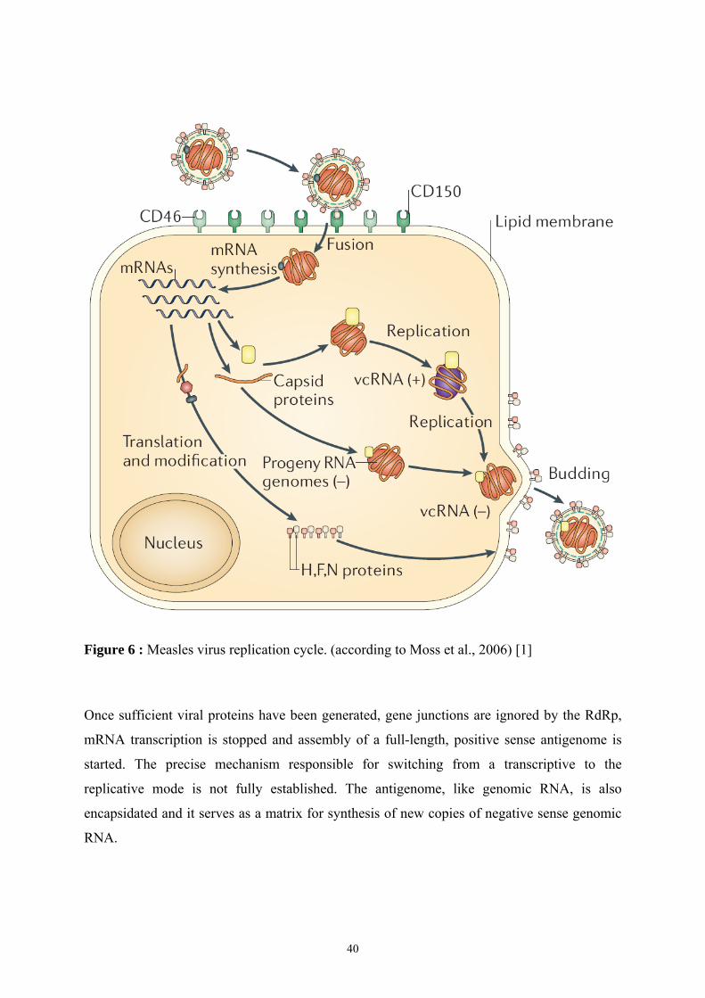
40
Figure 6 : Measles virus replication cycle. (according to Moss et al., 2006) [1]
Once sufficient viral proteins have been generated, gene junctions are ignored by the RdRp,
mRNA transcription is stopped and assembly of a full-length, positive sense antigenome is
started. The precise mechanism responsible for switching from a transcriptive to the
replicative mode is not fully established. The antigenome, like genomic RNA, is also
encapsidated and it serves as a matrix for synthesis of new copies of negative sense genomic
RNA.

41
1.3.3 Replication of viral genome, assembly and release of the viral particles
The virus assembly involves association of free N subunits with newly synthesized viral
genomic RNA. Next is formed the association between P-L protein forming a complex that
binds with N-RNA, resulting in formation of RNP complex [77]. The H and F proteins are
transported to the surface of cell membrane for incorporation on the outside of the envelope
for the newly budding virions. While the M protein plays the role of a bridge, by associating
on the one hand with the surface of plasma membrane, and cytoplasmic tails of the H and F
glycoproteins, and on the other hand it also interacts with RNP complex [96]. Finally the
interaction between viral H and F proteins on the cell membrane and RNP causes the
membrane to envelope the particle and a new virion is liberated into external environment by
budding. This process of budding does not kill the host cell thus allowing for (re-infection
and) formation of more viral particles.
1.3.4 Host immune response
The specific antibodies may be detectable within a week of infection [97, 98] and the immune
response is directed against all viral proteins. The most rapid and abundant antibodies
produced by the host during an outbreak are directed against the N protein, most likely
because of the location of the N gene at the 3′-end of the genome, the start–stop mechanism of
the RNA-dependent RNA polymerase [99] and because the N protein is the major structural
viral protein [73]. Antibodies to the morbillivirus N protein account for most of the
complement-fixing activity [99], while virus neutralizing (VN) antibodies are mostly directed
against the H glycoprotein and, to a lesser extent, against the F protein [99, 100]. Moreover,
maternal antibodies are also transmitted to the neonates via colostrum and can protect
newborns against RPV and PPRV for up to six months [101, 102]. Cell mediated immunity
also plays a role against morbilliviral infections and the studies in CDV and MV show a role
of cytotoxic T cells in convalescence, an immunity obtained by activation of CD4+ and CD8+
lymphocytes. A vaccination against RPV, by a Plowright vaccine strain (RBOK) of RPV [37],
was found to produce a strong CD4 T cell response [103].
Although morbillivirus infections generate strong immune responses with resulting long term
immunity, paradoxically, during temporary phase post-infection they also produce an
immunosuppression with the emergence of secondary infections [104, 105]. PPRV infection
in small ruminants also produces an impairment of cellular and humoral immune functions,
resulting in frequent opportunistic infections [106, 107]. The leucopenia and
immunosuppression result from a combination of factors like cell death caused by infection of

42
lymphocytes and apoptosis, and the cell cycle arrest, caused by exposure to the viral
haemagglutinin and fusion surface glycoproteins as well as viral nucleoprotein, expressed
from the damaged cells [106-108]. The immune suppression is not only observed in case of
natural infections but also after vaccination with attenuated morbilliviruses [103, 109, 110].
2. Peste de Petites Ruminants : The disease, its diagnosis and control
The disease has been differently named as Kata, goat plague, pseudo-rinderpest,
pneumenteritis complex and stomatitis-pneumoenteritis syndrome. However its French name
has been finally adopted as Peste des Petits Ruminants (PPR). Peste des petits ruminants
(literally meaning plague of small ruminants) is an acute viral disease of small ruminants
clinically characterized by fever, occulo-nasal discharges, stomatitis, diarrhea and pneumonia
with foul offensive breath [99]. Clinical signs are very similar to those of rinderpest.
2.1 Host range
PPR is primarily a disease of small ruminants i.e., sheep (Ovis aries) and goats (Capra
hircus). Goats are more susceptible to the disease which causes heavy losses, while it can be
occasionally severe in sheep [84]. However, susceptibility varies in different breeds. Apart
from domestic small ruminants, PPR has also been confirmed in captive wild ungulates
comprising gemsbok (Oryx gazelle), Dorcas gazelle (Gazella dorcas), Nubian ibex (Capra
ibex nubiana), Laristan sheep (Ovis orientalis laristanica), and Nilgai (Boselaphus
tragocemalus) [2]. Experimentally, the American white-tailed deer (Odocoileus virginianus)
is fully susceptible [111]. The only case of wild animal being seropositive with PPRV is that
of African grey duikers (Sylvicapra grimmia) which were found to be seropositive in Nigeria
[112]. PPR is primarily a disease of small ruminants, but infected cattle, buffalos, and pigs
that develop unapparent infections and seroconversion are considered as epidemiological
dead-ends. However, PPRV virus was isolated from pathological samples in buffaloes in
India [5]. Camels in Ethiopia and in Sudan showed clinical signs of the disease and proved to
be infected by PPRV [3, 4].
2.2 Transmission
The infected animal can excrete PPRV in discharges about 24-48 hours before onset of
clinical signs [97, 113]. The secretions from eyes, nose and mouth as well as feces contain
large quantities of the virus [113]. In an experimental infection of goats with PPRV, virus
could be detected in ocular secretions on day 2 and in nasal and oral secretions on day 3 post-
infection through RT-PCR [91]. Since the virus cannot survive long outside the host body and

43
the fact that upper respiratory tract is the entry point of the virus in to the host, close contact
of infected animal with the susceptible animals is an essential requirement for transmission as
described for RPV [114, 115]. The transmission occurs either through inhalation aerosol
dispersed virus or by ingestion of infected material [116-118]. There is no carrier state.
2.3 Pathogenesis
Like other viruses in the Morbillivirus genus, PPRV is also lymphotropic and epitheliotropic
[61]. The viral tropism is also manifested by the clinical signs like occulo-nasal discharges,
diarrhea, oral erosive lesions, marked immuno-suppression, and leucopenia [119]; while viral
antigen can be found in lymphoid organs and epithelial tissues of the affected animals [106].
The SLAM or CD150 is the primary receptor for PPRV [120], which is found on activated T
cells, B cells, thymocytes, macrophages and dendritic cells [121, 122]. The viral H protein
interacts with the SLAM receptor molecule on the cell surface while the F protein inserts its
fusogenic domain into target cell membrane, thus initiating the membrane fusion process
which is needed for entry of viral core complex into the host cell [95]. For PPRV infection,
respiratory route is the portal of entry as is the case of measles. It has been found that in
primate models as well as in mice expressing the MV receptor human SLAM the sub-
epithelial dendritic cells are initially infected by MV in the air ways [123]. However DCs in
respiratory epithelium express very low level of CD150 receptors [124]. These DCs express
C-type lectin called DC-SIGN which has been postulated to have a role in MV capture and
transmission to the lymphocytes in the regional lymph nodes by in trans–infection
(independent of de novo synthesis of virus in infected DCs) and also in cis infection (by the
newly synthesized of viruses in infected DCs) in combination with CD150 receptor [124,
125]. Similar role of DC-SIGN receptors have also been found in pathogenesis of several
other viruses like human immunodeficiency virus (HIV) [126] and herpes simplex virus
(HSV) [127]. Infected DCs also carry partly processed viral antigen to the lymphocytes in the
lymph glands [42, 128]. The immunological synapse formation, with lymphocytes in the
lymph glands, enhances viral transmission due to prolonged interactions during antigen
presentation. Furthermore antigen presentation initially activates them and activated
lymphocytes expressing greater CD150 are later infected [124]. While primary targets of
morbilliviruses are the CD150 (SLAM) positive cells, for release from the host in the later
stages of infection, the virus uses an additional unknown receptor(s) on SLAM-negative
epithelial and endothelial cells [129, 130]. Interestingly, the epithelial cells are infected
basolaterally only upon contact with underlying already infected leukocytes [128]. Since, the

44
epithelial cells can only be infected by MV via basolateral surface; role of respiratory
epithelium in initiation of infection is not an essential pre-requisite [95]. C
Figure 7: Possible strategy used by measles virus to cross epithelium of lung (A) and endothelial barriers (B). Antigen presentation and transmission of measles virus by the infected DCs to lymphocytes in the lymph glands (C) [42, 95] Modified.
Moreover, after intra nasal inoculation of rhesus monkeys with a recombinant MV selectively
unable to recognize human SLAM (SLAM-blind), it induced neither clinical signs nor
significant viremia, proving that efficient SLAM recognition is necessary for viral virulence
and pathogenesis [131]. Additionally, in experimental infection of ferrets with canine
distemper virus expressing GFP, it was found that CDV replication in epithelial cells was not
detectable initially, but was found substantial before death of the host [132]. In the same
experiment, 40% of T and 60% of B lymphocytes were infected [132].
Even after recovery from PPRV infection, there is a long lasting and important inhibition and
impairment of cellular and humoral immune functions characterized by immuno-suppression,
lymphocyte loss and leucopenia rendering animals highly susceptible to opportunistic
infections [106, 107]. The marked immune-suppression and leucopenia may be due to a
combination of cell death, caused by infection of lymphocytes and apoptosis, and the cell

45
cycle arrest, caused by exposure to the viral haemagglutinin and fusion surface glycoproteins
as well as viral nucleoprotein, expressed from the damaged cells [106, 107].
2.4 Gross and microscopic pathology
Animals dying from the peracute form of PPR do not show any gross pathological findings
except for congestion of oral mucosa and ileo-caecal valve and occasional oral erosions.
However, major gross lesions are mostly found after death with an acute form of the disease.
On digestive system, erosive and necrotic lesions are found on the lips, tongue, gingiva, hard
palate, and oral mucosa. Occasionally, lesions may be even found on pharynx and erosions on
the ruminal pillars and abomasum. Congestion of intestinal mucosa, colon, rectum with some
hemorrhages and thickening of mucosa giving a zebra-stripe appearance, are also observed.
The gut-associated lymphoid tissues are necrotic and mesenteric lymph nodes and spleen may
be swollen. Respiratory mucosa is congested with erosions in nares and trachea. There is
fibrinous or suppurative pneumonia in cardiac and apical lobes of lungs. Pulmonary lymph
nodes are found swollen and oedematous. In females, vulvo-vaginitis may be seen. Atypical
renal lesions have also been reported [133].
The histopathological findings in gastrointestinal tract include degeneration and necrosis of
labial mucosa, mucosal and submucosal congestion, degeneration and necrosis of intestinal
epithelium and lymphoid cell depletion in Peyer’s patches. Mucosal alterations are
surrounded by a mild inflammatory infiltration of macrophages and lymphocytes at the
borders of the lesions [133]. There is lymphocytolysis and occasional syncytia formation in
lymphoid tissues. The viral antigen can be found in conjunctival, tracheal, bronchial,
bronchiolar and ileal epithelial cells. It is also found in pneumocytes, macrophages, and
lymphoid and reticular cells in the lymphoid organs [106]. The viral antigen is mostly found
on the apical portions of the proliferating surface epithelium on the luminal side [134].
2.5 Clinical signs
The length and severity of disease may vary according to species or breed involved, along
with age and immune status of the animal. The PPR clinical description [54, 135, 136] can be
divided in four forms, i.e., peracute, acute, subacute, and subclinical.
The peracute form is seen when PPR occurs in young and naive populations of sheep and
goats. It starts after an incubation period of about 1 to 3 days and is clinically characterized by
high fever (40-42°C), dullness, anorexia, and constipation. There is congestion of oral and
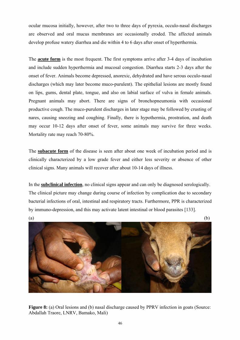
46
ocular mucosa initially, however, after two to three days of pyrexia, occulo-nasal discharges
are observed and oral mucus membranes are occasionally eroded. The affected animals
develop profuse watery diarrhea and die within 4 to 6 days after onset of hyperthermia.
The acute form is the most frequent. The first symptoms arrive after 3-4 days of incubation
and include sudden hyperthermia and mucosal congestion. Diarrhea starts 2-3 days after the
onset of fever. Animals become depressed, anorexic, dehydrated and have serous occulo-nasal
discharges (which may later become muco-purulent). The epithelial lesions are mostly found
on lips, gums, dental plate, tongue, and also on labial surface of vulva in female animals.
Pregnant animals may abort. There are signs of bronchopneumonia with occasional
productive cough. The muco-purulent discharges in later stage may be followed by crusting of
nares, causing sneezing and coughing. Finally, there is hypothermia, prostration, and death
may occur 10-12 days after onset of fever, some animals may survive for three weeks.
Mortality rate may reach 70-80%.
The subacute form of the disease is seen after about one week of incubation period and is
clinically characterized by a low grade fever and either less severity or absence of other
clinical signs. Many animals will recover after about 10-14 days of illness.
In the subclinical infection, no clinical signs appear and can only be diagnosed serologically.
The clinical picture may change during course of infection by complication due to secondary
bacterial infections of oral, intestinal and respiratory tracts. Furthermore, PPR is characterized
by immuno-depression, and this may activate latent intestinal or blood parasites [133].
(a) (b)
Figure 8: (a) Oral lesions and (b) nasal discharge caused by PPRV infection in goats (Source: Abdallah Traore, LNRV, Bamako, Mali)

47
2.6 Diagnosis
PPR can be diagnosed on the basis of clinical signs, as well as laboratory tests. The clinical
diagnosis is based upon major clinical signs of PPR like which include fever, oral erosive
lesions, dyspnoea, and diarrhoea. However, definitive diagnosis is possible only with
laboratory tests.
2.6.1 Differential diagnosis
Certain clinical signs manifested in PPR in small ruminants can also be found in some other
infectious diseases of sheep and goats.
- Rinderpest: PPR has long been confused with rinderpest however unlike PPR there
no respiratory problems associated with RP. Moreover, RP is almost eradicated now.
- Pasteurellosis: It does not have involvement of digestive system, however may exist
as a secondary infection alongside PPR.
- Contagious ecthyma: similar to subacute form of PPR, however the lesions are not
erosive but are in the form of papules or pustules which sometimes be found on mouth
and nose.
- Foot and Mouth Disease: Oral lesions are present but there is absence of respiratory
problems and diarrhoea
- Bluetongue: affects mainly the sheep but rarely the goats and is characterized by signs
of congestion of oral and nasal congestion and edema of lips and tongue there is
oedema of the face which are not found in PPR.
- Contagious Caprine Pleuropneumonia: is not characterized by diarrhea or oral
erosive lesions.
In case of suspicion, the confirmatory diagnosis can only be based on laboratory tests. The
samples can be collected either from the living sick or freshly died animals. Blood samples
with and without anticoagulant should be taken for obtaining infected peripheral blood
mononuclear cells (PBMC) and serology, respectively. Swabs or nasal and ocular discharges
should be taken. Tissue samples should also be taken from thymus, mesenteric and bronchial
lymph nodes, lung and intestines. The samples must be rapidly transported at 4°C to
laboratory for diagnosis. For rapid sample collection, blood or nasal and ocular discharges can
also be sampled on filter papers, dried and stored over long periods (up to 9 months) at
ambient temperatures [137].
48
The laboratory tests for diagnosis of PPR are based upon isolation of virus, detection of viral
genome, and detection of viral antigen or antibodies. Virus isolation is the classical technique
for diagnosis and still serves as a gold standard. In past, primary kidney or lung cells of goat
or Vero cell (kidney cells of African green monkey) cultures were used, which required
numerous blind passages and the isolation was often not efficient [138]. However, since
discovery of the fact that SLAM is the common receptor of morbilliviruses [139],
Vero.DogSLAMtag (expressing canine SLAM receptor) [140] and CV1 cell line (expressing
sheep SLAM receptor) [141] have been developed in recent years, which have improved
PPRV isolation in vitro. Cytopathic effects (CPE) induced by the virus in these cells can be
observed much earlier, in fact within two days after infection instead of 2-3 weeks for the
normal vero cells [141]. However, for virus isolation good quality and well conserved
samples are still required.
Various diagnostic tests are available which are based upon detection of viral antigens like
agar gel immunodiffusion (AGID), and immunocapture ELISA (Enzyme-linked
Immunosorbent Assay [142]. While for viral antibody detection tests like virus neutralization
test (VN), indirect ELISA and competitive ELISA are used [143]. Among these test, AGID is
incapable of discriminating between PPR and RP but immunocapture ELISA based
monoclonal antibodies [144, 145] against PPRV or RPV can efficiently discriminate between
the two viruses and have proven to be rapid, sensitive and specific tests (115). Numerous
diagnostic tests which use nucleic acid based detection of the pathogen which includes use of
radioactive or non-radioactive DNA probes, conventional reverse transcription-PCR (RT-
PCR), quantitative RT-PCR (QRT-PCR) [142, 146]. PCR based diagnostic tests are highly
specific and, using specific primers, can not only discriminate between PPRV and RPV, but
are also capable of discriminating between lineages of PPRV.
2.7 Prophylaxis and treatment
To prevent the spread of PPR to new areas, measures like controlling the movement of
animals from the areas endemic for PPRV is of primary importance. However, in case of an
outbreak of the disease in a new area, the most effective solution would be to kill the infected
animals and destroy the cadavers [36]. However, PPR is endemic in developing countries
therefore stamping-out policy is seldom accepted due to high costs. The only viable
prophylactic measure is vaccination of the susceptible animals around the affected area.
Effective vaccines are available against PPRV since long time. Rinderpest vaccine, being
cross protective against PPRV, was used initially but it can no longer be used due to Global

49
Rinderpest Eradication Program. Indeed, rinderpest vaccination was banned in the process of
eradication to allow effective epidemio-surveillance of the circulation of any residual virus
and avoid confusion between infected and vaccinated animals using the available antibody
detection tests. An effective homologous vaccine is available since 1989 [147], which
provides immunity against the disease during the economic life of small ruminants.
There is no effective treatment against PPR, once animals are infected. The sick animals can
only be provided supportive and symptomatic therapy. Antibiotics are also given to prevent
secondary bacterial infections. Not much research has been done for discovery of antiviral
agents effective against PPRV. As for as small molecular drugs are concerned, there is only
one report of in vitro testing of a synthetic compound 4,4′-(arylmethylene)bis(1H-pyrazol-5-
ols) for its ability to inhibit CPE produced by PPRV and this compound was found to be more
potent than the standard drug ribavirin [148]. The in vivo use of classical small molecular
antiviral drugs has serious limitations due to toxic side effects and emergence of resistant
strains [149]. The antivirals based RNA interference (RNAi) are a promising approach as they
are highly specific end effective in their action. However, even after more than a decade after
their discovery, their development as a therapeutic tool is hindered by problems of in vivo
delivery. In fact, problems are somewhat similar as the gene therapy has been facing since
1980s.
3. Interfering RNAs as antivirals and their delivery vectors
3.1 RNAi
RNAi is a highly specific, evolutionarily conserved, post transcriptional gene silencing
mechanism used by cells for gene regulation, protection of genome against transposable
elements and the attack of RNA viruses. While RNAi plays an important innate antiviral
defense mechanism in plants and insects [150, 151], whether RNAi as antiviral immune
mechanism really exists in mammals, remains unclear [152, 153]. However, the enzymatic
machinery for RNAi is used by the other endogenous non-coding small RNAs like
microRNAs (miRNAs), for regulation of gene expression [154, 155]. It is estimated that
RNAi mediated gene regulation controls the expression of about 30% of mammalian genes,
many of which are involved in functions like cell fate, proliferation and death [156].

50
3.1.1 Discovery
The phenomenon of RNAi was first observed by Napoli et al in 1990, when they were trying
to over express an enzyme responsible for plant coloration by introduction of the exogenous
gene in petunias [157]. Surprisingly, the introduced gene resulted in a blockage of pigment
synthesis. The phenomenon of gene suppression, however, was not understood at that time. It
was Andrew Fire and Craig Mello who finally established the mechanism in 1998, by
demonstrating that injection of short stretches of 23mer to 25mer nucleotide sequence of
double stranded RNA (dsRNA) into the nematode Caenorhabditis elegans resulted in
silencing of endogenous genes [158]. Fire and Mello were awarded the Nobel Prize for
Medicine or Physiology in 2006 for their discovery. This mode of gene silencing was
different from the already established anti-sense mode of gene silencing in that, although both
are based upon sequence complementarity to messenger RNA (mRNA). RNAi has a catalytic
component that makes it possible for a single small interfering RNA (siRNA) molecule to
bind with and destroy thousands of copies of mRNA molecules and thus is 1000 times more
effective than antisense oligonucleotides (ODNs) in silencing target gene [159-161].
Subsequently, it was reported that RNAi could take place in numerous other organisms
including invertebrates, and vertebrates [125, 126]. Elbashir et al in 2001, showed that
transfection of synthetic siRNAs could silence genes in mammalian cells [127]. Since then
RNAi has found widespread applications ranging from use in functional genomics and gene
knock-down, to treatment of various medical conditions like macular degeneration, cancers
and viral infections.
3.1.2 Mechanism
RNAi machinery in mammals is used by miRNAs for regulation of gene expression. The
miRNAs are transcribed by polymerase II as long primary transcripts or pri-miRNAs [162].
The pri-miRNAs undergo a sequential maturation process in which they first bind to a
microprocessor, a complex of two proteins in the nucleus which contains the DiGeorge
Syndrome critical region-8 (DGCR8) protein and a nuclear RNase III called Drosha [163-
165]. Next the Drosha cleaves pri-miRNAs into pre-miRNAs, which are the imperfectly
paired stem-loop miRNA precursors having 60 to 80 nucleotides. The pre-miRNAs are then
exported to the cytoplasm by Ran GTP-dependent Exporting-5 transporter [166, 167]. The
Dicer (an RNase III family enzyme) at first, interacts with its double stranded RNA-binding
protein partner (TAR RNA binding protein; TRBP) and other partners to cleave pre-miRNAs
into mature double-stranded miRNAs of 19-25 bp with 2 nucleotide 3’ overhangs [168].
Then, Dicer together with R2D2 protein and its partners, couple with miRNAs duplex to form

51
RISC loading complex (RLC), which helps in the loading of the miRNA duplex into another
multiprotein complex, having Argonaute protein as its core component. This new complex is
called RNA-induced silencing complex (RISC or miRNA protein complex (miRNP)) [155,
169, 170]. After attachment to RISC, the passenger strand of miRNA is lost, while antisense
strand guides RISC to the target mRNA. The miRNAs pair perfectly or imperfectly with the
target mRNAs and thus may either result in endonuclease Argonaute 2 mediated cleavage and
destruction of guide strand-mRNA duplex or blockage ribosome movement halting mRNA
translation, respectively [171].
Figure 9: Mechanism of RNA interference (Schematic representation - according to Keck, 2008) [172] The degree of miRNA-mRNA complementarity has been considered to be the key
determinant of the regulatory mechanism. Thus, in case of a perfect match, mRNA is cleaved
by RISC, while central mismatches may result in repression of mRNA translation [154]. The
plasmid or viral vector expressed short hairpin RNAs (shRNAs) and chemically synthesized
siRNAs mimic mammalian pre-miRNAs and miRNAs respectively and thus use cellular
machinery for gene silencing. The only difference being that, the antisense strand of siRNA,
acting as a guiding strand pairs perfectly with the target mRNA resulting in RISC-mediated
cleavage of the target mRNA [173]. Viral/plasmid vector based shRNAs, transcribed from a
polymerase III promoter, are produced in the nucleus and exported to the cytoplasm by
exportin-5 together with GTP-bound form of its cofactor Ran, and are processed by Dicer like
pre-microRNAs to produce siRNAs [167].

52
3.1.3 Interfering RNAs as an antiviral therapeutics
The infection by negative sense RNA viruses requires the transfer of viral genome into the
host cells, transcription of the viral mRNA, and translation of the viral proteins that are
needed for viral genome replication, assembly, and budding. Targeting viral mRNA with
siRNA is an attractive strategy since firstly, one can prevent synthesis of critical viral proteins
to disrupt viral life cycle, secondly with siRNAs, being highly specific, there is little chance
of side effects, and finally, understanding of gene function is not required: only viral genome
sequences are needed [174]. Soon after discovery of RNAi, its potential as effective antiviral
therapy was recognized. RNAi antiviral treatment was first used by Bitko and Barik in 2001
against respiratory syncytial virus (RSV) [175]. Since then the approach has been variously
exploited for antiviral purposes, not only through the use of chemically synthesized siRNAs
but also by expressing the shRNAs from plasmid or viral vectors [176-178]. RNAi through
these various approaches could effectively block viral replication in vitro but for in vivo use,
delivery issue is still a major hurdle.
3.1.4 Difficulties in use of RNAi as an antiviral approach
The therapeutic use of RNAi in vivo faces other practical difficulties than those related with
delivery, like off-target effects, RNAi suppression and viral escape.
3.1.4.1 Off-target effects:
Although RNAi is highly specific, in addition to the intended mRNA suppression, it also
produces unintended effects on gene expression. For in vivo delivery, off-target effects are
major concerns since the down-regulation of self genes could have serious biological
consequences. They may result from either a partial sequence complimentarity of RNAi
construct to non-targeted mRNA or induction of a variety of immune and toxicity related
effects emanating from certain motifs or patterns in the RNAi construct itself [179].
3.1.4.1.1 Specific off-target effects dependent on siRNA sequences
Although initially thought to be highly specific, RNAi was shown by expression profiling to
produce off-target effects whereby a particular siRNA will bind to other mRNAs than the one
originally targeted. Partial sequence complementarity in both the passenger or guide strands
of RNAi construct can produce off-target gene suppression [180]. The off-target silencing has
been reported for transcripts with as low as 7 nucleotides complementarity with the guide
strand [146]. siRNAs like miRNAs can also bind to sequences with partial complementarity
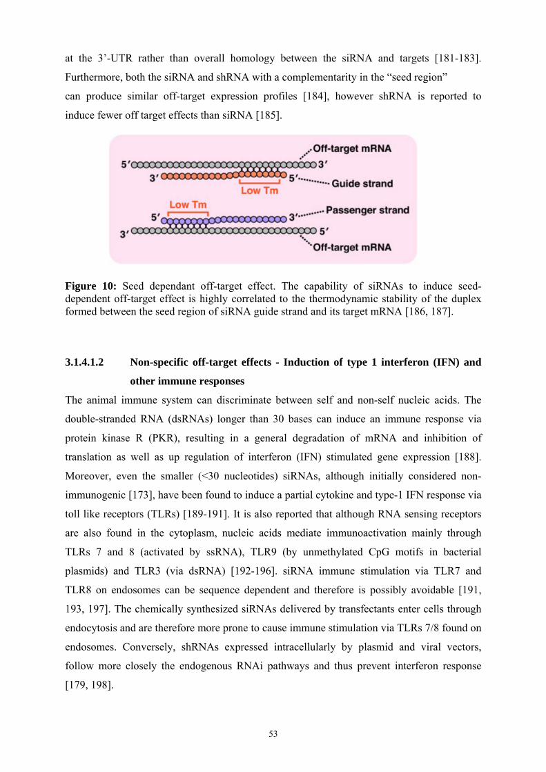
53
at the 3’-UTR rather than overall homology between the siRNA and targets [181-183].
Furthermore, both the siRNA and shRNA with a complementarity in the “seed region”
can produce similar off-target expression profiles [184], however shRNA is reported to
induce fewer off target effects than siRNA [185].
Figure 10: Seed dependant off-target effect. The capability of siRNAs to induce seed-dependent off-target effect is highly correlated to the thermodynamic stability of the duplex formed between the seed region of siRNA guide strand and its target mRNA [186, 187].
3.1.4.1.2 Non-specific off-target effects - Induction of type 1 interferon (IFN) and
other immune responses
The animal immune system can discriminate between self and non-self nucleic acids. The
double-stranded RNA (dsRNAs) longer than 30 bases can induce an immune response via
protein kinase R (PKR), resulting in a general degradation of mRNA and inhibition of
translation as well as up regulation of interferon (IFN) stimulated gene expression [188].
Moreover, even the smaller (<30 nucleotides) siRNAs, although initially considered non-
immunogenic [173], have been found to induce a partial cytokine and type-1 IFN response via
toll like receptors (TLRs) [189-191]. It is also reported that although RNA sensing receptors
are also found in the cytoplasm, nucleic acids mediate immunoactivation mainly through
TLRs 7 and 8 (activated by ssRNA), TLR9 (by unmethylated CpG motifs in bacterial
plasmids) and TLR3 (via dsRNA) [192-196]. siRNA immune stimulation via TLR7 and
TLR8 on endosomes can be sequence dependent and therefore is possibly avoidable [191,
193, 197]. The chemically synthesized siRNAs delivered by transfectants enter cells through
endocytosis and are therefore more prone to cause immune stimulation via TLRs 7/8 found on
endosomes. Conversely, shRNAs expressed intracellularly by plasmid and viral vectors,
follow more closely the endogenous RNAi pathways and thus prevent interferon response
[179, 198].

54
3.1.4.2 Saturation of endogenous RNAi pathway components
The enzymatic machinery involved in RNAi in mammalian cells is naturally used by miRNAs
which play an important role in cell physiology. Since miRNA and shRNA share common
enzymatic pathway for their processing, the over-expression of shRNA in vivo is known to
produce toxicity due to over-saturation of endogenous miRNA pathway involving exportin-5
and RISC component Argonaute-2 [199]. However, in vivo toxicity can be alleviated by the
selection of efficient but safe shRNA expression cassettes, and applying minimal effective
vector doses [199].
3.1.4.3 Viral encoded suppressors of RNAi
Several plants and invertebrates use RNAi as an immune mechanism to counter viral
infections [150, 151]. Therefore, viruses infecting these plants and insects have evolved their
countermeasures by producing various suppressors of RNAi silencing (SRS) [200, 201].
Some recent studies suggest that RNAi in mammals is also involved in antiviral responses and
cellular regulatory miRNAs have a function in restricting virus replication in the cells [152].
Moreover, several mammalian viruses like hepatitis C virus (HCV), influenza virus, ebola
virus, human immunodeficiency virus (HIV), vaccinia virus, and adenoviruses type 2 and 5
are known to produce SRS factors that inhibit the RNAi mechanism. Many of these factors
are multifunctional proteins which are known to inhibit interferon response as well [152,
153]. Since the viruses producing SRS can be efficiently inhibited by RNAi, it appears that
viral-suppression activity does not pose a serious difficulty for therapeutic RNAi [202]. None
of the viruses belonging to the morbillivirus genus is reported to produce RNAi suppression
through SRS factors.
3.1.4.4 Viral escape
Several applications of a single siRNA can lead to the emergence of viral escape mutants due
to mutations in the target sites making viral genomes resistant to RNAi [203]. Since, unlike
eukaryotic DNA polymerase, RNA viruses lack proofreading activity, they tend to have a
high error rate during replication which facilitates development of viral mutants [204]. After
discovery of RNAi, an early study by Jacque et al., 2002, showed that even a single
nucleotide mismatch in the siRNA target region may be sufficient to reduce the silencing
effect of an siRNA on HIV-1 [205]. Interestingly, viral escape to RNAi can also occur by a
point mutation outside the target sequence of siRNA, if this mutation changes local RNA
folding into a structure that reduces the accessibility of the target sequence [206]. Several
viruses have been reported to escape suppression by effective siRNAs, which include HIV-1,

55
HCV, HAV, and Poliovirus [207-210]. Studies of HIV-1 and HBV populations have shown
that many resistant mutants may pre-exist before they have been exposed to inhibitors and the
siRNAs may also exert selection pressure on these pre-existing resistant mutants [211, 212].
The problem of RNAi escape can be more severe in chronic diseases as these require an
extended antiviral therapy but may be less crucial in treatment of acute diseases [202].
However, design and selection of siRNAs targeting conserved regions of virus and
combination of siRNAs targeting multiple genes or regions in viral genome can help avoid
problem of resistance to siRNA therapy [213]. In a study using a T cell line expressing three
potent shRNAs against HIV-1, it was found that even after an extended culturing for more
than 100 days there was no viral replication [214]. A combination of four shRNAs that target
different sequences of HBV genome has entered a phase I clinical trial [215]. However, use of
multiple siRNA/shRNA doses must be optimized before use as some studies have shown that
it is possible to saturate the RNAi pathway with high levels of shRNA expression [199].
Another possible alternative to targeting multiple conserved viral genomic regions is to use
siRNAs that recognize the mutated target sites through use of siRNAs or shRNAs that target
the most likely escape variants [216]. In vitro studies are needed to study the frequency of
emergence of PPRV escape mutants and the viral sequences that are likely to mutate in the
course of therapy. Since viruses tend to mutate, an alternative means could be to down-
regulate cellular factors which are required by the virus to either enter a cell or to replicate.
3.2 In vivo delivery of interfering RNAs
Compared to the large number of studies using siRNAs in cell culture, there have been
relatively few studies in vivo. The progress is slower due to difficulties in delivery, especially
by systemic route of administration [217]. The issue of drug delivery is not unique to RNAi
based therapeutics, but here it is a major obstacle as the drug cargo (siRNA) is nuclease
sensitive, has a net negative charge, and is hydrophilic, thus making difficult to keep it stable
in serum and capable to cross the anionic and hydrophobic plasma membranes [218-220].
Moreover, for diseases like PPR, systemic delivery of siRNA is a necessity. In order to use
RNAi as an antiviral approach, the siRNAs can either be chemically synthesized [175] or
shRNA expression cassettes with Poll III promoters can be inserted into plasmid [176] or viral
vectors [177, 221]. The viral vectors can efficiently use their own machinery to reach the
target cells, transfer their genome into the cells and express shRNA molecules.
The naked siRNAs and plasmid vectors, however, can not readily cross the cell membrane
and therefore need delivery vehicles to help them to enter the cell cytoplasm. Bitko and
colleagues successfully delivered naked siRNAs intranasally in a mouse model against

56
respiratory syncitial virus [19]. Interestingly, they compared delivery of siRNA with and
without a transfection reagent, and noted only a marginal enhancement (20%) in the
knockdown of the respiratory syncytial virus (RSV) target gene when lipid agent was used.
However, the mechanism by which these cells take up these siRNA molecules remains
unknown [222]. The systemic delivery route, however, is more difficult to approach. The
unmodified siRNA has a half-life of less than an hour in human plasma and the siRNA
molecules circulating in blood, are rapidly excreted by kidneys due to their small size [223].
The vectors are therefore needed not only to improve the bio-availability of siRNAs but also
to provide protection from nucleases and help in penetration of anionic plasma membranes
[224, 225]
3.2.1 Physical methods of siRNA delivery
The physical methods for in vivo delivery of siRNA involve mainly electroporation and
hydrodynamic injection. The hydrodynamic injection involves a quick injection of siRNA or
plasmid DNA (pDNA) in a large volume of physiological buffer, around one-tenth the mass
of the animal, within a few seconds in the tail vein of the animal and this results in a delivery
mainly to the liver [226, 227]. The early attempts for systemic delivery of siRNAs in vivo
used the hydrodynamic injection method that was already tested by Liu et al., for pDNA
delivery in mice [228]. McCaffrey et al., in 2002, in their studies on in vivo siRNA delivery
by hydrodynamic method, demonstrated that co-injection of luciferase expressing plasmid
with siRNA targeting luciferase produced efficient silencing of luciferase expression in the
mouse liver [229]. Various studies using this method could deliver naked siRNA to
hepatocytes, demonstrating functional knockdown of specific genes in the livers of mice
[229-231]. Most studies are using hydrodynamic method for delivery of siRNA to liver as it
can be much more easily transfected by this method as compared to other organs. This
procedure is not clinically viable because of potential damage to liver, as well as due to
danger of volume overload side effects like right sided heart failure [232]. Moreover, it
would be very difficult to scale up this method for use in large animals.
The electroporation involves administration of an electric current for short duration, which
temporarily increases the permeability of cell membranes, and thus allowing for passing of
nucleic acids through membranes. The electric field polarizes the membrane molecules and
temporarily destabilizes the membrane integrity, which results in greater permeability for the
exogenous materials [233]. This technique has been successfully used to transfer siRNAs
locally [234], but it cannot be used for systemic administration. Similarly, ultrasound is also

57
used for nucleic acid delivery [235]. These physical methods of delivery of siRNAs either
cannot be used systemically or are too dangerous for clinical use.
3.2.2 Chemical vectors for delivery
The development of chemical vectors for siRNA delivery has been influenced by the studies
on intracellular DNA delivery [236]. However, there are important differences between
delivery of siRNA and DNA: i) size and charge of siRNA are lower than those of DNA; ii)
cytosol is the place of action for siRNA while DNA has to enter nucleus to be effective [237].
The chemical methods of siRNA delivery involve formulation of negatively charged siRNA
molecules with various polycations, like cationic lipids, peptides, polymers, or inorganic
nano-structured materials, either through electrostatic or ionic interactions, with the aim of
enhancing pharmacokinetic behavior, nuclease resistance, cellular uptake, target specificity
and safety [18]. The general principle is based on complex formation between anionic siRNA
molecules and cationic polymers resulting in complexes with net positive charge for
facilitating interaction with anionic cell membranes. Moreover, the physico-chemical
properties of the complexes like size, morphology, surface charge and stability, are the major
determinants of transfection efficiency which in turn are determined by structure of
polycation, the polycation/nucleic acid stochiometry (+/- charge ratio), pH and order of
mixing of nucleic acids and the vectors [238]. Though most delivery vectors are cationic, the
net surface charge of the vector-siRNA complex, has to be near neutral to avoid toxicity,
excessive uptake by reticulo-endothelial system (RES) and aggregation by serum proteins [18,
239]. In order to deliver siRNA cargo into target cell cytoplasm, the formulation has to
surpass various physiological barriers (Fig. 11) by shielding siRNA from serum nucleases,
avoiding aggregation by anionic serum proteins, escaping from RES, penetrating
endothelium, getting internalized through endocytosis, and finally being able to deliver
siRNA from the endosomes to the cytoplasm before degradation by lysosomes [18, 239]. The
in vivo use of siRNA by chemical delivery systems is limited by siRNA packaging
efficiency, colloidal stability of the complex, internalization, and endosomal escape [240].
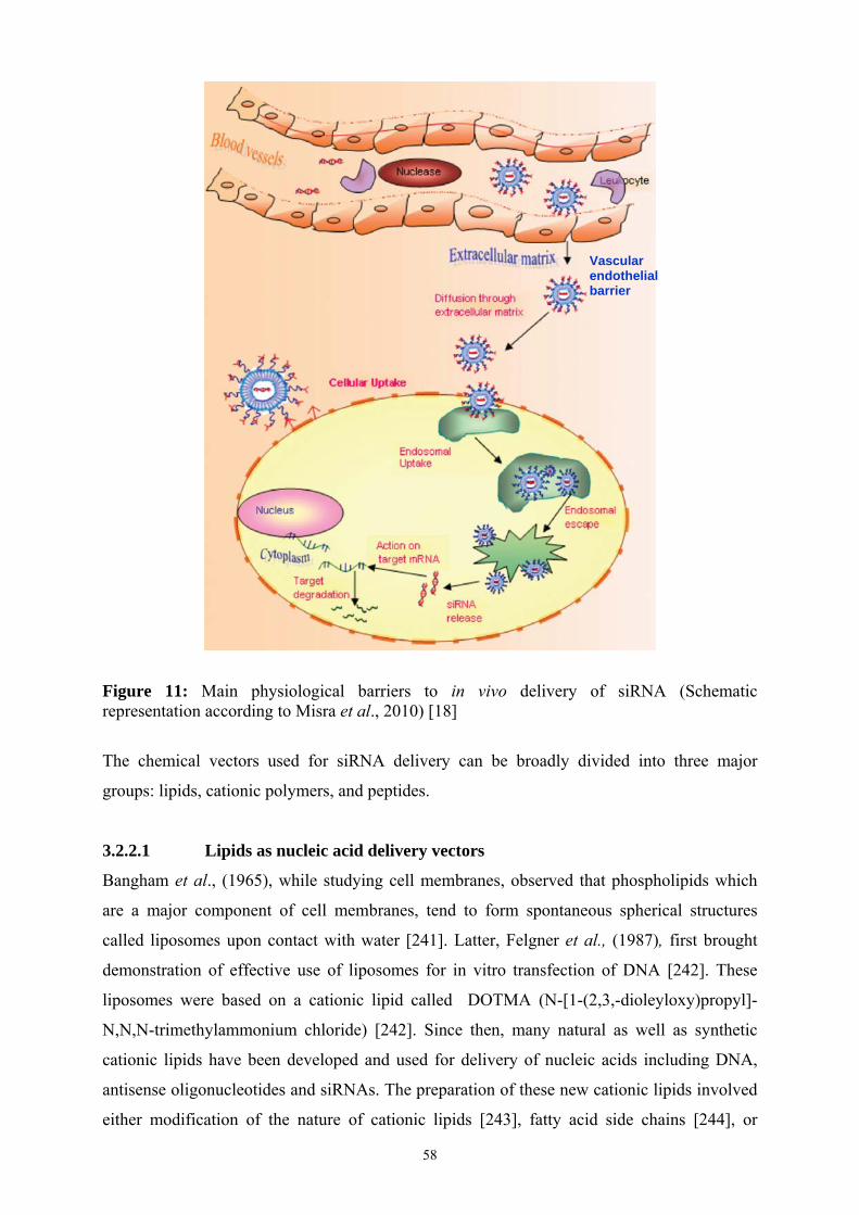
58
Figure 11: Main physiological barriers to in vivo delivery of siRNA (Schematic representation according to Misra et al., 2010) [18]
The chemical vectors used for siRNA delivery can be broadly divided into three major
groups: lipids, cationic polymers, and peptides.
3.2.2.1 Lipids as nucleic acid delivery vectors
Bangham et al., (1965), while studying cell membranes, observed that phospholipids which
are a major component of cell membranes, tend to form spontaneous spherical structures
called liposomes upon contact with water [241]. Latter, Felgner et al., (1987), first brought
demonstration of effective use of liposomes for in vitro transfection of DNA [242]. These
liposomes were based on a cationic lipid called DOTMA (N-[1-(2,3,-dioleyloxy)propyl]-
N,N,N-trimethylammonium chloride) [242]. Since then, many natural as well as synthetic
cationic lipids have been developed and used for delivery of nucleic acids including DNA,
antisense oligonucleotides and siRNAs. The preparation of these new cationic lipids involved
either modification of the nature of cationic lipids [243], fatty acid side chains [244], or
Vascular endothelial barrier

59
formulations with some additional lipids [245]. Despite initial difficulties found in in vivo
transfection, the death of a human patient participating in gene therapy trials based on an
adenoviral vector in 1999 [246] and the resulting temporary ban on use of viral vectors for
gene therapy by US Food and Drug Administration [247], lead to a renewed interest in
chemical vectors.
Typically, the cationic lipid molecules are amphiphilic or amphipathic in nature and are made
up of three parts: a cationic head-group, a hydrophobic anchor, and a linker (Fig. 12b). The
cationic head-group is required for binding and complexation of nucleic phosphate groups,
whereas hydrophobic part probably assists in assembling the lipids into polycationic scaffold
and facilitating absorptive endocytosis or fusion with plasma membranes [248]. The cationic
lipids are classified on the basis of number of positive charges, nature of linker bond, and
nature of hydrophobic anchor [248]. Although cationic lipids are the most common lipids
used for liposome based transfections, often neutral and/or anionic lipids are mixed with these
in variable ratios to neutralize excessive cationic charge and to improve endosomal escape
[249]. The anionic lipids when added to nucleic acid-cationic liposome complexes, not only
reduce cellular toxicity but also reduce nonspecific interaction with anionic serum proteins
like albumin as well as extracellular matrix [250, 251]. Mastrobattista et al. (2001) were able
to improve transfection by preparing positively charged polyplexes coated with an anionic
lipid [251].
The neutral lipids play a role of a helper when formulated with cationic lipids for preparation
of liposomes. There are three neutral lipids which are often incorporated in the formulations:
dioleyl phosphatidylethanolamine (DOPE), cholesterol and dioleoyl phosphatidyl choline
(DOPC). The DOPE is known to destabilize lipid bilayers and is believed to be involved in
endosomal disruption and thus enabling nucleic acids to escape endosomes before being
destroyed by lysosomes [252]. Cholesterol is also used as a helper lipid. Although it forms
more stable but less efficient complexes with nucleic acids than those containing DOPE in
vitro, cholesterol containing lipoplexes show higher biological activity due to greater cell
uptake and stability in serum, compared to lipoplexes with DOPE when these complexes are
utilized in vivo [248]. The liposome mediated transfection has been shown to take place
mostly by endocytosis [236]. However, probably a minor pathway mediated by fusion
between siRNA containing lipoplexes and the plasma membrane is responsible for around 5%
of siRNA delivery in to cell cytoplasm [236].

60
Cationic (hydrophilic) Polar Head
Spacer arm
Hydrophobic lipid tail
DOTMA
3.2.2.1.1 Cationic liposomes
Liposomes are vesicles composed of a phospholipid bilayers with an aqueous core [253].
They are classified according to their size and their number of bilayers: Small Unilamellar
Vesicles (SUV), Large Unilamellar Vesicles (LUV), Multilamellar Vesicles (MLV), and
Giant Unilamellar Vesicles (GUV) (Fig. 12b) [254]. The complex formation between anionic
phosphate group from nucleic acids and cationic amine head group of cationic liposomes
results in formation of lipoplexes, which are quite different from liposomes in structure as the
final charge as well particle size changes and these changes are determined largely by lipid /
nucleic acid (+ / -) ratios [255].
a b
c
Figure 12: Structure of a cationic liposome. (a) formed by phospholipids in an aqueous medium [256]; (b) Classification of liposomes according to size and number of bilayers [254]; (c) Representative structure of cationic lipid DOTMA modified [257]

61
3.2.2.1.2 Liposome preparation
The liposomes can be prepared by numerous methods, however the basic principle of their
formation involves hydrophilic/hydrophobic interactions between lipid-lipid and lipid-water
molecules [258]. Moreover, there is always a need for energy input (sonication,
homogenisation, shaking, etc) which promotes the arrangement of lipid molecules in bilayers
and achieves a thermodynamic equilibrium in the aqueous phase [258]. The classical method
of liposome preparation used by Bangham et al. (1965), consists in evaporing the organic
solvent into which lipids are dissolved, and then adding an aqueous solvent [241]. The
hydration of lipid film causes phospholipids to self-associate so as to hide their hydrophobic
chains to the aqueous solvent. It results in formation of bilayers enclosing the aqueous
solvent.
3.2.2.1.3 In vivo delivery of siRNA with liposomes
DOTAP (N-[1-(2,3-dioleoyloxy)]-N-N-N trimethyl ammonium propane) and Oligofectamine
were some of the first lipid formulations used for in vivo delivery of siRNA and effective
gene silencing of TNF-alpha and beta-catenin in mice [259, 260]. Since then siRNAs have
been successfully delivered locally but there are not many reports of successful systemic
liposome based delivery of siRNAs against systemic viral infections. However, there are
some studies showing promising results in terms of successful delivery of siRNAs and
effective viral suppression in vivo by formulations based up on neutral lipids. For example,
Morrissey et al. (2005) using three daily intravenous injections in mice of siRNA (3
mg/kg/day) complexed with the neutral liposomes called “stable nucleic acid-lipid particles”
(SNALPs) stabilized with PEG (polyethylene glycol) , could reduce serum DNA of hepatitis
B virus by more than 1.0 log10 and the effect lasted for up to 7 days after dosing [20]. In
another experiment using intraperitoneal delivery of siRNAs complexed with SNALPs for
seven days against Ebola virus has been shown to protect guinea pigs against viremia and
death shortly after the virus challenge [261]. Using a lactosylated liposome based upon
neutral lipid the phosphatidylcholine, Watanabe et al., (2007) could effectively deliver siRNA
targeting hepatitis C virus (HCV) in a transgenic murine model, resulting in suppression of
intrahepatic HCV expression without interferon response [21]. SNALPs developed by Protiva
Biotherapeutics and Alnaylam, have also been successfully used to deliver siRNA in non
human primates, whereby using a SNALP-formulated siRNA dose of 2.5 mg/kg, could
markedly suppress the Apolipoprotein B (APOB) [262]. Despite encouraging results in
murine models, there are no known reports of non-viral delivery of siRNAs in farm animals.
62
Systemic application of siRNA molecules by lipid based carriers is still challenging and
issues of toxicity and need for targeted delivery still need to be addressed.
3.2.2.2 Cell penetrating peptides (CPPs)
CPPs or protein transduction domains (PTDs) are short peptides having fewer than 30 residue
peptides, derived from natural or unnatural protein or chimeric sequences [263]. Most of the
CPPs possess a high density basic amino acids (arginines and/or lysines), which are proposed
to interact with the anionic surface of the cell membrane and enhance internalization of the
peptides [264]. Among chemical delivery vectors CPPs are unique in that the concentrations
of CPPs that are used for molecular delivery, produce very low or undetectable cytotoxicity
[264].
CPPs were first discovered when Frank and Pabo in 1988 observed that HIV-1 Trans-
Activator of Transcription (Tat) protein, which transactivates transcription of the HIV-1
genome, crossed the cell membrane by itself [265]. Later, a minimal peptide fragment of Tat
(49-59 amino acids) involved in cellular uptake was identified by a French group [266]). The
Tat peptide has been shown to successfully deliver siRNAs in vitro [267]. Another major
discovery in CPP domain was made by Joliot et al (1991), who demonstrated that Drosophila
Antennapedia home domain could be internalized by neuronal cells [268]. This work lead to
discovery of the first CPP derived from a non-viral protein which consisted of a 16-mer
peptide derived from the third helix of the Drosophila Antennapedia homeodomain protein
and was named PenetratinTM [269]. The Penetratin is capable of transfecting nucleic acids
antisense oligonucleotides [270], peptide nucleic acids (PNAs) [271]. A MPG peptide was
used to perform the first non-covalent delivery of nucleic acids in 1997 [272]. The MPG
peptide is an artificially constructed 27 amino acid amphipathic CPP containing a lysine rich
basic region derived from the nuclear localization signal (NLS) of the SV40 large T antigen
and a hydrophobic region derived from the HIV-gp41 coat protein [272]. Both the MPG
peptide and a modified MPGΔNLS (having a single mutation of the second lysine residue in
the NLS motif to serine) can transfect siRNA in vitro however latter is more efficient as it
delivers siRNA into cytoplasm instead of nucleus [273]. MPG peptides have been also used to
deliver siRNA into mice by systemic route [274]. Another milestone was achieved when a
chimeric CPP “Transportan”, derived from the N-terminal fragment of the neuropeptide
galanin linked to a wasp venom peptide called mastoparan was successfully used in vivo for
delivery of peptide nucleic acids (PNAs), in mice [271]. The first CPP based transfection of
siRNA was performed with a MPG peptide [275] and since then numerous natural and as well

63
as engineered CPPs have been tested for siRNA delivery [276]. Polyarginine-based peptides
have also been successfully used for siRNA delivery. Majority of the CPPs possess basic
amino acids (arginines and/or lysines), through which these interact with cell membranes and
help in the internalization of CPPs [264]. The experiments on Tat and penetratin revealed that
the role of positive charges is crucial for translocation [277]. After studying various cationic
polypeptides, CPPs with polyarginines were found to be more efficient than other cationic
polypeptides like polyhistidines and polylysines and among polyarginine peptides, Arg7 and
Arg9 have been the most widely used for in vitro and in vivo delivery [277].
Although cellular uptake mechanism for CPPs has been reported to be associated with
endosomal pathway [278], there is no unified mechanism established for CPP uptake
mechanism up to now, and probably numerous factors are involved [263]. However, there is a
consensus that the initial contact between the CPPs and the cell membrane take place by
electrostatic interactions with negatively charged proteoglycans [263]. Moreover, as CPPs are
diverse in chemical and physical structures, it is suggested that different properties like CPP
molecule length, charge delocalization as well as size and charge of the cargo can have impact
upon peptide uptake mechanism [279]. Cellular uptake of CPPs, therefore, does not occur by
any single mechanism thus, energy independent direct diffusion, macropinocytosis, clathrin-
mediated endocytosis, and caveloe/lipid raft mediated endocytosis may all occur [280].
CPPs can be classified in two ways. These are classified either on the basis of mode of
bonding with cargo into, those requiring covalent linkage with siRNAs and others which,
being amphipathic, can form stable non-covalent bonds. Secondly, these can also be
subdivided structurally into those which polycationic having clusters of polyarginine in the
primary sequence or those which are amphipathic [263].
3.2.2.2.1 Covalent attachment of CPP to siRNA
Tat conjugated to the modified antisense strand of siRNA was successfully used to inhibit
EGFP gene in vitro [281]. However, most of Tat-siRNA was found localized in endosomes
and there was a concern that the attachment of Tat to siRNA may disrupt functionality of
siRNA molecules [282]. To address this concern, CPPs have also been linked to siRNA
through disulfide bond which is cleaved when conjugates reach reducing environment of
cytosol, resulting in successful RNAi [283, 284]. The covalent strategy usually requires

64
complex chemistry for conjugation and there is also risk of alteration of biological activity of
siRNA therefore non-covalent strategy of delivery appears more suitable [263].
3.2.2.2.2 Non-covalent CPP-siRNA complex formation
Assembling siRNA/CPP complexes through non-covalent interactions is advantageous as it
simplifies conjugation protocols but also eliminates the need for optimization of individual
syntheses schemes. Additionally, there is also lower likelihood that CPP will interfere with
the bioactivity of the cargo [263]. The non-covalent delivery of siRNA is based up on
electrostatic interactions between siRNA and amphipathic peptides [264]. The amphipathic
peptides possess hydrophobic (polar) and hydrophilic (non-polar) domains. The amphipathic
property of CPPs may arise from either primary or secondary structure [277]. Primary
amphipathic peptides consists of the sequential assembly of a domain of hydrophobic residues
with a domain of hydrophilic residues while secondary amphipathic peptides are produced by
conformational state that allows positioning of hydrophobic and hydrophilic residues on
opposite sides of the molecule [277].
Since siRNAs are negatively charged, they can bind with positively charged CPPs through
nonspecific electrostatic interactions and provide permeability by covering the siRNA surface
with positive charges from the CPP [264]. In 2003, a non-covalent strategy based on MPG
was found to efficiently deliver siRNA into cell lines [285]. Similarly, this non-covalent mode
of delivery has also been used for other CPPs like, Tat, polyarginine, and transportan-derived
peptides [263]. A novel 20-amino acid amphipathic peptide, CADY, has been recently
described which combines both cationic arginine and aromatic tryptophan residues into its
design [286]. It forms stable complexes with siRNA through electrostatic interactions and
interestingly uses a non-endocytic mechanism to pass through plasma membrane, thus
avoiding endosomal entrapment [286]. This CPP can transfect a variety of cell lines,
including difficult to transfect primary cell lines [286]. Since CADY-siRNA is stable in 50%
serum for 24 hours and leads to significant knockdown with sub-nanomolar concentration, it
may be effective in vivo as well [286].
3.2.2.2.3 In vivo delivery with CPPs
CPPs have been delivered in vivo with some successes and failures. MPG peptide was used
for systemic in vivo delivery of siRNA targeting essential cell cycle protein cyclin B1,
resulting in efficient blockage of tumor growth [287]. In another experiment, siRNA targeting
HIV was successfully delivered through a CD7-specific single-chain antibody conjugated to

65
the oligoarginine peptide (scFvCD7-9R) could effectively suppress HIV infection in a mouse
model [22]. The CD7 receptor is rapidly internalized after antibody binding, therefore it has
been exploited for the targeted delivery of several monoclonal antibody (mAb) [22]. Kumar et
al., [27] using a nonamer arginine was introduced at the carboxy terminus of a peptide
derived from rabies virus glycoprotein (RVG) were able to transfer siRNA into neuronal cells
in vivo, resulting in efficient gene silencing after intravenous injection into mice.
Furthermore, systemically delivering of RVGR9 conjugated antiviral siRNA complex could
protect mice from encephalitis induced by Japanese encephalitis virus (JEV) infection, which
is the first report on a nontoxic method to deliver siRNA across the blood brain barrier [288].
CPP based siRNA delivery has also entered at level of preclinical and clinical trials, with
Traversa Inc., are testing HIV Tat-based and Panomics Inc., testing secondary amphipathic
peptide-based non-covalent delivery of siRNA [263].
3.2.2.3 Other chemical vectors
Aptamers are RNA or DNA oligonucleotides that fold by intramolecular interaction into
unique three-dimensional conformations capable of binding to target antigens with high
affinity and specificity [289]. The aptamers have been used as siRNA vectors in a mouse
tumor xenograft model, however their systemic use requires addition of nuclease stabilizing
agents and endosmolytic functionalities [18]. Dendrimers molecules are repeated branched
species characterized by structural perfection [18]. Dendrimers have been used successfully
for in vitro delivery of siRNAs [290]. Polyethyleneimines (PEI) are polycation-containing
block copolymers like [291], which have also been used for delivery of siRNA molecules in
vivo, although PEI use in vivo has some toxicity issues [292]. Chitosan is a biodegradable,
biocompatible and non-toxic cationic polymer obtained from deacetylation of chitin, which
have been proposed as biocompatible alternative cationic polymers, suitable for nonviral
nucleic acid delivery[293]. Chitosan have been used for siRNA delivery in vitro as well as in
vivo [294]. Antibody-protamine fusion carriers have also been shown to be efficient in
delivery of siRNA to HIV-infected or envelope-transfected leukocytes [295].
3.2.3 Viral vectors
Like chemical vectors, the viral vector based delivery of shRNA also profited from
technology that already existed for gene therapy. Soon after delivery of siRNAs in vitro [175],
it was demonstrated that siRNAs can also be expressed from plasmid DNA as “short hairpin
RNAs” (shRNAs) [296, 297]. This finding paved the way for viral vector based RNAi

66
therapeutics. U6 [297] and H1 [296] were the first polymerase III promoters described that
express functional siRNAs [298]. However U6 promoters have been reported to be more
efficient in vivo [299]. Typically, shRNA transcription starts in a position outside the
promoter sequence, continues along 19-29 nucleotide long top strand, the 4-19 nucleotide
long hairpin loop and finally the bottom strand and terminates after the second or third residue
of track of 4-6 thymidines [298]. The termination is so designed that it results in a 3’ two-
nucleotide overhang after RNAse III cleavage as is the case with natural pre-miRNAs [298].
The shRNA sequences are selected and translated to DNA, and these are normally
synthesized in the form of two complimentary oligonucleotides that are annealed and cloned
downstream of the selected promoter and regulatory sequences [298]. The constructed shuttle
plasmids expressing shRNA are next tested for inhibition of target gene in vitro. Moreover,
the cassettes may be inserted into expression clones for production of required recombinant
virus vectors. In addition to standard shRNA resulting in production of a 21 nt long siRNAs,
numerous other variations of shRNA have also been used. Longer shRNAs producing 27- 29
nt long siRNAs [300, 301], and multiple shRNAs simultaneously expressing different
shRNAs against different target regions have been used [302, 303]. The viral vectors have
certain advantages over chemical vectors for delivery of siRNAs like [298];
1. tissue specific delivery of shRNAs.
2. possibility of inducible or tissue specific promoters.
3. both transient as well as stable expression of shRNA is possible, according to the viral
vector chosen.
4. better transduction efficiency compared to non-viral transfections even in cells like
lymphocytes that are traditionally hard to transduce.
5. cost effectiveness.
6. most of the available viral vectors have already been tested clinically in phase I safety
trials.
The most widely used viral vectors for shRNA delivery include adenovirus, adeno associated
virus (AAV), lentivirus, retrovirus, baculovirus. In contrast to the gene therapy, shRNA
expressing cassettes tend to be of small length and their expression is possible even by the
smallest vectors [298]. However, viral vectors are selected on the basis of tissues required to
be transduced and also whether stable or transient transduction is required. For chronic
infections like HIV stable transduction is desirable to avoid repeated administration of vectors
while for acute infections, transient transductions with shRNA expressing vectors would
suffice. The RNA viruses such as retrovirus and lentivirus produce stable transduction as they

67
integrate into host genomes while non-integrating DNA viruses like adenovirus, baculovirus
and AAV maintain their genome episomally in the host cells.
3.2.3.1 Adenovirus vectors
Adenoviruses (Ads) are non-enveloped viruses having a linear double stranded 36 kbp DNA
genome with non-enveloped icosahedral protein capsid. Adenoviral capsids are made up of
three major protein components – the hexon, penton base and fiber (Fig. 13) [304]. In general,
the fiber knob functions as the major attachment site for cellular receptors, while the penton
base is involved in secondary interactions that are required for virus entry into the cell [304].
More than 50 serotypes have been identified which are divided into six subgroups (A-F). Out
of the numerous serotypes, Ad2 and Ad5 belonging to subgroup C were first shown to serve
as efficient vectors gene delivery in vitro [305] and still are the most commonly used
adenoviral vectors [306]. For gene therapy as well as for RNAi, replication defective
adenoviruses can be used since they are non-pathogenic and can transiently transduced a
variety of both dividing and non-dividing cells without integration to the host genome [298].
Figure 13: Structure of Adenovirus [307].
In the past it has been considered that adenovirus serotypes belonging to species A, C, D, E
and F use the coxsackie and adenovirus receptor (CAR) as a docking site providing a high
affinity virus-to-host association [308]. CAR was previously thought to be a primary Ad5
receptor, but now there are studies according to which adenovirus type 5 also uses many other
receptors for docking like the heparan sulfate proteoglycan (HSPG), vascular cell adhesion
molecule 1 (VCAM-1), major histocompatibility complex class I (MHC 1), scavenger
68
receptors (SR), while indirect binding it also uses dipalmitoyl phosphatidylcholine receptors
(DPPCRs), coagulation factor X (FXs), and Lactoferrin receptors (LfRs) [309]. Although
receptor binding is thought to play a major role in adenoviral tropism, this alone cannot
explain all aspects of in vivo host-virus interactions like enhanced transductions of liver cells
or adenoviral uptake by Kupffer cells [310]. Excessive adenoviral binding to hepatocytes may
be partly explained by binding of central depression of adenoviral hexon with Gla domain of
coagulation factor X (FX) which has been found to produce efficient transduction of
hepatocytes [311]. That may explain excessive liver tropism of adenovirus where FX could be
the main determinant of in vivo liver transduction [311].
The internalization of adenovirus occurs by a secondary interaction between RGD motifs on
penton base protein and integrins αvβ3 and αvβ5 [312] through clathrin-coated pits mediated
endocytosis [313]. The virus then escapes by lysing endosomal membrane and with the help
of microtubule mediated translocation, enter the nuclear pore complex where viral DNA is
released for expression [314]. The transcription of wild type virus initiates with expression of
early E1 genes, which activate synthesis of viral genes and replication. E1 and the non-
essential E3 genes were deleted in first generation adenoviral vectors to render them
replication deficient [298]. But these first-generation adenoviruses express viral genes to low
levels, including virus associated (VA) RNAs which have been described to saturate the
cellular silencing machinery, leading to toxicity [315]. Although functional inhibitory
exogenous shRNAs have been expressed for first generation adenoviral vectors without
saturation of silencing machinery [316]. The second generation vectors, which have deletions
in the E1-E4 locus are comparatively less immunogenic and show prolonged expression of
recombinant genes [317]. Whereas third generation adenoviruses are produced by deleting all
the viral genes, as a result of which expression of viral genes is avoided resulting in lesser
immunogenicity [318].
The recombinant replication deficient adenoviruses have been successfully used in vitro as
shRNA delivery vectors against numerous viruses like hepatitis B virus [221], measles virus
[319] and hepatitis C virus [320]. The adenoviral vectors expressing shRNA have been
successfully used in vivo against several viral infections. After pre-treatment of guinea pigs
and swine with adenoviral vectors expressing shRNA against foot and mouth disease virus
(FMDV), and challenging 24 hours later with the target virus, Chen et al., could protect the
animals from major clinical manifestation of the disease [23]. Similarly adenoviral vectors
have also been used to protect mice against porcine circovirus type 2 [225].

69
3.2.3.2 Baculovirus vectors
The baculoviruses belong to the family Baculoviridae which is subdivided into two genera:
the granuloviruses (GV) and nucleopolyhedroviruses (NPV). The GV contain one
nucleocapsid per envelop but the NPV may either contain single (SNPV) or multiple (MNPV)
nucleocapsids for each envelop in the occlusion body [321]. Furthermore, the NPV capsids
are occluded in polyhedron matrix and the polyhedra may contain multiple embedded virions
[322]. Among the numerous baculoviruses, Autographa californica multiple
nucleopolyhedrovirus (AcMNPV) is the most well studied and extensively used virus in
foreign gene expression [323]. AcMNPV possesses a circular double stranded DNA genome
which is condensed into a nucleocapsid core by a protamine-like protein. Naturally AcMNPV
are occluded in a polyhedron which after being ingested by insects is dissolved in alkaline
midgut, releasing infectious virions [323].
Figure 14: Structure of a Budded baculovirus. According to Blissard, 1996. [324]
The AcMNPVs only replicate in insect cells and naturally infect insects belonging to the order
Lepidoptera. Baculoviruses primarily enter insect cells through clathrin-mediated, low-pH
dependent endocytic pathway while they may enter mammalian cells through multiple
pathways including caveola-dependent mechanism as well [325]. However, AcMNPV can
infect insect cells and transduce mammalian cells even in the absence of endocytosis, by
direct fusion at cell surface under low pH conditions [326].
The baculoviruses can enter the mammalian cells but they are unable to express their genes
because the baculovirus promoters are inactive in these cells [327]. One of the interesting
consequences of this is the absence of pre-existing antibodies against baculovirus in mammals
[327, 328]. For gene transfer and expression in mammalian cells, so-called BacMam viruses

70
have been generated by incorporation of mammalian cell-active expression cassettes [329].
BacMam baculoviruses are capable of transducing wide variety of cells including non-
dividing cells [330] and primary cells [331]. They have been used to reduce viral infections
in vitro of numerous viruses like porcine arterivirus [332], hepatitis C virus [333], hepatitis B
virus [334], and influenza viruses A and B [335].
The baculovirus can prove to be good shRNA delivery vectors in vivo. Although delivery of
siRNA to the appropriate cells or tissues is a major challenge, problem of inactivation by
complement is resolved [336]. Chemical or genetic modification can also overcome problem
of serum inactivation [337, 338]. Furthermore, it is reported that baculoviruses do not have
deleterious effects on mammalian cells even when used at very high multiplicity of infections
(MOIs) [339]. Moreover, not only can the baculoviruses be easy and cheap to produce, they
can be grown to high titers in cell cultures as well. Although baculovirus vectors have not
been used thus far for shRNA delivery in vivo, but these have been successfully used for gene
transfer into mouse brain and rabbit retina [340, 341].
3.2.2.3 Other viral vectors
Many other viral vectors have been used for RNAi mediated silencing of viral infections like
adeno-associated virus (AAV) [342], herpes simplex virus [24], retroviruses [296], and
lentiviruses [177]. Moreover, these viral vectors have also been used against infectious
disease of veterinary importance. Infectious bursal disease virus (IBDV) by an avian adeno
associated virus delivered miRNAs targeting VP1 and VP2 genes in vitro [343]. Recombinant
herpesvirus of turkey (HVT) expressing shRNAs against genes gB and UL29 of the Marek’s
disease virus (MDV) moderately reduced viremia in chicken [24]. In another study, avian
leukosis virus-based retroviral vectors expressing shRNA against MDV gB glycoprotein gene
and ICP4 transcriptional regulatory gene could significantly reduce MDV viremia in vivo
[25].

71
4. Control of Morbillivirus Replication by RNAi – State of the art
RNA interference has become the method of choice for suppression of gene expression in
vitro. The synthetic short interfering RNAs (siRNAs) represent a new class of molecules with
a significant potential for therapeutic applications. This technology promises to bring benefits
in this field that far surpass the antivirals currently available in the market, as it offers new
possibilities to easily design highly selective antiviral drugs capable of targeting specific
genes in order to suppress its expression and this with a very low toxicity as its natural
nucleotide components are easily metabolized by cellular systems [344].
This method is now emerging as a powerful tool for research with many published studies
using siRNA in mammalian cell culture. They show that synthetic siRNA are particularly
effective in the areas of oncology and the treatment of viral infections. siRNAs have been
widely used against viruses of various families to inhibit the intracellular viral life cycle. The
first experiments carried out on an unsegmented, negative sense, RNA virus, the respiratory
syncytial virus (RSV), go back to 2001 [175]. A publication by Reuter et al., [345], describes
the generation of multiple siRNAs directed against 6 genes of a morbillivirus, measles virus
(MV) can effectively deregulate gene expression in general with siRNAs against transcripts of
the nucleocapsid (N), phosphoprotein (P) and polymerase (L), which form ribonucleoprotein
(RNP) complex. Two other publications describe the use of siRNA to inhibit replication in
cell culture of MV and subacute sclerosing panencephalitis (SSPE) virus [346, 347].
However, these studies show inhibition of measles replication only in vitro but siRNA against
morbilliviruses have not been successfully delivered in vivo so far.
In the case of morbilliviruses, despite existence of efficient vaccines against morbillivirus
diseases, no effective treatments exist for infected animals or humans. RNA interference
(RNAi) has a potential of being turned into an effective and specific antiviral therapy for the
control of these diseases. In case of disease outbreaks, the emergency vaccination alone, in
poor countries does not prevent heavy economic losses arising from animal mortality as well
as morbidity. Moreover, even after administration, the vaccines require several days before a
sufficiently strong protective immune response is present. Under these circumstances, an
antiviral that can block viral replication in the early stages of infection could be an important
complementary tool in the control strategy. A combination of antiviral agent together with
vaccination may reduce the economic losses incurred during an epidemic.

72
The in vitro tests conducted by the CIRAD have helped in identification of three sites
susceptible to RNAi, which are located on the conserved regions of nucleoprotein gene of the
morbilliviruses, through an siRNA design methodology that allows the rapid prediction of
potential sequences on target mRNAs [348]. These strategies for predicting design done on
the conserved regions of the genome should lead to the use of lower doses of siRNA while
reducing non-specific off-target effects. Indeed, knowledge of morbillivirus genomes allowed
us to develop an antiviral approach based upon RNAi against these three morbilliviruses.
Briefly, sixty two 19 nt long siRNA sequences targeting N, M, and F genes of three genes of
peste des petits ruminants virus (PPRV), rinderpest virus (RPV) and MV were designed,
chemically synthesized and tested for efficacy in vitro [348-350]. Three sequences directed
against nucleoprotein (N) gene, namely NPPRV1, NPPRV6, and NPPRV7, were found to be
most effective and resulted in reduction of viral titer to about 1,000 to 10,000 times and also
an inhibition of up to 90% of the nucleoprotein expression by PPRV when transfected at a
final concentration of 100 nM (Tab. 1).
However, for systemic administration of siRNA in vivo to be successful, numerous
constraints, as were described in the review of literature of this document, including poor
transfection in vivo, which is also the major issue with other nucleic acid based strategies, a
low serum stability and toxicity need to be overcome. Even after more than a decade of
discovery of RNAi and despite excellent knock down of viral or cancer genes in vitro, its
therapeutic application has not been possible due to these difficulties involved in the efficient
in vivo delivery. Although some siRNA based therapeutics have entered clinical trials, these
are mostly for local use only, whereas systemic delivery has proven to be a significant hurdle.
Since PPR is an acute and systemic infection of small ruminants, transient transfection or
transduction are required for systemic delivery of siRNA/shRNA in vivo. For that purpose, in
this work several chemical delivery vehicles as well as viral vectors were considered for their
ability to deliver the siRNA NPPRV1 directed against the nucleoprotein gene of PPRV,
previously selected as the most effective in inhibiting PPRV replication after delivery by a
commercial liposome in cell cultures.
A process of reflection by the laboratory, on the selection and development of delivery
vehicles was crucial for the success of in vivo studies. Indeed, due to their intrinsic
mechanisms, the viruses prove efficient delivery vectors, while despite certain drawbacks like
toxicity, the cationic lipids are also delivery systems of choice. Thus the laboratory had
therefore developed a replication-deficient human adenovirus type 5 expressing shRNA

73
48h 60h 72h
PPRV
PPRV +
shRNA NPPRV1
84h 96h
NPPRV1 (rAd_NPPRV1shRNA) against the nucleoprotein of PPRV and tested it in vitro
(personal communication), showing it was capable of reducing PPRV induced cytopathic
effects (CPE) as well as PPRV titers (Fig.15).
Table 1: Inhibition of PPRV N gene expression and PPRV titers by three effective siRNAs
siRNA
name
siRNA Sequence Position Fold Inhibition of
PPRV Titer
% Inhibition of
NPPRV expression
NPPRV1 Sense 5′-GGAUCAACUGGUUUGAGAAtt-3′ 480-498 10,000 90+- 2
NPPRV6 Sense 5′- GGCGGUUCAUGGUAUCUCUtt-3′ 741-759 100 >80
NPPRV7 sense 5′-GCAUUAGGCCUUCACGAGUtt-3′ 899-917 100 >80
(a)
(b)
Figure 15: (a) Inhibition of cytopathic effects (CPE) in Vero cells, and (b) PPRV progeny by rAd_NPPRVshRNA, at various time intervals after challenge with PPRV MOI 0.1.
100
1000
10000
100000
1000000
10000000
12h 24h 36h 48h 60h 72h 84h 96h
TCID
50/0
.1 m
l PPR
V
Hours post-challenge
shRNA MOI 80 + PPRV PPRV

74
AIMS AND OBJECTIVES
The aim of this project was the effective delivery of siRNA or shRNA in vivo against peste
des petits ruminants virus. For the purpose, firstly the viral and chemical vectors were tested
in the course of study to find efficient delivery vectors in vitro. The in vivo experiments were
initially conducted on goats under L3 bio-security conditions. However, due to ethical,
economical, and bio-security issues, a mouse model based upon bioluminescent imagery is
developed for in vivo testing of various siRNA/shRNA delivery vectors.

75
PART. 1:
In vivo delivery of siRNA/shRNA against PPRV infection by adenoviral and cationic liposome vectors

76
1.1 Introduction
RNAi based antivirals can be administered in vivo either in the form of chemically
synthesized siRNA molecules which are delivered by chemical vectors or these can be
delivered in the form of shRNA through viral vectors (for further information, see Review of
the Literature chapter). At the start of this work, siRNAs capable of effective inhibition of
PPRV replication in vitro were available. Since commercial chemical vectors prove too
expensive when used for in vivo delivery of large quantities of siRNAs in animals of large
size like goats, here a cationic lipid based formulation was developed for delivery of siRNAs.
In this study, this cationic lipid as well as a recombinant adenovirus expressing the shRNA
NPPRV1 (rAd_NPPRV1shRNA) were tested about their capacity to deliver the siRNA/shRNA
NPPRV1 and inhibit PPRV replication in goats.
1.2 Material and Methods
1.2.1 Cell culture
Vero cells (European Collection of Cell Culture, France 84113001) were maintained in
Eagle’s Minimum Essential Medium (EMEM) supplemented with 10% (vol/vol) heat-
inactivated foetal bovine serum (FBS), 2 mM L-glutamine at 37°C with 5% CO2 in a
humidified incubator. Vero cells were used for the production and titration of PPRV and
transfection experiments. The 293A cells (Invitrogen) were propagated in Dulbecco’s
modified Eagle’s medium supplemented with 10% (vol/vol) FBS at 37°C with 5% CO2.
1.2.2 Production of PPRV
The vaccine strain of PPRV (Nigeria 75/1) [351] and a virulent strain of PPRV Côte d’Ivoire
89 (CI 89) were propagated by infecting Vero cells, cultured in 75 cm2 cell culture flasks with
EMEM having 5% FBS at multiplicity of infection (MOI) of 0.1 CCID50/ml. When viral
cytopathic effect (CPE) was almost complete (~80%), the cells were freeze-thawed 3 times
and aliquots were stored at -80°C. Viral titration was performed in 96 well cell culture plates,
in which 100 µl of 10 fold serial dilutions of PPRV in EMEM were added to 100 µl of Vero
cell suspension (20,000 cells). For each viral dilution, 10 wells were used. The viral titers
were calculated by end point titration method of Reed and Muench [352] and expressed as
cell culture infectious cytopathic doses for 50% of the tests (CCID50 /ml).
77
1.2.3 Preparation of liposomes for in vitro validation of siRNA delivery
A liposome formulation (M2b) based up on a 1/1/0.5 (m/m) combination of cationic lipid 1,2-
dioleoyl-3-trimethylammonium-propane-chloride salt (DOTAP), the neutral lipid 1,2-
dioleoyl-sn-glycero-3-phosphoethanolamine (DOPE), and an anionic lipid cardiolipin was
developed and tested in vitro for delivery of siRNA NPPRV1. DOTAP, DOPE, and
cardiolipin were each dissolved in ethanol (90%) to have a concentration of 25µg/µl. Then,
20µl each of DOTAP and DOPE were mixed in ratio of 1/1 (m/m). Solution was
homogenized by vortexing for 10 seconds and incubated at room temperature for 15 minutes.
The tube containing the lipid mixtures was vacuum centrifuged overnight at room temperature
to obtain a dry lipid film. Then, 10 µl of cardiolipin was added to the tube with lipid film and
vortexed until film was completely dissolved. Next, 190 µl of sterile water (filtered by 0.2
μm) was rapidly added with pipette, in a single step, under hood and vortexed immediately
for 10 seconds. The solution was incubated overnight at 4oC before using the liposomes for
making complexes.
1.2.3.1 Transfection in vitro of siRNA with cationic liposomes and challenge with
PPRV
Vero cells of 48 hours of growth were trypsinized and 105 cells were cultured in 24 well cell
culture plates using EMEM supplemented with 10% (vol/vol) FBS, 2 mM L-glutamine at
37°C with 5% CO2 in a humidified incubator. At the time of transfections, cell layers were
more than 80% confluent. Before use, liposome solution (6.25 mg/ml) was homogenized by
light vortexing and placed at room temperature for 15 minutes. Next, it was used either
undiluted for high siRNA/lipid mass ratios (1/90, 1/70, and 1/56) or diluted 1/9 in NaCl (150
mM, pH 5.2) for lower siRNA/lipid ratios (1/12.4, 1/10, and 1/6.2) to have a concentration of
0.694 mg/ml.
The synthetic siRNAs, NPPRV1 which targets N gene of PPRV and the irrelevant siRNA
NRPV1 [3] targeting N gene of rinderpest virus, were used for transfection. For complex
formation, 0.41 µg of each siRNA was diluted to a final volume of 45 µl/well in RNAse free
water. The liposomes were diluted variably (according to variable mass ratios to be used
against siRNA) in NaCl solution to a final volume of 45 µl/well and mixed. For lipoplex
formation, the siRNA solution was rapidly injected upon liposome solution in one step,
pipetted three times, and lightly vortexed. The complexes were allowed to form at room
temperature for 30 minutes. Finally, 90 µl of complexes were added to each well of 24 well
cell culture plates, already having 210 µl of EMEM, resulting in a final siRNA concentration
of 100 nM. After five hours of incubation at 37°C and 5% CO2, the complexes were removed

78
and fresh EMEM supplemented with 5% FBS and glutamine was added to each well.
Lipofectamine™ 2000 (Invitrogen) was used as control liposome, according to the supplier's
protocol.
Twenty four hours post-transfection, the cells were infected with the PPRV vaccine strain
Nigeria 75/1 using a MOI of 0.1 CCID50 per cell. For that, cell culture medium was removed
and viral inocula were added in EMEM without serum in a final volume of 200 µl in a 24 well
cell culture plates. After one hour of incubation at 37°C with 5 % CO2, EMEM with 5% FBS
was added to have a final volume of 1 ml per well and plates were again incubated. The
positive controls for transfection consisted of the Vero cells transfected with the irrelevant
siRNA NRPV1 and infected with the same MOI of PPRV. Moreover, the cells serving as
negative controls were similarly transfected but not infected with PPRV. Efficacy of siRNA
was assessed on the inhibition of N protein and CPE produced by PPRV compared to the
positive control. Efficacy of M2b liposomes was compared to Lipofectamine™ 2000.
1.2.3.2 Measurement of PPRV N protein expression and cytopathic effects (CPE)
of PPRV in siRNA transfected cells
The relative expression of PPRV N protein was used as a measure of the N gene knock-down
by siRNA NPPRV1 transfected with different liposome formulations. The expression of the N
protein was quantified by immunofluorescence staining and reading by flow cytometry 96
hours post-infection, as described by Servan de Almeida et al. [353]. To measure the
inhibition of CPE, cell layer was checked by microscopy 96 hours after PPRV challenge.
Scores ranging from 0% to 100% were attributed depending on the severity of CPE (Fig. 16).
Figure 16: Percentage scale of CPE produced by PPRV on Vero cells.
0 % 25 % 50%
75 100 %

79
1.2.4 Construction of recombinant adenovirus rAd_NPPRV1shRNA
Recombinant adenovirus expressing shRNA NPPRV1 (rAd_NPPRV1shRNA) available at the
beginning of this work, was constructed using the commercial kit BLOCK-iTTM Adenoviral
RNAi Expression System according to the supplier instructions (Invitrogen). Briefly, the
double-stranded oligo encoding the shRNA NPPRV1 was cloned into the pENTRTM/U6
plasmid (Invitrogen) encompassing the human U6 promoter, and a polymerase III terminator.
Recombination was performed using pENTR/U6/ NPPRV1shRNA clone and pAD/ BLOCK-
iTTM –DEST vector to generate an adenovirus expression clone. The expression clone was
transfected into 293A cells to produce the rAd_NPPRV1shRNA stock (Fig. 17).
Figure 17: Schematic representation of (a) siRNA NPPRV1 sequence (b) shRNA sequence, (c) plasmid shuttle vector, and (d) expression plasmids for rAd_NPPRV1shRNA.
Pol III terminatorshRNA NPPRV1u6 Promoter
Sense: 5'-GGAUCAACUGGUUUGAGAAtt-3'; Antisense: 3'-ttCCUAGUUGACCAAACUCUU-5'
Antisense Sequence Loop SequenceSense SequenceLinker
Top Strand 5'-CACCGGATCAACTGGTTTGAGAACGAATTCTCAAACCAGTTGATCC-3' Bottom Strand 5'-AAAAGGATCAACTGGTTTGAGAATTCGTTCTCAAACCAGTTGATCC-3'
b. shRNA Sequence
a. siRNA sequence NPPRV1
c. Plasmid Shuttle Vector pENTRshRNA_NPPRV1
d. Expression construct for rAd NPPRV1shRNA

80
1.2.5 In vivo delivery of siRNA/shRNA by “M2b” liposome and rAd_NPPRV1shRNA
vectors
1.2.5.1 Preparation of liposomes and lipoplexes for in vivo application
DOTAP, cardiolipin sodium salt from bovine heart, and DOPE were purchased from Avanti
Polar Lipids, Inc. Endotoxin free water was used for siRNA dilution as well liposome
preparation. An inherent problem occurred in scaling-up experiments from cell cultures to
large animals like goats is the sense that larger volumes as well as concentrated reagents had
to be used to achieve rational injectable doses. A siRNA/lipid ratio of 1/6.2 was intended to
be used for experiment in vivo but due to problem of precipitation at this ratio, a ratio of
1/2.08 was retained.
For this preparation, 20 ml of ethanol (90%) were added to 500 mg of cardiolipin lipid and
vortexed for one minute. 1 mg of each DOPE and DOTAP powders were diluted in
cardiolipin solution resulting in a final lipid concentration of 125 mg/ml. The mixture was
then heated in a water bath at 50°C for 1h. Eighty milliliters of water pre-warmed to 50°C was
rapidly injected onto 20 ml of dissolved lipids to have a liposome solution in a final lipid
concentration of 25 mg/ml. To prepare working solution of the liposomes, original liposome
solution (DOTAP+DOPE+cardiolipin 25 mg/ml) was diluted three times in NaCl (0.9%, pH
5.2) to have a final lipid concentration of ~8.33 mg/ml. The solution was vortexed for mixing.
The 50 mg lyophilized siRNA NPPRV1 was diluted in endotoxin free water to a final
concentration of 2 mg/ml and solution was well mixed.
For lipoplex preparation, liposomes were first extruded three times with a discontinuous
extruder (Lipofast-extruder, Avanti Polar Lipids, Inc.) through 100 nm filter. The lipoplexes
were prepared using siRNA/lipid mass ratio 1/2.08 (12mg of siRNA for 25 mg of lipids). The
complexes were allowed to form at room temperature for 30 minutes. Finally, Dulbecco’s
Minimum Essential Medium (DMEM) was added to complete 30 ml, which was the volume
injected intravenously to the goats.
1.2.5.2 Experimental design
For the in vivo experiment, thirty goats were confined in the Experimental Infectiology
Platform (PFIE) of INRA (Nouzilly, France). The animals were pre-treated with
rAd_SCRshRNA, rAd_NPPRV1shRNA or lipoposomes M2b complexed with siRNA NPPRV1
and subsequentlychallenged with the virulent strain of PPRV - Côte d’Ivoire-89 (CI 89). The
81
animals were divided in 3 groups of 10 animals each. Group-1 and Group-2 were injected
intravenously with 0.5x10¹⁰ CCID50/ml of an irrelevant adenovirus rAd_SCRshRNA (Vector
Biolabs) and rAd_NPPRV1shRNA, respectively (Fig. 18). Forty-eight hours later, the animals
were challenged with 0.5x10⁵ CCID50/goat of the virulent strain of PPRV - Côte d’Ivoire-89
(CI 89). Group-3 was injected with 3 doses of M2b liposome+siRNA NPPRV (12 mg
siRNA/dose/day) and similarly challenged 48 hours after first dose administration. Animals
were examined daily by a veterinarian for clinical signs and a score according to the severity
of signs was accorded. Blood samples for virus detectionwere collected on day 1 to day 14
and the day 20 post-challenge. At the end of experiment, animals were euthanized by
injection of barbiturates.
Figure 18: Experimental design for in vivo delivery of shRNA/siRNA.
0.5 x 10¹⁰ particles 0.5 x 10¹⁰ particles siRNA 12 mg D0
D1 siRNA 12 mg + PPRV (IC-89) 0.5 x 10⁵ TCID50
D2 siRNA 12 mg PPRV (IC-89) 0.5 x 10⁵ TCID50PPRV (IC-89) 0.5 x 10⁵ TCID50
Group-1
rAd_SCRshRNA
Group-2
rAd_NPPRV1shRNA
Group-3
Liposomes+siRNA
10 Goats 10 Goats 10 Goats

82
1.2.5.2.1. Assignment of clinical scores
Different clinical scores were assigned according to severity of clinical signs/lesions
• Hyperthermia (equal or greater than 39°C) = 1 point (pt)
• Respiratory difficulties :
o nasal discharge and lacrimation (mild) = 1 pt
o nasal discharge and lacrimation (severe) = 2 pts
o cough= 1 pt
• Disorder of digestive system :
o mild stomatitis (oral lesions) = 1 pts
o necrotic stomatitis (oral lesions) = 3 pts
o diarrhea = 3 pts
• General behavior :
o good = 0 pt
o apathy = 1 pt
o prostration = 2 pts
o decubitus = 3 pts
• Mortality = 20 pts
1.2.5.2.1.1 Association of scores for ocular and nasal discharges:
Since the mild nasal discharge can be confusing for scoring, more weight was given to scores
for ocular discharge than for nasal discharge and similarly if the two signs coexist.
Ocular discharge Nasal discharge Combined score
0 0 0
0 1 0
1 0 1
1 1 1
0 2 1
2 0 2
2 1 2
1 2 2
2 2 3

83
1.3 Results
1.3.1 In vitro inhibition of PPRV N protein expression by siRNA NPPRV1 transfected
with liposome formulation using various siRNA/lipid mass ratios
Efficacy of siRNA NPPRV1 transfection was measured as % inhibition of nucleoprotein
expression and reduction of CPE by PPRV as compared to a positive control which was cell
transfected by an irrelevant siRNA and PPRV-infected. Initially, high siRNA/lipid mass ratios
(1/90, 1/70, and 1/56) were tested for transfection efficiency in the absence of serum. The
transfection time was three hours. The M2b formulation at siRNA/lipid ratio of 1/56 could
efficiently transfect siRNA NPPRV1 resulting in an inhibition of PPRV N protein expression
up to 64.5% as measured by flow cytometry (Fig. 19). M2b formulation at high siRNA/lipid
ratio of 1/56 showed visible toxic effects upon Vero cells. As ultimate objective was to use
liposome formulation for siRNA delivery in vivo, low toxicity was considered.
Figure 19: Inhibition of PPRV N protein expression by siRNA NPPRV1 after transfection with liposome formulation M2b. Therefore, lower siRNA/lipid ratios (1/12.4, 1/10, and 1/6.2) were tested for transfection
efficiency of siRNA NPPRV1, both in the absence and in the presence of various
concentrations of goat serum. The siRNA/lipid ratio of 1/6.2 was the lowest which could
inhibit CPE produced by PPRV to about 80% when transfection was performed in absence of
goat serum, while CPE inhibition was about 45% upon transfection in presence of 60% goat
serum (Tab. 2 and Fig. 20). Therefore, the liposome formulation M2b with siRNA/lipid ratio
of 1/6.2 was selected for in vivo delivery of siRNA NPPRV1.
1.6
80.9
31.0 32.6 29.8
0
10
20
30
40
50
60
70
80
90
100
N vi
ral p
rote
in e
xpre
ssio
n (%
)
Vero PPRV M2b 1/90 M2b 1/70 M2b 1/56
84
Figure 20: Effect of M2b liposome mediated transfection of siRNA NPPRV1, using siRNA/lipid ratio of 1/6.2 (m/m), upon CPE production by PPRV 96 hours after challenge with MOI 0.1. (a) non-infected Vero cells (b) PPRV infected Vero cells (c) transfection in medium without FBS (d) transfection in presence of 30 goat serum Table 2: Inhibition of CPE produced by PPRV in Vero cells transfected with siRNA NPPRV1 transfected by M2b liposome formulation using various siRNA/lipid (m/m) ratios.
M2b (DOPE+DOTAP+Cardiolipin) 1/1/0.5 % Goat serum % Inhibition of CPE
siRNA/M2b 1/12.4
10 75
30 50
60 40
siRNA/M2b 1/10
10 80
30 50
60 45
siRNA/M2b 1/6.2
10 80
30 50
60 45
Lipofectamine™ 2000 0% 85
(b)(a)
(d) (c)
Cells transfected with irrelevant siRNA
Cells transfected with siRNA NPPRV1

85
Stomatitis
0
1
2
3
4
5
6
7
D0- D6 D7 - D13 D14 - D20 D21 - D23
Weeks
Scor
es
rAd_ SCRshRNA rAd_ NPPRV1shRNA Liposomes+siRNA
Oculo-nasal discharges
0
1
2
3
4
5
6
7
D0- D6 D7 - D13 D14 - D20 D21 - D23
Weeks
Scor
es
rAd_ SCRshRNA rAd_ NPPRV1shRNA Liposomes+siRNA
Temperature
0
1
2
3
4
5
6
7
D0- D6 D7 - D13 D14 - D20 D21 - D23
Weeks
Scor
es
rAd_ SCRshRNA rAd_ NPPRV1shRNA Liposomes+siRNA
Diarrhea
0
1
2
3
4
5
6
7
D0- D6 D7 - D13 D14 - D20 D21 - D23
Weeks
Scor
es
rAd_ SCRshRNA rAd_ NPPRV1shRNA Liposomes+siRNA
1.3.2 In vivo delivery of siRNA by M2b liposome formulation and recombinant
adenoviruses
1.3.2.1 Clinical scores of animals treated with siRNA+M2b, rAd_NPPRV1shRNA
and rAd_SCRshRNA.
The group of animals receiving siRNA in the form of lipoplexes (Group-3), had lower scores
for occulo-nasal discharges and stomatitis at day 14 to day 20 post-siRNA/shRNA
administration as compared to the positive control group (Group-1) and to the group receiving
rAd_NPPRV1shRNA (Fig. 21), but the difference was not statistically significant (Mann–
Whitney U test, p<0.01). While for diarrhea and body temperatures, scores were in fact higher
for both treatment groups (Group-2 and Group-3) as compared to the positive control group
(Group-1) (Fig. 21).
Figure 21: Clinical scores for occulo-nasal discharges, stomatitis, temperature and diarrhea for the group of animals treated with M2b liposome+siRNA NPPRV, rAd_NPPRV1shRNA and rAd_SCRshRNA.

86
1.4 Discussion
There could be numerous possible reasons for absence of significant therapeutic effect of
liposome mediated siRNA delivery and shRNA delivery by the recombinant adenovirus.
Firstly, due to problem of precipitation, it was not possible to form lipoplexes using an
optimal siRNA/lipid ratio of 1/6.2 and therefore a ratio of 1/2.08 had to be used which is three
times lower than expected. This ratio was later tested in vitro and it was actually less active
and could merely inhibit siRNA mediated inhibition of CPE to about 20 %.
Secondly, the fact that morbilliviruses are primarily lymphotropic and secondarily
epitheliotropic [61, 124, 132], highlights the importance to deliver siRNA/shRNA into the
lymphatic tissues in vivo. The wild-type morbilliviruses use the SLAM or CD150 as a
primary receptor [8, 9], which is found on activated T cells, B cells, thymocytes, macrophages
and dendritic cells (DCs) [121, 122]. It is reported that measles virus selectively unabled to
recognize human SLAM (SLAM-blind) receptors, induced neither clinical signs nor
significant viremia, proving that efficient SLAM recognition (on lymphatic tissues) is
necessary for viral virulence and pathogenesis [131]. Thus the infection of lymphatic tissues
plays a major role in measles virus pathogenesis. However, the lymphatic tissues are not only
hard to transfect with liposomes [13] but are also difficult to transduce with adenovirus
vectors [354]. Liu et al., tested four different liposome based transfectants, including
Lipofectamine™ 2000, for siRNA delivery into human T lymphocytes but found no silencing
effect [13]. Similarly, in our attempt at transfecting siRNA NPPRV1 with Lipofectamine™
2000 and subsequent infection of peripheral blood mononuclear cells (PBMCs) of goats with
vaccine strain of PPRV, we did not find any inhibitory effect upon NPPRV expression (data
not shown). Moreover, it is reported that human adenovirus type 5 could transduce only 4%
of human T cells [354]. Similarly we tested transduction of goat PBMCs with human
adenovirus type 5 expressing green fluorescent protein (GFP) and found that only 3% of
PBMCs could be transduced (data not shown).
There are studies that report successful therapeutic effect of adenoviruses expressing shRNA
in vivo against foot and mouth disease virus (FMDV) [15, 16]. Chen et al., could protect
swine from a major clinical disease through adenoviral vectors targeting FMDV, however
they could only achieve a limited inhibition of FMDV infection, and they sited the different
tissue distribution of rAd5 and FMDV as the reason [23]. The better efficiency of adenoviral
vectors against FMDV infection in vivo may be due to relative similarity of tropism of the two
viruses. Both the adenovirus type 5 and field isolates of FMDV use arginine-glycine-aspartate

87
(RGD) motifs on their capsids to interact with the αvβ subclass of integrins for internalization
into host cells, although FMDV uses αvβ6 as its primary receptor and it can use αvβ3 [355,
356], while adenovirus type 5 uses αvβ3 and αvβ5 [312]. Finally, the fact that lymphatic tissues
are primary sites of replication for morbilliviruses and are very difficult to transduce or
transfect transiently as compared to epithelial tissues, can preclude therapeutic implications.
Thus from therapeutic point of view, in vivo delivery of siRNAs/shRNAs lymphatic tissues,
in addition to epithelial tissues, may also be indispensable to control morbillivirus infections.
This experiment in vivo showed that better viral and chemical vectors are needed for future in
vivo delivery and experiments on animals of large size like goats under L3 bio-security, are
not only cumbersome, but are also prohibitively expensive. Therefore, a more economical and
practical small animal model is needed for initial screening of vectors before their ultimate
test in small ruminants.

88
PART 2:
Potential of adenovirus and baculovirus vectors and cell penetrating peptides (CPPs) for the delivery of shRNA/siRNA against peste des petits
ruminants virus. 2.1 Introduction
89
Therapeutic application of siRNAs requires correct delivery of the molecules into the cell
cytoplasm which poses significant problems in vivo. Successful delivery of siRNAs/shRNAs
by chemical and viral vectors in vitro is not always reflected in the form of effective in vivo
results. Our preliminary attempt at in vivo delivery of siRNA/shRNA through liposome and
adenovirus vectors did not give promising results (for further information, see Chapter 1 of
this document). This entailed us to search for more efficient viral or non-viral vectors which
could be possibly used for future siRNA/shRNA delivery in vivo.
The adenoviruses were one of the first viral vectors to be used for gene therapy in vitro [305]
and are the most commonly utilized viral vectors [306]. Replication deficient adenoviruses
have been successfully used in vitro and also in vivo as shRNA delivery vectors against
numerous viruses including measles virus [221, 319, 320] . Adenoviruses have advantages
over other viral vectors as they can be produced in high titers and can transduce a broad range
of cell types [357]. Whereas use of baculoviruses like Autographa californica multiple
nucleopolyhedrovirus (AcMNPV), as shRNA delivery agents have shown promising results
in vitro against viral infections [332, 333] [334, 335]. Moreover, these recombinant
Baculovirus gene transfer into Mammalian cells, known as BacMam baculoviruses, having
mammalian cell-active expression cassettes, are capable of transducing a wide variety of cells
including non-dividing cells [330] and primary cells [331].
Among the various chemical vectors used for siRNA delivery, cell penetrating peptides
(CPPs) are unique since they produce very low or undetectable cytotoxicity [264]. PepFect6
(PF6) and PepFect14 (PF14) are new chemically modified CPPs, developed by team of Ülo
Langel (Department of Neurochemistry, Stockholm University, Sweden), which form stable
non-covalent complexes with siRNA and are capable of efficient transfection of siRNAs
[358] (Fig. 22). Most of siRNA molecules transfected by chemicals, remain trapped in
endosomes and are ultimately destroyed, therefore the trafficking of siRNAs from endosomes
into the cytoplasm represents a major rate-limiting step for many delivery approaches [359].
The presence of a modified stearyl moiety on both PF6 and PF14 help these peptides to have
better interaction with plasma membrane whereas a chloroquine analogue coupled to
PepFect6 results in an efficient endosomal escape [358]. Among the two peptides, the PF14 is
more serum resistant [33], whereas PF6 can deliver the siRNAs more efficiently into various
cell types including lymphocyte suspension cells and primary embryonic stem cells [360].
The PF6 mediated delivery of siRNAs is independent of cell confluence and does not induce

90
transcriptomic or proteomic changes [360]. Moreover, it is capable of efficient delivery of
siRNA in vivo in mouse models as well [360].
Peptide Sequence
PepFect 6 Stearyl-AGYLLGK[KK2sa4qn4]INLKALAALAKKIL-NH2
PepFect 14 Stearyl-LLOOLAAAALOOLL -NH2
Figure 22: Sequences of Pepfect6 and PepFect14 peptides [358]
In this study, a recombinant replication deficient human adenovirus type 5 and a recombinant
baculovirus expressing shRNA (NPPRV1) were compared for their inhibition activity against
PPRV replication in vitro, referring to the inhibition previously achieved with siRNA
delivered by transfecting reagents. Additionally, two novel chemically modified CPPs, the
PF6 and PF14 [358], have also been tested for delivery of siRNA NPPRV1 and inhibition of
N gene expression by PPRV in vitro. These three delivery systems are potential candidates for
in vivo studies after validation in vitro.
2.2 Material and Methods
2.2.1 Cell culture
Vero cells were maintained in Eagle’s Minimum Essential Medium (EMEM) supplemented
with 10% (vol/vol) heat-inactivated bovine calf serum (FBS), 2 mM L-glutamine at 37°C and
5% CO2 in a humidified incubator. Vero cells were used for the production and titration of
PPRV and for transduction with the recombinant baculovirus (rBac_NPPRV1shRNA) and the
recombinant adenovirus (rAd_ NPPRV1shRNA) vectors. The 293A cells (Invitrogen) were used
for the amplification and titration of rAd_NPPRV1shRNA. These cells were propagated in
Dulbecco’s modified Eagle’s medium supplemented with 10% (vol/vol) FBS at 37°C with 5%
CO2. For production and titration of rBac_NPPRV1shRNA and rBac_eGFPshRNA, SF-21 cells
were cultured in Grace's insect cell culture medium supplemented with 10% (vol/vol) FBS at
27°C.
2.2.2 Preparation of recombinant viruses
2.2.2.1 Construction of adenovirus rAd_NPPRV1shRNA
The construction of the replication deficient adenovirus vector expressing shRNA NPPRV1
(rAd_ NPPRV1shRNA) is already described in Chapter 1.
91
2.2.2.2 Construction of the baculovirus rBac_NPPRV1shRNA
Baculovirus vectors expressing shRNA NPPRV1 (rBac_NPPRV1shRNA) and a shRNA against
GFP gene (rBac_EGFPshRNA) were constructed and provided to us by Dr. Günther M. Keil
(Friedrich-Loeffler-Institut, Germany). Briefly, an insect cell-active GFP-expression cassette
was integrated into the commercially available pFastBac Dual to yield plasmid
pBacPH_GFPpolyA [361]. This plasmid was used to integrate a blunt ended BsrG1 fragment
(400 bp) from pENTR/U6/ NPPRV1shRNA (Fig. 23). The resulting plasmid
pBacPH_GFP_NPPRV-1shRNA was used to generate the recombinant baculovirus
(rBac_NPPRV1shRNA), as recommended in the Bac-to-Bac® Baculovirus Expression Systems
kit (Invitrogen). rBac_NPPRV1shRNA was amplified on SF21 cells. The budded baculovirus
from insect cell culture medium was concentrated by ultracentrifugation on sucrose gradient
and the viral titers were determined by end-point titration method of Reed and Muench (1938)
and expressed as CCID50 /ml.
Figure 23: Schematic representation of (a) siRNA NPPRV1 sequence (b) shRNA sequence, (c) plasmid shuttle vector, and (d) expression plasmid for rBac_shRNA_NPPRV1.
Pol III terminatorshRNA NPPRV1u6 Promoter
Sense: 5'-GGAUCAACUGGUUUGAGAAtt-3'; Antisense: 3'-ttCCUAGUUGACCAAACUCUU-5'
AntisenseLoop SequenceSense SequenceLinke
Top Strand 5'-CACCGGATCAACTGGTTTGAGAACGAATTCTCAAACCAGTTGATCC-3' Bottom Strand 5'-AAAAGGATCAACTGGTTTGAGAATTCGTTCTCAAACCAGTTGATCC-3'
b. shRNA Sequence
a. siRNA sequence NPPRV1
c. Plasmid Shuttle Vector pENTRshRNA_NPPRV1
d. Expression construct for rBac_NPPRV1

92
2.2.3 Adenoviral and baculoviral transductions and PPRV challenge:
To investigate the interfering activity of shRNA expressed by rBac_NPPRV1shRNA and
rAd_NPPRV1shRNA, the Vero cells were first transduced with various transduction doses
(MOIs of 25, 50, 100, and 200) and challenged with a low dose of PPRV (MOI of 0.01).
Later, the effect of transduction with higher MOIs of the two recombinant viruses (MOIs of
100, 200, 300, and 400) and a challenge with both a low (MOI 0.01) as well as a high (MOI
of 0.1) dose of PPRV were assessed.
For transductions with the recombinant adenoviruses and baculoviruses, Vero cells of 48
hours were trypsinized and 105 cells were plated in 24 well cell culture plates. Twenty four
hours after plating, culture medium was removed. Then, various MOIs of rAd_NPPRV1shRNA
and an adenovirus expressing scrambled shRNA, rAd_ SCRshRNA (Vector Biolabs), were
added in a final volume of 400 µl per well and the plates were incubated overnight at 37°C,
5% CO2. Next, the inocula were removed and 1 ml of EMEM with 5% of FBS was added to
each well. For baculovirus transduction, the culture medium was removed and the cells were
washed twice with Dulbecco’s phosphate-buffered saline containing calcium and magnesium
(Sigma Aldrich). Then, rBac_NPPRV1shRNA and rBac_eGFPshRNA expressing an irrelevant
shRNA were added to the wells using various MOIs in a final volume of 300 µl per well. The
plates were incubated for 30 min at 27°C under agitation (100 rpm) and then centrifuged at
27°C for 1 hour at 600 g. Inocula were discarded and 1 ml EMEM was added to each well.
Cells were finally challenged with PPRV (MOIs 0.01 or 0.1), 24 and 72 hours post-
transduction with the recombinant baculovirus and adenovirus, respectively. To assess the
antiviral effect, PPRV cytopathic effects (CPE) were scored from 0 to 100%, at 72 and 96
hours post-infection with PPRV MOI 0.1. In addition, cell supernatants were collected 48, 72
and 96 hours post-infection and titrated using 10 fold serial dilutions according to Reed &
Muench [352]. Viral titers were expressed in CCID50/ml. The relative expression of PPRV
nucleoprotein was also measured 96 hours post-infection by flow cytometry as described by
Servan de Almeida et al. [353]. Moreover, to confirm that the inhibitory effect upon PPRV
replication was due to specific silencing by the two recombinant viruses, Vero cells were
transduced and challenged with rinderpest virus (RPV). The two recombinant viruses never
showed any inhibitory effect upon CPE produced by RPV (data not shown).
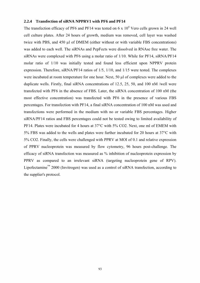
93
2.2.4 Transfection of siRNA NPPRV1 with PF6 and PF14
The transfection efficacy of PF6 and PF14 was tested on 6 x 10⁴ Vero cells grown in 24 well
cell culture plates. After 24 hours of growth, medium was removed, cell layer was washed
twice with PBS, and 450 µl of DMEM (either without or with variable FBS concentrations)
was added to each well. The siRNAs and PepFects were dissolved in RNAse free water. The
siRNAs were complexed with PF6 using a molar ratio of 1/10. While for PF14, siRNA/PF14
molar ratio of 1/10 was initially tested and found less efficient upon NPPRV protein
expression. Therefore, siRNA/PF14 ratios of 1/5, 1/10, and 1/15 were tested. The complexes
were incubated at room temperature for one hour. Next, 50 µl of complexes were added to the
duplicate wells. Firstly, final siRNA concentrations of 12.5, 25, 50, and 100 nM /well were
transfected with PF6 in the absence of FBS. Later, the siRNA concentration of 100 nM (the
most effective concentration) was transfected with PF6 in the presence of various FBS
percentages. For transfection with PF14, a final siRNA concentration of 100 nM was used and
transfections were performed in the medium with no or variable FBS percentages. Higher
siRNA/PF14 ratios and FBS percentages could not be tested owing to limited availability of
PF14. Plates were incubated for 4 hours at 37°C with 5% CO2. Next, one ml of EMEM with
5% FBS was added to the wells and plates were further incubated for 20 hours at 37°C with
5% CO2. Finally, the cells were challenged with PPRV at MOI of 0.1 and relative expression
of PPRV nucleoprotein was measured by flow cytometry, 96 hours post-challenge. The
efficacy of siRNA transfection was measured as % inhibition of nucleoprotein expression by
PPRV as compared to an irrelevant siRNA (targeting nucleoprotein gene of RPV).
Lipofectamine™ 2000 (Invitrogen) was used as a control of siRNA transfection, according to
the supplier's protocol.

94
2.3 Results
2.3.1 rBac_NPPRV1shRNA and rAd_NPPRV1shRNA challenged with PPRV MOI of 0.01
inhibit PPRV progeny production 96 h post challenge
Both recombinant viruses could inhibit PPRV progeny in vitro when transduced cells were
challenged with PPRV at MOI of 0.01. However, the rAd_NPPRV1shRNA at MOI of 200 could
only reduce PPRV titers to 0.79 log10 with (Fig. 24). In contrast, the rBac_NPPRV1shRNA in
the same MOI could inhibit PPRV titers by more than 2 log10 (Fig. 24).
Figure 24: Inhibition of PPRV progeny production by rAd_NPPRV1shRNA and rBac_NPPRV1shRNA 96h post-challenge with PPRV MOI of 0.01.
2.3.2 Effect of higher transduction doses of rBac_NPPRV1shRNA and
rAd_NPPRV1shRNA and challenge doses of PPRV, upon inhibition of PPRV
progeny production after over time
The effects of transduction with higher MOIs of the two recombinants on a higher challenge
dose of PPRV and the time/effect relationship upon PPRV progeny production were assessed.
Vero cells were transduced with the two recombinant viruses using MOIs of 100, 200, 300,
and 400 and challenged with a low (MOI of 0.01) and high (MOI of 0.1) doses of PPRV.
The rAd_NPPRV1shRNA transduction at MOI of 200, after challenge with PPRV MOI of 0.01
and 0.1, inhibited PPRV progeny titers to 0.5 and 0.7 log10, respectively (Fig. 25). The CPE
1.0E+051.6E+05
3.2E+055.0E+056.3E+05
3.2E+03
4.0E+04
1.0E+051.6E+05
4.0E+05
1.0E+02
1.0E+03
1.0E+04
1.0E+05
1.0E+06
PPRV 25 50 100 200
MOIs
CC
ID50
/ml
rAd_NPPRV1_shRNA rBac_NPPRV1_shRNA
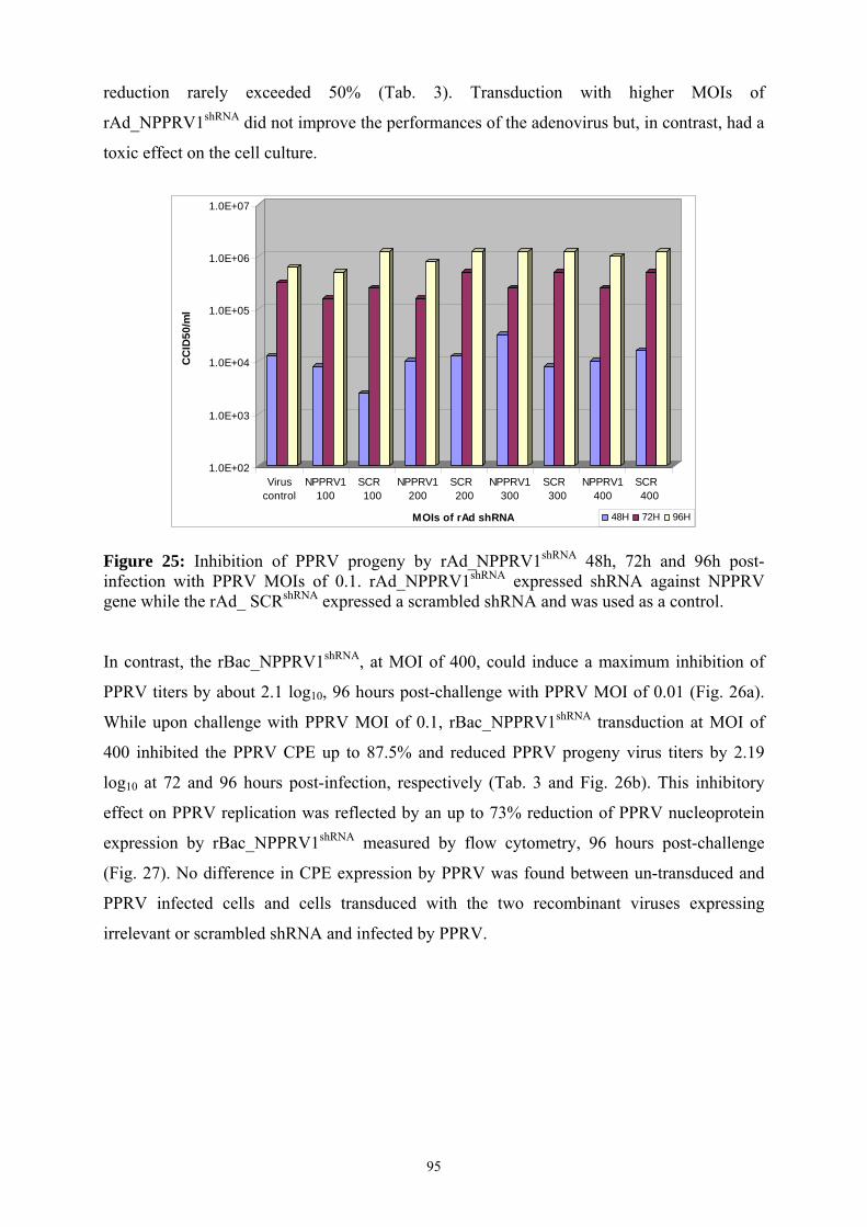
95
reduction rarely exceeded 50% (Tab. 3). Transduction with higher MOIs of
rAd_NPPRV1shRNA did not improve the performances of the adenovirus but, in contrast, had a
toxic effect on the cell culture.
Figure 25: Inhibition of PPRV progeny by rAd_NPPRV1shRNA 48h, 72h and 96h post-infection with PPRV MOIs of 0.1. rAd_NPPRV1shRNA expressed shRNA against NPPRV gene while the rAd_ SCRshRNA expressed a scrambled shRNA and was used as a control. In contrast, the rBac_NPPRV1shRNA, at MOI of 400, could induce a maximum inhibition of
PPRV titers by about 2.1 log10, 96 hours post-challenge with PPRV MOI of 0.01 (Fig. 26a).
While upon challenge with PPRV MOI of 0.1, rBac_NPPRV1shRNA transduction at MOI of
400 inhibited the PPRV CPE up to 87.5% and reduced PPRV progeny virus titers by 2.19
log10 at 72 and 96 hours post-infection, respectively (Tab. 3 and Fig. 26b). This inhibitory
effect on PPRV replication was reflected by an up to 73% reduction of PPRV nucleoprotein
expression by rBac_NPPRV1shRNA measured by flow cytometry, 96 hours post-challenge
(Fig. 27). No difference in CPE expression by PPRV was found between un-transduced and
PPRV infected cells and cells transduced with the two recombinant viruses expressing
irrelevant or scrambled shRNA and infected by PPRV.
1.0E+02
1.0E+03
1.0E+04
1.0E+05
1.0E+06
1.0E+07
CC
ID50
/ml
Viruscontrol
NPPRV1100
SCR 100
NPPRV1200
SCR 200
NPPRV1300
SCR 300
NPPRV1400
SCR 400
MOIs of rAd shRNA 48H 72H 96H

96
1.0E+02
1.0E+03
1.0E+04
1.0E+05
1.0E+06
1.0E+07
CC
ID50
/ml
Viruscontrol
NPPRV1100
eGFP 100
NPPRV1200
eGFP 200
NPPRV1300
eGFP 300
NPPRV1400
eGFP 400
MOIs of rBac shRNA 48H 72H 96H
(a)
(b) Figure 26: Inhibition of PPRV progeny virus production by rBac_NPPRV1shRNA 48h, 72h and 96h post-infection with PPRV MOIs of (a) 0.01 (b) 0.1. rBac_ eGFPshRNA expressing an irrelevant shRNA was used as control.
1.0E+02
1.0E+03
1.0E+04
1.0E+05
1.0E+06
1.0E+07C
CID
50/m
l
Viruscontrol
NPPRV1100
eGFP 100
NPPRV1200
eGFP 200
NPPRV1300
eGFP 300
NPPRV1400
eGFP 400
MOIs of rBac shRNA 48H 72H 96H

97
0.4
88.3
52.5
85.0
37.4
84.0
29.0
86.4
23.4
86.3
0
10
20
30
40
50
60
70
80
90
100
N v
iral p
rote
in e
xpre
ssio
n (%
)
Vero PPRV NPPRV1100
eGFP100
NPPRV1200
eGFP200
NPPRV1300
eGFP300
NPPRV1400
eGFP400
MOIs of rBac_shRNA
Table 3: Effect of rBac_NPPRV1shRNA and rAd_NPPRV1shRNA upon percentage of CPE induced by PPRV at 72h and 96h post infection.
72h 96h Virus control 75 100
rBac_NPPRV1shRNA MOI 100 25 50
rBac_NPPRV1shRNA MOI 200 25 25
rBac_NPPRV1shRNA MOI 300 25 25
rBac_NPPRV1shRNA MOI 400 12.5 25
Virus control 100 100
rAd _NPPRV1shRNA MOI 100 75 100
rAd _NPPRV1shRNA MOI 200 75 100
rAd _NPPRV1shRNA MOI 300 75 100
rAd_NPPRV1shRNA MOI 400 75 100
Figure 27: Inhibition of PPRV N protein expression by rBac_NPPRV1shRNA measured by flow cytometry. 2.3.4 siRNA NPPRV1 delivered by PepFect6 and PepFect14 inhibits nucleoprotein
expression by PPRV
PF6 is capable of efficiently transfecting siRNA at siRNA/PF6 molar ratio of 1/10.
Transfection of siRNA NPPRV1 by PF6 at final concentration of 100 nM inhibits N gene
expression by more than 99% while transfection of the irrelevant siRNA NRPV1 has no
inhibitory effect (Fig. 28). However, its efficiency declines when transfection is performed in
the presence of increasing FBS percentages and no inhibitory effect of siRNA NPPRV1 is

98
0.89
59.98
19.46
5.26
19.4528.19
52.76
60.01
0
10
20
30
40
50
60
70
80
90
100
N v
iral
pro
tein
exp
ress
ion
(%)
Vero PPRV Lipofect+NPPRV1
PF6+NPPRV1+0%FCS
PF6+NPPRV1
+10%FCS
PF6+NPPRV1
+30%FCS
PF6+NPPRV1
+60%FCS
PF6 +100%FCS
0.22
74.46
19.52
64.09
75.97
42.44
76.98
10.20
73.89
0.51
75.46
0
10
20
30
40
50
60
70
80
90
100
N vi
ral p
rote
in e
xpre
ssio
n (%
)
Vero PPRV Lipofect+NPPRV1 100nM
PF6+ NPPRV1 12.5 nM
PF6+ NRPV1 12.5 nM
PF6+ NPPRV1
25nM
PF6+ NRPV1
25nM
PF6+ NPPRV1
50nM
PF6+ NRPV1
50nM
PF6+ NPPRV1100nM
PF6+ NRPV1
100nM
found when transfection is performed in 100% of FBS (Fig. 29). PF14 is apparently less
efficient in transfecting siRNAs compared to PF6 at siRNA/PF14 ratio of 1/10 (Fig. 30).
However an increase in siRNA/PF14 ratio does increase the inhibitory effect of siRNA (Fig.
31). Moreover, transfection of siRNA with PF14 appears to be less affected by presence FBS.
Figure 28: Inhibition of PPRV N protein expression by various siRNA NPPRV1 doses transfected by PF6, measured by flow cytometry.
Figure 29: Inhibition of PPRV N protein expression by siRNA NPPRV1 transfected by PF6 upon transfection in the absence or presence of various FBS percentages, measured by flow cytometry.

99
0.83
83.88
60.81 64.55
76.33
82.06
0
10
20
30
40
50
60
70
80
90
100
N vi
ral p
rote
in e
xpre
ssio
n (%
)
Vero PPRV siRNA/PF1/10
+0%FBS
siRNA/PF1/10
+10%FBS
siRNA/PF1/10
+30%FBS
siRNA/PF1/10
+60%FBS
Figure 30: Inhibition of PPRV N protein expression by siRNA NPPRV1 transfected by PF14 upon transfection in the absence or presence of various FBS percentages, measured by flow cytometry.
Figure 31: Inhibition of PPRV N protein expression by siRNA NPPRV1 transfected by PF14 in absence or presence of 30% FBS, measured by flow cytometry.
0.60 0.70
74.19
79.04
24.71
40.6344.45
53.42
63.33 65.06
73.51
0
10
20
30
40
50
60
70
80
90
100
N v
iral
pro
tein
exp
ress
ion
(%)
Vero+0%FBS
Vero+30%FBS
PPRV+0%FBS
PPRV+30%FBS
Lipofect siRNA/PF1/15 +
0%FBS
siRNA/PF1/15 +
30%FBS
siRNA/PF1/10 +
0%FBS
siRNA/PF1/10 +
30%FBS
siRNA/PF1/5 +0%FBS
siRNA/PF1/5 +
30%FBS

100
2.4 Discussion
This study provides evidence that the recombinant adenovirus or baculovirus expressing
active shRNAs can interfere with the replication of PPRV in vitro. However, the baculovirus
proved to be more efficient than adenovirus vector when used at same MOIs. The low
efficiency of the adenovirus against PPRV replication differs from other studies in which
other pathogens were targeted. In vitro, shRNA-expressing adenoviruses achieved titer
reductions of 3 log10 and 1.39 log10 for porcine picornavirus and circovirus when used at
MOIs of 80 and 1000, respectively [225, 362]. Moreover, the results obtained in this study
differ also from the results obtained previously by our group using the same recombinant
adenovirus in the same conditions (see “State of the art” chapter of this document). The
reason for this efficacy discrepancy is not really known. The initial experiments were
performed with the rAd_NPPRV1shRNA vector which was produced at CIRAD while later in
vitro as well as in vivo experiments were performed using the rAd_NPPRV1shRNA vector
which was produced and purified by another laboratory (using bio-fermentor growth and
purification by chromatography). The process may have modified the virus. Moreover it is
known that many chromatographic elution buffers used for Adenovirus purification
procedures are not suitable for in vivo manipulations [363]. Thus the two lots of
rAd_NPPRV1shRNA were not the same and it could be at the origin of this discrepancy.
Anyhow, it remains to be evaluated. Here, the rAd_NPPRV1shRNA did not reduce PPRV titers
more than 0.7 log10 (Fig. 25) and higher concentrations of this vector showed to be deleterious
to transduced cells. Whereas, the rBac_NPPRV1shRNA tested in this study, when used at MOI
of 400, could reduce PPRV replication by 2.19 log10 (Fig. 26b), which is higher than other
comparable studies using baculoviral vector for delivery of shRNA against other viral
pathogens in vitro. Lu et al., [332] using baculoviral vector expressing shRNA against
nucleoprotein of porcine arterivirus, at MOI of 625, could inhibit viral titers up to 0.6 log10.
While Suzuki et al., [335, 364], reported a decrease in titer for human influenza viruses to a
maximum of 0.9 log10, after in vitro transduction with baculoviral vector expessing
bifunctional shRNA at MOI of 100.
Inhibition of PPRV titer and N viral protein expression by 2.1 log10 and 73% respectively, by
rBac_NPPRV1shRNA is lower than the N protein inhibition by 90% and a reduction of PPRV
progeny by 3-4 log10 that our group have obtained by siRNA NPPRV1 delivered by
Lipofectamine 2000™ [350]. Moreover, highest inhibition of PPRV progeny titers was
obtained 72h post-challenge for the higher challenge dose of PPRV (MOI 0.1) but the
inhibitory effect tends to decline by 96h (Fig. 26b). This result is surprising since as far as

101
longevity of action is concerned shRNA expressing viral vectors normally have advantages
over chemically synthesized siRNAs. It is reported that less than 1% of the introduced siRNA
duplexes remain in the cell 48 hours after administration, whereas the shRNA can be
continuously synthesized by the vector in the host cells, therefore, its effect should be much
more durable [185]. However, as the viral vectors expressing shRNA used here are replication
deficient, shRNA expression is limited and can get diluted over time due to continuous cell
division, while the target virus may continue to replicate in non-transduced cells producing
ever more mRNA and can also re-infect other cells. Maximum inhibition was observed 96
hours post challenge, when the cells transduced cells with high baculovirus MOIs were
challenged with the low PPRV challenge dose (MOI 0.01) (Fig. 26a).
Anyhow, the in vivo application of this viral vector can be advantageous when compared to
liposomes since baculoviruses do not have deleterious effects on mammalian cells even when
used at very high concentrations [339]. Indeed, we did not find any adverse effects of
rBac_NPPRV1shRNA in Vero cells even when used at an MOI of 400. On the other hand,
baculoviruses seem to be susceptible to complement inactivation [336] that could make it
impracticable to in vivo application. However, chemical or genetic modification, can
overcome this problem [337, 338]. Furthermore, morbillivirus infections occasionally lead to
infection of nervous system as well [16, 17], while the baculoviral vectors are not only
capable of transducing nervous tissues in mouse upon being injected into brain but have also
been reported to cross blood brain barrier [340].
Finally, two new chemical vectors, which are CPPs named the PF6 and PF14, were tested for
their ability to transfect the siRNA NPPRV1 in Vero cells. The siRNA NPPRV1 has already
been validated to be effective against PPRV at concentration of 100 nM when transfected
with Lipofectamine 2000™ [350]. Transfection of siRNA NPPRV1 at 100 nM with PF6
could silence viral protein expression by 99% (Fig. 28) which is higher than around 90%
inhibition obtained with Lipofectamine 2000™ [350]. Interestingly, even at 50 nM, siRNA
NPPRV1 transfected with PF6 could inhibit protein expression by 86.5% while it was only
78% with Lipofectamine 2000™ [350]. Therefore, PF6 has a better efficacy compared to
Lipofectamine 2000™.
The PF14 is less efficient as reflected by a lower inhibition of N gene expression by PPRV
(Fig. 30). However a higher siRNA/PF14 ratio of 1/15 had better effects, thus suggesting that
a further increase of siRNA/PF14 ratio may improve antiviral activity (Fig. 31). Moreover,

102
PF14 is less affected in the presence of serum compared to PF6 (Fig. 31 and Fig. 29).
However, at the siRNA/PF ratio of 1/10, the PF6 is still around 2.6 and 5.5 times more
efficient, when transfection is performed in the presence of 30% and 60% of FBS,
respectively. The inhibitory effect of higher concentration of serum in the PF6 and PF14
activities remains to be evaluated.
In conclusion, a recombinant replication deficient adenovirus and a baculovirus expressing
shRNA against nucleoprotein of PPRV were constructed and tested in vitro. This study shows
that both recombinants can inhibit PPRV replication in vitro. However, the baculoviral vector
was more efficient. A CPP PF6 can deliver siRNA NPPRV1 effectively in vitro resulting in
an almost complete inhibition of N gene expression by PPRV. PF14, although has a lower
transfection efficiency in vitro, appears to be more serum resistant.
The PF6 and rBac_NPPRV1shRNA vectors were efficient in vitro however whether these would
prove equally efficient for inhibition of PPRV replication also in vivo needs to be elucidated.
.
103
PART. 3:
First contributions towards the development of a small animal model for
the assessment of siRNA activity in vivo

104
3.1 Introduction
Final potential beneficiaries of an antiviral therapy based on siRNA against morbilliviruses
are large species like ruminants, dogs and possibly humans. However, the demonstration of in
vivo efficacy can hardly be established directly in the target species for ethical, safety and
economic reasons. Therefore, a small animal model is highly desirable to generate the proof
of concept and also demonstrate the safety and efficacy of such an approach. Validated small
animal models for morbillivirus infection are not frequent. This is probably due to a long
virus-host co-evolution, limiting cross-species transmission.
However, for measles virus, a mouse model for experimental challenge was developed
recently [365]. Since we have identified active siRNA against this virus, the use of this model
was initially considered. Nevertheless, for intellectual property reasons, this model could not
be transferred to our laboratory. Another small animal model based on Marmosets could be
used as these have been shown to be susceptible to measles [366, 367]. In addition, the model
requires the manipulation of virulent measles virus strains. Although zoonotic diseases are
currently investigated in our laboratory, we are not primarily in a human health research unit.
The permission to use virulent measles virus strains is therefore a long and tricky process. A
lapinized strain of rinderpest virus was shown to induce disease in rabbits [368]. However, the
use of rinderpest virus strains is not recommended since this disease is about to be eradicated
globally and the propagation of the virus in target or non target species always increases the
risk for pathogen escape. In addition, the in vivo validation of siRNA against an eradicated
disease would have a limited impact.
For PPRV, the third virus for which we have demonstrated an in vitro activity of siRNA, no
animal model is available. For this reason, we decided to launch a strategy for developing a
mouse model for PPRV infection. Recently, mice that are knocked out for the interferon type
I receptor (IFNAR-/-) were shown to be highly susceptible to bluetongue virus [369]. The
research team that established this model was contacted and collaboration was initiated to test
a virulent PPRV challenge on these IFNAR-/- mice. However, these mice never developed
disease after PPRV challenge or sero-converted against the virus (data not shown).
An alternative strategy was then designed to enable an in vivo assessment of systemic siRNA
delivery for efficacious knock down of a morbillivirus gene.

105
3.2 Development of a non-infectious model to test in vivo the delivery of siRNA
In a first approach, a non-infectious model was designed with the objective of generating
kinetic and quantitative measurements of RNA interfering activity in a peripheral tissue. This
model consisted of the expression of a luminescent Firefly reporter gene downstream of the
target sequence of a siRNA directed against nucleoprotein gene of PPRV (named NPPRV1).
The expression of this reporter gene is self-contained in the tibialis anterior (TA) muscle of
mice after intramuscular injection of the plasmid containing the expression cassette driven by
the human cytomegalovirus promoter (pCMV). For the kinetic and quantitative measurement
of luminescence, we selected a method based on bioluminescence imaging (BLI) of mice
allowing a measurement once a day over a week for all treated animals. Bioluminescence is a
chemo-luminescence reaction only achievable by a living organism and produces light that
can be imaged by non-invasive techniques. It has been used to monitor specific cellular or
genetic activities in the animals. This system is therefore perfectly adapted to test different
delivery systems for our siRNA, with a dynamic approach in live animals. In this method, a
cooled charged couple detector (CCD) camera is used to quantify photons emitted from a
light source localized in deep tissues. Generally, luminescence is preferred for that purpose
because there is no such light emission in natural tissues and therefore the signal/noise ratio is
very high.
The most often used luminescence marker is the Firefly luciferase which cleaves the luciferin
substrate and generates photons that are detected by the camera. We decided to use this
reporter gene in our approach. However, the normalization of the signal required later on the
use of a second “house-keeping” marker to minimize variability among mice.
106
3.2.1 Material and methods
3.2.1.1 Production and in vitro validation of a siRNA-NPPR1-Firefly_luciferase-2
reporter system
The reporter system was constructed according to the strategy depicted in figure 32. Briefly,
the Firefly luciferase-2 (luc2) gene was amplified from a commercial reporter plasmid
pGL4.51[luc2/CMV/Neo] Vector (Promega) using PhusionTM High-Fidelity DNA polymerase
(FINNZYMES, Finland) with the following primers: forward sequence 5’-GGA-TCA-ACT-
GGT-TTG-AGA-AAT-GGA-AGA-TGC-CAA-AAA-CAT-TAA-GAA-GGG-C-3’, reverse
sequence 5’-TTA-CAC-GGC-GAT-CTT-GCC-GCC-CTT-C-3’. The forward primer
consisted in the target sequence of the siRNA-NPPRV1 (in bold), followed by the
complementary sequence of the Firefly luciferase-2 gene. This modified gene was then
inserted into an expression plasmid vector (pCI-neo Mammalian Expression Vector,
Promega) by blunt ended ligation. The expression capacity of this construction was then
assessed in vitro by transfection of Vero cells with 100 ng of the plasmid and Lipofectamine™
2000 (Invitrogen), according to the manufacturer’s protocol. A positive control of expression
consisted of the original commercial plasmid having the luc2 gene (pGL4.51, Promega). The
luciferase gene expression was measured at 24-48 hours post-transfection by ONE-Glo™
Luciferase Assay System (Promega) with a luminometer (VictorII, Berthold-Wallac). Results
were expressed as relative luminescent units (RLU). The down-regulation of the siRNA-
NPPRV1-Firefly luciferase-2 construction, later on named psiRNA-Fluc, by siRNA NPPRV1
was then validated in vitro by co-transfection of both molecules in Vero cells with
Lipofectamine™ 2000 (Invitrogen)
3.2.1.2 In vivo validation of Firefly luciferase reporter systems
In order to test the in vivo delivery of siRNA against the psiRNA-Fluc, a model consisting in
a dynamic monitoring of the luciferase expression in mice by bioluminescence imaging (BLI)
was developed. The siRNA-Fluc gene in pCIneo plasmid (psiRNA-Fluc) was appropriately
diluted in PBS as indicated in the results section and injected under a volume of 60 µl in the
TA muscle of mice as previously reported by others [370]. A positive control, consisting of
the commercial Firefly luciferase gene under the same pCMV promoter (pGL4.51, Promega),
was also used at the optimal dose of 10 µg/60 µl as previously defined (results not shown).
Expression of the reporter gene was measured 48 hours after injection. Measurement was
done between 8 to 15 minutes after two separate intra-peritoneal injections of 100 µl of D-

107
Luciferin potassium salt (150 mg/kg, Caliper Life Sciences) using an IVIS-Lumina (Caliper
Life Sciences). For imaging, anesthetized animals were placed in the light-tight chamber of
the IVIS-Lumina. Photons emitted from luciferase-expressing plasmids were collected and
integrated over three-minute periods. Pseudocolor images indicating photon counts were
overlaid on photographs of the mice using the Living Image software version 4.0 (Caliper
Life Sciences). Regions of interest (ROIs) were selected manually over the signal intensity.
3.2.1.3 In vivo assessment of siRNA delivery against siRNA-Fluc
Ten mice were split into two groups of five. One group received over three consecutive days,
the siRNA-NPPRV1 at 40 µg/dose/mouse in a liposome-based emulsion (Aonys, Medesis
Pharma, Montpellier) administrated by the intra-rectal route. This delivery system has been
previously validated for siRNA delivery in mice targeting other genes (unpublished results).
The second group of 5 mice received an irrelevant siRNA (MPPRV10) targeting the matrix
protein of PPRV. Twenty four hours after the first administration of the siRNA-NPPRV1-
Aonys, all mice received in the left leg 10 µg/60 µl of pGL4.51 and 30 µg/60 µl of psiRNA-
Fluc (dose chosen after the previous in vivo test). All mice were imaged as previously
described, 48 hours after plasmid injection (72 hours after first administration of the siRNA).
3.2.1.4 Inclusion of a second reporter gene to normalize the in vivo psiRNA-Fluc
The initial in vivo test of siRNA delivery showed that the signal intensity between mice was
highly variable and thus required further normalization. The observed variability was ascribed
to the lack of accuracy of pDNA injection/expression in the mouse TA muscle. It was decided
to develop a protocol based on a co-injection of the Firefly luciferase reporter gene with
another reporter gene retaining a stable expression over time. This stable reporter gene would
then be considered as a sort of house-keeping gene and serve for normalization.
The Renilla luciferase gene was selected as a second luminescent reporter gene. The enzyme
expressed by this gene has a specific substrate that does not cross react with that of the Firefly
luciferase gene. Conversely, the D-Luciferine is not used by Renilla. However, the emission
spectra of the two substrates are overlapping, thus requiring a sequential injection of the
substrates in mice. The plasmid vector pGL4.75[hRluc/CMV] expressing Renilla luciferase
gene under the pCMV promoter was bought from a commercial company (Promega). After in
vitro validation using the same approach than for the Firefly luciferase gene, the Renilla was
probed in vivo. Three mice received in both tibialis muscles a co-injection of pGL4.51
(Firefly luciferase, 10 µg) and pGL4.75[hRluc/CMV] Vector (Promega) (Renilla luciferase,

108
100 µg) in 60 µl of PBS. Seventy two hours later, the mice were imaged 4 minutes after
intravenous injection of Coelenterazine h (the substrate of Renilla, 22 µg in 150 µl). Twenty
minutes later, the mice were injected with D-Luciferin as previously described and again
imaged.
Figure 32: Schematic presentation of siRNA-NPPR1-Firefly luciferase-2 reporter gene “psiRNA-Fluc” construct.
siRNA NPPRV1 target sequence
Luciferase-2 gene

109
3.2.2 Results
The psiRNA-Fluc construction was achieved and tested in vitro in comparison with the
commercial Firefly luciferase gene (pGL4.51, Promega). Both constructions have an
expression driven by the pCMV promoter. The luciferase expression by psiRNA-Fluc
construction was first found to be around 20 times more than the expression of pGL4.51
(data not shown). In a second step, the psiRNA-Fluc was probed in presence of the siRNA-
NPPRV1 or an irrelevant siRNA in comparison with PGL4.51. The table 4 shows that
siRNA-NPPRV1 specifically inhibited the expression of luciferase by psiRNA-Fluc and not
by the original pGL4.51 Firefly reporter. In addition, the irrelevant siRNA could not down-
regulate any construction, thus illustrating that the sequence placed upstream of the Firefly
luciferase reporter gene was efficaciously targeted by our siRNA.
Table 4: In vitro inhibition of luciferase expression by psiRNA-Fluc after co-transfection with siRNA NPPRV1.
pGL4.51 (100 ng)
psiRNA-Fluc (100 ng)
Negative control
24h 48h 24h 48h 24h 48h 20 pmole siRNA NPPRV1 16924 3183 13672 2584 134 174 20 pmole -siRNA IR* 26527 7440 634592 92659 40 pmole siRNA NPPRV1 17954 6166 9592 2344 40 pmole-siRNA IR 21335 3047 257299 72207 *IR: irrelevant
Before testing the delivery of siRNA NPPRV1 in vivo, we tried first to calibrate the amount
of psiRNA-Fluc plasmid to be injected in the TA muscle. Forty eight hours post injection of
the reporter plasmids, the mice were injected with D-Luciferin and imaged. Results are shown
in figure 33. The reproducibility of the positive control was not perfect in this assay since the
second mouse showed almost no signal. However, the dose/effect of psiRNA-Fluc was
considered as acceptable. Since, the production of micrograms of plasmid is expensive and
time-consuming, we considered from this trial that a dose of 30 µg/60 µl for psiRNA-Fluc
would be satisfactory enough for the next studies although the signal at this dose may be
lower than the one resulting from 10 µg/60 µl of pGL4.51 (see mouse 3 in Fig. 33).

110
(a)
(b)
Figure 33: Determination of the optimal dose for psiRNA-Fluc in vivo. (a) In the three mice, PGL4.51, considered as the positive control, was injected at a constant dose (10 µg/60 µl) in the left leg. As depicted from left to right, the psiRNA-Fluc was injected in the right leg at 3, 10 and 30 µg/ 60 µl. (b) The histogram shows the total photons measured at each ROI per second and surface (p/s/cm²).
1.0E+00
1.0E+01
1.0E+02
1.0E+03
1.0E+04
1.0E+05
1.0E+06
p/s/
cm2
Mouse 1 Mouse 2 Mouse 3
psiRNA-FlucPGL4.51

111
The RNA interference against our psiRNA-Fluc reporter system was tested using a liposome-
based formulation developed by a private company (Medesis Pharma, Montpellier). The
delivery of siRNA with this system requires three consecutive deposits in contact with the
rectal mucosa. Results are shown in Figure 34. For all mice, whatsoever the treatment
received, luciferase expression increased from day 1 to day 2 after the plasmid injection.
Afterwards, the increase became inconsistent. The expression of pGL4.51 was comparable
between the group treated with the specific siRNA-NPPRV1 and the group treated with the
irrelevant siRNA. A difference was observed between the two groups when considering the
expression of psiRNA-Fluc. However, this difference was not significant because of a high
heterogeneity within each group with some non–responding individuals (Fig. 34). This
observation confirmed what was already observed in the previous test (Fig. 33). This
variability resulted from the quality of plasmid injection and not from the substrate
bioavailability after intraperitoneal injection as it persisted over days and after repeated
substrate injections. Therefore, we decided to develop a double labeled system to assess the
efficacy of plasmid injections and to normalize the test signals.

112
(a)
(b)
Figure 34: In vivo imagery of mice treated with siRNA-NPPRV1 (inside the green zone) or with an irrelevant siRNA. All mice were injected in the right and left legs with psiRNA-Fluc (30 µg/60 µl) and pGL4.51 (10 µg/60 µl), respectively. They were imaged at different time points after plasmid injections. The picture (a) shows the results at day 3 post-injection and the histogram (b) shows average total photons (p/s/cm²) measured in each group for the two siRNAs administered at different days after plasmid injection.
p/s/cm2
Days
113
For the normalization of the signals, we selected a second bioluminescent marker. Although,
it is possible to use a fluorescent marker in the IVIS-Lumina bioimaging system, in our hands,
fluorescence signals were hardly detected from the TA muscle compared to the Firefly
luciferase-2 gene (data not shown). The Renilla reporter gene was therefore evaluated. Three
mice were co-injected with the two reporter genes in both legs and sequentially imaged, first
after IV injection of Coelenterazine h and then after IP injection with D-Luciferin. Results are
shown in Figure 35. Renilla luciferase was found to be 1.5 to 2 log10 less bright than Firefly
luciferase-2, which is an expected result since the former is known to give less signal. For
both signals, we could reproduce the variable success of intramuscular injections of the
plasmid vectors. However, the dot plot shows a good correlation between the two markers. In
conclusion to this trial, it appears that the results obtained from both legs of mouse 1, the left
leg of mouse 2 and the right leg of mouse 3 could be exploited in a siRNA in vivo assay since
normalization can be envisaged.

114
(a)
(b)
Figure 35: In vivo measurement of the co-expression of Renilla and Firefly luciferase genes. (a) Top picture shows Renilla activity, 4 minutes after IV injection of Coelenterazine h and on bottom picture the Firefly luciferase-2 activity, 10 minutes after IP injection of D-Luciferin. (b) The dot plot illustrates the correlation between the two signals.

115
GENERAL DISCUSSION
116
The viruses belonging to the Morbillivirus genus cause devastating diseases affecting humans,
marine mammals, domestic and wild canines, small ruminants and large ruminants. They are
emerging or re-emerging pathogens because of their high contagiosity and the difficulty to
achieve a high enough immunity cover to prevent the appearance of epidemics. In spite of the
availability of an effective vaccine, more than 30 million cases of acute measles with
approximately 345,000 cases of infant deaths are annually reported [42]. For the control of
measles, 95% of the population should ideally be vaccinated to avoid any new outbreak. But
this objective is extremely difficult to achieve, even in Europe, because a small proportion of
children is still not vaccinated or do not develop a sufficient immunity. Although effective
vaccines against canine distemper are also available, a resurgence of the disease has been
reported even in vaccinated dogs [49, 50]. After massive international efforts, rinderpest has
been eradicated worldwide and the world will be declared rinderpest free by 2011 [38].
Eradication of rinderpest has brought PPR to the limelight, highlighting the need for its
control. Although an efficacious vaccine is available and the fact that the disease hits the
economically most vulnerable sections of world population [65], there is no international
program for eradication of PPR.
The need for antiviral treatments concerns more particularly the diseases for which there are
no efficacious vaccines. However, it is now possible to foresee antiviral drugs as an
alternative or a complementary line of defence against infections. Thus, an RNAi based
antiviral therapy against PPR may prove economically useful for control of epidemics not
only if used alone but also along with vaccination. The vaccination against PPR is actually
resorted to as an emergency measure in case of an epidemic. However, only the healthy
animals can be vaccinated and it takes around a week for the development of the protection.
In case of a PPR outbreak, use of an effective antiviral therapy along with ring vaccination in
and around the disease pockets could help not only to control the epidemic but also to prevent
extensive economic losses.
In a recent past, there has been a spectacular expansion of the comprehension of the molecular
mechanisms of the viral cycles. Further, the efforts to contain the sudden rise of HIV have
lead to the development of numerous antiviral molecules whose number has increased tenfold
over the last ten years. Strategies developed to counter viral diseases have identified new
molecular targets. Interfering RNA constitute an interesting trail to follow because of the in
vitro selective degradation of the viral RNA renders them highly specific and effective in their
action. However, even after more than a decade after its discovery, RNAi therapeutics are still
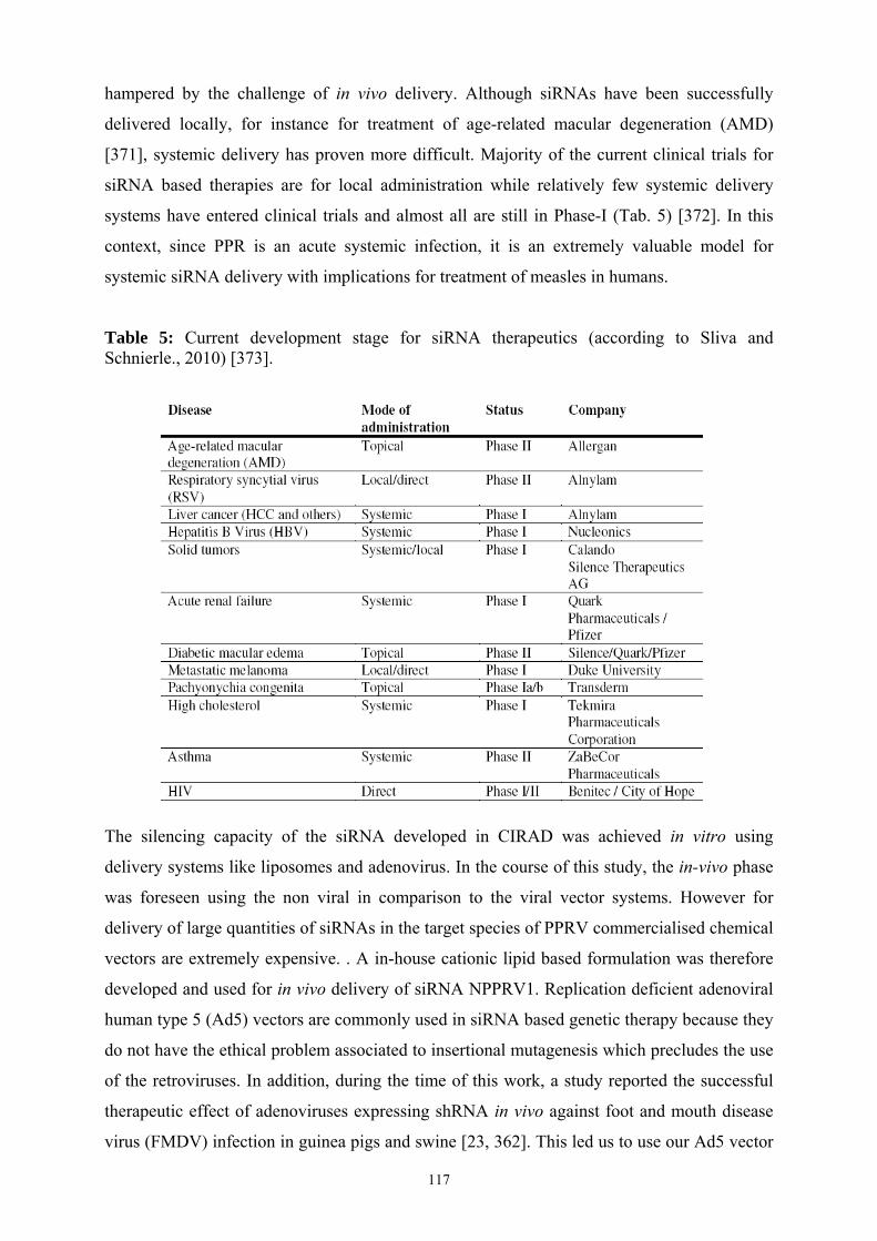
117
hampered by the challenge of in vivo delivery. Although siRNAs have been successfully
delivered locally, for instance for treatment of age-related macular degeneration (AMD)
[371], systemic delivery has proven more difficult. Majority of the current clinical trials for
siRNA based therapies are for local administration while relatively few systemic delivery
systems have entered clinical trials and almost all are still in Phase-I (Tab. 5) [372]. In this
context, since PPR is an acute systemic infection, it is an extremely valuable model for
systemic siRNA delivery with implications for treatment of measles in humans.
Table 5: Current development stage for siRNA therapeutics (according to Sliva and Schnierle., 2010) [373].
The silencing capacity of the siRNA developed in CIRAD was achieved in vitro using
delivery systems like liposomes and adenovirus. In the course of this study, the in-vivo phase
was foreseen using the non viral in comparison to the viral vector systems. However for
delivery of large quantities of siRNAs in the target species of PPRV commercialised chemical
vectors are extremely expensive. . A in-house cationic lipid based formulation was therefore
developed and used for in vivo delivery of siRNA NPPRV1. Replication deficient adenoviral
human type 5 (Ad5) vectors are commonly used in siRNA based genetic therapy because they
do not have the ethical problem associated to insertional mutagenesis which precludes the use
of the retroviruses. In addition, during the time of this work, a study reported the successful
therapeutic effect of adenoviruses expressing shRNA in vivo against foot and mouth disease
virus (FMDV) infection in guinea pigs and swine [23, 362]. This led us to use our Ad5 vector

118
rAd_NPPRV1shRNA for in vivo delivery of shRNA NPPRV1. Although the incidence of oculo-
nasal discharges and stomatitis appeared to be reduced for animals treated with liposomes, the
diarrhoea and pyrexia were in contrast increased and in any case, the differences were
statistically significant. One of the possible reasons for this, was the insufficient relative
siRNA/lipids concentration administrated to the animals resulting from the necessity to
achieve acceptable injectable doses. Indeed, the use of diluted siRNA and lipid solutions for
complex formation as were used for in vitro study, would have resulted in large volumes for
intravenous injection which would not be feasible for injecting goats. However, while trying
to use more concentrated siRNA and liposome solutions for achieving final injectable doses, a
problem of precipitation occurred. Then, the siRNA/lipids ratio of had to be reduced by three
for complex formation to avoid precipitation. This siRNA/lipids ratio tested in vivo was later
on tested in vitro and was found to be less efficacious in PPRV CPE inhibition in vitro.
The lack of adenovirus effect in vivo could probably be due to differences in the tropism of
the two viruses. While morbilliviruses, are primarily lymphotropic and secondarily
epitheliotropic [61, 124, 132], lymphatic tissues are not transduced efficiently by adenovirus
type 5 vectors [354] and are not correctly transfected by liposomes as well [374]. Whether the
in vivo transfection or transduction of the epithelial tissues alone, with insufficient
transfection/transduction of lymphatic tissues, can suffice for having a therapeutic effect
cannot be confirmed. Similarly, Chen et al., could protect swine from a major clinical disease
through adenoviral vectors targeting FMDV, however they incriminated the different tissue
distribution of rAd5 and FMDV as responsible for the limited inhibition of FMDV infection
in vivo [23]. For effective delivery of siRNA/shRNA against PPRV, the
transfection/transduction of lymphatic tissues is desirable for improved therapeutic effect. As
perspectives, ligands targeting lymphocytes, could be attached to metabolically biotinylated
adenoviral [375], baculoviral vectors, liposomes and CPPs for improving
transduction/transfection of the target tissues. These modified viral and chemical vectors
could be tested in vitro on goat PBMCs. However for initial screening of unmodified vectors
in vitro, the Vero cells remain ideal as they lack genes for interferon (IFN), while if
lymphocytes or other cell lines are used the effects of IFN may over shadow the contribution
of RNAi upon PPRV replication [376-378]. RNAi experiments in goats create several
practical difficulties. In our in vivo experiment, we could not test several doses for adenoviral
vector in vivo as it would have required large number of goats; therefore in future in vivo dose
optimization is still needed. The results of Chen et al., indicated that all three swine receiving
a low adenoviral dose (4 X 10⁹ PFU) were completely protected against FMDV challenge.

119
However, the antiviral effect was somewhat impaired in the animals treated with a high dose
of adenoviruses (8 X 10⁹ PFU) whereby only one animal was protected [23]. They suggested
that this may have been due to inhibitory effect of adenoviral VA1 non-coding RNA, which
they think should be removed in future for more efficient delivery [23].
Since the first attempt at in vivo delivery of siRNA did not give the expected results, it
entailed us to search for more efficient viral or non-viral vectors which could be possibly used
for future siRNA/shRNA delivery in vivo. For this purpose, recombinant adenovirus and
baculovirus vectors expressing short hairpin RNA (shRNA) against the PPR virus (PPRV)
were prepared and compared for efficiency in vitro. We found a lower efficiency of the
adenovirus against PPRV replication compared to previous studies in which other pathogens
were targeted [362]. In contrast, the recombinant baculovirus tested in this study proved to be
more efficient than adenovirus vector when used at same MOIs. A reduction of 2.19 log10 on
PPRV replication was observed which is more than 1 log10 higher than other comparable
studies involving baculoviral vectors [335, 364]. However, the reduction of NPPRV protein
expression by 73% and PPRV progeny titre by 2.19 log10 with rBac_NPPRV1shRNA was lower
than the one previously observed by our group when the siRNA were delivered to cell
cultures by Lipofectamine 2000™. The latter reduced the PPRV nucleoprotein expression by
90% and progeny titre by 3-4 log10 [350]. Although recombinant baculoviruses are easier and
cheaper to produce in high quantities than liposomes and do not have deleterious effects up on
mammalian cells even at very high MOIs [339], they are susceptible to complement
inactivation [336]. Chemical or genetic modifications, however, has been reported to be able
to overcome this problem [337, 338]. In addition to baculovirus, we also investigated two new
peptides (CPPs) named PF6 and PF14 for siRNA delivery. Transfection of siRNA NPPRV1
with PF6 at 100 nM resulted in the silencing of viral protein expression by up to 99% which
was higher than the inhibition achieved with Lipofectamine 2000™ (90%, [350]). In contrast,
the PF14 was less efficient for the transfection of siRNA NPPRV1. Transfection could be
improved by using higher siRNA/PF14 ratio but still it never reached efficacy of PF6.
Unfortunately, the availability of PF14 was limited and we could not increase the ratio
siRNA/PF14 as required. This was a pity since our results tended to show that PF14 was less
prone to the negative effect of the presence of serum in the environment of the cells where
had to be delivered the siRNA. In provisional conclusion, we could not discriminate clearly
the suitability of two CPPs for future studies in vivo. This will require new in vitro trials or to
test the two CPPs in vivo with the recombinant baculovirus. However, such an in vivo test is
not feasible in the target species since it requires many large animals and large quantity of

120
expensive siRNA. Therefore, development of a small animal model to assess the in vivo
performance of different delivery systems became an absolute necessity in the course of this
work.
To decrease the technical leap between the in vitro and in vivo evaluation and also improve
the ethical, bio-security and economic aspects of the in vivo experiments the mouse model
was investigated. From a technical point of view, a BALB/c mouse is more than 1000 times
smaller than a goat and thus requires much lower quantities of siRNAs and vectors for
experiments. Furthermore, the RNAi experiments with virulent PPRV require high
containment animal facilities which are not available at CIRAD. Although PPRV infection of
mice could be possible at the high containment laboratory of CIRAD, normal mice are
resistant to PPRV infection. We tried to identify a susceptible mouse that could be infected by
PRRV. IFN knockout mice were tested because they were recently shown to be highly
susceptible to another ruminant virus, an orbivirus responsible of bluetongue disease [379].
However, PPRV challenge on these mice was unsuccessful since neither clinical signs nor
viremia was detected. This failure is probably due to inability of PPRV to use mouse SLAM
or CD150 receptors. To continue in this direction, the production of transgenic mice
expressing sheep SLAM receptor will be required. Such a model was successfully developed
for measles virus [380]. In the absence of a model for PPRV, a non-infectious strategy was
developed instead to enable in vivo assessment of siRNA delivery. For this purpose, a
luciferase expressing reporter plasmid having siRNA NPPRV1 target sequence was
developed. Aim was to inject this construct into the mouse muscle and deliver siRNA via
various vectors and measure effect of siRNA on luciferase expression by CDD camera. The
siRNA NPPRV1 could knock down luciferase expression by psiRNA-Fluc in vitro, thus
illustrating that the siRNA target sequence placed upstream of the Firefly luciferase 2 reporter
gene was efficaciously targeted by our siRNA. The amount of psiRNA-Fluc required for
injection into the mouse tibialis anterior (TA) muscle was determined for best in vivo imagery
by CDD camera. Next, a liposome-based formulation developed by a private company was
tested against our psiRNA-Fluc reporter system. However, luciferase expression was found to
be inconsistent between legs and mice with some non-responding individuals. The variability
was assumed to arise from the plasmid injection and not from the substrate bioavailability
after intraperitoneal injection (performed in two steps at different sites to avoid possible
failures of intraperitoneal injections). Owing to this variability, the difference observed
between the treatment and control groups, considering the expression of psiRNA-Fluc, was
not significant. In order to manage this variability, a double labeled system was introduced,

121
using Renilla luciferase as a second house-keeping bioluminescent reporter to check the
quality of plasmid injections and to normalize the results of siRNA activity. For both signals,
the variability in accuracy of intramuscular injection of plasmid vectors was observed,
however, a good correlation was obtained between the two markers, thus allowing
normalization. Since normalization could be achieved, it appears that this mouse model can
now be used for testing of siRNA delivery in vivo. Use of this bioluminescent reporter based
mouse model would allow us to test not only the various siRNA or shRNA expressing viral
vector doses but also would enable us to test other parameters like vector toxicity. The ethical
aspects are also considered in this model since the mice are not infected but only receive
reporter plasmids by the intramuscular route followed by daily injections of luminescent
substrates, either intravenously or intraperitoneally. All administrations are practiced under
gas anesthesia. Therefore, the animals suffer less as compared to a PPRV infection in small
ruminants.
122
CONSCLUSION AND PERSPECTIVES
A recombinant replication deficient adenovirus and a baculovirus expressing shRNA against
nucleoprotein of PPRV were tested in vitro. This study showed that both recombinants can
inhibit PPRV replication in vitro. However, the baculoviral vector was found to be more
efficient. A CPP PF6 can deliver siRNA NPPRV1 effectively in vitro resulting in an almost
complete inhibition of N gene expression by PPRV. Although PF14 has a lower transfection
efficiency in vitro at the siRNA/PF14 ratio of 1/15, use of higher ratios may improve
transfection. Furthermore, it is relatively serum resistant compared to PF6. Whether these
vectors which have been found to be efficient in vitro, would equally be effective in siRNA
delivery in vivo for inhibition of PPRV replication, needs to be elucidated.
Since the systemic delivery of siRNA is the key issue for future therapeutic application, we
have investigated a strategy based on the use of a non-infectious mouse model and a dynamic
follow up of siRNA treatment by live imaging. We show in this work that it is possible to
measure and standardize the expression of a bioluminescent reporter gene and thus, to
quantify a down-regulation of such gene. The work is on-going to calibrate this experiment
and then siRNA delivery will be tested again circumventing the initial problems of variability
that result from inconsistent plasmid injections. This model will be very useful for comparing
various vectors for systemic delivery of siRNAs. At the moment, in addition to the Aonys
system by Medesis Pharma, our recombinant baculovirus and the CPP PF6 or PF14, described
in the part 2, are promising candidates.
Once the best candidates will have been identified in this non infectious model, the objective
will be to confirm our observation in an infectious mouse model. Since mice are naturally
resistant to PPRV, our approach will be based on the generation of a transgenic mouse
expressing the sheep SLAM receptor for PPRV and being deficient for interferon type I
responses (double knock-out mouse for interferon type I receptor: IFNAR-/-). A similar
model (e.g. IFNAR-/- mice expressing human SLAM receptor for the measles virus) was
developed for measles virus [365]. The idea is to adapt this model to PPRV. Once established,
we will be able to test the RNA interference in mice in vivo: the inhibitory effect of siRNA
will be measured in terms of reduction of clinical signs or mortality. In addition, the
laboratory is also developing a bioluminescent PPR virus by reverse genetics. The final goal
will be to test the control of PPRV replication by siRNA through bioluminescence imagery.

123
REFERENCES 1. Moss, W.J. and D.E. Griffin, Global measles elimination. Nat Rev Microbiol, 2006. 4(12): p. 900-8. 2. Furley, C.W., W.P. Taylor, and T.U. Obi, An outbreak of peste des petits ruminants in a zoological
collection. Vet Rec, 1987. 121(19): p. 443-7. 3. Roger, F., et al., Detection of antibodies of rinderpest and peste des petits ruminants viruses
(Paramyxoviridae, Morbillivirus) during a new epizootic disease in Ethiopian camels (Camelus dromedarius). Rev Méd Vét., 2001: p. 152:265.
4. Khalafalla, A.I., et al., An outbreak of peste des petits ruminants (PPR) in camels in the Sudan. Acta Trop, 2010. 116(2): p. 161-5.
5. Govindarajan, R., et al., Isolation of pestes des petits ruminants virus from an outbreak in Indian buffalo (Bubalus bubalis). Vet Rec, 1997. 141(22): p. 573-4.
6. Lalanne, L.G.a.A., La peste des petits ruminants. Bull. Serv. Zoo. A.O.F, 1942. 5: p. 15– 21. 7. El Hag Ali, B. and W.P. Taylor, Isolation of peste des petits ruminants virus from the Sudan. Research
in Veterinary Science, 1984. 36(1): p. 1-4. 8. Wamwayi, H.M., et al., Peste des petits ruminants antibodies in east Africa. Vet Rec, 1995. 136(8): p.
199-200. 9. Shaila, M.S., et al., Peste des petits ruminants of sheep in India. Vet Rec, 1989. 125(24): p. 602. 10. Shaila, M.S., et al., Geographic distribution and epidemiology of peste des petits ruminants virus. Virus
Research, 1996. 43(2): p. 149-53. 11. Wang, Z.L., et al., Peste des Petits Ruminants Virus in Tibet, China. Emerging Infectious Diseases,
2009. 15(2): p. 299-301. 12. Kwiatek, O., et al., Peste des petits ruminants (PPR) outbreak in Tajikistan. J Comp Pathol, 2007.
136(2-3): p. 111-9. 13. Albayrak, H. and F. Alkan, PPR virus infection on sheep in blacksea region of Turkey: Epidemiology
and diagnosis by RT-PCR and virus isolation. Veterinary Research Communications, 2009. 33(3): p. 241-249.
14. Ayari-Fakhfakh, E., et al., First serological investigation of peste-des-petits-ruminants and Rift Valley fever in Tunisia. Vet J, 2010.
15. FAO (2008) Outbreak of 'peste des petits ruminants' in Morocco. 16. Cosby, S.L., et al., Approaches in the understanding of morbillivirus neurovirulence. J Neurovirol,
2002. 8 Suppl 2: p. 85-90. 17. Schneider-Schaulies, J., V. Meulen, and S. Schneider-Schaulies, Measles infection of the central
nervous system. J Neurovirol, 2003. 9(2): p. 247-52. 18. Misra, A., S. Trehan, and G. Sharma, siRNA: Sojourn from discovery to delivery challenges and clinics.
Systematic Reviews in Pharmacy, 2010. 1(1): p. 1. 19. Bitko, V., et al., Inhibition of respiratory viruses by nasally administered siRNA. Nat Med, 2005. 11(1):
p. 50-5. 20. Morrissey, D.V., et al., Potent and persistent in vivo anti-HBV activity of chemically modified siRNAs.
Nature Biotechnology, 2005. 23(8): p. 1002-7. 21. Watanabe, T., et al., Liver target delivery of small interfering RNA to the HCV gene by lactosylated
cationic liposome. J Hepatol, 2007. 47(6): p. 744-50. 22. Kumar, P., et al., T cell-specific siRNA delivery suppresses HIV-1 infection in humanized mice. Cell,
2008. 134(4): p. 577-86. 23. Chen, W.Z., et al., Adenovirus-mediated RNA interference against foot-and-mouth disease virus
infection both in vitro and in vivo. Journal of Virology, 2006. 80(7): p. 3559-3566. 24. Lambeth, L.S., et al., Targeting Marek's disease virus by RNA interference delivered from a herpesvirus
vaccine. Vaccine, 2009. 27(2): p. 298-306. 25. Chen, M., et al., Retroviral delivery of RNA interference against Marek's disease virus in vivo. Poult
Sci, 2009. 88(7): p. 1373-80. 26. Barrett, T., Morbillivirus infections, with special emphasis on morbilliviruses of carnivores. Veterinary
Microbiology, 1999. 69(1-2): p. 3-13. 27. Tatsuo, H., N. Ono, and Y. Yanagi, Morbilliviruses use signaling lymphocyte activation molecules
(CD150) as cellular receptors. J Virol, 2001. 75(13): p. 5842-50. 28. Banyard, A., B.K. Rima, and T. Barrett, The morbilliviruses. Rinderpest and Peste des Petits
Ruminants. Virus Plagues of Large and Small Ruminants ed. T. Barrett, P. Banyard, and W.P. Taylor. 2006, Amsterdam: Elsevier Academic Press. 13-30. 29. Pomeroy, L.W., O.N. Bjornstad, and E.C. Holmes, The evolutionary and epidemiological dynamics of the paramyxoviridae. J Mol Evol, 2008. 66(2): p. 98-106.
30. Black, F.L., Measles endemicity in insular populations: critical community size and its evolutionary implication. J Theor Biol, 1966. 11(2): p. 207-11.
31. Keeling, M.J., Modelling the persistence of measles. Trends Microbiol, 1997. 5(12): p. 513-8.

124
32. James, A.D. and P.B. Rossiter, An epidemiological model of rinderpest. I. Description of the model. Trop Anim Health Prod, 1989. 21(1): p. 59-68.
33. BARTON, A., Plagues and contagions in antiquity. J Am Vet Med Assoc, 1956 129((11)): p. 503-5. 34. Pastoret, P.P., et al., Rinderpest - an old and worldwide story: history to c. 1902. Rinderpest and Peste
des Petits Ruminants. Virus Plagues of Large and Small Ruminants, ed. T. Barrett, P. Banyard, and W.P. Taylor. 2006, Amsterdam: Elsevier Academic Press. 86-104.
35. Pastoret, P.P. and E. NICOLAS, La peste bovine et la profession vétérinaire. De l'art à la science ou 150 ans de médecine vétérinaire à Cureghem. Annales de Médecine Vétérinaire, ed. P. P., G. Mees, and M. Mammerickx. 1986, Bruxelles. 117-122.
36. Roeder, P.L., W.P. Taylor, and M.M. Rweyemamu, Rindrpest in twentieth and twenty-first centuries. Rinderpest and Peste des Petits Ruminants. Virus Plagues of Large and Small Ruminants, ed. T. Barrett, P. Banyard, and W.P. Taylor. 2006, Amsterdam: Elsevier Academic Press. 105-142.
37. Plowright, W., The application of monolayer tissue culture techniques in rinderpest research. II. The use of attenuated cultured virus as a vaccine for cattle. Bull. I'Office Int. epizooties, 1962)(57): p. 253–277.
38. FAO. The Global Rinderpest Eradication Programme. Status report on progress made to May 2010 in the eradication of rinderpest: highlighting success stories and actions required prior to Global Declaration in 2011. 2010 27/7/2010]; Available from: http://www.fao.org/ag/againfo/resources/documents/AH/GREP_flyer.pdf.
39. Norrby, E., et al., Is rinderpest virus the archevirus of the Morbillivirus genus? Intervirology, 1985. 23(4): p. 228-32.
40. Norling, B., Plagues and Peoples William H. McNeill: Plagues and Peoples. (Garden City, New York: Anchor Press, Doubleday, 1976. Pp. 369. $10.00.). The Review of Politics, 1977. 39(04): p. 557-560.
41. Furuse, Y., A. Suzuki, and H. Oshitani, Origin of measles virus: divergence from rinderpest virus between the 11th and 12th centuries. Virol J, 2010. 7: p. 52.
42. Avota, E., E. Gassert, and S. Schneider-Schaulies, Measles virus-induced immunosuppression: from effectors to mechanisms. Med Microbiol Immunol, 2010.
43. Gee, S., S. Cotter, and D. O'Flanagan, Spotlight on measles 2010: measles outbreak in Ireland 2009-2010. Euro Surveill, 2010. 15(9).
44. WHO. Measles Surveillance Data - Number of reported measles cases with onset date from Feb 2010 to Aug 2010. 2010 18 March 2010 08/10/2010]; Available from: https://www.who.int/immunization_monitoring/diseases/measles_monthlydata/en/index.html.
45. ProMED-mail. MEASLES - INDIA (JAMMU AND KASHMIR). 2010 12/10/2010]; Available from: http://www.promedmail.org/pls/apex/f?p=2400:1202:2508937063540372::NO::F2400_P1202_CHECK_DISPLAY,F2400_P1202_PUB_MAIL_ID:X,84162.
46. ProMED-mail. MEASLES - AFRICA (25): ZIMBABWE. 2010; Available from: http://www.promedmail.org/pls/apex/f?p=2400:1202:2508937063540372::NO::F2400_P1202_CHECK_DISPLAY,F2400_P1202_PUB_MAIL_ID:X,84981.
47. ProMED-mail. MEASLES - BRAZIL: . 2010; Available from: http://www.promedmail.org/pls/apex/f?p=2400:1202:2508937063540372::NO::F2400_P1202_CHECK_DISPLAY,F2400_P1202_PUB_MAIL_ID:X,84031.
48. Woma, T.Y., et al., Phylogenetic analysis of the haemagglutinin gene of current wild-type canine distemper viruses from South Africa: lineage Africa. Veterinary Microbiology, 2010. 143(2-4): p. 126-32.
49. Lan, N.T., et al., Comparative analyses of canine distemper viral isolates from clinical cases of canine distemper in vaccinated dogs. Veterinary Microbiology, 2006. 115(1-3): p. 32-42.
50. Hashimoto, M., Y. Une, and M. Mochizuki, Hemagglutinin genotype profiles of canine distemper virus from domestic dogs in Japan. Archives of virology, 2001. 146(1): p. 149-55.
51. Sekulin, K., et al., Emergence of canine distemper in Bavarian wildlife associated with a specific amino acid exchange in the haemagglutinin protein. Vet J, 2010.
52. McCarthy, A.J., M.A. Shaw, and S.J. Goodman, Pathogen evolution and disease emergence in carnivores. Proc Biol Sci, 2007. 274(1629): p. 3165-74.
53. Gilbert, J.M.a.Y., Adaptation des virus de la peste des petits ruminants aux cultures cellularaires. Revue d’Elevage et de Medecine Veterinaire des Pays Tropicaux, 1962. 15: p. 321–335.
54. Roeder, P.L., et al. (1999) RECOGNIZING PESTE DES PETITS RUMINANTS - A field manual. 5. 55. Diallo, A., et al., The threat of peste des petits ruminants: progress in vaccine development for disease
control. Vaccine, 2007. 25(30): p. 5591-7. 56. Mornet, P., Orue, J., Gilbert, Y., Thiery, G. and Mamadore, S., La peste des petits ruminants en Afrique
occidentale française; ses rapports avec la peste bovine. . Rev. Elev. Méd. Vét. Pays Trop. , 1956. 9: p. 313–342.
57. Rowland, A.C., et al., A comparative study of peste des petits ruminants and Kata in West African Dwarf goats. Trop Anim Health Prod, 1971. 3(4): p. 241-7.

125
58. Gibbs, E.P., et al., Classification of peste des petits ruminants virus as the fourth member of the genus Morbillivirus. Intervirology, 1979. 11(5): p. 268-74.
59. Hamdy, F.M., et al., Etiology of the stomatitis pneumoenteritis complex in Nigerian dwarf goats. Can J Comp Med, 1976. 40(3): p. 276-84.
60. Diallo, A., et al., Comparison of proteins induced in cells infected with rinderpest and peste des petits ruminants viruses. Journal of General Virology, 1987. 68 ( Pt 7): p. 2033-8.
61. Scott, G.R., Rinderpest and peste des petits ruminants. Virus Diseases of Food Animals, 1981. II: p. 401 - 432.
62. Roeder, P.L., et al., Peste des petits ruminants in Ethiopian goats. Trop Anim Health Prod, 1994. 26(2): p. 69-73.
63. Minet, C., et al., Morbillivirus infections in ruminants: rinderpest eradication and peste des petits ruminants spreading towards the North. Virologie, 2009. 13(2): p. 103-113.
64. Taylor, W.P. and T. Barrett, Rinderpest and Peste Des Petits Ruminants. Diseases of Sheep. 2008: Blackwell Publishing Ltd. 460-469.
65. Perry, B.D., T. F. Randolph, J. J. McDermott, K. R. Sones, and P. K. and Thornton, Investing in Animal Health Research to Alleviate Poverty. 2002(ILRI (International Livestock Research Institute), Nairobi, Kenya,): p. 148
66. Food and Agriculture Organization, U.N., Peste des Petits Ruminants (PPR): A challenge for Small Ruminant Production. 2009.
67. Diallo, A., Control of peste des petits ruminants and poverty alleviation? J Vet Med B Infect Dis Vet Public Health, 2006. 53 Suppl 1: p. 11-3.
68. Food and Agriculture Organization, U.N., FAO supports study on the socioeconomic impacts of peste de petits ruminants in Kenya.
69. Barrett, T., A. Banyard, and A. Diallo, Molecular biology of the morbilliviruses. Rinderpest and Peste des Petits Ruminants. Virus Plagues of Large and Small Ruminants, ed. T. Barrett, P. Banyard, and W.P. Taylor. 2006, Amsterdam: Elsevier Academic Press. 31–67.
70. Bourdin, P. and A. Laurent Vautier, Note sur la structure du virus de la peste des petits ruminants. Revue d'élevage et de médecine vétérinaire des pays tropicaux, 1967. 20(3): p. 383-386.
71. Iwasaki, M., et al., The matrix protein of measles virus regulates viral RNA synthesis and assembly by interacting with the nucleocapsid protein. Journal of Virology, 2009. 83(20): p. 10374-83.
72. Bailey, D., et al., Full genome sequence of peste des petits ruminants virus, a member of the Morbillivirus genus. Virus Research, 2005. 110(1-2): p. 119-24.
73. Rima, B.K., The proteins of morbilliviruses. Journal of General Virology, 1983. 64 (Pt 6): p. 1205-19. 74. Graves, M., et al., Development of antibody to measles virus polypeptides during complicated and
uncomplicated measles virus infections. J Virol, 1984. 49(2): p. 409-12. 75. Diallo, A., et al., Cloning of the nucleocapsid protein gene of peste-des-petits-ruminants virus:
relationship to other morbilliviruses. Journal of General Virology, 1994. 75 ( Pt 1): p. 233-7. 76. Banyard, A.C., et al., Sequence of the nucleocapsid gene and genome and antigenome promoters for an
isolate of porpoise morbillivirus. Virus Research, 2008. 132(1-2): p. 213-219. 77. Knipe, D.M. and P.M. Howley, Fundamental Virology. 4 ed. Vol. 1 & 2. 2001, Philadelphia:
Lippencott Williams and Wilkins. 78. Barrett, T., B. A.C., and A. Diallo, Rinderpest and Peste des Petits Ruminants - Molecular biology of
morbilliviruses. Rinderpest and Peste des Petits Ruminants. Biology of Animal Infections, ed. T. Barrett, P.-P. Pastoret, and W.P. Taylor. 2006: Elsevier Academic Press, Amsterdam. 31-67.
79. Mahapatra, M., et al., Sequence analysis of the phosphoprotein gene of peste des petits ruminants (PPR) virus: editing of the gene transcript. Virus Research, 2003. 96(1-2): p. 85-98.
80. Bellini, W.J., et al., Measles virus P gene codes for two proteins. Journal of Virology, 1985. 53(3): p. 908-19.
81. Cattaneo, R., et al., Measles virus editing provides an additional cysteine-rich protein. Cell, 1989. 56(5): p. 759-64.
82. Boxer, E.L., S.K. Nanda, and M.D. Baron, The rinderpest virus non-structural C protein blocks the induction of type 1 interferon. Virology, 2009. 385(1): p. 134-42.
83. Hirano, A., et al., Functional analysis of matrix proteins expressed from cloned genes of measles virus variants that cause subacute sclerosing panencephalitis reveals a common defect in nucleocapsid binding. Journal of Virology, 1993. 67(4): p. 1848-53.
84. Reuter, T., et al., RNA interference with measles virus N, P, and L mRNAs efficiently prevents and with matrix protein mRNA enhances viral transcription. Journal of Virology, 2006. 80(12): p. 5951-7.
85. Riedl, P., et al., Measles virus matrix protein is not cotransported with the viral glycoproteins but requires virus infection for efficient surface targeting. Virus Research, 2002. 83(1-2): p. 1-12.
86. Spielhofer, P., et al., Chimeric measles viruses with a foreign envelope. Journal of Virology, 1998. 72(3): p. 2150-9.
87. Cathomen, T., et al., A matrix-less measles virus is infectious and elicits extensive cell fusion: consequences for propagation in the brain. EMBO J, 1998. 17(14): p. 3899-908.

126
88. Coronel, E.C., et al., Nucleocapsid incorporation into parainfluenza virus is regulated by specific interaction with matrix protein. Journal of Virology, 2001. 75(3): p. 1117-1123.
89. Seth, S. and M.S. Shaila, The fusion protein of Peste des petits ruminants virus mediates biological fusion in the absence of hemagglutinin-neuraminidase protein. Virology, 2001. 289(1): p. 86-94.
90. Seth, S. and M.S. Shaila, The hemagglutinin-neuraminidase protein of peste des petits ruminants virus is biologically active when transiently expressed in mammalian cells. Virus Research, 2001. 75(2): p. 169-77.
91. Langedijk, J.P.M., F.J. Daus, and J.T. VanOirschot, Sequence and structure alignment of Paramyxoviridae attachment proteins and discovery of enzymatic activity for a morbillivirus hemagglutinin. Journal of Virology, 1997. 71(8): p. 6155-6167.
92. de Swart, R.L., et al., Depletion of measles virus glycoprotein-specific antibodies from human sera reveals genotype-specific neutralizing antibodies. Journal of General Virology, 2009. 90(Pt 12): p. 2982-9.
93. Pratelli, A., Canine distemper virus: The emergence of new variants. Vet J, 2010. 94. Schnell, M.J. and K.K. Conzelmann, Polymerase activity of in vitro mutated rabies virus L protein.
Virology, 1995. 214(2): p. 522-30. 95. Ludlow, M., I. Allen, and J. Schneider-Schaulies, Systemic spread of measles virus: overcoming the
epithelial and endothelial barriers. Thromb Haemost, 2009. 102(6): p. 1050-6. 96. Iwasaki, M., et al., The matrix protein of measles virus regulates viral RNA synthesis and assembly by
interacting with the nucleocapsid protein. J Virol, 2009. 83(20): p. 10374-83. 97. Couacy-Hymann, E., et al., Early detection of viral excretion from experimentally infected goats with
peste-des-petits ruminants virus. Preventive Veterinary Medicine, 2007. 78(1): p. 85-88. 98. Lefèvre, P.C., Peste bovine. 1 ed. Principales maladies infectieuses et parasitaires du bétail : Europe et
régions chaudes 2003 285-305. 99. Griffin, D.E. and C.H. Pan, Measles: old vaccines, new vaccines. Curr Top Microbiol Immunol, 2009.
330: p. 191-212. 100. Santibanez, S., et al., Probing neutralizing-antibody responses against emerging measles viruses
(MVs): immune selection of MV by H protein-specific antibodies? Journal of General Virology, 2005. 86(Pt 2): p. 365-74.
101. Libeau, G., et al., A Competitive Elisa Using Anti-N Monoclonal-Antibodies for Specific Detection of Rinderpest Antibodies in Cattle and Small Ruminants. Veterinary Microbiology, 1992. 31(2-3): p. 147-160.
102. Brown, R.D., Rinderpest immunity in calves. I. The acquisition and persistence of maternally derived antibody. J Hyg (Lond), 1958. 56(4): p. 427-34.
103. Lund, B.T., et al., Vaccination of cattle with attenuated rinderpest virus stimulates CD4(+) T cell responses with broad viral antigen specificity. Journal of General Virology, 2000. 81(Pt 9): p. 2137-46.
104. Beckford, A.P., R.O. Kaschula, and C. Stephen, Factors associated with fatal cases of measles. A retrospective autopsy study. S Afr Med J., 1985. 12(68): p. 858-63.
105. Griffin, D.E., Measles virus-induced suppression of immune responses. Immunological Reviews, 2010. 236(1): p. 176-189.
106. Kumar, P., et al., Pathological and immunohistochemical study of experimental peste des petits ruminants virus infection in goats. Journal of Veterinary Medicine Series B-Infectious Diseases and Veterinary Public Health, 2004. 51(4): p. 153-159.
107. Mondal, B., et al., Apoptosis induced by peste des petits ruminants virus in goat peripheral blood mononuclear cells. Virus Res, 2001. 73(2): p. 113-9.
108. Griffin, D.E., Measles virus-induced suppression of immune responses. Immunological Reviews, 2010. 236: p. 176-189.
109. Hussey, G.D., et al., The effect of Edmonston-Zagreb and Schwarz measles vaccines on immune responses in infants. Journal of Infectious Diseases, 1996. 173(6): p. 1320-1326.
110. Stevenson, P., et al., Rinderpest vaccination and the incidence and development of trypanosomosis in cattle. Tropical Animal Health and Production, 1999. 31(2): p. 65-73.
111. Hamdy, F.M. and A.H. Dardiri, Response of white-tailed deer to infection with peste des petits ruminants virus. J Wildl Dis, 1976. 12(4): p. 516-22.
112. Ogunsanmi, A.O., et al., Peste des petits ruminants (PPR) virus antibodies in African grey Duiker (Sylvicapra grimmia. African Journal of Biomedical Research, 2003. 6 ((1)): p. 59-61.
113. Couacy-Hymann, E., et al., The early detection of peste-des-petits-ruminants (PPR) virus antigens and nucleic acid from experimentally infected goats using RT-PCR and immunocapture ELISA techniques. Research in Veterinary Science, 2009. 87(2): p. 332-5.
114. Provost, A. and G. Borredon, Les différents aspects du diagnostic clinique et expérimental de la peste bovine. Rev. Elev. Méd. vét Pays trop. , 1963. 16: p. 445-526.
115. Plowright, W., ed. Rinderpest virus. In: Virolology Monographs 3. . ed. S. Gard, C. Hallauer, and K.F. Meyer. 1968, Springer-Verlag, Vienne New-York. 25-110.

127
116. Elizabeth, S.W. and I.K. Barker, Infectious Diseases of Wild Mammals. 2001, Blackwell Publishing. p. 45-49.
117. FAO animal health manual - RECOGNIZING PESTE DES PETITS RUMINANTS - A field manual http://www.fao.org/docrep/003/x1703e/x1703e00.htm#The%20disease.
118. The Merck Veterinary Manual http://www.merckvetmanual.com/mvm/index.jsp?cfile=htm/bc/56100.htm&word=peste%2coetits%2cruminants.
119. Rajak, K.K., et al., Experimental studies on immunosuppressive effects of peste des petits ruminants (PPR) virus in goats. Comp Immunol Microbiol Infect Dis, 2005. 28(4): p. 287-96.
120. Pawar, R.M., et al., Effect of siRNA mediated suppression of signaling lymphocyte activation molecule on replication of peste des petits ruminants virus in vitro. Virus Res, 2008. 136(1-2): p. 118-23.
121. Kruse, M., et al., Signaling lymphocytic activation molecule is expressed on mature CD83(+) dendritic cells and is up-regulated by IL-1 beta. Journal of Immunology, 2001. 167(4): p. 1989-1995.
122. McQuaid, S., et al., Distribution of measles virus in the central nervous system of HIV-seropositive children. Acta Neuropathol, 1998. 96(6): p. 637-42.
123. Ferreira, C.S., et al., Measles virus infection of alveolar macrophages and dendritic cells precedes spread to lymphatic organs in transgenic mice expressing human signaling lymphocytic activation molecule (SLAM, CD150). J Virol, 2010. 84(6): p. 3033-42.
124. de Witte, L., et al., DC-SIGN and CD150 have distinct roles in transmission of measles virus from dendritic cells to T-lymphocytes. Plos Pathogens, 2008. 4(4): p. -.
125. de Witte, L., et al., Measles virus targets DC-SIGN to enhance dendritic cell infection. J Virol, 2006. 80(7): p. 3477-86.
126. Zhou, T., et al., DC-SIGN and immunoregulation. Cell Mol Immunol, 2006. 3(4): p. 279-83. 127. de Jong, M.A.W.P., et al., Dendritic cells mediate herpes simplex virus infection and transmission
through the C-type lectin DC-SIGN. Journal of General Virology, 2008. 89: p. 2398-2409. 128. Ludlow, M., et al., Wild-type measles virus infection of primary epithelial cells occurs via the
basolateral surface without syncytium formation or release of infectious virus. J Gen Virol, 2010. 91(Pt 4): p. 971-9.
129. de Swart, R.L., et al., Predominant infection of CD150+ lymphocytes and dendritic cells during measles virus infection of macaques. PLoS Pathog, 2007. 3(11): p. e178.
130. Leonard, V.H., et al., Measles virus blind to its epithelial cell receptor remains virulent in rhesus monkeys but cannot cross the airway epithelium and is not shed. J Clin Invest, 2008. 118(7): p. 2448-58.
131. Leonard, V.H., et al., Measles virus selectively blind to signaling lymphocytic activation molecule (SLAM; CD150) is attenuated and induces strong adaptive immune responses in rhesus monkeys. J Virol, 2010. 84(7): p. 3413-20.
132. von Messling, V., D. Milosevic, and R. Cattaneo, Tropism illuminated: lymphocyte-based pathways blazed by lethal morbillivirus through the host immune system. Proc Natl Acad Sci U S A, 2004. 101(39): p. 14216-21.
133. Wohlsein, P. and J. Saliki, Rinderpest and Peste des Petits Ruminants - the diseases: clinical signs and pathology. Rinderpest and Peste des Petits Ruminants. Biology of Animal Infections, ed. T. Barrett, P.-P. Pastoret, and W.P. Taylor. 2006. 68-85.
134. Brown, C.C., J.C. Mariner, and H.J. Olander, An Immunohistochemical Study of the Pneumonia Caused by Peste des Petits Ruminants Virus. Veterinary Pathology, 1991. 28(2): p. 166-170.
135. Lefèvre, P.C. (1987) Peste des petits ruminants et infection bovipestique des ovins et caprins. (Etudes et Synthèses de l'IEMVT; 5).
136. Rossiter, P.B., Peste des petits ruminants. 2 ed. Infectious diseases of livestock., ed. J.A.W. Coetzer and R.C. Tustin. Vol. 2. 2004, Cape Town: Oxford University Press. 660-672.
137. Michaud, V., et al., Long-term storage at tropical temperature of dried-blood filter papers for detection and genotyping of RNA and DNA viruses by direct PCR. Journal of Virological Methods, 2007. 146(1-2): p. 257-265.
138. Diallo, A., et al., Recent developments in the diagnosis of rinderpest and peste des petits ruminants. Veterinary Microbiology, 1995. 44(2-4): p. 307-17.
139. Tatsuo, H., N. Ono, and Y. Yanagi, Morbilliviruses use signaling lymphocyte activation molecules (CD150) as cellular receptors. Journal of Virology, 2001. 75(13): p. 5842-5850.
140. Seki, F., et al., Efficient isolation of wild strains of canine distemper virus in Vero cells expressing canine SLAM (CD150) and their adaptability to marmoset B95a cells. J Virol, 2003. 77(18): p. 9943-50.
141. Diallo, A., et al., Research on Diagnostic Tests: Key Topics and New applications, in Second Global Conference of OIE Reference Laboratories and Collaborating Centres. 2010: Paris.
142. Anderson, J., M. Corteyn, and G. Libeau, Diagnosis of rinderpest virus and peste des petits ruminants. Rinderpest and Peste des Petits Ruminants. Virus Plagues of Large and Small Ruminants ed. T. Barrett, P. Banyard, and W.P. Taylor. 2006, Amsterdam: Elsevier Academic Press. 163-184.
128
143. Wohlsein, P. and J. Saliki, Rinderpest and Peste des Petits Ruminants- the diseases: clinical signs and pathology. Rinderpest and Peste des Petits Ruminants. Virus Plagues of Large and Small Ruminants, ed. T. Barrett, P. Banyard, and W.P. Taylor. 2006, Amsterdam: Elsevier Academic Press. 68-85.
144. Saliki, J.T., Structural and functional characterization of various isolates of peste des petits ruminants virus using monoclonal antibodies. 1993, Cornell University: Ithaca, N.Y.
145. Libeau, G., et al., Rapid differential diagnosis of rinderpest and peste des petits ruminants using an immunocapture ELISA. Vet Rec, 1994. 134(12): p. 300-4.
146. Couacy-Hymann, E., et al., Rapid and sensitive detection of peste des petits ruminants virus by a polymerase chain reaction assay. Journal of Virological Methods, 2002. 100(1-2): p. 17-25.
147. Diallo, A., et al., [Attenuation of a strain of rinderpest virus: potential homologous live vaccine]. Rev Elev Med Vet Pays Trop, 1989. 42(3): p. 311-9.
148. Sujatha, K., et al., Synthesis and antiviral activity of 4,4'-(arylmethylene)bis(1H-pyrazol-5-ols) against peste des petits ruminant virus (PPRV). Bioorganic & Medicinal Chemistry Letters, 2009. 19(15): p. 4501-3.
149. Stiver, G., The treatment of influenza with antiviral drugs. CMAJ, 2003. 168(1): p. 49-56. 150. Zamore, P.D., et al., RNAi: Double-stranded RNA directs the ATP-dependent cleavage of mRNA at 21
to 23 nucleotide intervals. Cell, 2000. 101(1): p. 25-33. 151. Hamilton, A.J. and D.C. Baulcombe, A species of small antisense RNA in posttranscriptional gene
silencing in plants. Science, 1999. 286(5441): p. 950-2. 152. Haasnoot, J. and B. Berkhout, Virus-encoded suppressors of RNA silencing and role of cellular miRNAs
in Mammalian antiviral immune responses. RNA Interference and Viruses - Current Innovations and Future Trends, ed. M.A. Martinez. 2010, Norfolk, UK: Caister academic Press - Cromwell Press Group 67-88.
153. Umbach, J.L. and B.R. Cullen, The role of RNAi and microRNAs in animal virus replication and antiviral immunity. Genes & Development, 2009. 23(10): p. 1151-1164.
154. Pfeifer, A. and H. Lehmann, Pharmacological potential of RNAi - focus on miRNA. Pharmacol Ther, 2010. 126(3): p. 217-27.
155. Hajeri, P.B. and S.K. Singh, siRNAs: their potential as therapeutic agents - Part I. Designing of siRNAs. Drug Discovery Today, 2009. 14(17-18): p. 851-858.
156. Lewis, B.P., C.B. Burge, and D.P. Bartel, Conserved seed pairing, often flanked by adenosines, indicates that thousands of human genes are microRNA targets. Cell, 2005. 120(1): p. 15-20.
157. Napoli, C., C. Lemieux, and R. Jorgensen, Introduction of a Chimeric Chalcone Synthase Gene into Petunia Results in Reversible Co-Suppression of Homologous Genes in trans. Plant Cell, 1990. 2(4): p. 279-289.
158. Fire, A., et al., Potent and specific genetic interference by double-stranded RNA in Caenorhabditis elegans. Nature, 1998. 391(6669): p. 806-11.
159. Grunweller, A., et al., Comparison of different antisense strategies in mammalian cells using locked nucleic acids, 2 '-O-methyl RNA, phosphorothioates and small interfering RNA. Nucleic Acids Research, 2003. 31(12): p. 3185-3193.
160. Miyagishi, M., M. Hayashi, and K. Taira, Comparison of the suppressive effects of antisense oligonucleotides and siRNAs directed against the same targets in mammalian cells. Antisense & Nucleic Acid Drug Development, 2003. 13(1): p. 1-7.
161. Bertrand, J.R., et al., Comparison of antisense oligonucleotides and siRNAs in cell culture and in vivo. Biochem Biophys Res Commun, 2002. 296(4): p. 1000-4.
162. Lee, Y., et al., MicroRNA genes are transcribed by RNA polymerase II. EMBO J, 2004. 23(20): p. 4051-60.
163. Lee, Y., et al., The nuclear RNase III Drosha initiates microRNA processing. Nature, 2003. 425(6956): p. 415-9.
164. Han, J., et al., The Drosha-DGCR8 complex in primary microRNA processing. Genes Dev, 2004. 18(24): p. 3016-27.
165. Denli, A.M., et al., Processing of primary microRNAs by the Microprocessor complex. Nature, 2004. 432(7014): p. 231-5.
166. Lund, E., et al., Nuclear export of microRNA precursors. Science, 2004. 303(5654): p. 95-8. 167. Yi, R., et al., Exportin-5 mediates the nuclear export of pre-microRNAs and short hairpin RNAs. Genes
Dev, 2003. 17(24): p. 3011-6. 168. Lee, Y., et al., MicroRNA maturation: stepwise processing and subcellular localization. EMBO J, 2002.
21(17): p. 4663-70. 169. Chendrimada, T.P., et al., TRBP recruits the Dicer complex to Ago2 for microRNA processing and gene
silencing. Nature, 2005. 436(7051): p. 740-4. 170. Gregory, R.I., et al., Human RISC couples microRNA biogenesis and posttranscriptional gene silencing.
Cell, 2005. 123(4): p. 631-40. 171. Eulalio, A., E. Huntzinger, and E. Izaurralde, Getting to the root of miRNA-mediated gene silencing.
Cell, 2008. 132(1): p. 9-14.

129
172. Keck, K., et al., Rational Design Leads to More Potent RNA Interference Against Hepatitis B Virus: Factors Effecting Silencing Efficiency. Molecular Therapy, 2009. 17(3): p. 538-547.
173. Elbashir, S.M., et al., Duplexes of 21-nucleotide RNAs mediate RNA interference in cultured mammalian cells. Nature, 2001. 411(6836): p. 494-8.
174. Spurgers, K.B., et al., Oligonucleotide antiviral therapeutics: antisense and RNA interference for highly pathogenic RNA viruses. Antiviral Res, 2008. 78(1): p. 26-36.
175. Bitko, V. and S. Barik, Phenotypic silencing of cytoplasmic genes using sequence-specific double-stranded short interfering RNA and its application in the reverse genetics of wild type negative-strand RNA viruses. BMC Microbiol, 2001. 1: p. 34.
176. McCaffrey, A.P., et al., Inhibition of hepatitis B virus in mice by RNA interference. Nature Biotechnology, 2003. 21(6): p. 639-44.
177. Banerjea, A., et al., Inhibition of HIV-1 by lentiviral vector-transduced siRNAs in T lymphocytes differentiated in SCID-hu mice and CD34+ progenitor cell-derived macrophages. Mol Ther, 2003. 8(1): p. 62-71.
178. Lee, N.S., et al., Expression of small interfering RNAs targeted against HIV-1 rev transcripts in human cells. Nature Biotechnology, 2002. 20(5): p. 500-5.
179. Rao, D.D., et al., siRNA vs. shRNA: similarities and differences. Adv Drug Deliv Rev, 2009. 61(9): p. 746-59.
180. Jackson, A.L., et al., Expression profiling reveals off-target gene regulation by RNAi. Nature Biotechnology, 2003. 21(6): p. 635-7.
181. Valencia-Sanchez, M.A., et al., Control of translation and mRNA degradation by miRNAs and siRNAs. Genes Dev, 2006. 20(5): p. 515-24.
182. Birmingham, A., et al., 3' UTR seed matches, but not overall identity, are associated with RNAi off-targets. Nat Methods, 2006. 3(3): p. 199-204.
183. Anderson, E., et al., Identifying siRNA-induced off-targets by microarray analysis. Methods Mol Biol, 2008. 442: p. 45-63.
184. Jackson, A.L., et al., Widespread siRNA "off-target" transcript silencing mediated by seed region sequence complementarity. RNA, 2006. 12(7): p. 1179-87.
185. Rao, D.D., et al., siRNA vs. shRNA: Similarities and differences. Advanced Drug Delivery Reviews, 2009. 61(9): p. 746-759.
186. Ui-Tei, K., et al., Thermodynamic stability and Watson-Crick base pairing in the seed duplex are major determinants of the efficiency of the siRNA-based off-target effect. Nucleic Acids Res, 2008. 36(22): p. 7100-9.
187. Naito, Y., et al., siDirect 2.0: updated software for designing functional siRNA with reduced seed-dependent off-target effect. BMC Bioinformatics, 2009. 10: p. 392.
188. Bumcrot, D., et al., RNAi therapeutics: a potential new class of pharmaceutical drugs. Nat Chem Biol, 2006. 2(12): p. 711-9.
189. Sledz, C.A., et al., Activation of the interferon system by short-interfering RNAs. Nat Cell Biol, 2003. 5(9): p. 834-9.
190. Kariko, K., et al., Small interfering RNAs mediate sequence-independent gene suppression and induce immune activation by signaling through toll-like receptor 3. J Immunol, 2004. 172(11): p. 6545-9.
191. Hornung, V., et al., Sequence-specific potent induction of IFN-alpha by short interfering RNA in plasmacytoid dendritic cells through TLR7. Nat Med, 2005. 11(3): p. 263-70.
192. Hornung, V., et al., Sequence-specific potent induction of IFN-alpha by short interfering RNA in plasmacytoid dendritic cells through TLR7. Nature Medicine, 2005. 11(3): p. 263-270.
193. Judge, A.D., et al., Sequence-dependent stimulation of the mammalian innate immune response by synthetic siRNA. Nature Biotechnology, 2005. 23(4): p. 457-62.
194. Diebold, S.S., et al., Innate antiviral responses by means of TLR7-mediated recognition of single-stranded RNA. Science, 2004. 303(5663): p. 1529-31.
195. Heil, F., et al., Species-specific recognition of single-stranded RNA via toll-like receptor 7 and 8. Science, 2004. 303(5663): p. 1526-9.
196. Karin, M., Y. Yamamoto, and Q.M. Wang, The IKK NF-kappa B system: a treasure trove for drug development. Nat Rev Drug Discov, 2004. 3(1): p. 17-26.
197. Sioud, M., Induction of inflammatory cytokines and interferon responses by double-stranded and single-stranded siRNAs is sequence-dependent and requires endosomal localization. Journal of Molecular Biology, 2005. 348(5): p. 1079-1090.
198. Schlee, M., V. Hornung, and G. Hartmann, siRNA and isRNA: two edges of one sword. Mol Ther, 2006. 14(4): p. 463-70.
199. Grimm, D., et al., Fatality in mice due to oversaturation of cellular microRNA/short hairpin RNA pathways. Nature, 2006. 441(7092): p. 537-541.
200. Voinnet, O., Y.M. Pinto, and D.C. Baulcombe, Suppression of gene silencing: a general strategy used by diverse DNA and RNA viruses of plants. Proc Natl Acad Sci U S A, 1999. 96(24): p. 14147-52.
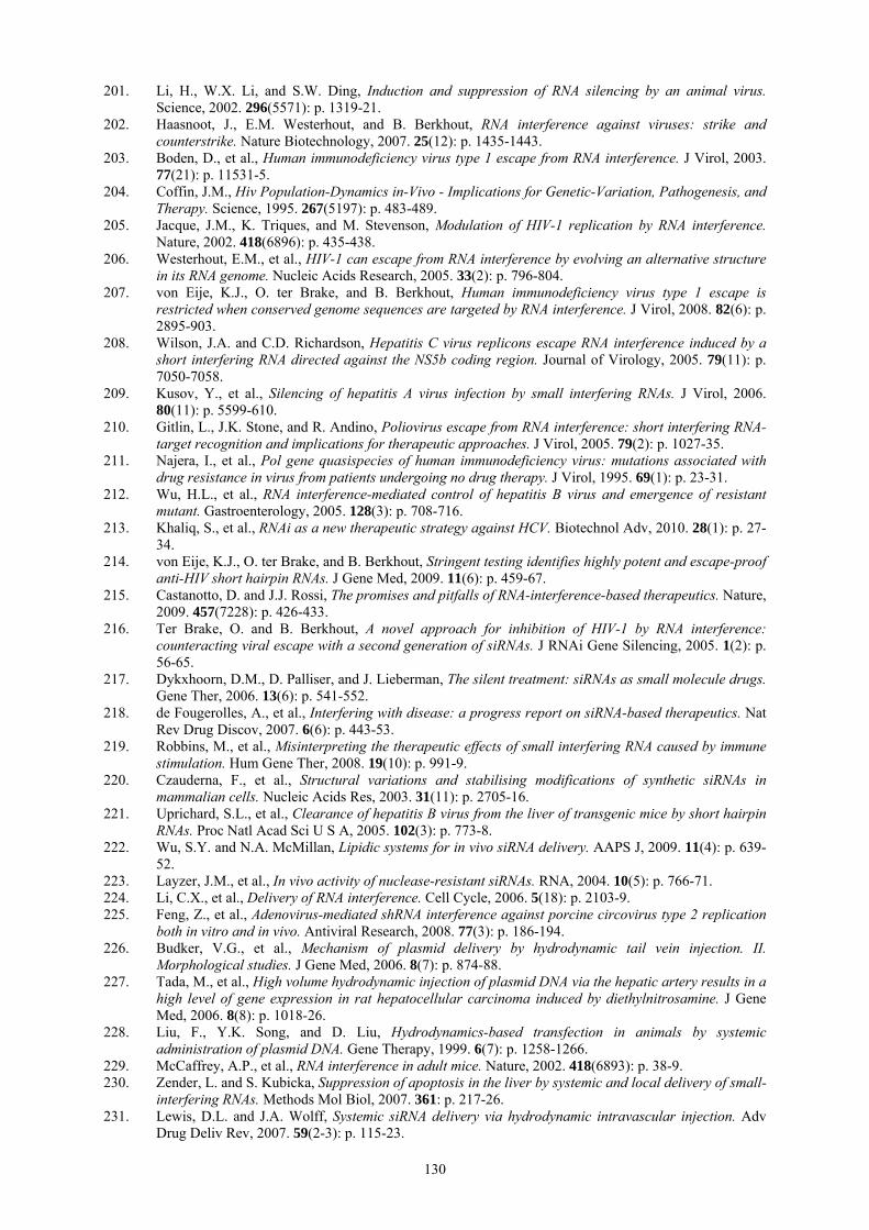
130
201. Li, H., W.X. Li, and S.W. Ding, Induction and suppression of RNA silencing by an animal virus. Science, 2002. 296(5571): p. 1319-21.
202. Haasnoot, J., E.M. Westerhout, and B. Berkhout, RNA interference against viruses: strike and counterstrike. Nature Biotechnology, 2007. 25(12): p. 1435-1443.
203. Boden, D., et al., Human immunodeficiency virus type 1 escape from RNA interference. J Virol, 2003. 77(21): p. 11531-5.
204. Coffin, J.M., Hiv Population-Dynamics in-Vivo - Implications for Genetic-Variation, Pathogenesis, and Therapy. Science, 1995. 267(5197): p. 483-489.
205. Jacque, J.M., K. Triques, and M. Stevenson, Modulation of HIV-1 replication by RNA interference. Nature, 2002. 418(6896): p. 435-438.
206. Westerhout, E.M., et al., HIV-1 can escape from RNA interference by evolving an alternative structure in its RNA genome. Nucleic Acids Research, 2005. 33(2): p. 796-804.
207. von Eije, K.J., O. ter Brake, and B. Berkhout, Human immunodeficiency virus type 1 escape is restricted when conserved genome sequences are targeted by RNA interference. J Virol, 2008. 82(6): p. 2895-903.
208. Wilson, J.A. and C.D. Richardson, Hepatitis C virus replicons escape RNA interference induced by a short interfering RNA directed against the NS5b coding region. Journal of Virology, 2005. 79(11): p. 7050-7058.
209. Kusov, Y., et al., Silencing of hepatitis A virus infection by small interfering RNAs. J Virol, 2006. 80(11): p. 5599-610.
210. Gitlin, L., J.K. Stone, and R. Andino, Poliovirus escape from RNA interference: short interfering RNA-target recognition and implications for therapeutic approaches. J Virol, 2005. 79(2): p. 1027-35.
211. Najera, I., et al., Pol gene quasispecies of human immunodeficiency virus: mutations associated with drug resistance in virus from patients undergoing no drug therapy. J Virol, 1995. 69(1): p. 23-31.
212. Wu, H.L., et al., RNA interference-mediated control of hepatitis B virus and emergence of resistant mutant. Gastroenterology, 2005. 128(3): p. 708-716.
213. Khaliq, S., et al., RNAi as a new therapeutic strategy against HCV. Biotechnol Adv, 2010. 28(1): p. 27-34.
214. von Eije, K.J., O. ter Brake, and B. Berkhout, Stringent testing identifies highly potent and escape-proof anti-HIV short hairpin RNAs. J Gene Med, 2009. 11(6): p. 459-67.
215. Castanotto, D. and J.J. Rossi, The promises and pitfalls of RNA-interference-based therapeutics. Nature, 2009. 457(7228): p. 426-433.
216. Ter Brake, O. and B. Berkhout, A novel approach for inhibition of HIV-1 by RNA interference: counteracting viral escape with a second generation of siRNAs. J RNAi Gene Silencing, 2005. 1(2): p. 56-65.
217. Dykxhoorn, D.M., D. Palliser, and J. Lieberman, The silent treatment: siRNAs as small molecule drugs. Gene Ther, 2006. 13(6): p. 541-552.
218. de Fougerolles, A., et al., Interfering with disease: a progress report on siRNA-based therapeutics. Nat Rev Drug Discov, 2007. 6(6): p. 443-53.
219. Robbins, M., et al., Misinterpreting the therapeutic effects of small interfering RNA caused by immune stimulation. Hum Gene Ther, 2008. 19(10): p. 991-9.
220. Czauderna, F., et al., Structural variations and stabilising modifications of synthetic siRNAs in mammalian cells. Nucleic Acids Res, 2003. 31(11): p. 2705-16.
221. Uprichard, S.L., et al., Clearance of hepatitis B virus from the liver of transgenic mice by short hairpin RNAs. Proc Natl Acad Sci U S A, 2005. 102(3): p. 773-8.
222. Wu, S.Y. and N.A. McMillan, Lipidic systems for in vivo siRNA delivery. AAPS J, 2009. 11(4): p. 639-52.
223. Layzer, J.M., et al., In vivo activity of nuclease-resistant siRNAs. RNA, 2004. 10(5): p. 766-71. 224. Li, C.X., et al., Delivery of RNA interference. Cell Cycle, 2006. 5(18): p. 2103-9. 225. Feng, Z., et al., Adenovirus-mediated shRNA interference against porcine circovirus type 2 replication
both in vitro and in vivo. Antiviral Research, 2008. 77(3): p. 186-194. 226. Budker, V.G., et al., Mechanism of plasmid delivery by hydrodynamic tail vein injection. II.
Morphological studies. J Gene Med, 2006. 8(7): p. 874-88. 227. Tada, M., et al., High volume hydrodynamic injection of plasmid DNA via the hepatic artery results in a
high level of gene expression in rat hepatocellular carcinoma induced by diethylnitrosamine. J Gene Med, 2006. 8(8): p. 1018-26.
228. Liu, F., Y.K. Song, and D. Liu, Hydrodynamics-based transfection in animals by systemic administration of plasmid DNA. Gene Therapy, 1999. 6(7): p. 1258-1266.
229. McCaffrey, A.P., et al., RNA interference in adult mice. Nature, 2002. 418(6893): p. 38-9. 230. Zender, L. and S. Kubicka, Suppression of apoptosis in the liver by systemic and local delivery of small-
interfering RNAs. Methods Mol Biol, 2007. 361: p. 217-26. 231. Lewis, D.L. and J.A. Wolff, Systemic siRNA delivery via hydrodynamic intravascular injection. Adv
Drug Deliv Rev, 2007. 59(2-3): p. 115-23.

131
232. Weinstein, S. and D. Peer, RNAi nanomedicines: challenges and opportunities within the immune system. Nanotechnology, 2010. 21(23): p. 232001.
233. Heller, L.C. and R. Heller, In vivo electroporation for gene therapy. Hum Gene Ther, 2006. 17(9): p. 890-7.
234. Golzio, M., et al., In vivo gene silencing in solid tumors by targeted electrically mediated siRNA delivery. Gene Ther, 2007. 14(9): p. 752-9.
235. Taniyama, Y., et al., Local delivery of plasmid DNA into rat carotid artery using ultrasound. Circulation, 2002. 105(10): p. 1233-1239.
236. Lu, J.J., R. Langer, and J. Chen, A novel mechanism is involved in cationic lipid-mediated functional siRNA delivery.
237. Schroeder, A., et al., Lipid-based nanotherapeutics for siRNA delivery. Journal of Internal Medicine, 2010. 267(1): p. 9-21.
238. Issa, M.M., Linear and Branched Chitosan Oligomers as Delivery Systems for pDNA and siRNA In Vitro and In Vivo [Elektronisk resurs]. 2006, Uppsala: Acta Universitatis Upsaliensis.
239. Rao, N.M., Cationic lipid-mediated nucleic acid delivery: beyond being cationic. Chemistry and Physics of Lipids, 2010. 163(3): p. 245-252.
240. Reischl, D. and A. Zimmer, Drug delivery of siRNA therapeutics: potentials and limits of nanosystems. Nanomedicine-Nanotechnology Biology and Medicine, 2009. 5(1): p. 8-20.
241. Bangham, A.D., M.M. Standish, and J.C. Watkins, Diffusion of univalent ions across the lamellae of swollen phospholipids. J Mol Biol, 1965. 13(1): p. 238-52.
242. Felgner, P.L., et al., Lipofection: a highly efficient, lipid-mediated DNA-transfection procedure. Proc Natl Acad Sci U S A, 1987. 84(21): p. 7413-7.
243. Felgner, J.H., et al., Enhanced gene delivery and mechanism studies with a novel series of cationic lipid formulations. J Biol Chem, 1994. 269(4): p. 2550-61.
244. Rosenzweig, H.S., et al., O-alkyl dioleoylphosphatidylcholinium compounds: The effect of varying alkyl chain length on their physical properties and in vitro DNA transfection activity. Bioconjugate Chemistry, 2000. 11(3): p. 306-313.
245. Budker, V., et al., pH-sensitive, cationic liposomes: a new synthetic virus-like vector. Nature Biotechnology, 1996. 14(6): p. 760-4.
246. Raper, S.E., et al., Fatal systemic inflammatory response syndrome in a ornithine transcarbamylase deficient patient following adenoviral gene transfer. Mol Genet Metab, 2003. 80(1-2): p. 148-58.
247. Couzin, J. and J. Kaiser, Gene therapy - As Gelsinger case ends, gene therapy suffers another blow. Science, 2005. 307(5712): p. 1028-1028.
248. Eliyahu, H., Y. Barenholz, and A.J. Domb, Polymers for DNA delivery. Molecules, 2005. 10(1): p. 34-64.
249. Zhang, S.B., et al., Cationic compounds used in lipoplexes and polyplexes for gene delivery. Journal of Controlled Release, 2004. 100(2): p. 165-180.
250. Lee, R.J. and L. Huang, Folate-targeted, anionic liposome-entrapped polylysine-condensed DNA for tumor cell-specific gene transfer. Journal of Biological Chemistry, 1996. 271(14): p. 8481-8487.
251. Mastrobattista, E., et al., Lipid-coated polyplexes for targeted gene delivery to ovarian carcinoma cells. Cancer Gene Ther, 2001. 8(6): p. 405-13.
252. Farhood, H., N. Serbina, and L. Huang, The role of dioleoyl phosphatidylethanolamine in cationic liposome mediated gene transfer. Biochim Biophys Acta, 1995. 1235(2): p. 289-95.
253. Reischl, D. and A. Zimmer, Drug delivery of siRNA therapeutics: potentials and limits of nanosystems. Nanomedicine, 2009. 5(1): p. 8-20.
254. LORIN, et al., Les liposomes : description, fabrication et applications. Vol. 8. 2004, Gembloux, BELGIQUE: Faculté universitaire des sciences agronomiques. 14.
255. Pires, P., et al., Interaction of cationic liposomes and their DNA complexes with monocytic leukemia cells. Biochimica Et Biophysica Acta-Biomembranes, 1999. 1418(1): p. 71-84.
256. Boutet, E. Scheme of a liposome formed by phospholipids in an aqueous solution. 2007 15 October 2007 [cited 2010 24/08/2010]; Available from: http://en.wikipedia.org/wiki/File:Liposome_scheme-en.svg.
257. Karmali, P.P. and A. Chaudhuri, Cationic Liposomes as non-viral carriers of gene medicines: Resolved issues, open questions, and future promises. Medicinal Research Reviews, 2007. 27(5): p. 696-722.
258. Mozafari, M.R., Liposomes: An overview of manufacturing techniques. Cellular & Molecular Biology Letters, 2005. 10(4): p. 711-719.
259. Verma, U.N., et al., Small interfering RNAs directed against beta-catenin inhibit the in vitro and in vivo growth of colon cancer cells. Clinical Cancer Research, 2003. 9(4): p. 1291-1300.
260. Sorensen, D.R., M. Leirdal, and M. Sioud, Gene silencing by systemic delivery of synthetic siRNAs in adult mice. Journal of Molecular Biology, 2003. 327(4): p. 761-766.
261. Geisbert, T.W., et al., Postexposure protection of guinea pigs against a lethal ebola virus challenge is conferred by RNA interference. J Infect Dis, 2006. 193(12): p. 1650-7.

132
262. Zimmermann, T.S., et al., RNAi-mediated gene silencing in non-human primates. Nature, 2006. 441(7089): p. 111-4.
263. Heitz, F., M.C. Morris, and G. Divita, Twenty years of cell-penetrating peptides: from molecular mechanisms to therapeutics. Br J Pharmacol, 2009. 157(2): p. 195-206.
264. Endoh, T. and T. Ohtsuki, Cellular siRNA delivery using cell-penetrating peptides modified for endosomal escape. Adv Drug Deliv Rev, 2009. 61(9): p. 704-9.
265. Frankel, A.D. and C.O. Pabo, Cellular uptake of the tat protein from human immunodeficiency virus. Cell, 1988. 55(6): p. 1189-93.
266. Vives, E., P. Brodin, and B. Lebleu, A truncated HIV-1 Tat protein basic domain rapidly translocates through the plasma membrane and accumulates in the cell nucleus. J Biol Chem, 1997. 272(25): p. 16010-7.
267. Meng, S., et al., TAT peptides mediated small interfering RNA delivery to Huh-7 cells and efficiently inhibited hepatitis C virus RNA replication. Intervirology, 2009. 52(3): p. 135-40.
268. Joliot, A., et al., Antennapedia homeobox peptide regulates neural morphogenesis. Proc Natl Acad Sci U S A, 1991. 88(5): p. 1864-8.
269. Derossi, D., et al., The third helix of the Antennapedia homeodomain translocates through biological membranes. J Biol Chem, 1994. 269(14): p. 10444-50.
270. Allinquant, B., et al., Downregulation of amyloid precursor protein inhibits neurite outgrowth in vitro. J Cell Biol, 1995. 128(5): p. 919-27.
271. Pooga, M., et al., Cell penetrating PNA constructs regulate galanin receptor levels and modify pain transmission in vivo. Nature Biotechnology, 1998. 16(9): p. 857-61.
272. Morris, M.C., et al., A new peptide vector for efficient delivery of oligonucleotides into mammalian cells. Nucleic Acids Res, 1997. 25(14): p. 2730-6.
273. Simeoni, F., et al., Insight into the mechanism of the peptide�based gene delivery system MPG: implications for delivery of siRNA into mammalian cells. Nucleic Acids Research, 2003. 31(11): p. 2717-2724.
274. Crombez, L., et al., Targeting cyclin B1 through peptide-based delivery of siRNA prevents tumour growth. Nucleic Acids Research, 2009. 37(14): p. 4559-4569.
275. Simeoni, F., et al., Insight into the mechanism of the peptide-based gene delivery system MPG: implications for delivery of siRNA into mammalian cells. 2003, Oxford University Press.
276. Meade, B.R. and S.F. Dowdy, Exogenous siRNA delivery using peptide transduction domains/cell penetrating peptides. Advanced Drug Delivery Reviews, 2007. 59(2-3): p. 134-140.
277. Deshayes, S., et al., Cell-penetrating peptides: tools for intracellular delivery of therapeutics. Cellular and Molecular Life Sciences, 2005. 62(16): p. 1839-49.
278. Richard, J.P., et al., Cell-penetrating peptides. A reevaluation of the mechanism of cellular uptake. J Biol Chem, 2003. 278(1): p. 585-90.
279. Mueller, J., et al., Comparison of Cellular Uptake Using 22 CPPs in 4 Different Cell Lines. Bioconjugate Chemistry, 2008. 19(12): p. 2363-2374.
280. Duchardt, F., et al., A comprehensive model for the cellular uptake of cationic cell-penetrating peptides. Traffic, 2007. 8(7): p. 848-66.
281. Chiu, Y.-L., et al., Visualizing a correlation between siRNA localization, cellular uptake, and RNAi in living cells. 2004, eScholarship@UMMS.
282. Juliano, R., et al., Mechanisms and strategies for effective delivery of antisense and siRNA oligonucleotides. Nucleic Acids Res, 2008. 36(12): p. 4158-71.
283. Davidson, T.J., et al., Highly efficient small interfering RNA delivery to primary mammalian neurons induces MicroRNA-like effects before mRNA degradation. J Neurosci, 2004. 24(45): p. 10040-6.
284. Muratovska, A. and M.R. Eccles, Conjugate for efficient delivery of short interfering RNA (siRNA) into mammalian cells. FEBS Lett, 2004. 558(1-3): p. 63-8.
285. Simeoni, F., et al., Insight into the mechanism of the peptide-based gene delivery system MPG: implications for delivery of siRNA into mammalian cells. Nucleic Acids Research, 2003. 31(11): p. 2717-2724.
286. Crombez, L., et al., A new potent secondary amphipathic cell-penetrating peptide for siRNA delivery into mammalian cells. Mol Ther, 2009. 17(1): p. 95-103.
287. Crombez, L., et al., A non-covalent peptide-based strategy for siRNA delivery. Biochem Soc Trans, 2007. 35(Pt 1): p. 44-6.
288. Anantpadma, M., D.A. Stein, and S. Vrati, Inhibition of Japanese encephalitis virus replication in cultured cells and mice by a peptide-conjugated morpholino oligomer. J Antimicrob Chemother, 2010. 65(5): p. 953-61.
289. Farokhzad, O.C., et al., Nanoparticle-aptamer bioconjugates: a new approach for targeting prostate cancer cells. Cancer Res, 2004. 64(21): p. 7668-72.
290. Patil, M.L., et al., Surface-modified and internally cationic polyamidoamine dendrimers for efficient siRNA delivery. Bioconjugate Chemistry, 2008. 19(7): p. 1396-1403.
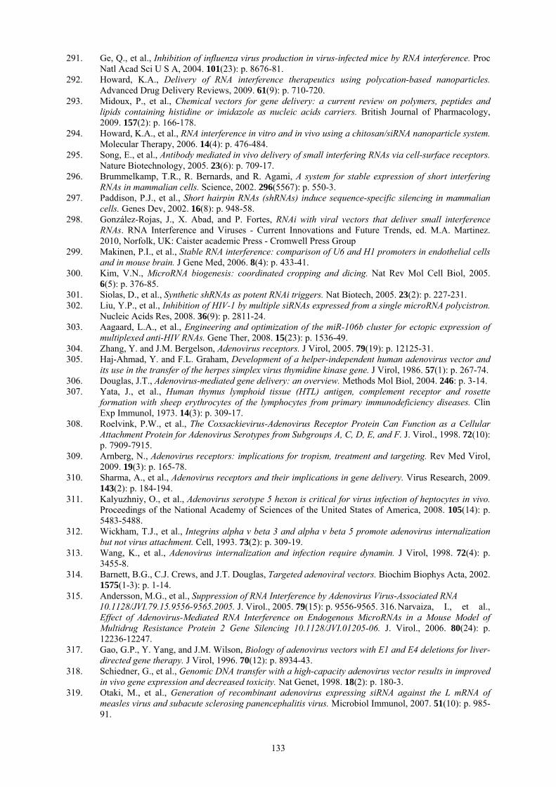
133
291. Ge, Q., et al., Inhibition of influenza virus production in virus-infected mice by RNA interference. Proc Natl Acad Sci U S A, 2004. 101(23): p. 8676-81.
292. Howard, K.A., Delivery of RNA interference therapeutics using polycation-based nanoparticles. Advanced Drug Delivery Reviews, 2009. 61(9): p. 710-720.
293. Midoux, P., et al., Chemical vectors for gene delivery: a current review on polymers, peptides and lipids containing histidine or imidazole as nucleic acids carriers. British Journal of Pharmacology, 2009. 157(2): p. 166-178.
294. Howard, K.A., et al., RNA interference in vitro and in vivo using a chitosan/siRNA nanoparticle system. Molecular Therapy, 2006. 14(4): p. 476-484.
295. Song, E., et al., Antibody mediated in vivo delivery of small interfering RNAs via cell-surface receptors. Nature Biotechnology, 2005. 23(6): p. 709-17.
296. Brummelkamp, T.R., R. Bernards, and R. Agami, A system for stable expression of short interfering RNAs in mammalian cells. Science, 2002. 296(5567): p. 550-3.
297. Paddison, P.J., et al., Short hairpin RNAs (shRNAs) induce sequence-specific silencing in mammalian cells. Genes Dev, 2002. 16(8): p. 948-58.
298. González-Rojas, J., X. Abad, and P. Fortes, RNAi with viral vectors that deliver small interference RNAs. RNA Interference and Viruses - Current Innovations and Future Trends, ed. M.A. Martinez. 2010, Norfolk, UK: Caister academic Press - Cromwell Press Group
299. Makinen, P.I., et al., Stable RNA interference: comparison of U6 and H1 promoters in endothelial cells and in mouse brain. J Gene Med, 2006. 8(4): p. 433-41.
300. Kim, V.N., MicroRNA biogenesis: coordinated cropping and dicing. Nat Rev Mol Cell Biol, 2005. 6(5): p. 376-85.
301. Siolas, D., et al., Synthetic shRNAs as potent RNAi triggers. Nat Biotech, 2005. 23(2): p. 227-231. 302. Liu, Y.P., et al., Inhibition of HIV-1 by multiple siRNAs expressed from a single microRNA polycistron.
Nucleic Acids Res, 2008. 36(9): p. 2811-24. 303. Aagaard, L.A., et al., Engineering and optimization of the miR-106b cluster for ectopic expression of
multiplexed anti-HIV RNAs. Gene Ther, 2008. 15(23): p. 1536-49. 304. Zhang, Y. and J.M. Bergelson, Adenovirus receptors. J Virol, 2005. 79(19): p. 12125-31. 305. Haj-Ahmad, Y. and F.L. Graham, Development of a helper-independent human adenovirus vector and
its use in the transfer of the herpes simplex virus thymidine kinase gene. J Virol, 1986. 57(1): p. 267-74. 306. Douglas, J.T., Adenovirus-mediated gene delivery: an overview. Methods Mol Biol, 2004. 246: p. 3-14. 307. Yata, J., et al., Human thymus lymphoid tissue (HTL) antigen, complement receptor and rosette
formation with sheep erythrocytes of the lymphocytes from primary immunodeficiency diseases. Clin Exp Immunol, 1973. 14(3): p. 309-17.
308. Roelvink, P.W., et al., The Coxsackievirus-Adenovirus Receptor Protein Can Function as a Cellular Attachment Protein for Adenovirus Serotypes from Subgroups A, C, D, E, and F. J. Virol., 1998. 72(10): p. 7909-7915.
309. Arnberg, N., Adenovirus receptors: implications for tropism, treatment and targeting. Rev Med Virol, 2009. 19(3): p. 165-78.
310. Sharma, A., et al., Adenovirus receptors and their implications in gene delivery. Virus Research, 2009. 143(2): p. 184-194.
311. Kalyuzhniy, O., et al., Adenovirus serotype 5 hexon is critical for virus infection of heptocytes in vivo. Proceedings of the National Academy of Sciences of the United States of America, 2008. 105(14): p. 5483-5488.
312. Wickham, T.J., et al., Integrins alpha v beta 3 and alpha v beta 5 promote adenovirus internalization but not virus attachment. Cell, 1993. 73(2): p. 309-19.
313. Wang, K., et al., Adenovirus internalization and infection require dynamin. J Virol, 1998. 72(4): p. 3455-8.
314. Barnett, B.G., C.J. Crews, and J.T. Douglas, Targeted adenoviral vectors. Biochim Biophys Acta, 2002. 1575(1-3): p. 1-14.
315. Andersson, M.G., et al., Suppression of RNA Interference by Adenovirus Virus-Associated RNA 10.1128/JVI.79.15.9556-9565.2005. J. Virol., 2005. 79(15): p. 9556-9565. 316. Narvaiza, I., et al., Effect of Adenovirus-Mediated RNA Interference on Endogenous MicroRNAs in a Mouse Model of Multidrug Resistance Protein 2 Gene Silencing 10.1128/JVI.01205-06. J. Virol., 2006. 80(24): p. 12236-12247.
317. Gao, G.P., Y. Yang, and J.M. Wilson, Biology of adenovirus vectors with E1 and E4 deletions for liver-directed gene therapy. J Virol, 1996. 70(12): p. 8934-43.
318. Schiedner, G., et al., Genomic DNA transfer with a high-capacity adenovirus vector results in improved in vivo gene expression and decreased toxicity. Nat Genet, 1998. 18(2): p. 180-3.
319. Otaki, M., et al., Generation of recombinant adenovirus expressing siRNA against the L mRNA of measles virus and subacute sclerosing panencephalitis virus. Microbiol Immunol, 2007. 51(10): p. 985-91.

134
320. Sakamoto, N., et al., Inhibition of hepatitis C virus infection and expression in vitro and in vivo by recombinant adenovirus expressing short hairpin RNA. J Gastroenterol Hepatol, 2008. 23(9): p. 1437-47.
321. Williams, T. and P. Faulkner, Cytological changes and viral morphogenesis during baculovirus infection, in The baculoviruses, L.K. Miller, Editor. 1997, Plenum Press, : New York, N.Y. p. 61-96.
322. Funk, C.J., S.C. Braunagel, and G.F. Rohrmann, Baculovirus structure, in The baculoviruses, L.K. Miller, Editor. 1997, Plenum Press, : New York, N.Y. p. 7-32.
323. Hu, Y.-c., Baculovirus as a highly efficient expression vector in insect and mammalian cells. Acta Pharmacol Sin, 2005. 26(4): p. 405-416.
324. BLISSARD, G.W., Baculovirus-insect cell interactions. 1996, Dordrecht, PAYS-BAS: Springer. 318. 325. Long, G., et al., Functional Entry of Baculovirus into Insect and Mammalian Cells Is Dependent on
Clathrin-Mediated Endocytosis. J. Virol., 2006. 80(17): p. 8830-8833. 326. Dong, S., et al., Autographa californica Multicapsid Nucleopolyhedrovirus Efficiently Infects Sf9 Cells
and Transduces Mammalian Cells via Direct Fusion with the Plasma Membrane at Low pH. J. Virol., 2010. 84(10): p. 5351-5359.
327. Kost, T.A. and J.P. Condreay, Recombinant baculoviruses as mammalian cell gene-delivery vectors. Trends in Biotechnology, 2002. 20(4): p. 173-180.
328. Volkman, L.E. and P.A. Goldsmith, In Vitro Survey of Autographa californica Nuclear Polyhedrosis Virus Interaction with Nontarget Vertebrate Host Cells. Appl Environ Microbiol, 1983. 45(3): p. 1085-93.
329. Kost, T.A., J.P. Condreay, and R.S. Ames, Baculovirus gene delivery: a flexible assay development tool. Current Gene Therapy, 2010. 10(3): p. 168-73.
330. van Loo, N.D., et al., Baculovirus infection of nondividing mammalian cells: mechanisms of entry and nuclear transport of capsids. Journal of Virology, 2001. 75(2): p. 961-70.
331. Sarkis, C., et al., Efficient transduction of neural cells in vitro and in vivo by a baculovirus-derived vector. Proc Natl Acad Sci U S A, 2000. 97(26): p. 14638-43.
332. Lu, L., Y. Ho, and J. Kwang, Suppression of porcine arterivirus replication by baculovirus-delivered shRNA targeting nucleoprotein. Biochem Biophys Res Commun, 2006. 340(4): p. 1178-83.
333. Suzuki, H., et al., Suppression of HCV RNA replication by baculovirus-mediated shRNA expression vectors. Antiviral Research, 2008. 78(2): p. A49-A49.
334. Starkey, J.L., E.F. Chiari, and H.C. Isom, Hepatitis B virus (HBV)-specific short hairpin RNA is capable of reducing the formation of HBV covalently closed circular (CCC) DNA but has no effect on established CCC DNA in vitro. J Gen Virol, 2009. 90(Pt 1): p. 115-26.
335. Suzuki, H., et al., Inhibition of influenza virus by baculovirus-mediated shRNA. Nucleic Acids Symp Ser (Oxf), 2009(53): p. 287-8.
336. Hofmann, C. and M. Strauss, Baculovirus-mediated gene transfer in the presence of human serum or blood facilitated by inhibition of the complement system. Gene Therapy, 1998. 5(4): p. 531-6.
337. Yang, Y., et al., Polyethylenimine coating to produce serum-resistant baculoviral vectors for in vivo gene delivery. Biomaterials, 2009. 30(29): p. 5767-74.
338. Kaname, Y., et al., Acquisition of Complement Resistance through Incorporation of CD55/Decay-Accelerating Factor into Viral Particles Bearing Baculovirus GP64. Journal of Virology, 2010. 84(7): p. 3210-3219.
339. Andersson, M., et al., Baculovirus-mediated gene transfer and recombinant protein expression do not interfere with insulin dependent phosphorylation of PKB/Akt in human SHSY-5Y and C3A cells. BMC Cell Biol, 2007. 8: p. 6.
340. Lehtolainen, P., et al., Baculoviruses exhibit restricted cell type specificity in rat brain: a comparison of baculovirus- and adenovirus-mediated intracerebral gene transfer in vivo. Gene Ther, 2002. 9(24): p. 1693-9.
341. Kinnunen, K., et al., Baculovirus is an efficient vector for the transduction of the eye: comparison of baculovirus- and adenovirus-mediated intravitreal vascular endothelial growth factor D gene transfer in the rabbit eye. The Journal of Gene Medicine, 2009. 11(5): p. 382-389.
342. Moore, M.D., et al., Stable inhibition of hepatitis B virus proteins by small interfering RNA expressed from viral vectors. J Gene Med, 2005. 7(7): p. 918-25.
343. Wang, Y., et al., Effective inhibition of infectious bursal disease virus replication by recombinant avian adeno-associated virus-delivered microRNAs. J Gen Virol, 2009. 90(Pt 6): p. 1417-22.
344. Ryther, R.C., et al., siRNA therapeutics: big potential from small RNAs. Gene Ther, 2005. 12(1): p. 5-11.
345. Reuter, T., et al., RNA interference with measles virus N, P, and L mRNAs efficiently prevents and with matrix protein mRNA enhances viral transcription. J Virol, 2006. 80(12): p. 5951-7.
346. Hu, L., et al., Inhibition of Measles virus multiplication in cell culture by RNA interference. Acta Virol, 2005. 49(4): p. 227-34.
347. Otaki, M., et al., Inhibition of measles virus and subacute sclerosing panencephalitis virus by RNA interference. Antiviral Res, 2006. 70(3): p. 105-11.
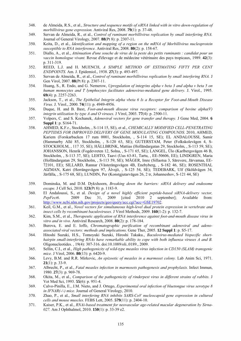
135
348. de Almeida, R.S., et al., Structure and sequence motifs of siRNA linked with in vitro down-regulation of morbillivirus gene expression. Antiviral Res, 2008. 79(1): p. 37-48.
349. Servan de Almeida, R., et al., Control of ruminant morbillivirus replication by small interfering RNA. Journal of General Virology, 2007. 88(Pt 8): p. 2307-11.
350. Keita, D., et al., Identification and mapping of a region on the mRNA of Morbillivirus nucleoprotein susceptible to RNA interference. Antiviral Res, 2008. 80(2): p. 158-67.
351. Diallo, A., et al., Atténuation d'une souche de virus de la peste des petits ruminants : candidat pour un vaccin homologue vivant. Revue d'élevage et de médecine vétérinaire des pays tropicaux, 1989. 42(3): p. 311-319.
352. REED, L.J. and H. MUENCH, A SIMPLE METHOD OF ESTIMATING FIFTY PER CENT ENDPOINTS. Am. J. Epidemiol., 1938. 27(3): p. 493-497.
353. Servan de Almeida, R., et al., Control of ruminant morbillivirus replication by small interfering RNA. J Gen Virol, 2007. 88(Pt 8): p. 2307-11.
354. Huang, S., R. Endo, and G. Nemerow, Upregulation of integrins alpha v beta 3 and alpha v beta 5 on human monocytes and T lymphocytes facilitates adenovirus-mediated gene delivery. J. Virol., 1995. 69(4): p. 2257-2263.
355. Jackson, T., et al., The Epithelial Integrin alpha vbeta 6 Is a Receptor for Foot-and-Mouth Disease Virus. J. Virol., 2000. 74(11): p. 4949-4956.
356. Duque, H. and B. Baxt, Foot-and-mouth disease virus receptors: comparison of bovine alpha(V) integrin utilization by type A and O viruses. J Virol, 2003. 77(4): p. 2500-11.
357. Volpers, C. and S. Kochanek, Adenoviral vectors for gene transfer and therapy. J Gene Med, 2004. 6 Suppl 1: p. S164-71.
358. AHMED, K.F.r., Stockholm, , S-114 15, SE), et al., CHEMICALLY MODIFIED CELL-PENETRATING PEPTIDES FOR IMPROVED DELIVERY OF GENE MODULATING COMPOUNDS. 2010, AHMED, Kariem (Forskarbacken 17 rum 0001, Stockholm, , S-114 15, SE); EL ANDALOUSSI, Samir (Hammarby Allé 85, Stockholm, , S-120 63, SE); GUTERSTAM, Peter (Folkskolevägen 4, S-STOCKHOLM, , 117 35, SE); HÄLLBRINK, Mattias (Holländargatan 29, Stockholm, , S-113 59, SE); JOHANSSON, Henrik (Fogdevreten 12, Solna, , S-171 65, SE); LANGEL, Ülo (Karlbergsvägen 46 B, Stockholm, , S-113 37, SE); LEHTO, Taavi (Uus 63-81, Tartu, , EE-50606, EE); LINDGREN, Maria (Holländargatan 29, Stockholm, , S-113 59, SE); MÄGER, Imre (Sillaotsa 3, Särevere, Järvamaa, EE-72101, EE); SILLARD, Rannar (Västerängsvägen 4B, Enebyberg, , S-182 46, SE); ROSENTHAL AIZMAN, Katri (Herrängsvägen 97, Älvsjö, , S-125 54, SE); TEDEBARK, Ulf (Sköldvägen 34, Järfälla, , S-175 68, SE); LUNDIN, Per (Konstgjutarvägen 26, 2 tr, Johanneshov, S-121 44, SE)
359. Dominska, M. and D.M. Dykxhoorn, Breaking down the barriers: siRNA delivery and endosome
escape. J Cell Sci, 2010. 123(Pt 8): p. 1183-9. 360. El Andaloussi, S., et al. Design of a novel highly efficient peptide-based siRNA-delivery vector,
PepFect6. 2009 Dec 31, 2009 [cited 2010 2 september]; Available from: http://www.ncbi.nlm.nih.gov/projects/geo/query/acc.cgi?acc=GSE19702.
361. Keil, G.M., et al., Novel vectors for simultaneous high-level dual protein expression in vertebrate and insect cells by recombinant baculoviruses. J Virol Methods, 2009. 160(1-2): p. 132-7.
362. Kim, S.M., et al., Therapeutic application of RNA interference against foot-and-mouth disease virus in vitro and in vivo. Antiviral Research, 2008. 80(2): p. 178-184.
363. Burova, E. and E. Ioffe, Chromatographic purification of recombinant adenoviral and adeno-associated viral vectors: methods and implications. Gene Ther, 2005. 12 Suppl 1: p. S5-17.
364. Hitoshi Suzuki, H.S., Tomoyuki Suzuki, Hiroshi Takaku., Baculovirus-mediated bispecific short-hairpin small-interfering RNAs have remarkable ability to cope with both influenza viruses A and B. Oligonucleotides. , 19(4): 307-316. doi:10.1089/oli..0189., 2009.
365. Sellin, C.I., et al., High pathogenicity of wild-type measles virus infection in CD150 (SLAM) transgenic mice. J Virol, 2006. 80(13): p. 6420-9.
366. Levy, B.M. and R.R. Mirkovic, An epizootic of measles in a marmoset colony. Lab Anim Sci, 1971. 21(1): p. 33-9.
367. Albrecht, P., et al., Fatal measles infection in marmosets pathogenesis and prophylaxis. Infect Immun, 1980. 27(3): p. 969-78.
368. Okita, M., et al., Comparison of the pathogenicity of rinderpest virus in different strains of rabbits. J Vet Med Sci, 1993. 55(6): p. 951-4.
369. Calvo-Pinilla, E., J.M. Nieto, and J. Ortego, Experimental oral infection of bluetongue virus serotype 8 in IFNAR(-/-) mice. Journal of General Virology, 2010.
370. Zhao, P., et al., Small interfering RNA inhibits SARS-CoV nucleocapsid gene expression in cultured cells and mouse muscles. FEBS Lett, 2005. 579(11): p. 2404-10.
371. Kaiser, P.K., et al., RNAi-based treatment for neovascular age-related macular degeneration by Sirna-027. Am J Ophthalmol, 2010. 150(1): p. 33-39 e2.

136
372. David, S., et al., Non-viral nanosystems for systemic siRNA delivery. Pharmacol Res, 2010. 62(2): p. 100-14.
373. Sliva, K. and B.S. Schnierle, Selective gene silencing by viral delivery of short hairpin RNA. Virol J, 2010. 7: p. 248.
374. Liu, Z., et al., siRNA delivery into human T cells and primary cells with carbon-nanotube transporters. Angewandte Chemie-International Edition, 2007. 46(12): p. 2023-2027.
375. Parrott, M.B., et al., Metabolically biotinylated adenovirus for cell targeting, ligand screening, and vector purification. Mol Ther, 2003. 8(4): p. 688-700.
376. Desmyter, J., J.L. Melnick, and W.E. Rawls, Defectiveness of interferon production and of rubella virus interference in a line of African green monkey kidney cells (Vero). J Virol, 1968. 2(10): p. 955-61.
377. Emeny, J.M. and M.J. Morgan, Regulation of the Interferon System: Evidence that Vero Cells have a Genetic Defect in Interferon Production 10.1099/0022-1317-43-1-247. J Gen Virol, 1979. 43(1): p. 247-252.
378. Matskevich, A.A., et al., Vero Cells as a Model to Study the Effects of Adenoviral Gene Delivery Vectors on the RNAi System in Context of Viral Infection. Journal of Innate Immunity, 2009. 1(4): p. 389-394.
379. Calvo-Pinilla, E., et al., Establishment of a Bluetongue Virus Infection Model in Mice that Are Deficient in the Alpha/Beta Interferon Receptor. PLoS One, 2009. 4(4): p. e5171.
380. Sellin, C.I., et al., High pathogenicity of wild-type measles virus infection in CD150 (SLAM) transgenic mice. Journal of Virology, 2006. 80(13): p. 6420-6429.