universidade federal da paraÍba centro de tecnologia … · 2018-09-06 · a moringa oleífera...
TRANSCRIPT

UNIVERSIDADE FEDERAL DA PARAÍBA
CENTRO DE TECNOLOGIA
PROGRAMA DE PÓS-GRADUAÇÃO EM CIÊNCIA E
TECNOLOGIA DE ALIMENTOS
JAQUELINE AZEVEDO NASCIMENTO BATISTA
POTENCIAL ANTIOXIDANTE DOS EXTRATOS DE
MORINGA OLEIFERA LAMARK EM SISTEMAS LIPÍDICOS
DE BAIXA ESTABILIDADE OXIDATIVA
JOÃO PESSOA – PB
2013

JAQUELINE AZEVEDO NASCIMENTO BATISTA
POTENCIAL ANTIOXIDANTE DOS EXTRATOS DE
MORINGA OLEIFERA LAMARK EM SISTEMAS LIPÍDICOS
DE BAIXA ESTABILIDADE OXIDATIVA
JOÃO PESSOA – PB
2013

JAQUELINE AZEVEDO NASCIMENTO BATISTA
POTENCIAL ANTIOXIDANTE DOS EXTRATOS DE
MORINGA OLEIFERA LAMARK EM SISTEMAS LIPÍDICOS
DE BAIXA ESTABILIDADE OXIDATIVA
Tese apresentada ao Programa de
Pós-Graduação em Ciência e
Tecnologia de Alimento, Centro de
Tecnologia, Universidade Federal
da Paraíba, em cumprimento aos
requisitos para obtenção do título de
Doutor em Ciência e Tecnologia de
Alimentos.
Orientador: Prof. Dr. Antônio Gouveia de Souza
JOÃO PESSOA – PB
2013

B333p Batista, Jaqueline Azevedo Nascimento.
Potencial antioxidante dos extratos de Moringa oleífera
Lamark em sistemas lipídicos de baixa estabilidade
oxidativa / Jaqueline Azevedo Nascimento Batista.- João
Pessoa, 2013.
95f. : il.
Orientador: Antônio Gouveia de Souza
Tese (Doutorado) – UFPB/CT
1. Tecnologia de Alimentos. 2. Moringa oleífera Lam.
3. Óleo de peixe. 4. Óleo de soja. 5. Estabilidade oxidativa.
UFPB/BC CDU: 664(043)
JAQUELINE AZEVEDO NASCIMENTO BATISTA
POTENCIAL ANTIOXIDANTE DOS EXTRATOS DE MORINGA OLEIFERA
LAMARK EM SISTEMAS LIPÍDICOS DE BAIXA ESTABILIDADE OXIDATIVA
Tese APROVADA em 25/02/2013.
BANCA EXAMINADORA
Prof. Dr. Antônio Gouveia de Souza – DQ/UFPB
Coordenador da Banca Examinadora
Profa. Dra. Marciane Magnani – DEA/UFPB
Examinador Interno
Profa. Dra. Rita de Cássia Ramos do Egypto Queiroga – DN/UFPB
Examinador Interno
Prof. Dr. Raul Rosenhaim – DEQ/UFPB
Examinador Externo
Profa. Dra. Marta Maria da Conceição – CES/UFCG
Examinador Externo
À Deus, fonte de toda minha força e
inspiração.
A Renê e Ramon, meus amados filhos, que me
alegram e motivam para aceitar os desafios.
Aos meus pais Elimar e Fátima que sempre
investiram e confiaram em mim.
Ao meu amado esposo Renato pelo incentivo e
paciência.
A minhas irmãs Janine, Alcerita e Alessandra,
companheiras e conselheiras.
Aos meus avos Alcebíades e Aurita, sempre
presentes, dando lições de amor e sabedoria.
Dedico.
AGRADECIMENTO
Agradeço a Deus, principio de todas as coisas, luz inspiradora de todas as horas, alento
nos momentos mais difíceis, esperança de importantes realizações e motivo de louvor e
exaltação.
Ao Prof. Dr. Antônio Gouveia de Souza, pela receptividade, confiança, incentivo,
amizade e pela orientação cercada de seriedade e de rigor cientifico, que foram
imprescindíveis tanto para realização deste trabalho como para construção de um valoroso
conhecimento, subsídio importante na atuação profissional. Serei eternamente grata...
A Profa. Dra. Antônia Lúcia de Souza pela co-orientação deste trabalho, capacidade e
competência, além de sua disposição ímpar em ajudar.
A Profa. Dra. Neide Queiroz, pela competência, ensinamentos e disponibilização.
A Profa. Dra. Marciane Magnani, pela sua importante contribuição na correção dos
artigos, orientações e amizade.
A Profa. Dra. Alessandra Azevedo do Nascimento, minha querida irmã, pela preciosa
contribuição nos ensaios de avaliação da bioatividade.
As amigas e companheiras de todas as horas, Kassandra, Poliana e Alline pela
amizade e importante ajuda na realização deste trabalho.
As amigas Ângela, Teta, Andréa e Maristela pela valorosa contribuição, competência
e amizade.
Aos amigos de pós-graduação do Laboratório de Combustíveis e Materiais – LACOM
contemporâneos durante o período do meu doutoramento e a todos os amigos de iniciação
científica e voluntários pela ajuda e amizade.
A amiga Ruth Gomes Gadelha, nestes quase dez anos de convivência desde o
mestrado até o Doutorado, sempre disposta a ajudar e repassar seus conhecimentos.

A Maria Lucia Braga de Carvalho, pelo excelente trabalho realizado no laboratório
de análises térmicas.
A Humberto Bandeira (Secretario da Pós-graduação) pelo seu trabalho desempelhado
com dedicação na secretaria e pela atenção e amizade em momentos difíceis.
A Coordenação, funcionários e a todos os professores que compõem o Programa de
Pós-Graduação em Ciência e Tecnologia de Alimentos, pela competência, seriedade,
honestidade, incentivo e apoio.
A todos que fazem o LACOM;
MUITO OBRIGADA.

“A persistência é o menor caminho do êxito”
Charles Chaplin
RESUMO
A Moringa oleífera Lamark é uma planta perene e tolerante a seca cultivada como planta
ornamental e medicinal. No presente estudo, os extratos etanólicos das folhas, flores, cascas
das vagens e sementes desta planta foram avaliados quanto a sua proteção contra a oxidação
dos óleos de soja e de peixe. Para isso, foram determinados o teor de fenólicos extraíveis
totais (FET) através do método Folin-Ciocalteau e o potencial antioxidante, empregando-se os
métodos de sequestro do radical DPPH (RSA-DPPH), poder de redução do ferro (FRAP) e
sistema β-caroteno/ácido linoléico. A estabilidade térmica dos referidos extratos foi avaliada
através da análise térmica (TG/DTA) e o efeito antioxidante investigado nos óleos de soja e de
peixe através dos métodos Rancimat, calorimetria exploratória diferencial pressurizada (P-
DSC) e teste de estocagem acelerada em estufa. A avaliação preliminar da toxicidade dos
extratos foi realizada utilizando larvas de Artemia salina. Os teores FET nos extratos das
folhas, flores, casca das vagens e sementes foram (53,69 ± 1,00); (45,85 ± 1,71); (41,75 ±
3,35) e (8,06 ± 0,47) mg GAE/g de extrato, respectivamente. Nos ensaios de determinação da
capacidade antioxidante, o extrato das folhas apresentou melhor RSA-DPPH, FRAP e
também maior atividade antioxidante no sistema β-caroteno/ácido linoleico. Na avaliação da
estabilidade térmica, o extrato das folhas também se mostrou mais estável do que os demais
extratos. Nos ensaios de avaliação do efeito antioxidante dos extratos, verificou-se que o
extrato das folhas foi o mais eficiente em ambos os óleos nos métodos Rancimat e PDSC,
com efeito protetor equivalente ao antioxidante sintético BHT no óleo de soja na técnica
Rancimat. No teste de estocagem acelerada em estufa, foi verificado que após 16 dias de
armazenamento o extrato das folhas foi o mais eficaz na inibição da formação dos produtos da
oxidação primária e secundária em ambos os óleos, sendo mais eficiente do que o BHT e
TBHQ no óleo de peixe. Os resultados da avaliação preliminar de toxicidade indicaram uma
baixa toxicidade dos extratos, sugerindo que os mesmos podem ser consumidos sem prejuízo
à saúde. Estes resultados sugerem que o extrato etanólico das folhas de moringa possui efeito
protetor eficiente quando aplicado a sistemas lipídicos de baixa estabilidade oxidativa,
podendo vir a ser uma fonte alternativa de potencial aplicação na indústria de óleos e
gorduras.
Palavras-chaves: Moringa oleífera Lam., óleo de peixe, óleo de soja, estabilidade oxidativa.
ABSTRACT
The species Moringa oleifera Lamarck is a perennial and drought-tolerant plant cultivated as
ornamental and medicinal plant. In this study, ethanolic extracts of leaves, flowers, and seed
pods of this plant were evaluated for their protective effect against soybean and fish oil
oxidation. For this, total extractable phenolics (TEP) were determined by Folin-Ciocalteau
method, as well as the antioxidant potential, using the DPPH radical scavenging activity
method (RSA-DPPH), iron power reduction (FRAP) and β-carotene/ linoleic acid system. The
thermal stability of extracts was measured by thermal analysis (TG / DTA) and the
antioxidant effect on soybean and fish oils was investigated by the Rancimat method,
pressurized differential scanning calorimetry (P-DSC) and accelerated storage test. The
preliminary toxicity assessment of extracts was performed using Artemia salina larvae. The
TEP levels in extracts of leaves, flowers and seed pods were (53.69 ± 1.00), (45.85 ± 1.71),
(41.75 ± 3.35) and (8.06 ± 0.47) mg GAE / g of extract, respectively. In tests to determine the
antioxidant capacity, extract from leaves showed better RSA-DPPH and FRAP and also
higher antioxidant activity in the β-carotene/ linoleic acid system. In the thermal stability
evaluation, extract from leaves was also more stable than the other extracts. In tests to
evaluate the antioxidant effect of extracts, it was found that extract from leaves was more
efficient in both oils in Rancimat and P-DSC methods with protective effect equivalent to
synthetic antioxidant BHT in soybean oil using the Rancimat method. In the accelerated
storage test, it was found that after 16 days of storage, extract from leaves was the most
effective in inhibiting the formation of oxidation products in both oils, being more effective
than BHT and TBHQ in fish oil. The results of the preliminary toxicity assessment showed
low toxicity, suggesting that the extracts may be consumed without health damage. These
results suggest that the ethanolic extract of moringa leaves has efficient protective effect when
applied to lipid systems of low oxidative stability and could be an alternative source of
potential application in the oils and fats industry.
Keywords: Moringa oleifera Lam., fish oil, soybean oil, oxidative stability.
LISTA DE ILUSTRAÇÕES
Figura 1 Moringa oleífera Lam. com suas folhas, flores, vagens e sementes 18
Figura 2 Mecanismo de ação dos antioxidantes primários 27
Figura 3 Estrutura fenólica de antioxidantes sintéticos 29
Figura 4 Estrutura química dos ácidos benzoicos 31
Figura 5 Estrutura química dos principais ácidos cinâmicos 31
Figura 6 Estrutura química das cumarinas 31
Figura 7 Estrutura química dos flavonoides 32

LISTA DE ABREVIATURAS E SIGLAS
AA Ácido araquidônico
BHA Butil hidroxianisol
BHT Butil hidroxitolueno
BST Brine shrimp lethality test
CD Dienos conjugados
CL50 Concentração letal média
DHA Ácido docosahexaenóico
DPPH 2,2-difenil-1-picrilidrazil
DSC Calorimetria exploratória diferencial
DTA Análise Térmica Diferencial
EDTA Ácido etileno diamino tetra acético
EF Extrato etanólicos das flores
EL Extrato etanólicos das folhas
EP Extrato etanólicos das cascas das vagens
EPA Ácido eicosapentaenoico
ES Extrato etanólicos das sementes
FRAP Poder redutor do ferro
FTIR Infravermelho com transformada de Fourier
GAE Equivalente de ácido gálico
IA Índice de anisidina
OIT Tempo de indução oxidativa
OSI Indice de estabilidade oxidativa
P-DSC Calorimetria Exploratória Diferencial Pressurizada
PG Propil galato
PI Período de indução
TBARS Ácidos tiobarbitúrico-reactivos
TBHQ terc-butilhidroquinona
TEP Fenólicos extraíveis totais
TG Termogravimetria
SUMÁRIO
1 INTRODUÇÃO.............................................................................................................. 15
1.1 OBJETIVO GERAL...................................................................................................... 17
1.2 OBJETIVOS ESPECÍFICOS........................................................................................ 17
2 REVISÃO DE LITERATURA...................................................................................... 18
2.1 MORINGA OLEIFERA LAM........................................................................................ 18
2.2 ÓLEOS DE SOJA E DE PEIXE................................................................................... 21
2.3 OXIDAÇÃO LIPÍDICA................................................................................................ 23
2.4 AVALIAÇÃO DA ESTABILIDADE OXIDATIVA DE ÓLEOS............................... 24
2.4.1 Teste de estocagem acelerada em estufa ou “Schaal Oven Test”......................... 25
2.4.2 Rancimat................................................................................................................... 25
2.4.3 Calorimetria exploratória diferencial pressurizada (P – DSC)........................... 26
2.5 ANTIOXIDANTES....................................................................................................... 27
2.5.1 Antioxidantes sintéticos............................................................................................ 28
2.5.2 Antioxidantes naturais............................................................................................. 30
2.6 BIOATIVIDADE E TOXICIDADE FRENTE À ARTEMIA SALINA......................... 32
3 MATERIAL E MÉTODOS........................................................................................... 34
3.1 MATERIAL.................................................................................................................. 34
3.2 AQUISIÇÃO E PREPARO DAS AMOSTRAS........................................................... 34
3.3 OBTENÇÃO DOS EXTRATOS.................................................................................. 34
3.4 DETERMINAÇÃO DE FENÓLICOS EXTRAÍVEIS TOTAIS.................................. 35
3.5 ESTUDO TÉRMICO DOS EXTRATOS..................................................................... 35
3.6 DETERMINAÇÃO DA ATIVIDADE ANTIOXIDANTE.......................................... 35
3.6.1 Atividade sequestrante do radical livre DPPH...................................................... 35
3.6.2 Capacidade antioxidante pelo método de redução do ferro (FRAP)................... 36
3.6.3 Auto-oxidação do sistema β-caroteno/ácido linoleico............................................ 36
3.7 BIOENSAIO COM ARTEMIA SALINA LEACH......................................................... 37
3.8 DETERMINAÇÃO DA ESTABILIDADE OXIDATIVA........................................... 38
3.8.1 Preparação das amostras......................................................................................... 38
3.8.2 Método Rancimat..................................................................................................... 38
3.8.3 Calorimetria exploratória diferencial pressurizada (P – DSC)........................... 38
3.8.4 Teste de estocagem acelerada em estufa................................................................. 39

3.8.4.1 Índice de peróxido................................................................................................... 39
3.8.4.2 Índice de ρ-anisidina............................................................................................... 40
3.8.4.3 Determinação do valor de dienos conjugados (CD)............................................... 40
3.8.4.4 Infravermelho com transformada de Fourier (FTIR)............................................. 41
3.8 ANÁLISES ESTATÍSTICAS....................................................................................... 41
REFERÊNCIAS................................................................................................................ 42
4 RESULTADOS............................................................................................................... 50
ARTIGO 1 - Ethanolic extracts of Moringa oleifera Lam.: Evaluation of its potential as
an antioxidant additive for fish oil....................................................................................... 51
ARTIGO 2 - Ethanolics extracts of moringa: antioxidant effect in soybean oil by P-DSC and
Rancimat .............................................................................................................................. 57
ARTIGO 3 - Oxidative stability of fish oil supplemented with ethanolic extracts of
Moringa oleifera Lam. compared with synthetic antioxidants during accelerated
test........................................................................................................................................ 65
ARTIGO 4 - Antioxidant effect of Moringa oleifera Lamark extracts in soybean oil during
accelerated storage test........................................................................................................... 76
ARTIGO 5 - Study of antioxidant activity and preliminary bioactivity determination of
ethanolic extracts of Moringa oleifera Lam........................................................................ 86
5 CONSIDERAÇÕES FINAIS........................................................................................ 95

15
1 INTRODUÇÃO
A Moringa oleifera Lamark, é uma planta originária do noroeste da Índia (BEZERRA;
MOMENTÉ; MEDEIROS FILHO, 2004) que têm sido referida como fonte de proteínas, β-
caroteno, vitamina C, cálcio e potássio, além de compostos fenólicos e carotenóides. A
moringa apresenta uma combinação rica e rara de zeatina, quercetina, β - sitosterol, ácido
cafeoilquínico e kaempferol, o que atribui também à planta uma potencial atividade
antioxidante ( ANWAR et al., 2007; BARRETO et al., 2009; IQBAL; BHANGER, 2006;
LAKO et al., 2007; REDDY; UROOJ; KUMAR, 2005; SIDDHURAJU; BECKER, 2003). É
perfeitamente adaptável ao clima do Nordeste brasileiro, pois, apresenta bom crescimento,
mesmo quando submetido a condições de seca e elevadas temperaturas, características desta
região (SILVA, et al., 2012).
A importância dos lipídios na nutrição e desenvolvimento humano é reconhecida há
muitas décadas, pois os ácidos graxos são constituintes estruturais das membranas celulares,
cumprem funções energéticas e de reservas metabólicas, além de formarem hormônios e sais
biliares. O homem é capaz de sintetizar todos os ácidos graxos, exceto os essenciais ácido
linoléico (ω-6) e o ácido linolênico (ω-3), que devem ser fornecidos pela dieta (KEY et al.,
2012). Estudos apontam que a ingestão regular de ácidos graxos poli-insaturados traz
benefícios para a saúde humana, prevenindo enfermidades cardiovasculares, câncer de cólon e
doenças imunológicas, além de favorecerem o desenvolvimento cerebral e da retina (BALK et
al., 2006; KNOTHE, 2008; NEMETS et al., 2006; PIVIK et al., 2009).
O óleo de soja é composto por 12 a 15% de ácidos graxos saturados (na maior parte o
palmítico) e 85 a 88% de ácidos graxos insaturados, com destaque para oléico, linoleico e
linolênico (RIBEIRO et al., 2009). No Brasil, o óleo de soja responde por cerca de 80,6 % do
consumo de óleos vegetais, sendo utilizado preferencialmente como óleo de fritura (USDA,
2011).
O óleo de peixe apresenta um alto teor dos ácidos graxos poli-insaturados, contendo
cerca de 18% de ácido eicosapentaenóico (EPA) e 12% de ácido docosahexaenóico (DHA)
(TAMJIDI; NASIRPOUR; SHAHEDI, 2012). Tal composição lhe confere uma notável
importância, já que é um dos únicos alimentos que aparece como fonte expressiva destes
ácidos graxos (BALK et al., 2006).
A presença de alto teor de ácidos graxos poli-insaturados torna esses óleos
extremamente susceptíveis a processos oxidativos durante o processamento e estocagem. A

16
oxidação lipídica compromete a integridade das ligações duplas desses ácidos graxos,
alterando sua concentração e funcionalidade, e representando risco à saúde humana, visto que
o consumo de produtos da peroxidação lipídica pode induzir a carcinogênese (HALVORSEN;
BLOMHOFF, 2011).
Para manter a qualidade e prolongar o tempo de vida útil dos óleos, o processo de
oxidação lipídica é usualmente prevenido através do emprego de antioxidantes sintéticos
como butilhidroxianisol (BHA), butilhidroxitolueno (BHT) e terc-butilhidroquinona (TBHQ).
Estas substâncias, entretanto, apesar de serem bastante eficazes como antioxidantes, têm seu
uso restrito devido efeitos tóxicos e cancerígenos em seres humanos (SUN-WATERHOUSE;
THAKORLAL; ZHOU, 2011). No Brasil, a concentração máxima de TBHQ e BHT permitida
é de 200 mg.kg-1
e 100 mg.kg-1
, respectivamente (BRASIL, 1988). Neste contexto,
antioxidantes provenientes de fontes naturais têm sido estudados, buscando obter produtos
mais seguros e eficientes que possam ser usados em alimentos, substituindo total ou
parcialmente os antioxidantes sintéticos (BREWER, 2011).
Considerando o potencial antioxidante da moringa, torna-se relevante investigar a sua
potencialidade como fonte de aditivo antioxidante em sistemas lipídicos de baixa estabilidade
oxidativa, tais como o óleo de soja, óleo mais consumido no Brasil, e o óleo de peixe, fonte
nutricional singular.

17
1.1 OBJETIVO GERAL
Avaliar o potencial antioxidante in vitro de extratos etanólicos de Moringa oleífera
Lam. e analisar seus efeito na estabilidade oxidativa dos óleos de soja e de peixe.
1.2 OBJETIVOS ESPECÍFICOS
Obter os extratos etanólicos das folhas (EL), flores (EF), cascas das vagens (EP) e
sementes (ES) de Moringa oleífera Lam.;
Avaliar o potencial antioxidante dos extratos etanólicos EL, EF, EP e ES através da
determinação do conteúdo de fenólicos extraíveis totais (TEP) pelo método Folin-Ciocalteau;
Determinar a capacidade antioxidante dos extratos EL, EF, EP e ES utilizando os
métodos: sequestro do radical livre DPPH, poder redutor do ferro (FRAP) e oxidação
acoplada ao sistema β-caroteno/ácido linoleico;
Avaliar através dos métodos acelerados Rancimat, Calorimetria Exploratória
Diferencial Pressurizada (P-DSC) e teste de estocagem acelerada em estufa o efeito
antioxidante dos extratos que apresentarem boa capacidade antioxidante nos testes de
sequestro do radical livre DPPH, poder redutor do ferro (FRAP) e oxidação acoplada ao
sistema β-caroteno/ácido linoleico, quando aplicados nos óleos de soja e de peixe.
Avaliar a bioatividade e toxicidade preliminar dos extratos EL, EF, EP e ES através do
teste de letalidade da Artemia salina.

18
2 REVISÃO DE LITERATURA
2.1 MORINGA OLEIFERA LAM
A Moringa oleífera Lamarck é uma árvore nativa do noroeste da Índia, sendo
cultivada ao longo dos trópicos. Trata-se de uma planta perene, amplamente distribuída nos
países da Ásia, Oriente médio e da África, podendo também ser encontrada na América
Central e América do Sul (BEZERRA; MOMENTÉ; MEDEIROS FILHO, 2004). É bem
tolerante à seca, florescendo e produzindo frutos mesmo com escassez de chuvas
(RAMACHANDRAN; PETER; GOPALAKRISHNAN, 1980).
Foi introduzida no Brasil por volta de 1950, sendo cultivada como planta ornamental e
medicinal, principalmente na região Nordeste, onde é conhecida como lírio-branco, quiabo de
quina ou simplesmente moringa (SILVA, et al., 2012). Pode atingir 10 metros de altura e
apresenta folhas grandes e flores perfumadas brancas ou creme, vagens longas, variando de
verde a marrom esverdeada, contendo de 10 a 20 sementes globoides (Figura 1). Em
condições ideais de cultivo, a moringa começa a frutificar entre o primeiro e o segundo ano
(TSAKNIS, et al., 1999).
Figura 1. Moringa oleífera Lam. com suas folhas (a), flores (b), vagens (c) e sementes (d)
19
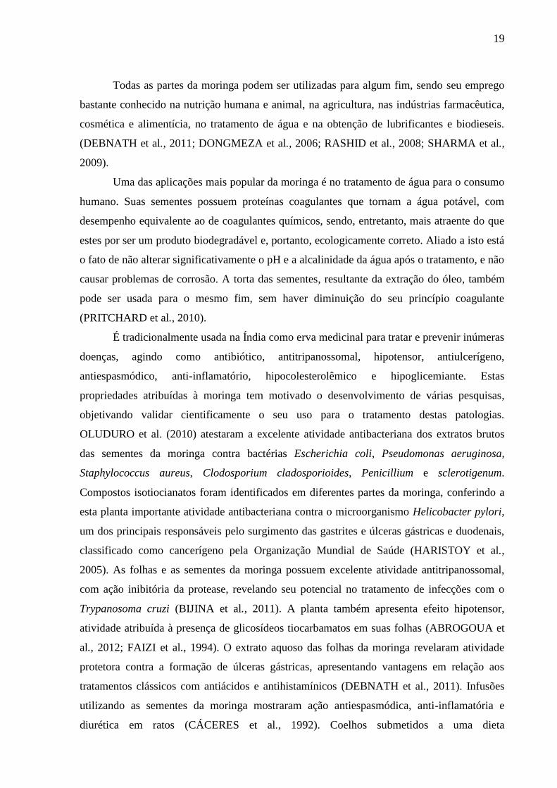
Todas as partes da moringa podem ser utilizadas para algum fim, sendo seu emprego
bastante conhecido na nutrição humana e animal, na agricultura, nas indústrias farmacêutica,
cosmética e alimentícia, no tratamento de água e na obtenção de lubrificantes e biodieseis.
(DEBNATH et al., 2011; DONGMEZA et al., 2006; RASHID et al., 2008; SHARMA et al.,
2009).
Uma das aplicações mais popular da moringa é no tratamento de água para o consumo
humano. Suas sementes possuem proteínas coagulantes que tornam a água potável, com
desempenho equivalente ao de coagulantes químicos, sendo, entretanto, mais atraente do que
estes por ser um produto biodegradável e, portanto, ecologicamente correto. Aliado a isto está
o fato de não alterar significativamente o pH e a alcalinidade da água após o tratamento, e não
causar problemas de corrosão. A torta das sementes, resultante da extração do óleo, também
pode ser usada para o mesmo fim, sem haver diminuição do seu princípio coagulante
(PRITCHARD et al., 2010).
É tradicionalmente usada na Índia como erva medicinal para tratar e prevenir inúmeras
doenças, agindo como antibiótico, antitripanossomal, hipotensor, antiulcerígeno,
antiespasmódico, anti-inflamatório, hipocolesterolêmico e hipoglicemiante. Estas
propriedades atribuídas à moringa tem motivado o desenvolvimento de várias pesquisas,
objetivando validar cientificamente o seu uso para o tratamento destas patologias.
OLUDURO et al. (2010) atestaram a excelente atividade antibacteriana dos extratos brutos
das sementes da moringa contra bactérias Escherichia coli, Pseudomonas aeruginosa,
Staphylococcus aureus, Clodosporium cladosporioides, Penicillium e sclerotigenum.
Compostos isotiocianatos foram identificados em diferentes partes da moringa, conferindo a
esta planta importante atividade antibacteriana contra o microorganismo Helicobacter pylori,
um dos principais responsáveis pelo surgimento das gastrites e úlceras gástricas e duodenais,
classificado como cancerígeno pela Organização Mundial de Saúde (HARISTOY et al.,
2005). As folhas e as sementes da moringa possuem excelente atividade antitripanossomal,
com ação inibitória da protease, revelando seu potencial no tratamento de infecções com o
Trypanosoma cruzi (BIJINA et al., 2011). A planta também apresenta efeito hipotensor,
atividade atribuída à presença de glicosídeos tiocarbamatos em suas folhas (ABROGOUA et
al., 2012; FAIZI et al., 1994). O extrato aquoso das folhas da moringa revelaram atividade
protetora contra a formação de úlceras gástricas, apresentando vantagens em relação aos
tratamentos clássicos com antiácidos e antihistamínicos (DEBNATH et al., 2011). Infusões
utilizando as sementes da moringa mostraram ação antiespasmódica, anti-inflamatória e
diurética em ratos (CÁCERES et al., 1992). Coelhos submetidos a uma dieta

20
hipercolesterolêmica e tratados por 12 semanas com extrato aquoso das folhas da moringa,
apresentaram redução nos níveis de colesterol em cerca de 50% e redução na formação de
placas ateroscleróticas em cerca de 86%, sendo esses efeitos de grau comparável aos da
sinvastatina (CHUMARK et al., 2008). As folhas também demonstraram capacidade em
promover uma redução considerável dos níveis de glicose no sangue e na urina, com aumento
da proteína sérica total, do peso corporal e da hemoglobina e redução da proteinúria, sendo
indicada para o tratamento do diabetes mellitus (JAISWAL et al., 2009). Extratos aquosos e
alcoólicos da raiz da moringa demonstraram sua eficiência no tratamento de litíase urinária de
oxalato de cálcio (KARADI et al., 2006).
A Moringa oleifera Lam. é uma boa alternativa na alimentação humana e animal, pois
apresenta importantes minerais, como o ferro, além de ser fonte de vitaminas, como o beta-
caroteno e de aminoácidos como a metionina e cistina, geralmente deficientes em outros
alimentos (ANWAR et al., 2007; MAKKAR; BECKER, 1996).
Mohammed et al. (2011), ao utilizarem as folhas frescas da moringa como suplemento
alimentar em galinhas, observaram o aumento da taxa de postura de ovos e a melhoria na
qualidade dos mesmos, com melhora na conversão alimentar e na cor da gema, quando
comparado ao controle, sugerindo que tal planta pode ser usadas como recurso alimentar
sustentável para galinhas criadas em áreas tropicais. Já o uso da farinha das sementes
desengorduradas da Moringa oleifera como aditivo em dietas de ovinos relevou resultados
que sugerem que este aditivo tem potencial para melhorar a fermentação no rúmen e a taxa de
crescimento dos cordeiros alimentados com feno-SBM (BEN SALEM; MAKKAR, 2009).
Com relação ao óleo extraído das sementes da moringa, o mesmo possui alta
resistência à oxidação por conter em sua composição o ácido graxo saturado behênico, apesar
de possuir elevados teores de ácidos graxos insaturados, principalmente o oleico, tendo
inclusive composição quimicamente semelhante ao azeite de oliva (TSAKNIS, JOHN;
LALAS, 2002).
A moringa possui excelente potencial como antioxidante, pois apresenta entre seus
constituintes químicos os ácidos fenólicos (ácido gálico, ácido clorogênico, ácido elágico e
ácido ferúlico), além dos flavonoides (campferol, quercetina e rutina), substâncias com
atividade antioxidante relevante atribuída à capacidade de sequestrarem radicais, além de
serem excelentes quelantes de metais (VERMA et al., 2009).
A atividade antioxidante in vitro de extratos aquosos de folhas da moringa em dois
estágios de maturação foi estudada, e os resultados mostraram que os extratos são potentes
fontes de substâncias sequestradoras de radicais livres, com ação comparável a de

21
antioxidantes de referência, sugerindo que esses extratos são capazes de prevenir os danos
oxidativos no organismo humano (SREELATHA; PADMA, 2009).
A presença de ácido gálico, ácido clorogénico, ácido elágico, ácido ferúlico,
kaempferol, quercetina e vanilina em extratos aquosos das folhas, frutos e sementes da
Moringa oleifera foi revelada através de análises em HPLC e MS, sendo o extrato da folha o
mais rico em compostos fenólicos totais, flavonóides e ácido ascórbico, com consequente
melhor atividade antioxidante (SINGH et al., 2009). A moringa é também uma excelente
fonte de tocoferóis (SÁNCHEZ-MACHADO; LÓPEZ-CERVANTES; VÁZQUEZ, 2006).
Hazra et al. (2012), avaliando o efeito dos extratos brutos das folhas de Moringa
oleifera sob a carne cozida de búfalo, evidenciou efeito antioxidante e antimicrobiano, além
de melhoria na qualidade da carne, aumentando a maciez, suculência e prevenindo a
descoloração.
Arabshahi-D; Vishalakshi Devi; Urooj (2007), avaliaram a atividade antioxidante de
extratos etanólicos de folhas de moringa (Moringa oleifera Lam.), folhas de hortelã (Mentha
spicata) e tubérculo de cenoura (Daucus carota), bem como seu pH e estabilidade durante o
armazenamento e concluiram que estes vegetais são fontes potenciais de compostos
antioxidantes, apresentando atividade antioxidante potente em sistemas lipidicos diferentes, e
que a atividade antioxidante dos extratos variou de acordo com o pH, tratamento térmico e
armazenamento. Tais descobertas confirmam que as propriedades anti e pró-oxidantes de
vegetais são fortemente influenciadas por uma série de fatores de processamento e pelas
condições de reação, sendo importante considerar as ótimas condições tecnológicas e os
fatores de processamento que influenciam a atividade e biodisponibilidade de antioxidantes
vegetais para utilização em alimentos e sistemas biológicos.
2.2 ÓLEOS DE SOJA E DE PEIXE
Por definição, óleos e gorduras são substâncias insolúveis em água e solúveis em
solventes orgânicos. Um óleo se diferencia de uma gordura pelo estado físico a temperatura
de 20 ºC, apresentando os óleos a forma líquida, enquanto que as gorduras apresentam forma
sólida nessa temperatura.
São compostos constituídos por uma mistura de tri, di e monoacilgliceróis, ácidos
graxos livres, glicolipídeos, fosfolipídeos, esteróis e outras substâncias, que apresentam
oxidação distinta, sendo que os ácidos graxos insaturados são os componentes mais

22
susceptíveis ao processo oxidativo (RAMALHO, V.; JORGE, 2006). Podem ser originários
de fontes animais, vegetais ou mesmo microbianas.
Estas substâncias exercem papel fundamental na alimentação humana, pois além de
fornecem calorias, agem como veículos de vitaminas lipossolúveis (A, E, D e K) e de ácidos
graxos essenciais como o linoleico e o linolênico e contribuem para melhorar a palatabilidade
dos alimentos (CASTRO; MENDES; SANTOS, 2004).
Fisiologicamente, atuam na estrutura, composição e permeabilidade das membranas e
das paredes celulares, e são os componentes majoritários do tecido adiposo, isolando o
organismo, protegendo os órgãos internos e contribuindo para a configuração do corpo.
Os óleos e gorduras também desempenham importantes papeis tecnológicos, podendo
ser empregados como emulsificantes, texturizantes, aromatizantes, umectantes e transmissores
de calor a alta temperatura. Segundo a ABIOVE (2012), são processados diariamente cerca de
173.441 toneladas de óleos vegetais no Brasil, incluindo óleo de soja, algodão, canola,
girassol, linhaça e babaçu.
O óleo de soja surgiu como um subproduto do processamento do farelo de soja, e
tornou-se um dos lideres mundiais no mercado de óleos devido as suas qualidades
nutricionais, abundância, valor econômico e larga aplicabilidade na formulação de outros
produtos. Destaca-se pelo alto teor de ácido graxos insaturados, cerca de 85 a 88%
(principalmente oleico, linoleico e linolênico) e apenas 12 a 15% de ácidos graxos saturados
(na maior parte palmítico) (RIBEIRO et al., 2009). Caracteriza-se também por ter vários
componentes menores que podem ser recuperados durante o processo de refino. Estes incluem
os fosfolipídios recuperados como lecitina, esterois mistos, que servem como matéria-prima
para a produção de produtos farmacêuticos valiosos, além dos tocoferóis (vitamina E)
(FERRARI et al., 1996).
A composição e o teor de cada ácido graxo do óleo de soja podem ser afetados pelas
diferenças de variedade e pelos vários fatores geográficos e do meio ambiente, principalmente
das condições climáticas (DAHMER et al., 1989). O teor elevado de ácidos graxos
insaturados e o teor relativamente elevado de ácido linolênico, torna o óleo de soja muito
suscetível às reações de oxidação (RAMALHO; JORGE, 2006).
Os óleos de peixes, em especial os de origem marinha, são ricos em ácidos graxos
poli-insaturados de cadeia longa eicosapentaenoico (EPA) e docosahexaenoico (DHA), sendo
estes conhecidos por seu importante papel na nutrição humana. Esses ácidos graxos, contidos
no óleo de peixe, competem com o ácido araquidônico (AA), nas vias enzimáticas, reduzindo
a formação de mediadores pró-inflamatórios e favorecendo a produção de mediadores com

23
reduzida atividade biológica (FAIZI et al., 1994). Dessa forma o consumo regular de óleos de
peixes trazem inúmeros benefícios ao organismo humano, dentre os quais a proteção contra
doenças como o mal de Alzheimer (LUKIW; BAZAN, 2008), depressão (NEMETS et al.,
2006), declínio cognitivo (MORRIS et al., 2005), artrite reumatoide (BYKERK;
KEYSTONE, 2005), doenças cardiovasculares (KRIS-ETHERTON et al., 2002), além de
ajudar no desenvolvimento do sistema nervoso e no crescimento (PIVIK et al., 2009).
O perfil de ácidos graxos dos peixes varia em função de vários fatores, como a
temperatura do meio ambiente, idade, sexo, espécie, tipo de peixe e principalmente em função
dos perfis de ácidos graxos dos componentes da cadeia alimentar, característicos do
ecossistema das espécies selvagens, ou do perfil de ácidos graxos da ração nos peixes
cultivados (AVERINA; KUTYREV, 2011).
Considera-se que o perfil lipídico constitui a principal diferença entre os peixes de
água doce e os peixes marinhos, mas existem algumas espécies de peixes de água doce que
são especialmente ricas em ácidos graxos ω-3 (AVERINA; KUTYREV, 2011).
2.3 OXIDAÇÃO LIPÍDICA
A oxidação lipídica é um processo afetado por diversos fatores como a sua
composição em ácidos graxos, a temperatura, a luz incidente, a presença de oxigênio, e a ação
de íons metálicos, dentre outros, sendo o fator primordial, a presença de oxigênio (NAZ et al.,
2005; REGITANO-D’ARCE, 2006). Tal processo pode ocorrer por várias vias, dependendo
do meio e dos agentes catalisadores, resultando em uma oxidação enzimática, uma
fotoxidação e/ ou em uma autoxidação, sendo esta última o principal mecanismo de
deterioração lipídica (SCHAICH, 2012).
A oxidação enzimática é resultante da ação das enzimas lipoxigenases sobre os ácidos
graxos poli-insaturados, catalisando a adição de oxigênio à cadeia hidrocarbonada poli-
insaturada, com formação de peróxidos e hidroperóxidos, com duplas ligações conjugadas que
podem envolver-se em diferentes reações degradativas, originando diversos produtos
secundários, semelhantes ao processo de autoxidação (RAMALHO; JORGE, 2006).
A fotoxidação ocorre através de reações fotoquímicas e/ou fotossensibilizadoras e
envolve a interação entre a dupla ligação e o oxigênio singlete, produzido pelo efeito da luz
sobre o oxigênio triplete, na presença de sensibilizadores como a clorofila, sendo a reação de
fotoxidação 1500 vezes mais rápida que a autoxidação (GUPTA, 2000). Assim, o produto

24
primário da fotoxidação é o hidroperóxido e sua velocidade de formação é de 10 a 30 vezes
maior que na autoxidação (KIM; DECKER; LEE, 2012).
A autoxidação lipídica ocorre segundo um mecanismo de reação em cadeia de radicais
livres, e divide-se em três fases: iniciação, propagação e terminação. Na fase de iniciação, a
molécula de ácido graxo sofre a retirada de um hidrogênio adjacente à dupla ligação, em
condições favorecidas por catalisadores como íons metálicos, enzimas, meio alcalino, calor,
luz e disponibilidade de oxigênio para reação, entre outros, levando a formação de um radical
livre. Na fase de propagação, o radical livre reage com molécula de oxigênio, formando
radical livre peróxido, que por sua vez, reage com outra molécula insaturada para formar um
hidroperóxido e um radical livre capaz de dar continuidade à reação, resultando em um
processo autocatalítico. A fase de terminação caracteriza-se pelas reações dos radicais livres
entre si, formando produtos estáveis (OSTROWSKA-LIGEZA et al., 2010).
Aldeídos, cetonas, álcoois e hidrocarbonetos também são formados, os quais são
voláteis e contribuem para as alterações de sabor, odor e cor dos óleos, gorduras e alimentos
oxidados, comprometendo a qualidade nutricional e a segurança alimentar (ZHANG et al.,
2010).
2.4 AVALIAÇÃO DA ESTABILIDADE OXIDATIVA DE ÓLEOS
Os métodos acelerados de determinação da estabilidade oxidativa surgiram a partir de
uma tentativa de predizer a vida útil de óleos e gorduras, pois o acompanhamento das
alterações ocorridas nestes produtos, nas condições de armazenamento, é lento e pode
consumir grande quantidade de reagentes (GORDON, 2003).
Neste contexto, estes métodos utilizam condições padronizadas de oxidação acelerada,
tais como oxigenação intensiva, tratamento térmico e/ou catálise metálica. Estes testes são
empregados na indústria, pois permitem estimar de forma rápida a estabilidade oxidativa de
uma matéria graxa ou a eficácia ‘teórica’ de um antioxidante, isolado ou em associação (TAN
et al., 2002). Dentre os testes de oxidação acelerada mais utilizados industrialmente,
destacam-se o teste de estocagem acelerada em estufa, o Rancimat e a calorimetrial
exploratória diferencial pressurizada (P-DSC), métodos que serão descritos a seguir.
25
2.4.1 Teste de estocagem acelerado em estufa ou “Schaal Oven Test”
O teste de estocagem acelerada em estufa ou “Schaal oven test” é um método que
reconhecidamente fornece uma boa correlação com o armazenamento ao ambiente
(REGITANO-D’ARCE, 2006). Este procedimento inicialmente envolvia a exposição de um
béquer contendo 100 g de amostra em uma estufa a temperatura de 70 °C ou 63 °C até o
desenvolvimento do ranço. A amostra era examinada em intervalos diários ou semanais e o
ponto final era determinado através de uma avaliação organoléptica do odor e índice de
peróxido (GRAY, 1978).
Atualmente, para a realização deste teste são utilizadas temperaturas variadas com
amostras com e sem aditivos, e a evolução do processo oxidativo é monitorado através do
acompanhado do índice de peróxido, como também por outras análises como o índice de
anisidina, conteúdo de ácidos graxos livres, ácido tiobarbitúrico-substâncias reativas
(TBARS), valor de dienos e trienos conjugados e determinação dos ácidos graxos por CG-
MS, entre outras.
Wang et al. (2011), compararam os efeitos da aditivivação do óleo de peixe com ácido
carnósico em diferentes concentrações (0,1, 0,2, e 0,3 mg.g-1
) e com antioxidantes sintéticos,
durante 66 dias nas temperaturas de 30 ºC e 4 ºC. Eles observaram o aumento dos valores de
peróxido e dos valores de dienos conjugados, assim como os ácidos graxos livres e os ácidos
tiobarbitúrico-reactivos (TBARS). A mensuração dos ácidos graxos trans e do conteúdo de
compostos aldeídos foram investigadas por espectroscopia de infravermelho com
transformada de Fourier, enquanto que as mudanças no conteúdo de ácido graxos poli-
insaturados foram monitoradas por cromatografia gasosa acoplada a espectrometria de massa.
Os resultados mostram que as três concentrações de ácido carnósico foram eficazes na
contenção da oxidação de óleo de peixe, sendo mais eficaz que a vitamina E, porém, não tão
eficiente quando o TBHQ.
2.4.2 Rancimat
O método Rancimat permite que a estabilidade oxidativa de uma matriz lipídica seja
determinada automaticamente, sob condições padronizadas. É muito utilizado na indústria de
óleos e gorduras, sendo um dos parâmetros mais importantes para a avaliação da qualidade
(KOWALSKI et al., 2004). O método baseia-se no aumento da condutividade elétrica da água
deionizada pelos produtos voláteis gerados de amostras de óleo aquecidas em altas

26
temperaturas em aeração constante. Os softwares dos aparelhos Rancimat® fornecem uma
curva de condutividade elétrica (µs.cm-1
) em função do tempo (min). As projeções de retas
passam pela linha de base e pela tangente, a partir do ponto de inflexão da curva se
interceptam num ponto que corresponde na escala de tempo ao período de indução (PI)
(VELASCO; ANDERSEN; SKIBSTED, 2004).
2.4.3 Calorimetria exploratória diferencial pressurizada (P-DSC)
A Calorimetria exploratória diferencial pressurizada (P-DSC) surgiu da evolução da
calorimetria exploratória diferencial (DSC) utilizando-se uma célula de pressão acoplada ao
equipamento de análise. As altas pressões utilizadas pela P-DSC inibem a taxa de
volatilização da amostra, elevando o seu ponto de ebulição, como também eleva a saturação
da fase líquida com o oxigênio, aumentando a interação do gás oxidante com a amostra;
permitindo, assim, o uso de baixas temperaturas de teste ou tempos de testes mais curtos às
mesmas temperaturas. Os resultados obtidos pela P-DSC são mais precisos que os obtidos por
DSC. Trata-se de uma técnica rápida, precisa e que utiliza uma pequena quantidade de
amostra em comparação com as outras técnicas de análise de oxidação de óleos (KODALI,
2005).
A curva P-DSC registra o fluxo calor (mW/mg) em função do tempo (min). Transições
endotérmicas ou exotérmicas são caracterizadas como picos e sua área é proporcional à
entalpia (∆H), expressa em Joule por grama (J/g). As curvas isotérmicas da P-DSC são úteis
para determinar o tempo de indução oxidativa (OIT), que é observado como um pico súbito
nas curvas P-DSC referente ao processo exotérmico gerado pela oxidação da amostra
(MOTHE; AZEVEDO, 2009).
Os métodos acelerados determinam estágios diferenciados da auto-oxidação. As
técnicas P-DSC e Rancimat apresentam um comportamento distinto, pois o P-DSC é medido
na etapa inicial da propagação oxidativa, relativa às reações de formação de peróxidos, que
levam ao pico exotérmico, representado pelo valor de OIT, enquanto que o Rancimat é
determinado entre a etapa final de propagação e o início da terminação do processo oxidativo,
caracterizado pelo aumento nos voláteis, representado pelo índice de estabilidade oxidativa
(OSI) (RAMALHO et al., 2011).

27
2.5 ANTIOXIDANTES
De acordo com Halliwell (2000), antioxidante é qualquer substância que, quando
presente em baixa concentração comparada à do substrato oxidável, regenera o substrato ou
previne significativamente a oxidação do mesmo.
Os antioxidantes, segundo o mecanismo de ação, são classificados em antioxidantes
primários e secundários. Os primários são compostos de estrutura fenólica que promovem a
remoção ou inativação dos radicais livres formados durante a iniciação ou propagação da
reação, através da doação de átomos de hidrogênio a estas moléculas, interrompendo a reação
em cadeia (SIMIC; JAVANOVIC, 1994). O mecanismo de ação para os antioxidantes
primários, é representado na Figura 2.
Figura 2. Mecanismo de ação dos antioxidantes primários.
FONTE: (RAMALHO; JORGE, 2006)
O átomo de hidrogênio ativo do antioxidante é sequestrado pelos radicais livres R• e
ROO• com maior facilidade que os hidrogênios alílicos das moléculas insaturadas. Assim,
formam-se espécies inativas para a reação em cadeia e um radical inerte (A•) procedente do
antioxidante. Este radical, estabilizado por ressonância, não tem a capacidade de iniciar ou
propagar as reações oxidativas.
Os principais antioxidantes primários são: butil-hidroxianisol (BHA), butil-
hidroxitolueno (BHT), propil galato (PG), terc-butilhidroquinona (TBHQ), e tocoferois
(DUBINSKY, 2000).
Os antioxidantes secundários contribuem para retardar a autoxidação por mecanismos
diferentes aos dos antioxidantes primários (DUBINSKY, 2000). Nesta categoria encontram-
se:
Agentes quelantes – complexam íons metálicos, principalmente cobre e ferro, que
catalisam a oxidação lipídica. Um par de elétrons não compartilhado na sua estrutura

28
molecular promove ação de complexação. Os mais comuns são ácido cítrico e seus sais,
fosfatos e sais de ácido etileno diamino tetra acético (EDTA).
Removedores de oxigênio – atuam capturando o oxigênio presente no meio, através de
reações químicas estáveis, tornando-os, conseqüentemente, indisponíveis para atuar como
propagadores da autoxidação. Ácido ascórbico e palmitato de ascorbila são os melhores
exemplos deste grupo.
Compostos que decompõem os hidroperóxidos – formam produtos finais estáveis,
como os fosfolipídios em determinadas condições.
Compostos que regeneram os antioxidantes primários – como o ácido ascórbico, que
regenera o α-tocoferol.
Na seleção de antioxidantes para a indústria alimentícia, devem ser levados em
consideração alguns aspectos: eficácia em baixas concentrações (0,001% a 0,01%); ausência
de interferências indesejáveis nos parâmetros sensoriais característicos do alimento;
compatibilidade com a matriz alimentar a qual será aplicado e facilidade de aplicação;
estabilidade nas condições de processo e armazenamento e segurança de ingestão do
composto e seus produtos de oxidação mesmo se ingeridos em doses maiores do que as
geralmente ingeridas nos alimentos. Além disso, é prudente considerar a legislação vigente,
custo de aplicação e preferência de mercado por antioxidantes sintéticos ou naturais
(RAMALHO; JORGE, 2006). Antioxidantes sintéticos como o BHT (butil hidroxitolueno),
BHA (butil hidroxianisol), PG (propil galato) e TBHQ (terc-butil hidroquinona) são
amplamente usados como aditivos alimentares a fim de aumentar a vida útil dos alimentos,
especialmente os ricos em óleos e gorduras, por meio do retardo do processo de peroxidação
lipídica (BAYDAR; OZKAN; YASAR, 2007).
2.5.1 Antioxidantes sintéticos
Os antioxidantes sintéticos de estrutura fenólica (Figura 3) como BHA, BHT, PG e
TBHQ são os mais utilizados na indústria de alimentos por diminuirem a fase de propagação
da reação de oxidação. Entretanto, apresentam o inconveniente de serem voláteis e facilmente
decompostos em altas temperaturas (MARTÍNEZ-TOMÉ et al., 2001).
29
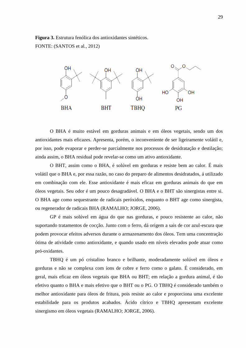
Figura 3. Estrutura fenólica dos antioxidantes sintéticos.
FONTE: (SANTOS et al., 2012)
O BHA é muito estável em gorduras animais e em óleos vegetais, sendo um dos
antioxidantes mais eficazes. Apresenta, porém, o inconveniente de ser ligeiramente volátil e,
por isso, pode evaporar e perder-se parcialmente nos processos de desidratação e destilação;
ainda assim, o BHA residual pode revelar-se como um ativo antioxidante.
O BHT, assim como o BHA, é solúvel em gorduras e resiste bem ao calor. É mais
volátil que o BHA e, por essa razão, no caso do preparo de alimentos desidratados, á utilizado
em combinação com ele. Esse antioxidante é mais eficaz em gorduras animais do que em
óleos vegetais. Seu odor é um pouco desagradável. O BHA e o BHT são sinergistas entre si.
O BHA age como sequestrante de radicais peróxidos, enquanto o BHT age como sinergista,
ou regenerador de radicais BHA (RAMALHO; JORGE, 2006).
GP é mais solúvel em água do que nas gorduras, e pouco resistente ao calor, não
suportando tratamentos de cocção. Junto com o ferro, dá origem a sais de cor azul-escura que
podem provocar efeitos adversos durante o armazenamento dos óleos. Tem uma concentração
ótima de atividade como antioxidante, e quando usado em níveis elevados pode atuar como
pró-oxidantes.
TBHQ é um pó cristalino branco e brilhante, moderadamente solúvel em óleos e
gorduras e não se complexa com íons de cobre e ferro como o galato. É considerado, em
geral, mais eficaz em óleos vegetais que BHA ou BHT; em relação a gordura animal, é tão
efetivo quanto o BHA e mais efetivo que o BHT ou o PG. O TBHQ é considerado também o
melhor antioxidante para óleos de fritura, pois resiste ao calor e proporciona uma excelente
estabilidade para os produtos acabados. Ácido cítrico e TBHQ apresentam excelente
sinergismo em óleos vegetais (RAMALHO; JORGE, 2006).
30
Entretanto, estudos toxicológicos associam estes compostos à carcinogênese, entre
outras doenças (SUN-WATERHOUSE; THAKORLAL; ZHOU, 2011).Tais evidências têm
restringido a utilização destes antioxidantes sintéticos, controlando seu emprego em alguns
países como Canadá e também na Comunidade Europeia, onde o uso de TBHQ não é
permitido e o Codex Alimentar limita a 200 mg.kg-1
o uso de BHT em óleos vegetais. No
Brasil, o uso de antioxidantes é controlado pelo Ministério da Saúde, sendo os limites
máximos permitidos de 200 mg.kg-1
para BHA e TBHQ e 100 mg.kg-1
para BHT (SILVA;
JORGE, 2012).
Nesse sentido, muitas pesquisas têm sido dirigidas com a finalidade de encontrar
produtos naturais com atividade antioxidante, porque estes são presumidamente seguros e
permitirão substituir os sintéticos ou fazer associações entre eles, com o intuito de diminuir
sua quantidade nos alimentos (SOARES, 2002).
2.5.2 Antioxidantes naturais
Compostos fenólicos e outros bioativos, como carotenoides, tocoferois e ácido
ascórbico, são exemplos de substâncias naturalmente presentes em algumas plantas, capazes
de interceptar radicais livres e evitar processos oxidativos (BREWER, 2011).
Entre os bioativos presentes nos vegetais, encontram-se os compostos fenólicos, que
são quimicamente definidos como substâncias que possuem anel aromático com um ou mais
substituintes hidroxílicos, incluindo seus grupos funcionais. Possuindo, assim, estrutura
variável e com isso apresentando características multifuncionais. Existem cerca de cinco mil
fenólicos divididos nas suas diversas classes, dentre estes se sobressaem os ácidos fenólicos,
os flavonoides e as cumarinas, que constituem a família dos compostos fenólicos largamente
distribuídos na natureza (ANGELO; JORGE, 2007).
Os ácidos fenólicos caracterizam-se pela presença de um anel benzênico, um
grupamento carboxílico e um ou mais grupamentos de hidroxila e/ou metoxila na molécula,
conferindo-lhes propriedades antioxidantes (SOARES, 2002). Consistem em três grupos: os
ácidos benzoicos, com fórmulas gerais e denominações representadas na Figura 4; os ácidos
cinâmicos, sendo sete os mais comumente encontrados na natureza, com fórmulas gerais e
denominações representadas na Figura 5; e as cumarinas que são derivadas do ácido cinâmico
por ciclização da cadeia lateral do ácido o-cumárico descrito na Figura 6 (RAMALHO;
JORGE, 2006).

31
Figura 4. Estrutura química dos ácidos benzoicos.
FONTE: (RAMALHO; JORGE, 2006)
Figura 5. Estrutura química dos principais ácidos cinâmicos.
FONTE: (RAMALHO; JORGE, 2006)
Figura 6. Estrutura química das cumarinas.
FONTE: (RAMALHO; JORGE, 2006)
Os flavonoides são compostos largamente distribuídos no reino vegetal, encontram-se
presentes em frutas, folhas, sementes e em outras partes da planta na forma de glicosídeos ou
agliconas. São compostos de baixo peso molecular, consistindo em 15 átomos de carbono,
organizados na configuração C6–C3–C6. Caracterizam-se pela presença de dois anéis
aromáticos, denominados anel A e B, unidos por três carbonos que formam um anel

32
heterocíclico, denominado anel C, como representado na Figura 7. Variações em substituição
do anel C padrão resultam em importantes classes de flavonoides, como flavonois, flavonas,
flavanonas, flavanois (ou catequinas), isoflavonas e antocianidinas. Substituições dos anéis A
e B originam diferentes compostos dentro de cada classe de flavonoides (RAMALHO;
JORGE, 2006).
Figura 7. Estrutura química dos flavonoides.
FONTE: (RAMALHO; JORGE, 2006)
Os flavonoides, assim como os ácidos fenólicos, funcionam como sequestradores de
radicais, e algumas vezes, como quelantes de metais, agindo tanto na etapa de iniciação como
na propagação do processo oxidativo (ANGELO; JORGE, 2007).
2.6 BIOATIVIDADE E TOXICIDADE FRENTE À ARTEMIA SALINA
Os metabólitos secundários presentes nas plantas são os responsáveis por atividades
biológicas tais como as toxicológicas, alelopática, antibacteriana, antioxidante, antiparasitária,
analgésica, anti-inflamatória, diurética, anticonvulsivante, miorelaxante e antiespasmódica
entre outras.
Compostos bioativos presentes nas plantas medicinais são quase sempre tóxicos em
altas doses. Desta maneira, a avaliação da letalidade em um organismo animal menos
complexo pode ser usada para um monitoramento simples e rápido. O ensaio de letalidade
para larvas de Artemia salina tem sido introduzido na rotina de muitos grupos de pesquisa
envolvidos com isolamento, purificação e elucidação estrutural, devido sua simplicidade
(KANWAR, 2007).
A Artemia salina é um crustáceo da classe Anostracea, de água salgada que é utilizado
como alimento vivo para peixes, sendo seus ovos facilmente encontrados em lojas de
aquaristas, permanecendo viáveis por anos no estado seco. Possui 4 estágios de

33
desenvolvimento (ovo, náuplio, metanáuplio e adulto) e alguns mecanismos de adaptação que
as tornam cosmopolitas, como a osmorregulação, a presença de pigmentos respiratórios como
a hemoglobina e a disponibilidade de alternativas reprodutivas que facilitam a dispersão e a
perpetuação dessa espécie (KANWAR, 2007).
Utilizando-se a concentração letal média (CL50) é possível determinar e avaliar a
atividade biológica (toxicidade) de um composto ou extrato natural. Diversos trabalhos
correlacionam a toxicidade sobre Artemia salina com atividades biológicas de extratos, como
pode ser visto no estudo desenvolvido por Kumbhare et al.(2012), onde extratos da casca do
caule da Moringa oleífera foram testados quanto à atividade antioxidante e citotoxidade. Um
outro estudo desenvolvido por Kannan et al. (2013), testou extratos de seis ervas marinhas
quanto a sua atividade antibacteriana, citotóxica e hemolítica, e seus resultados revelaram
uma boa atividade antibacteriana aliada a baixa toxicidade, o que sugere seu uso como
aditivos alimentares.

34
3 MATERIAL E MÉTODOS
Os experimentos foram realizados no Laboratório de Combustíveis e Materiais do
Departamento de Química do Centro de Ciências Exatas e da Natureza da Universidade
Federal da Paraíba (LACOM/DQ/CCEN/UFPB)
3.1 MATERIAL
O óleo de soja refinado sem adição de antioxidante foi adquirido da indústria Cargill
Agrícola S. A., Brasil. O óleo de peixe refinado sem adição de antioxidante e os reagentes
butil-hidroxitolueno (BHT), terc-butil-hidroquinona (TBHQ), 2,2-difenil-1-picrilidrazil
(DPPH) foram adquiridos na Sigma-Aldrich (Steinheim, Germany) e o reagente de Folin-
Ciocalteau, ácido gálico e todos os outros reagentes foram adquiridos na Merck (Dusseldorf,
Germany).
3.2 AQUISIÇÃO E PREPARO DAS AMOSTRAS
As folhas, flores, casca das vagens e sementes da moringa (Moringa oleífera Lam.)
foram coletadas durante o mês de abril de 2010, no Campus I da Universidade Federal da
Paraíba, João Pessoa/PB, Brasil e submetidas à secagem, em estufa com circulação de ar a 45
°C (Modelo MA 035, Marconi) durante 24 h. Após desidratação, cada parte foi triturada
separadamente, em moinho de facas, e posteriormente acondicionada em sacos de polietileno
a vácuo, sendo mantidas, à temperatura de -18 ºC durante a execução dos experimentos. Um
exemplar da exsicata foi depositado no Herbário do Departamento de Botânica da UFPB com
o registro– J.A.N. Batista 01 (JPB).
3.3 OBTENÇÃO DOS EXTRATOS
Os extratos foram obtidos através de agitação de 50 g do material vegetal em pó e 500
mL de etanol em banho termostatizado por 2 horas a 25 ºC, seguido de filtração à vácuo. Os
extratos das folhas (EL), das flores (EF), da casca das vagens (EP) e das sementes (ES) foram
concentrados em evaporador rotativo (Modelo TE-221 Tecnal) a 60 °C até a retirada total do

35
solvente. Em seguida acondicionados em frascos âmbar em atmosfera inerte (N2) sob
refrigeração (aproximadamente 8 ºC) até o momento de sua utilização.
3.4 DETERMINAÇÃO DE FENÓLICOS EXTRAÍVEIS TOTAIS
Os teores de fenólicos extraíveis totais (FET) foram determinados colorimetricamente
pelo método de Folin-Ciocalteau (SLINKARD; SINGLETON, 1977). Uma alíquota de 0,05
mL das amostras diluídas em 3,95 mL de água foi adicionada de 0,25 mL de reagente de
Folin-Ciocalteau e posteriormente de 0,75 mL de solução de carbonato de sódio 20%. A
mistura foi agitada e mantida no escuro por 2 h. A absorbância foi medida a 765 nm em
espectrofotômetro UV-vis (modelo UV-2550, Shimadzu), juntamente com o controle, que
continha somente os reagentes e água. A concentração de compostos fenólicos foi estimada,
usando curva de calibração de ácido gálico (50-500 mg.L-1
).Os resultados foram expressos
como média ± desvio-padrão de mg equivalente de ácido gálico (GAE) em cada grama de
extrato.
3.5 ESTUDO TÉRMICO DOS EXTRATOS
A curva Termogravimétrica (TG) e a Análise Térmica Diferencial (DTA) dos extratos
desidratados foram obtidas em Analisador Térmico Simultâneo DTA-TG, DTG modelo H-60
da Shimadzu, utilizando aproximadamente 10 mg de amostra em cadinho de alumina,
atmosfera de ar sintético com fluxo de 50 mL/min, razão de aquecimento de 10 °C.min-1
e
temperatura de 25 - 600 °C.
3.6 DETERMINAÇÃO DA ATIVIDADE ANTIOXIDANTE
3.6.1 Atividade sequestrante do radical livre DPPH
A capacidade dos extratos etanólicos e dos antioxidantes sintéticos BHT e TBHQ em
sequestrar o radical livre DPPH foi analisada através de metodologia proposta por Blois
(1958) com modificações de Brand-Williams et al. (1995). Alíquotas de 3,0 mL das amostras
nas diluições de 20, 40 e 80 µg/mL foram adicionadas a 0,1 mL de solução etanólica de
DPPH● 0,1 mol.L
-1. O controle das amostras consistiu de 3,0 mL de cada amotra e 0,1 mL de
etanol e o controle negativo de 3,0 mL de etanol e 0,1 mL de solução de DPPH●. O
36
decréscimo da absorbância a 515 nm foi mensurado após 120 min. A atividade sequestrante
do radical DPPH foi calculada usando a seguinte fórmula:
Atividade de sequestro do radical DPPH (%) = [(1-Aa-A
Ac)×100] EQUAÇÃO 1
em que Aa é a absorbância das amostras, Ab é a absorbância do controle das amostras de cada
extrato e Ac é a absorbância do controle negativo.
A partir dos resultados, foi construído um gráfico para o percentual de atividade
sequestradora do radical DPPH em cada concentração de extrato (µg.mL-1
) testada. Para o
cálculo do EC50 (concentração do extrato com capacidade de reduzir 50% do DPPH● inicial),
foi utilizada a equação da reta, substituindo o valor de y por 50. A eficiência antirradical foi
definida como a relação inversa do EC50, ou seja, (1/ EC50).
3.6.2 Capacidade antioxidante pelo método de redução do ferro (FRAP)
A capacidade antioxidante foi estimada pelo ensaio do poder antioxidante de redução
do ferrro (FRAP), seguindo o procedimento descrito por Benzie e Strain (1996) com as
modificações de Pulido et al. (2000). Uma alíquota de 2,7 mL do reagente FRAP recém
preparado (TPTZ 10 mM, FeCl3 20 mM e tampão de acetato) foi adicionado a 90 µL de cada
extrato e 270 µL de água destilada. A mistura foi homogeinizada em banho termostatizado a
37 ºC por 30 mim e em seguida a leitura da absorbância a 595 nm foi mensurada usando o
reagente FRAP como controle negativo para calibrar o espectrofotômetro. Concentrações de
500-2000 µmol.L-1
de sulfato ferroso (FeSO4●7H2O) foram utilizadas para a determinação da
curva-padrão. Os resultados foram expressos como média ± desvio-padrão de µM sulfato
ferroso/g de extrato.
3.6.3 Auto-oxidação do sistema β-caroteno/ácido linoleico
A atividade antioxidante pelo sistema modelo β-caroteno/ácido linoleico foi
determinado conforme método descrito por Marco (1968) com modificações de Miller (1971).
O ensaio baseia-se na oxidação do β-caroteno induzida pelos produtos de degradação
oxidativa do ácido linoleico. As soluções foram preparadas pela mistura de 5 mL da solução
sistema de β-caroteno/ácido linoleico e 0,4 mL de cada extrato ou solução de trolox (200
37
µg.mL-1
). Após leitura inicial da absobância a 470 nm, a mistura foi mantida em banho
termostatizado a 40 ºC, e a absorbância medida em intervalos de 15 min até 120 min. A
amostra controle negativo consistiu de 5 mL da solução sistema de β-caroteno/ácido linoleico
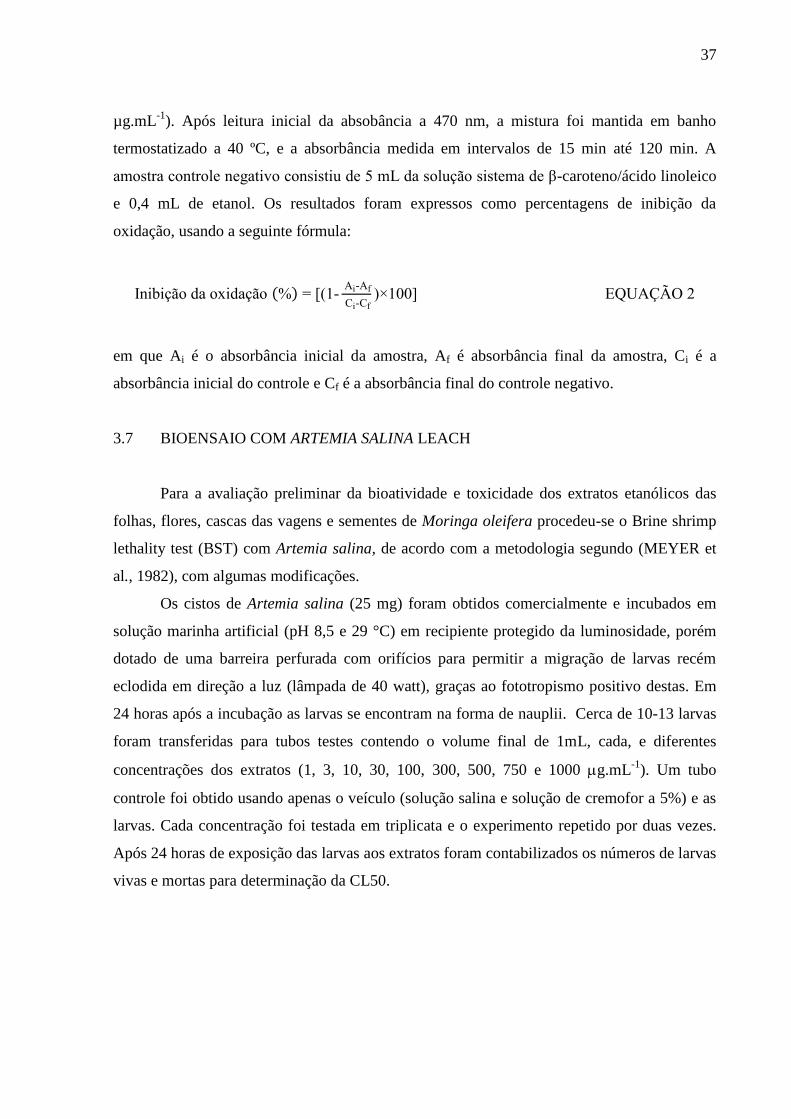
e 0,4 mL de etanol. Os resultados foram expressos como percentagens de inibição da
oxidação, usando a seguinte fórmula:
ni ição da oxidação (%) = [(1- Ai-Af
Ci-Cf)×100] EQUAÇÃO 2
em que Ai é o absorbância inicial da amostra, Af é absorbância final da amostra, Ci é a
absorbância inicial do controle e Cf é a absorbância final do controle negativo.
3.7 BIOENSAIO COM ARTEMIA SALINA LEACH
Para a avaliação preliminar da bioatividade e toxicidade dos extratos etanólicos das
folhas, flores, cascas das vagens e sementes de Moringa oleifera procedeu-se o Brine shrimp
lethality test (BST) com Artemia salina, de acordo com a metodologia segundo (MEYER et
al., 1982), com algumas modificações.
Os cistos de Artemia salina (25 mg) foram obtidos comercialmente e incubados em
solução marinha artificial (pH 8,5 e 29 °C) em recipiente protegido da luminosidade, porém
dotado de uma barreira perfurada com orifícios para permitir a migração de larvas recém
eclodida em direção a luz (lâmpada de 40 watt), graças ao fototropismo positivo destas. Em
24 horas após a incubação as larvas se encontram na forma de nauplii. Cerca de 10-13 larvas
foram transferidas para tubos testes contendo o volume final de 1mL, cada, e diferentes
concentrações dos extratos (1, 3, 10, 30, 100, 300, 500, 750 e 1000 g.mL-1
). Um tubo
controle foi obtido usando apenas o veículo (solução salina e solução de cremofor a 5%) e as
larvas. Cada concentração foi testada em triplicata e o experimento repetido por duas vezes.
Após 24 horas de exposição das larvas aos extratos foram contabilizados os números de larvas
vivas e mortas para determinação da CL50.

38
3.8 DETERMINAÇÃO DA ESTABILIDADE OXIDATIVA
3.8.1 Preparação das amostras
Alíquotas de 50 mL dos óleos de soja e de peixe foram adicionadas dos extratos
etanólicos das folhas (EL), flores (EF) e cascas das vagens (EP) na concentração de 100
mg.kg-1
em relação ao seu conteúdo de fenólicos extraíveis totais (TEP) ou de BHT e TBHQ
na concentração de 100 mg.kg-1
. As misturas foram homogeneizadas em agitador magnético
durante 30 min, acondicionadas em frascos âmbar em atmosfera inerte a -18 ºC até o
momento das análises. Os óleos de soja e de peixe sem adição de antioxidantes foram
submetidos ao mesmo procedimento e utilizados como controle.
3.8.2 Método Rancimat
A estabilidade oxidativa dos óleos de soja e de peixe adicionado dos extratos ou dos
antioxidantes sintéticos foi analisada pelo emprego do método proposto pela AOCS CD 12b-
92 (AOCS, 2003) em equipamento Rancimat (873 Biodiesel, Metrohm). Um total de 2 g de
cada amostra foram submetidos a 110 ºC sob fluxo constante de ar (10 L.h-1
). O resultado foi
determinado a partir do ponto de inflexão da curva tempo em função da condutividade. Os
cálculos dos períodos de indução foram realizados com o auxílio do Software 873-Rancimat®,
sendo os resultados expressos como índice de estabilidade oxidativa (OSI) em h e como
percentual de aumento do OSI.
3.8.3 Calorimetria exploratória diferencial pressurizada (P-DSC)
As curvas de P-DSC das amostras foram obtidas através de um calorímetro
exploratório diferencial acoplado a uma célula de pressão da TA Instruments, modelo
DSC Q1000.
A curva dinâmica do óleo de peixe puro foi realizada utilizando cadinho de alumínio
com aproximadamente 10 mg da amostra e as condições de análise no calorímetro foram
atmosfera de oxigênio, pressão de 1400 kPa e razão de aquecimento de 10 °C.min-1
no
intervalo de 25 a 500 °C. As curvas isotermas foram realizadas a 100 °C, nas mesmas
condições descritas.

39
As análises no modo isotérmico (110 ºC) do óleo de soja foram processadas utilizando
cadinho de alumínio com aproximadamente 10 mg da amostra, sob atmosfera de oxigênio
com pressão de 1400 kPa.
Os valores do OIT foram determinados pela diferença entre os tempos onset e do
inicial e os resultados expressos como tempo de indução oxidativa (OIT) em minutos e como
percentual de aumento da OIT.
3.8.4 Teste de estocagem acelerada em estufa
Os extratos de moringa na concentração de 100 mg.kg-1
em relação ao seu conteúdo de
fenólicos extraíveis totais (TEP), o BHT e o TBHQ nesta mesma concentração foram
adicionadas em 15 g de óleo de soja e de peixe e homogeneizadas em agitador magnético
durante 30 min. O teste de estocagem acelerada em estufa do óleo de soja e do óleo de peixe
foi conduzido em estufa com circulação de ar durante 16 dias a temperatura de 60 ± 2 oC. A
cada quatro dias, foram realizadas as análises dos índices de peróxido, índice de p-anisidina,
valor de dienos conjugados e espectroscopia de infravermelho por transformada de Fourier.
Os óleos de soja e de peixe sem adição de antioxidantes submetidos ao teste de estocagem
foram utilizados como controle.
3.8.4.1 Índice de peróxido
O índice de peróxido foi determinado pelo método Cd 8-53 da AOCS (2003).
Alíquotas de 2 g dos óleos foram dissolvidas em 30 mL de uma solução de ácido acético-
clorofórmio (3:2 v/v), e adicionadas de 0,5 mL de solução saturada de iodeto de potássio.
Após 1 min em repouso foram adicionados 30 mL de água e 0,5 mL de uma solução de
amido a 1 %. Na sequência foi realizada titulação com solução de tiossulfato de sódio 0,01 N,
até o desaparecimento da coloração azulada. Uma prova em branco foi conduzida nas mesmas
condições, sem a presença da amostra. Os cálculos foram realizados de acordo com Equação:
ndice de peróxido (meq / g) = x ( A-
) x 1000
P da amostra (g) EQUAÇÃO 3
40
em que N é a normalidade da solução de Na2S2O3, Va é o volume da solução de Na2S2O3
consumido pela amostra (mL), Vb é o volume da solução de Na2S2O3 consumido pelo branco
(mL) e m é a massa da amostra (g).
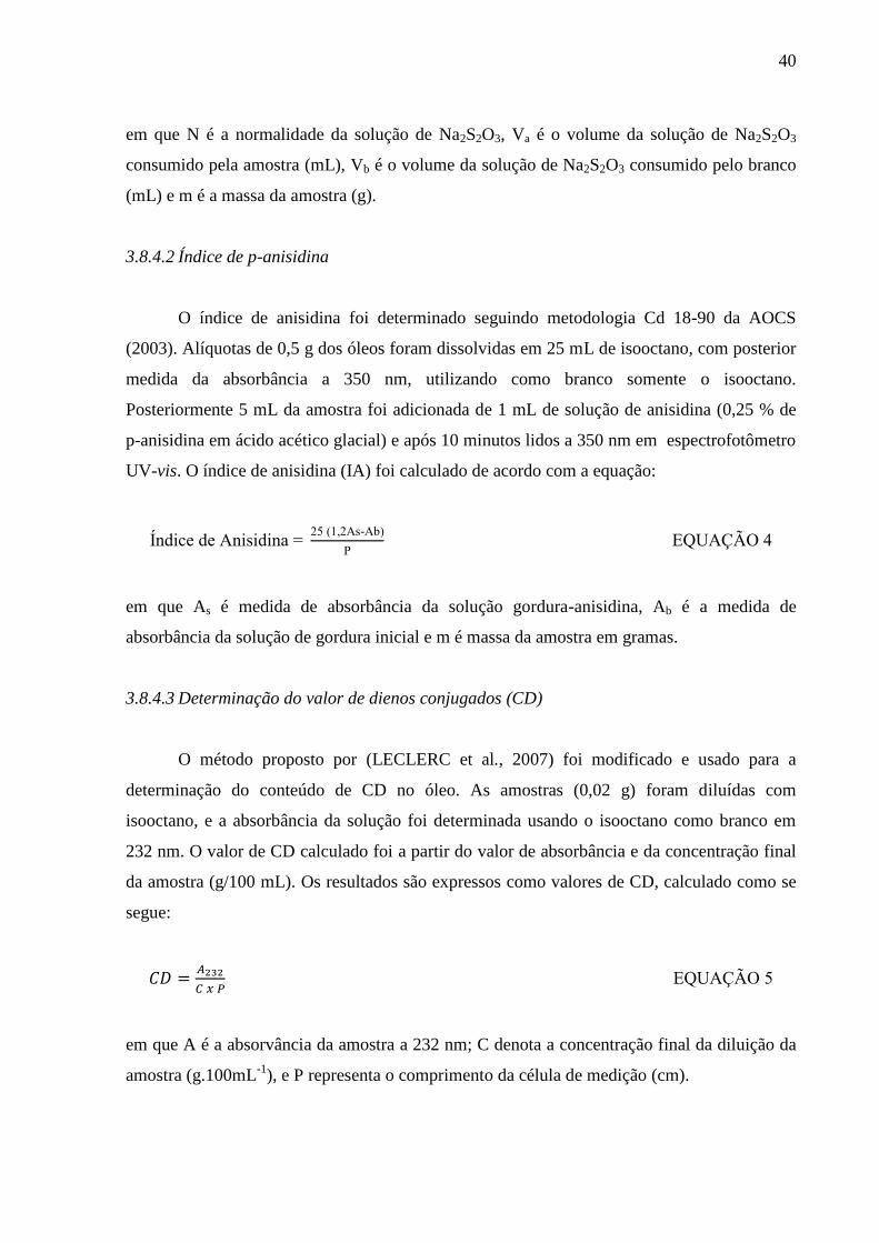
3.8.4.2 Índice de p-anisidina
O índice de anisidina foi determinado seguindo metodologia Cd 18-90 da AOCS
(2003). Alíquotas de 0,5 g dos óleos foram dissolvidas em 25 mL de isooctano, com posterior
medida da absorbância a 350 nm, utilizando como branco somente o isooctano.
Posteriormente 5 mL da amostra foi adicionada de 1 mL de solução de anisidina (0,25 % de
p-anisidina em ácido acético glacial) e após 10 minutos lidos a 350 nm em espectrofotômetro
UV-vis. O índice de anisidina (IA) foi calculado de acordo com a equação:
ndice de Anisidina = 25 (1,2As-A )
P EQUAÇÃO 4
em que As é medida de absorbância da solução gordura-anisidina, Ab é a medida de
absorbância da solução de gordura inicial e m é massa da amostra em gramas.
3.8.4.3 Determinação do valor de dienos conjugados (CD)
O método proposto por (LECLERC et al., 2007) foi modificado e usado para a
determinação do conteúdo de CD no óleo. As amostras (0,02 g) foram diluídas com
isooctano, e a absorbância da solução foi determinada usando o isooctano como branco em
232 nm. O valor de CD calculado foi a partir do valor de absorbância e da concentração final
da amostra (g/100 mL). Os resultados são expressos como valores de CD, calculado como se
segue:
EQUAÇÃO 5
em que A é a absorvância da amostra a 232 nm; C denota a concentração final da diluição da
amostra (g.100mL-1
), e P representa o comprimento da célula de medição (cm).

41
3.8.4.4 Infravermelho com transformada de Fourier (FTIR)
Os espectros de absorção na região do infravermelho foram obtidos em
espectrofotômetro de IV com transformada de Fourier modelo IR Prestige-21, Class 1, Laser
Product da marca Shimadzu. A análise foi realizada na região de 4.000 a 650 cm-1
utilizando
acessório ATR de uma reflexão com prisma de ZnSe, na resolução de 4 cm-1
.
3.9 ANÁLISES ESTATÍSTICAS
Os ensaios foram conduzidos em triplicata e os resultados expressos como média ±
desvio-padrão. Os resultados foram analisados no Statistica 7.0 (Statsoft®) utilizando
ANOVA e teste de Tukey considerando P ˂ 0,05.
Os resultados da bioatividade foram expressos como média de análises em triplicata,
sendo os ensaios repetidos para obtenção de nova média. A CL50 foi estabelecida com um
intervalo de confiança de 95% para todos os experimentos, sendo os resultados considerados
significativos quando P < 0,05, de acordo com a análise estatística de Probit, segundo Finney
(1971), e usando o programa GraphPad Prism 4.03.

42
REFERÊNCIAS
ABIOVE. Pesquisa de Capacidade Instalada da Indústria de Óleos Vegetais - 2012.
Disponível em: <http://www.abiove.org.br. Acesso em: 4 jan. 2013.
ABROGOUA, D. P.; DANO, D. S.; MANDA, P.; ADEPO, A. J. B.; KABLAN, B. J.; GOZE,
N. B.; EHABNTOULÉ, K. Effect on blood pressure of a dietary supplement containing
traditional medicinal plants of Côte d’ voire. Journal of Ethnopharmacology, v. 141, p.
840-847, 2012.
ANGELO, P. M.; JORGE, N. Compostos fenólicos em alimentos – Uma breve revisão.
Revista do Instituto Adolfo Lutz, v. 66, n. 1, p. 232-240, 2007.
ANWAR, F.; LATIF, S.; ASHRAF, M.; GILANI, A. H. Moringa oleifera : A Food Plant with
Multiple Medicinal Uses. Phytotherapy Research, v. 21, p. 17-25, 2007.
AOCS. Fats, Oils and Lipid Related Analytical Methods. In: FIRESTONE, D. (Org.). Official
Methods and Recommended Practices of the AOCS. 6. ed. [S.l.]: AOCS, 2003. p. Method Cd
12b-92.
ARABSHAHI-D, S.; VISHALAKSHI DEVI, D.; UROOJ, A. Evaluation of antioxidant
activity of some plant extracts and their heat, pH and storage stability. Food Chemistry, v.
100, n. 3, p. 1100-1105, 2007.
AVERINA, E. S.; KUTYREV, I. A. Perspectives on the use of marine and freshwater
hydrobiont oils for development of drug delivery systems. Biotechnology Advances, v. 29, n.
5, p. 548-557, 2011.
BALK, E. M.; LICHTENSTEIN, A. H.; CHUNG, M.; KUPELNICK, B.; CHEW, P.; LAU, J.
Effects of omega-3 fatty acids on serum markers of cardiovascular disease risk: a systematic
review. Atherosclerosis, v. 189, n. 1, p. 19-30, 2006.
BARRETO, M. B.; FREITAS, J. V. B.; SILVEIRA, E. R.; BEZERRA, A. M. E.; NUNES, E.
P.; GRAMOSA, N. V. Constituintes químicos voláteis e não-voláteis de Moringa oleifera
Lam ., Moringaceae. Brazilian Journal of Pharmacognosy, v. 19, n. 4, p. 893-897, 2009.
BAYDAR, N. G.; OZKAN, G.; YASAR, S. Evaluation of the antiradical and antioxidant
potential of grape extracts. Food Control, v. 18, p. 1131-1136, 2007.
BEN SALEM, H.; MAKKAR, H. P. S. Defatted Moringa oleifera seed meal as a feed
additive for sheep. Animal Feed Science and Technology, v. 150, n. 1-2, p. 27-33, 2009.
BENZIE, I. F.; STRAIN, J. J. The ferric reducing ability of plasma (FRAP) as a measure of
“antioxidant power”: the FRAP assay. Analytical Biochemistry, v. 239, p. 70-76, 1996.
BEZERRA, A. M. E.; MOMENTÉ, V. G.; MEDEIROS FILHO, S. Germinação de sementes
e desenvolvimento de plântulas de moringa (Moringa oleifera Lam.) em função do peso da
semente e do tipo de substrato. Horticultura Brasileira, v. 22, n. 2, p. 295-299, 2004.

43
BIJINA, B.; CHELLAPPAN, S.; KRISHNA, J. G.; BASHEER, S. M.; ELYAS, K. K.;
BAHKALI, A. H.; CHANDRASEKARAN, M. Protease inhibitor from Moringa oleifera with
potential for use as therapeutic drug and as seafood preservative. Saudi Journal of Biological
Sciences, v. 18, p. 273-281, 2011.
BLOIS, M. S. Antioxidant Determinations by the Use of a Stable Free Radical. Nature, v.
181, p. 1199-1200, 1958.
BRAND-WILLIAMS, W.; CUVELIER, M. E.; BERSET, C. Use of a free radical method to
evaluate antioxidant activity. LWT - Food Science and Technology, v. 28, n. 1, p. 25-30,
1995.
BRASIL. Resolução no 04, de 24 de novembro de 1988. Brasília: Ministério da Saúde, 1988.
BREWER, M. S. Natural Antioxidants: Sources, Compounds, Mechanisms of Action, and
Potential Applications. Comprehensive Reviews in Food Science and Food Safety, v. 10, n.
4, p. 221-247, 2011.
BYKERK, V. P.; KEYSTONE, E. C. What are the goals and principles of management in the
early treatment of rheumatoid arthritis? Best practice & research. Clinical rheumatology, v.
19, n. 1, p. 147-61, 2005.
CASTRO, H. F.; MENDES, A. A.; SANTOS, J. C. Modificação de óleos e gorduras por
biotransformação. Química Nova, p. 146-156, 2004.
CHUMARK, P.; KHUNAWAT, P.; SANVARINDA, Y.; PHORNCHIRASILP, S.;
MORALES, N. P.; PHIVTHONG-NGAM, L.; RATANACHAMNONG, P.; SRISAWAT, S.;
PONGRAPEEPORN, K. U. S. The in vitro and ex vivo antioxidant properties,
hypolipidaemic and antiatherosclerotic activities of water extract of Moringa oleifera Lam.
leaves. Journal of Ethnopharmacology, v. 116, n. 3, p. 439-46, 2008.
CÁCERES, A.; SARAVIA, A.; RIZZO, S.; ZABALA, L.; DE LEON, E.; NAVE, F.
Pharmacologic properties of Moringa oleifera. 2: Screening for antispasmodic,
antiinflammatory and diuretic activity. Journal of Ethnopharmacology, v. 36, p. 233-237,
1992.
DAHMER, M. L.; FLEMING, P. D.; COLLINS, G. B.; HILDEBRAND, D. F. A rapid
screening technique for determining the lipid composition of soybean seeds. Journal of the
American Oil Chemists’ Society, v. 66, n. 4, p. 543-548, 1989.
DEBNATH, S.; BISWAS, D.; RAY, K.; GUHA, D. Moringa oleifera induced potentiation of
serotonin release by 5-HT(3) receptors in experimental ulcer model. Phytomedicine, v. 18, p.
91-95, 2011.
DONGMEZA, E.; SIDDHURAJU, P.; FRANCIS, G.; BECKER, K. Effects of dehydrated
methanol extracts of moringa (Moringa oleifera Lam.) leaves and three of its fractions on
growth performance and feed nutrient assimilation in Nile tilapia (Oreochromis niloticus
(L.)). Aquaculture, v. 261, n. 1, p. 407-422, 2006.
44
DUBINSKY, E. Utilización de antioxidantes en aceites y grasas. Aceites y Grasas, v. 12, n.
1, p. 191-199, 2000.
FAIZI, S.; SIDDIQUI, B. S.; SALEEM, R.; SIDDIQUI, S.; AFTAB, K.; GILANI, A. H.
Isolation and structure elucidation of new nitrile and mustard oil glycosides from Moringa
oleifera and their effect on blood pressure. Journal of Natural Products, v. 57, n. 9, p. 1256-
1261, 1994.
FERRARI, R. A.; SCHULTE, E.; ESTEVES, W.; BRIIHL, L.; MUKHERJEE, K. D. Minor
Constituents of Vegetable Oils During Industrial Processing 1. Journal of the American Oil
Chemists` Society, v. 73, n. 5, p. 587-592, 1996.
FINNEY, D. J. Probit analysis. Cambridge: Cambridge University, 1971. p. 333
GORDON, M. Measuring antioxidant activity practical applications. In: POKORNY, J.;
YANISHLIEVA, N.; GORDON, M. (Org.). Antioxidants in food: practical applications.
Cambridge: Woodhead Publishing, 2003. p. 71-84.
GRAY, J. I. Measurement of lipid oxidation: a review. Journal of the American Oil
Chemists` Society, v. 55, n. 6, p. 539-546, 1978.
GUPTA, M. K. Oil quality improvement through processing. n: O’ R E , R. D.; FARR, W.
E.; WAN, P. J. (Org.). Introduction to Fats and Oils Technology. 2a. ed. [S.l.]:
Champaign :AOCS press, 2000.
HALLIWELL, B. Lipid peroxidation, antioxidants and cardiovascular disease: how should
we move forward? Cardiovascular Research, v. 47, n. 3, p. 410-418, 2000.
HALVORSEN, B. L.; BLOMHOFF, R. Determination of lipid oxidation products in
vegetable oils and marine omega-3 supplements. Food & Nutrition Research, v. 55, p. 5792-
801, 2011.
HARISTOY, X. W.; FAHEY, J. W.; SCHOLTUS, I. W.; LOZNIEWSKI, A. W. Adicionar ao
Meu Espaço Evaluation of the Antimicrobial Effects of Several Isothiocyanates on
Helicobacter pylori. Planta Medica, v. 71, n. 4, p. 326-330, 2005.
HAZRA, S.; BISWAS, S.; BHATTACHARYYA, D.; DAS, S. K.; KHAN, A. Quality of
cooked ground buffalo meat treated with the crude extracts of Moringa oleifera (Lam.) leaves.
Journal of Food Science and Technology, v. 49, n. 2, p. 240-245, 2012.
IQBAL, S.; BHANGER, M. I. Effect of season and production location on antioxidant
activity of Moringa oleifera leaves grown in Pakistan. Journal of Food Composition and
Analysis, v. 19, n. 6-7, p. 544-551, 2006.
JAISWAL, D.; RAI, P. K.; KUMAR, A.; MEHTA, S.; WATAL, G. Effect of Moringa
oleifera Lam. leaves aqueous extract therapy on hyperglycemic rats. Journal of
Ethnopharmacology, v. 123, p. 392-396, 2009.
KANNAN, R. R. R.; ARUMUGAM, R.; IYAPPARAJ, P.; THANGARADJOU, T.;
ANANTHARAMAN, P. In vitro antibacterial, cytotoxicity and haemolytic activities and

45
phytochemical analysis of seagrasses from the Gulf of Mannar, South India. Food chemistry,
v. 136, p. 1484-9, 2013.
KANWAR, A. S. Brine shrimp (Artemia salina) – a marine animal for simple and rapid
biological assays. Journal of Chinese Clinical Medicine, v. 2, n. 4, p. 236-240, 2007.
KARADI, R. V; GADGE, N. B.; ALAGAWADI, K. R.; SAVADI, R. V. Effect of Moringa
oleifera Lam. root-wood on ethylene glycol induced urolithiasis in rats. Journal of
Ethnopharmacology, v. 105, n. 1-2, p. 306-11, 2006.
KEY, T. J.; APPLEBY, P. N.; MASSET, G.; BRUNNER, E. J.; CADE, J. E.;
GREENWOOD, D. C.; STEPHEN, A. M.; KUH, D.; BHANIANI, A.; POWELL, N.;
KHAW, K.-T. Vitamins, minerals, essential fatty acids and colorectal cancer risk in the
United Kingdom Dietary Cohort Consortium. International journal of cancer, v. 131, n. 3,
p. 320-325, 2012.
KIM, T. S.; DECKER, E. A.; LEE, J. Effects of chlorophyll photosensitisation on the
oxidative stability in oil-in-water emulsions. Food Chemistry, v. 133, n. 4, p. 1449-1455,
2012.
K OTHE, G. “ Designer ” iodiesel : Optimizing Fatty Ester Composition to mprove Fuel
Properties. Energy & Fuels, v. 22, p. 1358-1364, 2008.
KODALI, D. R. Oxidative stability measurement of high-stability oils by pressure differencial
scanning calorimeter (PDSC). Journal agricultural and food chemistry, v. 53, p. 7649-
7653, 2005.
KOWALSKI, B.; RATUSZ, K.; KOWALSKA, D.; BEKAS, W. Determination of the
oxidative stability of vegetable oils by Differential Scanning Calorimetry and Rancimat
measurements. European Journal of Lipid Science and Technology, v. 106, n. 3, p. 165-
169, 2004.
KRIS-ETHERTON, P. M.; HECKER, K. D.; BONANOME, A.; COVAL, S. M.;
BINKOSKI, A. E.; HILPERT, K. F.; GRIEL, A. E.; ETHERTON, T. D. Bioactive
Compounds in Foods : Their Role in the Prevention of Cardiovascular Disease and Cancer.
The American Journal of Medicine, v. 113, n. 9, p. 71-88, 2002.
KUMBHARE, M.; GULEHA, V.; SIVAKUMAR, T. Estimation of total phenolic content,
cytotoxicity and in–vitro antioxidant activity of stem bark of Moringa oleifera. Asian Pacific
Journal of Tropical Disease, v. 2, n. 2, p. 144-150, 2012.
LAKO, J.; TRENERRY, V.; WAHLQVIST, M.; WATTANAPENPAIBOON, N.;
SOTHEESWARAN, S.; PREMIER, R. Phytochemical flavonols, carotenoids and the
antioxidant properties of a wide selection of Fijian fruit, vegetables and other readily available
foods. Food Chemistry, v. 101, n. 4, p. 1727-1741, 2007.
LECLERC, L.; ZHAO, X.; SIMPSON, B. K.; ZUTA, P. C. The effect of α-tocopherol on the
oxidation of mackerel oil.pdf. Food Chemistry, v. 100, p. 800-807, 2007.

46
LUKIW, W. J.; BAZAN, N. G. Docosahexaenoic Acid and the Aging Brain. American
Society for Nutrition, v. 138, n. 12, p. 2510-2514, 2008.
MAKKAR, H. P. S.; BECKER, K. Nutrional value and antinutritional components of whole
and ethanol extracted Moringa oleifera leaves. Animal Feed Science and Technology, v. 63,
n. 1-4, p. 211-228, 1996.
MARCO, G. A rapid method for evaluation of antioxidants. Journal of the American Oil
Chemists’ Society, v. 45, p. 594-598, 1968.
MARTÍNEZ-TOMÉ, M.; JIMÉNEZ, A. M.; RUGGIER, S.; FREGA, N.; STRABBIOLI, R.;
M.A., M. Antioxidant properties of mediterranean spices compared to common food
additives. Journal of Food Protection, v. 64, n. 9, p. 1412-1419, 2001.
MEYER, B. N.; FERRIGNI, N. R.; PUTNAM, J. E.; JACOBSEN, L. B.; NICHOLS, D. E.;
MCLAUGHLIN, J. L. Brine shrimp: A convenient general bioassay for active plant
constituents. Planta Medica, v. 45, p. 31-34, 1982.
MILLER, H. E. A simplified method for the evaluation of antioxidants. Journal of the
American Oil Chemists’ Society, v. 48, p. 91, 1971.
MOHAMMED, K. A. E. F.; SARMIENTO-FRANCO, L.; SANTOS-RICALDE, R.;
SOLORIO-SANCHEZ, J. F. The nutritional effect of Moringa oleifera fresh leaves as feed
supplement on Rhode Island Red hen egg production and quality. Tropical animal health
and production, 2011.
MORRIS, M. C.; EVANS, D.; TANGNEY, C. C.; BIENIAS, J. L.; WILSON, R. S. Fish
consumption and cognitive decline with age in a large community study. Archives of
neurology, v. 62, n. 12, p. 1849-53, 2005.
MOTHE, C. G.; AZEVEDO, A. D. Análise Termica de Materiais. 1o. ed. [S.l: s.n.], 2009.
NAZ, S.; SIDDIQI, R.; SHEIKH, H.; SAYEED, S. Deterioration of olive, corn and soybean
oils due to air, ligth, heat and deep-frying. Food Research International, v. 38, n. 2, p. 127-
134, 2005.
NEMETS, H.; NEMETS, B.; APTER, A.; BRACHA, Z.; BELMAKER, R. H. Omega-3
treatment of childhood depression: a controlled, double-blind pilot study. The American
journal of psychiatry, v. 163, n. 6, p. 1098-100, 2006.
OSTROWSKA-LIGEZA, E.; BEKAS, W.; KOWALSKA, D.; LOBACZ, M.; WRONIAK,
M. Kinetics of commercial olive oil oxidation : Dynamic differential scanning calorimetry
and Rancimat studies. European Journal Lipid Science Techonology, v. 112, p. 268-274,
2010.
PIVIK, R. T.; DYKMAN, R. A.; JING, H.; GILCHRIST, J. M.; BADGER, T. M. Early Infant
Diet and the Omega 3 Fatty Acid DHA: Effects on Resting Cardiovascular Activity and
Behavioral Development During the First Half-Year of Life. Developmental
Neuropsychology, v. 34, n. 2, p. 139-158, 2009.

47
PR TCHARD, M.; CRA E , T.; MKA DAW RE, T.; EDMO DSO , A. S.; O’ E LL, J.
G. A study of the parameters affecting the effectiveness of Moringa oleifera in drinking water
purification. Physics and Chemistry of the Earth, Parts A/B/C, v. 35, n. 13-14, p. 791-797,
2010.
PULIDO, R.; BRAVO, L.; FULGENCIO, S. C. Antioxidant Activity of Dietary Polyphenols
As Determined by a Modified Ferric Reducing/Antioxidant Power Assay. Journal of
Agricultural and Food Chemistry, v. 48, p. 3396-3402, 2000.
RAMACHANDRAN, C.; PETER, K. V; GOPALAKRISHNAN, P. K. Drumstick (Moringa
oleifera) a multipurpose Indian vegetable. Economic Botany, v. 34, n. 3, p. 276–283, 1980.
RAMALHO, E. F. S. M.; ALBUQUERQUE, A. R.; SOUZA, A. L.; BARRO, A. K.; MAIA,
A. S.; SANTOS, I. M. G.; SOUZA, A. G. Use of different techniques in the evaluation of the
oxidative stability of poultry fat biodiesel. Journal of Thermal Analysis and Calorimetry,
v. 106, n. 3, p. 787-791, 2011.
RAMALHO, V.; JORGE, N. Antioxidantes utilizados em óleos, gorduras e alimentos
gordurosos. Química Nova, v. 29, n. 4, p. 755-760, 2006.
RASHID, U.; ANWAR, F.; MOSER, B. R.; KNOTHE, G. Moringa oleifera oil: a possible
source of biodiesel. Bioresource technology, v. 99, n. 17, p. 8175-9, 2008.
REDDY, V.; UROOJ, A.; KUMAR, A. Evaluation of antioxidant activity of some plant
extracts and their application in biscuits. Food Chemistry, v. 90, p. 317-321, 2005.
REGITANO-D’ARCE, M. A. . Deterioração de lipídeos – ranço. In: OETTERER, M.;
REGITANO-D’ARCE, M. A. .; SPOTTO, M. H. F. (Org.). Fundamentos de ciência e
tecnologia de alimentos. Barueri: Manole, 2006. p. 243-270.
RIBEIRO, A. P. B.; GRIMALDI, R.; GIOIELLI, L. A.; GONÇALVES, L. A. G. Zero trans
fats from soybean oil and fully hydrogenated soybean oil: Physico-chemical properties and
food applications. Food Research International, v. 42, n. 3, p. 401-410, 2009.
SANTOS, N. A.; CORDEIRO, A. M. T. M.; DAMASCENO, S. S.; AGUIAR, R. T.;
ROSENHAIM, R.; CARVALHO FILHO, J. R.; SANTOS, I. M. G.; MAIA, A. S.; SOUZA,
A. G. Commercial antioxidants and thermal stability evaluations. Fuel, v. 97, p. 638-643,
2012.
SCHAICH, K. M. Thinking outside the classical chain reaction box of lipid oxidation. Lipid
Technology, v. 24, n. 3, p. 55-58, 2012.
SHARMA, B. K.; RASHID, U.; ANWAR, F.; ERHAN, S. Z. Lubricant properties of
Moringa oil using thermal and tribological techniques. Journal of Thermal Analysis and
Calorimetry, v. 96, n. 3, p. 999-1008, 2009.
SIDDHURAJU, P.; BECKER, K. Antioxidant properties of various solvent extracts of total
phenolic constituents from three different agroclimatic origins of drumstick tree (Moringa
oleifera Lam.) leaves. Journal of Agricultural and Food Chemistry, v. 51, p. 2144-2155,
2003.

48
SILVA, A. C.; JORGE, N. Influence of Lentinus edodes and Agaricus blazei extracts on the
prevention of oxidation and retention of tocopherols in soybean oil in an accelerated storage
test. Journal of Food Science and Technology, 2012.
SILVA, A. V. C.; SANTOS, A. R. F.; LÉDO, A. S.; ALMEIDA, R. B. F. C. S.; SILVA, G.
M.; RANGEL, M. S. A. Moringa genetic diversity from germplasm bank using RAPD
markers. Tropical and Subtropical Agroecosystems, v. 15, n. 1, p. 31-39, 2012.
SIMIC, M. G.; JAVANOVIC, S. V. Inactivation of oxygen radicals by dietary phenolic
compounds in anticarcinogenesis. In: HO, C. T. et al. (Org.). Food phytochemicals for cancer
prevention. Washington: American Chemical Society, 1994. p. 20-33.
SINGH, B. N.; SINGH, B. R.; SINGH, R. L.; PRAKASH, D.; DHAKAREY, R.;
UPADHYAY, G.; SINGH, H. B. Oxidative DNA damage protective activity, antioxidant and
anti-quorum sensing potentials of Moringa oleifera. Food and Chemical Toxicology, v. 47,
p. 1109-16, 2009.
SLINKARD, K.; SINGLETON, V. L. Total Phenol Analysis: Automation and Comparison
with Manual Methods. American Journal of Enology and Viticulture, v. 28, n. 1, p. 49-55,
1977.
SOARES, S. E. Ácidos fenólicos como antioxidantes. Revista de Nutrição, v. 15, n. 1, p. 71-
81, 2002.
SREELATHA, S.; PADMA, P. R. Antioxidant activity and total phenolic content of Moringa
oleifera leaves in two stages of maturity. Plant foods for human nutrition, v. 64, n. 4, p.
303-11, 2009.
SUN-WATERHOUSE, D.; THAKORLAL, J.; ZHOU, J. Effects of added phenolics on the
storage stability of avocado and coconut oils. International Journal of Food Science &
Technology, v. 46, n. 8, p. 1575-1585, 2011.
SÁNCHEZ-MACHADO, D. I.; LÓPEZ-CERVANTES, J.; VÁZQUEZ, N. J. R. High-
performance liquid chromatography method to measure alpha- and gamma-tocopherol in
leaves, flowers and fresh beans from Moringa oleifera. Journal of chromatography. A, v.
1105, n. 1-2, p. 111-4, 2006.
TAMJIDI, F.; NASIRPOUR, A.; SHAHEDI, M. Physicochemical and sensory properties of
yogurt enriched with microencapsulated fish oil. Food science and technology
international, From Duplicate 1 ( Physicochemical and sensory properties of yogurt
enriched with microencapsulated fish oil. - Tamjidi, F; Nasirpour, A; Shahedi, M ) , v. 18, n.
4, p. 381-90, 2012.
TAN, C. P.; CHE MAN, Y. B.; SELAMAT, J.; YUSOFF, M. S. A. Comparative studies of
oxidative stability of edible oils by differential scanning calorimetry and oxidative stability
index methods. Food Chemistry, v. 76, n. 3, p. 385-389, 2002.
TSAKNIS, J; LALAS, S.; GERGIS, V.; DOURTOGLOU, V.; SPILIOTIS, V.
Characterisation of Moringa oleifera variety Mbololo seed oil of Kenya. Journal of
agricultural and food chemistry, v. 47, p. 4495-4499, 1999.

49
TSAKNIS, JOHN; LALAS, S. Stability During Frying of Moringa oleifera Seed Oil Variety
“Periya ulam 1”. Journal of Food Composition and Analysis, v. 15, n. 1, p. 79-101, 2002.
USDA, U. S. D. O. A. Foreign Agricultural Service. Global Agricultural Information
Network: Brazil, Oilseeds and Products Annual, 2010.
VELASCO, J.; ANDERSEN, M. L.; SKIBSTED, L. H. Evaluation of oxidative stability of
vegetable oils by monitoring the tendency to radical formation. A comparison of electron spin
resonance spectroscopy with the Rancimat method and differential scanning calorimetry.
Food Chemistry, v. 85, n. 4, p. 623-632, 2004.
VERMA, A. R.; VIJAYAKUMAR, M.; MATHELA, C. S.; RAO, C. V. In vitro and in vivo
antioxidant properties of different fractions of Moringa oleifera leaves. Food and Chemical
Toxicology, v. 47, n. 9, p. 2196-2201, 2009.
WANG, H.; LIU, F.; YANG, L.; ZU, Y.; WANG, H.; QU, S.; ZHANG, Y. Oxidative stability
of fish oil supplemented with carnosic acid compared with synthetic antioxidants during long-
term storage. Food Chemistry, v. 128, n. 1, p. 93-99, 2011.
ZHANG, Y.; YANG, L.; ZU, Y.; CHEN, X.; WANG, F.; LIU, F. Oxidative stability of
sunflower oil supplemented with carnosic acid compared with synthetic antioxidants during
accelerated storage. Food Chemistry, v. 118, n. 3, p. 656-662, 2010.

50
4 RESULTADOS
Os resultados do presente estudo estão apresentados na forma de artigos. São eles:
Artigo 1 Ethanolic extracts of Moringa oleifera Lam.: evaluation of its potential as an
antioxidant additive for fish oil
Publicado em: Journal of Thermal Analysis and Calorimetry
Artigo 2 Ethanolics extracts of moringa: antioxidant effect in soybean oil by P-DSC and
Rancimat
Submetido em: Journal of Thermal Analysis and Calorimetry
Artigo 3 Oxidative stability of fish oil supplemented with ethanolic extracts of Moringa
oleifera Lam. compared with synthetic antioxidants during accelerated test
Submetido em: Journal of Agricultural and Food Chemistry
Artigo 4 Antioxidant effect of Moringa oleifera Lamark extracts in soybean oil during
accelerated storage test
Submetido em: Journal of the American Oil Chemists' Society
Artigo 5 Study of antioxidant activity and preliminary bioactivity determination of
ethanolic extracts of Moringa oleifera Lam.
Submetido em: Natural Product Communications

51
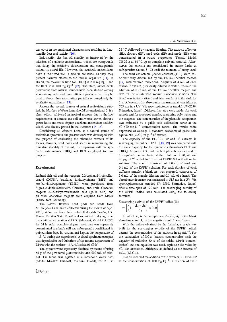
52

53
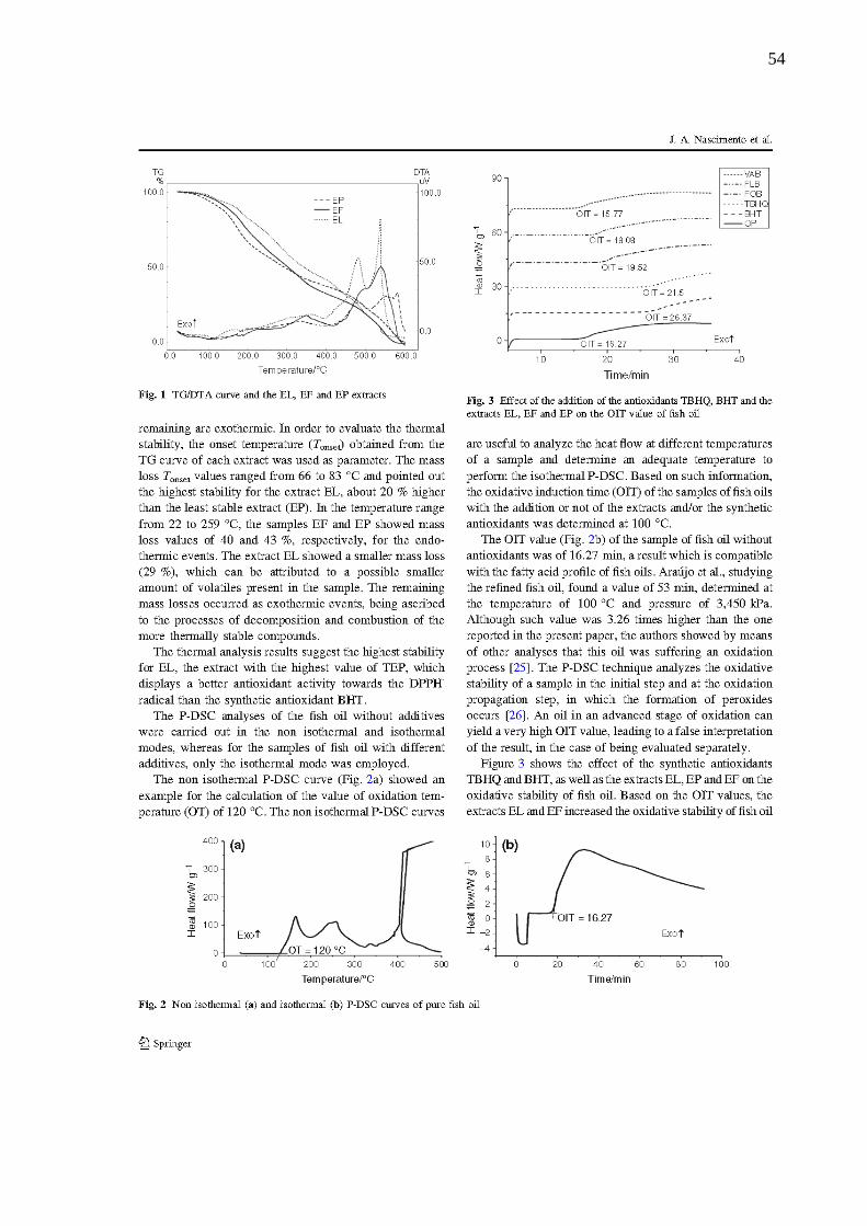
54

55
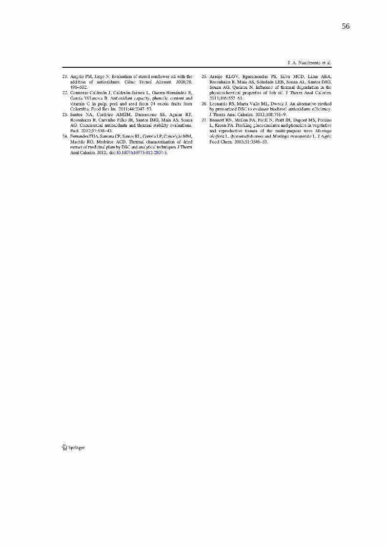
56
57
ETHANOLICS EXTRACTS OF MORINGA: ANTIOXIDANT EFFECT IN SOYBEAN OIL BY PDSC
AND RANCIMAT
Jaqueline A. Nascimento1*
, Kassandra L. G. V. Araújo1, Poliana S. Epaminondas
1,3, Alline L. S. Pontes
1,
Antonia L. Souza2, Neide Queiroz
2, Antonio G. Souza
2
1Programa de Pós-Graduação em Ciência e Tecnologia de Alimentos, CT, Campus I, Universidade Federal da
Paraíba, 58051-970, João Pessoa, Paraíba, Brazil.
2Laboratório de Combustíveis e Materiais – LACOM, Departamento de Química, CCEN, Universidade Federal
da Paraíba, 58059-900, Campus I, João Pessoa, Paraíba, Brazil.
3Setor de Agroindústria, Instituto Federal de Educação, Ciência e Tecnologia (IFPB), 58800-970, Campus
Sousa, Sousa - Paraíba, Brasil.
*Corresponding author: [email protected]
Fax: +55-83-32167441; Phone:+55-83-32167441
Abstract
The use of natural antioxidants has been widely promoted in the food industry because it is economically viable
and very attractive with consumers. In this study we determined the extractable total phenolic content (FET) of
crude ethanol extracts of the leaves (EL), flowers (EF) and seed pods (EP) of Moringa oleifera Lam. We also
evaluated the antioxidant effect of the extracts on oxidative stability of soybean oil, through the accelerated
PDSC and Rancimat® techniques using the synthetic antioxidants butylatedhydroxytoluene (BHT) and tert-
Butylhydroquinone (TBHQ) as positive control. The values of FET in the extracts ranged from 41.75 ±3.35 to
53.69 ± 1.00 mg GAE/g, and EL extract exhibited best result. The results of oxidative stability in both
techniques showed that the EL extract provided greater protection to the oil, indicating a correlation between the
amount of FET and the protective effect. Comparison made between the synthetic antioxidants and extracts
revealed that in the Rancimat technique, extracts were less effective than the synthetic antioxidants TBHQ and
BHT, however, in the PDSC technique the EL extract was more effective than BHT, proving to be a good
alternative for applications in the soybean oil, replacing this synthetic antioxidant.
Keywords: Oxidative stability, Moringa oleifera Lam., accelerated techniques, PDSC, Racimat

58
Introduction
Soybean is a culture of great commercial value in the world and is used mainly for the production of oil,
which comprises 12 to 15% of saturated fatty acids (mostly palmitic acid) and 85 to 88% acid unsaturated fatty
acids, mainly oleic, linoleic and linolenic acids [1]. The high percentage of unsaturated fatty acids makes the oil
highly susceptible to oxidative processes [2], leading to its rancidity and impaired quality [3].
To maintain the quality and prolong the shelf life of the oils, the lipid oxidation process is usually
prevented by employing synthetic antioxidants as butylated hydroxyanisole (BHA), the Butylated
hydroxytoluene (BHT) and tert-butyl hydroquinone (TBHQ). These compounds, although very satisfactory as
antioxidants, have its use restricted for having assigned to them carcinogenic and toxic effects in humans [4]. In
Brazil, the maximum concentration of BHT and TBHQ allowed is 200 mg kg-1
and 100 mg kg-1
, respectively [5].
Given the above, antioxidants from natural sources have been studied in the search for safer and more
effective products for use in foods, replacing all or part of synthetic antioxidants [6].
Moringa oleifera Lam, is a plant that has, among its chemical constituent, high content of phenolic
compounds distributed in leaves, flowers, roots and unripe fruits [7–9]. The plant is native to northern India, but
widely cultivated in tropical regions around the world. It is perfectly adapted to the climate of northeastern
Brazil, because it presents good growth, even when subjected to drought conditions and high temperatures,
characteristic of this region.
Considering the Moringa as a natural source of antioxidant compounds, the present work was to
evaluate the antioxidant potential of crude ethanol extracts of leaves, flowers and seed pods on the oxidative
stability of soybean oil compared to synthetic antioxidant TBHQ and BHT, by employing the technique of
pressurized differential scanning calorimetry (PDSC) and Rancimat ®.
Experimental
Refined soybean oil was acquired from the local market. According to the manufacturer specifications,
the soybean oil was free from antioxidant. Reagents butylatedhydroxytoluene (BHT) and tert-
Butylhydroquinone (TBHQ) were purchased from Sigma-Aldrich (Steinheim, Germany). Folin-Ciocalteau
reagent, 3,4,5-trihydroxybenzoic acid (gallic acid) and all other reagents and solvents of analytical grade were
purchased from Merck (Düsseldorf, Germany).
The leaves, flowers and seed pods from moringa (Moringa oleifera Lam.) were collected at Campus I,
Federal University of Paraíba, in the city of João Pessoa/PB, Brazil, during the month of April 2010. The plant

59
material was submitted to drying in an oven air circulation at 45 °C (Marconi, Model MA 035) for 24 h. After
complete drying, each part was separately comminuted in a knife mill and subsequently conditioned in
polyethylene bags in vacuum and kept at the temperature of -18 ºC during the experiments. A copy of exsiccate
was deposited in the Herbarium of the Botany of UFPB with the register – J.A.N. 01 Batista (JPB).
The extracts were separately obtained by means of using 50 g of the powdered plant material and 500
mL of ethanol. The bland was agitated in a metabolic water bath (Dubnoff Model MA-093, Marconi, Piracicaba,
Brazil) for 2 hours at 25 °C, followed by vacuum filtering. The extracts of leaves (EL), flowers (EF) and seed
pods (EP) were concentrated in a rotary evaporator (Model TE-221 Tecnal Piracicaba, Brazil) at 60 °C up to
complete solvent removal. Afterwards he extracts are conditioned in amber at refrigeration (about 8 ºC) until the
moment of being used.
The level of total extractable phenolics (FET) was colorimetrically determined by the Folin-Ciocalteau
method [10] with volume reductions. Aliquots of 4 mL of each ethanolic extract diluted in water, received the
addition of 0.25 mL of Folin-Ciocalteau reagent and 0.75 mL of saturated sodium carbonate. The mixture was
stirred and later was kept in the dark for 2 hours. The absorbance was measurement at 765 nm in a UV-vis
spectrophotometer (model UV-2550, Shimadzu, Japan). A sample containing only water and the reagents (white)
was used as a reference. The concentration of the phenolic compounds was estimated by using a gallic acid
calibration curve at the 50-500 mg L-1
concentration range. The results were expressed as mean ± standard
deviation of gallic acid equivalents mg (GAE)/g of extract.
Soybean oil (SO) received the addition of the extracts EL, EF and EP at the concentration of 100 mg kg-
1, in relation of their extractable total phenolic contents of each extract. The synthetic antioxidant BHT and
TBHQ were also used at a concentration of 100 mg kg-1
.
The PDSC curves of the soybean oil, with and without additives were obtained by a model DSC Q1000,
TA Instruments, pressurized differential scanning calorimeter. The isothermal curves were carried out at 110 ºC,
utilizing an aluminum crucible with approximately 10 mg sample, under oxygen atmosphere with initial
pressure of 1,400 kPa and heating rate of 10 ºC min-1
. Oxidation induction time (OIT) values were determined by
the difference of the onset time and the initial time.
The oxidative stability of soybean oil without and with antioxidant additives was determined employing
the method proposed by AOCS CD 12b-92 [11], in 873 Biodiesel Rancimat equipment from Metrohm, utilizing
a sample of 2 grams, temperature of 110 ºC, under a constant air flow (10 L h-1
). The result, expressed as

60
induction period (IP), is automatically determined from the inflection point of the curve by the software that
accompanies the equipment.
Results and Discussion
From the determination of the levels of total extractable phenolics (FET) it was possible to determine
the necessary amount of each extract equivalent to one additive 100 mg kg-1
GAE in oil. In Table 1, we can see
the total extractable phenolic content (FET) expressed as gallic acid equivalents (GAE), of the extracts studied.
The content of FET in the extracts ranged from 41.75 to 53.69 ± 3:35 ± 1.00 mg GAE g-1
, with EL extract higher
levels. EF extracts and EP showed no significant difference in the content of FET.
Table 1 FET Values of EL, EF and EP extracts
Samples mg GAE g-1 of extract
EL 53.69 ± 1.00a
EF 45.85 ± 1.71b
EP 41.75 ± 3.35b
Values are mean ± standard deviation, n=9
Mean followed by different letters in the same column differs significantly (P ≤ 0.05)
Analyzing the calorimetric curves (PDSC) of soybean oil with and without additives by the isothermal
method (110 °C) in oxygen atmosphere, there were obtained the values of the oxidation induction time (OIT) of
each sample (Figure 1).

61
Fig. 1 PDSC Curves of all samples with their respective OIT
According to the OIT values, it is found that addition of the antioxidants BHT and TBHQ, as well as
extracts of Moringa promoted increased oxidative stability of soybean oil. The percentages increase of OIT
additives oils, figure, 2, was calculated according formula:
% increase percentage = (OIT sample – OIT control) x 100/OIT control
According to the results the EL extract provided better protective effect, increasing 9.78% on the value
of the OIT in relation to oil without additive. This result, although seemingly small, was superior to the synthetic
antioxidant BHT, which increased 9.16% in the value of OIT oil without additives. The extracts EF and EP were
increased by 8.37% and 0.19% respectively. Although not having shown significant difference in the content of
FET, it was observed that EF and EP exhibited differentiated antioxidant effects when added to soybean oil. This
fact may be related to the type of antioxidant present in the extracts, which can not differentiate quantitatively,
but structurally [6].
Fig. 2 Percentage increase in OIT of BHT, TBHQ, EL, EF and EP compared to soybean oil without additive.
Different letters differs significantly (P ≤ 0.05)
The results of oxidative stability of soybean oil with and without additives by the Rancimat® Method
are shown in Table 2. Extracts of Moringa oleifera Lam showed protective effect against oxidation of soybean
oil, with emphasis on the EL. The synthetic antioxidant TBHQ showed the best protective effect among

62
additives, corroborating the results obtained by the method PDSC. Unlike calorimetric test results, however, it
was observed that BHT protects more soybean oil against oxidation than EL. These conflicting results EL and
BHT for the two assessment methods of oxidative stability leads us to suggest that EL would promote a greater
protective effect of soybean oil in the early spread of rust, while BHT would cause greater protection in the step
of lipid oxidation termination, and this enhances the antioxidant effect of the natural extract. Thus, the increase
percentage sequence of IP value was TBHQ> BHT> EL> EF> EP. Study with basil and thyme oleoresin
additives in soybean oil at a concentration of 500 mg kg-1
showed an increase of IP of 10.44 to 11.32 and 11.38
respectively, results similar to those obtained in this work with EL extract at a concentration of 100 mg kg-1
[12].
Table 2 Oxidative Stability of samples soybean oil with additives of the extracts of Moringa, BHT and TBHQ
(IP) expressed in hours (h) and as percentage of increase (%)
Samples IP / h % increase
SO 10.56f N.A.
SO + BHT 12.97b 22.78
SO + TBHQ 19.40a 83.73
SO + EL 11.42c 8.18
SO + EF 11.06d 4.75
SO + EP 10.99e 4.10
N.A. = not applicable because it is the control
Values IP Followed by different letters in the same column Differs Significantly (P ≤ 0.05)
The comparison between the oxidative stability of the samples determined by both technique shows that
not always there a correlation between these. In PDSC technique, the extrat EL was more effective than BHT
antioxidant synthetic. In Rancimat method, however, does not show the same efficacy, 14.6% less effective than
BHT. The difference in the effectiveness of the antioxidants can be attributed to different variables used in each
technique, and the principles evaluated. In the PDSC exothermic events are measured and the Rancimat, the
conductivity of the volatiles formed during oxidation [13]. According to data reported in the literature the

63
techniques determine different stages of lipid autoxidation. The technique PDSC evaluates the beginning of the
propagation of the oxidation step in which peroxides are formed, leading to an exothermic peak which is
expressed by the value OIT [14-15]. The Rancimat method, evaluates the final stage of oxidation, where volatile
compounds, such as carboxylic acids, ketones and aldehydes, increase the electrical conductivity of water,
expressed by OSI value [16]. By correlating the results obtained by these two techniques into fatty acid esters
that showed OSI less than 40 hours, R2 0.5852 was found, a result close to that found in this study [17].
Conclusions
The ethanol extracts of leaf, flower and of Moringa oleifera Lam husks increased the oxidative stability
of soybean oil, demonstrating that its application as an antioxidant in polyunsaturated lipid systems is viable.
However, it is necessary to develop complementary research with investigations of a possible synergism between
extracts and synthetic antioxidants in order to obtain a more potent antioxidant composition of the extracts used
individually. An antioxidant composition involving the extracts of Moringa, besides being economically viable
for the industry, since the costs of production are low, is attractive for the consumer, for being a natural product,
with low toxicity, and therefore a safer intake when compared to synthetic antioxidants.
Acknowledgement
The authors would like to thank the Brazilian agencies National Council for Scientific and
Technological Development (CNPq) and Coordination for the Improvement of Higher Education Personnel
(CAPES) for the financial support.
References
1 Ribeiro APB, Grimaldi R, Gioielli LA, Gonçalves LAG. Zero trans fats from soybean oil and fully
hydrogenated soybean oil: Physico-chemical properties and food applications. Food Res
Int.2009;42:401–10.
2 Hou JC, Jiang LZ, ZhangCW. Effects of frying on the trans-fatty acid formation in soybean oils. Eur J
Lipid Sci Technol.2012;114:287–93.
3 Lee KW, Hong Z, Piao F, Kim YW, Chung KW. Bond reactivity and antioxidant effect on the
autoxidation of soybean oil. J Ind Eng Chem.2010;16:419–24.
4 Sun-Waterhouse D, Thakorlal J, Zhou J. Effects of added phenolics on the storage stability of avocado
and coconut oils. Int J Food Sci Technol.2011;46:1575–85.
5 Brasil. Ministério da Saúde. Comissão Nacional de Normas e Padrões para Alimentos. Resolução no
04/88. Brasília.1988.

64
6 Brewer MS. Natural Antioxidants: Sources, Compounds, Mechanisms of Action, and Potential
Applications. Compr Rev Food Sci F.2011;10:221–47.
7 Iqbal S, Bhanger MI. Effect of season and production location on antioxidant activity of Moringa
oleifera leaves grown in Pakistan. J Food Compos Anal. 2006;19:544–51.
8 Lako J, Trenerry V, Wahlqvist M, Wattanapenpaiboon N, Sotheeswaran S, Premier R. Phytochemical
flavonols, carotenoids and the antioxidant properties of a wide selection of Fijian fruit, vegetables and
other readily available foods. Food Chem.2007;101:1727–41.
9 Reddy V, Urooj A, Kumar A. Evaluation of antioxidant activity of some plant extracts and their
application in biscuits. Food Chem.2005;90:317–21.
10 Slinkard K, Singleton VL. Total Phenol Analysis: Automation and Comparison with Manual Methods.
Am. J. Enol. Vitic. 1977;28:49–55.
11 American Oil Chemists Society. Official methods and recomended pratices of the American Oil
Chemists Society. 4th
ed. Champaign:AOCS Press;1993.
12 Del Ré PV, Jorge N. Antioxidant potential of oregano (Oreganum vulgare L.), basil (Ocimum basilicum
L.) and thyme (Thymus vulgaris L.): application of oleoresins in vegetable oil. Cien Tecnol Aliment.
2011;31:955–59.
13 Ramalho EFSM, Albuquerque AR, Souza AL, Barro AK, Maia AS, Santos IMG, Souza AG. Use of
different techniques in the evaluation of the oxidative stability of poultry fat biodiesel. J Therm Anal
Calorim. 2011;106:787–91.
14 Diniz ZN, Bora PS, NetoVQ, Cavalheiro JMO. Sterculia striata seed kernel oil: Characterization and
thermal stability. Gras Aceit, vol. 59, no. 2, pp. 160–165, Jun. 2008.
15 Candeia RA, Sinfrônio FSM, Bicudo TC, Queiroz N, Barros Filho AKD, Soledade LEB, Santos IMG,
Souza AL, Souza AG. Influence of the storage on the thermo-oxidative stability of methyl and ethyl
esters by PDSC.J Therm Anal Calorim.2011;106:581–6.
16 Leonardo RS, Valle MLM, Dweck J. An alternative method by pressurized DSC to evaluate biodiesel
antioxidants efficiency. J Therm Anal Calorim.2012;108:751–9.
17 Moser BR. Comparative Oxidative Stability of Fatty Acid Alkyl Esters by Accelerated Methods. J Am
Oil Chem Soc.2009;86:699–706.

65
Oxidative stability of fish oil supplemented with ethanolic extracts of Moringa oleifera 1
Lam. compared with synthetic antioxidants during accelerated test 2
3
Jaqueline A. Nascimento1*
, Kassandra L. G. V. Araújo1, Poliana S. Epaminondas
1,3, Alline S. 4
Souza1, Antônia L. Souza
2, M. C. D. Silva
4, Antônio G. Souza
2 5
6
1Graduate Program in Food Science and Technology, CT, Campus I, Federal University of 7
Paraíba, CEP 58051-970, João Pessoa, Paraíba, Brazil. 8
2 Laboratory of Fuels and Materials - LACOM, Department of Chemistry, CCEN, Campus I, 9
Federal University of Paraíba, CEP 58059-900, João Pessoa, Paraíba, Brazil. 10
3 Department of Agribusiness, Federal Institute of Education, Science and Technology 11
(IFPB), 58800-970, Campus Sousa, Sousa - Paraiba, Brazil. 12
4 Center for Social Science, Health and Technology, Federal University of Maranhão, Campus 13
Imperatriz 14
*Corresponding author: [email protected] 15
Fax: +55-83-32167441; Phone:+55-83-32167441 16
17
Abstract 18
The effects of ethanolic extracts of leaves, flowers and seed pods from Moringa oleifera Lam 19
and synthetic antioxidants TBHQ and BHT on the oxidative stability of fish oil stored for 16 20
days at 60°C were compared. The investigation focused on the increased peroxide, anisidine 21
and conjugated diene contents. Changes in trans fatty acid and aldehyde contents were 22
investigated by Fourier transform infrared spectroscopy. Results showed that extracts were 23
effective in delaying oxidation and among these, extracts of leaves was the most efficient, 24
being more protective than TBHQ and BHT in controlling the formation of peroxides and 25
hydroperoxides. In controlling secondary oxidation, synthetic antioxidants were more 26
effective; however, extracts of leaves preserved better PUFAS than TBHQ. Results showed 27
that the extracts of leaves is an antioxidant as effective as BHT and TBHQ for use in fish oil 28
and its natural origin possibly makes it a safe additive for use in foods. 29
30
Keywords: Fish oil, Moringa oleifera Lam, Oxidative stability 31
32

66
1. Introduction 33
Marine fish oils are composed of long-chain polyunsaturated fatty acids, 34
eicosapentaenoic acid (EPA) and docosahexaenoic acid (DHA). These fatty acids have 35
beneficial properties for human health, protecting against diseases such as Alzheimer's disease 36
(Lukiw & Bazan, 2008), depression (Nemets, Nemets, Apter, Bracha, & Belmaker, 2006), 37
cognitive decline (Morris, Evans, Tangney, Bienias, & Wilson, 2005), rheumatoid arthritis 38
(Bykerk & Keystone, 2005) and cardiovascular disease (Kris-Etherton et al., 2002), besides 39
contributing in the development of the nervous system and growth (Pivik, Dykman, Jing, 40
Gilchrist, & Badger, 2009). However, the presence of such acids in fish oil makes it highly 41
susceptible to oxidation (Leclerc, Zhao, Simpson & Zuta, 2007). 42
Autoxidation is a process that occurs in three stages. In the initial and propagation 43
stage, there is the formation of free radicals and neutral molecules such as hydroperoxides, 44
which without interruption by antioxidants, the reaction proceeds resulting in the formation of 45
secondary oxidation products, small-chain carboxylic acids, aldehydes and ketones. 46
Secondary oxidation products compromise not only color, odor and flavor but also the 47
nutritional characteristics of the oil, resulting in loss of functionality and toxicity (Rehman & 48
Salariya, 2006; Siriwardhana, Lee, Kim, Ha, Park & Jeonl, 2004). 49
In industry, the stability of fish oil is improved by the use of synthetic antioxidants, 50
capable of delaying oxidative deterioration and, consequently, extending its shelf life. 51
However, the use of synthetic antioxidants is restricted in many countries due to possible 52
adverse effects on human health (Hras, Hadolin, Knez, & Bauman, 2000). In Brazil, the use of 53
TBHQ is limited to 200 mg.kg-1
and BHT to 100 mg.kg-1
(Brasil, Saúde, & Conselho 54
Nacional de Saúde, 1988). In this context, anti-oxidants from natural sources have been 55
studied in the attempt to obtain safer and more effective products which can be used in foods, 56
fully or partially replacing synthetic antioxidants (Brewer, 2011). 57
Among the various sources of natural antioxidants studied, Moringa oleifera Lam. 58
Stands out, a plant originating from northwest India and widely cultivated in tropical regions 59
due to the low demands of climate and soil and whose leaves, flowers, unripe fruits and roots 60
have a rich and rare combination of zeatin, quercetin, β - sitosterol, caffeoylquinic acid and 61
kaempferol, giving the plant a potential antioxidant activity (Iqbal & Bhanger, 2006; Lako, 62
Trenerry, Wahlqvist, Wattanapenpaiboon, Sotheeswaran & Premier, 2007; Reddy, Urooj, & 63
Kumar, 2005; Siddhuraju & Becker, 2003). In southern India, its fresh leaves are traditionally 64
used in the preparation of foods rich in fat in order to increase their shelf life (Siddhuraju & 65
Becker, 2003). Considering Moringa as a natural source of antioxidant compounds, the 66
objective of this study was to evaluate the ethanol extracts of its leaves, flowers and seed 67
pods, to assess the technical feasibility of its constituents as antioxidant additives in fish oil 68
and to compare its efficacy compared with synthetic antioxidants industrially used for this 69
purpose. 70
71
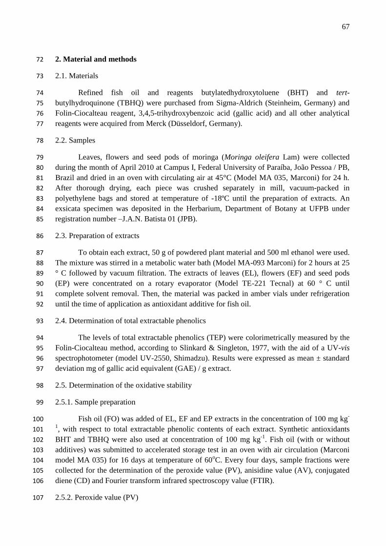
67
2. Material and methods 72
2.1. Materials 73
Refined fish oil and reagents butylatedhydroxytoluene (BHT) and tert-74
butylhydroquinone (TBHQ) were purchased from Sigma-Aldrich (Steinheim, Germany) and 75
Folin-Ciocalteau reagent, 3,4,5-trihydroxybenzoic acid (gallic acid) and all other analytical 76
reagents were acquired from Merck (Düsseldorf, Germany). 77
2.2. Samples 78
Leaves, flowers and seed pods of moringa (Moringa oleifera Lam) were collected 79
during the month of April 2010 at Campus I, Federal University of Paraíba, João Pessoa / PB, 80
Brazil and dried in an oven with circulating air at 45°C (Model MA 035, Marconi) for 24 h. 81
After thorough drying, each piece was crushed separately in mill, vacuum-packed in 82
polyethylene bags and stored at temperature of -18ºC until the preparation of extracts. An 83
exsicata specimen was deposited in the Herbarium, Department of Botany at UFPB under 84
registration number –J.A.N. Batista 01 (JPB). 85
2.3. Preparation of extracts 86
To obtain each extract, 50 g of powdered plant material and 500 ml ethanol were used. 87
The mixture was stirred in a metabolic water bath (Model MA-093 Marconi) for 2 hours at 25 88
° C followed by vacuum filtration. The extracts of leaves (EL), flowers (EF) and seed pods 89
(EP) were concentrated on a rotary evaporator (Model TE-221 Tecnal) at 60 ° C until 90
complete solvent removal. Then, the material was packed in amber vials under refrigeration 91
until the time of application as antioxidant additive for fish oil. 92
2.4. Determination of total extractable phenolics 93
The levels of total extractable phenolics (TEP) were colorimetrically measured by the 94
Folin-Ciocalteau method, according to Slinkard & Singleton, 1977, with the aid of a UV-vis 95
spectrophotometer (model UV-2550, Shimadzu). Results were expressed as mean ± standard 96
deviation mg of gallic acid equivalent (GAE) / g extract. 97
2.5. Determination of the oxidative stability 98
2.5.1. Sample preparation 99
Fish oil (FO) was added of EL, EF and EP extracts in the concentration of 100 mg kg-100
1, with respect to total extractable phenolic contents of each extract. Synthetic antioxidants 101
BHT and TBHQ were also used at concentration of 100 mg kg-1
. Fish oil (with or without 102
additives) was submitted to accelerated storage test in an oven with air circulation (Marconi 103
model MA 035) for 16 days at temperature of 60oC. Every four days, sample fractions were 104
collected for the determination of the peroxide value (PV), anisidine value (AV), conjugated 105
diene (CD) and Fourier transform infrared spectroscopy value (FTIR). 106
2.5.2. Peroxide value (PV) 107

68
The peroxide value was determined by the Cd 8-53 method of AOCS (AOCS, 2003). 108
Fish oil samples (2g) were dissolved in 30 ml of acetic acid: chloroform solution (3:2 v / v), 109
followed by the addition of 0.5 ml of saturated potassium iodide solution. The mixture was 110
allowed to stand for exactly one minute and then added of 30 ml of freshly boiled water and 111
0.5 mL of 1% starch. The iodine released was titrated with sodium thiosulfate solution 0.01 N 112
until the disappearance of blue coloration. A blank test was performed under the same 113
conditions, but without the sample. The calculations were performed according to Equation: 114
PV (meq/kg) = N x (VA – VB) x 1000 / W sample (g) 115
Where: N is the normality of the Na2S2O3 solution, VA is the Na2S2O3 solution volume 116
consumed by the sample (mL), VB is the Na2S2O3 solution volume consumed by the blank 117
(mL), and W is the sample weight (g). 118
2.5.3. Anisidine value (AV) 119
The anisidine value was determined according to Cd 8-53 method of AOCS (AOCS, 120
2003). Fish oil sample (0.5g) was dissolved in 25 mL of isooctane and its absorbance was 121
measured at 350 nm using isooctane with blank. About 5 ml of fat and isooctane solution was 122
added of 1 mL of reagent anisidine (0.25% p-anisidine solution in glacial acetic acid) and 123
after 10 minutes in the dark, the absorbance of the fat-anisidine solution against solvent 124
anisidine solution was measured at 350 nm in UV-vis spectrophotometer (model UV-2550, 125
Shimadzu). The anisidine value (AV) was calculated according to the equation: 126
AV = 25 (1.2 AS – AB) / P 127
Where As is absorbance of the fat-anisidine solution, Ab is the absorbance of the initial fat 128
solution and P is the sample weight in grams. 129
2.5.4. Conjugated diene (CD) value 130
The method Proposed by Leclerc, Zhao, Simpson & Zuta (2007) was slightly modified 131
and used for the measurement of the CD content in the oil. Samples (0.02 g) were diluted with 132
isooctane, and the solution absorbance was determined against a blank made from isooctane 133
at 232 nm. The CD value was calculated from the absorbance value and the final sample 134
concentration (g/100 mL). The results are expressed as CD values, computed as follows: 135
CD = A232 / C x P 136
where A is the sample absorbance at 232 nm, C denotes the final dilution of the sample 137
concentration (g 100 mL), and P represents the length of the measuring cell (cm). 138
2.5.5. Fourier transform infrared spectroscopy (FTIR) 139
The absorption spectra in the infrared region were obtained from IR 140
spectrophotometer with Fourier transform model IR Prestige-21, Class 1 Laser Product from 141
Shimadzu. The analysis was performed in the region from 4000 to 650 nm using a reflection 142
ATR accessory with ZnSe prism, at resolution of 4 cm-1
. 143

69
2.5.6. Statistical analysis 144
Results were expressed as mean ± standard deviation, compared by analysis of 145
variance (ANOVA) and Duncan test, considering p <0.05, using the Statistica 7.0 software 146
(Statsoft ®). 147
148
3. Results and discussion 149
3.1. Total extractable phenolics (TEP) 150
From the determination of total extractable phenolics (TEP), it was possible to 151
determine the necessary amount of each extract equivalent to additivation of 100 mg kg-1
152
GAE in the oil. The TEP content expressed as gallic acid equivalents (GAE) of extracts 153
studied was 41.75 ± 3.35, 45.85 ± 1.71 and 53.69 ± 1.00 mg GAE / g, respectively for EP, EF 154
and EL extracts. 155
3.2. Peroxide value (PV) 156
Primary oxidation products (peroxides and hydroperoxides) from fish oil samples with 157
and without additives (100 mg kg-1
) were determined by measuring the PV, which is one of 158
the most widely used tests for evaluating the initial oxidation in oils and fats. Figure 1 shows 159
the influence of different additives on the increase in PV in fish oil during the storage period. 160
The results indicated that all samples showed increasing trend in the peroxide value and the 161
control sample (FO) showed greater increase in PV compared to other treatments, 162
demonstrating that all additives provide protection to fish oil against oxidation. Samples 163
added of EL showed the smallest increase in PV, revealing a greater antioxidant effect of this 164
extract in the initial oxidation phase, exceeding synthetic antioxidants BHT and TBHQ, which 165
did not differ significantly from each other (p> 0.5). It was observed that in the control sample 166
(FO) and in samples added of EL, EF and EP extracts, the maximum PV was reached at about 167
the 12th
day of storage, and that after this day, the PV began to fall, which means that the 168
primary oxidation products (hydroperoxides) began to decompose, yielding stable products 169
such as aldehydes, ketones, small-chain carboxylic acids, among others (Rehman & Salariya, 170
2006; Siriwardhana et al. 2004). Thus, the effectiveness sequence as antioxidant in the initial 171
oxidation stages was EL> BHT = TBHQ> EP> EF. Wang et al. (2011) evaluated the 172
oxidative stability of fish oil supplemented with carnosic acid compared with synthetic 173
antioxidants during storage at different temperatures and observed that the synthetic 174
antioxidant TBHQ was more effective than carnosic acid, since TBHQ showed lower PV. 175
3.3. Anisidine value (AV) 176
The anisidine value is useful in assessing secondary lipid oxidation. It determines the 177
aldehyde levels in animal fats and vegetable oils. According to Figure 2, AV values increased 178
in all samples during the storage period. The increase in AV in control samples (FO) was 179
significantly higher (p <0.05) than that obtained in samples with additives. Samples 180
containing antioxidants TBHQ and BHT did not differ significantly from each other (p> 181

70
0.05), and showed the lowest increase in AV, followed by samples with EL. The highest AV 182
was observed in samples containing EF and EP, which showed no significant difference from 183
each other (p> 0.05). The AV data corroborate the PV results, indicating that all additives 184
studied provided oxidative stability to fish oil; however, the most effective were synthetic 185
antioxidants BHT and TBHQ, followed EL extract. 186
3.4. Conjugated diene (CD) value 187
The conjugated diene value is often used to measure hydroperoxides and together with 188
PV, evaluates primary oxidation products. The peroxidation of unsaturated fatty acids leads to 189
the displacement of the double bond, forming a conjugated diene system which strongly 190
absorbs ultraviolet light at wavelength between 232 and 234 nm. The CD determination is a 191
strong indicator of the oxidative status of oils, and can be used to evaluate the effectiveness of 192
antioxidants (Iqbal & Bhanger, 2007). The CD values of fish oil samples with and without 193
additives increased during the storage period, and from the 12th
day of storage, the control 194
sample (FO), showed a higher increase compared to samples with additives (Fig. 3). On the 195
16th
day of storage, CD values of FO sample increased by 55.63%, while samples added of 196
TBHQ, BHT and EL showed the smallest increase in the CD value, with 26.40%, 27.00% and 197
27.09%, respectively, with no significant difference (p> 0.05) between the results of these 198
samples. Samples added of EF and EP showed increased in CD value by 40.24% and 40.98%, 199
differing significantly (p <0.05) from value shown by the control sample, but with no 200
significant difference from each other. These results show that all extracts used in the 201
experiment exhibited effective inhibition of the CD form in the fish oil, especially EL, which 202
was as effective as synthetic antioxidants BHT and TBHQ, which is consistent with the 203
results obtained for PV and AV tests previously shown. Studies using carnosic acid as fish oil 204
additive also exhibited effective inhibition of the CD form (Wang et al. 2011). 205
3.5. Fourier Transform Infrared Spectroscopy (FTIR) 206
In the evaluation of the fish oil degradation by FTIR, the following spectral regions 207
were observed: (1) absorption peaks around 982 cm-1
corresponding to absorption of trans 208
fatty acids (TFA) relating to the C-H vibration outside the trans configuration plane of the 209
double bond, (2) absorption peaks around 1734 cm-1
corresponding to carbonyl absorption, 210
(3) absorption peaks around 3015 cm-1
, corresponding to cis double bonds which are related 211
to the PUFA contents present in the fish oil, and (4) absorption peaks at 3500 cm-1
, 212
corresponding to carbonyl frequency multiplication. Since the absorption peak of the carbonyl 213
frequency multiplication was broad and the noise was greater nearby, quantification was 214
difficult. Content changes in the carbonyl cis double bond, and trans double bond of fish oil 215
during storage at 60 ° C are shown in Fig 4 (AC). The initial stage of the oxidation process of 216
fish oil results in a reduction in the cis double bonds of polyunsaturated fatty acids. Figure 4 – 217
A shows that on the 16th
day of storage, the control sample (FO) showed a decrease in the 218
absorption peak at 3015 cm-1
of 15.60%. The sample added of EF showed identical reduction, 219
indicating that this extract did not preserve PUFAS present in the sample. On the other hand, 220
EL and EP extracts decreased by only 7.72% and 11.87% respectively. Samples added of 221
synthetic antioxidants BHT and TBHQ showed a decrease of 3.29% and 11.82%, 222

71
respectively, showing that EL was more effective than TBHQ in the preservation of PUFAs. 223
While the content of cis double bonds decreases throughout the storage time, Figure 4 – C 224
shows that the content of trans double bonds relating to trans fatty acids (TFA) increased by 225
49.04% in the control sample (FO), 27.41% for sample added of EL, 20:43% for sample 226
added of BHT and 14.94% for sample added of TBHQ. Samples added of EF showed an 227
increase of 38.76%, while samples added of EP increased by 50.68%, higher than values 228
observed in the control sample. The carbonyl content is related to the oxidation degree, 229
increasing during the storage time with the formation of products arising from the oxidation 230
of DHA, EPA or other constituents present in fish oil. Figure 4 – B shows that the carbonyl 231
content increased by 11.50% in the control samples. Samples added of EL (8.71%), EP 232
(10.10%), BHT (6.29%) and TBHQ (5.56%) showed increase in carbonyl content lower than 233
the control, while samples added of EF increased by 13.13%. 234
235
4. Conclusions 236
The ethanol extracts of moringa showed inhibitory effect against fish oil oxidation, 237
and EL extract was the most effective, whose protection was more effective than that 238
performed by synthetic antioxidants BHT and TBHQ in the formation of peroxides and 239
hydroperoxides. The effect of the EL extract on fish oil was also significant in reducing 240
secondary oxidation products and in protecting polyunsaturated fatty acids, reducing the 241
formation of trans fatty acids during storage at 60°C, which demonstrates its potential use in 242
edible oils, as an alternative to replace synthetic antioxidants. 243
244
5. Acknowledgement 245
The authors would like to thank the Brazilian agencies National Council for Scientific 246
and Technological Development (CNPq) and Coordination for the Improvement of Higher 247
Education Personnel (CAPES) for the financial support. 248
249
6. References 250
AOCS. (2003). Official Methods and Recommended Pratices of the American Oil Chemists 251
Society.Champaign Illinois, USA:AOCS. 252
Brasil (1988). Resolução CNS/MS no 04 de 24 de novembro de 1988. Diário Oficial da 253
União, 19 de dezembro de 1988. 254
Brewer, M. S. (2011). Natural Antioxidants: Sources, Compounds, Mechanisms of Action, 255
and Potential Applications. Comprehensive Reviews in Food Science and Food Safety, 256
10(4), 221–247. 257

72
Bykerk, V. P., & Keystone, E. C. (2005). What are the goals and principles of management in 258
the early treatment of rheumatoid arthritis? Best practice & research. Clinical 259
rheumatology, 19(1), 147–61. 260
Hras, A. R., Hadolin, M., Knez, Z., & Bauman, D. (2000). Comparison of antioxidative and 261
synergistic effects of rosemary extract with α-tocopherol, ascorbyl palmitate and citric 262
acid in sunflower oil. Food Chemistry, 71, 229–233. 263
Iqbal, S., & Bhanger, M. I. (2006). Effect of season and production location on antioxidant 264
activity of Moringa oleifera leaves grown in Pakistan. Journal of Food Composition and 265
Analysis, 19(6-7), 544–551. 266
Iqbal, S., & Bhanger, M. I. (2007). Stabilization of sunflower oil by garlic extract during 267
accelerated storage. Food Chemistry, 100(1), 246–254. 268
Kris-etherton, P. M., Hecker, K. D., Bonanome, A., Coval, S. M., Binkoski, A. E., Hilpert, K. 269
F., Griel, A. E., Etherton, T. D. (2002). Bioactive Compounds in Foods: Their Role in 270
the Prevention of Cardiovascular Disease and Cancer. The American Journal of 271
Medicine, 113(9), 71–88. 272
Lako, J., Trenerry, V., Wahlqvist, M., Wattanapenpaiboon, N., Sotheeswaran, S., & Premier, 273
R. (2007). Phytochemical flavonols, carotenoids and the antioxidant properties of a wide 274
selection of Fijian fruit, vegetables and other readily available foods. Food Chemistry, 275
101(4), 1727–1741. 276
Leclerc, L., Zhao, X., Simpson, B. K., & Zuta, P. C. (2007). The effect of α-tocopherol on the 277
oxidation of mackerel oil.pdf. Food Chemistry, 100, 800–807. 278
Lukiw, W. J., & Bazan, N. G. (2008). Docosahexaenoic Acid and the Aging Brain. American 279
Society for Nutrition, 138(12), 2510–2514. 280
Morris, M. C., Evans, D., Tangney, C. C., Bienias, J. L., & Wilson, R. S. (2005). Fish 281
consumption and cognitive decline with age in a large community study. Archives of 282
neurology, 62(12), 1849–53. 283
Nemets, H., Nemets, B., Apter, A., Bracha, Z., & Belmaker, R. H. (2006). Omega-3 treatment 284
of childhood depression: a controlled, double-blind pilot study. The American journal of 285
psychiatry, 163(6), 1098–100. 286
Pivik, R. T., Dykman, R. A., Jing, H., Gilchrist, J. M., & Badger, T. M. (2009). Early Infant 287
Diet and the Omega 3 Fatty Acid DHA: Effects on Resting Cardiovascular Activity and 288
Behavioral Development During the First Half-Year of Life. Developmental 289
Neuropsychology, 34(2), 139–158. 290

73
Reddy, V., Urooj, a, & Kumar, a. (2005). Evaluation of antioxidant activity of some plant 291
extracts and their application in biscuits. Food Chemistry, 90(1-2), 317–321. 292
Rehman, Z., & Salariya, A. (2006). Effect of synthetic antioxidants on storage stability of 293
Khoa – a semi-solid concentrated milk product. Food Chemistry, 96(1), 122–125. 294
Siddhuraju, P., & Becker, K. (2003). Antioxidant properties of various solvent extracts of 295
total phenolic constituents from three different agroclimatic origins of drumstick tree 296
(Moringa oleifera Lam.) leaves. Journal of agricultural and food chemistry, 51(8), 297
2144–55. 298
Siriwardhana, N., Lee, K.W., Kim, S.H., Ha, J.H., Park, G.T., & Jeon, Y.J. (2004). Lipid 299
Peroxidation Inhibitory Effects of Hizikia Fusiformis Methanolic Extract on Fish Oil and 300
Linoleic Acid. Food Science and Technology International, 10(2), 65–72. 301
Slinkard, K., & Singleton, V. L. (1977). Total Phenol Analysis: Automation and Comparison 302
with Manual Methods. American Journal of Enology and Viticulture, 28(1), 49–55. 303
Wang, H., Liu, F., Yang, L., Zu, Y., Wang, H., Qu, S., & Zhang, Y. (2011). Oxidative 304
stability of fish oil supplemented with carnosic acid compared with synthetic 305
antioxidants during long-term storage. Food Chemistry, 128(1), 93–99. 306
307
308
309
310
311
312
313
314
315
316
317
318
319
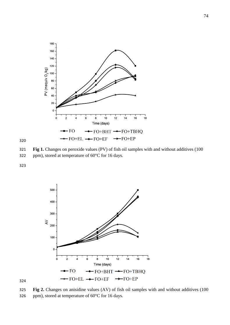
74
320
Fig 1. Changes on peroxide values (PV) of fish oil samples with and without additives (100 321
ppm), stored at temperature of 60°C for 16 days. 322
323
324
Fig 2. Changes on anisidine values (AV) of fish oil samples with and without additives (100 325
ppm), stored at temperature of 60°C for 16 days. 326

75
327
Fig 3. Changes on conjugated diene (CD) values of fish oil samples with and without 328
additives (100 ppm), stored at temperature of 60°C for 16 days. 329
330
331
Fig. 4 Changes of the main spectral peaks of fish oil by FTIR. The spectrogram was fish oil at 332
the time 0 with no antioxidant analysed by FTIR. (A) cis double bond, (B) C=O vibration, (C) 333
trans double bond. All the samples for analyse were stored at the temperature of 60oC. 334

76
Antioxidant effect of Moringa oleifera Lamark extracts in soybean oil during accelerated 1
storage test 2
3
Jaqueline A. Nascimento1*
, Kassandra L. G. V. Araújo1, Poliana S. Epaminondas
1,3, Alline S. 4
Souza1, Antônia L. Souza
2, Marciane Magnani
1, Neide Queiroz
2, Antônio G. Souza
2 5
6 1Graduate Program in Food Science and Technology, CT, Campus I, Federal University of 7
Paraíba, CEP 58051-970, João Pessoa, Paraíba, Brazil. 8 2 Laboratory of Fuels and Materials - LACOM, Department of Chemistry, CCEN, Campus I, 9
Federal University of Paraíba, CEP 58059-900, João Pessoa, Paraíba, Brazil. 10 3 Department of Agribusiness, Federal Institute of Education, Science and Technology 11
(IFPB), 58800-970, Campus Sousa, Sousa - Paraiba, Brazil. 12
*Corresponding author: [email protected] 13
Fax: +55-83-32167441; Phone:+55-83-32167441 14
15
Abstract 16
17
The effects of ethanolic extracts of leaves (EL), flowers (EF) and seed pods (EP) from 18
MoringaoleiferaLamark on the oxidative stability of soybean oil stored for 16 days at 60°C 19
were evaluated. The investigation was focused on the increase in peroxide, anisidine and 20
conjugated diene values. The changes in trans fatty acid and aldehyde contents were 21
investigated by Fourier transform infrared spectroscopy. The results show that the moringa 22
extracts tested have protective effects against soybean oil oxidation, showing higher activity 23
compared to synthetic antioxidant BHT in inhibiting peroxide formation and controlling 24
secondary oxidation. The most efficient extract was EL, with protective effects equal to or 25
better than BHT against oxidation of PUFAs, and the formation of carbonyl and trans fatty 26
acids, respectively. In addition, this extract was more effective than TBHQ in the inhibition of 27
trans fatty acids. The results suggest that EL extract has potential to be used as an antioxidant 28
for preserving soybean oil. 29
30
Keywords: Soybean oil, Moringa oleifera Lam., Oxidative stability 31
32
1. Introduction 33
34
The species Moringa oleifera Lamarck is a perennial and drought-tolerant plant 35
cultivated as ornamental and medicinal plant (Sánchez-Machado, Núñez-Gastélum, Reyes-36
Moreno, Ramirez-Wong, & Lopez-Cervantes, 2009). In Brazil, it is found mainly in the 37
Northeastern region, where it is known as lily-white or simply moringa (Silva et al., 2012). 38
All parts of this plant can be used for some purpose, and its use is known in human and 39
animal nutrition, agriculture, pharmaceuticals, cosmetics and food, in water treatment and in 40
the production of biodiesel and lubricant. (Debnath, Biswas, Ray, & Guha, 2011; Rashid 41
Anwar, Moser, & Knothe, 2008; Sharma, Rashid Anwar, & Erhan, 2009). This plant has a 42
high content of phenolic acids and carotenoids, and its constitution has a rare combination of 43

77
zeatin, quercetin, β-sitosterol, kaempferol caffeoylquinic acid, which provides moringa a high 44
antioxidant power (Anwar Hussain, Iqbal, & Bhanger, 2007; Iqbal & Bhanger, 2006; Lako et 45
al., 2007). 46
Antioxidants from natural sources have been studied with the goal of identifying 47
effective compounds that can fully or partially replace synthetic antioxidants in foods, such as 48
butylated hydroxyanisole (BHA), butylated hydroxytoluene (BHT) and tert-butyl 49
hydroquinone (TBHQ ) (Brewer, 2011), which have restricted use due to their toxicity and 50
potential carcinogenic effects to humans (Sun-Waterhouse, Thakorlal, & Zhou, 2011). 51
Since moringa is a natural source of compounds with antioxidant potential, and that 52
soybean oil is the most consumed edible oil worldwide, the present work aimed to evaluate 53
the oxidative stability of soybean oil added to ethanolic extracts of leaves, flowers and seed 54
pods of moringa during accelerated storage test. 55
56
2. Material and methods 57
58
2.1. Material 59
60
Refined soybean oil without additives were purchased from Cargill Agricultural S.A., 61
Brazil. Reagents butylatedhydroxytoluene (BHT) e tert-Butylhydroquinone (TBHQ) were 62
purchased from Sigma-Aldrich (Steinheim, Germany) and Folin-Ciocalteau reagent, 3,4,5-63
trihydroxybenzoic acid (gallic acid) and all other analitical reagentes were acquired from 64
Merck (Düsseldorf, Germany). 65
66
2.2. Samples 67
68
Leaves, flowers and seed pods of moringa (Moringa oleifera Lam) were collected 69
during the month of April 2010 at Campus I, Federal University of Paraíba, João Pessoa / PB, 70
Brazil and dried in an oven with circulating air at 45°C (Model MA 035, Marconi) for 24 h. 71
After thorough drying, each piece was crushed separately in mill, vacuum-packed in 72
polyethylene bags and stored at temperature of -18ºC until the preparation of extracts. 73
74
2.3. Extracts 75
76
To obtain each extract, 50 g of powdered plant material and 500 ml ethanol were used. 77
The mixture was stirred in a metabolic water bath (Model MA-093 Marconi) for 2 hours at 25 78
° C followed by vacuum filtration. The extracts of leaves (EL), flowers (EF) and seed pods 79
(EP) were concentrated on a rotary evaporator (Model TE-221 Tecnal) at 60 ° C until 80
complete solvent removal. Then, the material was packed in amber vials under refrigeration 81
until the time of application as antioxidant additive for soybean oil. 82
83
2.4. Determination of total extractable phenolics 84
85

78
The levels of total extractable phenolics (TEP) were colorimetrically measured by the 86
Folin-Ciocalteau method, according to Slinkard & Singleton, 1977, with the aid of a UV-vis 87
spectrophotometer (model UV-2550, Shimadzu). Results were expressed as mean ± standard 88
deviation mg of gallic acid equivalent (GAE) / g extract. 89
90
2.5. Determination of the oxidative stability 91
92
2.5.1. Sample preparation 93
94
Soybean oil (SO) was added of EL, EF and EP extracts in the concentration of 100 mg 95
kg-1
, with respect to total extractable phenolic contents of each extract. Synthetic antioxidants 96
BHT and TBHQ were also used at concentration of 100 mg kg-1
. Soybean oil (with or without 97
additives) was submitted to accelerated storage test in an oven with air circulation (Marconi 98
model MA 035) for 16 days at temperature of 60oC. Every four days, sample fractions were 99
collected for the determination of the peroxide value (PV), anisidine value (AV), conjugated 100
diene (CD) and Fourier transform infrared spectroscopy value (FTIR). 101
102
2.5.2. Peroxide value (PV) 103
104
The peroxide value was determined by the Cd 8-53 method of AOCS (AOCS, 2003). 105
Soybean oil samples (2g) were dissolved in 30 ml of acetic acid: chloroform solution (3:2 v / 106
v), followed by the addition of 0.5 ml of saturated potassium iodide solution. The mixture was 107
allowed to stand for exactly one minute and then added of 30 ml of freshly boiled water and 108
0.5 mL of 1% starch. The iodine released was titrated with sodium thiosulfate solution 0.01 N 109
until the disappearance of blue coloration. A blank test was performed under the same 110
conditions, but without the sample. The calculations were performed according to Equation: 111
PV (meq/kg) = N x (VA – VB) x 1000 / W sample (g) 112
In which: N is the normality of the Na2S2O3 solution, VA is the Na2S2O3 solution volume 113
consumed by the sample (ml), VB is the Na2S2O3 solution volume consumed by the blank 114
(mL), and W is the sample weight (g). 115
116
2.5.3. Anisidine value (AV) 117
118
The anisidine value was determined according to Cd 8-53 method of AOCS (AOCS, 119
2003). Soybean oil sample (0.5g) was dissolved in 25 ml of isooctane and its absorbance was 120
measured at 350 nm using isooctane with blank. About 5 ml of fat and isooctane solution was 121
added of 1 mL of reagent anisidine (0.25% p-anisidine solution in glacial acetic acid) and 122
after 10 minutes in the dark, the absorbance of the fat-anisidine solution against solvent 123
anisidine solution was measured at 350 nm in UV-vis spectrophotometer (model UV-2550, 124
Shimadzu). The anisidine value (AV) was calculated according to the equation: 125
AV = 25 (1.2 AS – AB) / P 126
In which AS is absorbance of the fat-anisidine solution, AB is the absorbance of the initial fat 127
solution and P is the sample weight in grams. 128
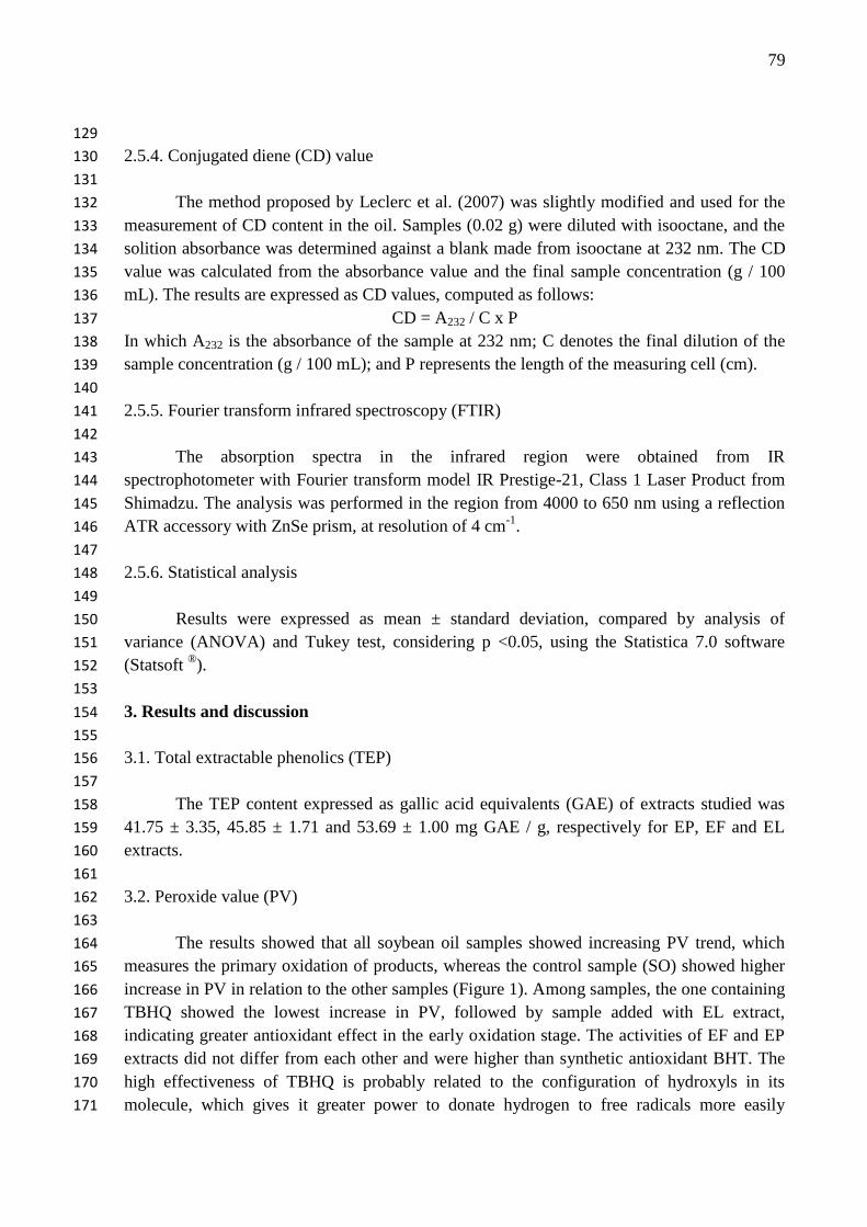
79
129
2.5.4. Conjugated diene (CD) value 130
131
The method proposed by Leclerc et al. (2007) was slightly modified and used for the 132
measurement of CD content in the oil. Samples (0.02 g) were diluted with isooctane, and the 133
solition absorbance was determined against a blank made from isooctane at 232 nm. The CD 134
value was calculated from the absorbance value and the final sample concentration (g / 100 135
mL). The results are expressed as CD values, computed as follows: 136
CD = A232 / C x P 137
In which A232 is the absorbance of the sample at 232 nm; C denotes the final dilution of the 138
sample concentration (g / 100 mL); and P represents the length of the measuring cell (cm). 139
140
2.5.5. Fourier transform infrared spectroscopy (FTIR) 141
142
The absorption spectra in the infrared region were obtained from IR 143
spectrophotometer with Fourier transform model IR Prestige-21, Class 1 Laser Product from 144
Shimadzu. The analysis was performed in the region from 4000 to 650 nm using a reflection 145
ATR accessory with ZnSe prism, at resolution of 4 cm-1
. 146
147
2.5.6. Statistical analysis 148
149
Results were expressed as mean ± standard deviation, compared by analysis of 150
variance (ANOVA) and Tukey test, considering p <0.05, using the Statistica 7.0 software 151
(Statsoft ®). 152
153
3. Results and discussion 154
155
3.1. Total extractable phenolics (TEP) 156
157
The TEP content expressed as gallic acid equivalents (GAE) of extracts studied was 158
41.75 ± 3.35, 45.85 ± 1.71 and 53.69 ± 1.00 mg GAE / g, respectively for EP, EF and EL 159
extracts. 160
161
3.2. Peroxide value (PV) 162
163
The results showed that all soybean oil samples showed increasing PV trend, which 164
measures the primary oxidation of products, whereas the control sample (SO) showed higher 165
increase in PV in relation to the other samples (Figure 1). Among samples, the one containing 166
TBHQ showed the lowest increase in PV, followed by sample added with EL extract, 167
indicating greater antioxidant effect in the early oxidation stage. The activities of EF and EP 168
extracts did not differ from each other and were higher than synthetic antioxidant BHT. The 169
high effectiveness of TBHQ is probably related to the configuration of hydroxyls in its 170
molecule, which gives it greater power to donate hydrogen to free radicals more easily 171
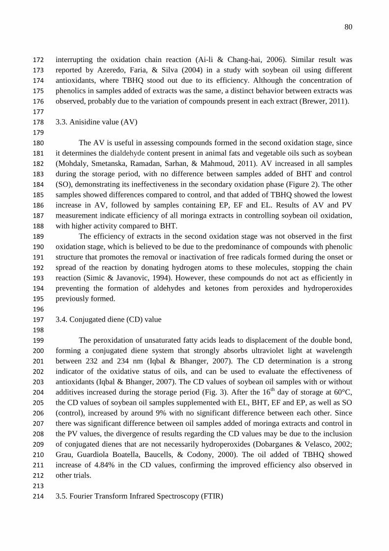
80
interrupting the oxidation chain reaction (Ai-li & Chang-hai, 2006). Similar result was 172
reported by Azeredo, Faria, & Silva (2004) in a study with soybean oil using different 173
antioxidants, where TBHQ stood out due to its efficiency. Although the concentration of 174
phenolics in samples added of extracts was the same, a distinct behavior between extracts was 175
observed, probably due to the variation of compounds present in each extract (Brewer, 2011). 176
177
3.3. Anisidine value (AV) 178
179
The AV is useful in assessing compounds formed in the second oxidation stage, since 180
it determines the dialdehyde content present in animal fats and vegetable oils such as soybean 181
(Mohdaly, Smetanska, Ramadan, Sarhan, & Mahmoud, 2011). AV increased in all samples 182
during the storage period, with no difference between samples added of BHT and control 183
(SO), demonstrating its ineffectiveness in the secondary oxidation phase (Figure 2). The other 184
samples showed differences compared to control, and that added of TBHQ showed the lowest 185
increase in AV, followed by samples containing EP, EF and EL. Results of AV and PV 186
measurement indicate efficiency of all moringa extracts in controlling soybean oil oxidation, 187
with higher activity compared to BHT. 188
The efficiency of extracts in the second oxidation stage was not observed in the first 189
oxidation stage, which is believed to be due to the predominance of compounds with phenolic 190
structure that promotes the removal or inactivation of free radicals formed during the onset or 191
spread of the reaction by donating hydrogen atoms to these molecules, stopping the chain 192
reaction (Simic & Javanovic, 1994). However, these compounds do not act as efficiently in 193
preventing the formation of aldehydes and ketones from peroxides and hydroperoxides 194
previously formed. 195
196
3.4. Conjugated diene (CD) value 197
198
The peroxidation of unsaturated fatty acids leads to displacement of the double bond, 199
forming a conjugated diene system that strongly absorbs ultraviolet light at wavelength 200
between 232 and 234 nm (Iqbal & Bhanger, 2007). The CD determination is a strong 201
indicator of the oxidative status of oils, and can be used to evaluate the effectiveness of 202
antioxidants (Iqbal & Bhanger, 2007). The CD values of soybean oil samples with or without 203
additives increased during the storage period (Fig. 3). After the 16th
day of storage at 60°C, 204
the CD values of soybean oil samples supplemented with EL, BHT, EF and EP, as well as SO 205
(control), increased by around 9% with no significant difference between each other. Since 206
there was significant difference between oil samples added of moringa extracts and control in 207
the PV values, the divergence of results regarding the CD values may be due to the inclusion 208
of conjugated dienes that are not necessarily hydroperoxides (Dobarganes & Velasco, 2002; 209
Grau, Guardiola Boatella, Baucells, & Codony, 2000). The oil added of TBHQ showed 210
increase of 4.84% in the CD values, confirming the improved efficiency also observed in 211
other trials. 212
213
3.5. Fourier Transform Infrared Spectroscopy (FTIR) 214

81
215
In assessing soybean oil degradation by FTIR, the following spectral regions were 216
observed: (1) absorption peaks around 982 cm-1
, corresponding to absorption of trans fatty 217
acids (TFA) related to the C-H vibration outside of plane in the trans configuration of the 218
double bond, (2) absorption peaks around 1734 cm-1
, corresponding to absorption of carbonyl, 219
(3) absorption peaks around 3015 cm-1
, corresponding to cis double bonds, which are related 220
to the PUFA content in the soybean oil (Wang et al. May 2011). Content changes in the cis 221
double bond, carbonyl and trans double bond of soybean oil during storage at 60 ° C are 222
shown in Fig 4 (A-C). The initial stage of the oxidation process of soybean oil leads to a 223
reduction in the cis double bonds of polyunsaturated fatty acids. Figure 4 – A shows that after 224
the 16th
day of storage, the control sample (SO) showed a decrease in the absorption peak at 225
3015 cm-1
of 14.97%. All samples added of moringa extracts and synthetic antioxidants 226
showed differences in peak reducing compared to control sample (SO), revealing preservation 227
of PUFAs in samples with additives. EL and BHT extracts showed reduction of 10.76% and 228
10.71% respectively, not significantly differing from each other. Samples added of EF and EP 229
extracts differed from each other, showing a reduction of 11.70% and 12.87% respectively. 230
Synthetic antioxidant TBHQ was the most effective in preserving PUFAS, with a 9% 231
reduction. While the content of cis double bonds decreased throughout the storage time, 232
Figure 4 – C shows that the content of trans double bonds related to trans fatty acids (TFA) 233
was increased by 26.68% in control (SO) and 26.75% the sample added of EP, with no 234
significant difference between samples. In sample added of EF, EL, BHT and TBHQ, the 235
increase was 19.82%, 20:16%, 23:27% and 24.49% respectively, differing from control 236
samples. The carbonyl content is related to the oxidation degree, increasing during the storage 237
time with the formation of products from DHA, EPA oxidation or other constituents present 238
in the soybean oil. Figure 4 – B shows that the control sample and those added of PE and 239
BHT had exactly the same increase in the carbonyl content (7.59%), while samples added of 240
EL, EF and TBHQ showed smaller increases, differing significantly from the control sample. 241
242
4. Conclusions 243
244
Ethanolic extracts of leaves, flowers and seed pods of Moringa oleífera Lamark 245
showed a protective effect against soybean oil oxidation, and the extract from leaves (EL) was 246
the most effective, with antioxidant activity higher than that of synthetic antioxidant BHT. 247
The action of this extract on soybean oil was also significant in reducing secondary oxidation 248
products and in protecting polyunsaturated fatty acids, reducing the formation of trans fatty 249
acids during storage at 60°C, suggesting their potential for use in the edible oils industry, as 250
an alternative to replace synthetic antioxidants. 251
252
5. Acknowledgement 253
254
The authors would like to thank the Brazilian agencies National Council for Scientific 255
and Technological Development (CNPq) and Coordination for the Improvement of Higher 256
Education Personnel (CAPES) for the financial support. 257

82
258
6. References 259
Ai-li, J., & Chang-hai, W. (2006). Antioxidant properties of natural components from Salvia 260
plebeia on oxidative stability of ascidian oil. Process Biochemistry, 41(5), 1111–1116. 261
Anwar, F., Hussain, A. I., Iqbal, S., & Bhanger, M. I. (2007). Enhancement of the oxidative 262
stability of some vegetable oils by blending with Moringa oleifera oil. Food Chemistry, 263
103(4), 1181–1191. 264
Azeredo, H. M. C., Faria, J. A. F., & Silva, M. A. A. P. (2004). Minimization of peroxide 265
formation rate in soybean oil by antioxidant combinations. Food Research International, 266
37(7), 689–694. 267
Brewer, M. S. (2011). Natural Antioxidants: Sources, Compounds, Mechanisms of Action, 268
and Potential Applications. Comprehensive Reviews in Food Science and Food Safety, 269
10(4), 221–247. 270
Debnath, S., Biswas, D., Ray, K., & Guha, D. (2011). Moringa oleifera induced potentiation 271
of serotonin release by 5-HT(3) receptors in experimental ulcer model. International 272
Journal of Phytotherapy and Phytopharmacology, 18(2-3), 91–5. 273
Dobarganes, M. C., & Velasco, J. (2002). Analysis of lipid hydroperoxides. European 274
Journal of Lipid Science and Technology, 104, 420–428. 275
Grau, A., Guardiola, F., Boatella, J., Baucells, M. D., & Codony, R. (2000). Evaluation of 276
lipid ultraviolet absorption as a parameter to measure lipid oxidation in dark chicken 277
meat. Journal of Agricultural and Food Chemistry, 48(9), 4128–4135. 278
Iqbal, S., & Bhanger, M. I. (2006). Effect of season and production location on antioxidant 279
activity of Moringa oleifera leaves grown in Pakistan. Journal of Food Composition and 280
Analysis, 19(6-7), 544–551. 281
Iqbal, S., & Bhanger, M. I. (2007). Stabilization of sunflower oil by garlic extract during 282
accelerated storage. Food Chemistry, 100(1), 246–254. 283
Lako, J., Trenerry, V., Wahlqvist, M., Wattanapenpaiboon, N., Sotheeswaran, S., & Premier, 284
R. (2007). Phytochemical flavonols, carotenoids and the antioxidant properties of a wide 285
selection of Fijian fruit, vegetables and other readily available foods. Food Chemistry, 286
101(4), 1727–1741. 287
Leclerc, L., Zhao, X., Simpson, B. K., & Zuta, P. C. (2007). The effect of α-tocopherol on the 288
oxidation of mackerel oil.pdf. Food Chemistry, 100, 800–807. 289
Mohdaly, A. a. a., Smetanska, I., Ramadan, M. F., Sarhan, M. a., & Mahmoud, A. (2011). 290
Antioxidant potential of sesame (Sesamum indicum) cake extract in stabilization of 291
sunflower and soybean oils. Industrial Crops and Products, 34(1), 952–959. 292
Rashid, U., Anwar, F., Moser, B. R., & Knothe, G. (2008). Moringa oleifera oil: a possible 293
source of biodiesel. Bioresource technology, 99(17), 8175–9. 294
Sharma, B. K., Rashid, U., Anwar, F., & Erhan, S. Z. (2009). Lubricant properties of Moringa 295
oil using thermal and tribological techniques. Journal of Thermal Analysis and 296
Calorimetry, 96(3), 999–1008. 297
Silva, A. V. C., Santos, A. R. F., Lédo, A. S., Almeida, R. B. F. C. S., Silva, G. M., & Rangel, 298
M. S. A. (2012). Moringa genetic diversity from germplasm bank using RAPD markers. 299
Tropical and Subtropical Agroecosystems, 15(1), 31–39. 300

83
Simic, M. G., & Javanovic, S. V. (1994). Inactivation of oxygen radicals by dietary phenolic 301
compounds in anticarcinogenesis. In C. T. HO, T. OSAWA, T. M. HUANG, & R. T. 302
ROSEN (Eds.), Food phytochemicals for cancer prevention (pp. 20–33). Washington: 303
American Chemical Society. 304
Sun-Waterhouse, D., Thakorlal, J., & Zhou, J. (2011). Effects of added phenolics on the 305
storage stability of avocado and coconut oils. International Journal of Food Science & 306
Technology, 46(8), 1575–1585. 307
Sánchez-Machado, D. I., Núñez-Gastélum, J. a., Reyes-Moreno, C., Ramírez-Wong, B., & 308
López-Cervantes, J. (2009). Nutritional Quality of Edible Parts of Moringa oleifera. 309
Food Analytical Methods, 3(3), 175–180. 310
Wang, H., Liu, F., Yang, L., Zu, Y., Wang, H., Qu, S., & Zhang, Y. (2011). Oxidative 311
stability of fish oil supplemented with carnosic acid compared with synthetic 312
antioxidants during long-term storage. Food Chemistry, 128(1), 93–99. 313
314
315
316
317
318
319
320
321
322
323
324
325
326
327
328
329
330
331
332
333
334
335
336
337
338
339
340
341
342
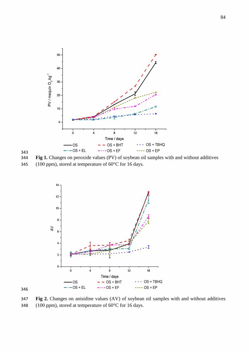
84
343 Fig 1. Changes on peroxide values (PV) of soybean oil samples with and without additives 344
(100 ppm), stored at temperature of 60°C for 16 days. 345
346
Fig 2. Changes on anisidine values (AV) of soybean oil samples with and without additives 347
(100 ppm), stored at temperature of 60°C for 16 days. 348
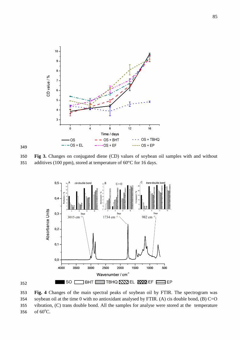
85
349
Fig 3. Changes on conjugated diene (CD) values of soybean oil samples with and without 350
additives (100 ppm), stored at temperature of 60°C for 16 days. 351
352
Fig. 4 Changes of the main spectral peaks of soybean oil by FTIR. The spectrogram was 353
soybean oil at the time 0 with no antioxidant analysed by FTIR. (A) cis double bond, (B) C=O 354
vibration, (C) trans double bond. All the samples for analyse were stored at the temperature 355
of 60oC. 356
86
Study of antioxidant activity and preliminary bioactivity determination of ethanolic extracts of Moringa oleifera
Lam.
Jaqueline A. Nascimento1*
, Kassandra L. G. V. Araújo1, Poliana S. Epaminondas
1,3, Alline S. Souza
1, Antonia L.
Souza2, Antonio G. Souza
2, André M. Santos
4, Alessandra A. Nascimento
4
1Graduate Program in Food Science and Technology, CT, Campus I, Federal University of Paraíba, CEP 58051-
970, João Pessoa, Paraíba, Brazil. 2 Laboratory of Fuels and Materials - LACOM, Department of Chemistry, CCEN, Campus I, Federal University
of Paraíba, CEP 58059-900, João Pessoa, Paraíba, Brazil. 3 Department of Agribusiness, Federal Institute of Education, Science and Technology (IFPB), 58800-970,
Campus Sousa, Sousa - Paraiba, Brazil. 4Laboratory of Animal Experimentation, Federal University of Amapá, 68906-970, Campus I, Macapá, Amapá,
Brazil.
*Corresponding author: [email protected]
Fax: +55-83-32167441; Phone:+55-83-32167441
Abstract
Moringa oleifera Lam is an Indian plant popularly known by its pharmacological properties still little explored
in Brazil. In the present study, it was determined the levels of totals extractables phenolics (TEP) and antioxidant
activity of ethanolic extracts from leaf, flowers, seed pods and seeds of Moringa oleifera Lam. A preliminary
assessment of the toxicity of above extracts was also performed using the bioassay with Artemia salina. The
content of TEP expressed as gallic acid equivalents (GAE) showed that “moringa” leaves are excellent sources
of phenolic compounds. The assays Ferric Reducing Antioxidant Power (FRAP) and β-Carotene bleaching
showed promising antioxidant activity for the extracts, being of the leaves extracts the most efficient in the two
methods, although the extracts were evaluated in the same concentration of TEP. All etanolic extracts from M.
oleifera showed bioactivity against Artemia salina, however, unrelated toxicity according to determined LC50
(394.07, 349.82, 391.97 e 444.83, respectively). These results suggest the viability of using M. oleifera Lam. as
source of antioxidant and alternative food.
Keywords: Moringa oleifera Lam., Antioxidant, Artemia salina, Toxicity

87
1. Introduction
Moringa oleifera Lamarck is a tree native to northwestern India and widely cultivated throughout the
tropics. It is a perennial and drought tolerant plant. It was introduced in Brazil by 1950, where it is cultivated as
an ornamental and medicinal plant. It is found mostly in the Northeast, where it is known as “lírio-branco”,
“quiabo de quina” or simply “moringa”. All parts of Moringa have applicability, being well known its use in
nutrition due to its important minerals and because it is a good source of vitamins, beta-carotene and proteins
(Anwar et al., 2007; Makkar and Becker, 1996). It is traditionally used in India as a medicinal herb to treat and
prevent numerous diseases, acting as antibiotic, antitripanossomal, hypotensive, anti-ulcer, antispasmodic, anti-
inflammatory, hypocholesterolemic and hypoglycemic (Abrogoua et al., 2012; Bijina et al., 2011; Debnath et al.,
2011; Oluduro et al., 2010). Moringa also stands out due to carotenoids and phenolic compounds and provides a
rich and rare combination of zeatin, quercetin, β-sitosterol, caffeoylquinic acid and kaempferol which also gives
the plant a potential antioxidant activity (Barreto et al., 2009; Iqbal and Bhanger, 2006; Lako et al., 2007; Reddy
et al., 2005; Siddhuraju and Becker, 2003).
Given the rich constitution of compounds found in "moringa", it should also be emphasized the need to
demonstrate the safe use of the plant, ie, the guarantee that it has no ability to produce toxic effects to the
consumer.
A bioassay which has been employed for evaluating the pharmacological activity of plant extracts is the
brine shrimp lethality test (BST). It uses Artemia salina to determine the LC50 (median lethal concentration) due
to the fact this microcrustacean be sensitive to a number of substances and also the tendency to limit the use of
laboratory animals in toxicity testing (Ghisalberti, 2008; Kanwar, 2007; Solis et al., 1993).
Given the excellent nutritional and pharmacological characteristics of Moringa and considering its
antioxidant potential, this study investigated the in vitro antioxidant activity of ethanol extracts of its leaves,
flowers and string beans as well as the bioactivity of ethanolic extracts of leaves, flowers, string beans and seeds
against Artemia salina Leach.
2. Materials and methods
2.1. Materials
Reagents tert-Butylhydroquinone (TBHQ), 6-hydroxy-2,5,7,8-tetramethylchoman-2-carboxylic acid
(Trolox), 2,4,6,tris(2-pyridul)-s-triazina) (TPTZ), linoleic acid, were purchased from Sigma-Aldrich (Steinheim,
Germany) while ß-carotene, ferric choride hexahydrate (FeCl3.6H20), ferrous sulphate heptahydrate
(FeSO4.7H2O), Folin-Ciocalteau reagent, 3,4,5-trihydroxybenzoic acid (gallicacid) and all other analytical
reagents were acquired from Merck (Düsseldorf, Germany).
2.2. Samples
Leaves, flowers, seed pods and seeds of “moringa” (Moringa oleifear Lam.) were collected during April,
2010, in Campus I of the Federal University of Paraíba (UFPB), João Pessoa/PB, Brazil, and subsequently dried
in air circulating oven at 45 °C (model MA 035, Marconi) for 24 hours. After complete drying, each part was
ground separately in mill knives and subsequently packed in polyethylene vacuum bags and kept at -18 °C
during the experiments.
2.3. Preparation of extracts
It was used 50 g of powdered plant material and 500 mL of ethanol to obtain each extract. The mixture
was stirred in metabolic water bath, for 2 hours, at 25 °C, followed by vacuum filtration. The extracts of leaves
(EL), flowers (EF) and shell of seed pods (EP) were concentrated on a rotatory evaporator at 60 °C until
complete removal of the solvent. Each one was packed in amber vials under refrigeration until the time of
analysis.
2.4. Determination of total extractable phenolics

88
The levels of total extractable phenolics (TEP) were measured colorimetrically by the Folin-Ciocalteau
according to Slinkard and Singleton (1977), with the assistance of a UV-vis spectrophotometer (model UV-2550,
Shimadzu). The results were expressed as mean ± standard deviation mg of gallic acid equivalents (GAE) / g
extract.
2.5. Ferric reducing antioxidant power (FRAP) assay
The antioxidant activity of extracts of “moringa” was estimated by the assay of ferric reducing
antioxidant power (FRAP), following the procedure described in literature (Benzie and Strain, 1996) with
modifications previously reported (Pulido et al., 2000). Initially a 2.7 mL aliquot of the freshly prepared FRAP
reagent (TPTZ 10 µM, FeCl3 20 µM and acetate buffer) was homogenized with 90 µL of extract and 270 µL of
distilled water in bath water at 37 °C for 30 minutes. The absorbance at 595 µm of the mixture was measured in
UV-vis spectrophotometer (model UV-2550, Shimadzu). The FRAP reagent was used as blank to calibrate the
spectrophotometer. Concentrations of 500-2000 µM of ferrous sulfate (FeSO4.7H2O) were used to determine the
standard curve. The results were expressed as mean ± standard deviation µM ferrous sulfate / g extract.
2.6. ß-carotene bleaching method
The antioxidant activity of the extracts of “moringa” by ß-carotene / linoleic acid model system was
determined as described in the literature by Marco (1968) with modifications by Miller (1971). The
spectrophotometric assay is based on oxidation (bleaching) of ß-carotene induced by products of the oxidative
degradation of linoleic acid. The solutions were prepared by mixing 5 mL of system solution of ß-
carotene/linoleic acid and 0.4 mL of “moringa” extracts or Trolox solution. The mixture was kept in water bath
at 40 ºC. Readings were taken in UV-vis spectrophotometer (model UV-2550, Shimadzu) at 470 nm 2 minutes
after mixing and then at intervals of 15-120 minutes. The control sample consisted of 5 mL of system solution of
ß-carotene/linoleic acid and 0.4 mL of solvent in place of the extracts. The reference substance (Trolox) was
used at a concentration of 200 µg.mL-1
. The results were expressed as percentages of inhibition of oxidation by
using the following formula:
Oxidation inhibition (%) = [(1- (Abs Ai – Abs Af / Abs Ci – Abs Cf)) x 100]
where Abs Ai is the initial absorbance of the sample, Abs Af is the final absorbance of the sample, Abs Ci is the
initial absorbance of the control and Abs Cf is the final absorbance of the control.
2.7. Brine shrimp lethality test (BST) with Artemia salina
For assessing the bioactivity of extracts of leaves, flowers, seed pods and seeds of Moringa oleifera it was
performed the Brine shrimp lethality test (BST) with Artemia salina according to Meyer et al. (1982).
Artemia salina cysts (25 mg) were obtained in Laboratory of Animal Experimentation, Amapá, Brazil and
were incubated in artificial sea solution (pH 8.5 and 29 °C) under light (40 watt lamp) for 24 hours to hatching
the nauplii. Approximately 10-13 larvae were transferred to test tubes containing final volume of 1 mL each and
different concentrations of extracts (1, 3, 10, 30, 100, 300, 500, 750 and 1000 g.mL-1
). A control tube was
obtained using 1 mL of vehicle (cremophor solution 5%) and larvae. Each concentration was tested in triplicate
and repeated in two experiments. It was counted the number of living and dead larvae to determine the LC50.
2.8. Statistical analysis
The results were expressed as mean ± standard deviation, compared by analysis of variance (ANOVA)
and Duncan test, considering p < 0.05, assisted by software Statistica 7.0 (Statsoft®).
In the assessment of bioactivity, the results were expressed as mean of triplicate analysis and the
experiment was repeated to obtain a new mean. CL50 was established with a 95% confidence interval for all
experiments, being significant when p < 0.05 according to Probit analysis (Finney, 1971) and assisted by
software GraphPad Prism 4.03.

89
3. Results and discussion
3.1. Total extractable phenolics (TEP)
From the determination of the levels of total extractable phenolics (TEP) it was possible to determine the
necessary amount of each extract equivalent to one additivation of 200 mg.kg1 of GAE for the assay in the
system ß-carotene / linoleic acid. The analyses of the TEP contents present in the extracts showed that the
highest values were observed for EL and the smallest for ES, and that it was not found a significant difference (p
< 0.05) in relation the amount of phenolics determined for EF and EP (Table 1). The highest value of TEP for
the EL extracts agrees with the data available in the literature, that report the presence of the flavonoids: gallic
acid, ellagic acid, ferulic acid, chlorogenic acid, kaempferol, quercetin, vanillin and rutin in the leaves of
Moringa oleifera Lam (Singh et al., 2009).
3.2. Ferric reducing antioxidant Power (FRAP) assay
The FRAP assay is based on the ability of an antioxidant to reduce Fe3+
to Fe2+
with maximum absorption
at 593 nm. This assay provides rapid and reproducible results, however, it cannot measure all the antioxidants
present in a complex matrix, as in the case of extracts. For this reason, many studies have used this method in
conjuction with other assays (Niki, 2010).
The results of the FRAP assay for the “moringa” extracts and TBHQ and Trolox standards are
summarized in Table 1. According to these data all samples exhibited ferric reducing activity. Among the
extracts, EL showed better activity, followed by EF and EP. These data clearly reveal a correlation between the
amount of phenolic compounds and antioxidant capacity of the extracts. TBHQ was more active than the
extracts.
3.3. ß-Carotene bleaching method
In the coupled oxidation assay of ß-carotene / linoleic acid, the system model when submitted to
oxidizing conditions generates a free radical from the oxidation of linoleic acid, which will abstract the hydrogen
from the molecule of unsaturated ß-carotene. As a result of the oxidation of this molecule, the system loses its
characteristic orange color which is monitored spectrophotometrically in order to quantify the degree of
inhibition of oxidation by the antioxidant to be tested (Abidille et al., 2005).
The kinetic curve of the potential antioxidant of the ethanolic extracts of “moringa” in the ß-
carotene/linoleic acid system is shown in Figure 1. According to that, it is possible to observe the kinetic of the
degradation of ß-carotene for 120 minutes using the extracts of leave (EL), flowers (EF) and seed pods (EP) at
concentration of 200 µg.mL-1
compared to total phenolic content. Trolox was used for comparison at
concentration of 200 µg.mL-1
. The extracts inhibited the oxidation of ß-carotene throughout the degradation
curve in both the beginning of oxidation (within 15-45 minutes) as the ending of oxidation (between 75-105
minutes). EL extract obtained better antioxidant activity compared to Trolox and it was more efficient than EF
and EP.
3.4. Artemia salina survival trials
According to Meyer et al. (1982) when the determined LC50 is less than 1000 g.mL-1
, it indicates that
the compound presents evidence of biological activity. Dolabela (1997) has also established a correlation
between LC50 value and the level of toxicity against Artemia salina, namely: CL50 < 80 g.mL-1
, highly toxic,
LC50 between 80 g.mL-1
and 250 g.mL-1
, moderately toxic, and LC50 > 250 g.mL-1
, nontoxic or low
toxicity.
Table 2 shows values of LC50 determined for the ethanolic extracts of leaves, flowers, seed pods and
seeds of “moringa” through bioassay with Artemia salina. The respective values show that such compounds has
pharmacological activity (Meyer et al., 1982), however, it allows to infer that such activity does not rank as

90
toxic, since for each tested part of the plant, the LC50 value found was greater than 250 g.mL-1
, which
classifies these extracts as nontoxic (Dolabela, 1997).
4. Conclusions
Moringa oleifera showed a promising antioxidant activity for all the tested extracts, which was evidenced
in vitro in different methods. The content of phenolic compounds is higher in extract EL, which stood out for its
greater effectiveness in producing antioxidant effect independently of the determination method employed. The
results of brine shrimp lethality test of the ethanolic extracts from M. oleifera (leaves, flowers, seed pods and
seeds) shows that in all tested parts of the plant it was found bioactive compounds, which corroborate the
findings of medicinal properties related to this species. Those results do not indicate toxicity in the extracts,
suggesting safety in the use of possible products formulated from this species for human consumption, or even as
alternative food and source of natural antioxidants.
5. Acknowledgement
The authors acknowledge the Brazilian agencies Conselho Nacional de Desenvolvimento Científico e
Tecnológico (CNPq) and Coordenação de Aperfeiçoamento de Pessoal de Nível Superior (CAPES) for financial
support.
6. References
Abidille, M.H., Singh, R.P., Jayaprakasha, G.K., Jena, B.S., 2005. Antioxidant activity of the extracts from
Dillenia indica fruits. Food Chemistry 90, 891–896.
Abrogoua, D.P., Dano, D.S., Manda, P., Adepo, A.J.B., Kablan, B.J., Goze, N.B., Ehoulé, K., 2012. Effect on
blood pressure of a dietary supplement containing traditional medicinal plants of Côte d’Ivoire. Journal of
Ethnopharmacology 141, 840–847.
Anwar, F., Latif, S., Ashraf, M., Gilani, A.H., 2007. Moringa oleifera : A Food Plant with Multiple Medicinal
Uses. Phytother. Res. 21, 17–25.
Barreto, M.B., Freitas, J.V.B., Silveira, E.R., Bezerra, A.M.E., Nunes, E.P., Gramosa, N. V., 2009. Constituintes
químicos voláteis e não-voláteis de Moringa oleifera Lam ., Moringaceae. Brazilian Journal of
Pharmacognosy 19, 893–897.
Benzie, I.F., Strain, J.J., 1996. The ferric reducing ability of plasma (FRAP) as a measure of “antioxidant
power”: the FRAP assay. Analytical Biochemistry 239, 70–76.
Bijina, B., Chellappan, S., Krishna, J.G., Basheer, S.M., Elyas, K.K., Bahkali, A.H., Chandrasekaran, M., 2011.
Protease inhibitor from Moringa oleifera with potential for use as therapeutic drug and as seafood
preservative. Saudi Journal of Biological Sciences 18, 273–281.
Chumark, P., Khunawat, P., Sanvarinda, Y., Phornchirasilp, S., Morales, N.P., Phivthong-Ngam, L.,
Ratanachamnong, P., Srisawat, S., Pongrapeeporn, K.U.S., 2008. The in vitro and ex vivo antioxidant
properties, hypolipidaemic and antiatherosclerotic activities of water extract of Moringa oleifera Lam.
leaves. Journal of Ethnopharmacology 116, 439–46.
Cáceres, A., Saravia, A., Rizzo, S., Zabala, L., De Leon, E., Nave, F., 1992. Pharmacologic properties of
Moringa oleifera. 2: Screening for antispasmodic, antiinflammatory and diuretic activity. Journal of
Ethnopharmacology 36, 233–237.

91
Debnath, S., Biswas, D., Ray, K., Guha, D., 2011. Moringa oleifera induced potentiation of serotonin release by
5-HT(3) receptors in experimental ulcer model. International Journal of Phytotherapy and
Phytopharmacology 18, 91–5.
Dolabela, M.F., 1997. Triagem in vitro para atividade antitumoral e anti-Tripanossoma cruzi de extratos
vegetais, produtos naturais e substâncias sintéticas.
Faizi, S., Siddiqui, B.S., Saleem, R., Siddiqui, S., Aftab, K., Gilani, A.H., 1994. Isolation and structure
elucidation of new nitrile and mustard oil glycosides from Moringa oleifera and their effect on blood
pressure. Journal of Natural Products 57, 1256–1261.
Finney, D.J., 1971. Probit analysis. Cambridge University, Cambridge.
Ghisalberti, E.L., 2008. Detection and isolation of bioactive natural products, in: COLEGATE, S.M.,
MOLYNEUX, R.J. (Eds.), Bioactive Natural Products – Detection, Isolation, and Structural
Determination. CRC Press, Boca Raton, pp. 11–76.
Haristoy, X.W., Fahey, J.W., Scholtus, I.W., Lozniewski, A.W., 2005. Adicionar ao Meu Espaço Evaluation of
the Antimicrobial Effects of Several Isothiocyanates on Helicobacter pylori. Planta Medica 71, 326–330.
Iqbal, S., Bhanger, M.I., 2006. Effect of season and production location on antioxidant activity of Moringa
oleifera leaves grown in Pakistan. Journal of Food Composition and Analysis 19, 544–551.
Jaiswal, D., Rai, P.K., Kumar, A., Mehta, S., Watal, G., 2009. Effect of Moringa oleifera Lam. leaves aqueous
extract therapy on hyperglycemic rats. Journal of Ethnopharmacology 123, 392–396.
Kanwar, A.S., 2007. Brine shrimp (Artemia salina) – a marine animal for simple and rapid biological assays.
Journal of Chinese Clinical Medicine 2, 236–240.
Karadi, R. V, Gadge, N.B., Alagawadi, K.R., Savadi, R. V, 2006. Effect of Moringa oleifera Lam. root-wood on
ethylene glycol induced urolithiasis in rats. Journal of Ethnopharmacology 105, 306–11.
Lako, J., Trenerry, V., Wahlqvist, M., Wattanapenpaiboon, N., Sotheeswaran, S., Premier, R., 2007.
Phytochemical flavonols, carotenoids and the antioxidant properties of a wide selection of Fijian fruit,
vegetables and other readily available foods. Food Chemistry 101, 1727–1741.
Makkar, H.P.S., Becker, K., 1996. Nutrional value and antinutritional components of whole and ethanol
extracted Moringa oleifera leaves. Animal Feed Science and Technology 63, 211–228.
Marco, G., 1968. A rapid method for evaluation of antioxidants. Journal of the American Oil Chemists’ Society
45, 594–598.
Meyer, B.N., Ferrigni, N.R., Putnam, J.E., Jacobsen, L.B., Nichols, D.E., McLaughlin, J.L., 1982. Brine shrimp:
A convenient general bioassay for active plant constituents. Planta Medica 45, 31–34.
Miller, H.E., 1971. A simplified method for the evaluation of antioxidants. Journal of the American Oil
Chemists’ Society 48, 91.
Niki, E., 2010. Assessment of antioxidant capacity in vitro and in vivo. Free Radical Biology & Medicine 49,
503–515.

92
Oluduro, O.A., Aderiye, B.I., Connolly, J.D., Akintayo, E.T., Famurewa, O., 2010. Characterization and
Antimicrobial Activity benzyl thiocarboxamide ; a Novel Bioactive Compound from Moringa oleifera
Seed Extract. Folia Microbiol 55, 422–426.
Pulido, R., Bravo, L., Fulgencio, S.C., 2000. Antioxidant Activity of Dietary Polyphenols As Determined by a
Modified Ferric Reducing/Antioxidant Power Assay. Journal of Agricultural and Food Chemistry 48,
3396–3402.
Reddy, V., Urooj, A., Kumar, A., 2005. Evaluation of antioxidant activity of some plant extracts and their
application in biscuits. Food Chemistry 90, 317–321.
Siddhuraju, P., Becker, K., 2003. Antioxidant properties of various solvent extracts of total phenolic constituents
from three different agroclimatic origins of drumstick tree (Moringa oleifera Lam.) leaves. Journal of
Agricultural and Food Chemistry 51, 2144–2155.
Singh, B.N., Singh, B.R., Singh, R.L., Prakash, D., Dhakarey, R., Upadhyay, G., Singh, H.B., 2009. Oxidative
DNA damage protective activity, antioxidant and anti-quorum sensing potentials of Moringa oleifera. Food
and chemical toxicology : an international journal published for the British Industrial Biological Research
Association 47, 1109–16.
Slinkard, K., Singleton, V.L., 1977. Total Phenol Analysis: Automation and Comparison with Manual Methods.
Am. J. Enol. Vitic 28, 49–55.
Solis, P.N., Wright, C.W., Anderson, M.M., Gupta, M.P., Phillipson, D., 1993. A microwell cytotoxicity assay
using Artemia salina (Brine shrimp). Planta Medica 59, 250–252.

93
Table 1
Antioxidant capacity determined by the FRAP method and expressed as mmol Fe2+
.g-1
of
extract and % of oxidation inhibition (OI) by the ß-carotene method.
Samples FRAP /mmol Fe2+
.g-1
OI ß-carotene*
%
EL 19.82 ± 0.69b 82.80 ± 0.88
a
EF 11.61 ± 0.65c 73.15 ± 0.52
c
EP 10.40 ± 0.59d 61.25 ± 0.74
d
TBHQ 30.29 ± 0.10 a ---
TROLOX --- 81.35 ± 0.79 b
Values are mean ± standard deviation, n=3.
Mean followed by different letters in the same column differs significantly (p≤0.05).
* Extracts at concentration of 200 µg.mL-1
/ mL compared to total phenolic contente. Trolox was
used at concentration of 200 µg.mL-1
.
Fig. 1. Kinetic curve of the potential antioxidante of ethanolic extracts of leaves (EL), flowers
(EF) and seed pods (EP) of “moringa” with phenolic concentration of 200 µg.mL-1
compared
to the control (no antioxidant) and with Trolox at concentration of 200 µg.mL-1
through ß-
carotene/linoleic acid system.

94
Table 2
Brine shrimp toxicity results of ethanolic extracts of parts of Moringa oleifera
Tested part LC50/g.mL-1 a
p value 95 % CI /g.mL-1 b
r2
Leaves 394.07 < 0.0001 185.51 – 512.37 0.9216
Flowers 349.82 0.0003 187.19 – 512.36 0.8653
Seed pods 391.97 0.0003 192.54 – 514.03 0.8681
Seeds 444.83 < 0.0001 385.87 – 503.32 0.9800
a median lethal concentration
b confidence interval

95
5 CONSIDERAÇÕES FINAIS
Os extratos etanólicos das folhas, flores e casca das vagens da moringa mostraram
que são fontes em potencial de compostos fenólicos, tendo exibido resultados expressivos no
sequestro de radicais livres pelo método DPPH, tendo sido, também, eficientes na redução do
ferro e no sistema β-caroteno/ácido linoleico.
Os extratos estudados exibiram ação protetora superior ao antioxidante sintético BHT,
quando aplicados aos óleos de peixe e de soja e avaliados pelos métodos Rancimat e P-DSC,
indicando que, do ponto de vista químico, os mesmos podem vir a serem fontes alternativas
de aditivos antioxidantes para uso na indústria de alimentos.
A avaliação do efeito antioxidante dos extratos pelo teste acelerado de estufa revelou
que os extratos das folhas e das flores são eficientes no controle da oxidação primária,
reduzindo consideravelmente os teores de peróxidos e de dienos conjugados, quando
comparados ao controle negativo de ambos os óleos.
Os resultados da avaliação preliminar da bioatividade e toxicidade sugerem que os
extratos testados possuem compostos bioativos supostamente atóxicos, o que é um indicativo
da viabilidade de utilização dos referidos extratos como fonte alternativa de alimentos e
antioxidantes naturais.
Estes resultados sugerem que o extrato etanólico das folhas de moringa possui efeito
protetor eficiente quando aplicado a sistemas lipídicos de baixa estabilidade oxidativa,
podendo vir a ser uma fonte alternativa de potencial aplicação na indústria de óleos e
gorduras.