united states environmental protect
TRANSCRIPT

8/7/2019 United States Environmental Protect
http://slidepdf.com/reader/full/united-states-environmental-protect 1/183
EPA/635/R-07/004F
www.epa.gov/iris
TOXICOLOGICAL REVIEW
OF
CHLORDECONE (KEPONE)
(CAS No. 143-50-0)
In Support of Summary Information on the Integrated Risk Information System (IRIS)
September 2009
U.S. Environmental Protection AgencyWashington, DC

8/7/2019 United States Environmental Protect
http://slidepdf.com/reader/full/united-states-environmental-protect 2/183
DISCLAIMER
This document has been reviewed in accordance with U.S. Environmental Protection
Agency policy and approved for publication. Mention of trade names or commercial products
does not constitute endorsement or recommendation for use.
ii

8/7/2019 United States Environmental Protect
http://slidepdf.com/reader/full/united-states-environmental-protect 3/183
CONTENTS —TOXICOLOGICAL REVIEW OF CHLORDECONE (CAS No. 143-50-0)
LIST OF TABLES .......................................................................................................................... vLIST OF FIGURES ..................................................................................................................... viiiLIST OF ABBREVIATIONS AND ACRONYMS ...................................................................... ixFOREWORD .................................................................................................................................. x
AUTHORS, CONTRIBUTORS, AND REVIEWERS ................................................................. xi1. INTRODUCTION ..................................................................................................................... 12. CHEMICAL AND PHYSICAL INFORMATION RELEVANT TO ASSESSMENTS .......... 33. TOXICOKINETICS .................................................................................................................. 5
3.1. ABSORPTION ................................................................................................................. 53.2. DISTRIBUTION............................................................................................................... 73.3. METABOLISM .............................................................................................................. 103.4. ELIMINATION .............................................................................................................. 133.5. PHYSIOLOGICALLY BASED TOXICOKINETIC MODELS.................................... 15
4. HAZARD IDENTIFICATION ................................................................................................ 184.1. STUDIES IN HUMANS―EPIDEMIOLOGY, CASE REPORTS, CLINICAL
CONTROLS .................................................................................................................. 184.2. SUBCHRONIC AND CHRONIC STUDIES AND CANCER BIOASSAYS INANIMALS―ORAL AND INHALATION ................................................................... 19
4.2.1. Subchronic Studies ............................................................................................... 204.2.1.1. Oral Exposure Studies........................................................................... 204.2.1.2. Inhalation Exposure Studies.................................................................. 20
4.2.2. Chronic Studies .................................................................................................... 204.2.2.1. Oral Exposure Studies........................................................................... 21
4.3. REPRODUCTIVE/DEVELOPMENTAL STUDIES ..................................................... 344.3.1. Reproductive Toxicity Studies ............................................................................. 344.3.2. Developmental Toxicity Studies .......................................................................... 464.3.3. Screening Studies ................................................................................................. 48
4.4. OTHER DURATION-OR ENDPOINT-SPECIFIC STUDIES...................................... 494.4.1. Acute Toxicity Studies ......................................................................................... 494.4.2. Potentiation of Halomethane Toxicity ................................................................. 494.4.3. Neurotoxicity Studies ........................................................................................... 514.4.4. Endocrine Disruption Studies .............................................................................. 514.4.5. Immunological Studies ........................................................................................ 53
4.5. MECHANISTIC DATA AND OTHER STUDIES IN SUPPORT OF THE MODE OFACTION ........................................................................................................................ 57
4.5.1. Genotoxicity ......................................................................................................... 574.5.2. Tumor Promotion and Mechanistic Studies ......................................................... 574.5.3. Structural Analog Data—Relationship to Mirex ................................................. 60
4.6. SYNTHESIS OF MAJOR NONCANCER EFFECTS ................................................... 634.6.1. Oral ...................................................................................................................... 664.6.2. Mode-of-Action Information—Glomerular Lesions ........................................... 69
4.7. EVALUATION OF CARCINOGENICITY................................................................... 704.7.1. Summary of Overall Weight of Evidence ............................................................ 704.7.2. Synthesis of Human, Animal, and Other Supporting Evidence ........................... 714.7.3. Mode-of-Action Information ............................................................................... 75
4.8. SUSCEPTIBLE POPULATIONS AND LIFE STAGES ............................................... 764.8.1. Possible Childhood Susceptibility ....................................................................... 76
iii
8/7/2019 United States Environmental Protect
http://slidepdf.com/reader/full/united-states-environmental-protect 4/183
4.8.2. Possible Gender Differences ................................................................................ 765. DOSE-RESPONSE ASSESSMENTS ..................................................................................... 78
5.1. ORAL REFERENCE DOSE (RfD) ................................................................................ 785.1.1. Choice of Principal Study and Critical Effect—with Rationale and Justification 785.1.2. Methods of Analysis ............................................................................................ 845.1.3. RfD Derivation—Including Application of Uncertainty Factors (UFs) .............. 865.1.4. Reference value (RfV) Comparison Information ................................................ 875.1.5. Previous RfD Assessment .................................................................................... 90
5.2. INHALATION REFERENCE CONCENTRATION (RfC) .......................................... 905.3. CANCER ASSESSMENT .............................................................................................. 91
5.3.1. Choice of Study/Data with Rationale and Justification ....................................... 915.3.2. Dose-Response Data ............................................................................................ 925.3.3. Dose Adjustments and Extrapolation Methods.................................................... 945.3.4. Derivation of the Oral Cancer Slope Factor ........................................................ 965.3.5. Uncertainties in Cancer Risk Values ................................................................... 97
6. MAJOR CONCLUSIONS IN THE CHARACTERIZATION OF ....................................... 102HAZARD AND DOSE RESPONSE .......................................................................................... 102
6.1. HUMAN HAZARD POTENTIAL ............................................................................... 102
6.2. DOSE RESPONSE ....................................................................................................... 1046.2.1. Noncancer .......................................................................................................... 1046.2.2. Cancer ................................................................................................................ 106
7. REFERENCES ...................................................................................................................... 108APPENDIX A. SUMMARY OF EXTERNAL PEER REVIEW AND PUBLIC COMMENTSAND DISPOSITION .................................................................................................................. A-1APPENDIX B. BENCHMARK DOSE CALCULATIONS FOR THE RfD ............................ B-1APPENDIX C. TIME-TO-TUMOR MODELING RESULTS FROM TOX_RISK BASED ONTHE INCIDENCE OF HEPATOCELLULAR CARCINOMAS ............................................... C-1
iv
8/7/2019 United States Environmental Protect
http://slidepdf.com/reader/full/united-states-environmental-protect 5/183
LIST OF TABLES
Table 2-1. Physicochemical properties of chlordecone ................................................................. 3
Table 3-1. Whole blood chlordecone level by group of exposed subjects .................................... 6
Table 3-2. Distribution of chlordecone in exposed workers .......................................................... 8
Table 4-1. Incidence and time to tumor of hepatocellular carcinoma in rats .............................. 24
Table 4-2. Summary of endocrine and reproductive system tumor incidence among rats exposedto chlordecone ............................................................................................................................... 25
Table 4-3. Percent body weight gain and percent survival of chlordecone-exposed rats andmice ............................................................................................................................................... 28
Table 4-4. Incidence and time to tumor of hepatocellular carcinoma in mice ............................ 30
Table 4-5. Testicular atrophy in male rats receiving chlordecone in the diet for 3 months ........ 32
Table 4-6. Incidence of histopathologic liver lesions (fatty changes and hyperplasia) and renalglomerulosclerosis in male and female Wistar rats following administration of chlordecone in thediet for 1–2 years .......................................................................................................................... 33
Table 4-7. Effects of dietary chlordecone on reproduction in male and female mice (of mixedparentage) treated for 1 month prior to mating and for 100 days following the initiation of mating ........................................................................................................................................... 36
Table 4-8. Effects of dietary chlordecone (0 or 40 ppm) on reproduction in BALB/cJaxGnMcmice during 100 days of treatment (preceded by 2 months of pre-mating treatment) and during100 days of a crossover-mating period following the termination of treatment ........................... 37
Table 4-9. Effects of dietary chlordecone for 1 month prior to mating on reproductive indices of male and female laboratory mice of mixed breeds ....................................................................... 38
Table 4-10. Effects of dietary chlordecone (0 or 5 ppm) 1 month prior to mating and up to 5months after initiation of mating on reproduction in BALB/c mice ............................................. 39
Table 4-11. Effects of chlordecone on estrous cyclicity and ovulation in CD-1 mice exposed tochlordecone by gavage 5 days/week for up to 6 weeks ................................................................ 42
Table 4-12. Abundance of various-sized follicles and the condition of large-sized follicles in theovaries of female CD-1 mice exposed to chlordecone by gavage 5 days/week for 4 weeks ....... 43
Table 4-13. Effects of chlordecone on adult female offspring of Sprague-Dawley rat damsadministered chlordecone by gavage on GDs 14–20 .................................................................... 44
v
8/7/2019 United States Environmental Protect
http://slidepdf.com/reader/full/united-states-environmental-protect 6/183
Table 4-14. Sperm parameters in male Sprague-Dawley rats following administration of chlordecone in the diet for 90 days ............................................................................................... 45
Table 4-15. Maternal and fetal effects following gavage dosing of pregnant rat dams withchlordecone on GDs 7–16 ............................................................................................................. 47
Table 4-16. Maternal and fetal effects following gavage dosing with chlordecone on GDs7–16............................................................................................................................................... 48
Table 4-17. Physiochemical properties of chlordecone and mirex .............................................. 61
Table 4-18. Summary of noncancer results for oral exposure studies of experimental animals tochlordecone ................................................................................................................................... 64
Table 5-1. Incidence of histopathologic renal lesions (glomerulosclerosis grades 1, 2, or 3combined) in male or female Wistar rats following administration of chlordecone in the diet for 1–2 years ....................................................................................................................................... 84
Table 5-2. Possible PODs with applied uncertainty factors and resulting Potential RfVs .......... 88
Table 5-3. Tumor incidence and time to first tumor for hepatocellular carcinomas observed inOsborne-Mendel rats following administration of chlordecone in the diet for 80 weeks ............ 93
Table 5-4. Tumor incidence and time to first tumor for hepatocellular carcinomas observed inB6C3F1 mice following administration of chlordecone in the diet for 80 weeks ......................... 93
Table 5-5. Summary of time-to-tumor dose-response modeling based on the incidence of liver tumors in Osborne-Mendel rats and B6C3F1 mice ....................................................................... 97
Table 5-6. Summary of uncertainty in the chlordecone cancer risk assessment ......................... 98
Table B-1. Incidence of histopathologic renal lesions (glomerulosclerosis grades 1, 2, or 3combined) in female Wistar rats following administration of chlordecone in the diet for 2years ............................................................................................................................................ B-1
Table B-2. BMD modeling results for the incidence of histopathologic renal lesions(glomerulosclerosis) in female Wistar rats, following administration of chlordecone in the dietfor 2 years ................................................................................................................................... B-2
Table B-3. Incidence of testicular atrophy in male rats receiving chlordecone in the diet for 3
months ......................................................................................................................................... B-5
Table B-4. BMD modeling results for the incidence of testicular atrophy in male Wistar rats,following administration of chlordecone in the diet for 3 months.............................................. B-6
Table B-5. Incidence of histopathologic liver lesions (fatty changes and hyperplasia) in Wistar rats, following administration of chlordecone in the diet for 1–2 years ..................................... B-9
vi
8/7/2019 United States Environmental Protect
http://slidepdf.com/reader/full/united-states-environmental-protect 7/183
Table B-6. BMD modeling results for the increased incidence of liver lesions in rats (both sexescombined), following administration of chlordecone in the diet for 1–2 years ........................ B-10
Table B-7. Cauda Epididymal sperm count in male Sprague-Dawley rats receiving chlordeconein the diet for 3 months ............................................................................................................. B-13
Table B-8. BMD modeling results for decreased epididymal sperm count in rats, followingadministration of chlordecone in the diet for 3 months ............................................................ B-13
vii

8/7/2019 United States Environmental Protect
http://slidepdf.com/reader/full/united-states-environmental-protect 8/183
LIST OF FIGURES
Figure 2-1. The structure of chlordecone. ...................................................................................... 3
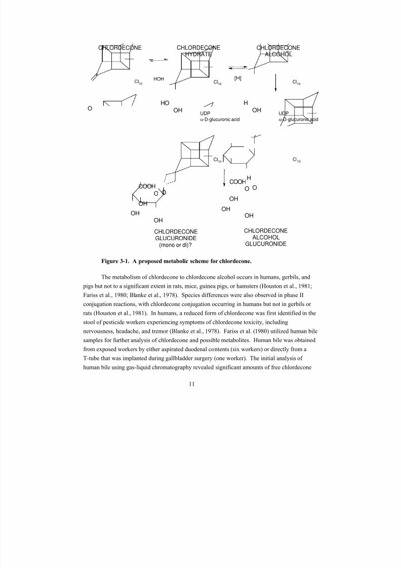
Figure 3-1. A proposed metabolic scheme for chlordecone. ....................................................... 11
Figure 4-1. Dosing regimen for male rats. ................................................................................... 22
Figure 4-2. Dosing regimen for female rats. ................................................................................ 22
Figure 4-3. Dosing regimen for male mice. ................................................................................. 27
Figure 4-4. Dosing regimen for female mice. .............................................................................. 27
Figure 5-1. Potential RfV comparison array for alternate points of departure. ........................... 89
Figure B-1. Observed and predicted incidence of histopathologic renal lesions(glomerulosclerosis grades 1, 2, or 3 combined) in female Wistar rats following administration of chlordecone in the diet for 1–2 years. ......................................................................................... B-2
Figure B-2. Observed and predicted incidence of testicular atrophy in male Wistar rats,following administration of chlordecone in the diet for 3 months.............................................. B-6
Figure B-3. Observed and predicted incidence of liver lesions in male and female Wistar ratsfollowing administration of chlordecone in the diet for 1–2 years. ......................................... B-10
Figure B-4. Observed and predicted epididymal sperm count in male rats, following
administration of chlordecone in the diet for 3 months. ........................................................... B-14
viii

8/7/2019 United States Environmental Protect
http://slidepdf.com/reader/full/united-states-environmental-protect 9/183
LIST OF ABBREVIATIONS AND ACRONYMS
AIC Akaike’s Information CriterionALP alkaline phosphataseALT alanine aminotransferase
AST aspartate aminotransferaseBMD benchmark doseBMD10 benchmark dose associated with a 10% extra risk BMDL10 benchmark dose lower 95% confidence limitBMDS Benchmark Dose SoftwareBMR benchmark responseBUN blood urea nitrogenCASRN Chemical Abstracts Service Registry Number CHO Chinese hamster ovarycon A concanavalin ACYP450 cytochrome P450
DEN diethynitrosamineEEG electroencephalogramELISA enzyme-linked immunosorbent assayFSH follicle-stimulating hormoneGGT γ-glutamyl transpeptidaseGPT glutamic pyruvic transferaseHDL high-density lipoproteinIRIS Integrated Risk Information SystemLD50 median lethal doseLOAEL lowest-observed-adverse-effect levelLSPC Life Science Products Company
MOA mode of actionNCI National Cancer InstituteNK natural killer NOAEL no-observed-adverse-effect levelNRC National Research CouncilPBTK physiologically based toxicokineticPFC plaque-forming cellPHA phytohemagglutininPND postnatal dayPOD point of departurePVE persistent vaginal estrus
RfC reference concentrationRfD reference doseRfV reference values.c. subcutaneousSER smooth endoplasmic reticulumSRBC sheep red blood cellSTM Salmonella typhimurium mitogenTD toxicodynamicUF uncertainty factor U.S. EPA U.S. Environmental Protection Agency
ix

8/7/2019 United States Environmental Protect
http://slidepdf.com/reader/full/united-states-environmental-protect 10/183
FOREWORD
The purpose of this Toxicological Review is to provide scientific support and rationale
for the hazard and dose-response assessment in IRIS pertaining to chronic exposure to
chlordecone. It is not intended to be a comprehensive treatise on the chemical or toxicological
nature of chlordecone.
The intent of Section 6, Major Conclusions in the Characterization of Hazard and Dose
Response, is to present the major conclusions reached in the derivation of the reference dose,
reference concentration, and cancer assessment, where applicable, and to characterize the overall
confidence in the quantitative and qualitative aspects of hazard and dose response by addressing
the quality of the data and related uncertainties. The discussion is intended to convey the
limitations of the assessment and to aid and guide the risk assessor in the ensuing steps of the
risk assessment process.
For other general information about this assessment or other questions relating to IRIS,
the reader is referred to EPA’s IRIS Hotline at (202) 566-1676 (phone), (202) 566-1749 (fax), or
[email protected] (email address).
x
8/7/2019 United States Environmental Protect
http://slidepdf.com/reader/full/united-states-environmental-protect 11/183
AUTHORS, CONTRIBUTORS, AND REVIEWERS
CHEMICAL MANAGER/AUTHOR
Kathleen Newhouse, M.S.
National Center for Environmental AssessmentU.S. Environmental Protection AgencyWashington, DC
CONTRIBUTING AUTHORS
Ted Berner, M.S.National Center for Environmental AssessmentU.S. Environmental Protection AgencyWashington, DC
Debdas Mukerjee, Ph.D.National Center for Environmental AssessmentU.S. Environmental Protection AgencyCincinnati, OH
Andrew Rooney, Ph.D.National Center for Environmental AssessmentU.S. Environmental Protection AgencyResearch Triangle Park, NC
CONTRACTING SUPPORT
Mark Follansbee, Ph.D.Syracuse Research CorporationN. Syracuse, NY
Julie Stickney, Ph.D.Syracuse Research CorporationN. Syracuse, NY
David Wohlers, Ph.D.Syracuse Research CorporationN. Syracuse, NY
REVIEWERS
This document has been reviewed by EPA scientists, interagency reviewers from other
federal agencies, and the public, and peer reviewed by independent scientists external to EPA. A
xi

8/7/2019 United States Environmental Protect
http://slidepdf.com/reader/full/united-states-environmental-protect 12/183
summary and EPA’s disposition of the comments received from the independent external peer
reviewers and from the public is included in Appendix A.
INTERNAL EPA REVIEWERS
Chao Chen, Ph.D.
National Center for Environmental AssessmentWashington, DC
Harlal Choudhury, Ph.D.National Center for Environmental AssessmentCincinnati, OH
Lynn Flowers, Ph.D.National Center for Environmental AssessmentWashington, DC
Karen Hogan M.S.National Center for Environmental AssessmentWashington, DC
Prasada Kodavanti, Ph.D.National Health and Environmental Effects Research LaboratoryResearch Triangle Park, NC
Jamie Strong, Ph.D.National Center for Environmental AssessmentWashington, DC
EXTERNAL PEER REVIEWERS
Harvey J. Clewell, Ph.D.The Hamner Institutes for Health SciencesResearch Triangle Park, NC
George P. Daston, Ph.D.Miami Valley Laboratories
The Proctor and Gamble CompanyCincinnati, OH
Gary L. Ginsberg, Ph.D. (Chair)Connecticut Department of Public HealthDivision of Environmental Epidemiology & Occupational HealthHartford, CT
xii

8/7/2019 United States Environmental Protect
http://slidepdf.com/reader/full/united-states-environmental-protect 13/183 xiii
Michael I. Luster, Ph.D.M. Luster Associates, LLCMorgantown, WV
Lauren Zeise, Ph.D.California Office of Environmental Health Hazard Assessment (OEHHA)Oakland, CA

8/7/2019 United States Environmental Protect
http://slidepdf.com/reader/full/united-states-environmental-protect 14/183
1. INTRODUCTION
This document presents background information and justification for the Integrated Risk
Information System (IRIS) Summary of the hazard and dose-response assessment of
chlordecone. IRIS Summaries may include oral reference dose (RfD) and inhalation reference
concentration (RfC) values for chronic and other exposure durations, and a carcinogenicity
assessment.
The RfD and RfC, if derived, provide quantitative information for use in risk assessments
for health effects known or assumed to be produced through a nonlinear (presumed threshold)
mode of action (MOA). The RfD (expressed in units of mg/kg-day) is defined as an estimate
(with uncertainty spanning perhaps an order of magnitude) of a daily exposure to the human
population (including sensitive subgroups) that is likely to be without an appreciable risk of
deleterious effects during a lifetime. The inhalation RfC (expressed in units of mg/m3) is
analogous to the oral RfD, but provides a continuous inhalation exposure estimate. The
inhalation RfC considers toxic effects for both the respiratory system (portal of entry) and for
effects peripheral to the respiratory system (extrarespiratory or systemic effects). Reference
values are generally derived for chronic exposures (up to a lifetime), but may also be derived for
acute (≤24 hours), short-term (>24 hours up to 30 days), and subchronic (>30 days up to 10% of
lifetime) exposure durations, all of which are derived based on an assumption of continuous
exposure throughout the duration specified. Unless specified otherwise, the RfD and RfC are
derived for chronic exposure duration.
The carcinogenicity assessment provides information on the carcinogenic hazard
potential of the substance in question and quantitative estimates of risk from oral and inhalation
exposure may be derived. The information includes a weight-of-evidence judgment of the
likelihood that the agent is a human carcinogen and the conditions under which the carcinogenic
effects may be expressed. Quantitative risk estimates may be derived from the application of a
low-dose extrapolation procedure. If derived, the oral slope factor is a plausible upper bound on
the estimate of risk per mg/kg-day of oral exposure. Similarly, a plausible inhalation unit risk is
an upper bound on the estimate of risk per μg/m3 air breathed.
Development of these hazard identification and dose-response assessments for
chlordecone has followed the general guidelines for risk assessment as set forth by the NationalResearch Council (NRC, 1983). U.S. Environmental Protection Agency (U.S. EPA) Guidelines
and Risk Assessment Forum Technical Panel Reports that may have been used in the
development of this assessment include the following: Guidelines for the Health Risk
Assessment of Chemical Mixtures (U.S. EPA, 1986a), Guidelines for Mutagenicity Risk
Assessment (U.S. EPA, 1986b), Recommendations for and Documentation of Biological Values
for Use in Risk Assessment (U.S. EPA, 1988), Guidelines for Developmental Toxicity Risk
1
8/7/2019 United States Environmental Protect
http://slidepdf.com/reader/full/united-states-environmental-protect 15/183
2
Assessment (U.S. EPA, 1991), Interim Policy for Particle Size and Limit Concentration Issues in
Inhalation Toxicity (U.S. EPA, 1994a), Methods for Derivation of Inhalation Reference
Concentrations and Application of Inhalation Dosimetry (U.S. EPA, 1994b), Use of the
Benchmark Dose Approach in Health Risk Assessment (U.S. EPA, 1995), Guidelines for
Reproductive Toxicity Risk Assessment (U.S. EPA, 1996), Guidelines for Neurotoxicity Risk
Assessment (U.S. EPA, 1998), Science Policy Council Handbook : Risk Characterization (U.S.EPA, 2000a), Benchmark Dose Technical Guidance Document (U.S. EPA, 2000b),
Supplementary Guidance for Conducting Health Risk Assessment of Chemical Mixtures (U.S.
EPA, 2000c), A Review of the Reference Dose and Reference Concentration Processes (U.S.
EPA, 2002), Guidelines for Carcinogen Risk Assessment (U.S. EPA, 2005a), Supplemental
Guidance for Assessing Susceptibility from Early-Life Exposure to Carcinogens (U.S. EPA,
2005b), Science Policy Council Handbook: Peer Review (U.S. EPA, 2006a), and A Framework
for Assessing Health Risks of Environmental Exposures to Children (U.S. EPA, 2006b).
The literature search strategy employed for this compound was based on the Chemical
Abstracts Service Registry Number (CASRN) and at least one common name. Any pertinent
scientific information submitted by the public to the IRIS Submission Desk was also considered
in the development of this document. The relevant literature was reviewed through August 2009.
8/7/2019 United States Environmental Protect
http://slidepdf.com/reader/full/united-states-environmental-protect 16/183
2. CHEMICAL AND PHYSICAL INFORMATION RELEVANT TO ASSESSMENTS
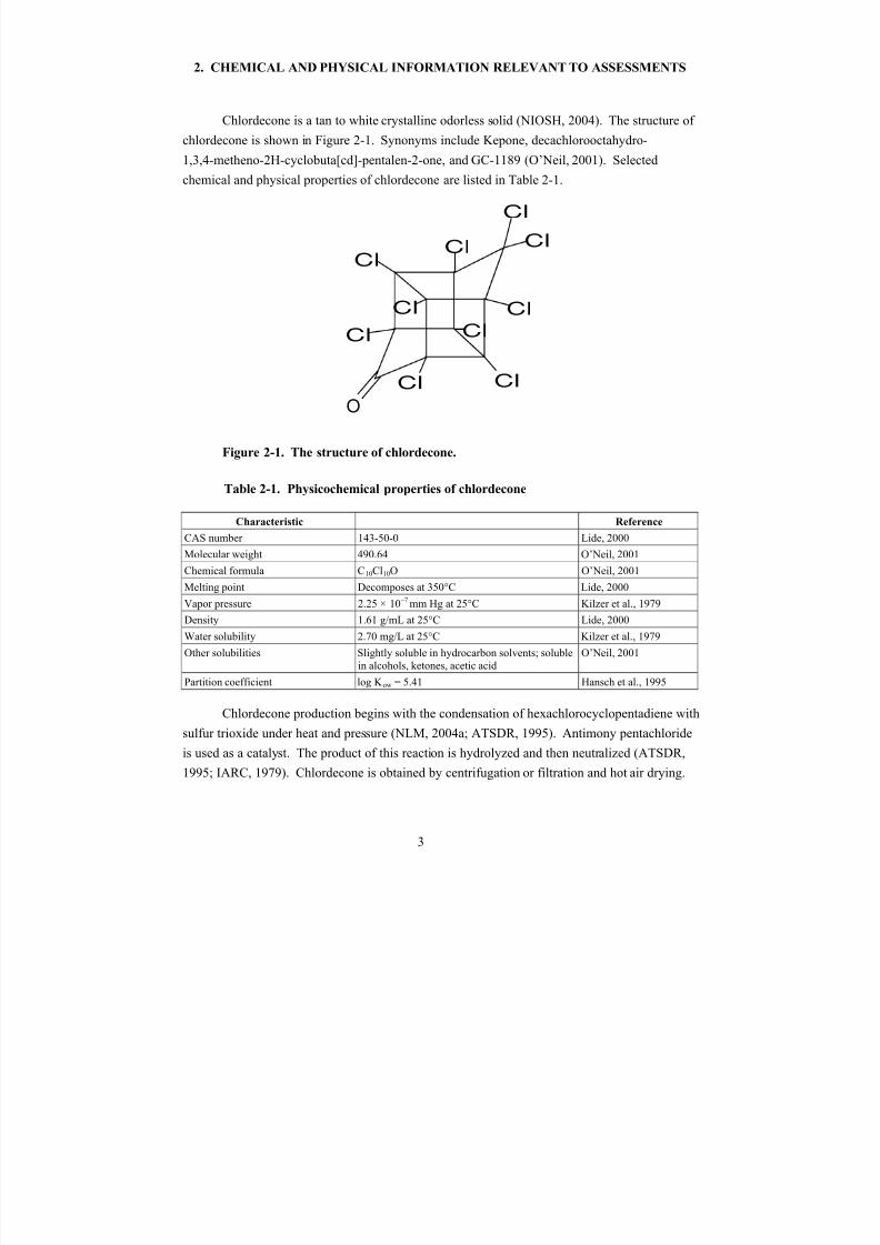
Chlordecone is a tan to white crystalline odorless solid (NIOSH, 2004). The structure of
chlordecone is shown in Figure 2-1. Synonyms include Kepone, decachlorooctahydro-
1,3,4-metheno-2H-cyclobuta[cd]-pentalen-2-one, and GC-1189 (O’Neil, 2001). Selected
chemical and physical properties of chlordecone are listed in Table 2-1.
Cl
Cl
Cl
Cl
Cl
Cl
Cl
Cl
Cl
O
Cl
Figure 2-1. The structure of chlordecone.
Table 2-1. Physicochemical properties of chlordecone
Characteristic Reference
CAS number 143-50-0 Lide, 2000
Molecular weight 490.64 O’Neil, 2001
Chemical formula C10Cl10O O’Neil, 2001
Melting point Decomposes at 350°C Lide, 2000
Vapor pressure 2.25 × 10⁻⁷ mm Hg at 25°C Kilzer et al., 1979
Density 1.61 g/mL at 25°C Lide, 2000
Water solubility 2.70 mg/L at 25°C Kilzer et al., 1979
Other solubilities Slightly soluble in hydrocarbon solvents; solublein alcohols, ketones, acetic acid
O’Neil, 2001
Partition coefficient log K ow = 5.41 Hansch et al., 1995
Chlordecone production begins with the condensation of hexachlorocyclopentadiene with
sulfur trioxide under heat and pressure (NLM, 2004a; ATSDR, 1995). Antimony pentachloride
is used as a catalyst. The product of this reaction is hydrolyzed and then neutralized (ATSDR,
1995; IARC, 1979). Chlordecone is obtained by centrifugation or filtration and hot air drying.
3
8/7/2019 United States Environmental Protect
http://slidepdf.com/reader/full/united-states-environmental-protect 17/183
4
Chlordecone is also a contaminant in mirex formulations and is a degradation product of mirex
(Bus and Leber, 2001).
Chlordecone was first produced in the United States in the early 1950s (IARC, 1979). It
was introduced commercially in 1958 (Bus and Leber, 2001). Approximately 3.6 million pounds
of chlordecone were produced in the United States between 1951 and 1975 (ATSDR, 1995).
Chlordecone production in the United States ended in 1975 after intoxication from severeindustrial exposure was observed in employees who worked at the only chlordecone
manufacturing plant in the country (Bus and Leber, 2001). Typical signs of chlordecone
intoxication include nervousness, headache, and tremor (Cannon et al., 1978).
Chlordecone was primarily used as an insecticide (IARC, 1979). Specific applications
have included control of the banana root borer, application on non-fruit-bearing citrus trees to
control rust mites, control of wireworms in tobacco fields, control of apple scab and powdery
mildew, control of the grass mole cricket, and control of slugs, snails, and fire ants (NLM,
2004a; ATSDR, 1995). Its registration was cancelled in 1978 (Metcalf, 2002; IARC, 1979).
Chlordecone is resistant to degradation in the environment. It is not expected to react
with hydroxyl radicals in the atmosphere or to hydrolyze or photolyze (NLM, 2004a).
Chlordecone in the air is likely to be removed by deposition of particles (NLM, 2004a). Studies
have shown that microorganisms degrade chlordecone slowly (NLM, 2004a). Chlordecone is
expected to adsorb to soil and to stick to suspended solids and sediments in water (NLM, 2004a).
Small amounts of chlordecone will evaporate from soil or water surfaces (NLM, 2004a).
Chlordecone has a high potential for bioaccumulation in fish and other aquatic organisms
(ATSDR, 1995).

8/7/2019 United States Environmental Protect
http://slidepdf.com/reader/full/united-states-environmental-protect 18/183
3. TOXICOKINETICS
The available data for humans and animals indicate that chlordecone is well absorbed
following oral exposure. Once absorbed, it is widely distributed and eventually concentrates in
the liver. It is metabolized by humans and some animal species to chlordecone alcohol.
Glucuronide conjugates of chlordecone and chlordecone alcohol, as well as unconjugated
chlordecone, are slowly excreted in the bile and eliminated in the feces. Fecal excretion is
limited by enterohepatic recirculation.
3.1. ABSORPTION
Chlordecone absorption in humans has been demonstrated by the measurement of
chlordecone concentrations in blood, subcutaneous (s.c.) fat, and other body fluids and tissues
following subchronic occupational exposure, presumably through ingestion, inhalation, and
dermal contact (Taylor, 1982; Adir et al., 1978; Cannon et al., 1978; Cohn et al., 1978). Workers
categorized as having subjective or objective neurological symptoms of chlordecone toxicity
(i.e., nervousness, tremulousness, ataxia) had whole blood concentrations ranging between
0.009 and 11.8 ppm (Cannon et al., 1978). Workers with subjective symptoms alone represented
36% of identified cases. Chlordecone blood levels of the subset of workers with clinically
confirmed neurological symptoms were not reported. Chlordecone blood concentrations for
workers without neurological symptoms were between 0.003 and 4.1 ppm. Chlordecone was
also detected in the blood of Hopewell community residents living near a pesticide plant with
concentrations ranging from 0.005 to 0.0325 ppm. Potential exposure routes for community
residents included inhalation of chlordecone associated with fine particulate matter and ingestion
of contaminated soil and drinking water. Neurological symptoms were reported by some
residents living near the plant site. In general, the highest blood chlordecone concentrations
were observed in affected workers, and lower concentrations were measured in unaffected
workers and community residents (Table 3-1) (Cannon et al., 1978).
5

8/7/2019 United States Environmental Protect
http://slidepdf.com/reader/full/united-states-environmental-protect 19/183
Table 3-1. Whole blood chlordecone level by group of exposed subjects
Group
Number
tested
Number with
detectable
level
Percent with
detectable
level
Range of
detectable level,
ppm
Mean of
detectable level,
ppm
Affected LSPCa workers 57 57 100 0.009–11.8 2.53
Unaffected LSPC workers 49 48 99 0.003–4.1 0.60
Family members, LSPC workers 32 30 94 0.003–0.39 0.10
Allied chlordecone workers 39 30 77 0.003–0.45 0.06
Neighborhood workers 32 23 72 0.003–0.031 0.011
Sewage treatment plant workers 10 6 60 0.004–0.014 0.006
Cab drivers 5 1 20 0.003 0.003
Truck drivers 2 1 50 0.004 0.004
Hopewell community residentsc 214 40 19 0.005–0.0325 0.011
aLSPC = Life Science Products Company workers with self reported or clinically observed neurological symptoms.bAllied Chemical Corporation.cExcludes chlordecone factory workers.
Source: Cannon et al. (1978).
No data were available in laboratory animals to evaluate chlordecone absorption
following inhalation exposure. Quantitative data on absorption of orally administered
chlordecone are limited; however, studies on the distribution and excretion of chlordecone in
rats, mice, gerbils, and pigs following oral administration of chlordecone indicate that this
chemical is readily absorbed from the gastrointestinal tract in animals (Hewitt et al., 1985;
Aldous et al., 1983; Fujimori et al., 1982a; Wang et al., 1981; Kavlock et al., 1980; Egle et al.,
1978). One study (Egle et al., 1978) attempted to estimate oral absorption quantitatively. Male
Sprague-Dawley rats received a single oral dose of 40 mg/kg-day [14C]-labeled chlordecone in
corn oil solution. The percentage of radioactivity excreted in the feces was measured over time.
Approximately 10% of the dose was detected in the feces on the first day after dosing,
suggesting that 90% of the orally administered dose was absorbed from the corn oil vehicle.
Animal studies suggest that chlordecone is absorbed only to a limited extent through the
skin (Heatherington et al., 1998; Shah et al., 1987). The in vivo percutaneous absorption of
chlordecone was evaluated in young (33 days old) and adult (82 days old) F344 rats (Shah et al.,
1987). Acetone solution that contained [14C]-labeled chlordecone was applied to the shaved
backs of animals, with the treatment area constituting 2–3% of the total body surface area.
Urine and feces were collected over a 72-hour period, after which animals were sacrificed
to determine the recovery of radioactivity and the percutaneous absorption of chlordecone.
6
8/7/2019 United States Environmental Protect
http://slidepdf.com/reader/full/united-states-environmental-protect 20/183
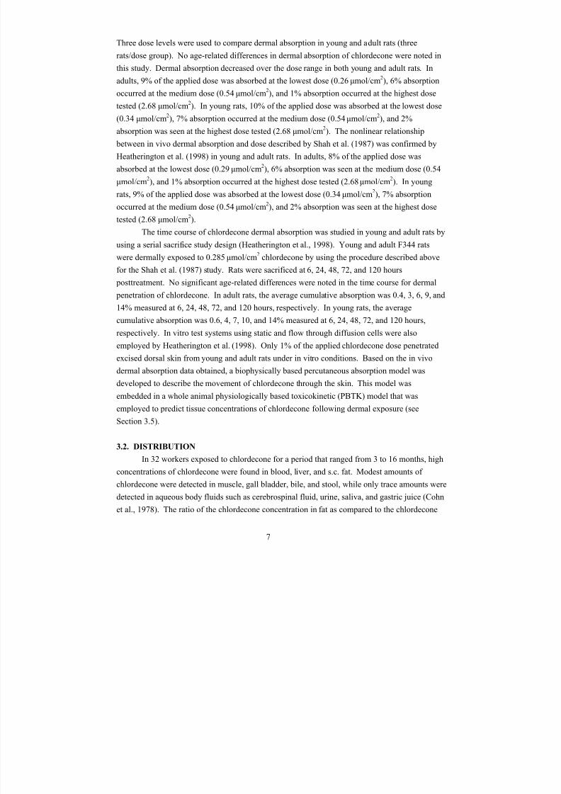
Three dose levels were used to compare dermal absorption in young and adult rats (three
rats/dose group). No age-related differences in dermal absorption of chlordecone were noted in
this study. Dermal absorption decreased over the dose range in both young and adult rats. In
adults, 9% of the applied dose was absorbed at the lowest dose (0.26 μmol/cm2), 6% absorption
occurred at the medium dose (0.54 μmol/cm2), and 1% absorption occurred at the highest dose
tested (2.68 μmol/cm2
). In young rats, 10% of the applied dose was absorbed at the lowest dose(0.34 μmol/cm
2), 7% absorption occurred at the medium dose (0.54 μmol/cm
2), and 2%
absorption was seen at the highest dose tested (2.68 μmol/cm2). The nonlinear relationship
between in vivo dermal absorption and dose described by Shah et al. (1987) was confirmed by
Heatherington et al. (1998) in young and adult rats. In adults, 8% of the applied dose was
absorbed at the lowest dose (0.29 μmol/cm2), 6% absorption was seen at the medium dose (0.54
μmol/cm2), and 1% absorption occurred at the highest dose tested (2.68 μmol/cm
2). In young
rats, 9% of the applied dose was absorbed at the lowest dose (0.34 μmol/cm2), 7% absorption
occurred at the medium dose (0.54 μmol/cm2), and 2% absorption was seen at the highest dose
tested (2.68 μmol/cm2).
The time course of chlordecone dermal absorption was studied in young and adult rats by
using a serial sacrifice study design (Heatherington et al., 1998). Young and adult F344 rats
were dermally exposed to 0.285 μmol/cm2
chlordecone by using the procedure described above
for the Shah et al. (1987) study. Rats were sacrificed at 6, 24, 48, 72, and 120 hours
posttreatment. No significant age-related differences were noted in the time course for dermal
penetration of chlordecone. In adult rats, the average cumulative absorption was 0.4, 3, 6, 9, and
14% measured at 6, 24, 48, 72, and 120 hours, respectively. In young rats, the average
cumulative absorption was 0.6, 4, 7, 10, and 14% measured at 6, 24, 48, 72, and 120 hours,respectively. In vitro test systems using static and flow through diffusion cells were also
employed by Heatherington et al. (1998). Only 1% of the applied chlordecone dose penetrated
excised dorsal skin from young and adult rats under in vitro conditions. Based on the in vivo
dermal absorption data obtained, a biophysically based percutaneous absorption model was
developed to describe the movement of chlordecone through the skin. This model was
embedded in a whole animal physiologically based toxicokinetic (PBTK) model that was
employed to predict tissue concentrations of chlordecone following dermal exposure (see
Section 3.5).
3.2. DISTRIBUTION
In 32 workers exposed to chlordecone for a period that ranged from 3 to 16 months, high
concentrations of chlordecone were found in blood, liver, and s.c. fat. Modest amounts of
chlordecone were detected in muscle, gall bladder, bile, and stool, while only trace amounts were
detected in aqueous body fluids such as cerebrospinal fluid, urine, saliva, and gastric juice (Cohn
et al., 1978). The ratio of the chlordecone concentration in fat as compared to the chlordecone
7
8/7/2019 United States Environmental Protect
http://slidepdf.com/reader/full/united-states-environmental-protect 21/183
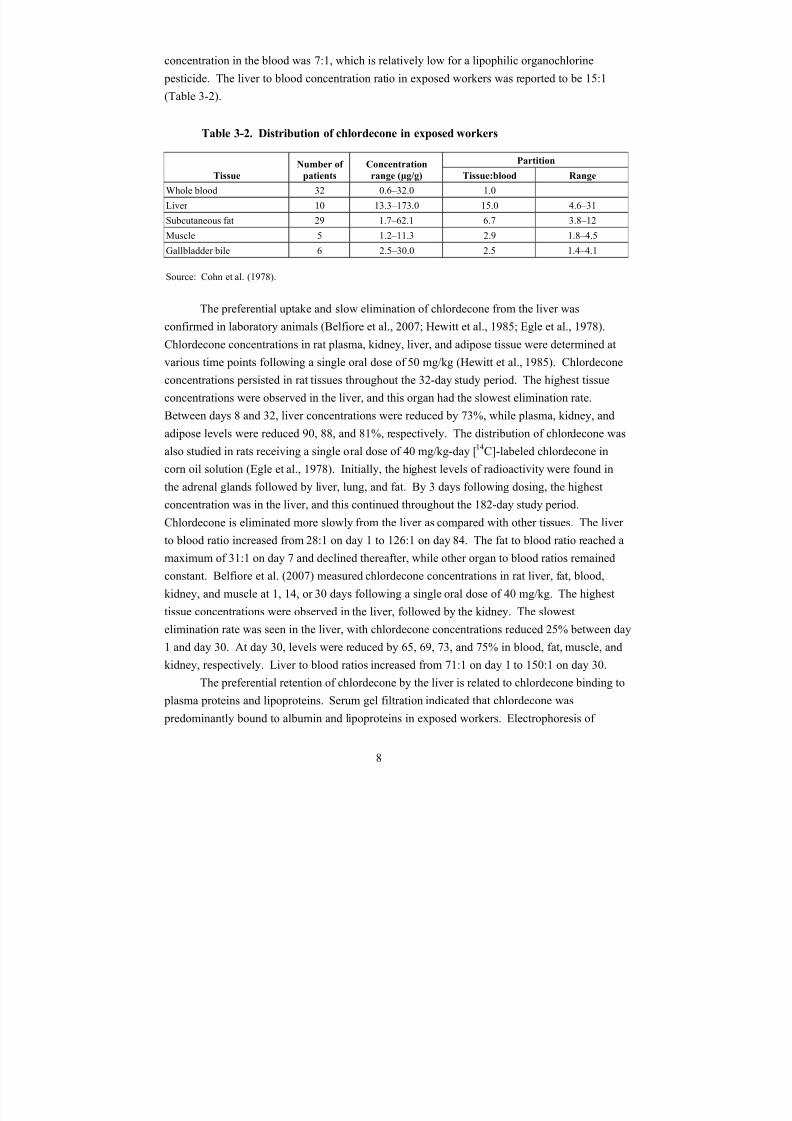
concentration in the blood was 7:1, which is relatively low for a lipophilic organochlorine
pesticide. The liver to blood concentration ratio in exposed workers was reported to be 15:1
(Table 3-2).
Table 3-2. Distribution of chlordecone in exposed workers
Tissue
Number of
patients
Concentration Partition
range (μg/g) Tissue:blood Range
Whole blood 32 0.6–32.0 1.0
Liver 10 13.3–173.0 15.0 4.6–31
Subcutaneous fat 29 1.7–62.1 6.7 3.8–12
Muscle 5 1.2–11.3 2.9 1.8–4.5
Gallbladder bile 6 2.5–30.0 2.5 1.4–4.1
Source: Cohn et al. (1978).
The preferential uptake and slow elimination of chlordecone from the liver was
confirmed in laboratory animals (Belfiore et al., 2007; Hewitt et al., 1985; Egle et al., 1978).
Chlordecone concentrations in rat plasma, kidney, liver, and adipose tissue were determined at
various time points following a single oral dose of 50 mg/kg (Hewitt et al., 1985). Chlordecone
concentrations persisted in rat tissues throughout the 32-day study period. The highest tissue
concentrations were observed in the liver, and this organ had the slowest elimination rate.
Between days 8 and 32, liver concentrations were reduced by 73%, while plasma, kidney, and
adipose levels were reduced 90, 88, and 81%, respectively. The distribution of chlordecone was
also studied in rats receiving a single oral dose of 40 mg/kg-day [
14
C]-labeled chlordecone incorn oil solution (Egle et al., 1978). Initially, the highest levels of radioactivity were found in
the adrenal glands followed by liver, lung, and fat. By 3 days following dosing, the highest
concentration was in the liver, and this continued throughout the 182-day study period.
Chlordecone is eliminated more slowly from the liver as compared with other tissues. The liver
to blood ratio increased from 28:1 on day 1 to 126:1 on day 84. The fat to blood ratio reached a
maximum of 31:1 on day 7 and declined thereafter, while other organ to blood ratios remained
constant. Belfiore et al. (2007) measured chlordecone concentrations in rat liver, fat, blood,
kidney, and muscle at 1, 14, or 30 days following a single oral dose of 40 mg/kg. The highest
tissue concentrations were observed in the liver, followed by the kidney. The slowest
elimination rate was seen in the liver, with chlordecone concentrations reduced 25% between day
1 and day 30. At day 30, levels were reduced by 65, 69, 73, and 75% in blood, fat, muscle, and
kidney, respectively. Liver to blood ratios increased from 71:1 on day 1 to 150:1 on day 30.
The preferential retention of chlordecone by the liver is related to chlordecone binding to
plasma proteins and lipoproteins. Serum gel filtration indicated that chlordecone was
predominantly bound to albumin and lipoproteins in exposed workers. Electrophoresis of
8
8/7/2019 United States Environmental Protect
http://slidepdf.com/reader/full/united-states-environmental-protect 22/183
normal human plasma following the addition of [14C]-labeled chlordecone demonstrated 80%
binding to lipoproteins, with most of this binding associated with high-density lipoproteins
(HDLs) (Skalsky et al., 1979). The preferential binding of chlordecone to albumin and HDL was
demonstrated in human, rat, and pig plasma (Soine et al., 1982). In human plasma, the in vitro
distribution of [14C]-labeled chlordecone was 46% protein, 30% HDL, 20% low density
lipoprotein, and 6% very low density lipoprotein. Similar distributions were seen for pig plasmaand for in vitro and in vivo distribution studies in rat plasma. Albumin was identified as the
major component of the protein fraction that binds chlordecone. Experiments in isolated
perfused pig liver demonstrated that an increase in HDL can affect the distribution of
chlordecone, favoring chlordecone uptake and retention in the liver and decreased chlordecone
elimination in the bile (Soine et al., 1984). Chlordecone and cholesterol have been shown to
compete for similar intracellular binding and transport proteins, which are inducible by
chlordecone pretreatment (Gilroy et al., 1994; Carpenter and Curtis, 1991, 1989).
The brain and plasma levels of chlordecone in mice were measured after daily oral dosing
with 10 or 50 mg/kg-day (Wang et al., 1981). At the lower dose, the plasma level of chlordecone
increased steadily throughout the 12-day treatment period, while the brain chlordecone level
reached a plateau on day 10. Brain and plasma levels decayed biphasically following
administration of 50 mg/kg-day chlordecone for 1 or 2 days. Brain and plasma concentrations
were correlated with loss of motor control at both administered dose levels. Chlordecone was
distributed to discrete areas of the mouse brain following a single gavage dose of 50 mg/kg
(Fujimori et al., 1982a). The striatum and the medulla/pons had significantly higher chlordecone
levels than the cortex, midbrain, or cerebellum.
The distribution of chlordecone following dermal absorption was studied byHeatherington et al. (1998) in young and adults rats (see Section 3.1 for study design
information). Less than 15% of the applied dose was absorbed within 120 hours. Organ
concentrations increased slowly over time, with the highest concentrations observed in the liver
followed by (in decreasing order) kidney, carcass, skin, and blood. Kinetic differences in liver
accumulation of chlordecone were suggested between young and adult rats, but all other organ
concentrations were comparable. Tissue levels did not appear to have reached steady-state
conditions by 120 hours of dermal exposure to chlordecone.
Kavlock et al. (1980) studied the distribution of chlordecone in fetal and neonatal rats.
Pregnant rats were given an oral dose of 5 mg/kg chlordecone on gestation days (GDs) 15, 18, or
20. For the prenatal study, animals were killed at 4, 24, or 48 hours after dosing, and maternal
and fetal tissues were obtained for chlordecone analysis. In the postnatal study, the dams were
given chlordecone at a dose of either 1 or 10 mg/kg-day on days 2–5 of the lactation period.
Maternal milk was obtained following an injection of oxytocin on GDs 5, 9, and 15. Pups were
sacrificed for chlordecone tissue analysis on days 3, 5, 7, 9, 12, 15, and 17 of lactation.
Chlordecone crossed the placenta and was observed in fetal tissues as early as 4 hours after
9
8/7/2019 United States Environmental Protect
http://slidepdf.com/reader/full/united-states-environmental-protect 23/183
maternal dosing. The maximum concentrations of chlordecone on the placenta were 3.5 and 4.0
ppm. Maternal tissue levels were 4 to 5 times higher than fetal concentrations, indicating some
retardation in distribution of chlordecone to the fetus. Chlordecone levels in the fetus were
highest in the liver, followed by the brain, heart, and kidneys. Chlordecone excretion into milk
was an important pathway for elimination in nursing dams. Neonatal organ concentrations of
chlordecone increased steadily over the lactation period. Tissue uptake for neonates was highestin the liver, followed by the brain and the eyes. Day 5 liver and brain levels rose from 2 to 23 μg
and from 16 to 150 μg, respectively, in pups nursed by 10 mg/kg-day dosed dams. Tissue
concentrations were correlated with chlordecone levels in milk.
The tissue distribution of chlordecone was investigated in rats following pretreatment
with phenobarbital, an inducer of hepatic metabolism (Aldous et al., 1983). Repeat doses of
phenobarbital (65 mg/kg) were administered intraperitoneally to adult male Sprague-Dawley rats
6, 12, and 24 hours prior to gavage administration of [14
C]-labeled chlordecone. Phenobarbital
pretreatment resulted in an increase in the specific activity in the liver and uniformly reduced the
specific activity in other tissues. In phenobarbital pretreated rats, 87% of the [14C]-labeled
chlordecone was found in the liver, compared to 55% in control rats not receiving phenobarbital.
Fecal and urinary excretion of chlordecone was reduced. A single dose of phenobarbital (12 or
24 hours prior to chlordecone administration) similarly altered the distribution of chlordecone;
however, changes were more marked with multiple dose administration.
3.3. METABOLISM
Based on available data, a proposed metabolic scheme for chlordecone is shown in Figure
3-1. Although chlordecone is not extensively metabolized in mammals, chlordecone alcohol isformed in humans and some laboratory animal species by reduction of the hydrated carbonyl
group (Fariss et al., 1980; Blanke et al., 1978). A cytosolic aldo-keto reductase enzyme appears
to be responsible for the formation of chlordecone alcohol (Molowa et al., 1986). Chlordecone
alcohol is excreted in bile primarily as a glucuronide conjugate, while chlordecone is excreted
into bile mostly in the unconjugated form (Fariss et al., 1980).
10
8/7/2019 United States Environmental Protect
http://slidepdf.com/reader/full/united-states-environmental-protect 24/183
O
Cl10
OH
OH
Cl10
OH
H
Cl10
O
OH
OH
OH
O
COOH
Cl10
Cl10
O
OH
OH
OH
O
COOHH
HOH
CHLORDECONE CHLORDECONEHYDRATE
CHLORDECONEALCOHOL
UDPα-D-glucuronic acid
UDPα-D-glucuronic acid
[H]
CHLORDECONEGLUCURONIDE
(mono or di)?
CHLORDECONEALCOHOL
GLUCURONIDE
Figure 3-1. A proposed metabolic scheme for chlordecone.
The metabolism of chlordecone to chlordecone alcohol occurs in humans, gerbils, and
pigs but not to a significant extent in rats, mice, guinea pigs, or hamsters (Houston et al., 1981;
Fariss et al., 1980; Blanke et al., 1978). Species differences were also observed in phase II
conjugation reactions, with chlordecone conjugation occurring in humans but not in gerbils or
rats (Houston et al., 1981). In humans, a reduced form of chlordecone was first identified in the
stool of pesticide workers experiencing symptoms of chlordecone toxicity, including
nervousness, headache, and tremor (Blanke et al., 1978). Fariss et al. (1980) utilized human bile
samples for further analysis of chlordecone and possible metabolites. Human bile was obtained
from exposed workers by either aspirated duodenal contents (six workers) or directly from a
T-tube that was implanted during gallbladder surgery (one worker). The initial analysis of
human bile using gas-liquid chromatography revealed significant amounts of free chlordecone
11
8/7/2019 United States Environmental Protect
http://slidepdf.com/reader/full/united-states-environmental-protect 25/183
and small amounts of free chlordecone alcohol in exposed workers. Subsequent treatment of bile
samples with β-glucuronidase prior to the analysis resulted in large amounts of measurable
chlordecone alcohol. It was estimated that >90% of the chlordecone alcohol in human bile is
present as a glucuronide conjugate, while <10% of the chlordecone parent compound is
conjugated prior to biliary excretion. The ratio of chlordecone to chlordecone alcohol following
β-glucuronidase, sulfatase, and acid hydrolysis treatments was between 1:2 and 1:4 in humanbile. In contrast, rat bile contained only trace amounts of chlordecone alcohol, with a
corresponding chlordecone to chlordecone alcohol ratio of 155:1.
Molowa et al. (1986) characterized a unique cytosolic aldo-keto reductase enzyme
responsible for the conversion of chlordecone to chlordecone alcohol. Chlordecone reductase
activity was detected in the liver cytosol of rabbits, gerbils, and humans but was absent in rats,
mice, hamsters, and guinea pigs. Pretreatment of gerbils with a single oral dose of chlordecone
(20 mg/kg) resulted in a 38% increase in the specific activity of chlordecone reductase 7 days
later. Soine et al. (1983) also demonstrated the metabolism of chlordecone to chlordecone
alcohol in the pig. Pigs were given an intraperitoneal dose of either 40 or 80 mg/kg-day, and
chlordecone and chlordecone alcohol concentrations in the blood and gallbladder bile were
measured at regular intervals over a 35-day study period. At the end of the study, hepatic bile,
liver, and feces were also analyzed for chlordecone and chlordecone alcohol levels. The plasma
half-life of chlordecone in the pig was determined to be 12 days at the higher dose and 22 days at
the lower dose. Chlordecone metabolites were generally not detected in the plasma; however,
free chlordecone, free chlordecone alcohol, and conjugated chlordecone alcohol were measured
in gallbladder bile at both doses. Conjugated chlordecone was only observed in gallbladder bile
at the high-dose level. The induction of chlordecone reductase in the pig was suggested by theobserved increase in the chlordecone alcohol to chlordecone ratio in the gallbladder bile over the
time course of the study. On the last day of the study, 20% of chlordecone was conjugated in the
plasma and bile, while only 3% of chlordecone was conjugated in the liver and feces.
Chlordecone alcohol was not detected in the plasma or the liver, but was 85% conjugated in the
bile and 15% conjugated in the feces.
Chlordecone has been shown to induce the cytochrome 450 (CYP450) mixed function
oxidase enzyme system in male and female rats (Gilroy et al., 1994; Hewitt et al., 1985;
Mehendale et al., 1978, 1977). Mehendale et al. (1978, 1977) exposed male and female rats to 0,
50, 100, or 150 ppm chlordecone in the diet for 16 days. A dose-related decrease in body weight
gain was observed, while liver weights were unaltered by chlordecone treatment. Enzyme
activities that were increased by chlordecone treatment at each dose level included aniline,
pentobarbital, and hexobarbital hydroxylation, and aminopyrine and ethylmorphine
demethylation. CYP450, cytochrome c reductase, and aniline binding were all increased, while
cytochrome b5 and NADPH dehydrogenase activity were unaffected by chlordecone treatment.
Hewitt et al. (1985) demonstrated increases in microsomal CYP450 and NADPH cytochrome c
12
8/7/2019 United States Environmental Protect
http://slidepdf.com/reader/full/united-states-environmental-protect 26/183
reductase following a single oral dose of 50 mg/kg (days 2 to 32). Cytochrome b5 was also
increased, but not until 24 to 32 days after chlordecone administration. A single oral dose of
15 mg/kg to Sprague-Dawley rats resulted in an increase in CYP450 and ethoxyresorufin-
O-deethylase and ethoxycoumarin-O-deethylase enzyme activities (Gilroy et al., 1994).
Weanling pups of Sprague-Dawley rat dams exposed to chlordecone from GD 2 to day 21
postpartum (0, 0.1, 1, or 1.5 mg/kg-day) exhibited a dose-related increase in metabolism andexcretion of lindane (Chadwick et al., 1979).
Chlordecone was shown to selectively induce CYP2B2 in adult rat hepatocyte cultures
(Kocarek et al., 1991). Chlordecone selectively increased the mRNA for CYP2B2, and both
chlordecone and chlordecone alcohol induced the immunoreactive protein levels for CYP2B2.
Chlordecone did not affect the mRNA or immunoreactive protein levels for CYP2B1 in isolated
rat hepatocytes. In addition to its selective induction of CYP2B2, chlordecone also suppressed
the induction of CYP2B1 and CYP2B2 when coincubated with phenobarbital in hepatocyte
culture. Mechanistic studies suggest that selective induction of CYP2B2 is not due to the
estrogenic properties of chlordecone, while the ability to suppress phenobarbital induction may
relate to the gem-diol configuration of chlordecone (Kocarek et al., 1994).
3.4. ELIMINATION
Chlordecone and chlordecone alcohol are eliminated from the body primarily through
biliary excretion into feces. In humans, chlordecone is eliminated slowly from the blood.
Estimates of the chlordecone serum half-life (t1/2) in chemical plant workers ranged from 63 to
128 days (Adir et al., 1978). Analysis of excretory fluids in exposed pesticide workers showed
that, while chlordecone was undetectable in sweat and present only in minor quantities in urine,saliva, and gastric juice concentrations in gallbladder bile were approximately equivalent to
chlordecone concentrations in blood (Cohn et al., 1978). The excretion rate of chlordecone into
hepatic bile was estimated from either aspirated duodenal contents (six workers) or bile collected
directly from a T-tube that was implanted during gallbladder surgery (one worker) (Cohn et al.,
1978). The biliary excretion rates varied widely among workers (~1–10 mg/day); however, the
daily excretion amount expressed as a percent of the total body content was relatively constant
(0.29–0.85%). For workers who underwent duodenal aspiration, only 5–10% of the chlordecone
that entered the duodenal lumen via the bile was detected in the feces. Similarly, the rate of
chlordecone excreted in bile collected from a surgically implanted T-tube was 19 times greater
than the rate of elimination in the stool. These results suggest that enterohepatic recycling plays
an important role in the slow excretion of chlordecone. In order to prevent the reabsorption of
chlordecone into the gastrointestinal tract, cholestyramine was investigated as a possible
treatment for chlordecone intoxication. Cholestyramine is an anion-exchange resin that binds
chlordecone but is not absorbed in the gastrointestinal tract. Treatment with cholestyramine
reduced the average t1/2 in the blood of workers from 165 to 80 days (Cohn et al., 1978).
13
8/7/2019 United States Environmental Protect
http://slidepdf.com/reader/full/united-states-environmental-protect 27/183
Gastrointestinal secretion of chlordecone also appears to play a role in fecal excretion in
humans (Boylan et al., 1979). Diversion of the bile stream from the intestine was accomplished
in a chlordecone-exposed worker with a surgically implanted T-tube. Chlordecone excretion in
stool increased eightfold when bile was diverted from the gut. This nonbiliary mechanism for
fecal excretion does not appear to be related to salivary or gastric juice, because chlordecone
concentrations in these fluids were minimal in exposed workers. Chlordecone is transferredfrom the bloodstream to gastrointestinal lumen via a secretory process governed by diffusion
(Bungay et al., 1979). High concentrations of chlordecone in the lumen inhibit gastrointestinal
secretion. Experimental data in rats confirmed the presence of a nonbiliary pathway for fecal
excretion of chlordecone. Bungay et al. (1979) evaluated the transport of chlordecone in and out
of the gut and utilized a PBTK model to describe the results (see Section 3.5). The transport of
chlordecone into and out of the gut was studied following intravenous administration to the bile
duct of cannulated rats and oral administration to intact rats.
Animal studies evaluated the elimination of chlordecone following oral exposure. Egle et
al. (1978) studied chlordecone excretion in male Sprague-Dawley rats receiving a single oral
dose of 40 mg/kg-day [14C]-labeled chlordecone in corn oil solution. The percentage of
radioactivity excreted in the feces was measured over time. Approximately 30% of the
administered chlordecone was excreted within the first 7 days, after which the rate of excretion
steadily declined. After 12 weeks, 65.5% of the dose had been excreted into the feces and after
26 weeks, the cumulative excretion in feces was only 69.8%. A small amount of the
administered chlordecone was excreted in the urine. Only 1.6% of the administered dose was
found in the urine by 12 weeks, one-third of which was excreted into urine in the first 24 hours.
Chlordecone was measured in expired air on days 1 and 9 after dosing, and less than 1% of theadministered dose was detected in expired air.
Heatherington et al. (1998) studied the excretion of chlordecone following dermal
absorption in young and adult rats (see Section 3.1 for study methods). Higher concentrations of
chlordecone were detected in the urine of young rats as compared with adults. Chlordecone
elimination was primarily in the feces, with limited urinary excretion. Feces to urine ratios
120 hours following dermal application of chlordecone were 3:1 and 3:8 in young and adult rats,
respectively.
Chlordecone treatment has been shown to decrease the biliary excretion of other
chemicals (Curtis and Mehendale, 1979). Male Sprague-Dawley rats were fed diets containing
0, 10, 50, or 150 ppm chlordecone for 15 days. Food consumption and body weight data were
used to estimate daily dose levels of 0, 0.69, 3.2, and 8.0 mg/kg-day. Clinical signs of
chlordecone toxicity were not apparent in the 10 or 50 ppm groups, but hyperexcitability and
tremors were observed at 150 ppm. Decreased body weight gain was observed at the two highest
dose levels. Biliary function was evaluated in bile-duct-cannulated intact animal preparations.
The highest dose of chlordecone reduced the biliary excretion of the polar metabolites of
14
8/7/2019 United States Environmental Protect
http://slidepdf.com/reader/full/united-states-environmental-protect 28/183
imipramine (31% of control) and phenolphthalein glucuronide (27% of control). These
decreases occurred despite an increase in cumulative bile flow at the 150 ppm dose level.
Oligomycin-sensitive mitochondrial ATPase activity was inhibited by chlordecone in this study;
however, the dose-response data do not suggest a direct correlation between enzyme inhibition
and hepatobiliary dysfunction.
Teo and Vore (1991) studied the effect of chlordecone on bile acid secretory function(i.e., bile flow, bile acid concentration, bile acid secretory rate) in the isolated perfused rat liver.
Rats were given an oral dose of 18.75 mg/kg-day chlordecone for 3 days prior to measurement of
bile secretory parameters. Chlordecone treatment resulted in an increase in bile flow, but a
decrease in bile acid concentration and bile acid secretory rate. These results suggest that
chlordecone acts primarily at the bile canalicular membrane to decrease biliary excretion.
Rochelle et al. (1990) demonstrated that chlordecone perturbs the membrane and inhibits the
active transport of glutamate at the bile canalicular membrane. Hepatobiliary dysfunction does
not appear to be related to the concentration of chlordecone associated with the liver plasma
membrane (Rochelle and Curtis, 1994); however, inhibition and recovery of 5'-nucleotidase
activity in the liver plasma membrane suggest that biochemical alterations in membrane function
may be involved.
3.5. PHYSIOLOGICALLY BASED TOXICOKINETIC MODELS
PBTK models have been used to describe the hepatic sequestration of chlordecone
(Belfiore et al., 2007), movement of chlordecone in and out of the gut (Bungay et al., 1979),
percutaneous absorption and disposition of chlordecone (Heatherington et al., 1998), and toxic
interactions between chlordecone and carbon tetrachloride (el-Masri et al., 1995) in laboratoryanimals. PBTK models are not available to describe toxicokinetic processes in humans.
Belfiore et al. (2007) developed a PBTK model to describe sequestration of chlordecone
in the liver of rats. Male Sprague-Dawley rats received a one-time treatment of 40 mg/kg-day of
chlordecone in corn oil by gavage. Rats were sacrificed at 1, 14, or 30 days following dosing,
and liver, fat, kidney, and muscle specimens were removed and assayed for chlordecone
concentration. Data from this time course and from distribution studies in the literature (Hewitt
et al., 1985; Egle et al., 1978) were used to develop and validate a toxicokinetic model to
describe the preferential sequestration of chlordecone in the liver. A model was constructed in
which liver, fat, and slowly perfused and rapidly perfused tissues were flow limited. Metabolism
was not included due to the low biotransformation rate for chlordecone. The model fit to the
experimental data was greatly improved by adding blood and liver binding coefficients derived
from data from Soine et al. (1984, 1982). This model provides additional support for the hepatic
sequestration of chlordecone in Sprague-Dawley rats; however, several factors limit its use in the
derivation of reference values. It is not known how the measured blood, fat, or liver tissue levels
would correlate other organ compartments not included in the model. This model also does not
15
8/7/2019 United States Environmental Protect
http://slidepdf.com/reader/full/united-states-environmental-protect 29/183
provide information on inhalation exposure that would be needed for route-to-route
extrapolation. Additionally, the model is not parameterized for humans, so it cannot be used to
evaluate interspecies toxicokinetic differences.
Bungay et al. (1979) conducted experiments comparing intravenous administration of
chlordecone in bile-duct-cannulated rats and oral administration in intact rats. The data were
used in the gut portion of a whole body PBTK model. The gastrointestinal tract was divided intosix segments, and the lumens of these segments were connected in series in the model. Flow
rates were measured in each segment, and the net secretion or absorption was determined for
each compartment. Diffusional processes were assumed to govern chlordecone exchange
between blood, gut tissue, and the lumen. In the rat, the PBTK model yields a maximum
clearance estimate for gut secretion of 25 mL/hour. Measurement of biliary clearance in bile-
duct-cannulated rats was 5 mL/hour, suggesting a total maximum clearance rate of 30 mL/hour.
Assuming that the permeability of the gut to chlordecone is similar in rats and humans, the
authors calculated a maximum human clearance rate of 1,000 mL/hour by using a body-weight
scaling factor (body-weight ratio raised to the 2/3 power). The chlordecone clearance rate
estimated for pesticide workers not receiving cholestyramine treatment (Cohn et al., 1978) was
only 40 mL/hour due to the presence of chlordecone in the lumens and the inhibition of diffusion
from the gut.
A PBTK model was developed to describe the percutaneous absorption and disposition of
chlordecone in young and adult rats (Heatherington et al., 1998). The experimental data for the
dose effect and time course of chlordecone dermal absorption are described in Section 3.1. The
distribution and excretion data for this study are reported in Sections 3.2 and 3.3. A
biophysically-based percutaneous absorption model was developed based on in vivo dermalabsorption data. The absorption model consisted of five first-order rate constants describing the
movement of chlordecone by diffusion from the site of application to the stratum corneum,
where it undergoes partitioning with the viable epidermis, followed by entry into the blood and
distribution throughout the body. The rate constants for movement among compartments were
based on chlordecone physical and chemical characteristics, skin physiology, and experimental
data. The absorption model was significantly limited by its inability to describe the nonlinear
dose effect of percutaneous exposure (i.e., decreasing percent absorption with increasing dose).
Therefore, the data for only one dose level could be used for PBTK disposition modeling (i.e.,
time course data for 0.285 μmol/cm2). The absorption model was embedded in the whole body
PBTK model to describe the distribution and excretion of chlordecone in young and adult rats.
The distribution of chlordecone from blood to various tissue compartments was described. The
PBTK model took into account chlordecone binding to albumin and lipoproteins in the blood,
preferential uptake by the liver, and the predominant fecal excretion pathway for chlordecone.
Once optimized using the experimental data for chlordecone, the PBTK model was used to
predict partition coefficients and excretion rates. Tissue concentrations at varying dose levels
16

8/7/2019 United States Environmental Protect
http://slidepdf.com/reader/full/united-states-environmental-protect 30/183
17
were reasonably well estimated if the nonlinear dermal absorption at high doses and the
nonlinear uptake of bound chlordecone into the liver were considered.
el-Masri et al. (1995) utilized PBTK and toxicodynamic (TD) modeling to evaluate the
toxic interaction between chlordecone and carbon tetrachloride. Chlordecone significantly
potentiates the hepatotoxicity and lethality of carbon tetrachloride by interfering with the
regeneration process in the liver (see Section 4.4.2). A PBTK model for carbon tetrachloridewas adapted and verified using experimental data. The PBTK model was then linked with a TD
model based on the mechanistic data for the interaction between chlordecone and carbon
tetrachloride in liver cells. The combined model yielded a time course simulation of mitotic,
injured, and pyknotic cells following treatment with carbon tetrachloride alone or in combination
with chlordecone. The PBTK/TD model was coupled with Monte Carlo simulation techniques to
predict the acute lethality of carbon tetrachloride under various exposure conditions. Predictions
of lethality were in agreement with experimentally derived values except at very high doses
where neurotoxicity led to significant mortality.
8/7/2019 United States Environmental Protect
http://slidepdf.com/reader/full/united-states-environmental-protect 31/183
4. HAZARD IDENTIFICATION
4.1. STUDIES IN HUMANS―EPIDEMIOLOGY, CASE REPORTS, CLINICAL
CONTROLS
Information regarding the health effects of chlordecone in humans comes from studies of
a single group of 133 men exposed occupationally to chlordecone at a facility in Hopewell,
Virginia (Taylor, 1985, 1982; Guzelian, 1982a; Guzelian et al., 1980; Sanborn et al., 1979;
Cannon et al., 1978; Martinez et al., 1978; Taylor et al., 1978). Of the 133 men, 76 experienced
neurological symptoms, especially nervousness, headaches, and tremors, sometimes persisting as
long as 9–10 months after cessation of exposure (Cannon et al., 1978). In addition, some of the
men experienced oligospermia. Sperm count and motility had returned to normal by 5 to 7 years
following the cessation of chlordecone exposure and treatment with cholestyramine to reduce
chlordecone blood levels (Taylor, 1982). Some workers exposed to high levels of chlordecone
developed skin rashes, enlarged livers, and joint pain. Liver enlargement developed in 20 out of
32 workers with high blood levels of chlordecone (>0.6 μg/mL) after an average duration period
of 5–6 months, although evidence of significant liver toxicity was not found (Guzelian, 1982a;
Guzelian et al., 1980; Taylor et al., 1978). Normal results were obtained in all patients for serum
bilirubin, albumin, globulin, prothrombin time, cholesterol, alanine aminotransferase (ALT),
aspartate aminotransferase (AST), and γ-glutamyl transpeptidase (GGT), and serum alkaline
phosphatase (ALP) was only minimally elevated in seven patients. Sulfobromophthalein
retention, a measure of liver clearance, was normal in a subset of 18 workers tested (Guzelian et
al., 1980). Liver biopsy samples taken from 12 workers with hepatomegaly showed histological
changes in the liver that were characterized as nonadverse in nature. These included
proliferation of the smooth endoplasmic reticulum (SER) and cytoplasmic accumulation of
lipofuscin. No evidence of liver neoplasia, fibrosis, cholestasis, or hepatocellular necrosis was
found. Neurological symptoms were reported in workers exposed to high doses of chlordecone
for a period of months to years (Taylor, 1985, 1982; Guzelian, 1982a; Guzelian et al., 1980;
Sanborn et al., 1979; Cannon et al., 1978; Martinez et al., 1978; Taylor et al., 1978). These
symptoms included tremor, headache, irritability, poor recent memory, rapid random eye
movements, muscle weakness, gait ataxia, incoordination, and slurred speech. The effects
persisted for as long as 9–10 months after cessation of exposure and the start of treatment(Cannon et al., 1978). Martinez et al. (1978) reported that nerve conduction velocity tests,
electroencephalography, radioisotope brain scans, computerized tomography, and analyses of
cerebral spinal fluid content from these workers were all normal. Sural nerve and skeletal
muscle biopsies in workers with detectable neurological impairment exhibited a reduction in the
number of unmyelinated axons and a disruption in Schwann cell metabolism (Martinez et al.,
1978).
18
8/7/2019 United States Environmental Protect
http://slidepdf.com/reader/full/united-states-environmental-protect 32/183
The factory did not follow good industrial hygiene practices. Substantial inhalation,
dermal, and oral exposures could have occurred to the workers (Guzelian, 1982a; Guzelian et al.,
1980; Cannon et al., 1978). Because of uncertainties regarding exposure routes and levels at the
facility and concomitant exposure to the precursors used to manufacture chlordecone, no-
observed-adverse-effect levels (NOAELs) or lowest-observed-adverse-effect levels (LOAELs)
could not be established for the adverse effects observed on the nervous systems, livers, andreproductive systems of these men. Liver biopsy samples taken from 12 workers with
hepatomegaly resulting from intermediate- or chronic-duration exposures to high levels of
chlordecone showed no evidence of significant liver toxicity or cancer (Guzelian et al., 1980);
however, conclusions from this study are limited by the very small number of workers sampled,
uncertainties concerning exposure dose and route, the relatively brief duration of exposures, and
the absence of a sufficient latency period for tumor development. The average exposure of the
subjects was 5–6 months, and they were examined immediately after exposure (Cannon et al.,
1978). A review of biological and epidemiological evidence of cancer found no population-
based studies on cancer in humans related to chlordecone exposure (Ahlborg et al., 1995). These
case reports of occupationally exposed workers at the pesticide plant (who were repeatedly
exposed to high but unmeasured levels for less-than-lifetime durations) indicate that primary
target organs for chlordecone toxicity in humans are the nervous system, reproductive organs,
skin, and liver.
4.2. SUBCHRONIC AND CHRONIC STUDIES AND CANCER BIOASSAYS IN
ANIMALS―ORAL AND INHALATION
Animal studies show effects similar to those reported in occupationally exposed humansincluding neurological effects, oligospermia, hepatomegaly, and skin rashes, as well as kidney
lesions (which were not reported in occupational studies). Chlordecone is moderately lethal by
single exposures; oral median lethal dose (LD50) values range from 71 mg/kg-bw for rabbits to
250 mg/kg-bw for dogs (Larson et al., 1979a). The oral LD50 value for rats is 125 mg/kg-bw
(Gaines, 1969). In experimental animals, the effects of chlordecone following short-term
exposures generally include nervous system effects (tremor and hyperexcitability), liver
hypertrophy (with induction of mixed-function oxidases), and structural and ultrastructural
changes in the liver, thyroid, adrenal glands, and testes (ATSDR, 1995; U.S. EPA, 1986c; WHO,
1984). In subchronic studies with experimental animals, chlordecone produced tremors and
other neurological symptoms, liver hypertrophy with induction of mixed function oxidases,
hepatobiliary dysfunction, and centrilobular hepatocellular necrosis. Chlordecone also interferes
with reproductive processes in both males (oligospermia) and females (disruption of estrous
cyclicity), and it is fetotoxic in experimental animals. Chronic testing of chlordecone in
laboratory animals is limited to two studies: NCI (1976a) and Larson et al. (1979a). In a dietary
cancer bioassay with chlordecone, NCI (1976a) found a statistically significant increase in the
19
8/7/2019 United States Environmental Protect
http://slidepdf.com/reader/full/united-states-environmental-protect 33/183
incidence of and a reduction in the time to detection of hepatic tumors among male (marginal)
and female Osborne-Mendel rats and male and female B6C3F1 mice. The Larson et al. (1979a)
study also reported hepatic proliferative lesions, but the determination of whether these
represented tumors was equivocal. No data are available concerning the toxicity of chlordecone
in animals following inhalation exposure. Studies demonstrating adverse effects in experimental
animals following oral exposures are presented below. No studies were available for inhalationor dermal routes of exposure.
4.2.1. Subchronic Studies
4.2.1.1. Oral Exposure Studies
Huang et al. (1980) administered chlordecone by gavage to adult male ICR mice (15/dose
group) at 0 (corn oil vehicle), 10, 25, or 50 mg/kg-day. Animals were gavaged daily for up to
24 days. Tremor and hyperexcitability were observed in all mice receiving chlordecone and time
to onset was dose-dependent. Loss of body weight (accompanied by reduced food and water
consumption) was also apparent in chlordecone-exposed animals, with the greatest loss of body
weight coincident with the onset of tremor. The authors speculated that the reduction in body
weight among treated mice was due to paralysis and loss of motor control, which resulted in a
decreased ability to eat. Upon termination of chlordecone administration, a diminution of tremor
and corresponding recovery of body weight were observed in surviving animals. Chlordecone-
treated mice showed a high degree of mortality. Time to onset of first death and slope of the
mortality curves (cumulative mortality per day) were dose-dependent. Mortality among mice
exposed to 50 mg/kg-day began on day 4, and all were dead by day 6. For the 25 mg/kg-day
group, mortality began on day 6 and reached 100% by day 11. For the 10 mg/kg-day group,mortality began on day 12 and reached nearly 90% by day 24 of treatment. The control group
had no deaths. The cumulative oral LD50 was estimated by the authors to be between 180 and
200 mg/kg. In a follow-up study, Fujimori et al. (1982b) administered chlordecone by gavage to
male ICR mice at 0 (corn oil vehicle), 10, 25, or 50 mg/kg-day for 9 consecutive days. The
results of this study also demonstrated a dose-response in the time to onset of chlordecone
impairment of motor coordination (days 2, 4, and 9 for 50, 25, and 10 mg/kg-day dose groups).
The authors examined dopamine, serotonin, and norepinephrine levels in the brain. Significant
decreases in whole brain and straital dopamine levels were seen in animals exhibiting tremors,
indicating the dopaminergic pathway may be involved in chlordecone-induced tremor and
neurotoxicity.
4.2.1.2. Inhalation Exposure Studies
No inhalation exposure studies were found in the literature.
4.2.2. Chronic Studies
20
8/7/2019 United States Environmental Protect
http://slidepdf.com/reader/full/united-states-environmental-protect 34/183
4.2.2.1. Oral Exposure Studies
Chu et al. (1981a) fed male Sprague-Dawley rats (10/group) diets containing 0 or 1 ppm
of chlordecone (0 or 0.07 mg/kg-day reported by the authors) for 21 months. Corn oil was used
to dissolve the chemicals, and control diets contained 4% corn oil. Survival and weight gain
were similar in treated and control rats. Hematology and clinical chemistry were also unaffected
by treatment. Histopathological findings included increases in the incidence of lesions in theliver (5/6 [83%] vs. 3/7 [43%] in controls) and thyroid (4/6 [67%] vs. 1/7 [14%] in controls).
The differences in incidences were not statistically significant (Fisher’s exact test conducted for
this review), although the power of the statistical test to detect a difference at such small sample
sizes is low. The lesion in the liver was described as pericentral cytoplasmic vacuolation with
mild anisokaryosis, while the thyroid lesion was described as a mild degenerative and
proliferative change in the epithelium. The authors reported that the severity of both lesions was
increased in chlordecone-treated rats in comparison with controls, although the nature and extent
of these differences were not described.
Osborne-Mendel rats and B6C3F1 mice were exposed to technical-grade chlordecone in
the diet for 80 weeks in a study by the National Cancer Institute (NCI, 1976a, b). The test
material was reported to contain no more than 2% impurities other than water. Chlordecone was
added to finely ground rat chow in acetone (to aid uniform dispersion of the chemical); the diets
were mixed for homogeneity and to allow the acetone to evaporate. Corn oil (2%) was added to
the diet as a dust suppressant. Dietary concentrations of chlordecone began at 0, 15, 30, or 60
ppm for male rats and 0, 30, or 60 ppm for female rats. Treatment groups comprised 50 rats/sex;
however, only 10 animals/sex were used in matched control groups. Pooled control groups
(from the same laboratory with birth dates within 3–4 months of the animals in the matchedcontrol and exposed groups) contained 105 male rats and 100 female rats. Overt clinical signs of
toxicity observed in the treated animals indicated that the initial doses exceeded the maximum
tolerated dose in the high exposure groups; consequently, concentrations of chlordecone in feed
were reduced (to one-third to one-sixth of the original concentration) during the experiment
(after durations ranging from as short as 42 days in high-dose female rats to as long as 386 days
in high-dose male rats). The specific dosing regimens for male and female rats are illustrated in
Figures 4-1 and 4-2.
21
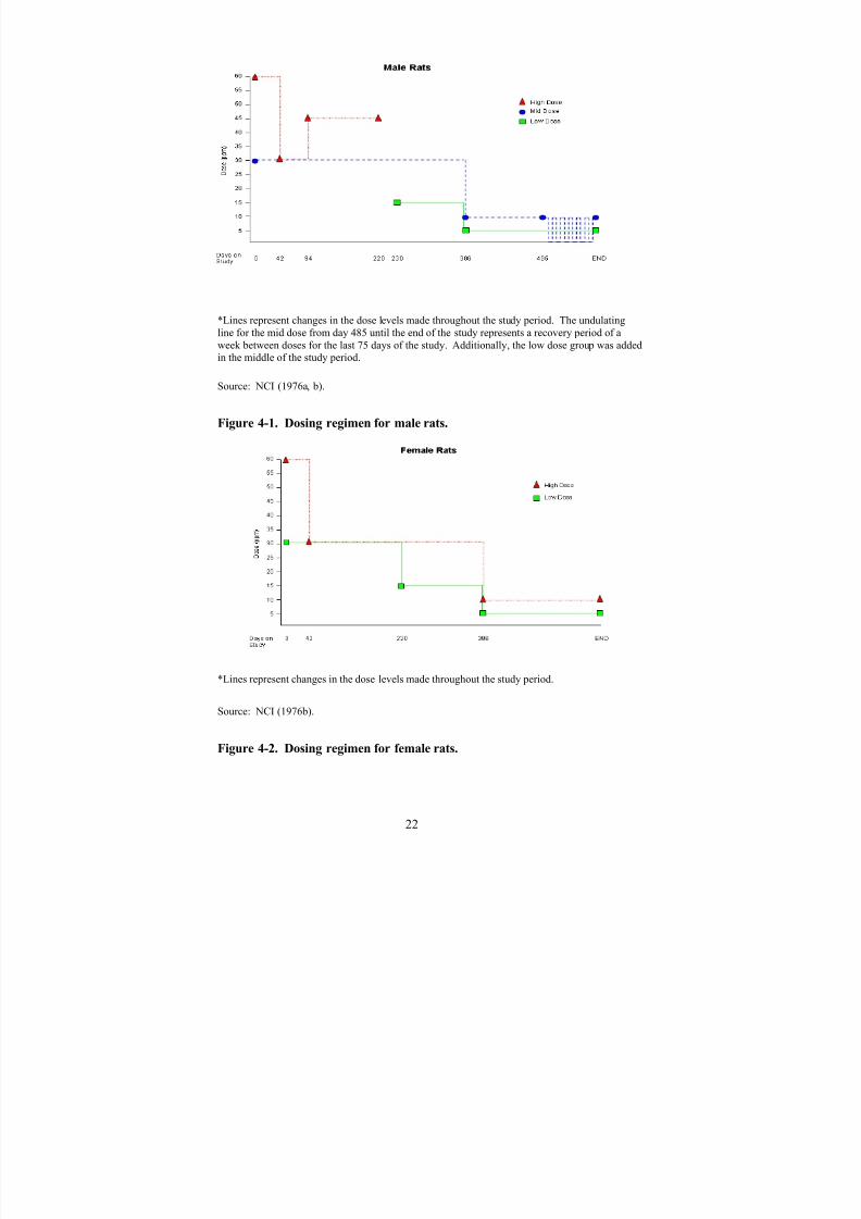
8/7/2019 United States Environmental Protect
http://slidepdf.com/reader/full/united-states-environmental-protect 35/183
*Lines represent changes in the dose levels made throughout the study period. The undulating
line for the mid dose from day 485 until the end of the study represents a recovery period of aweek between doses for the last 75 days of the study. Additionally, the low dose group was addedin the middle of the study period.
Source: NCI (1976a, b).
Figure 4-1. Dosing regimen for male rats.
*Lines represent changes in the dose levels made throughout the study period.
Source: NCI (1976b).
Figure 4-2. Dosing regimen for female rats.
22
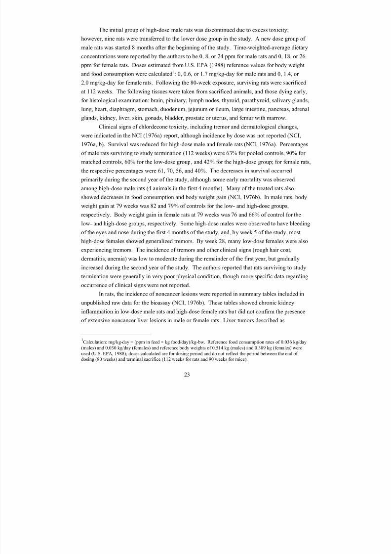
8/7/2019 United States Environmental Protect
http://slidepdf.com/reader/full/united-states-environmental-protect 36/183
The initial group of high-dose male rats was discontinued due to excess toxicity;
however, nine rats were transferred to the lower dose group in the study. A new dose group of
male rats was started 8 months after the beginning of the study. Time-weighted-average dietary
concentrations were reported by the authors to be 0, 8, or 24 ppm for male rats and 0, 18, or 26
ppm for female rats. Doses estimated from U.S. EPA (1988) reference values for body weight
and food consumption were calculated1
: 0, 0.6, or 1.7 mg/kg-day for male rats and 0, 1.4, or 2.0 mg/kg-day for female rats. Following the 80-week exposure, surviving rats were sacrificed
at 112 weeks. The following tissues were taken from sacrificed animals, and those dying early,
for histological examination: brain, pituitary, lymph nodes, thyroid, parathyroid, salivary glands,
lung, heart, diaphragm, stomach, duodenum, jejunum or ileum, large intestine, pancreas, adrenal
glands, kidney, liver, skin, gonads, bladder, prostate or uterus, and femur with marrow.
Clinical signs of chlordecone toxicity, including tremor and dermatological changes,
were indicated in the NCI (1976a) report, although incidence by dose was not reported (NCI,
1976a, b). Survival was reduced for high-dose male and female rats (NCI, 1976a). Percentages
of male rats surviving to study termination (112 weeks) were 63% for pooled controls, 90% for
matched controls, 60% for the low-dose group, and 42% for the high-dose group; for female rats,
the respective percentages were 61, 70, 56, and 40%. The decreases in survival occurred
primarily during the second year of the study, although some early mortality was observed
among high-dose male rats (4 animals in the first 4 months). Many of the treated rats also
showed decreases in food consumption and body weight gain (NCI, 1976b). In male rats, body
weight gain at 79 weeks was 82 and 79% of controls for the low- and high-dose groups,
respectively. Body weight gain in female rats at 79 weeks was 76 and 66% of control for the
low- and high-dose groups, respectively. Some high-dose males were observed to have bleedingof the eyes and nose during the first 4 months of the study, and, by week 5 of the study, most
high-dose females showed generalized tremors. By week 28, many low-dose females were also
experiencing tremors. The incidence of tremors and other clinical signs (rough hair coat,
dermatitis, anemia) was low to moderate during the remainder of the first year, but gradually
increased during the second year of the study. The authors reported that rats surviving to study
termination were generally in very poor physical condition, though more specific data regarding
occurrence of clinical signs were not reported.
In rats, the incidence of noncancer lesions were reported in summary tables included in
unpublished raw data for the bioassay (NCI, 1976b). These tables showed chronic kidney
inflammation in low-dose male rats and high-dose female rats but did not confirm the presence
of extensive noncancer liver lesions in male or female rats. Liver tumors described as
1Calculation: mg/kg-day = (ppm in feed × kg food/day)/kg-bw. Reference food consumption rates of 0.036 kg/day
(males) and 0.030 kg/day (females) and reference body weights of 0.514 kg (males) and 0.389 kg (females) wereused (U.S. EPA, 1988); doses calculated are for dosing period and do not reflect the period between the end of dosing (80 weeks) and terminal sacrifice (112 weeks for rats and 90 weeks for mice).
23
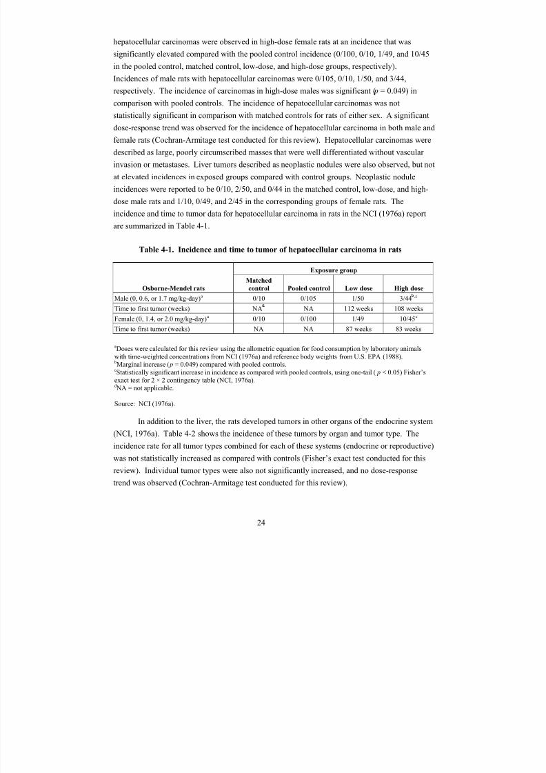
8/7/2019 United States Environmental Protect
http://slidepdf.com/reader/full/united-states-environmental-protect 37/183
hepatocellular carcinomas were observed in high-dose female rats at an incidence that was
significantly elevated compared with the pooled control incidence (0/100, 0/10, 1/49, and 10/45
in the pooled control, matched control, low-dose, and high-dose groups, respectively).
Incidences of male rats with hepatocellular carcinomas were 0/105, 0/10, 1/50, and 3/44,
respectively. The incidence of carcinomas in high-dose males was significant (p = 0.049) in
comparison with pooled controls. The incidence of hepatocellular carcinomas was notstatistically significant in comparison with matched controls for rats of either sex. A significant
dose-response trend was observed for the incidence of hepatocellular carcinoma in both male and
female rats (Cochran-Armitage test conducted for this review). Hepatocellular carcinomas were
described as large, poorly circumscribed masses that were well differentiated without vascular
invasion or metastases. Liver tumors described as neoplastic nodules were also observed, but not
at elevated incidences in exposed groups compared with control groups. Neoplastic nodule
incidences were reported to be 0/10, 2/50, and 0/44 in the matched control, low-dose, and high-
dose male rats and 1/10, 0/49, and 2/45 in the corresponding groups of female rats. The
incidence and time to tumor data for hepatocellular carcinoma in rats in the NCI (1976a) report
are summarized in Table 4-1.
Table 4-1. Incidence and time to tumor of hepatocellular carcinoma in rats
Osborne-Mendel rats
Exposure group
Matched
control Pooled control Low dose High dose
Male (0, 0.6, or 1.7 mg/kg-day)a 0/10 0/105 1/50 3/44 ,c
Time to first tumor (weeks) NA NA 112 weeks 108 weeks
Female (0, 1.4, or 2.0 mg/kg-day)a 0/10 0/100 1/49 10/45c
Time to first tumor (weeks) NA NA 87 weeks 83 weeks
aDoses were calculated for this review using the allometric equation for food consumption by laboratory animalswith time-weighted concentrations from NCI (1976a) and reference body weights from U.S. EPA (1988).bMarginal increase (p = 0.049) compared with pooled controls.cStatistically significant increase in incidence as compared with pooled controls, using one-tail (p < 0.05) Fisher’sexact test for 2 × 2 contingency table (NCI, 1976a).dNA = not applicable.
Source: NCI (1976a).
In addition to the liver, the rats developed tumors in other organs of the endocrine system
(NCI, 1976a). Table 4-2 shows the incidence of these tumors by organ and tumor type. The
incidence rate for all tumor types combined for each of these systems (endocrine or reproductive)
was not statistically increased as compared with controls (Fisher’s exact test conducted for this
review). Individual tumor types were also not significantly increased, and no dose-response
trend was observed (Cochran-Armitage test conducted for this review).
24
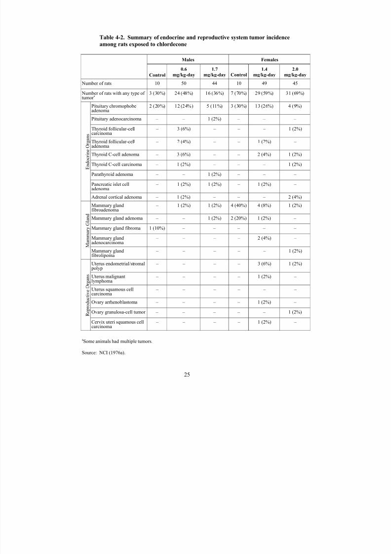
8/7/2019 United States Environmental Protect
http://slidepdf.com/reader/full/united-states-environmental-protect 38/183
Table 4-2. Summary of endocrine and reproductive system tumor incidence
among rats exposed to chlordecone
Males Females
Control
0.6
mg/kg-day
1.7
mg/kg-day Control
1.4
mg/kg-day
2.0
mg/kg-day
Number of rats 10 50 44 10 49 45
Number of rats with any type of tumor a
3 (30%) 24 (48%) 16 (36%) 7 (70%) 29 (59%) 31 (69%)
Endocrine Organs
Pituitary chromophobeadenoma
2 (20%) 12 (24%) 5 (11%) 3 (30%) 13 (26%) 4 (9%)
Pituitary adenocarcinoma – – 1 (2%) – – –
Thyroid follicular-cellcarcinoma
– 3 (6%) – – – 1 (2%)
Thyroid follicular-celladenoma
– 2 (4%) – – 1 (2%) –
Thyroid C-cell adenoma – 3 (6%) – – 2 (4%) 1 (2%)
Thyroid C-cell carcinoma – 1 (2%) – – – 1 (2%)
Parathyroid adenoma – – 1 (2%) – – –
Pancreatic islet celladenoma
– 1 (2%) 1 (2%) – 1 (2%) –
Adrenal cortical adenoma – 1 (2%) – – – 2 (4%)
Mammary G
land
Mammary glandfibroadenoma
– 1 (2%) 1 (2%) 4 (40%) 4 (8%) 1 (2%)
Mammary gland adenoma – – 1 (2%) 2 (20%) 1 (2%) –
Mammary gland fibroma 1 (10%) – – – – –
Mammary glandadenocarcinoma
– – – – 2 (4%) –
Mammary glandfibrolipoma
– – – – – 1 (2%)
Reproductive Organs
Uterus endometrial/stromalpolyp
– – – – 3 (6%) 1 (2%)
Uterus malignantlymphoma
– – – – 1 (2%) –
Uterus squamous cellcarcinoma
– – – – – –
Ovary arrhenoblastoma – – – – 1 (2%) –
Ovary granulosa-cell tumor – – – – – 1 (2%)
Cervix uteri squamous cellcarcinoma
– – – – 1 (2%) –
aSome animals had multiple tumors.
Source: NCI (1976a).
25

8/7/2019 United States Environmental Protect
http://slidepdf.com/reader/full/united-states-environmental-protect 39/183
In mice, dietary concentrations of chlordecone began at 0 or 40 ppm (two groups at this
concentration) for males and 0, 40, or 80 ppm for females. Treatment groups comprised
50 mice/sex; however, only 10 female mice and 19 male mice were used as matched controls.
Pooled control groups (from the same laboratory with birth dates within 3–4 months of the
animals in the matched control and exposed groups) contained 49 male mice and 40 female mice.
Overt clinical signs of toxicity observed in the high-dose male and female mice indicated that themaximum tolerated dose was exceeded in those exposure groups; consequently, concentrations
of chlordecone in feed for all dose groups were reduced (one-fourth to one-half of the original
concentration) during the experiment. The specific dosing regimens for male and female mice
were described in detail in unpublished raw data for the bioassay (NCI, 1976b) and are illustrated
in Figures 4-3 and 4-4. The initial high-dose group of male mice was discontinued due to excess
toxicity, and a new group was started 7 months later after the beginning of the study. Time-
weighted-average dietary concentrations were reported by the authors to be 0, 20, or 23 ppm for
male mice and 0, 20, or 40 ppm for female mice. Doses estimated from U.S. EPA (1988)
reference values for body weight and food consumption were calculated2: 0, 3.4, or 3.9 mg/kg-
day for male mice and 0, 3.5, or 7.0 mg/kg-day for female mice. Following the 80-week
exposure, surviving mice were sacrificed at 90 weeks. Histological examination was similar to
that described previously for rats.
2Calculation: mg/kg-day = (ppm in feed × kg food/day)/kg-bw. Reference food consumption rates of 0.0064 kg/day
(males) and 0.0061 kg/day (females) and reference body weights of 0.0373 (males) and 0.0353 kg (females) wereused (U.S. EPA, 1988).
26

8/7/2019 United States Environmental Protect
http://slidepdf.com/reader/full/united-states-environmental-protect 40/183
*Lines represent changes in the dose levels made throughout the study period.
Source: NCI (1976a, b).
Figure 4-3. Dosing regimen for male mice.
*Lines represent changes i
n the dose levels made throughout the study period.
ource: NCI (1976a, b).
igure 4-4. Dosing regimen for female mice.
S
F
27
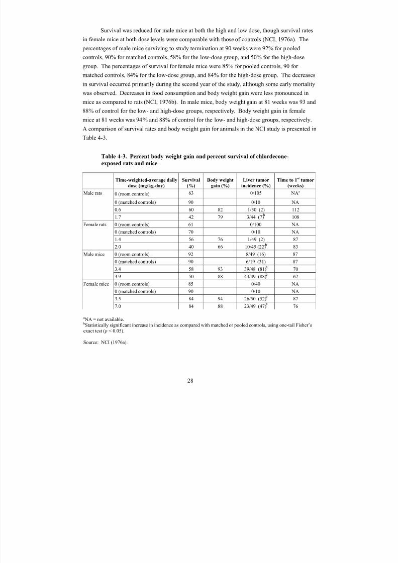
8/7/2019 United States Environmental Protect
http://slidepdf.com/reader/full/united-states-environmental-protect 41/183
Survival was reduced for male mice at both the high and low dose, though survival rates
in female mice at both dose levels were comparable with those of controls (NCI, 1976a). The
percentages of male mice surviving to study termination at 90 weeks were 92% for pooled
controls, 90% for matched controls, 58% for the low-dose group, and 50% for the high-dose
group. The percentages of survival for female mice were 85% for pooled controls, 90 for
matched controls, 84% for the low-dose group, and 84% for the high-dose group. The decreasesin survival occurred primarily during the second year of the study, although some early mortality
was observed. Decreases in food consumption and body weight gain were less pronounced in
mice as compared to rats (NCI, 1976b). In male mice, body weight gain at 81 weeks was 93 and
88% of control for the low- and high-dose groups, respectively. Body weight gain in female
mice at 81 weeks was 94% and 88% of control for the low- and high-dose groups, respectively.
A comparison of survival rates and body weight gain for animals in the NCI study is presented in
Table 4-3.
Table 4-3. Percent body weight gain and percent survival of chlordecone-
exposed rats and mice
Time-weighted-average daily
dose (mg/kg-day)
Survival
(%)
Body weight
gain (%)
Liver tumor
incidence (%)
Time to 1st
tumor
(weeks)
Male rats 0 (room controls) 63 0/105 NAa
0 (matched controls) 90 0/10 NA
0.6 60 82 1/50 (2) 112
1.7 42 79 3/44 (7) 108
Female rats 0 (room controls) 61 0/100 NA
0 (matched controls) 70 0/10 NA
1.4 56 76 1/49 (2) 87
2.0 40 66 10/45 (22) 83
Male mice 0 (room controls) 92 8/49 (16) 87
0 (matched controls) 90 6/19 (31) 87
3.4 58 93 39/48 (81) 70
3.9 50 88 43/49 (88) 62
Female mice 0 (room controls) 85 0/40 NA
0 (matched controls) 90 0/10 NA
3.5 84 94 26/50 (52) 87
7.0 84 88 23/49 (47) 76
aNA = not available.bStatistically significant increase in incidence as compared with matched or pooled controls, using one-tail Fisher’sexact test (p < 0.05).
Source: NCI (1976a).
28

8/7/2019 United States Environmental Protect
http://slidepdf.com/reader/full/united-states-environmental-protect 42/183
Clinical signs of chlordecone toxicity were reported in mice; however, the incidence by
dose was not reported (NCI, 1976a, b). High-dose female mice developed tremors during the
first week of the study that persisted to study termination. Tremors were also observed in some
high-dose male mice, and about 20% of high-dose males were highly excitable during the second
year of the study. Abdominal distention was first observed in high-dose males at week 45 and
high-dose females at week 68, presumably associated with hepatic hypertrophy. Palpableabdominal masses were found in high- and low-dose males during the second year of the study.
Alopecia, rough hair coats, and tail sores were seen primarily in males and were thought to be
due to fighting. More specific data regarding occurrence of clinical signs were not reported.
In mice, statistically significant elevated incidences of hepatocellular carcinomas were
found in both exposed groups compared with matched and pooled control incidences (NCI,
1976a). Incidences for matched control, low-, and high-dose groups were 6/19, 39/48, and 43/49
for male mice and 0/10, 26/50, and 23/49 for female mice. The incidence in control male mice
was reported as abnormally high. Two of the pooled control male mice had hepatocellular
carcinomas. Combining the matched and pooled control male mouse groups resulted in an
overall incidence of 8/49 for control male mice. Hepatocellular carcinomas in mice were
described as varying from demarcated nodules to large masses that were well differentiated
without vascular invasion or metastases. Extensive liver hyperplasia also was found in both
sexes in both low- and high-dose mouse groups. Incidences for liver hyperplasia were not
specified, but the report noted that “a few matched controls of each sex also had liver hyperplasia
although the incidence was quite low as compared to the treated groups.” No tumors of other
endocrine organs were reported, aside from one ovary cystadenoma in a single high-dose female
(1/49 or 2% incidence rate). No elevated incidences of tumors at other tissue sites were found inexposed mice compared with controls. The incidence and time-to-tumor data for hepatocellular
carcinoma in the NCI (1976a) report are summarized in Table 4-4. No exposure-related
noncancer lesions were mentioned other than the liver atypia and nodular and diffuse hyperplasia
(NCI, 1976a, b). Induction of noncancerous liver lesions (i.e., hyperplasia) was observed at all
dose levels for each sex and species. Thus, freestanding LOAELs identified for this study are
0.6, 1.4, 3.4, and 3.5 mg/kg-day for male rats, female rats, male mice, and female mice,
respectively.
29
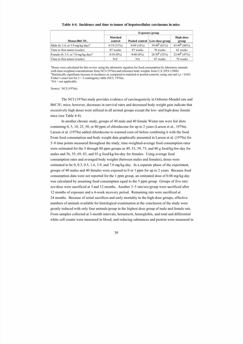
8/7/2019 United States Environmental Protect
http://slidepdf.com/reader/full/united-states-environmental-protect 43/183
Table 4-4. Incidence and time to tumor of hepatocellular carcinoma in mice
Mouse/B6C3F1
Exposure group
Matched
control Pooled control Low-dose group
High-dose
group
Male (0, 3.4, or 3.9 mg/kg-day)
a
6/19 (31%) 8/49 (16%) 39/48 (81%) 43/49 (88%)Time to first tumor (weeks) 87 weeks 87 weeks 70 weeks 62 weeks
Female (0, 3.5, or 7.0 mg/kg-day)a 0/10 (0%) 0/40 (0%) 26/50 (52%) 23/49 (47%)
Time to first tumor (weeks) NAc NA 87 weeks 76 weeks
aDoses were calculated for this review using the allometric equation for food consumption by laboratory animalswith time-weighted concentrations from NCI (1976a) and reference body weights from U.S. EPA (1988).bStatistically significant increase in incidence as compared to matched or pooled controls, using one-tail (p < 0.05)Fisher’s exact test for 2 × 2 contingency table (NCI, 1976a).cNA = not applicable.
Source: NCI (1976a).
The NCI (1976a) study provides evidence of carcinogenicity in Osborne-Mendel rats and
B6C3F1 mice; however, decreases in survival rates and decreased body weight gain indicate that
excessively high doses were utilized in all animal groups except the low- and high-dose female
mice (see Table 4-4).
In another chronic study, groups of 40 male and 40 female Wistar rats were fed diets
containing 0, 5, 10, 25, 50, or 80 ppm of chlordecone for up to 2 years (Larson et al., 1979a).
Larson et al. (1979a) added chlordecone to warmed corn oil before combining it with the food.
From food consumption and body weight data graphically presented in Larson et al. (1979a) for
5–8 time points measured throughout the study, time-weighted-average food consumption rates
were estimated for the 5 through 80 ppm groups as 49, 53, 59, 73, and 80 g food/kg-bw-day for
males and 56, 55, 69, 83, and 93 g food/kg-bw-day for females. Using average food
consumption rates and averaged body weights (between males and females), doses were
estimated to be 0, 0.3, 0.5, 1.6, 3.9, and 7.0 mg/kg-day. In a separate phase of the experiment,
groups of 40 males and 40 females were exposed to 0 or 1 ppm for up to 2 years. Because food
consumption data were not reported for the 1 ppm group, an estimated dose of 0.06 mg/kg-day
was calculated by assuming food consumption equal to the 5 ppm group. Groups of five rats/
sex/dose were sacrificed at 3 and 12 months. Another 3–5 rats/sex/group were sacrificed after
12 months of exposure and a 4-week recovery period. Remaining rats were sacrificed at
24 months. Because of serial sacrifices and early mortality in the high-dose groups, effective
numbers of animals available for histological examination at the conclusion of the study were
greatly reduced with only four animals/group in the highest dose group of male and female rats.
From samples collected at 3-month intervals, hematocrit, hemoglobin, and total and differential
white cell counts were measured in blood, and reducing substances and protein were measured in
30
8/7/2019 United States Environmental Protect
http://slidepdf.com/reader/full/united-states-environmental-protect 44/183
urine. Additional blood studies were performed at 3 months for platelet count, prothrombin
clotting time, and serum calcium. Oxygen consumption was measured by spirometry at
9 months. Organ-to-body-weight ratios (liver, kidneys, heart, spleen, and testes) were
determined in sacrificed rats. The following tissues were taken from sacrificed rats for
histopathological study: brain, spinal cord, heart, lung, liver, kidney, spleen, gut, urinary
bladder, bone marrow, skeletal muscle, skin, pancreas, thyroid, adrenal, pituitary, and gonad.Tremors developed in the 3.9 and 7.0 mg/kg-day groups within a few weeks of the start
of the study and became progressively more severe with time (Larson et al., 1979a). Slight
tremors were noted in some rats at 1.6 mg/kg-day after 3 months, becoming moderate in severity
after 5–6 months, but then regressing. Tremors were not observed at ≤0.5 mg/kg-day. The
incidence of tremors was not reported. All rats in the 3.9 and 7.0 mg/kg-day groups died during
the first 6 months. Long-term survival was reduced in the 1.6 mg/kg-day females (measured at 1
and 2 years, data not shown). Body weights were depressed after 3 weeks of study in males at
>1.6 mg/kg-day and in females at >0.3 mg/kg-day. Food consumption (per body weight) tended
to increase with concentration of chlordecone in the feed. Metabolic rate (measured by oxygen
consumption) increased with dose in both males and females, although statistical significance
was achieved only in males at 1.6 mg/kg-day (the highest dose with survivors remaining when
tested at 9 months). Hematology analyses revealed no differences related to treatment. Increases
in urinary protein concentrations or proteinuria (a clinical indicator of glomerular dysfunction)
were reported in both male and female rats exposed to >0.3 mg/kg-day for 6–24 months, though
statistical analysis was not performed on these data because of incomplete data reporting.
Proteinuria was not observed in rats exposed to 0.06 mg/kg-day in a separate phase of the
experiment (the time of analysis and other details were not reported). Relative liver weightincreased with dose at 3, 12, and 24 months in both male and female rats. The difference from
controls was statistically significant at >1.6 mg/kg-day in males and >0.5 mg/kg-day in females.
Relative testes weights were significantly decreased in the 3.9 and 7.0 mg/kg-day groups at the
3-month sacrifice. Relative weight changes in the kidneys and other organs were not
remarkable. Absolute organ weights were not reported.
Histopathological examination of five rats (randomly selected) from each sex at each
feeding level at 13 weeks revealed minimal congestion of the liver at 0.5 mg/kg-day and more
degenerative changes in the liver at higher doses (Larson et al., 1979a). There was a trend in
dose-response for degenerative liver changes. Swollen liver cells were noted in 4/5 males and
5/5 females in the 3.9 mg/kg-day group and 5/5 males and 3/5 females in the 7.0 mg/kg-day
group (compared with 0/10 males and 0/10 females in the control groups). The liver-to-body-
weight ratios were significantly increased in the 3.9 and 7.0 mg/kg-day groups for both sexes.
Histological examination also uncovered a dose-related increase in the incidence and severity of
testicular atrophy at 13 weeks, though not at 1–2 years. The study authors did not speculate as to
why testicular atrophy was observed after 13 weeks but not at the chronic time point. Interim (3-
31
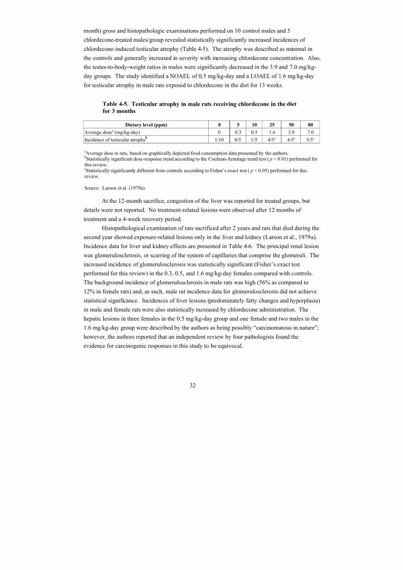
8/7/2019 United States Environmental Protect
http://slidepdf.com/reader/full/united-states-environmental-protect 45/183
month) gross and histopathologic examinations performed on 10 control males and 5
chlordecone-treated males/group revealed statistically significantly increased incidences of
chlordecone-induced testicular atrophy (Table 4-5). The atrophy was described as minimal in
the controls and generally increased in severity with increasing chlordecone concentration. Also,
the testes-to-body-weight ratios in males were significantly decreased in the 3.9 and 7.0 mg/kg-
day groups. The study identified a NOAEL of 0.5 mg/kg-day and a LOAEL of 1.6 mg/kg-dayfor testicular atrophy in male rats exposed to chlordecone in the diet for 13 weeks.
Table 4-5. Testicular atrophy in male rats receiving chlordecone in the diet
for 3 months
Dietary level (ppm) 0 5 10 25 50 80
Average dosea (mg/kg-day) 0 0.3 0.5 1.6 3.9 7.0
Incidence of testicular atrophy 1/10 0/5 1/5 4/5c 4/5c 5/5c
a
Average dose to rats, based on graphically depicted food consumption data presented by the authors.bStatistically significant dose-response trend according to the Cochran-Armitage trend test (p < 0.01) performed for this review.cStatistically significantly different from controls according to Fisher’s exact test (p < 0.05) performed for thisreview.
Source: Larson et al. (1979a).
At the 12-month sacrifice, congestion of the liver was reported for treated groups, but
details were not reported. No treatment-related lesions were observed after 12 months of
treatment and a 4-week recovery period.
Histopathological examination of rats sacrificed after 2 years and rats that died during thesecond year showed exposure-related lesions only in the liver and kidney (Larson et al., 1979a).
Incidence data for liver and kidney effects are presented in Table 4-6. The principal renal lesion
was glomerulosclerosis, or scarring of the system of capillaries that comprise the glomeruli. The
increased incidence of glomerulosclerosis was statistically significant (Fisher’s exact test
performed for this review) in the 0.3, 0.5, and 1.6 mg/kg-day females compared with controls.
The background incidence of glomerulosclerosis in male rats was high (56% as compared to
12% in female rats) and, as such, male rat incidence data for glomerulosclerosis did not achieve
statistical significance. Incidences of liver lesions (predominately fatty changes and hyperplasia)
in male and female rats were also statistically increased by chlordecone administration. The
hepatic lesions in three females in the 0.5 mg/kg-day group and one female and two males in the
1.6 mg/kg-day group were described by the authors as being possibly “carcinomatous in nature”;
however, the authors reported that an independent review by four pathologists found the
evidence for carcinogenic responses in this study to be equivocal.
32
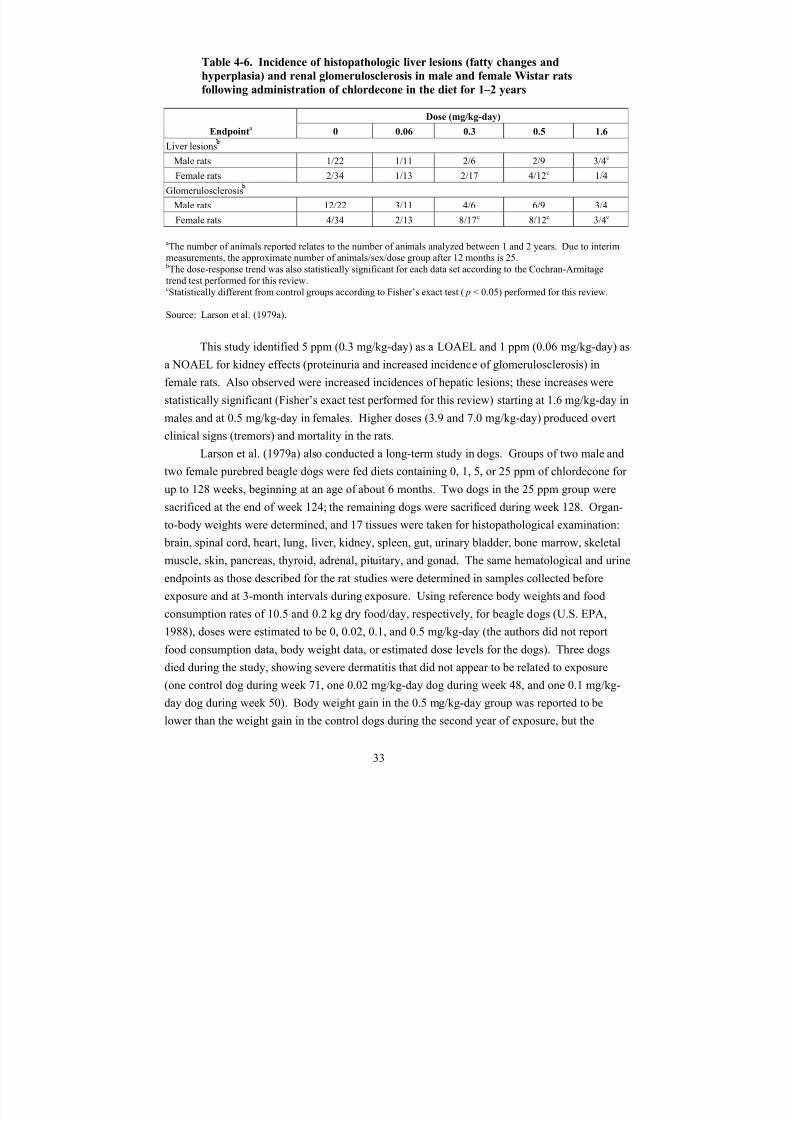
8/7/2019 United States Environmental Protect
http://slidepdf.com/reader/full/united-states-environmental-protect 46/183
Table 4-6. Incidence of histopathologic liver lesions (fatty changes and
hyperplasia) and renal glomerulosclerosis in male and female Wistar rats
following administration of chlordecone in the diet for 1–2 years
Endpointa
Dose (mg/kg-day)
0 0.06 0.3 0.5 1.6
Liver lesions
Male rats 1/22 1/11 2/6 2/9 3/4c
Female rats 2/34 1/13 4/12c 2/17 1/4
Glomerulosclerosis
Male rats 12/22 3/11 4/6 6/9 3/4
Female rats 4/34 2/13 8/17c 8/12c 3/4c
aThe number of animals reported relates to the number of animals analyzed between 1 and 2 years. Due to interimmeasurements, the approximate number of animals/sex/dose group after 12 months is 25.bThe dose-response trend was also statistically significant for each data set according to the Cochran-Armitagetrend test performed for this review.cStatistically different from control groups according to Fisher’s exact test (p < 0.05) performed for this review.
Source: Larson et al. (1979a).
This study identified 5 ppm (0.3 mg/kg-day) as a LOAEL and 1 ppm (0.06 mg/kg-day) as
a NOAEL for kidney effects (proteinuria and increased incidence of glomerulosclerosis) in
female rats. Also observed were increased incidences of hepatic lesions; these increases were
statistically significant (Fisher’s exact test performed for this review) starting at 1.6 mg/kg-day in
males and at 0.5 mg/kg-day in females. Higher doses (3.9 and 7.0 mg/kg-day) produced overt
clinical signs (tremors) and mortality in the rats.
Larson et al. (1979a) also conducted a long-term study in dogs. Groups of two male andtwo female purebred beagle dogs were fed diets containing 0, 1, 5, or 25 ppm of chlordecone for
up to 128 weeks, beginning at an age of about 6 months. Two dogs in the 25 ppm group were
sacrificed at the end of week 124; the remaining dogs were sacrificed during week 128. Organ-
to-body weights were determined, and 17 tissues were taken for histopathological examination:
brain, spinal cord, heart, lung, liver, kidney, spleen, gut, urinary bladder, bone marrow, skeletal
muscle, skin, pancreas, thyroid, adrenal, pituitary, and gonad. The same hematological and urine
endpoints as those described for the rat studies were determined in samples collected before
exposure and at 3-month intervals during exposure. Using reference body weights and food
consumption rates of 10.5 and 0.2 kg dry food/day, respectively, for beagle dogs (U.S. EPA,
1988), doses were estimated to be 0, 0.02, 0.1, and 0.5 mg/kg-day (the authors did not report
food consumption data, body weight data, or estimated dose levels for the dogs). Three dogs
died during the study, showing severe dermatitis that did not appear to be related to exposure
(one control dog during week 71, one 0.02 mg/kg-day dog during week 48, and one 0.1 mg/kg-
day dog during week 50). Body weight gain in the 0.5 mg/kg-day group was reported to be
lower than the weight gain in the control dogs during the second year of exposure, but the
33
8/7/2019 United States Environmental Protect
http://slidepdf.com/reader/full/united-states-environmental-protect 47/183
magnitude of the decrease was not reported and the data were not shown. Decreased food
efficiency (kg body weight gain/kg food consumed) was suggested by measurements of food
consumption, but again the data were not shown. The only statistically significant changes
associated with exposure to chlordecone were a moderate (37%) increase in relative liver weight
in dogs from the 0.5 mg/kg-day group (males and females combined) and slight changes (less
than about 25%) in relative kidney (increase), heart (increase), and spleen (decrease) weight inthe same group. Absolute organ weights were not reported. No exposure-related changes were
reported for clinical signs of toxicity; hematological, histopathological, or urinalysis endpoints;
sulfobromophthalein retention; or serum cholinesterase. Interpretation of this study is limited by
the small number of dogs tested, the deaths of three dogs during the study for reasons not
apparently related to treatment, and the reporting of results, including failure of the researchers to
present data to support the reported decrease in body weight in dogs from the 0.5 mg/kg-day
group during the second year of the study. Nevertheless, the statistically significant changes in
organ-to-body-weight ratios support occurrence of an adverse effect on body weight, and the
increase in relative liver weight is consistent with other studies demonstrating hepatic toxicity
with chlordecone exposure. Therefore, the results of this study suggest a LOAEL of 25 ppm
(0.5 mg/kg-day) and a NOAEL of 5 ppm (0.1 mg/kg-day), based on decreased body weight and
organ-to-body-weight changes (without histological changes) in beagle dogs fed chlordecone in
the diet for up to 128 weeks.
4.3. REPRODUCTIVE/DEVELOPMENTAL STUDIES
4.3.1. Reproductive Toxicity Studies
Information on reproductive effects in humans is restricted to findings of oligospermia,reduced sperm motility, and decreased libido in a group of men who were occupationally
exposed to chlordecone for periods up to 1.5 years (Taylor, 1985, 1982; Guzelian, 1982a; Taylor
et al., 1978). Sperm concentration and motility had returned to normal upon follow-up 5 years
following cessation of chlordecone exposure. Even though two of seven workers sired children,
there is no indication of the true denominator of how many were trying to conceive and/or the
fertility rate. In one worker, low sperm count persisted (Taylor, 1985). No information is
available concerning chlordecone-induced reproductive effects in women.
Reproductive toxicity has been assessed in some animal studies, but not in adequately
designed multiple generation studies. Available animal data suggest that chlordecone is a male
reproductive toxicant, causing alteration of sperm parameters at low doses and testicular atrophy
at higher doses. Persistent vaginal estrus (PVE) is reported to occur in exposed females, and
decreased reproductive success has been demonstrated. No animal studies are available to assess
the developmental or reproductive toxicity of chlordecone by the inhalation route of exposure.
Huber (1965) performed a series of experiments designed to assess reproductive toxicity
in mice exposed to chlordecone in the diet. In a pilot reproduction study (group A), 3-month-old
34
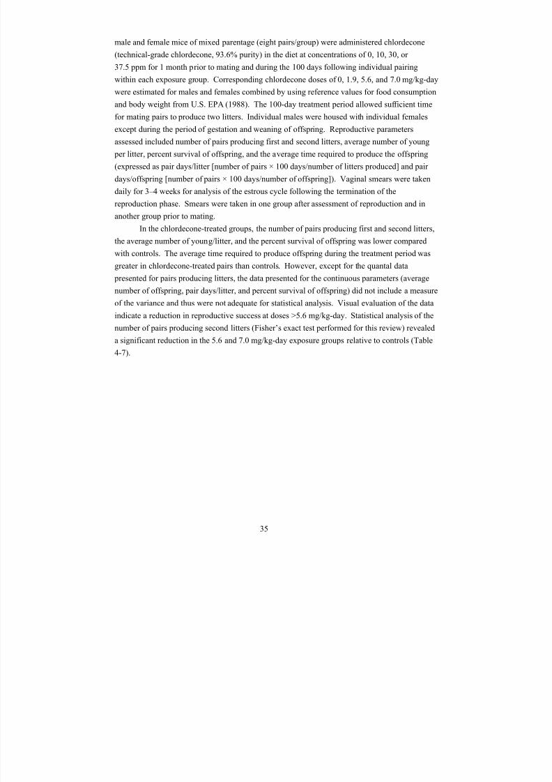
8/7/2019 United States Environmental Protect
http://slidepdf.com/reader/full/united-states-environmental-protect 48/183
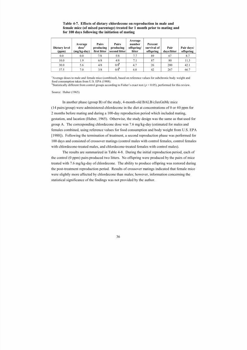
8/7/2019 United States Environmental Protect
http://slidepdf.com/reader/full/united-states-environmental-protect 49/183
Table 4-7. Effects of dietary chlordecone on reproduction in male and
female mice (of mixed parentage) treated for 1 month prior to mating and
for 100 days following the initiation of mating
Dietary level(ppm)
Average
dosea (mg/kg-day)
Pairs
producingfirst litter
Pairs
producingsecond litter
Average
number
offspring/litter
Percent
survival of offspring
Pairdays/litter
Pair days/offspring
0.0 0.0 7/8 5/8 7.7 89 67 8.7
10.0 1.9 6/8 4/8 7.1 87 80 11.3
30.0 5.6 4/8 0/8 4.7 26 200 42.1
37.5 7.0 3/8 0/8 4.0 42 267 66.7
aAverage doses to male and female mice (combined), based on reference values for subchronic body weight andfood consumption taken from U.S. EPA (1988).bStatistically different from control groups according to Fisher’s exact test (p < 0.05), performed for this review.
Source: Huber (1965).
In another phase (group B) of the study, 4-month-old BALB/cJaxGnMc mice
(14 pairs/group) were administered chlordecone in the diet at concentrations of 0 or 40 ppm for
2 months before mating and during a 100-day reproduction period which included mating,
gestation, and lacation (Huber, 1965). Otherwise, the study design was the same as that used for
group A. The corresponding chlordecone dose was 7.6 mg/kg-day (estimated for males and
females combined, using reference values for food consumption and body weight from U.S. EPA
[1988]). Following the termination of treatment, a second reproduction phase was performed for
100 days and consisted of crossover matings (control males with control females, control femaleswith chlordecone-treated males, and chlordecone-treated females with control males).
The results are summarized in Table 4-8. During the initial reproduction period, each of
the control (0 ppm) pairs produced two litters. No offspring were produced by the pairs of mice
treated with 7.6 mg/kg-day of chlordecone. The ability to produce offspring was restored during
the post-treatment reproduction period. Results of crossover matings indicated that female mice
were slightly more affected by chlordecone than males; however, information concerning the
statistical significance of the findings was not provided by the author.
36
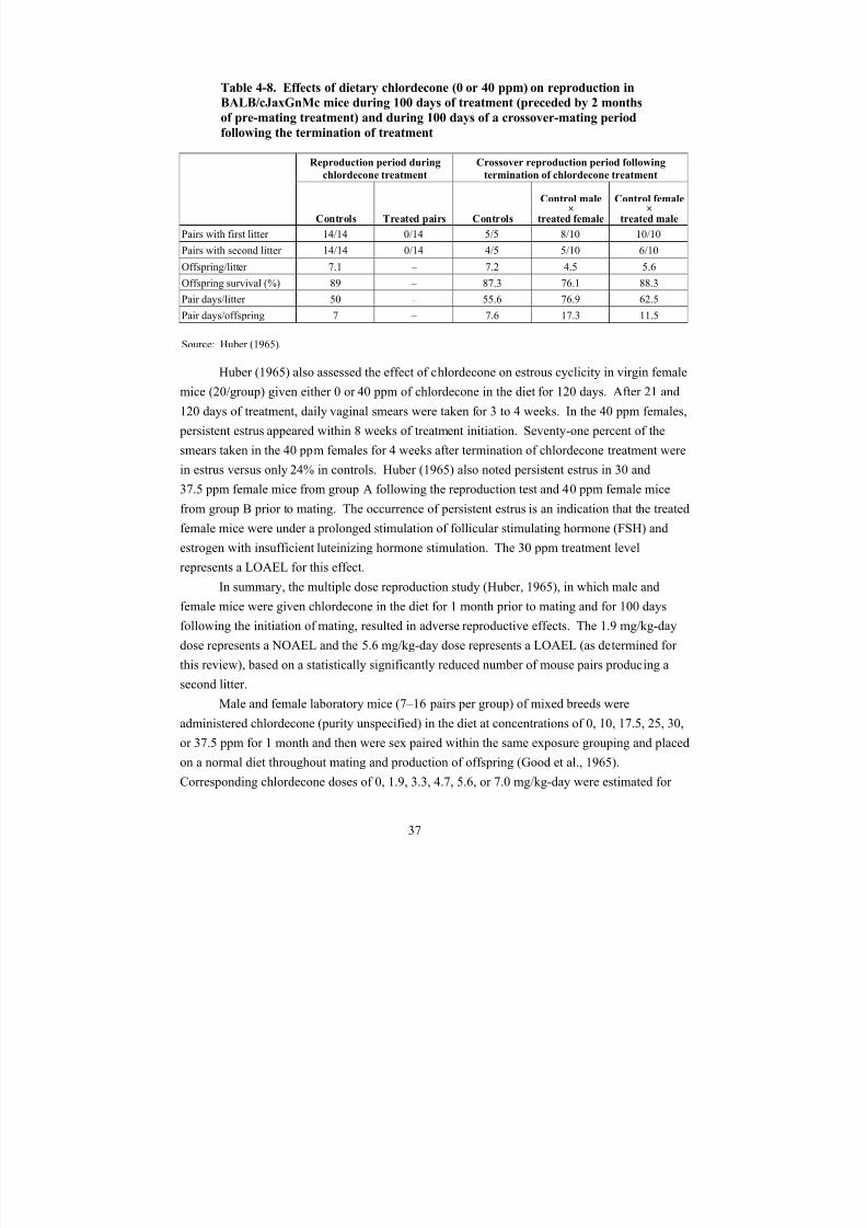
8/7/2019 United States Environmental Protect
http://slidepdf.com/reader/full/united-states-environmental-protect 50/183
8/7/2019 United States Environmental Protect
http://slidepdf.com/reader/full/united-states-environmental-protect 51/183
males and females combined by using reference values for food consumption and body weight
from U.S. EPA (1988). Reproductive indices (number of litters produced, average number of
young/litter, pair days/litter, and pair days/young produced) were assessed for approximately
5 months following the initiation of mating. As shown in Table 4-9, the results suggest a dose-
related effect on reproductive success (decreases in number of litters and average number of
young per litter, increases in pair days per litter and per young). Though the data presented inthe study were not adequate for statistical analysis (no measures of variance were provided for
the reproductive parameters), visual evaluation of the data indicates a reduction in reproductive
success at doses >5.6 mg/kg-day.
Table 4-9. Effects of dietary chlordecone for 1 month prior to mating on
reproductive indices of male and female laboratory mice of mixed breeds
Dietary level
(ppm)
Average dosea
(mg/kg-day)
Number of
pairs
Number of
litters
Number of
offspring/litter
Pair
days/litter
Pair
days/offspring
0 0.0 9 15 7.93 65.3 8.3
10 1.9 13 26 7.62 54.46 7.15
17.5 3.3 16 25 7.0 72.16 13.09
25 4.7 11 12 6.08 100.42 16.51
30 5.6 7 2 3.0 241.5 80.5
37.5 7.0 10 2 5.0 555.0 111.0
aAverage doses to male and female mice (combined), based on reference values for subchronic body weight andfood consumption taken from U.S. EPA (1988).
Source: Good et al. (1965).
In separate experiments by Good et al. (1965), impaired reproductive success, expressed
as significantly (p < 0.05) reduced production of a second litter, was observed in mice that were
administered chlordecone (purity unspecified) in the diet at a concentration of 5 ppm for 1 month
prior to mating and for up to 5 months following initiation of mating (shown in Table 4-10). The
corresponding chlordecone dose of 0.94 mg/kg-day was estimated for males and females
combined by using reference values for food consumption and body weight from U.S. EPA
(1988). The authors reported that continued treatment of offspring of chlordecone-treated mice
with either control or 5 ppm chlordecone diets resulted in significantly reduced production of a
first litter (p ≤ 0.05), compared with untreated offspring of untreated parental mice, though
reduced production of the second litter did not achieve statistical significance. The results of
these studies identified a LOAEL of 0.94 mg/kg-day for impaired reproductive success; a
NOAEL was not identified.
38

8/7/2019 United States Environmental Protect
http://slidepdf.com/reader/full/united-states-environmental-protect 52/183
Table 4-10. Effects of dietary chlordecone (0 or 5 ppm) 1 month prior to
mating and up to 5 months after initiation of mating on reproduction in
BALB/c mice
First generation Second generation
Control Treated Control
Offspring of
treated mice on
control diet Treated
Number of pairs 24 36 21 23 20
Number of litters 40 52 21 9 10
Number of offspring 275 314 123 42 40
% producing 1st litter 96 81 71 30a 25a
% producing 2nd litter 78 50a 29 9 15
First litter size 6.2 6.2 5.6 4.3 4.4
Second litter size 7.3 5.7 6.5 6.0 5.3
Pair days/litter 70.1 86 120 307 240
Pair days/offspring 10.2 14.2 21 66 60
aReported as significant at p < 0.05, using binomial distribution.
Source: Good et al. (1965).
As in the previous data reported by Good et al. (1965), reproductive parameters,
including litter size, pair days/litter, and pair days/young produced, were all reported as averages
for the treatment or control group without any measure of variance given (i.e., standard
deviation). Therefore the degree of variability for the reported reproductive parameters is
unclear. Additionally, there was reduced fertility of the BALB/c untreated controls just one
generation apart. For instance, 96 and 78% of untreated control animals produced first and
second litters, respectively, whereas only 71 and 29% of their untreated progeny produced first
and second litters. These inconsistencies limit confidence in this study and the reproducibility of
the data.
In a reproductive and neurodevelopmental toxicity study, female F344 rats (10/group)
were fed diets containing 0, 1, or 6 ppm of chlordecone (purity unspecified) for 60 days prior to
mating (with a nonexposed male rat) through lactation day 12 (Squibb and Tilson, 1982).
Corresponding maternal doses of 0, 0.1, and 0.6 mg/kg-day were estimated by using reference
values for food consumption from U.S. EPA (1988) and the average of reported body weights of the dams prior to mating and on the day after parturition. Chlordecone treatment did not produce
adverse effects on litter size or sex ratio of the offspring. Litters were culled to three male and
three female offspring per dam on postpartum day 3. Pup body weights were similar to those of
controls at 1, 7, 14, and 30 days of age, but after 100 days, body weight was significantly
reduced in male pups at 0.6 mg/kg-day (19% decrease relative to controls) and female pups at
0.1 mg/kg-day (27% decrease relative to controls) and 0.6 mg/kg-day (27% decrease relative to
39
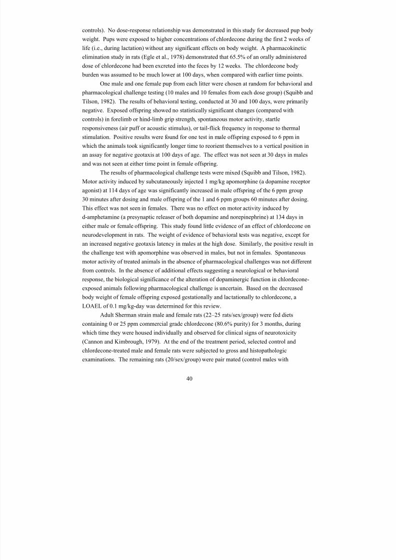
8/7/2019 United States Environmental Protect
http://slidepdf.com/reader/full/united-states-environmental-protect 53/183
controls). No dose-response relationship was demonstrated in this study for decreased pup body
weight. Pups were exposed to higher concentrations of chlordecone during the first 2 weeks of
life (i.e., during lactation) without any significant effects on body weight. A pharmacokinetic
elimination study in rats (Egle et al., 1978) demonstrated that 65.5% of an orally administered
dose of chlordecone had been excreted into the feces by 12 weeks. The chlordecone body
burden was assumed to be much lower at 100 days, when compared with earlier time points.One male and one female pup from each litter were chosen at random for behavioral and
pharmacological challenge testing (10 males and 10 females from each dose group) (Squibb and
Tilson, 1982). The results of behavioral testing, conducted at 30 and 100 days, were primarily
negative. Exposed offspring showed no statistically significant changes (compared with
controls) in forelimb or hind-limb grip strength, spontaneous motor activity, startle
responsiveness (air puff or acoustic stimulus), or tail-flick frequency in response to thermal
stimulation. Positive results were found for one test in male offspring exposed to 6 ppm in
which the animals took significantly longer time to reorient themselves to a vertical position in
an assay for negative geotaxis at 100 days of age. The effect was not seen at 30 days in males
and was not seen at either time point in female offspring.
The results of pharmacological challenge tests were mixed (Squibb and Tilson, 1982).
Motor activity induced by subcutaneously injected 1 mg/kg apomorphine (a dopamine receptor
agonist) at 114 days of age was significantly increased in male offspring of the 6 ppm group
30 minutes after dosing and male offspring of the 1 and 6 ppm groups 60 minutes after dosing.
This effect was not seen in females. There was no effect on motor activity induced by
d-amphetamine (a presynaptic releaser of both dopamine and norepinephrine) at 134 days in
either male or female offspring. This study found little evidence of an effect of chlordecone onneurodevelopment in rats. The weight of evidence of behavioral tests was negative, except for
an increased negative geotaxis latency in males at the high dose. Similarly, the positive result in
the challenge test with apomorphine was observed in males, but not in females. Spontaneous
motor activity of treated animals in the absence of pharmacological challenges was not different
from controls. In the absence of additional effects suggesting a neurological or behavioral
response, the biological significance of the alteration of dopaminergic function in chlordecone-
exposed animals following pharmacological challenge is uncertain. Based on the decreased
body weight of female offspring exposed gestationally and lactationally to chlordecone, a
LOAEL of 0.1 mg/kg-day was determined for this review.
Adult Sherman strain male and female rats (22–25 rats/sex/group) were fed diets
containing 0 or 25 ppm commercial grade chlordecone (80.6% purity) for 3 months, during
which time they were housed individually and observed for clinical signs of neurotoxicity
(Cannon and Kimbrough, 1979). At the end of the treatment period, selected control and
chlordecone-treated male and female rats were subjected to gross and histopathologic
examinations. The remaining rats (20/sex/group) were pair mated (control males with
40

8/7/2019 United States Environmental Protect
http://slidepdf.com/reader/full/united-states-environmental-protect 54/183
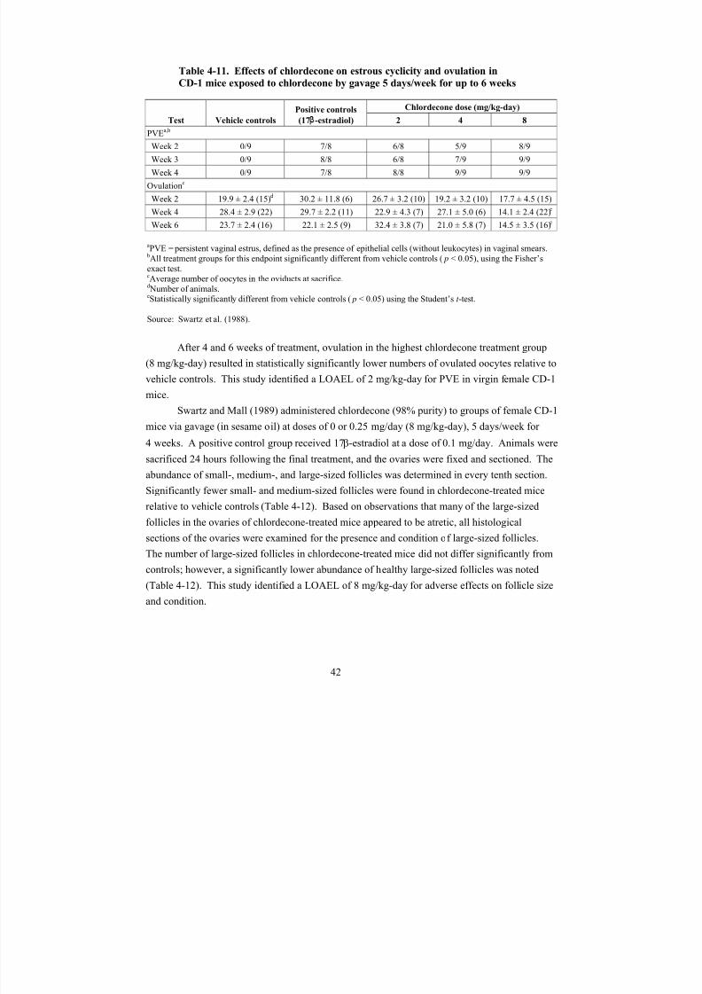
8/7/2019 United States Environmental Protect
http://slidepdf.com/reader/full/united-states-environmental-protect 55/183
Table 4-11. Effects of chlordecone on estrous cyclicity and ovulation in
CD-1 mice exposed to chlordecone by gavage 5 days/week for up to 6 weeks
Test Vehicle controls
Positive controls Chlordecone dose (mg/kg-day)
(17β-estradiol) 2 4 8
PVEa,b
Week 2 0/9 7/8 6/8 5/9 8/9
Week 3 0/9 8/8 6/8 7/9 9/9
Week 4 0/9 7/8 8/8 9/9 9/9
Ovulationc
Week 2 19.9 ± 2.4 (15)d 30.2 ± 11.8 (6) 26.7 ± 3.2 (10) 19.2 ± 3.2 (10) 17.7 ± 4.5 (15)
Week 4 28.4 ± 2.9 (22) 29.7 ± 2.2 (11) 22.9 ± 4.3 (7) 27.1 ± 5.0 (6) 14.1 ± 2.4 (22)e
Week 6 23.7 ± 2.4 (16) 22.1 ± 2.5 (9) 32.4 ± 3.8 (7) 21.0 ± 5.8 (7) 14.5 ± 3.5 (16)e
aPVE = persistent vaginal estrus, defined as the presence of epithelial cells (without leukocytes) in vaginal smears.bAll treatment groups for this endpoint significantly different from vehicle controls (p < 0.05), using the Fisher’s
exact test.cAverage number of oocytes in the oviducts at sacrifice.dNumber of animals.eStatistically significantly different from vehicle controls (p < 0.05) using the Student’s t -test.
Source: Swartz et al. (1988).
After 4 and 6 weeks of treatment, ovulation in the highest chlordecone treatment group
(8 mg/kg-day) resulted in statistically significantly lower numbers of ovulated oocytes relative to
vehicle controls. This study identified a LOAEL of 2 mg/kg-day for PVE in virgin female CD-1
mice.
Swartz and Mall (1989) administered chlordecone (98% purity) to groups of female CD-1
mice via gavage (in sesame oil) at doses of 0 or 0.25 mg/day (8 mg/kg-day), 5 days/week for
4 weeks. A positive control group received 17β-estradiol at a dose of 0.1 mg/day. Animals were
sacrificed 24 hours following the final treatment, and the ovaries were fixed and sectioned. The
abundance of small-, medium-, and large-sized follicles was determined in every tenth section.
Significantly fewer small- and medium-sized follicles were found in chlordecone-treated mice
relative to vehicle controls (Table 4-12). Based on observations that many of the large-sized
follicles in the ovaries of chlordecone-treated mice appeared to be atretic, all histological
sections of the ovaries were examined for the presence and condition of large-sized follicles.The number of large-sized follicles in chlordecone-treated mice did not differ significantly from
controls; however, a significantly lower abundance of healthy large-sized follicles was noted
(Table 4-12). This study identified a LOAEL of 8 mg/kg-day for adverse effects on follicle size
and condition.
42
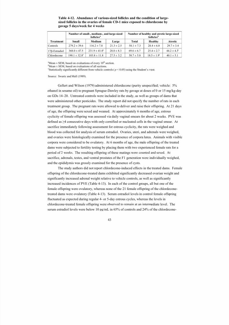
8/7/2019 United States Environmental Protect
http://slidepdf.com/reader/full/united-states-environmental-protect 56/183
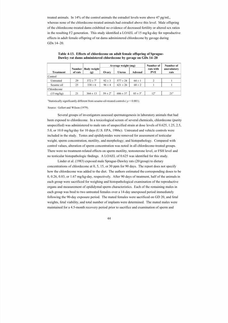
8/7/2019 United States Environmental Protect
http://slidepdf.com/reader/full/united-states-environmental-protect 57/183
treated animals. In 14% of the control animals the estradiol levels were above 47 pg/mL,
whereas none of the chlordecone-treated animals had estradiol above this level. Male offspring
of the chlordecone-treated dams exhibited no evidence of decreased fertility or altered sex ratios
in the resulting F2 generation. This study identified a LOAEL of 15 mg/kg-day for reproductive
effects in adult female offspring of rat dams administered chlordecone by gavage during
GDs 14–20.
Table 4-13. Effects of chlordecone on adult female offspring of Sprague-
Dawley rat dams administered chlordecone by gavage on GDs 14–20
Treatment
Number
of rats
Body weight
(g)
Average weight (mg) Number of
rats with
PVE
Number of
anovulatory
ratsOvary Uterus Adrenal
Control
Untreated 29 372 ± 7a 92 ± 3 577 ± 24 64 ± 1 2 1
Sesame oil 25 338 ± 6 96 ± 4 621 ± 26 68 ± 2 1 1
Chlordecone
(15 mg/kg) 21 364 ± 13 59 ± 2a 686 ± 37 85 ± 3a 12a 21a
aStatistically significantly different from sesame-oil-treated controls (p < 0.001).
Source: Gellert and Wilson (1979).
Several groups of investigators assessed spermatogenesis in laboratory animals that had
been exposed to chlordecone. In a toxicological screen of several chemicals, chlordecone (purity
unspecified) was administered to male rats of unspecified strain at dose levels of 0.625, 1.25, 2.5,
5.0, or 10.0 mg/kg/day for 10 days (U.S. EPA, 1986c). Untreated and vehicle controls wereincluded in the study. Testes and epididymides were removed for assessment of testicular
weight, sperm concentration, motility, and morphology; and histopathology. Compared with
control values, alteration of sperm concentration was noted in all chlordecone-treated groups.
There were no treatment-related effects on sperm motility, testosterone level, or FSH level and
no testicular histopathologic findings. A LOAEL of 0.625 was identified for this study.
Linder et al. (1983) exposed male Sprague-Dawley rats (20/group) to dietary
concentrations of chlordecone at 0, 5, 15, or 30 ppm for 90 days. The report does not specify
how the chlordecone was added to the diet. The authors estimated the corresponding doses to be
0, 0.26, 0.83, or 1.67 mg/kg-day, respectively. After 90 days of treatment, half of the animals in
each group were sacrificed for weighing and histopathological examination of the reproductive
organs and measurement of epididymal sperm characteristics. Each of the remaining males in
each group was bred to two untreated females over a 14-day unexposed period immediately
following the 90-day exposure period. The mated females were sacrificed on GD 20, and fetal
weights, fetal viability, and total number of implants were determined. The mated males were
maintained for a 4.5-month recovery period prior to sacrifice and examination of sperm and
44
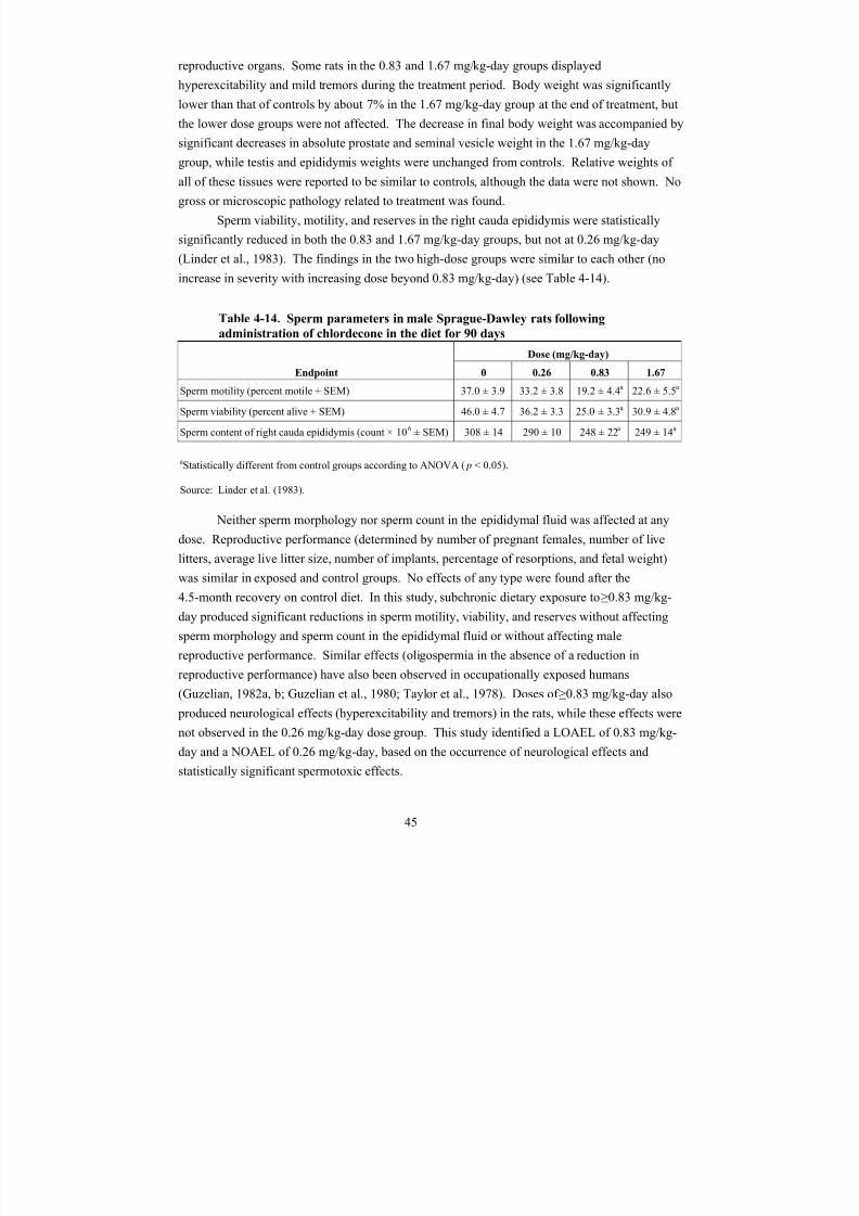
8/7/2019 United States Environmental Protect
http://slidepdf.com/reader/full/united-states-environmental-protect 58/183
reproductive organs. Some rats in the 0.83 and 1.67 mg/kg-day groups displayed
hyperexcitability and mild tremors during the treatment period. Body weight was significantly
lower than that of controls by about 7% in the 1.67 mg/kg-day group at the end of treatment, but
the lower dose groups were not affected. The decrease in final body weight was accompanied by
significant decreases in absolute prostate and seminal vesicle weight in the 1.67 mg/kg-day
group, while testis and epididymis weights were unchanged from controls. Relative weights of all of these tissues were reported to be similar to controls, although the data were not shown. No
gross or microscopic pathology related to treatment was found.
Sperm viability, motility, and reserves in the right cauda epididymis were statistically
significantly reduced in both the 0.83 and 1.67 mg/kg-day groups, but not at 0.26 mg/kg-day
(Linder et al., 1983). The findings in the two high-dose groups were similar to each other (no
increase in severity with increasing dose beyond 0.83 mg/kg-day) (see Table 4-14).
Table 4-14. Sperm parameters in male Sprague-Dawley rats following
administration of chlordecone in the diet for 90 days
Dose (mg/kg-day)
Endpoint 0 0.26 0.83 1.67
Sperm motility (percent motile + SEM) 37.0 ± 3.9 33.2 ± 3.8 19.2 ± 4.4a 22.6 ± 5.5a
Sperm viability (percent alive + SEM) 46.0 ± 4.7 36.2 ± 3.3 25.0 ± 3.3a 30.9 ± 4.8a
Sperm content of right cauda epididymis (count × 106 ± SEM) 308 ± 14 290 ± 10 248 ± 22a 249 ± 14a
aStatistically different from control groups according to ANOVA (p < 0.05).
Source: Linder et al. (1983).
Neither sperm morphology nor sperm count in the epididymal fluid was affected at any
dose. Reproductive performance (determined by number of pregnant females, number of live
litters, average live litter size, number of implants, percentage of resorptions, and fetal weight)
was similar in exposed and control groups. No effects of any type were found after the
4.5-month recovery on control diet. In this study, subchronic dietary exposure to ≥0.83 mg/kg-
day produced significant reductions in sperm motility, viability, and reserves without affecting
sperm morphology and sperm count in the epididymal fluid or without affecting male
reproductive performance. Similar effects (oligospermia in the absence of a reduction inreproductive performance) have also been observed in occupationally exposed humans
(Guzelian, 1982a, b; Guzelian et al., 1980; Taylor et al., 1978). Doses of ≥0.83 mg/kg-day also
produced neurological effects (hyperexcitability and tremors) in the rats, while these effects were
not observed in the 0.26 mg/kg-day dose group. This study identified a LOAEL of 0.83 mg/kg-
day and a NOAEL of 0.26 mg/kg-day, based on the occurrence of neurological effects and
statistically significant spermotoxic effects.
45
8/7/2019 United States Environmental Protect
http://slidepdf.com/reader/full/united-states-environmental-protect 59/183
Additional reproductive studies exist that evaluate the effect of acute injected
chlordecone (at doses of 20–80 mg/kg) in experimental animals. Effects observed in these
studies were similar to studies of repeat oral administration of chlordecone and generally
included changes in estrous cyclicity and fertility (Williams and Uphouse, 1991; Johnson et al.,
1990; Pinkston and Uphouse, 1987–1988). These acute injection studies provide information to
support reproductive effects at high doses of chlordecone but do not generally contributeadditional dose-response information regarding the most sensitive effects of chlordecone
exposure.
4.3.2. Developmental Toxicity Studies
The developmental toxicity of chlordecone in humans is not known. Chlordecone
produces developmental toxicity in rats and mice at dose levels that also produce maternal
toxicity (Seidenberg et al., 1986; Chernoff and Rogers, 1976). Chernoff and Rogers (1976)
administered chlordecone (purity unspecified) to groups of pregnant CD rats at gavage doses of
0, 2, 6, or 10 mg/kg-day on GDs 7–16. Dams were observed for clinical signs and weight gain,
and sacrificed on GD 21 for assessment of liver/body weight and evaluation of fetuses. Fetal
parameters evaluated include number of implants, mortality, weight, and gross developmental
abnormalities. Study results are depicted in Table 4-15. Significant maternal toxicity was
observed in high-dose dams. All groups of dosed dams exhibited significantly depressed weight
gain, and the average liver/body weight ratio was significantly increased in the two highest dose
groups (6 and 10 mg/kg-day). Fetotoxicity was observed as significantly depressed fetal body
weight and delayed ossification in 6 and 10 mg/kg-day dose groups and significantly increased
incidences of litters with fetuses having enlarged renal pelvis, edema, undescended testes, or enlarged cerebral ventricles in the 10 mg/kg-day group relative to controls. The study identified
a LOAEL of 2 mg/kg-day for maternal toxicity, based on significantly depressed maternal body
weight gain (16% lower than controls). The study identified a NOAEL of 2 mg/kg-day and a
LOAEL of 6 mg/kg-day for fetotoxicity.
46
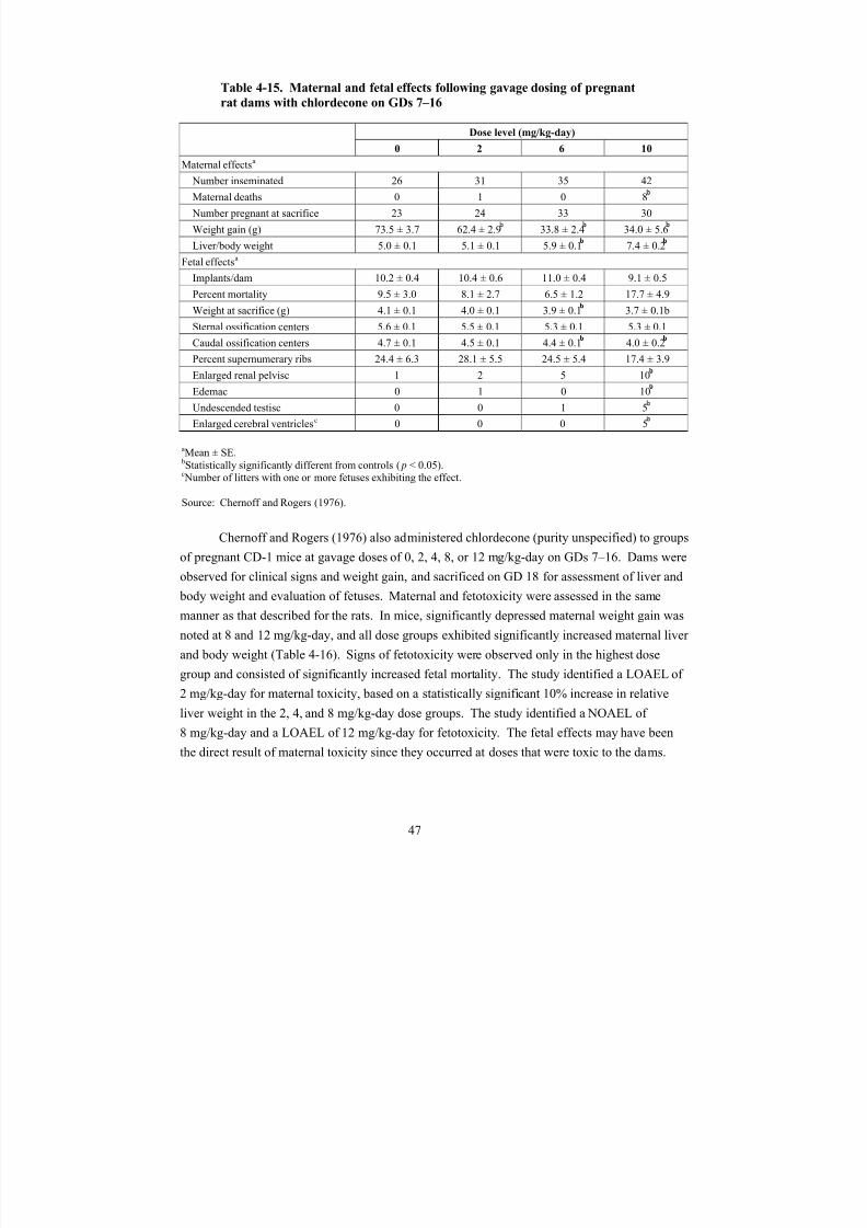
8/7/2019 United States Environmental Protect
http://slidepdf.com/reader/full/united-states-environmental-protect 60/183
Table 4-15. Maternal and fetal effects following gavage dosing of pregnant
rat dams with chlordecone on GDs 7–16
Dose level (mg/kg-day)
0 2 6 10
Maternal effectsa
Number inseminated 26 31 35 42
Maternal deaths 0 1 0 8
Number pregnant at sacrifice 23 24 33 30
Weight gain (g) 73.5 ± 3.7 62.4 ± 2.9 33.8 ± 2.4 34.0 ± 5.6
Liver/body weight 5.0 ± 0.1 5.1 ± 0.1 5.9 ± 0.1 7.4 ± 0.2
Fetal effectsa
Implants/dam 10.2 ± 0.4 10.4 ± 0.6 11.0 ± 0.4 9.1 ± 0.5
Percent mortality 9.5 ± 3.0 8.1 ± 2.7 6.5 ± 1.2 17.7 ± 4.9
Weight at sacrifice (g) 4.1 ± 0.1 4.0 ± 0.1 3.9 ± 0.1 3.7 ± 0.1b
Sternal ossification centers 5.6 ± 0.1 5.5 ± 0.1 5.3 ± 0.1 5.3 ± 0.1Caudal ossification centers 4.7 ± 0.1 4.5 ± 0.1 4.4 ± 0.1 4.0 ± 0.2
Percent supernumerary ribs 24.4 ± 6.3 28.1 ± 5.5 24.5 ± 5.4 17.4 ± 3.9
Enlarged renal pelvisc 1 2 5 10
Edemac 0 1 0 10
Undescended testisc 0 0 1 5
Enlarged cerebral ventriclesc 0 0 0 5
aMean ± SE. bStatistically significantly different from controls (p < 0.05).cNumber of litters with one or more fetuses exhibiting the effect.
Source: Chernoff and Rogers (1976).
Chernoff and Rogers (1976) also administered chlordecone (purity unspecified) to groups
of pregnant CD-1 mice at gavage doses of 0, 2, 4, 8, or 12 mg/kg-day on GDs 7–16. Dams were
observed for clinical signs and weight gain, and sacrificed on GD 18 for assessment of liver and
body weight and evaluation of fetuses. Maternal and fetotoxicity were assessed in the same
manner as that described for the rats. In mice, significantly depressed maternal weight gain was
noted at 8 and 12 mg/kg-day, and all dose groups exhibited significantly increased maternal liver
and body weight (Table 4-16). Signs of fetotoxicity were observed only in the highest dose
group and consisted of significantly increased fetal mortality. The study identified a LOAEL of
2 mg/kg-day for maternal toxicity, based on a statistically significant 10% increase in relative
liver weight in the 2, 4, and 8 mg/kg-day dose groups. The study identified a NOAEL of
8 mg/kg-day and a LOAEL of 12 mg/kg-day for fetotoxicity. The fetal effects may have been
the direct result of maternal toxicity since they occurred at doses that were toxic to the dams.
47

8/7/2019 United States Environmental Protect
http://slidepdf.com/reader/full/united-states-environmental-protect 61/183
Table 4-16. Maternal and fetal effects following gavage dosing with
chlordecone on GDs 7–16
Dose level (mg/kg-day)
0 2 4 8 12
Maternal effectsa
Number inseminated 26 16 24 25 12
Maternal deaths 0 0 0 0 1
Number pregnant at sacrifice 16 14 16 19 5
Weight gain (g) 4.3 ± 0.5 4.1 ± 0.4 3.3 ± 0.4 0.7 ± 0.9 -2.8 ± 0.9
Liver/body weight 6.8 ± 0.3 7.5 ± 0.2 7.9 ± 0.1 8.6 ± 0.3 7.6 ± 0.6
Fetal effectsa
Implants/dam 12.8 ± 0.6 12.0 ± 0.8 12.4 ± 0.7 11.3 ± 0.7 11.8 ± 1.4
Percent mortality 15.6 ± 3.3 12.4 ± 3.5 11.8 ± 2.1 16.9 ± 5.1 53.4 ± 19.4
Weight at sacrifice (g) 1.0 ± 0.1 1.0 ± 0.1 1.1 ± 0.1 1.0 ± 0.1 1.3 ± 0.1
Sternal ossification centers 5.5 ± 0.1 5.3 ± 0.2 5.6 ± 0.2 5.1 ± 0.3 6.0 ± 0.0
Caudal ossification centers 4.0 ± 0.3 3.5 ± 0.5 4.5 ± 0.4 4.1 ± 0.5 6.4 ± 0.4
Percent supernumerary ribs 33.0 ± 6.8 20.9 ± 9.4 13.8 ± 5.1 26.2 ± 6.4 12.3 ± 4.8
aMean ± SE.bStatistically significantly different from controls (p < 0.05).
Source: Chernoff and Rogers (1976).
Additional developmental studies exist on chlordecone administered by injection (of 5–
100 mg/kg) during gestation or postnatally. Effects associated with chlordecone exposure
generally included alterations in neurological function, as well as impaired learning and
behavioral changes, alterations in sexual differentiation, and weak estrogenic effects (Laessig et
al., 2007 Sierra and Uphouse, 1986; Cooper et al., 1985; Mactutus and Tilson, 1985; Rosecrans
et al., 1985). These acute injection studies help provide information to support developmental
effects at high doses of chlordecone, but do not generally contribute additional dose-response
information regarding the most sensitive effects of chlordecone exposure during development.
4.3.3. Screening Studies
In a neonatal survival screen, chlordecone (purity unspecified) was administered to
pregnant F344 rats at a gavage dose level of 0 or 10.0 mg/kg-day during GDs 7–16 (U.S. EPA,
1986c). Neonatal survival was assessed on days 1 and 3 postpartum. Significantly (p < 0.5)
reduced survival was noted on day 3 (but not day 1) postpartum (U.S. EPA, 1986c). In a
48
8/7/2019 United States Environmental Protect
http://slidepdf.com/reader/full/united-states-environmental-protect 62/183
developmental toxicity screen in the ICR/SIM mouse, chlordecone was administered by gavage
at a dose of 0 or 24 mg/kg-day during GDs 8–12 (Seidenberg et al., 1986). Maternal toxicity
was observed with decreased body weight gain and mortality in 18% of treated dams. Decreases
were also observed in neonatal body weight gain and percent survival (Seidenberg et al., 1986).
4.4. OTHER DURATION-OR ENDPOINT-SPECIFIC STUDIES4.4.1. Acute Toxicity Studies
Oral LD50 values for chlordecone range from 71 mg/kg-bw for rabbits to 250 mg/kg-bw
for dogs (Larson et al., 1979a). The oral LD50 value for rats is 125 mg/kg-bw (Gaines, 1969). In
experimental animals, the systemic effects of chlordecone following short-term exposures
generally include nervous system effects (tremor and hyperexcitability), reproductive system
toxicity (effects on estrous cyclicity and sperm parameters), liver effects (hypertrophy,
microsomal enzyme induction, and ultrastructural changes), musculoskeletal effects (resulting
from alterations in ATPase activity and calcium homeostasis), and thyroid and adrenal effects
(ATSDR, 1995; U.S. EPA, 1986c; WHO, 1984). The effects observed in the liver following
exposure to chlordecone are similar to those generally produced by halogenated hydrocarbons;
these effects include increase in liver weight or size and induction of the mixed function oxidase
enzyme system (ATSDR, 1995). Chlordecone was also shown to alter lipid storage and
metabolism in mice (Carpenter et al., 1996; Chetty et al., 1993a, b), and hepatobiliary excretion
of certain chemicals was inhibited by chlordecone following acute exposure (see Section 3.4).
Other systemic effects reported following acute chlordecone exposure include decreases
in food intake and body weight gain (ATSDR, 1995; Williams et al., 1992; U.S. EPA, 1986c;
Chernoff and Kavlock, 1982; Chernoff and Rogers, 1976), altered thermoregulation resulting ina decrease in core temperature that persisted for up to 12 days following ingestion of a single
dose of 55 or 75 mg/kg in rats (Swanson and Woolley, 1982), and slight hyperthermia in rats
following 12 weeks of exposure at 7.1 mg/kg-day (Pryor et al., 1983). The cardiovascular
effects in rats after acute-duration exposure to chlordecone are limited to biochemical changes in
cardiac tissue, such as membrane enzyme inhibitions and altered protein phosphorylation
(Kodavanti et al., 1990); however, the toxicological implications of these changes are unknown.
4.4.2. Potentiation of Halomethane Toxicity
Laboratory studies of chlordecone potentiation of halomethane liver toxicity provide
insight into potential mechanisms of chlordecone-induced liver toxicity, though doses used in
these studies are not considered environmentally relevant.
Chlordecone potentiates the liver toxicity and lethality of carbon tetrachloride (CCl4) and
other halomethanes (e.g., chloroform, bromotrichloromethane) in rats and mice, and this
interaction has been widely studied and reviewed (Mehendale, 1994, 1990; Mehendale et al.,
1989; Plaa et al., 1987; Curtis et al., 1981). The exposure of rats to 10 ppm chlordecone in the
49
8/7/2019 United States Environmental Protect
http://slidepdf.com/reader/full/united-states-environmental-protect 63/183

8/7/2019 United States Environmental Protect
http://slidepdf.com/reader/full/united-states-environmental-protect 64/183
to liver injury (measured by [3H]-thymidine incorporation into hepatocellular DNA). The
resiliency of postnatal rats was abolished by administration of the antimitotic agent colchicine,
highlighting the importance of cell turnover in liver tissue repair (Dalu et al., 1998). Aged rats
(2 years old) were also shown to be resistant to the potentiation of CCl4 liver toxicity by
chlordecone due to the robust and early liver tissue repair in old rats as compared with young
adult rats (3 months) (Murali et al., 2002). Gender effects were noted, with female rats beingmore sensitive to chlordecone-CCl4 hepatotoxicity than male rats (Blain et al., 1999).
4.4.3. Neurotoxicity Studies
With tremor being a cardinal feature of chlordecone intoxication in humans, research into
the mode of action of the neurological changes has been the focus of several studies. A number
of studies have associated alterations in neurotransmitter activity (e.g., α-noradrenergic,
dopaminergic, and serotonergic systems) with chlordecone-induced tremor and exaggerated
startle response (Vaccari and Saba, 1995; Brown et al., 1991; Herr et al., 1987; Desaiah, 1985;
Hong et al., 1984; Fujimori et al., 1982b). At the cellular level, changes in ATPase activity and
calcium homeostasis in the nervous system have been related to chlordecone exposure across
species (ATSDR, 1995). The reported effects of chlordecone exposure on calcium balance in
whole animal studies include decreased calcium uptake in rats following a single oral dose of
40 mg/kg (End et al., 1981); decreased total protein-bound, myelin, and synaptosomal calcium
following eight consecutive daily oral doses of 25 mg/kg-day in 4- to 6-week-old male ICR mice
(Hoskins and Ho, 1982); decreased total protein-bound and mitochondrial calcium content with
increased nuclear calcium content in 24-week-old male ICR mice following a single oral dose of
25 mg/kg (Hoskins and Ho, 1982); and decreased brain calmodulin in rats exposed to 2.5 mg/kg-day orally for 10 consecutive days (Desaiah et al., 1985). In vitro studies have supported that
chlordecone may alter calcium regulation of neuronal function (Bondy and McKee, 1990; Vig et
al., 1989; End et al., 1981).
4.4.4. Endocrine Disruption Studies
Specific mechanisms of chlordecone-induced reproductive effects are not known,
although it is believed that an estrogenic mode of action may be involved. Observed
chlordecone-induced reproductive effects include oligospermia, reduced sperm motility, and
decreased libido in occupationally exposed males (Taylor, 1985, 1982; Guzelian, 1982a; Taylor
et al., 1978) and decreased offspring production in laboratory animals (Cannon and Kimbrough,
1979; Good et al., 1965; Huber, 1965). Testicular atrophy, altered sperm characteristics,
persistent vaginal estrus, and anovulation observed in chlordecone-treated laboratory animals
mimic similar effects produced by excessive estrogen (Swartz et al., 1988; U.S. EPA, 1986c;
Uphouse, 1985; Linder et al., 1983; Larson et al., 1979a; Huber, 1965). Estrogens can alter gene
expression in reproductive tissues through interaction with nuclear estrogen receptors.
51
8/7/2019 United States Environmental Protect
http://slidepdf.com/reader/full/united-states-environmental-protect 65/183
Mechanistic studies, therefore, have been designed to assess the potential of chlordecone to
mimic the action of estrogen.
In cell-free preparations containing rat uterine estrogen receptors, 8 μM chlordecone
inhibited the binding of [3H]estradiol by nearly 50% (Bulger et al., 1979). It was further
demonstrated that chlordecone caused the translocation of estrogen receptors from the cytosolic
to the nuclear fraction in both isolated rat uteri and ovariectomized immature rats. These resultsindicate that chlordecone may act directly on the uterus. In another study, chlordecone-induced
uterine effects observed in ovariectomized immature rats were enhanced by coadministration of
estradiol, an indication that chlordecone and estradiol act at the same site in uterine tissue
(Johnson, 1996). Chlordecone demonstrated a relatively high affinity for recombinant human
estrogen receptors; 5.7 μM (Bolger et al., 1998) and 9 μM chlordecone (Scippo et al., 2004)
caused 50% inhibition of 17β-estradiol binding. Chlordecone exhibits approximately equal
affinity for both subtypes of human estrogen receptors (ER α and ER β) (Kuiper et al., 1998). In
one study, uterine levels of adenosine 3'5'-cyclic monophosphate (cAMP) decreased with
increasing uterine weight following repeated exposure to chlordecone in ovariectomized
immature rats (Johnson et al., 1995). The levels of cAMP were not decreased in similarly treated
rats that were also given an antiestrogen (ICI-182,780), indicating that the chlordecone-induced
effect on cAMP is estrogen receptor-dependent.
The affinity of chlordecone for estrogen appears to be tissue-dependent. Although
competition between [3H]estradiol and chlordecone was comparable in magnitude within
estrogen receptor preparations from brain or uterine tissues of rats, in vivo binding of
chlordecone in the brain of ovariectomized rats was much less than that observed in the uterus
(Williams et al., 1989). The basis for this in vivo tissue-specific difference is not clear but mayresult, at least in part, from a greater time requirement for chlordecone to reach a concentration
in the brain that could result in a significant estrogenic effect. Furthermore, although
chlordecone may mimic the effect of estrogen in uterine tissue, chlordecone appears to function
as an estrogen antagonist in central nervous tissue (Huang and Nelson, 1986; Uphouse et al.,
1986).
Chlordecone interacts in vitro and in vivo with the estrogen receptor system in the rat
uterus. Hammond et al. (1979) found that it competes with estradiol for binding to the
cytoplasmic receptor in vitro and also induces nuclear accumulation of estrogen receptor sites in
uteri in vitro. Chlordecone translocates estrogen receptor sites to the uterine nucleus, increases
uterine weight, and stimulates the synthesis of the progesterone receptor when it is injected into
immature female rats (Hammond et al., 1979). Chlordecone has been shown to increase growth
of rat leoma cell leiomyoma, however, not to the extent of estradial (Hodges et al., 2000).
Results of a recent study indicate that chlordecone-induced uterine effects may also be
induced via a pathway other than that which includes the estrogen receptor. Chlordecone up-
regulated uterine expression of an estrogen-responsive gene, lactoferrin, in ER α knockout mice,
52
8/7/2019 United States Environmental Protect
http://slidepdf.com/reader/full/united-states-environmental-protect 66/183
whereas these effects were not elicited by 17β-estradiol (Das et al., 1997). Neither the estrogen
receptor antagonist ICI-182,780 nor 17β-estradiol inhibited the chlordecone-induced uterine
expression of lactoferrin in these mice.
Chlordecone has been tested for its potential to bind to other receptors. The chemical
exhibited relatively high affinity for recombinant human progesterone receptors (Scippo et al.,
2004); 11 μM chlordecone resulted in 50% inhibition of progesterone binding. Treatment withchlordecone in ovariectomized (NBZ × NZW)F1 mice have indicated diminished prolactin levels
in contrast with estrogen treatment which elevates prolactin levels (Wang et al., 2007).
Chlordecone exhibited characteristics of a partial androgen antagonist, based on 50% reduction
of inhibition of 5α-dihydroxytestosterone-mediated activation of luciferase activity by 6.9 μM
chlordecone in the human PC-3 prostate carcinoma cell line (Schrader and Cooke, 2000).
4.4.5. Immunological Studies
Several studies have examined the potential for general immunotoxicity associated with
chlordecone exposure, and two studies have investigated chlordecone effects on the acceleration
of an autoimmune disease. Smialowicz et al. (1985) exposed male F344 rats to technical grade
chlordecone (87% pure) in corn oil by gavage for 10 days at doses of 0.625, 1.25, 2.5, 5.0, and
10 mg/kg-day (10 rats/dose). Dose groups also included a vehicle control group (corn oil), an
untreated cage-matched control group, and cyclophosphamide (1.5–24 mg/kg-day) exposure
groups as positive controls for immunosuppression. Blood samples were taken for total and
differential white blood cell counts, and the spleen and thymus weights were recorded. Single
cell suspensions were prepared from the spleen, and the lymphoproliferative response of
splenocytes to the T-cell mitogens phytohemagglutinin (PHA) and concanavalin A (con A), theT- and B-cell mitogen pokeweed mitogen, and the B-cell mitogen Salmonella typhimurium
mitogen (STM) were assayed. A single functional immune test assessing natural killer (NK) cell
activity of splenocytes was also performed. NK activity was measured against W/Fu-G1 rat
lymphoma cells and YAC-1 mouse lymphoma cells as the target cell population. The high dose
(10 mg/kg-day) of chlordecone caused a 20% reduction in body weight as well as reduced
relative spleen and thymus weights (8 and 24% respectively). The high dose was also associated
with a 69% reduction in the concentration of circulating neutrophils, but no change was seen in
the number of lymphocytes, monocytes, or overall leukocytes. A reduced mitogenic response to
PHA was observed only in the 2.5 mg/kg-day chlordecone group. The high dose of chlordecone
was associated with a 45% reduced mitogenic response to con A, a 66% increased mitogenic
response to STM, and an almost threefold increase in background mitogenic response. In rats
exposed to the high dose of chlordecone, NK cell activity was reduced by 62–73% against both
target cell lines. The authors suggested that the observed effects in the high-dose animals
(10 mg/kg-day) were due to overt toxicity. The authors also noted that at 10 mg/kg-day, rats
displayed tremors characteristic of chlordecone intoxication, and therefore the decreased body
53
8/7/2019 United States Environmental Protect
http://slidepdf.com/reader/full/united-states-environmental-protect 67/183
weight, decreased spleen and thymus weight, altered lymphoproliferative response, and
decreased NK cell activity may have been secondary to overt toxicity.
The effects of chlordecone exposure on antibody response were examined as part of a
study of the consequences of malnutrition on antibody response in male Sprague-Dawley rats
(Chetty et al., 1993c). For the purpose of this review, only data from the control and
chlordecone-treated rats fed nutritionally sufficient diets are presented. Rats (six/group) wereexposed to 0, 10, or 100 ppm (doses calculated as 0, 0.96, or 9.6 mg/kg-day)
3 chlordecone in the
diet for 2 or 4 weeks. Rats were immunized by injection of sheep red blood cells (SRBCs)
4 days before the end of chlordecone exposure. In addition to measuring body weight, the
authors measured spleen weight and antibody response to SRBCs as determined by the plaque-
forming cell (PFC) assay. Chlordecone exposure for either 2 or 4 weeks increased the PFC
response. Although the results are only presented graphically, dietary exposure of 10 ppm
chlordecone increased the PFC response about two- to threefold over controls. At this dose,
chlordecone treatment significantly reduced body weight by 15% and increased relative spleen
weight by 29%. Average body weight and spleen weight were not reported for animals exposed
to 100 ppm.
No additional studies of general immunotoxicity of chlordecone were found. As part of
an acute neurotoxicity study, however, a single dose of 75 mg/kg chlordecone to Sprague-
Dawley rats resulted in significant reductions in thymus weights (Swanson and Woolley, 1982).
As with the results from Smialowicz et al. (1985), the dose associated with thymus weight
reduction was also associated with overt toxicity toxicity.
Several studies from the same laboratory have investigated the potential effects of
chlordecone treatment on autoimmune disease (Wang et al., 2007; Sobel et al., 2006, 2005).Sobel et al. (2005) investigated the effect of chlordecone in female (NZB × NZW)F1 mice, a
murine model of systemic lupus erythematosus in which the principal clinical manifestation of
lupus is renal disease, specifically immune-mediated glomerulonephritis. In this study, female
8-week-old (NZB × NZW)F1 control, ovariectomized, or sham-operated mice were implanted
with 60-day sustained-release pellets containing doses of 0, 0.01, 0.1, 0.5, or 1 mg chlordecone
(99.2% pure). Pellets were replaced every 60 days throughout the experiment. For this phase of
the experiment, treatment groups consisted of 10 animals/group, whereas the control group
consisted of 20 animals. Urine protein, blood urea nitrogen (BUN), and body weight were
evaluated monthly for all animals. Mice were euthanized at the conclusion of the experiment if
BUN exceeded 50 mg/dL or if proteinuria exceeded 2,000 mg/dL. IgG double-strand DNA
antibody (anti-dsDNA) titers in serum of some treatment groups were determined by indirect
enzyme-linked immunosorbent assay (ELISA). Kidneys were removed for histological
examination and glomerular damage was scored by light microscopy. Additionally, a subset of
3Calculation: mg/kg-day = (ppm in feed × kg food/day)/kg-bw. Reference food consumption rates of 0.0179 kg/day(U.S. EPA, 1988) and reported average body weight of 0.188 kg (males) were used.
54

8/7/2019 United States Environmental Protect
http://slidepdf.com/reader/full/united-states-environmental-protect 68/183
treatment groups were examined for IgG-mediated immune complex deposition in glomeruli by
using immunohistofluorescence.
Mice treated with 1.0 or 0.5 mg chlordecone pellets developed renal disease significantly
earlier than did ovariectomized controls (p < 0.05). This observation was also correlated with
proteinuria and the early appearance of immune complex glomerulonephritis. Additionally, mice
treated with chlordecone developed elevated anti-dsDNA titers earlier than ovariectomizedcontrols. Immunohistofluorescence analysis of renal sections from a subset of animals treated
for 8 weeks with 1 mg chlordecone showed enhanced deposits of IgG immune complexes as
compared with untreated controls. The lowest dose per pellet found to produce a significant
decrease in time to onset of renal disease was found to be 0.5 mg. Based on average body
weight, the authors calculated a dosing rate per unit body weight of 0.2 mg/kg-day. However,
blood levels of chlordecone were not examined, and the equivalent oral dose needed to achieve
this effect is uncertain.
After the demonstration that chronic chlordecone exposure accelerates the development
of autoimmunity in ovariectomized female (NZB × NZW)F1 mice (Sobel et al., 2005), additional
studies were designed to examine the effect of chlordecone on autoimmunity and renal disease in
ovary-intact female (NZB × NZW)F1 mice and female BALB/c mice, a mouse strain that is not
predisposed to the development of autoimmune-related renal disease (Sobel et al., 2006). As in
the previous study, 8-week-old female mice were implanted with 60-day sustained-release pellets
containing 0, 0.001, 0.01, 0.1, 0.5, 1, or 5 mg chlordecone subcutaneously above the shoulders.
Blood and urine were collected once per month for the assessment of renal function by BUN
analysis and urine protein content. Mice were euthanized at the conclusion of the experiment if
BUN exceeded 50 mg/dL or if proteinuria exceeded 2,000 mg/dL. Blood was taken for serumanalysis and kidneys were removed for later histological analysis by light microscopy. Antigen-
specific antibody levels for anti-dsDNA and antichromatin were determined by indirect ELISA.
In the first half of the experiment, involving chlordecone treatment in ovary-intact
(NZB × NZW)F1 mice, Sobel et al. (2006 ) reported that chlordecone shortened survival,
decreased the time to onset of elevated autoantibody titers, and accelerated glomerulonephritis in
a dose-dependent manner. Median survival of control groups was 25 weeks, compared with
21 and 18 weeks in mice implanted with the 1 mg and 5 mg chlordecone pellets, respectively.
Survival curves for mice treated with chlordecone were significantly different from controls by
log rank test for trend (p = 0.01). Time to development of renal disease in mice treated with the
5 mg pellets was significantly shorter than in controls (p < 0.05). However, histopathology
associated with renal disease was similar between the treated and untreated groups. Mice
implanted with either 1 or 5 mg chlordecone pellets developed anti-dsDNA and antichromatin
autoantibody titers significantly earlier than controls (p ≤ 0.005).
In the second half of the experiment, involving chlordecone treatment of BALB/c mice,
Sobel et al. (2006) performed the same assays as for the (NZB × NZW)F1 mice. No treatment-
55
8/7/2019 United States Environmental Protect
http://slidepdf.com/reader/full/united-states-environmental-protect 69/183
related effects were seen in mortality, and none of the chlordecone-exposed BALB/c mice
developed renal disease. Autoantibody titers (anti-dsDNA and antichromatin) were not different
from controls. Total serum IgG2a and IgG1 were statistically increased in mice implanted with
the 1 and 5 mg chlordecone pellets (p < 0.01). The failure of chlordecone to induce renal disease
or autoantibodies in BALB/c mice (a strain not predisposed to the development of autoimmunity
or renal disease) emphasizes the importance of genetic background on the effects of chlordeconeon autoimmunity.
The mechanism by which chlordecone accelerates autoimmunity in female (NZB ×
NZW)F1 mice is unknown. The (NZB × NZW)F1 mouse is a model of systemic lupus
erythematosus, an autoimmune disorder that affects women more frequently than men (Lahita,
1997). Estrogen receptor binding may play a role in some forms of autoimmune disease in
rodents and humans (Ahmed et al., 1999), and, in the (NZB × NZW)F1 mouse model of systemic
lupus erythematosus, 17β-estradiol accelerates the development of glomerulonephritis with
similar results to the effects observed following chlordecone treatment (Sobel et al., 2005).
Sobel et al. (2005) hypothesized that chlordecone’s acceleration of autoimmunity may be related
to its estrogenic properties and ability of chlordecone to bind the estrogen receptor. However,
the poor correlation between autoimmune effects and estrogenic activity of chlordecone as
measured by uterine hypertrophy suggests that a non-estrogen-receptor-mediated mechanism
may be important (Sobel et al., 2005). Further studies by this lab have supported mechanisms of
autoimmune effects in this model system which are distinct from estradiol (Wang et al., 2008,
2007). An additional study by the same laboratory was performed to compare the mechanism of
chlordecone-accelerated autoimmunity to that of 17β estradiol-accelerated autoimmunity in
(NZB × NZW)F1 mice by examining gene and protein expression of B cells (Wang et al., 2007).As with the earlier experiments, 6–8-week-old ovariectomized female (NZB × NZW)F1 mice
were implanted with 60-day sustained-release pellets. In this experiment, pellets contained 1 mg
chlordecone, 5 mg chlordecone, 0.05 mg estradiol, or matrix only for controls. Mice were
euthanized 5–6 weeks after implantation in order to evaluate the development of autoimmune
pathology rather than overt effects. Spleens were removed and splenic tissue and cells were
prepared for analysis. Splenocytes were analyzed for proliferation, apoptosis, and mRNA and
cDNA expression. The following immunological markers were analyzed for expression: B220,
IgM, CD19, CD21, CD24, CD44, CD69, CXCR4, CXCR5, ICAM-1, VCAM-1`, MHC II, B7.2,
and GL7. The authors stated that germinal center activity (the area in the lymph nodes where B
lymphocytes rapidly divide) and cell surface markers of B cells were examined because of the
importance of the germinal center in negative selection for autoreactive B cells. Both
chlordecone exposure and estradiol treatment activated splenic B cells and enhanced germinal
center activity as shown by upregulated protein expression of GL7, CXCR5, and CXCR4. Both
treatments also resulted in reduced B cell apoptosis and increased patterns of protein and gene
expression that may increase survival of autoreactive B cells (i.e., B cell expression of ICAM-1
56
8/7/2019 United States Environmental Protect
http://slidepdf.com/reader/full/united-states-environmental-protect 70/183
8/7/2019 United States Environmental Protect
http://slidepdf.com/reader/full/united-states-environmental-protect 71/183
initiator chemical diethynitrosamine (DEN) in water. Ten days following initiation, rats began to
receive biweekly s.c. injections of chlordecone in corn oil at doses of 0.17, 0.34, 1.7, and
3.4 mg/kg for a total of 44 weeks. Controls for this experiment included rats given DEN after
partial hepatectomy without chlordecone administration, rats receiving biweekly administration
of chlordecone without DEN initiation, and rats receiving corn oil vehicle only. Chlordecone
(30 mg/kg) was also administered by corn oil gavage as an initiating chemical given 24 hoursafter partial hepatectomy. This treatment was followed 10 days later by administration of the
tumor promoter sodium phenobarbital in the drinking water at a daily concentration of 0.05% for
44 weeks. A second experiment was conducted that compared promotion in the two-stage assay
in male and female rats. A similar study design was used; however, chlordecone was
administered biweekly by s.c. injection at higher doses (3 or 9 mg/kg) and the treatment was
continued for only 27 weeks.
At the end of each experiment, rats were killed and their livers were evaluated
histologically for the presence of preneoplastic lesions (hyperplastic hepatocellular foci) and
tumors (hepatocellular carcinomas). Histological staining for GGT was used to identify
preneoplastic foci in nontumorous liver sections. Morphometric measurements of GGT-positive
foci were determined, and the total number of foci/cm3
of liver were quantified. The
concentration of chlordecone in the liver was measured by gas-liquid chromatography.
Body weight gain was not altered in male rats receiving chlordecone at doses between
0.17 and 3.4 mg/kg biweekly for 44 weeks (with or without DEN initiation). Higher doses did
affect body weight gain (3 and 9 mg/kg in females and 9 mg/kg only in males) when
administered biweekly for 27 weeks. The depression in body weight gain was independent of
DEN initiation. Doses greater than 3 mg/kg lead to increased irritability in male and female rats,but no obvious tremors, dermatologic changes, or liver enlargement were observed.
Nonneoplastic liver lesions were observed histologically in both male and female rats given
chlordecone doses of 3 and 9 mg/kg biweekly (s.c.) for 27 weeks. The lesions included
hypertrophy of Zone 3 hepatocytes, congestion, mild fatty change, focal necrosis, and occasional
small nests of proliferated sinusoidal cells. The severity of these lesions appeared to be dose-
related, although the incidence and severity of noncancer lesions was not quantitatively
evaluated.
A dose-related increase in the number of GGT-positive foci/cm3 of liver was observed in
male rats given chlordecone at doses between 0.17 and 3.4 mg/kg biweekly (s.c) for 44 weeks
following hepatectomy and initiation with DEN (as compared with control groups that were
receiving either initiating or promoting treatment alone). Hyperplastic nodules were also
observed in 19% of male rats given the initiation and promotion treatments, while nodular liver
lesions were not observed in control rats. Chlordecone (30 mg/kg) was not effective as an
initiating chemical following partial hepatectomy and promotion with sodium phenobarbital for
44 weeks. A significant sex difference was noted in the chlordecone promotion response at
58
8/7/2019 United States Environmental Protect
http://slidepdf.com/reader/full/united-states-environmental-protect 72/183
doses of 3 and 9 mg/kg. Both the median number and the size of the GGT-positive foci were
increased in female rats as compared to males rats following DEN initiation and 27 weeks of
chlordecone promotion. In addition, hepatocellular carcinomas were observed in female rats
(11% at 3 mg/kg and 62% at 9 mg/kg) but were not found in male rats given the same initiation-
promotion treatment. Male rats exhibited only preneoplastic foci and nodular hyperplasia under
the condition of the two-stage assay. Similar concentrations of chlordecone were measured inthe livers of male and female rats, suggesting that enhancement of the tumor promotion response
is due to increased sensitivity of females and not altered pharmacokinetics.
Chlordecone was demonstrated to be a liver tumor promoter in a two-stage assay of
hepatocarcinogenesis (Sirica et al., 1989). The mode of action for liver tumor promotion by
chlordecone is unclear; however, liver toxicity and the subsequent repair/regeneration response
may play a role at high doses. Liver toxicity (i.e., focal necrosis, hypertrophy, congestion, and
fatty change) and decreased body weight gain were evident in male and female rats at doses that
induced liver tumor promotion. However, this study did not evaluate histological evidence of
liver toxicity at lower dose levels that were shown to cause an increase in GGT-positive foci in
male rats. Therefore, the study did not provide an indication of whether liver toxicity precedes
liver tumor promotion (Sirica et al., 1989).
Some in vitro evidence suggests that the promotion of liver tumors by chlordecone may
be related to suppression of proliferative control through inhibition of gap junctional cell-to-cell
communication. The metabolic cooperation between co-cultivated 6-thioguanine-sensitive and
resistant Chinese hamster V79 cells was used to evaluate intracellular communication via gap
junctions (Tsushimoto et al., 1982). 6-Thioguanine-sensitive cells are wild-type V79 cells that
are capable of metabolizing 6-thioguanine to a lethal substrate for nucleic acids that causes celldeath. Resistant cells lack the enzyme for 6-thioguanine metabolism; however, cell death can be
induced in these cells by a transfer of the lethal 6-thioguanine metabolite across gap junctions
from sensitive cells (i.e., metabolic cooperation). Chlordecone was shown to inhibit metabolic
cooperation in co-cultivated Chinese hamster V79 cells.
Chlordecone inhibition of cell-to-cell communication was also demonstrated in a dye
transfer study in embryonic palatal mesenchymal cells (Caldwell and Loch-Caruso, 1992).
Lucifer yellow was scrape-loaded into cell monolayers in the presence or absence of
chlordecone. The lucifer yellow dye is too large to cross the plasma membrane but can enter
cells through gap junctions. Junctional communication was demonstrated by the movement of
lucifer yellow fluorescence away from the scrape line. Chlordecone (20 μg/mL) inhibited dye
transfer as demonstrated by the restriction of dye to cells near the scrape line. This effect was
reversible with a recovery of dye transfer ability 15 minutes after incubation with control culture
medium.
Chlordecone was shown to disrupt adherens junctions in human breast epithelial cells
(Starcevic et al., 2001). Human breast epithelial cells cultured on Matrigel (an extracellular
59
8/7/2019 United States Environmental Protect
http://slidepdf.com/reader/full/united-states-environmental-protect 73/183
matrix) form lattice-like structures that were disrupted by incubation with 0.1 and 1.0 μM
chlordecone (0.01 μM chlordecone had no effect). Chlordecone was also demonstrated to
decrease the levels of the transmembrane proteins E-cadherin and β-catenin. These proteins are
components of the adherens junctions that mediate cell-to-cell interaction and may play a role in
development of neoplastic lesions.
The available data suggest that chlordecone, like many other halogenated hydrocarbons,is not genotoxic, but may act as an epigenetic carcinogen and a tumor promoter. Chlordecone
shares similar characteristics with several other well-known tumor promoters. These features
include the following: (1) chlordecone induces hepatic enzyme induction (Trosko et al., 1983;
Williams, 1980); (2) tumors are found predominantly in rat or mouse livers (NCI, 1976a);
(3) chlordecone lacks reactive functional groups and is not genotoxic; (4) there is no evidence of
covalent binding to DNA; (5) chlordecone induces ornithine decarboxylase activity (ATSDR,
1995; Mitra et al., 1990; Kitchin and Brown, 1989); and (6) chlordecone inhibits gap-junctional-
mediated intercellular communication (Caldwell and Loch-Caruso, 1992; Tsushimoto et al.,
1982).
Most of the effects of chlordecone are thought to be produced by the parent compound,
primarily by interfering with the function of mitochondrial and cellular membranes. Disruption
of cellular homeostasis and energy production within the cell eventually leads to impaired
cellular function. In the liver, membrane perturbation and inhibition of transport proteins at the
bile canalicular membrane is thought to be related to chlordecone-induced hepatobiliary
dysfunction.
4.5.3. Structural Analog Data—Relationship to MirexInformation on structural analogs can be instructive in predicting biological activity and
carcinogenic potential of an agent. Confidence in the conclusions of such a chemical
relationship is a function of how similar the analogs are in structure, metabolism, and biological
activity. Chlordecone is closely related to the chlorinated pesticide, mirex, in structure,
physiochemical properties, and biological activity.
Mirex is a fully chlorinated molecule, whereas chlordecone has a similar structure with
only the substitution of two chlorine atoms for a carbonyl group (a double-bonded oxygen atom).
This substitution imparts more water solubility as compared to mirex. Both compounds have
very low vapor pressures and very high melting points and are crystalline solids at standard
conditions. A comparison of physiochemical properties of chlordecone and mirex is presented
below in Table 4-17.
60

8/7/2019 United States Environmental Protect
http://slidepdf.com/reader/full/united-states-environmental-protect 74/183
8/7/2019 United States Environmental Protect
http://slidepdf.com/reader/full/united-states-environmental-protect 75/183
Mirex has been shown to induce liver tumors in both sexes of rats and mice in chronic
feeding studies at similar dose levels as chlordecone. Incidence of liver tumors in chlordecone-
treated male and female rats and mice were found to be significantly elevated at 1.7, 2, 3.4, and
3.5 mg/kg-day. Increased incidence of liver tumors with chronic mirex exposure has been shown
in rats and mice at 3.8 and 7 mg/kg-day (NTP, 1990; Innes et al., 1969). In F344/N rats exposed
to 0, 0.007, 0.07, 0.7, 1.8, 3.8, and 7.7 mg/kg-day mirex in the diet, statistically significantlyincreased incidences of combined liver adenomas and carcinomas were found in male and
female rats exposed to ≥3.8 mg/kg-day mirex (PWG, 1992; NTP, 1990). Incidences for liver
adenomas alone were statistically significantly elevated at concentrations ≥1.8 mg/kg-day in
male and ≥3.9 mg/kg-day in female F344/N rats compared with controls. In CD rats exposed
chronically in the diet to 0, 4, 7 (males), or 8 (females) mg/kg-day mirex, males showed
statistically significantly increased incidences of liver neoplastic nodules and hepatocellular
carcinomas at 7 mg/kg-day, whereas females showed increased incidences of neoplastic nodules
at 4 and 8 mg/kg-day, with no significant increases in hepatocellular carcinomas at either
exposure level (Ulland et al., 1977). In B6C3F1 and B6AKF1 mice exposed for life to 0 or
7 mg/kg-day mirex in the diet, liver tumors reported as hepatomas were found at statistically
significantly increased incidence in exposed males and females.
Liver tumors resulting from mirex and chlordecone exposure are generally described as
well-differentiated masses without vascular invasion or metastases (PWG, 1992; NTP, 1990;
Ulland et al., 1977; NCI, 1976a, b; Innes et al., 1969). The available studies on mirex or
chlordecone classified liver tumors as either neoplastic nodules or hepatocellular carcinomas
(Ulland et al., 1977; NCI, 1976a, b). In vivo and in vitro genotoxicity studies for mirex and
chlordecone were generally negative. However, the available evidence for chlordecone andmirex is inadequate to establish a mode of action by which these chemicals induce liver tumors
in rats and mice.
Mirex and chlordecone have exhibited similarities in reproductive effects. Decreased
sperm counts and testicular degeneration have been observed in animals (Yarbrough et al., 1981;
Larson et al., 1979a). Additionally, decreased production of litters in animals was observed for
both mirex and chlordecone (Cannon and Kimbrough, 1979; Gaines and Kimbrough, 1970).
It should be noted that although chlordecone and mirex have similar biological activity in
the liver at comparable dose levels, some of the observed noncancer effects for these structurally
related chemicals are dissimilar. For example, chlordecone exposure results in neurological
symptoms, most notably tremors, in experimental animals and in occupationally exposed humans
(Taylor, 1985, 1982; Linder et al., 1983; Guzelian, 1982a, b; Larson et al., 1979a; Taylor et al.,
1978). However, neurological effects have not been observed with mirex exposure (NTP, 1990;
Ulland et al., 1977; Innes et al., 1969). In addition, one of the most sensitive effects of mirex
exposure is the development of cataracts in offspring exposed in utero and lactationally, whereas
the development of cataracts in offspring does not occur as a result of chlordecone exposure.
62
8/7/2019 United States Environmental Protect
http://slidepdf.com/reader/full/united-states-environmental-protect 76/183
63
Differences in distribution between chlordecone and mirex may contribute to differences in their
low-dose biological effects. For instance, it is known that mirex primarily localizes in adipose
tissue, whereas chlordecone predominantly accumulates in the liver (Hewitt et al., 1985; Morgan
et al., 1979; Cohn et al., 1978; Egle et al., 1978; Wiener et al., 1976; Kennedy et al., 1975).
4.6. SYNTHESIS OF MAJOR NONCANCER EFFECTSTable 4-18 presents a summary of the noncancer results for the repeated-dose oral studies
of chlordecone toxicity in experimental animals. The primary noncancer health effects of
occupational exposure to chlordecone in humans and oral exposure in animals include liver
lesions, neurotoxicity, and male reproductive toxicity. Kidney effects were also observed in oral
exposure studies in animals. Female reproductive effects (i.e., PVE and impaired reproductive
sucess) and developmental effects also occur; however, the doses required to elicit these effects
were generally higher than those that resulted in other key effects.

8/7/2019 United States Environmental Protect
http://slidepdf.com/reader/full/united-states-environmental-protect 77/183
Table 4-18. Summary of noncancer results for oral exposure studies of experimental anima
Species Sex Average daily dose
(mg/kg-day) Exposure
duration
NOAEL
(mg/kg-day)
LOAEL
(mg/kg-day) Responses C
Rat MF
0, 0.6, 1.70, 1.4, 2.0
20 months NDND
0.61.4
Liver histopathology,neurotoxicity
Hyperplasia aninflammation doses
Mouse MF
0, 3.4, 3.90, 3.5, 7.0
20 months NDND
3.43.5
Liver histopathology,neurotoxicity
Hyperplasia an
Rat M 0, 0.07 21 months ND ND Liver and thyroidhistopathology
No statisticallyin incidence duanimals testedcontrols
Rat M/F 0, 0.06, 0.3, 0.5, 1.6,3.9, 7.0
2 years 0.06 0.3 Kidneyhistopathology
Glomerulosclecause fatty chathe liver, and t
Dog M/F 0, 0.02, 0.1, 0.5 128 weeks 0.1 0.5 Decreased bodyweight; organ to
body weightchanges
Magnitude of reduction not r
number of animreliability of st
Reproductive and Developmental Studies
Rat M 0, 0.625, 1.25, 2.5,5, 10
10 days ND 0.625 Reproductivetoxicity
Decreased spe
Mouse F 0, 2, 4, 8 4 weeks ND 2 Reproductivetoxicity
PVE; higher dfollicle size an
Rat M/F 0, 0.3, 0.5, 1.6, 3.9,7.0
13 weeks 0.5 1.6 Reproductivetoxicity
Testicular atroanimals from t
Rat M
F
0, 1.4
0, 1.7
3 months 1.4
ND
ND
1.7
Reproductive
toxicity
Impaired repro
females; tremolesions
Rat M 0, 0.26, 0.83, 1.67 3 months 0.26 0.83 Sperm parameters,neurotoxicity
Decreased speviability, hypemild tremors
Mouse M/F 0, 1.9, 5.6, 7.0 1 month prior tomating, 100 daysafter pairing
1.9 5.6 Reproductivetoxicity
Decrease in thproducing a se
64

8/7/2019 United States Environmental Protect
http://slidepdf.com/reader/full/united-states-environmental-protect 78/183
65
Table 4-18. Summary of noncancer results for oral exposure studies of experimental anima
Species Sex Average daily dose
(mg/kg-day) Exposure
duration
NOAEL
(mg/kg-day)
LOAEL
(mg/kg-day) Responses C
Mouse M/F 0, 0.94 1 month prior tomating, 5 monthsafter pairing
ND 0.94 Reproductivetoxicity
Decrease in thproducing a se
Rat F 0, 0.1, 0.6 60 days prior tomating andthroughoutgestation andlactation
0.1 0.6 Developmentaltoxicity
WOE for neurnegative; decrbody weight a
Rat F 0, 15 GDs 14–20 ND 15 Reproductive anddevelopmentaltoxicity
PVE in offspriweight, increa
Rat F 0, 2, 6, and 10 GDs 7–16 2 6 Developmentaltoxicity
Fetotoxicity (dweight); materdoses
Mouse F 0, 2, 4, 8, and 12 GDs 7–16 8 12 Developmental
toxicity
Fetotoxicity (f
maternal toxic ND = not determined; WOE= weight of evidence
8/7/2019 United States Environmental Protect
http://slidepdf.com/reader/full/united-states-environmental-protect 79/183
4.6.1. Oral
Liver enlargement developed in 20 out of 32 workers exposed to high levels of
chlordecone for an intermediate to chronic exposure duration; however, evidence of significant
liver toxicity was not found (Guzelian, 1982a; Guzelian et al., 1980; Taylor et al., 1978).
Normal results were obtained for serum biochemistry, and liver biopsy samples showed
histological changes in the liver that were characterized as nonadverse in nature by study authors(see Section 4.1). Histological changes included proliferation of the SER and cytoplasmic
accumulation of lipofuscin. No evidence of fibrosis, cholestasis, or hepatocellular necrosis was
found; however, the exposure duration and latency period before examination were relatively
short.
Histological changes in the liver have also been demonstrated in laboratory animals.
These effects include increased liver size and weight, hepatocellular hypertrophy, proliferation of
the SER, increased microsomal protein, CYP450 content, cytochrome c reductase activity, and
microsomal enzyme activity (see Section 3.3) (Gilroy et al., 1994; Hewitt et al., 1985;
Mehendale et al., 1978, 1977). Histopathological evidence of hepatotoxicity was also
demonstrated in animals following chronic exposure to chlordecone. The liver lesions observed
in male and female rats given chlordecone doses of 3 and 9 mg/kg biweekly (s.c.) for 27 weeks
(average daily doses of 0.86 and 2.6 mg/kg-day) included hepatocellular hypertrophy,
congestion, mild fatty change, focal necrosis, and occasional small nests of proliferated
sinusoidal cells (Sirica et al., 1989). Fatty changes and hyperplasia were also observed in rats
given doses >0.5 mg/kg-day for up to 2 years (Larson et al., 1979a).
Kidney toxicity was reported in laboratory animals, but was not observed in
occupationally exposed pesticide workers (Taylor, 1985, 1982; Guzelian, 1982a, b; Guzelian etal., 1980; Sanborn et al., 1979; Cannon et al., 1978; Martinez et al., 1978; Taylor et al., 1978). It
is possible that the clinical signs of glomerulosclerosis (including proteinuria) were not observed
in occupationally exposed pesticide workers because of the relatively short exposure duration
(average exposure duration was 5–6 months), which may not be a sufficient duration for the
development of more obvious renal disease (nephropathy and frank proteinuria). It is unclear
whether clinical tests sufficient to detect glomerular damage were performed on the exposed
workers. Furthermore, a definitive diagnosis of glomerulosclerosis can only be diagnosed
through a kidney biopsy, which was not performed on any occupationally exposed worker.
Larson et al. (1979a) identified a chronic LOAEL of 0.3 mg/kg-day for proteinuria and increased
incidence of glomerulosclerosis in female Wistar rats with a corresponding NOAEL of
0.06 mg/kg-day. Renal effects were also reported in other studies at higher dose levels. NCI
(1976b) included summary tables in which chronic kidney inflammation in male Osborne-
Mendel rats (at 0.6 mg/kg-day) and female Osborne-Mendel rats (at 2.0 mg/kg-day) was
reported. Chu et al. (1980) reported that 28 days of dietary exposure to chlordecone (at
66

8/7/2019 United States Environmental Protect
http://slidepdf.com/reader/full/united-states-environmental-protect 80/183
8/7/2019 United States Environmental Protect
http://slidepdf.com/reader/full/united-states-environmental-protect 81/183
et al., 1965; Huber, 1965). The mechanism responsible for impaired reproductive success is
unknown; however, chlordecone has been demonstrated to affect estrous cyclicity in female mice
(Swartz and Mall, 1989; Swartz et al., 1988; Huber, 1965). Huber (1965) demonstrated that PVE
occurs within 8 weeks of chlordecone treatment at doses of >5.6 mg/kg-day. Similar effects on
estrous cyclicity were noted by Swartz and Mall (1989) and Swartz et al. (1988) within 2 weeks
of chlordecone administration at dose levels of 2, 4, and 8 mg/kg-day. After 4 and 6 weeks of treatment, ovulation was reduced in the highest chlordecone treatment group (8 mg/kg-day),
which resulted in statistically significantly lower numbers of ovulated oocytes relative to vehicle
controls (Swartz et al., 1988). PVE was also observed in offspring of female rats given
15 mg/kg-day chlordecone by gavage on GDs 14–20 (Gellert and Wilson, 1979). Female
offspring also exhibited significantly decreased ovarian weight, significantly increased adrenal
weight (relative to vehicle controls), and a decrease in the number of animals ovulating.
No information is available concerning developmental effects of chlordecone exposure in
humans. Laboratory animal studies demonstrated developmental toxicity in rats and mice at
dose levels that also produced maternal toxicity (Chernoff and Rogers, 1976). Chernoff and
Rogers (1976) demonstrated that chlordecone administration via gavage during GDs 7–16
induced maternal toxicity in mice and rats at doses >2 mg/kg-day, while fetotoxicity did not
occur until doses of >6 mg/kg-day in rats and >12 mg/kg-day in mice. Maternal toxicity was
evidenced by decreased body weight and increased liver to body weight ratios. Fetotoxicity in
rats was observed as significantly depressed fetal body weight and delayed ossification in 6 and
10 mg/kg-day dose groups and significantly increased incidences of fetuses with enlarged renal
pelvis, edema, undescended testes, or enlarged cerebral ventricles in the 10 mg/kg-day group
relative to controls. Signs of fetotoxicity in mice were observed only in the highest dose groupand consisted of significantly increased fetal mortality.
The mode of action of chlordecone-induced toxicity is not completely understood.
However, limited evidence suggests that chlordecone may interact with cell membranes and
affect the membrane transport proteins (e.g., Mg2+-ATPase, Ca2+-ATPase) that are responsible
for cellular homeostasis and energetics. Disruption of cellular homeostasis and energy
production within the cell leads to impaired cellular function. In the central nervous system,
altered calcium homeostasis leads to changes in neurotransmitter activity (e.g., alpha-
noradrenergic, dopaminergic, and serotonergic systems) that may be related to chlordecone-
induced tremor and exaggerated startle response (Vaccari and Saba, 1995; Brown et al., 1991;
Herr et al., 1987; Desaiah, 1985; Fujimori et al., 1982b; Squibb and Tilson, 1982). In the liver,
membrane perturbation and inhibition of the active transport of glutamate at the bile canalicular
membrane may be related to chlordecone-induced hepatobiliary dysfunction (Teo and Vore,
1991). Additionally, chlordecone alters calcium homeostasis in hepatocytes, leading to a decline
in glycogen storage and a reduced energy status (Kodavanti et al., 1993, 1990).
68

8/7/2019 United States Environmental Protect
http://slidepdf.com/reader/full/united-states-environmental-protect 82/183
8/7/2019 United States Environmental Protect
http://slidepdf.com/reader/full/united-states-environmental-protect 83/183
dermal study of chlordecone organ distribution found that kidney concentration was second only
to liver concentration (Heatherington et al., 1998).
Uncertainty surrounds the mechanism for the observed glomerular damage following
chlordecone exposure. It is conceivable that chlordecone may not cause glomerular damage per
se but that it may accelerate or increase the severity of the disease in animals with preexisting
susceptibility to glomerular damage. For example, though a significant dose-responserelationship was seen in the principal study between glomerulosclerosis and increasing doses of
chlordecone, the control animals also exhibited a background incidence of glomerular lesions,
which was particularly high in the male rats (12% incidence in females and 55% in males). In
addition, Sobel et al. (2006, 2005) indicated that chlordecone exposure increased the severity and
accelerated the development of renal damage and autoantibodies in a susceptible mouse strain,
(NZB × NZW)F1. However, a follow-up experiment by Sobel et al. (2006) treated BALB/c
mice, a strain in which spontaneous development of glomerular damage is rare, and found that
treatment of these mice with chlordecone for up to 1 year did not produce elevated autoantibody
titers or renal disease.
4.7. EVALUATION OF CARCINOGENICITY
4.7.1. Summary of Overall Weight of Evidence
Under the U.S. EPA Guidelines for Carcinogen Risk Assessment (U.S. EPA, 2005a),
chlordecone is “likely to be carcinogenic to humans” based on data from an oral cancer bioassay
in rats and mice demonstrating an increase in the incidence of hepatocellular carcinomas in both
sexes of both species (NCI, 1976a, b). NCI (1976a, b) reported a statistically significant increase
in hepatocellular carcinomas in both sexes of mice. Male and female rats exhibited increasedincidences of hepatocellular carcinomas at high doses that were statistically significant when
compared with pooled controls. The incidence of hepatocellular carcinomas was not statistically
significant in comparison with matched controls for rats of either sex. The tumor response was
particularly robust in male and female mice at the highest doses (Table 4-1). NCI (1976a, b) also
demonstrated a decrease in the time to tumor in both sexes of both species. No other tumor types
were significantly increased in either rats or mice in this study.
There are no studies in humans that assess the carcinogenic potential of chlordecone.
Other chronic animal studies of chlordecone (Chu et al., 1981a; Larson et al., 1976a) lacked
adequate power to detect carcinogenicity. Chu et al. (1981a) included only one dose group of 10
animals/sex and did not use an adequately high dose (0.07 mg/kg-day). The study by Larson et
al. (1979a) also was limited in power. Only four animals/sex were examined in the highest dose
group (1.6 mg/kg-day) at the termination of the study.
Similarities in the tumor profile of chlordecone and mirex, a structurally related chemical,
have been observed in animals. Mirex has been shown to induce hepatocellular adenomas or
carcinomas in both sexes of rats and mice (PWG, 1992; NTP, 1990; Ulland et al., 1977; NCI,
70
8/7/2019 United States Environmental Protect
http://slidepdf.com/reader/full/united-states-environmental-protect 84/183
1976a, b; Innes et al., 1969). A statistically significantly increased incidence of liver tumors in
F344/N and CD rats and B6C3F1 and B6AKF1 mice has been observed following chronic oral
exposure to mirex at similar dose levels as chlordecone. The liver tumors resulting from
exposure to mirex, similar to exposure to chlordecone, are described as predominantly well-
differentiated masses without vascular invasion or metastases (PWG, 1992; NTP, 1990; Ulland
et al., 1977; NCI, 1976a, b; Innes et al., 1969). Mirex and chlordecone also produce noncancer effects in the liver at similar doses. It should be noted that, though chlordecone and mirex
appear to have closely related biological activity and carcinogenicity in the liver at similar dose
levels (though the mode of action for each is unknown), several noncancer effects reported
following exposure to mirex and chlordecone are dissimilar. For example, the characteristic
neurotoxicity observed following exposure to chlordecone has not been described for mirex.
The mode of carcinogenic action of chlordecone in the livers of rats and mice is
unknown. Most genotoxicity tests for chlordecone are negative. For the liver tumors in rats and
mice, some data suggest that chlordecone may induce cell proliferation and lead to a promotion
in the growth of preinitiated cells. However, key precursor events linked to observed cell
proliferation have not been identified.
U.S. EPA’s Guidelines for Carcinogen Risk Assessment (U.S. EPA, 2005a) indicate that
for tumors occurring at a site other than the initial point of contact the weight of evidence for
carcinogenic potential may apply to all routes of exposure that have not been adequately tested at
sufficient doses. An exception occurs when there is convincing toxicokinetic data that
absorption does not occur by other routes. For chlordecone, systemic tumors were observed in
rats and mice following oral exposure. No animal cancer bioassay data following inhalation or
dermal exposure to chlordecone are available. Data evaluating absorption by the inhalation routeare unavailable and limited data are reported for dermal absorption (Heatherington et al., 1998;
Shah et al., 1987). However, based on the observance of systemic tumors following oral
exposure, and in the absence of information to indicate otherwise, it is assumed that an internal
dose will be achieved regardless of the route of exposure. Therefore, chlordecone is “likely to be
carcinogenic to humans” by all routes of exposure.
4.7.2. Synthesis of Human, Animal, and Other Supporting Evidence
Few studies are available that directly assess the carcinogenic potential of chlordecone.
Limited data on the carcinogenic potential in humans can be garnered from observational studies
of a single group of 133 workers occupationally exposed to chlordecone at a chlordecone
manufacturing plant in Hopewell, Virginia, in the late 1970s (Taylor, 1985, 1982; Guzelian,
1982a; Guzelian et al., 1980; Sanborn et al., 1979; Cannon et al., 1978; Martinez et al., 1978;
Taylor et al., 1978). A subset of 32 of these workers with clinical signs or symptoms of
chlordecone toxicity and high chlordecone blood levels (>0.6 μg/mL at the time of diagnosis)
were examined specifically for hepatotoxicity (Guzelian et al., 1980). Hepatomegaly was
71
8/7/2019 United States Environmental Protect
http://slidepdf.com/reader/full/united-states-environmental-protect 85/183
observed in 20/30 of these workers. However, liver biopsy samples taken from 12 of these
workers showed no evidence of liver hyperplasia (Guzelian, 1982a; Guzelian et al., 1980). The
average exposure duration of these subjects was 5–6 months, and they were physically examined
for this study within 10 months of exposure cessation. Upon follow-up of the exposed workers
2–3 years after exposure cessation, hepatomegaly had resolved in all workers and biopsies were
negative for abnormal histopathological findings (Guzelian et al., 1980). Conclusions regardingcancer from this study are limited by the small number of workers examined, uncertainties
concerning exposure dose and route, the relatively brief duration of exposures, and the absence
of a sufficient latency period for tumor development.
Occupational exposures to chlordecone also provide evidence for the preferential
accumulation of chlordecone in the liver. For example, in 32 workers exposed to chlordecone
for a period that ranged from 3 to 16 months, high concentrations of chlordecone were found in
blood, liver, and s.c. fat (Cohn et al., 1978). The ratio of the chlordecone concentration in fat as
compared to the chlordecone concentration in the blood was 7:1. However, the liver to blood
concentration ratio in exposed workers was reported to be 15:1 (Table 3-2). Chlordecone has
also been shown to bind plasma proteins and lipoproteins and preferentially accumulate in the
liver, where it is slowly eliminated, in experimental animals and exposed workers (Cohn et al.,
1978; Egle et al., 1978). Thus, due to the preferential accumulation of chlordecone in the liver,
humans may be susceptible to chlordecone-induced liver toxicity.
The human case reports and clinical observations of occupational chlordecone exposure
lack sufficient design, power, and follow-up to determine carcinogenic potential of chlordecone
in humans; however, the observations from these studies provide valuable information on human
susceptibility to chlordecone. A review of biological and epidemiological evidence of cancer found no population-based studies on cancer in humans related to chlordecone exposure
(Ahlborg et al., 1995).
Animal studies provide evidence for the carcinogenic potential of chlordecone.
Chlordecone has been shown to induce liver tumors in Osborne-Mendel rats and B6C3F1 mice in
a study performed by the NCI (NCI, 1976a, b). B6C3F1 mice (50/sex/group) and Osborne-
Mendel rats (50/sex/group) were exposed to chlordecone in the diet for 20 months. Dietary
concentrations of chlordecone began at 0, 15, 30, or 60 ppm for male rats and 0, 30, or 60 ppm
for female rats. In mice, dietary concentrations of chlordecone began at 0 or 40 ppm (two groups
at this concentration) for males and 0, 40, or 80 ppm for females. During the course of the study,
concentrations were reduced at least once in each treatment group due to toxicity (see Figures 4-
1 to 4-4). Time-weighted-average dietary concentrations reported by the study authors were 0, 8,
or 24 ppm (0, 0.6, or 1.7 mg/kg-day) for male rats and 0, 18, or 26 ppm (0, 1.4, or 2.0 mg/kg-
day) for female rats. In mice, time-weighted-average dietary concentrations were 0, 20, or 23
ppm (0, 3.4, or 3.9 mg/kg-day) for male mice and 0, 20, or 40 ppm (0, 3.5, or 7.0 mg/kg-day) for
female mice. Liver tumors described as hepatocellular carcinomas were observed in high-dose
72
8/7/2019 United States Environmental Protect
http://slidepdf.com/reader/full/united-states-environmental-protect 86/183
female rats at an incidence that was significantly elevated compared with the pooled control
incidence (0/100, 0/10, 1/49, and 10/45 in the pooled control, matched control, low-dose and
high-dose groups, respectively). Incidences of male rats with hepatocellular carcinomas were
lower at 0/105, 0/10, 1/50, and 3/44, respectively. The incidence of carcinomas in high-dose
males was statistically significant (p = 0.049) in comparison with pooled controls. The incidence
of hepatocellular carcinomas was not statistically significant in comparison with matchedcontrols (n = 10) for rats of either sex. A significant dose-response trend was observed for the
incidence of hepatocellular carcinoma in both male and female rats (Cochran-Armitage test
conducted for this review). In mice, statistically significant elevated incidences of hepatocellular
carcinomas were found in both exposed groups compared with matched and pooled control
incidences (NCI, 1976a). Incidences for matched control, low-, and high-dose groups were 6/19,
39/48, and 43/49 for male mice and 0/10, 26/50, and 23/49 for female mice. No other tumor
types in rats or mice were found to be significantly elevated in this study.
Decreases in survival rates and decreased body weight gain were observed in all animal
groups except the low- and high-dose female mice (see Table 4-4). A robust liver tumor
incidence of 26/50 (52%) was observed in the low-dose group (3.5 mg/kg-day) of female mice, a
group that had survival rates and body weight gains that were comparable with controls. While
is it is true that high toxicity was observed in the high-dose groups (specifically of male rats and
mice), the conclusion that high toxicity is required for tumor induction may not be warranted.
The primary limitation of the NCI (1976a, b) bioassay relates to the dose selection. The
initial dietary concentrations in the high-dose groups were excessively high and induced early
mortality, tremors, anemia, and dermatitis in both sexes of both species. During the course of the
study, concentrations were reduced at least once in each treatment group due to overt toxicity(see Figures 4-1 to 4-4). In both male rats and mice, the initial high-dose group was
discontinued due to excessive toxicity and mortality (animals were sacrificed). Because of
changes in chlordecone dietary exposure levels, the dose metric related to the development of
liver tumors is uncertain.
Conclusions from cancer bioassays utilizing potentially excessive doses are regarded with
caution for several reasons. Doses of an agent that cause high toxicity to the animals may result
in early deaths directly resulting from toxicity, which could decrease the ability of the assay to
detect tumor effects. Animal mortality in the NCI (1976a, b) study was high in comparison to
controls; however, this did not prevent the detection of elevated rates of hepatocellular
carcinoma in the high-dose groups. Alternately, there is concern that high doses may result in
tumor effects that are secondary to toxic effects (e.g., cytotoxicity) or altered toxicokinetics (U.S.
EPA, 2005a). It is possible that high doses of chlordecone used in the NCI study resulted in
tumor effects that were secondary to liver cytotoxicity and thus would not be likely at low doses.
However, there is not sufficient data to support this mode of action. In the absence of data that
indicate that direct liver cytotoxicity at high doses precedes tumor development, the increased
73

8/7/2019 United States Environmental Protect
http://slidepdf.com/reader/full/united-states-environmental-protect 87/183
incidence of liver tumors observed in the NCI cancer bioassay cannot be discounted. There are
no data to support the concern that elevated levels of hepatocellular carcinoma detected by the
NCI study in rats and mice are the direct result of altered toxicokinetics from excessive
chlordecone levels. In fact, animal data support the conclusion that the liver is especially
sensitive to chlordecone-induced lesions even at very low doses that do not result in overt
toxicity to the animal or decreased survival (Chu et al., 1981a; Larson et al., 1979a).Additionally, chlordecone has been demonstrated in humans and animals to preferentially
accumulate in the liver (Hewitt et al., 1985; Cohn et al., 1978; Egle et al., 1978). Therefore it is
not likely that liver tumors arising after high exposures to chlordecone are due to altered
toxicokinetics.
Besides the NCI (1976a, b) cancer bioassay, Larson et al. (1979a) and Chu et al. (1981a)
are the only additional chronic dietary studies of chlordecone exposure in animals. Larson et al.
(1979a) fed groups of Wistar rats (40/sex/group) diets estimated to result in dose levels of 0,
0.06, 0.3, 0.5, 1.6, 3.9, or 7.0 mg/kg-day for up to 2 years. Increased incidence of liver lesions
(characterized as fatty changes and hyperplasia) were seen in females at 0.5 mg/kg-day and in
males at 1.6 mg/kg-day. Liver lesions in three females in the 0.5 mg/kg-day group and one
female and two males in the 1.6 mg/kg-day group were described by the authors as being
possibly carcinomatous in nature, though the authors reported that an independent review by four
pathologists was equivocal. However, it should be noted that very few animals were available
for pathological examination at the end of the study, limiting the study’s power to detect
carcinogenic effects.
A 21-month dietary exposure study by Chu et al. (1981a) detected an increase in liver
lesions in rats in the single chlordecone exposure group (5/6 compared to 3/7) of 0.07 mg/kg-daybut did not report tumors. However, the very small number of animals and the use of only a
single low-dose group severely limit this study’s power to assess carcinogenic potential.
Additionally, neither toxicity nor changes in body weight gain were observed in the dose tested.
Therefore, the dose utilized cannot be considered adequately high to detect carcinogenic
potential for chlordecone.
The structurally related chemical mirex has been shown to induce liver tumors in both
sexes of rats and mice in chronic feeding studies at similar dose levels as chlordecone.
Incidences of liver tumors in chlordecone-treated male and female rats and mice were found to
be significantly elevated at 1.7, 2, 3.4, and 3.5 mg/kg-day. Increased incidence of liver tumors
(adenomas or carcinomas) with chronic mirex exposure has been shown in rats and mice at 3.8
and 7 mg/kg-day (PWG, 1992; NTP, 1990; Ulland et al., 1977; Innes et al., 1969). Liver tumors
resulting from mirex and chlordecone exposure are generally described as well-differentiated
masses without vascular invasion or metastases (PWG, 1992; NTP, 1990; Ulland et al., 1977;
NCI, 1976a, b; Innes et al., 1969). The available studies on mirex or chlordecone classified liver
tumors as either neoplastic nodules or hepatocellular carcinomas (Ulland et al., 1977; NCI,
74

8/7/2019 United States Environmental Protect
http://slidepdf.com/reader/full/united-states-environmental-protect 88/183
1976a, b). In vivo and in vitro genotoxicity studies for mirex and chlordecone were generally
negative. However, the available evidence for chlordecone and mirex is inadequate to establish a
mode of action by which these chemicals induce liver tumors in rats and mice. Chlordecone and
mirex exposure in experimental animals results in similar noncancerous liver lesions that may be
precursor lesions to the development of liver tumors. Liver lesions common to mirex and
chlordecone include hypertrophy, hyperplasia, fatty changes, cytoplasmic vacuolation, andanisokaryosis (NTP, 1990; Chu et al., 1981b, c; Larson et al., 1979a, b; NCI, 1976a, b).
However, though chlordecone and mirex appear to have related biological activity and
carcinogenicity in the liver, this evidence is limited by the observation of several dissimilar
noncancer effects.
In summary, under U.S. EPA’s Guidelines for Carcinogen Risk Assessment (U.S. EPA,
2005a), chlordecone is likely to be carcinogenic to humans. This characterization is based on a
statistically significant increase in liver tumors in both sexes of rats and mice that was observed
following oral exposure to chlordecone in a cancer bioassay by the NCI (NCI, 1976a, b).
4.7.3. Mode-of-Action Information
The majority of studies on chlordecone were negative for genotoxic activity in a variety
of short-term in vitro and in vivo assays (see Section 4.4.1). One hypothesis for the mode of
action of chlordecone induced tumorigenicity is sustained proliferation of spontaneously
transformed liver cells, resulting in the eventual formation of liver tumors. Proliferative liver
lesions (hyperplasia) were found in a chronic dietary study in Wistar rats at doses greater than
0.5 mg/kg-day in females and 1.6 mg/kg-day in males (Larson et al., 1979a). Additionally, the
NCI (1976a, b) chronic dietary cancer bioassay that reported increased incidences of liver tumorsin both sexes of rats and mice also noted extensive liver hyperplasia in both sexes of both
species. Though the incidence of hyperplasia was not noted in the study, the authors reported
that the incidence of hyperplasia in the matched control mice was low as compared to the treated
groups. In rats, the authors reported that no liver hyperplasia was seen in the matched controls.
Chlordecone was demonstrated to be a liver tumor promoter, rather than an initiator or a
complete hepatic carcinogen, in a two-stage tumor promotion assay in male and female Sprague-
Dawley rats (Sirica et al., 1989). This study also demonstrated a greater tumor response in
female rats, suggesting that hormonal involvement may be important in the promotion of
chlordecone-induced liver tumors. The NCI (1976a, b) study provides further support for this
potential mode of action for chlordecone. The authors reported an increased incidence of liver
tumors and shorter time to tumor formation in female rats exposed to the high dose compared to
male rats exposed to the high dose (NCI, 1976a).
Chlordecone is one of a large number of organochlorine chemicals that produce liver
tumors in rodents and do not exhibit genotoxicity in short-term tests. Many of these pesticides
(including chlordane, heptachlor, and hexachlorocyclohexane) have been shown to promote liver
75
8/7/2019 United States Environmental Protect
http://slidepdf.com/reader/full/united-states-environmental-protect 89/183
8/7/2019 United States Environmental Protect
http://slidepdf.com/reader/full/united-states-environmental-protect 90/183
77
The extent to which men and women differ in susceptibility to chlordecone toxicity is not
known. No human data are available to suggest that there are gender differences in the toxicity
or carcinogenicity of chlordecone.
In the NCI (1976a) bioassay of chlordecone carcinogenicity, a strong liver tumor
response was seen in female rats, and only a weak response was noted among male rats. Tumors
were seen in both genders of mice; however, mortality in female mice at high doses was lower compared to males. A significant sex difference was noted in the liver tumor promotion
response in a two-stage assay of hepatocarcinogenesis (Sirica et al., 1989). Both the median
number and the size of the GGT-positive foci were increased in female rats as compared to males
following DEN initiation and 27 weeks of chlordecone promotion. In addition, hepatocellular
carcinomas were observed in female rats but were not found in male rats given the same
initiation-promotion treatment. Similar concentrations of chlordecone were measured in the
livers of male and female rats, suggesting that enhancement of the tumor promotion response is
due to increased sensitivity of females and not altered pharmacokinetics. It is possible that the
estrogenic properties of chlordecone may play a role in the sensitivity of female rats to tumor
promotion. Female rats in this study were also more susceptible to decreases in body weight
gain, suggesting that enhanced toxicity may play a role in tumor promotion; however,
histological examination of noncancerous portions of the liver did not indicate significant gender
differences in liver toxicity.
Chlordecone induces reproductive effects in both male and female laboratory animals.
However, some evidence exists to suggest that female reproductive toxicity has a larger effect on
reproductive success at the same chlordecone dose level. Reproductive toxicity has been
demonstrated by altered sperm parameters, testicular atrophy, altered estrous cyclicity, andimpaired reproductive success in animals. Although the most sensitive endpoint evaluated
appeared to be alterations in sperm parameters induced by subchronic chlordecone exposure in
male rats (Linder et al., 1983), these decreases were observed at doses where reproductive
success was unaffected. A crossover study in rats that paired control males with treated females
and control females with treated males suggests that female reproductive toxicity had a larger
effect on reproductive success at the same chlordecone dose level (Cannon and Kimbrough,
1979). In male and female rats fed diets containing 25 ppm chlordecone (1.4 or 1.7 mg/kg-day)
for 3 months, 12/20 pairs of treated males and control females produced offspring, while none of
the 20 pairs of treated females and control males produced offspring.
8/7/2019 United States Environmental Protect
http://slidepdf.com/reader/full/united-states-environmental-protect 91/183
5. DOSE-RESPONSE ASSESSMENTS
5.1. ORAL REFERENCE DOSE (RfD)
5.1.1. Choice of Principal Study and Critical Effect—with Rationale and Justification
The only available data concerning health effects of chlordecone in humans are derived
from studies of a single group of 133 men exposed occupationally to chlordecone in the late
1970s at a chlordecone manufacturing facility in Hopewell, Virginia (Taylor, 1985, 1982;
Guzelian, 1982a; Guzelian et al., 1980; Sanborn et al., 1979; Cannon et al., 1978; Martinez et al.,
1978; Taylor et al., 1978). Due to inadequate industrial safety measures at the factory,
substantial inhalation, dermal, and oral exposures likely occurred (Cannon et al., 1978). Toxicity
observed in the exposed workers included effects on the nervous system, liver, and reproductive
system. Of the 133 men, 76 experienced neurological symptoms, especially tremors,
nervousness, and headaches, sometimes persisting for as long as 9–10 months after cessation of
exposure and the start of treatment (Cannon et al., 1978). In addition, a subset of the men
experienced reproductive effects, including oligospermia, reduced sperm motility, and decreased
libido (Taylor, 1982). A subset of 32 of the occupationally exposed workers with clinical signs
or symptoms of chlordecone toxicity and high chlordecone blood levels (>0.6 μg/mL at the time
of diagnosis) were examined specifically for hepatotoxicity (Guzelian et al., 1980).
Hepatomegaly was observed in 20/32 workers. Minimal elevation (less than twofold) of ALP
was noted in seven patients; however, other liver enzymes were normal including ALT, AST,
and GGT (Guzelian et al., 1980). Sulfobromophthalein retention, a measure of liver clearance,
was normal in a subset of 18 workers tested (Guzelian et al., 1980). Upon biopsy of 12 workers
with hepatomegaly, histological changes included proliferation of the SER and cytoplasmic
accumulation of lipofuscin. These changes in the liver were characterized by the authors as
nonadverse in nature and were suggested to be adaptive changes rather than a reflection of
hepatotoxicity (Guzelian, 1982a, b; Guzelian et al., 1980; Taylor et al., 1978). Upon follow-up
of the exposed workers 2–3 years after exposure cessation, hepatomegaly had resolved in all
workers and biopsies were negative for abnormal histopathological findings (Guzelian et al.,
1980).
Because of uncertainties regarding exposure routes and exposure levels at the facility,
NOAELs or LOAELs could not be established for the observed neurological, liver, andreproductive effects in the occupationally exposed workers. Additionally, workers may have had
concomitant exposure to the chemical precursors used to manufacture chlordecone. Because of
these major uncertainties, health effects data in these workers are unsuitable for derivation of an
RfD.
78
8/7/2019 United States Environmental Protect
http://slidepdf.com/reader/full/united-states-environmental-protect 92/183
The toxicity database for oral exposure in laboratory animals includes three chronic
duration studies (Chu et al., 1981a; Larson et al., 1979a; NCI, 1976a) and several reproductive
and developmental toxicity studies (see Section 4.3 and Table 4-18).
Chu et al. (1981a) fed rats (10/group) chlordecone at 0.07 mg/kg-day for 21 months. The
authors reported an increase in liver lesions (described as pericentral cytoplasmic vacuolation
with mild anisokaryosis) compared to the control group (5/6 compared to 3/7). Chu et al.(1981a) also reported an increase in thyroid lesions (described as mild degenerative and
proliferative changes in the epithelium). However, because of small study size and high
incidence of effects in the controls, these increases were not statistically significant (Chu et al.,
1981a). Thus, due to limited study size and high incidence of effects in the control group, this
study was not selected as the principal study.
NCI (1976a, b) conducted a 20-month feeding study in B6C3F1 mice and Osborne-
Mendel rats. Though treatment groups consisted of 50/sex/group for both rats and mice, only
10 (19 for male mice) matched controls/sex were used. Pooled control groups (from the same
laboratory with birth dates within 3–4 months of the treatment groups) contained about
100/sex/group. During the course of the study, toxicity and mortality in the high-dose groups
prompted the investigators to reduce dietary chlordecone concentrations one-half to one-sixth of
the previous levels. The resulting time-weighted-average dietary concentrations reported by the
study authors were 0, 8, or 24 ppm (0, 0.6, or 1.7 mg/kg-day) for male rats and 0, 18, or 26 ppm
(0, 1.4, or 2.0 mg/kg-day) for female rats. In mice, time-weighted-average dietary
concentrations were 0, 20, or 23 ppm (0, 3.4, or 3.9 mg/kg-day) for male mice and 0, 20, or 40
ppm (0, 3.5, or 7.0 mg/kg-day) for female mice. Noncancer effects reported in response to
chlordecone treatment included tremors, dermatologic changes, and liver lesions. The observedliver lesions were characterized as extensive hyperplasia and atypia in both male and female
mice in both dose groups. However, due to the lack of incidence data or statistical testing of
non-cancer effects, this study was not selected as the principal study.
Larson et al. (1979a) fed groups of Wistar rats (40/sex/group) diets estimated (based on
graphically depicted food consumption and body weight data) to result in dose levels of 0, 0.06,
0.3, 0.5, 1.6, 3.9, or 7.0 mg/kg-day for up to 2 years. All rats in the highest two dose groups died
within the first 6 months. Though the two highest dose groups were uninformative because of
high mortality, four acceptable low-dose exposure groups exist. However due to serial sacrifices
and early mortality in several dose groups, effective numbers of animals available for
histological examination at the conclusion of the study were greatly reduced with only four
animals/sex available in the 1.6 mg/kg-day dose group. The most sensitive effects observed in
this study include kidney lesions in females, testicular atrophy in males, and liver lesions in both
sexes. The authors reported increased incidence of liver lesions and an increase in relative liver
weights in female rats at 0.5 mg/kg-day and male rats at 1.6 mg/kg-day. The liver lesions
79

8/7/2019 United States Environmental Protect
http://slidepdf.com/reader/full/united-states-environmental-protect 93/183
observed were characterized primarily as fatty changes and hyperplasia. Testicular atrophy was
observed in male rats treated with chlordecone at dose levels of ≥1.6 mg/kg-day.
In addition to liver lesions and testicular effects, Larson et al. (1979a) also observed a
significant, dose-related increase in the incidence and severity of renal lesions in female Wistar
rats in the 0.3, 0.5, and 1.6 mg/kg-day dose groups. The background incidence of renal lesions
in male rats was high (55% as compared to 12% in female rats) and, as such, renal effects inmale animals did not achieve statistical significance. An increase in proteinuria, a clinical sign
of glomerular damage, was observed in female rats, starting at 0.3 mg/kg-day, though data from
individual animals were not reported, precluding statistical analysis for this endpoint. Larson et
al. (1979a) identified a LOAEL of 0.3 mg/kg-day for proteinuria and increased incidence of
glomerulosclerosis in female Wistar rats with a corresponding NOAEL of 0.06 mg/kg-day.
A supporting study by Sobel et al. (2005) found that chlordecone, at doses estimated to
be ≥0.2 mg/kg-day, increased the severity and decreased the latency of glomerular disease in
subcutaneously treated mice of a strain known to be susceptible to autoimmunity mediated
glomerulonephritis, (NZB × NZW)F1. Female ovariectomized mice were exposed
subcutaneously to sustained-release pellets containing 0.01, 0.1, 0.5, or 1.0 mg chlordecone for
up to 30 weeks. Mice treated with 0.5 mg chlordecone pellets (calculated by the authors as an
average exposure level of 0.20 mg/kg-day) developed renal impairment (proteinuria and
glomerulonephritis) significantly earlier than did ovariectomized controls (p < 0.05). Renal
sections from the chlordecone-treated mice demonstrated severe proliferative glomerulonephritis
with the deposition of immune complexes. A follow-up study by the same group (Sobel et al.,
2006), utilizing the same doses and protocol, found that chlordecone treatment of BALB/c mice
(a strain not prone to autoimmune disease or glomerular lesions) for up to a year did not produceelevated autoantibody titers or renal disease. Due to the use of s.c. dosing, these studies are
considered supportive of the kidney effects, but are not appropriate for the derivation of an oral
RfD.
A short-term study in rats provides some additional support for the use of kidney and
liver effects as critical effects with chlordecone exposure as observed in the chronic study by
Larson et al. (1979a). Chetty et al. (1993c) found significantly elevated serum indicators of
kidney (specifically glomerular) and liver damage in male Sprague-Dawley rats (6/group) treated
for 15 days with 0, 1, 10, 50, or 100 ppm chlordecone (0.1, 1.0, 4.9, and 9.7 mg/kg-day) in the
diet. After 15 days of chlordecone exposure, serum levels of total protein, urea nitrogen, uric
acid, creatinine, glutamic oxaloacetic transaminase, glutamic pyruvic transaminase (GPT), ALP,
and creatine kinase were measured. GPT was elevated at doses starting at 1.0 mg/kg-day,
additionally all other serum enzymes tested were statistically significantly elevated at the highest
dose tested (9.7 mg/kg-day). The alterations of serum enzyme levels of liver enzymes suggest
chlordecone-induced liver damage. Urea nitrogen was statistically significantly elevated over
controls at doses ≥4.9 mg/kg-day. At 9.7 mg/kg-day, urea nitrogen, uric acid and creatinine were
80
8/7/2019 United States Environmental Protect
http://slidepdf.com/reader/full/united-states-environmental-protect 94/183
statistically significantly elevated. The increased concentrations of urea nitrogen and creatinine
in the serum of chlordecone-treated animals suggest kidney dysfunction, likely glomerular in
nature (Hart and Kinter, 2005).
Renal effects with chlordecone exposure were also reported in other studies. NCI
(1976b) reported chronic kidney inflammation in male (at 0.6 mg/kg-day) and female Osborne-
Mendel rats (at 2.0 mg/kg-day). Chu et al. (1980) reported that 28 days of dietary exposure tochlordecone (at 0.07 mg/kg-day) produced eosinophilic inclusions in proximal tubules in
2/10 male Sprague-Dawley rats. A 32-month oral exposure study in beagles (Larson et al.,
1979a) reported increased relative kidney weights in the 0.5 mg/kg-day chlordecone exposure
group, though renal histology findings were negative. Furthermore, a 3-month oral study
observed increased relative kidney weight in female rats exposed to 1.6–1.7 mg/kg-day, though
no histological findings were noted (Cannon and Kimbrough, 1979).
Support in the chlordecone database exists for a variety of reproductive effects with
chlordecone exposure. Larson et al. (1979a) observed testicular atrophy in male rats treated with
chlordecone for 13 weeks at dose levels of ≥1.6 mg/kg-day. The incidence of testicular atrophy
at 13 weeks was reported as 1/10, 0/5, 1/5, 4/5, 4/5, and 5/5 at 0, 0.3, 0.5, 1.6, 3.9, and
7.0 mg/kg-day. Testicular effects were not noted for the longer exposure durations (1–2 years)
in the same study. Other animal studies have shown male reproductive effects, such as
decreased sperm viability, motility, and concentration, following exposure to chlordecone (U.S.
EPA, 1986c; Linder et al., 1983). U.S. EPA (1986c) reported decreased sperm concentration in
male rats treated orally for 10 days with 0.625 mg/kg-day chlordecone. Linder et al. (1983) saw
sperm effects (decreased viability, motility, and concentration) in rats at 0.83 and 1.67 mg/kg-
day (90 days of treatment); however, the authors did not see any treatment-related histologicallesions or an effect on reproductive performance (number of pregnant females, number of live
litters, average live litter size, number of implants, percentage of resorptions, and fetal weight)
when treated males were mated to untreated females. This study and a study by Cannon and
Kimbrough (1979) indicate that decreased reproductive success in experimental animals may not
be solely attributable to male reproductive effects. Cannon and Kimbrough (1979) reported that
treated female rats (1.6–1.7 mg/kg-day for 3 months) mated to control rats failed to produce
litters, whereas treated males (1.2–1.6 mg/kg-day for 3 months) mated to control females had
reproductive success similar to controls. Good et al. (1965) reported in a continuous breeding
study that male and female mice treated with 0.94 mg/kg-day for 1 month prior to mating and
5 months during mating had impaired reproductive success; reduced production of litters was
seen in treated mice and the mated offspring of treated mice. However, the general confidence in
this study is limited by incomplete reporting of the variance of reproductive parameters and
decreased fertility of the control mice one-generation apart. Another reproductive study (Huber
et al., 1965) treated outbred mice in the diet for 1 month prior to mating and 3 months during the
mating period with doses of chlordecone starting at 1.9 mg/kg-day and did not see a depression
81
8/7/2019 United States Environmental Protect
http://slidepdf.com/reader/full/united-states-environmental-protect 95/183
8/7/2019 United States Environmental Protect
http://slidepdf.com/reader/full/united-states-environmental-protect 96/183
with exposure including mating, gestation, and lactation. Thus the full gestational exposure and
postnatal lactation exposure may have resulted in increased sensitivity of the offspring. It is
possible that developmental exposure to chlordecone resulted in a subtle alteration of the
endocrine system manifesting in latent decreased growth of adult animals, perhaps linked to the
hormonal activity of chlordecone. MOA data to support this hypothesis, however, are not
available. In consideration of the uncertainties regarding the finding of decreased body weight inadult female offspring gestationally and lactationally exposed to chlordecone, including the
latency, isolation, and lack of dose response, this effect was not considered the most appropriate
effect on which to base the derivation of the RfD.
In consideration of the available studies reporting effects of chronic and subchronic
chlordecone exposure in humans and animals, Larson et al. (1979a) was chosen as the principal
study. This study was designed with several acceptable dose groups and adequate numbers of
animals (though numbers of animals in high dose groups were greatly reduced following serial
sacrifices and early mortality). Results were sufficiently reported for most endpoints. Sensitive
endpoints identified in this study include glomerulosclerosis, liver lesions, and testicular atrophy.
Though testicular atrophy was observed at 13 weeks, the only lesions observed chronically that
were reported to be treatment related were in the liver and kidney. This observation coupled
with the lack of support for testicular lesions in other studies in rats of similar dose and duration
(Linder et al., 1983; Cannon and Kimbrough, 1979) decreases confidence in this endpoint.
Additionally, the liver lesions observed in the principal study (characterized as fatty changes and
hyperplasia) occurred at higher doses as compared with the observed kidney lesions. After
consideration of all endpoints, the increased incidence of glomerulosclerosis in female rats was
determined to be the most sensitive and biologically significant effect detected in this study.Furthermore, the chlordecone database contains additional support for the specific endpoint of
glomerular damage (Sobel et al., 2006, 2005; Chetty et al., 1993c) and general support for the
kidney as a target organ as determined by increased kidney weights seen in other studies
(Cannon and Kimbrough, 1979; NCI, 1976a).
Glomerulosclerosis is believed to be an irreversible effect that can result in renal
impairment (Medical College of Wisconsin, 1999). The mechanism by which chlordecone
causes kidney lesions is not known; however, there is no indication that kidney lesions would not
occur in humans chronically exposed to chlordecone. Though clinical indications of kidney
dysfunction were not detected in workers occupationally exposed to chlordecone, this may be
because the relatively short average exposure duration of workers (5–6 months) was not
sufficient for the development of detectable kidney impairment. Therefore, for the above
reasons, Larson et al. (1979a) was chosen as the principal study and renal lesions as the critical
effect.
Several studies described above demonstrate reproductive effects following chlordecone
exposure at levels slightly higher than the level reported to cause renal lesions in chronically
83

8/7/2019 United States Environmental Protect
http://slidepdf.com/reader/full/united-states-environmental-protect 97/183
treated rats (Linder et al., 1983; Cannon and Kimbrough, 1979; Larson et al., 1979a; Good et al.,
1965; Huber et al., 1965). Therefore, reproductive effects were not selected as the critical effect
of chlordecone exposure. However, potential points of departure (PODs) for reproductive
endpoints from Linder et al. (1983), Squibb and Tilson (1982), Larson et al. (1979a), and Good
et al. (1965) are presented for comparison (see Section 5.1.2 and Appendix B).
5.1.2. Methods of Analysis
All available models in the U.S. EPA Benchmark Dose Software (BMDS) version 1.3.2
were fit to quantal incidence data for histopathologic renal lesions in female Wistar rats from a
2-year dietary study (Larson et al., 1979a). The data modeled are shown below in Table 5-1.
Table 5-1. Incidence of histopathologic renal lesions (glomerulosclerosis
grades 1, 2, or 3 combined) in male or female Wistar rats following
administration of chlordecone in the diet for 1–2 years
Gender
Dose (mg/kg-day)
0 0.06 0.3 0.5 1.6
Male 12/22 (55%) 3/11 (27%) 4/6 (67%) 6/9 (67%) 3/4 (75%)
Femalea 2/13 (15%) 8/17 (47%) 8/12 (67%) 3/4 (75%)4/34 (12%)
aStatistically significant trend for increased incidence by Cochran-Armitage test (p < 0.01).bStatistically significantly different from controls according to Fisher’s exact test (p < 0.05) performed for thisreview.
Source: Larson et al. (1979a).
Biological and statistical considerations were taken into account in the selection of abenchmark response (BMR) level for this data set. Statistically, a 10% level of response is
intended to represent a response level near the lower range of detectable observations in typical
studies conducted with 50 animals per dose group (U.S. EPA, 2000c). The data set for the
critical effect from Larson et al. (1979a) relies on notably smaller groups of animals (4–
22 animals/group); therefore, use of a BMR below 10% would result in a POD further outside of
the observable range and would involve greater uncertainty. Biologically speaking, a BMR of a
10% increase in glomerulosclerosis was selected under an assumption that it represents a
minimal biologically significant change (U.S. EPA, 2000c). Therefore, for this dataset, a
response level of 10% was used. The results of benchmark dose (BMD) modeling of the data are
discussed below.
Statistical analysis of the incidence of glomerulosclerosis (grades 1, 2, or 3 combined) in
each dose group by sex revealed that the incidence of glomerulosclerosis in female rats exhibited
a significant dose response trend (according to the Cochran-Armitage test). Therefore, the
models within the BMDS were fit to the incidence data for renal lesions in female rats in the 0,
0.06, 0.3, 0.5, and 1.6 mg/kg-day dose groups to derive BMD10s and 95% lower confidence limit
84

8/7/2019 United States Environmental Protect
http://slidepdf.com/reader/full/united-states-environmental-protect 98/183
on the BMD10 (BMDL10s). It should be noted that all animals from the two highest dose groups
(3.9 and 7.0 mg/kg-day) died within the first 6 months of the study, and thus data from these
animals were not available for use in the dose-response assessment.
As shown in Appendix B, most of the models in BMDS (version 1.3.2.) provided
adequate fits to the incidence data for histopathologic renal lesions (glomerulosclerosis) in
female rats from the Larson et al. (1979a) study (Table 5-1), as assessed by a chi-squaregoodness-of-fit test (i.e., models with p < 0.1 failed to meet the goodness-of-fit criterion) and the
Akaike’s Information Criterion (AIC) value (i.e., a measure of the deviance of the model fit that
allows for comparison across models for a particular endpoint). BMDL10 estimates from these
models were within a factor of three of each other suggesting no appreciable model dependence.
The log-probit model provided the best fit to the female rat data as assessed by the AIC. Thus,
the log-probit model was selected to estimate the BMD for glomerulosclerosis data in female rats
from Larson et al. (1979a). The BMD10 associated with a 10% extra risk for glomerulosclerosis
in female rats was 0.12 mg/kg-day, and the BMDL10 was 0.08 mg/kg-day. The incidence of liver
lesions (fatty change and hyperplasia) in rats was also modeled yielding BMD10 and BMDL10
estimates of 0.23 and 0.14 mg/kg-day, respectively.
Reproductive effects observed following oral exposure to chlordecone were also
evaluated as potential PODs. Reproductive endpoints, such as decreased sperm concentration
(Linder et al., 1983) and testicular atrophy (Larson et al., 1979a) along with functional
reproductive outcomes, such as decreases in first and second litters (Good et al., 1965; Huber et
al., 1965), were investigated. The incidence of testicular atrophy in male Wistar rats, following
3 months of dietary chlordecone exposure (Larson et al., 1979a) was also modeled. The BMD10
associated with a 10% extra risk for testicular atrophy in rats was 0.21 mg/kg-day, and itsBMDL10 was 0.12 mg/kg-day. BMD modeling of the decreased sperm concentration associated
with one standard deviation from the control mean observed in Linder et al. (1983) identified a
BMD1SD of 1.36 mg/kg-day and a BMDL1SD of 0.86 mg/kg-day. Modeling results for these
endpoints are included as part of Appendix B.
The continuous reproductive endpoints reported (percent of pairs producing first and
second litters, pair days/litter) by Good et al. (1965) were averages and did not include any
measure of the variability, such as standard deviations, thus it was determined that these data
were not amenable to BMD modeling. A freestanding LOAEL of 0.94 mg/kg-day was identified
for the reduced production of second litters in chlordecone treated BALB/c mice and reduced
reproduction in offspring of treated mice (reduced production of first litters) (Good et al., 1965).
Due to identical response levels in female offspring at both doses (0.1 and 0.6 mg/kg-day), the
data reported by Squibb and Tilson (1982) were not amenable to BMD modeling. However, a
freestanding LOAEL of 0.1 mg/kg-day was identified based on decreased body weight in female
offspring of treated rats (Squibb and Tilson, 1982).
85
8/7/2019 United States Environmental Protect
http://slidepdf.com/reader/full/united-states-environmental-protect 99/183
5.1.3. RfD Derivation—Including Application of Uncertainty Factors (UFs)
Of the endpoints shown in Table 4-18, the increased incidence of histopathological renal
lesions (glomerulosclerosis) among female Wistar rats receiving chlordecone in the diet
continuously for 2 years (Larson et al., 1979a) is the most sensitive endpoint. BMD modeling
revealed that the BMDL10 associated with this effect is 0.08 mg/kg-day. The BMDL10 provides
the POD for the RfD.A total UF of 300 was applied to the POD of 0.08 mg/kg-day: 10 for interspecies
extrapolation from animals to humans (UFA); 10 for human intraspecies variability (UFH); and
3 to account for database deficiencies (UFD).
An UF of 10 was used to account for uncertainties in extrapolating from laboratory rats to
humans. Aside from a difference in metabolism (humans produce chlordecone alcohol, whereas
rats do not), the available data do not suggest differential toxicity of these forms, nor do the
toxicity data from various animal species provide evidence that rats or any other species are
more sensitive to chlordecone than humans. Consequently, the default UF of 10 for
extrapolating from laboratory animals to humans was applied.
An UF of 10 was used to account for variation in susceptibility among members of the
human population (i.e., interindividual variability). Insufficient information is available to
predict potential variability in human susceptibility.
An UF of 3 was applied to account for deficiencies in the chlordecone toxicity database.
The database includes limited human data from observational studies of occupationally exposed
workers. The database also includes several studies in laboratory animals, including chronic and
subchronic dietary exposure studies and several subchronic reproductive and developmental
studies, as well as one study specifically assessing developmental neurotoxicity. Thechlordecone database does not have a standard multigenerational reproductive study, but
includes approximately 10 oral repeat-exposure studies assessing reproductive and
developmental toxicity, including several single-generation reproductive toxicity studies and
three developmental studies in rats and mice (Linder et al., 1983; Squibb and Tilson, 1982;
Cannon and Kimbrough, 1979; Chernoff and Rogers, 1976; Good et al., 1965; Huber et al.,
1965). Several of these reproductive studies have indicated decreased reproductive success in
chlordecone-treated animals at doses higher than those associated with kidney lesions (Cannon
and Kimbrough, 1979; Good et al., 1965; Huber et al., 1965). The database also includes two
nonstandard multigenerational studies that evaluated reproductive success of chlordecone-treated
animals (Gellert and Wilson, 1979; Good et al., 1965). Due to limited scope and design, these
studies are not considered adequate for the assessment of potential multigenerational
reproductive toxicity. Therefore, in consideration of the entire database for chlordecone, a
database UF of 3 is considered appropriate to account for the lack of a two-generational
reproductive study.
86
8/7/2019 United States Environmental Protect
http://slidepdf.com/reader/full/united-states-environmental-protect 100/183
Because the POD was selected from a dose associated with an endpoint identified by a
chronic dietary study (Larson et al., 1979a), no uncertainty factor is needed for exposure duration
(subchronic to chronic). An UF for LOAEL-to-NOAEL extrapolation was not used because the
current approach is to address this factor as one of the considerations in selecting a BMR for
BMD modeling. In this case, a BMR of a 10% increase in glomerulosclerosis was selected under
an assumption that it represents a minimal, biologically significant change.
The oral RfD for chlordecone was calculated as follows:
RfD = BMDL10 ÷ UF
= 0.08 mg/kg-day ÷ 300
= 0.0003 or 3×10⁻⁴ mg/kg-day
5.1.4. Reference value (RfV) Comparison Information
Kidney (glomerular) lesions, liver lesions, testicular atrophy, and decreased fertility are
observed low-level effects, following subchronic or chronic oral exposure to chlordecone
(Larson et al., 1979a; Good et al., 1965). Table 5-2 provides a tabular summary of alternate
PODs and resulting potential RfVs for these endpoints. Additionally, Figure 5-1 provides a
graphical representation of this information. This figure should be interpreted with caution since
the PODs across studies are not necessarily comparable, nor is the confidence the same in the
data sets from which the PODs were derived. The PODs presented in this figure are based on
either a BMDL10 (for kidney, testicular, or liver lesions), a BMDL1SD (for decreased sperm
concentration), or a LOAEL (for decreased production of litters or decreased female offspringweight). Some indication of the confidence associated with the resulting potential RfVs are
reflected in the magnitude of the total UF applied to the POD (i.e., the size of the bar); however,
the text of Sections 5.1.1 and 5.1.2 should be consulted for a more complete understanding of the
issues associated with each dataset and the rationale for the selection of the principal study and
the critical effect used to derive the RfD. As discussed in Section 5.1.1, among the studies
considered, the chronic study by Larson et al. (1979a) provided the data set most appropriate for
the derivation of the RfD.
87
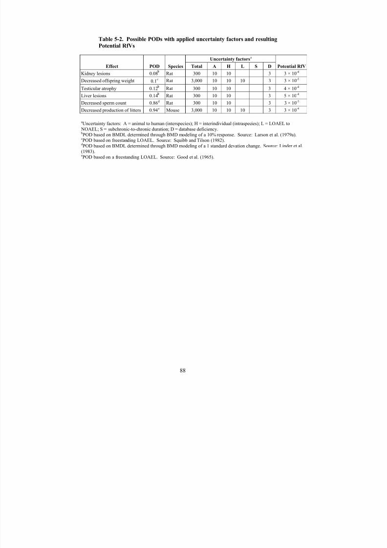
8/7/2019 United States Environmental Protect
http://slidepdf.com/reader/full/united-states-environmental-protect 101/183
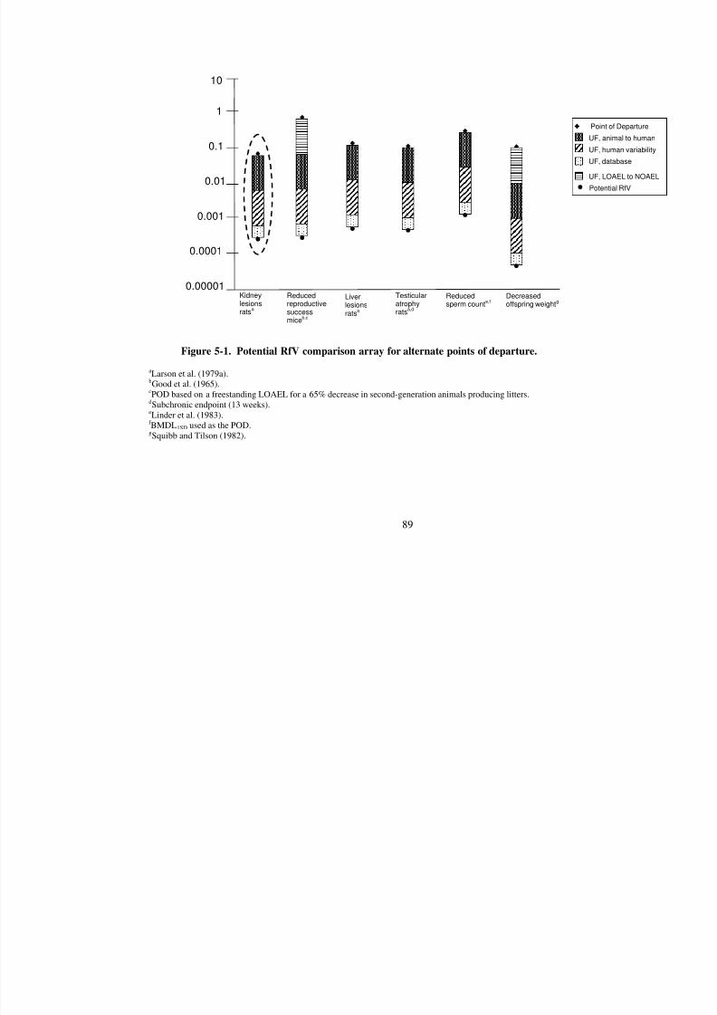
8/7/2019 United States Environmental Protect
http://slidepdf.com/reader/full/united-states-environmental-protect 102/183
89
0.00001
0.0001
0.001
0.01
0.1
1
10
Kidneylesionsrats
a
Liverlesionsrats
a
Reducedreproductivesuccessmice
b,c
Testicularatrophyrats
a,d
Po
UF
UF
UF
UF
Pot
Reducedsperm count
e,fDecreasedoffspring weight
g
Figure 5-1. Potential RfV comparison array for alternate points of departure.
aLarson et al. (1979a).
bGood et al. (1965).cPOD based on a freestanding LOAEL for a 65% decrease in second-generation animals producing litters.dSubchronic endpoint (13 weeks).eLinder et al. (1983).f BMDL1SD used as the POD.gSquibb and Tilson (1982).

8/7/2019 United States Environmental Protect
http://slidepdf.com/reader/full/united-states-environmental-protect 103/183
The PODs presented for kidney, liver, and testicular lesions were derived through BMD
modeling of the dichotomous data using a 10% response level. The POD presented for reduced
sperm count was derived through BMD modeling of continuous data using a response level of
one standard deviation from the control mean. BMD modeling outputs for these endpoints are
included in Appendix B. The PODs based on BMD methods have an inherent advantage over
the use of a NOAEL or LOAEL by making greater use of all the dose-response data from a givendata set. The PODs for reduced reproductive success in mice and decreased offspring weight in
rats were based on freestanding LOAELs.
The POD for testicular atrophy was derived from a 3-month exposure duration (within
the chronic study by Larson et al. [1979a]); however, testicular effects were not noted for the
longer exposure durations (1–2 years) in the same study, nor were testicular lesions detected in
other studies in rats treated with similar doses for the same duration (Linder et al., 1983; Cannon
and Kimbrough, 1979). Because testicular atrophy was not detected at the chronic timepoint in
the same study, an uncertainty factor to account for the use of a subchronic duration was not
applied to the potential POD for this endpoint.
5.1.5. Previous RfD Assessment
An oral assessment for chlordecone was not previously available on IRIS.
5.2. INHALATION REFERENCE CONCENTRATION (RfC)
Although adverse health effects from an occupational exposure incident may have
resulted from inhalation exposure (in combination with oral and dermal exposures), the data do
not identify exposure concentrations at which the effects occur (Taylor, 1985, 1982; Guzelian,1982a; Guzelian et al., 1980; Sanborn et al., 1979; Cannon et al., 1978; Martinez et al., 1978;
Taylor et al., 1978). Consequently, the human data cannot be used to define an exposuree-
response relationship for inhalation exposure to chlordecone. No studies on the toxicity of
chlordecone following inhalation exposure in laboratory animals were located. This lack of data
precludes the derivation of an RfC.
Consideration was given to route-to-route extrapolation to derive inhalation doses from
existing oral dose-response data for development of an RfC. Route-to-route extrapolation from
the oral database, however, is precluded by deficiencies in the database. The available rat PBTK
models for chlordecone do not include the inhalation route of exposure (see Section 3.5), and
human PBTK models with both oral and inhalation portals of entry have not yet been developed.
In the absence of PBTK models that include oral and inhalation routes of exposure, and lacking
inhalation absorption efficiency data in humans and rats, a route-to-route extrapolation from oral
to inhalation for chlordecone would be highly uncertain. As discussed in Chapter 2, only very
small amounts of chlordecone will evaporate from soil or water surfaces, and any chlordecone in
the air is likely to be removed by deposition of particles.
90
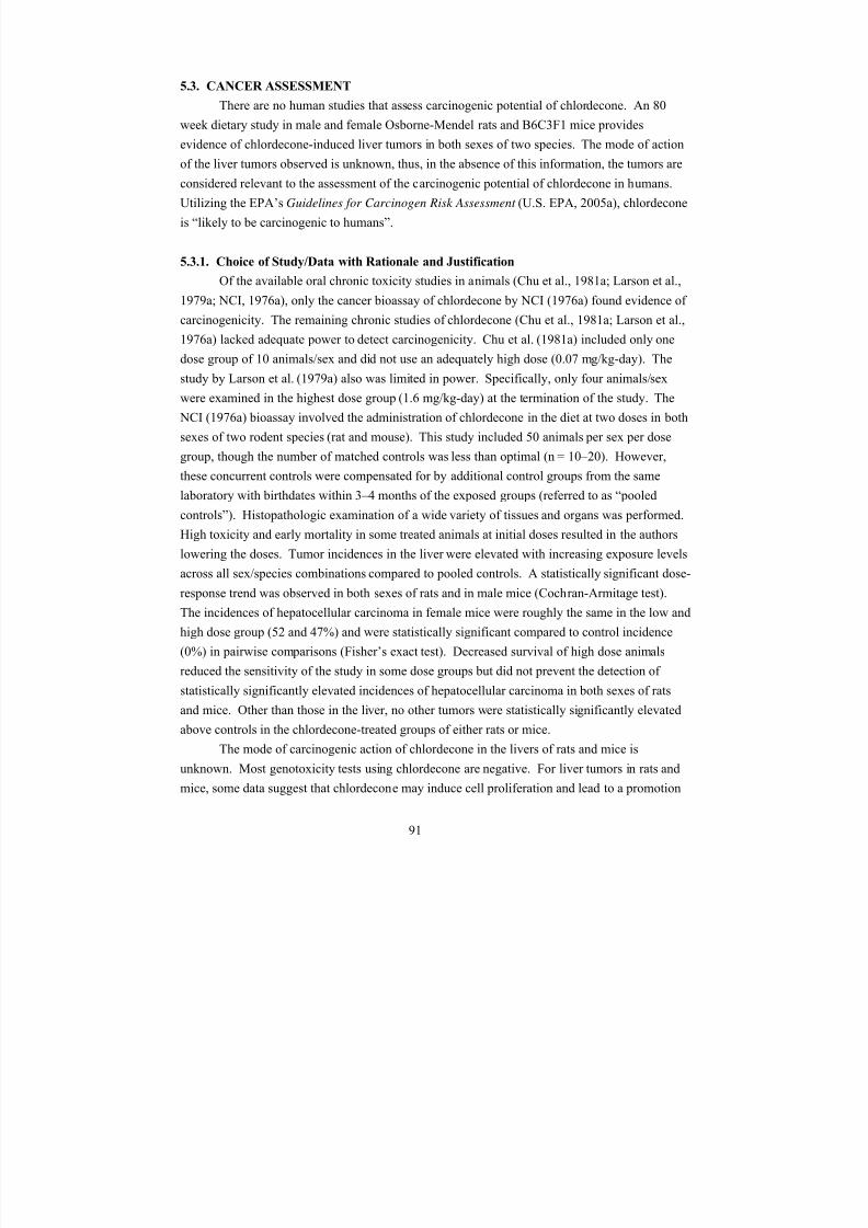
8/7/2019 United States Environmental Protect
http://slidepdf.com/reader/full/united-states-environmental-protect 104/183
5.3. CANCER ASSESSMENT
There are no human studies that assess carcinogenic potential of chlordecone. An 80
week dietary study in male and female Osborne-Mendel rats and B6C3F1 mice provides
evidence of chlordecone-induced liver tumors in both sexes of two species. The mode of action
of the liver tumors observed is unknown, thus, in the absence of this information, the tumors areconsidered relevant to the assessment of the carcinogenic potential of chlordecone in humans.
Utilizing the EPA’s Guidelines for Carcinogen Risk Assessment (U.S. EPA, 2005a), chlordecone
is “likely to be carcinogenic to humans”.
5.3.1. Choice of Study/Data with Rationale and Justification
Of the available oral chronic toxicity studies in animals (Chu et al., 1981a; Larson et al.,
1979a; NCI, 1976a), only the cancer bioassay of chlordecone by NCI (1976a) found evidence of
carcinogenicity. The remaining chronic studies of chlordecone (Chu et al., 1981a; Larson et al.,
1976a) lacked adequate power to detect carcinogenicity. Chu et al. (1981a) included only one
dose group of 10 animals/sex and did not use an adequately high dose (0.07 mg/kg-day). The
study by Larson et al. (1979a) also was limited in power. Specifically, only four animals/sex
were examined in the highest dose group (1.6 mg/kg-day) at the termination of the study. The
NCI (1976a) bioassay involved the administration of chlordecone in the diet at two doses in both
sexes of two rodent species (rat and mouse). This study included 50 animals per sex per dose
group, though the number of matched controls was less than optimal (n = 10–20). However,
these concurrent controls were compensated for by additional control groups from the same
laboratory with birthdates within 3–4 months of the exposed groups (referred to as “pooledcontrols”). Histopathologic examination of a wide variety of tissues and organs was performed.
High toxicity and early mortality in some treated animals at initial doses resulted in the authors
lowering the doses. Tumor incidences in the liver were elevated with increasing exposure levels
across all sex/species combinations compared to pooled controls. A statistically significant dose-
response trend was observed in both sexes of rats and in male mice (Cochran-Armitage test).
The incidences of hepatocellular carcinoma in female mice were roughly the same in the low and
high dose group (52 and 47%) and were statistically significant compared to control incidence
(0%) in pairwise comparisons (Fisher’s exact test). Decreased survival of high dose animals
reduced the sensitivity of the study in some dose groups but did not prevent the detection of
statistically significantly elevated incidences of hepatocellular carcinoma in both sexes of rats
and mice. Other than those in the liver, no other tumors were statistically significantly elevated
above controls in the chlordecone-treated groups of either rats or mice.
The mode of carcinogenic action of chlordecone in the livers of rats and mice is
unknown. Most genotoxicity tests using chlordecone are negative. For liver tumors in rats and
mice, some data suggest that chlordecone may induce cell proliferation and lead to a promotion
91
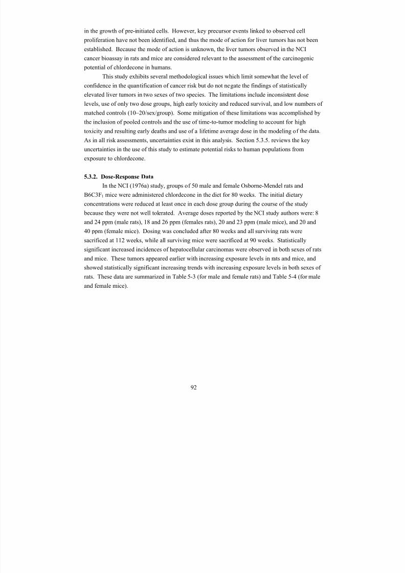
8/7/2019 United States Environmental Protect
http://slidepdf.com/reader/full/united-states-environmental-protect 105/183
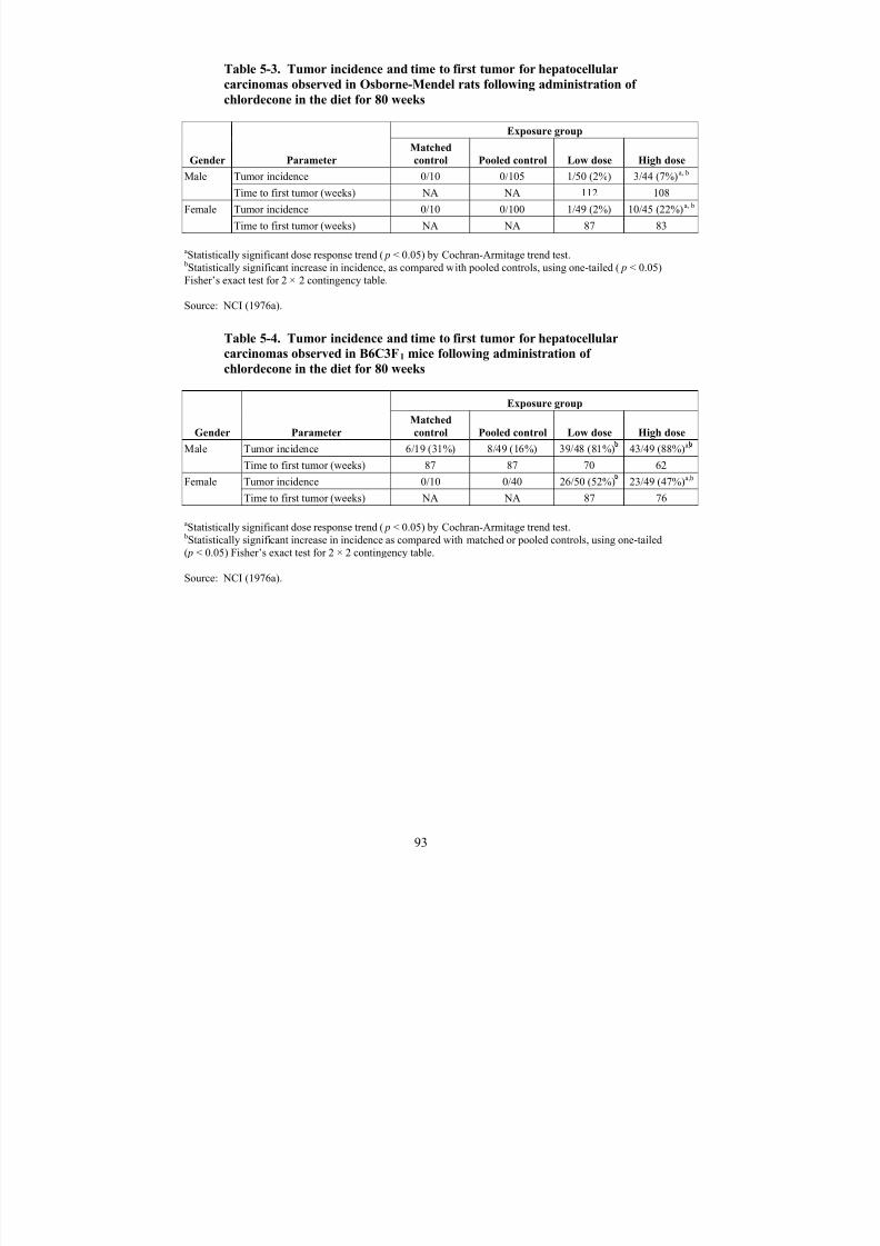
8/7/2019 United States Environmental Protect
http://slidepdf.com/reader/full/united-states-environmental-protect 106/183

8/7/2019 United States Environmental Protect
http://slidepdf.com/reader/full/united-states-environmental-protect 107/183
5.3.3. Dose Adjustments and Extrapolation Methods
The U.S. EPA Guidelines for Carcinogen Risk Assessment (U.S. EPA, 2005a)
recommend that the method used to characterize and quantify cancer risk from a chemical is
determined by what is known about the mode of action of the carcinogen and the shape of the
cancer dose-response curve. The linear approach is used as a default option if the mode-of-
action of carcinogenicity is not understood (U.S. EPA, 2005a). In the case of chlordecone, themode of carcinogenic action of chlordecone in the livers of rats and mice is unknown.
Therefore, a linear low-dose extrapolation approach was used to estimate human carcinogenic
risk associated with chlordecone exposure.
Due to the earlier occurrence of tumors with increasing exposure and the mortality
observed (especially in the high-dose groups in the second year of the study), dose-response
methodologies which can account for the influence of competing risks and intercurrent mortality
on site-specific tumor incidence rates are preferred. The U.S. EPA has generally used a model
which incorporates the time at which death-with-tumor occurred as well as the dose; the
multistage-Weibull model is multistage in dose and Weibull in time, and has the form:
P(d) = 1 - exp[-(q0 + q1d + q2d 2
+ ... + qk d k ) × (t - t 0)
z],
where P(d) represents the lifetime risk (probability) of cancer at dose d (i.e., human equivalent
exposure in this case); parameters qi ≥ 0, for i = 0, 1, ..., k; t is the time at which the tumor was
observed; and z is a parameter which characterizes the change in response with age. The
parameter t 0 represents the time between when a potentially fatal tumor becomes observable and
when it causes death, and is generally set to 0 either when all tumors are considered incidental or because of a lack of data to estimate the time reliably. The dose-response analyses in this
assessment were conducted using the computer software program TOX_RISK, Version 5.3 (ICF,
Fairfax, VA), which is based on Weibull models drawn from Krewski et al. (1983). Parameters
were estimated using the method of maximum likelihood.
Time-to-tumor analysis, as implemented by the TOX_RISK software program, allows the
distinction between tumor types that are fatal versus incidental in order to adjust for competing
risks. Incidental tumors are those tumors thought not to have caused the death of an animal,
while fatal tumors are thought to have resulted in animal death. The NCI (1976a) study did not
report individual causes of death, which would be preferable for time-to-tumor analysis. Thus,
all liver tumors observed were classified as incidental for the purpose of this analysis, and
consequently t 0 was set to zero. The data input into TOX_RISK, as well as the model output, are
provided in Appendix C.
Lifetime average dietary concentrations were calculated by TOX-RISK. Dietary
concentrations were reported as follows: 5.2 and 15.7 ppm (male rats), 12.2 and 17.9 ppm
(female rats), 16.8 and 19.6 ppm (male mice), and 16.8 and 33.7 ppm (female mice).
94
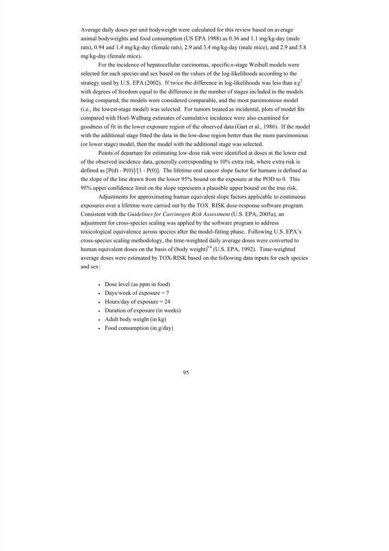
8/7/2019 United States Environmental Protect
http://slidepdf.com/reader/full/united-states-environmental-protect 108/183
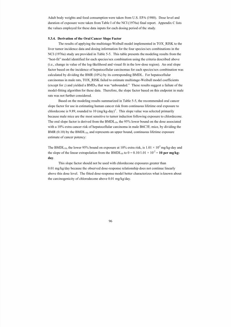
8/7/2019 United States Environmental Protect
http://slidepdf.com/reader/full/united-states-environmental-protect 109/183
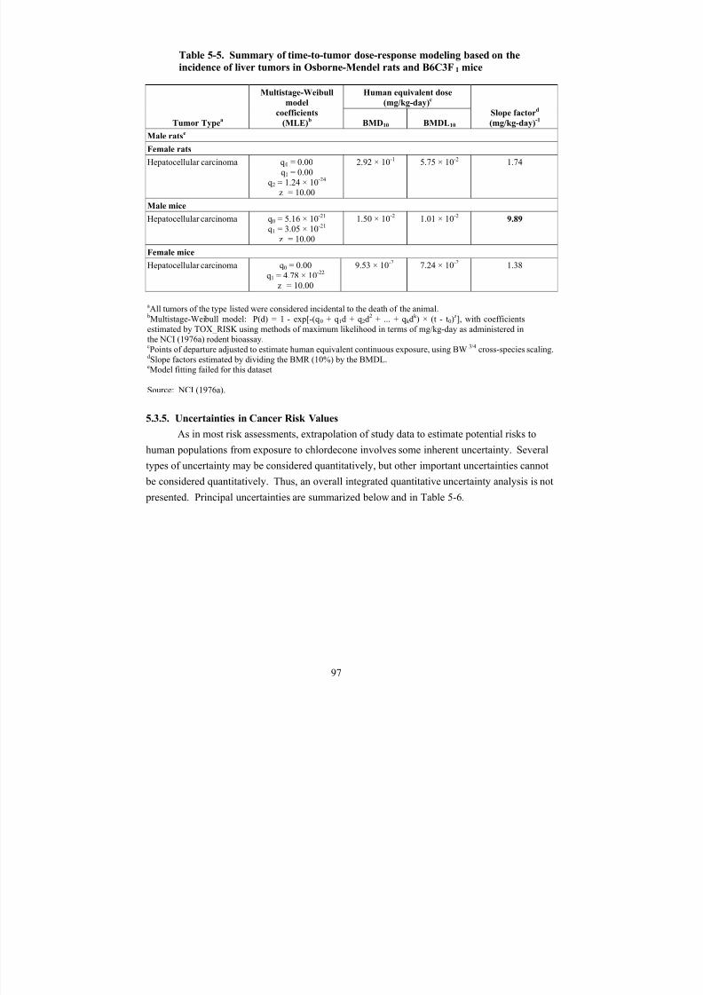
8/7/2019 United States Environmental Protect
http://slidepdf.com/reader/full/united-states-environmental-protect 110/183
Table 5-5. Summary of time-to-tumor dose-response modeling based on the
incidence of liver tumors in Osborne-Mendel rats and B6C3F1 mice
Tumor Typea
Multistage-Weibull
model
Human equivalent dose
(mg/kg-day)c
Slope factord coefficients
(MLE)b
(mg/kg-day)-1
BMD10 BMDL10
Male ratse
Female rats
Hepatocellular carcinoma q0 = 0.00q1 = 0.00
q2 = 1.24 × 10⁻²⁴
z = 10.00
2.92 × 10⁻¹ 5.75 × 10⁻² 1.74
Male mice
q0 = 5.16 × 10⁻²¹Hepatocellular carcinomaq1 = 3.05 × 10⁻²¹
z = 10.00
1.50 × 10⁻² 1.01 × 10-29.89
Female mice
q0 = 0.00q1 = 4.78 × 10⁻²²
z = 10.00
9.53 × 10
-2
7.24 × 10
-2
1.38Hepatocellular carcinoma
aAll tumors of the type listed were considered incidental to the death of the animal.bMultistage-Weibull model: P(d) = 1 - exp[-(q0 + q1d + q2d
2 + ... + qk dk ) × (t - t0)
z], with coefficientsestimated by TOX_RISK using methods of maximum likelihood in terms of mg/kg-day as administered inthe NCI (1976a) rodent bioassay.cPoints of departure adjusted to estimate human equivalent continuous exposure, using BW3/4 cross-species scaling.dSlope factors estimated by dividing the BMR (10%) by the BMDL.eModel fitting failed for this dataset
Source: NCI (1976a).
5.3.5. Uncertainties in Cancer Risk Values
As in most risk assessments, extrapolation of study data to estimate potential risks to
human populations from exposure to chlordecone involves some inherent uncertainty. Several
types of uncertainty may be considered quantitatively, but other important uncertainties cannot
be considered quantitatively. Thus, an overall integrated quantitative uncertainty analysis is not
presented. Principal uncertainties are summarized below and in Table 5-6.
97

8/7/2019 United States Environmental Protect
http://slidepdf.com/reader/full/united-states-environmental-protect 111/183
8/7/2019 United States Environmental Protect
http://slidepdf.com/reader/full/united-states-environmental-protect 112/183
8/7/2019 United States Environmental Protect
http://slidepdf.com/reader/full/united-states-environmental-protect 113/183
Cross-species scaling
An adjustment for cross-species scaling (BW3/4) was applied to address toxicological
equivalence of internal doses between each rodent species and humans, consistent with the 2005
Guidelines for Carcinogen Risk Assessment (U.S. EPA, 2005a). Without evidence to the
contrary, it is assumed that equal risks result from equivalent constant lifetime exposures.
Statistical uncertainty at the POD
Measures of statistical uncertainty require assuming that the underlying model and
associated assumptions are valid for the data under consideration. For the multistage-Weibull
model applied to the male mice data, there is a reasonably typical degree of uncertainty at the
10% extra incidence level (the POD for linear low-dose extrapolation). The lower bound on the
BMD10 (BMDL10) for hepatocellular carcinoma in male mice is approximately 1.5-fold lower
than the BMD10.
Choice of species/gender
The oral slope factor for chlordecone was quantified using the tumor incidence data for
male mice, which were found to be more sensitive than female mice or female rats to the
carcinogenicity of chlordecone. The oral slope factor calculated from male mice was 6–7 times
higher than the slope factors calculated from female mice and female rats. Liver tumor incidence
in the high-dose group of male rats was far less robust (7%) than the high-dose groups of female
rats, female mice, or male mice which had liver tumor incidences of 22, 47, and 88%,
respectively. As there is no information to inform which species or gender of animals would be
most applicable to humans, the most sensitive group was selected for the basis of the oral slopefactor. The human relevance of the observed liver tumors is unknown. However, data in
occupationally exposed workers indicate chlordecone predominantly accumulates in the liver
(Cohn et al., 1978). Additionally, similarities in liver effects have been shown in occupationally
exposed workers and in experimental animals including hypertrophy, hepatomegaly, and
proliferation of metabolic enzymes. Though the MOA for observed liver tumors in rodents is
unknown, the evidence suggesting the liver as a target organ of chlordecone toxicity and the
concordance of liver tumors across both sexes of rats and mice lends strength to the concern for
human carcinogenic potential.
Human population variability
The extent of inter-individual variability or sensitivity to the potential carcinogenicity of
chlordecone is unknown. There are no data exploring whether there is differential sensitivity to
chlordecone carcinogenicity across life stages. This lack of understanding about potential
susceptibility differences across exposed human populations thus represents a source of
uncertainty. Humans are expected to be more heterogenous than laboratory animals, and this
100

8/7/2019 United States Environmental Protect
http://slidepdf.com/reader/full/united-states-environmental-protect 114/183
101
variability is likely to be influenced by ongoing or background exposures, diseases, and
biological processes.

8/7/2019 United States Environmental Protect
http://slidepdf.com/reader/full/united-states-environmental-protect 115/183
8/7/2019 United States Environmental Protect
http://slidepdf.com/reader/full/united-states-environmental-protect 116/183
Chlordecone also causes tremors, decreased motor coordination, hyperexcitability, and an
exaggerated startle response in laboratory animals.
Chlordecone exposure in humans caused oligospermia, reduced sperm motility, and
decreased libido in a group of men who were occupationally exposed to chlordecone for periods
of up to 1.5 years. Upon re-examination of workers 5–7 years following the cessation of
chlordecone exposure and treatment with cholestyramine, male reproductive parameters hadreturned to normal. Chlordecone also induces reproductive toxicity in male and female
laboratory animals, as demonstrated by altered sperm parameters, testicular atrophy, altered
estrous cyclicity, and impaired reproductive success.
Kidney toxicity was reported in laboratory animals but was not observed in
occupationally exposed pesticide workers. However, it is unclear if clinical indicators of renal
damage were specifically examined in occupationally exposed workers or whether signs of
kidney impairment would be expected following the relatively short (5–6 month) average
exposure durations. Several animal studies reported kidney effects from chlordecone exposure.
Proteinuria and increased incidence of kidney lesions were observed in female Wistar rats and in
(NZB × NZW)F1 mice. Chronic kidney inflammation was observed in male and female
Osborne-Mendel rats. Twenty-eight days of dietary exposure to chlordecone produced
eosinophilic inclusions in proximal tubules in male Sprague-Dawley rats. Most of the effects of
chlordecone are thought to be produced by the parent compound, primarily by interfering with
the function of mitochondrial and cellular membranes. Disruption of cellular homeostasis and
energy production within the cell eventually leads to impaired cellular function. In the central
nervous system, altered calcium homeostasis leads to changes in neurotransmitter activity. In the
liver, membrane perturbation and inhibition of transport proteins at the bile canalicular membrane are thought to be related to chlordecone-induced hepatobiliary dysfunction. The
reproductive and developmental effects of chlordecone are most likely related to endocrine
disruption. Chlordecone exhibits estrogenic properties that may be related to impaired
reproductive success and adverse effects on sperm.
There are no reports of cancer in humans associated with exposure to chlordecone.
Increased incidence of hepatocellular carcinoma was observed in rats and mice following oral
exposure to chlordecone (NCI, 1976a, b). Significantly increased incidence of hepatocellular
carcinoma was observed in both sexes of mice compared to matched controls. The incidence of
liver tumors in male and female rats was comparatively less robust but did reach statistical
significance when compared to pooled laboratory controls.
103

8/7/2019 United States Environmental Protect
http://slidepdf.com/reader/full/united-states-environmental-protect 117/183
8/7/2019 United States Environmental Protect
http://slidepdf.com/reader/full/united-states-environmental-protect 118/183

8/7/2019 United States Environmental Protect
http://slidepdf.com/reader/full/united-states-environmental-protect 119/183
identified cases. Since potential variability in responses to chlordecone in the greater human
population is unknown, the default uncertainty factor of 10 for intrahuman variability was not
reduced. Human variation may be larger or smaller; however, chlordecone-specific data to
examine the potential magnitude of human variability of response are unknown.
Uncertainties associated with data gaps in the chlordecone database have been identified.
Data more fully characterizing potential multigenerational reproductive effects are lacking.Several one-generational reproductive studies have indicated decreased reproductive success in
chlordecone-treated animals (Cannon and Kimbrough, 1979; Good et al., 1965; Huber et al.,
1965). In addition, two nonstandard multigenerational studies exist that evaluate reproductive
success of chlordecone-treated animals (Gellert and Wilson, 1979; Good et al., 1965). However,
due to limited scope and design, these studies are not considered adequate for the assessment of
multigenerational reproductive toxicity. Therefore, for the above data gaps in the chlordecone
database, an UF of 3 was applied to the POD in the derivation of the RfD.
The overall confidence in the RfD and the principal study (Larson et al., 1979a) is
medium. The principal study involves a sufficient number of animals per group, several dose
levels, and a wide range of tissues and endpoints assessed. Confidence in the database is
medium. The chlordecone database includes case studies of occupationally exposed workers,
chronic and subchronic dietary exposure studies in laboratory animals, and several subchronic
reproductive and developmental studies, including one developmental neurotoxicity study.
However, the database is lacking a multigenerational reproductive toxicity study. Therefore,
reflecting medium confidence in both the database and the principal study, confidence in the RfD
is medium.
6.2.2. Cancer
Under the Guidelines for Carcinogen Risk Assessment (U.S. EPA, 2005a), the database
for chlordecone indicates that it is “likely to be carcinogenic to humans”. This determination is
primarily based on the NCI (1976a, b) study, which found positive evidence of liver tumors in
both sexes of rats and mice after chronic chlordecone dietary exposure. Additionally, data on
mirex, a structurally similar chemical, also demonstrates an increase in hepatocellular adenomas
or carcinomas in both sexes of rats and mice. However, unlike the observed cancer effects, some
but not all of the noncancer effects noted for these two chemicals are similar as described in
Section 4.5.3. This weight of evidence conclusion collectively takes into consideration the NCI
(1976a, b) cancer bioassay, the available human studies, and other chronic animal bioassays.
The increased incidence of hepatocellular carcinoma in both sexes of rats and mice
observed in the NCI (1976a, b) 80 week dietary study was used to calculate the oral slope factor
for chlordecone. Due to high toxicity and resulting early deaths in some dose groups, methods
which account for the influence of early mortality on tumor incidence rates were utilized. In this
case, the multistage-Weibull model was used because it incorporates the time at which death-
106
8/7/2019 United States Environmental Protect
http://slidepdf.com/reader/full/united-states-environmental-protect 120/183
8/7/2019 United States Environmental Protect
http://slidepdf.com/reader/full/united-states-environmental-protect 121/183
8/7/2019 United States Environmental Protect
http://slidepdf.com/reader/full/united-states-environmental-protect 122/183
Caldwell, V; Loch-Caruso, R. (1992) Chlordecone rapidly and reversibly inhibits gap junctional communication inhuman embryonic palatal mesenchyme cells. In Vitro Toxicol 5(2):113–122.
Cannon, SB; Kimbrough, RD. (1979) Short-term chlordecone toxicity in rats including effects on reproduction,pathological organ changes, and their reversibility. Toxicol Pharmacol 47:469–476.
Cannon, SB; Veazey, JM; Jackson, RS; et al. (1978) Epidemic Kepone poisoning in chemical workers. Am JEpidemiol 107:529–537.
Carpenter, HM; Curtis, LR. (1989) A characterization of chlordecone pretreatment-altered pharmacokinetics inmice. Drug Metab Dispos 17(2):131–138.
Carpenter, HM; Curtis, LR. (1991) Low dose chlordecone pretreatment altered cholesterol disposition withoutinduction of cytochrome P-450. Drug Metab Dispos 19(3):673–678.
Carpenter, HM; Hedstrom, OR; Siddens, LK; et al. (1996) Ultrastructural, protein, and lipid changes in liver associated with chlordecone treatment of mice. Fundam Appl Toxicol 34(1):157–164.
Chadwick, RW; Copeland, MF; Rosenstein, L. (1979) The effect of Kepone exposure during gestation and lactationon the metabolism of lindane by weanling rats. Toxicol Lett 4:247–252.
Chernoff, N; Kavlock, RJ. (1982) An in vivo teratology screen utilizing pregnant mice. J Toxicol Environ Health10:541–550.
Chernoff, N; Rogers, EH. (1976) Fetal toxicity of Kepone in rats and mice. Toxicol Appl Pharmacol 38:189–194.
Chetty, KN; Walker, J; Brown, K; et al. (1993a) The effects of dietary calcium and chlordecone on cholinesterase,triglycerides, low density lipoproteins, and cholesterol in serum of rat. Arch Environ Contam Toxicol 24(3):365– 367.
Chetty, KN; Brown, K; Walker, J; et al. (1993b) Effects of chlordecone and malnutrition on immune response inrats. Life Sci 52(18):PL175–180.
Chetty, KN; Walker, J; Brown, K; et al. (1993c) Influence of dietary calcium on chlordecone-induced biochemicalchanges in serum of rat. Ecotoxicol Environ Saf 26(2):248–252.
Chu, I; Villeneuve, DC; Becking, GC; et al. (1980) Short-term study of the combined effects of mirex, photomirex,and Kepone with halogenated biphenyls in rats. J Toxicol Environ Health 6:421–432.
Chu, I; Villeneuve, DC; Valli, VE; et al. (1981a) Chronic toxicity of photomirex in the rat. Toxicol Appl Pharmacol59:268–278.
Chu, I; Villeneuve, DC; MacDonald, BL; et al. (1981b) Reversibility of the toxicological changes induced byphotomirex and mirex. Toxicology 21(3):235–250.
Chu, I; Villeneuve, DC; Secours, VE; et al. (1981c) Effects of photomirex and mirex on reproduction in the rat.Toxicol Appl Pharmacol 60(3):549–556.
Cohn, WJ; Boylan, JJ; Blanke, RV; et al. (1978) Treatment of chlordecone (Kepone) toxicity with cholestyramine:results of a controlled trial. N Engl J Med 298:243–248.
Cooper, JR; Vodicnik, MJ; Gordon JH. (1985) Effects of perinatal Kepone exposure on sexual differentiation of therat brain. Neurotoxicology 6(1):183–190.
Curtis LR, Mehendale HM. (1979) The effects of kepone pretreatment on biliary excretion of xenobiotics in the
male rat. Toxicol Appl Pharmacol 47(2):295-303.
109
8/7/2019 United States Environmental Protect
http://slidepdf.com/reader/full/united-states-environmental-protect 123/183
8/7/2019 United States Environmental Protect
http://slidepdf.com/reader/full/united-states-environmental-protect 124/183
Gilroy, DJ; Carpenter, HM; Curtis, LR. (1994) Chlordecone pretreatment alters [14C]chlordecone and[14C]cholesterol transport kinetics in the perfused rat liver. Fundam Appl Toxicol 22(2):286–292.
Good, EE; Ware, GW; Miller, DF. (1965) Effects of insecticides on reproduction in the laboratory mouse: I.Kepone. J Econ Entomol 58(4):754–757.
Guzelian, PS. (1982a) Chlordecone poisoning: a case study in approaches for the detoxification of humans exposedto environmental chemicals. Drug Metab Rev 13:663–679.
Guzelian, PS. (1982b) Comparative toxicology of chlordecone (Kepone) in humans and experimental animals. AnnRev Pharmacol Toxicol 22:89–113.
Guzelian, PS; Vranian, G; Boylan, JJ; et al. (1980) Liver structure and function in patients poisoned withchlordecone (Kepone). Gastroenterology 78(2):206–213.
Hammond B; Katyzenellenbogen BS; Krauthammer N; et al. (1979). Estrogenic activity of the insecticidechlordecone (Kepone) and interaction with uterine estrogen receptors. Proc Nat1 Acad Sci 76:6641–6645.
Hansch, C; Leo, A; Hoekman, D. (1995) Exploring QSAR: hydrophobic, electronic, and steric constants. ACSProfessional Reference Book. Washington, DC: American Chemical Society; p. 65.
Hart, SE; Kinter, LB. (2005). Assessing renal effects of toxicants in vivo. In: Tarloff, JB; Lawrence, LH; eds.Toxicology of the kidney. New York, NY: CRC Press; pp. 81–147.
Heatheringon, AC; Fisher, HL; Sumler, MR; et al. (1998) Percutaneous absorption and disposition of [14C]chlordecone in young and adult female rats. Environ Res 79(2):138–155.
Herr DW, Gallus JA, Tilson HA. (1987) Pharmacological modification of tremor and enhanced acoustic startle by
chlordecone and p,p’-DDT. Psychopharmacology. 91(3):320-5.
Hewitt, LA; Caille, G; Plaa, GL; et al. (1985) Temporal relationships between biotransformation, detoxication, andchlordecone potentiation of chloroform-induced hepatotoxicity. Can J Physiol Pharmacol 64:477–482.
Hodges LC, Bergerson JS, Hunter DS, et al. (2000). Estrogenic effects of organochlorine pesticides on uterine
leiomyoma cells in vitro. Toxicol Sci. 54(2):355–364.
Hoskins B, Ho IK. (1982) Chlordecone-induced alterations in content and subcellular distribution of calcium inmouse brain. J Toxicol Environ Health. 9(4):535-44.
Houston, TE; Mutter, LC; Blanke, RV; et al. (1981) Chlordecone alcohol formation in the Mongolian gerbil(Meriones unguiculatus): A model for human metabolism of chlordecone (Kepone). Fundam Appl Toxicol1(3):293–298.
Huang, ESR; Nelson, FR. (1986) Anti-estrogenic action of chlordecone in rat pituitary gonadotrophs in vitro.Toxicol Appl Pharmacol 82:62–69.
Huang, TP; Ho, IK; Mehendale, HM. (1980) Assessment of neurotoxicity induced by oral administration of
chlordecone (Kepone) in the mouse. Neurotoxicology 2:113–124.
Huber, JJ. (1965) Some physiological effects of the insecticide Kepone in the laboratory mouse. Toxicol ApplPharmacol 7:516–524.
IARC (International Agency for Research on Cancer). (1979) Chlordecone. IARC monographs on the evaluation of carcinogenic risk of chemicals to humans. Vol. 20. Some halogenated hydrocarbons. Lyon, France: InternationalAgency for Research on Cancer; p. 67.
Innes, JRM; Ulland, BM; Valerio, MG; et al. (1969) Bioassay of pesticides and industrial chemicals for tumorigenicity in mice: a preliminary note. J Natl Cancer Inst 42(6):1101–1114.
111

8/7/2019 United States Environmental Protect
http://slidepdf.com/reader/full/united-states-environmental-protect 125/183
8/7/2019 United States Environmental Protect
http://slidepdf.com/reader/full/united-states-environmental-protect 126/183
Larson, PS; Egle, JL, Jr; Hennigar, GR. (1979a) Acute, subchronic, and chronic toxicity of chlordecone. ToxicolAppl Pharmacol 48:29–41.
Larson, PS; Egle, JL, Jr; Hennigar, GR; et al. (1979b) Acute and subchronic toxicity of mirex in the rat, dog andrabbit. Toxicol Appl Pharmacol 49(2):271–277.
Lide, DR; ed. (2000) CRC handbook of chemistry and physics. 81st edition. Boca Raton, FL: CRC Press; pp. 3– 204.
Linder, RE; Scotti, TM; McElroy, WK; et al. (1983) Spermotoxicity and tissue accumulation of chlordecone(Kepone) in male rats. J Toxicol Environ Health 12:183–192.
Mactutus, CF; Tilson, HA. (1985) Evaluation of long-term consequences in behavioral and/or neural functionfollowing neonatal chlordecone exposure. Teratology 31(2):177–186.
Martinez, AJ; Taylor, JR; Dyck, PJ; et al. (1978) Chlordecone intoxication in man: II. Ultrastructure of peripheralnerves and skeletal muscle. Neurology 28:631–635.
Medical College of Wisconsin. (1999) Glomerulosclerosis. Healthlink. Medical College of Wisconsin. Availableonline at: http://healthlink.mcw.edu/article/943054092.html (accessed June 25, 2009).
Mehendale, HM. (1990) Potentiation of halomethane hepatotoxicity by chlordecone: a hypothesis for themechanism. Med Hypotheses 33(4):289–299.
Mehendale, HM. (1994) Amplified interactive toxicity of chemicals at nontoxic levels: mechanistic considerationsand implications to public health. Environ Health Perspect 102(Suppl. 9):139–149.
Mehendale, HM; Klingensmith, JS. (1988) In vivo metabolism of CCl4 by rats pretreated with chlordecone, mirex,or phenobarbitol. Toxicol Appl Pharmacol 93(2):247–256.
Mehendale, HM; Takanaka, A; Desaiah, D; et al. (1977) Kepone induction of hepatic mixed function oxidases. LifeSci 20(6):991–997.
Mehendale, HM; Takanaka, A; Desaiah, D; et al. (1978) Effect of preexposure to Kepone on hepatic mixed functionoxidases in the female rat. Toxicol Appl Pharmacol 44:171–180.
Mehendale, HM; Purushotham, KR; Lockard, VG. (1989) The time course of liver injury and [3H]thymidineincorporation in chlordecone-potentiated CHCl3 hepatotoxicity. Exp Mol Pathol 51:31–47.
Metcalf, RL. (2002) Insect control. In: Ullmann’s encyclopedia of industrial chemistry. Wiley Interscience.Available online at http://www.mrw.interscience.wiley.com/ueic/ueic_search_fs.html (accessed March 22, 2004).
Mitra, A; Richards, I; Kitchin, K; et al. (1990) Mirex induces ornitine decarboxylase in female rat liver. J BiochemToxicol 5(2):119–124.
Molowa, DT; Wrighton, SA; Blanke, RV; et al. (1986) Characterization of a unique aldo-keto reductase responsiblefor the reduction of chlordecone in the liver of the gerbil and man. J Toxicol Environ Health 17:375–384.
Morgan DP, Sandifier SH, Hetzler HL et al; (1979) Test for in vivo conversion of mirex to kepone. Bull EnvironContam Toxicol 22(1-2):238-44.
Mortelmans, K; Haworth, S; Lawlor, T; et al. (1986) Salmonella mutagenicity tests: II. Results from the testing of 270 chemicals. Environ Mutagen 8(Suppl. 7):1–119.
Murali, B; Korrapati, MC; Anand, SS; et al. (2002) Age-dependent susceptibility of F344 rats to chlordeconepotentiated CCl4 hepatotoxicity and lethality. Int J Toxicol 21(6):524.
113

8/7/2019 United States Environmental Protect
http://slidepdf.com/reader/full/united-states-environmental-protect 127/183
8/7/2019 United States Environmental Protect
http://slidepdf.com/reader/full/united-states-environmental-protect 128/183

8/7/2019 United States Environmental Protect
http://slidepdf.com/reader/full/united-states-environmental-protect 129/183
8/7/2019 United States Environmental Protect
http://slidepdf.com/reader/full/united-states-environmental-protect 130/183
U.S. DHHS (Department of Health and Human Services). (2006) Glomerular diseases. National Kidney andUrologic Diseases Information Clearinghouse, Bethesda, MD; NIH Publication No. 07-4358. Available online athttp://kidney.niddk.nih.gov/kudiseases/pubs/glomerular/ (accessed June 25, 2009).
U.S. EPA (Environmental Protection Agency). (1986a) Guidelines for the health risk assessment of chemicalmixtures. Federal Register 51(185):34014-34025. Available online at http://www.epa.gov/iris/backgr-d.htm(accessed January 15, 2009).
U.S. EPA (Environmental Protection Agency). (1986b) Guidelines for mutagenicity risk assessment. FederalRegister 51(185):34006-34012. Available online at http://www.epa.gov/iris/backgr-d.htm (accessed January 15,2009).
U.S. EPA. (Environmental Protection Agency). (1986c) Final report on the evaluation of four toxic chemicals in anin vivo/in vitro toxicological screen: acrylamide, chlordecone, cyclophosphamide, and diethylstilbestrol.EPA/600/1-86/002.
U.S. EPA. (Environmental Protection Agency). (1988) Recommendations for and documentation of biologicalvalues for use in risk assessment. Environmental Criteria and Assessment Office, Office of Health andEnvironmental Assessment, Cincinnati, OH; EPA/600/6-87/008. Available from the National Technical InformationService, Springfield, VA; PB88-179874, and online at http://cfpub.epa.gov/ncea/cfm/recordisplay.cfm?deid=34855(accesses June 25, 2009).
U.S. EPA (Environmental Protection Agency). (1991) Guidelines for developmental toxicity risk assessment.Federal Register 56(234):63798-63826. Available online at http://www.epa.gov/iris/backgr-d.htm (accessed January15, 2009).
U.S. EPA (Environmental Protection Agency). (1992) Draft report: a cross-species scaling factor for carcinogen risk assessment based on equivalence of mg/kg3/4/day. Federal Register 57(109):24152–24173.
U.S. EPA. (Environmental Protection Agency). (1994a) Interim policy for particle size and limit concentrationissues in inhalation toxicity: notice of availability. Federal Register 59(206):53799. Available online athttp://www.epa.gov/EPA-PEST/1994/October/Day-26/pr-11.html (accessed June 25, 2009).
U.S. EPA. (Environmental Protection Agency). (1994b) Methods for derivation of inhalation referenceconcentrations and application of inhalation dosimetry. Environmental Criteria and Assessment Office, Office of Health and Environmental Assessment, Cincinnati, OH; EPA/600/8-90/066F. Available from the National TechnicalInformation Service, Springfield, VA, PB2000-500023, and online athttp://cfpub.epa.gov/ncea/raf/recordisplay.cfm?deid=71993 (accessed June 25, 2009).
U.S. EPA. (Environmental Protection Agency). (1995) Use of the benchmark dose approach in health risk assessment. Risk Assessment Forum, Washington, DC; EPA/630/R-94/007. Available from the National TechnicalInformation Service, Springfield, VA, PB95-213765, and online athttp://cfpub.epa.gov/ncea/raf/raf_pubtitles.cfm?detype=document&excCol=archive (accessed June 25, 2009).
U.S. EPA (Environmental Protection Agency). (1996) Guidelines for reproductive toxicity risk assessment. FederalRegister 61(212):56274-56322. Available online at http://www.epa.gov/iris/backgr-d.htm (accessed January 15,2009).
U.S. EPA. (Environmental Protection Agency). (1998) Guidelines for neurotoxicity risk assessment. FederalRegister 63(93):26926–26954. Available online at http://www.epa.gov/ncea/raf/rafguid.htm (accessed January 15,2009).
U.S. EPA. (Environmental Protection Agency). (2000a) Science policy council handbook: risk characterization.Office of Science Policy, Office of Research and Development, Washington, DC. EPA/100-B-00-002. Availablefrom: <http://www.epa.gov/iris/backgr-d.htm>.
U.S. EPA (Environmental Protection Agency). (2000b) Benchmark dose technical guidance document. Externalreview draft. Risk Assessment Forum, Washington, DC; EPA/630/R-00/001. Available online athttp://www.epa.gov/iris/backgr-d.htm (accessed January 15, 2009).
117
8/7/2019 United States Environmental Protect
http://slidepdf.com/reader/full/united-states-environmental-protect 131/183

8/7/2019 United States Environmental Protect
http://slidepdf.com/reader/full/united-states-environmental-protect 132/183

8/7/2019 United States Environmental Protect
http://slidepdf.com/reader/full/united-states-environmental-protect 133/183
APPENDIX A. SUMMARY OF EXTERNAL PEER REVIEW AND PUBLIC
COMMENTS AND DISPOSITION
The Toxicological Review of Chlordecone has undergone a formal external peer review
performed by scientists in accordance with U.S. EPA guidance on peer review (U.S. EPA,2006a; 2000a). The external peer reviewers were tasked with providing written answers to
general questions on the overall assessment and on chemical-specific questions in areas of
scientific controversy or uncertainty. A summary of significant comments made by the external
reviewers and U.S. EPA’s responses to these comments arranged by charge question follow. In
many cases the comments of the individual reviewers have been synthesized and paraphrased in
development of Appendix A. U.S. EPA also received scientific comments from the public.
These comments and U.S. EPA’s responses are included in a separate section of this appendix.
On April 10, 2008, U.S. EPA introduced revisions to the IRIS process for developing
chemical assessments. As part of the revised process, the disposition of peer reviewer and public
comments, as found in this Appendix, and the revised IRIS Toxicological Review was provided
to the external peer review panel members for their comment on May 4, 2009. No additional
substantive comments were received as part of this second review.
EXTERNAL PEER REVIEW PANEL COMMENTS
The reviewers made several editorial suggestions to clarify specific portions of the text.These changes were incorporated in the document as appropriate and are not discussed further.
(A) General Comments
1. Is the Toxicological Review logical, clear, and concise? Has U.S. EPA accurately, clearly,
and objectively represented and synthesized the scientific evidence for noncancer and cancer
hazard?
Comments: All reviewers found the document to be generally logical and clear. One reviewer
considered the Toxicological Review to be repetitive due to the format of the document. Two of
the reviewers offered specific suggestions to improve the clarity of the document.
Response: U.S. EPA has reviewed and modified the Toxicological Review to address reviewer
concerns about the repetitious nature of the document and to improve clarity.
A-1
8/7/2019 United States Environmental Protect
http://slidepdf.com/reader/full/united-states-environmental-protect 134/183
8/7/2019 United States Environmental Protect
http://slidepdf.com/reader/full/united-states-environmental-protect 135/183
Response: U.S. EPA agrees that the above research recommendations would improve future
hazard identifications of chlordecone.
4. Please comment on the identification and characterization of sources of uncertainty in
sections 5 and 6 of the assessment document. Please comment on whether the key sources of uncertainty have been adequately discussed. Have the choices and assumptions made in the
discussion of uncertainty been transparently and objectively described? Has the impact of the
uncertainty on the assessment been transparently and objectively described?
Comments: Four reviewers found the identification and characterization of sources of
uncertainty to be reasonable. However, some of the reviewers identified additional areas of
uncertainty that they believed were not addressed or should be discussed in more detail in
Sections 5 and 6 of the Toxicological Review. These areas included:
-lack of mechanistic data and NOAELs for many observed effects
-potential early life vulnerabilities
-uncertainties between human and animal pharmacokinetics
-ability of existing studies to describe potential developmental toxicity
-wide range of blood levels associated with effects in exposed workers
-potential additive or synergistic effects of low doses of chlordecone with other similarly
acting chemicals
-lack of establishment of NOAELs for female reproductive effects
-impact of the choice of BMD model chosen.
Response: Additional effort has been made to expand discussions of key areas of uncertainty in
the Toxicological Review. Several of the uncertainties listed above have been addressed in
section 6.2.1. of the document including the impact of the BMD model chosen and uncertainties
between animal and human pharmacokinetics. A paucity of additional data to inform these
uncertainties limits their further discussion in the document. Uncertainties regarding early life
vulnerabilities were noted in section 4.8.1 and the lack of NOAELs for female reproductive
effects was noted in section 6.2.1.
The uncertainty identified by a reviewer regarding potential additive effects of low doses
of chlordecone with similarly acting chemicals is acknowledged as biologically plausible,
however data on the mode(s) of action for cancer and noncancer effects of chlordecone are
limited, as are data regarding the interactions of chlordecone with other chemicals which may
share similar mechanisms. Therefore, further discussion of this point is considered speculative
in light of the limited database for chlordecone.
A-3
8/7/2019 United States Environmental Protect
http://slidepdf.com/reader/full/united-states-environmental-protect 136/183
8/7/2019 United States Environmental Protect
http://slidepdf.com/reader/full/united-states-environmental-protect 137/183
8/7/2019 United States Environmental Protect
http://slidepdf.com/reader/full/united-states-environmental-protect 138/183
8/7/2019 United States Environmental Protect
http://slidepdf.com/reader/full/united-states-environmental-protect 139/183

8/7/2019 United States Environmental Protect
http://slidepdf.com/reader/full/united-states-environmental-protect 140/183

8/7/2019 United States Environmental Protect
http://slidepdf.com/reader/full/united-states-environmental-protect 141/183
8/7/2019 United States Environmental Protect
http://slidepdf.com/reader/full/united-states-environmental-protect 142/183

8/7/2019 United States Environmental Protect
http://slidepdf.com/reader/full/united-states-environmental-protect 143/183
8/7/2019 United States Environmental Protect
http://slidepdf.com/reader/full/united-states-environmental-protect 144/183

8/7/2019 United States Environmental Protect
http://slidepdf.com/reader/full/united-states-environmental-protect 145/183
A-13
Response: In response to the comments of the external peer reviewers, U.S. EPA has re-
evaluated the WOE for carcinogenicity of chlordecone and the feasibility of quantifying an oral
slope factor from the data while considering and acknowledging the uncertainties inherent in the
data.
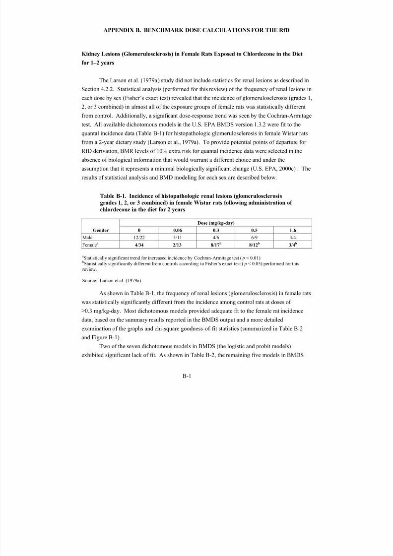
8/7/2019 United States Environmental Protect
http://slidepdf.com/reader/full/united-states-environmental-protect 146/183
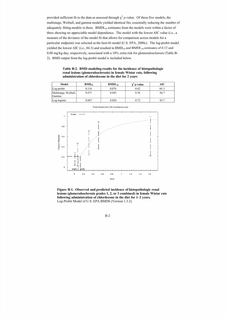
8/7/2019 United States Environmental Protect
http://slidepdf.com/reader/full/united-states-environmental-protect 147/183
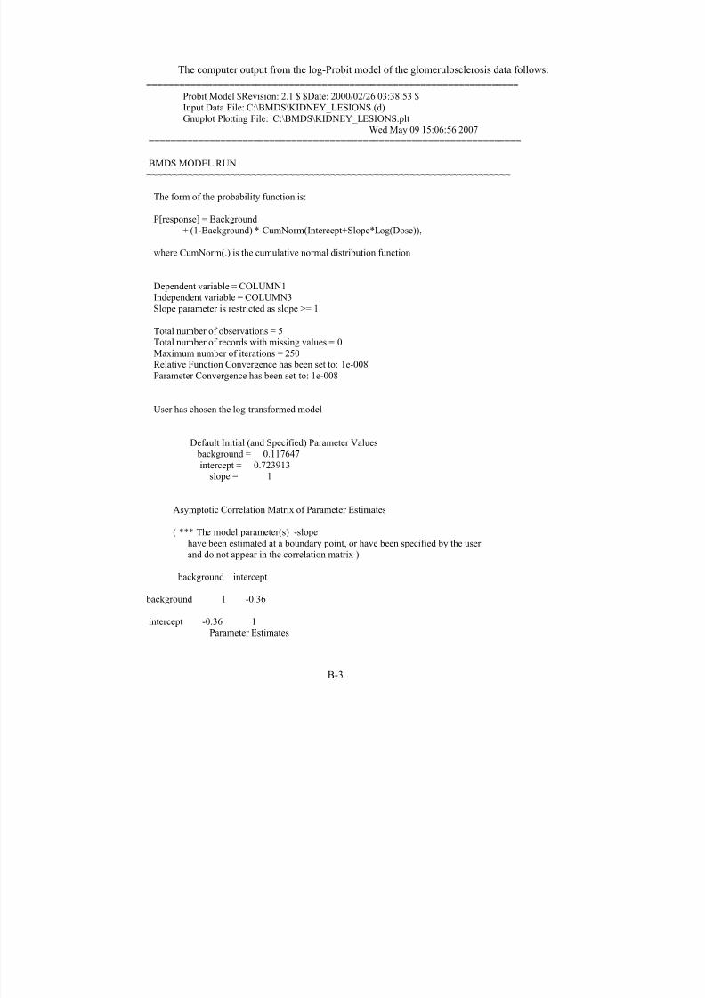
8/7/2019 United States Environmental Protect
http://slidepdf.com/reader/full/united-states-environmental-protect 148/183
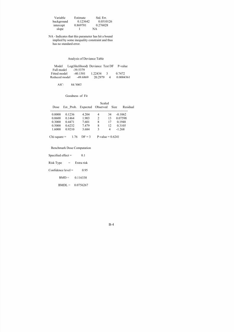
8/7/2019 United States Environmental Protect
http://slidepdf.com/reader/full/united-states-environmental-protect 149/183
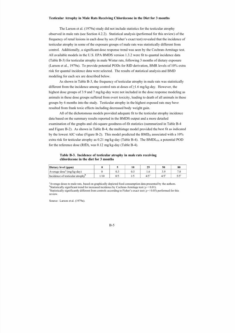
8/7/2019 United States Environmental Protect
http://slidepdf.com/reader/full/united-states-environmental-protect 150/183
Testicular Atrophy in Male Rats Receiving Chlordecone in the Diet for 3 months
The Larson et al. (1979a) study did not include statistics for the testicular atrophy
observed in male rats (see Section 4.2.2). Statistical analysis (performed for this review) of the
frequency of renal lesions in each dose by sex (Fisher’s exact test) revealed that the incidence of
testicular atrophy in some of the exposure groups of male rats was statistically different fromcontrol. Additionally, a significant dose response trend was seen by the Cochran-Armitage test.
All available models in the U.S. EPA BMDS version 1.3.2 were fit to quantal incidence data
(Table B-3) for testicular atrophy in male Wistar rats, following 3 months of dietary exposure
(Larson et al., 1979a). To provide potential PODs for RfD derivation, BMR levels of 10% extra
risk for quantal incidence data were selected. The results of statistical analysis and BMD
modeling for each sex are described below.
As shown in Table B-3, the frequency of testicular atrophy in male rats was statistically
different from the incidence among control rats at doses of ≥1.6 mg/kg-day. However, the
highest dose groups of 3.9 and 7 mg/kg-day were not included in the dose response modeling as
animals in these dose groups suffered from overt toxicity, leading to death of all animals in these
groups by 6 months into the study. Testicular atrophy in the highest exposed rats may have
resulted from frank toxic effects including decreased body weight gain.
All of the dichotomous models provided adequate fit to the testicular atrophy incidence
data based on the summary results reported in the BMDS output and a more detailed
examination of the graphs and chi-square goodness-of-fit statistics (summarized in Table B-4
and Figure B-2). As shown in Table B-4, the multistage model provided the best fit as indicated
by the lowest AIC value (Figure B-2). This model predicted the BMD10 associated with a 10%extra risk for testicular atrophy as 0.21 mg/kg-day (Table B-4). The BMDL10, a potential POD
for the reference dose (RfD), was 0.12 mg/kg-day (Table B-4).
Table B-3. Incidence of testicular atrophy in male rats receiving
chlordecone in the diet for 3 months
Dietary level (ppm) 0 5 10 25 50 80
Average dosea (mg/kg-day) 0 0.3 0.5 1.6 3.9 7.0
Incidence of testicular atrophy 1/10 0/5 1/5 4/5c 4/5c 5/5c
aAverage doses to male rats, based on graphically depicted food consumption data presented by the authors.bStatistically significant trend for increased incidence by Cochran-Armitage test (p < 0.01).cStatistically significantly different from controls according to Fisher’s exact test (p < 0.05) performed for thisreview.
Source: Larson et al. (1979a).
B-5
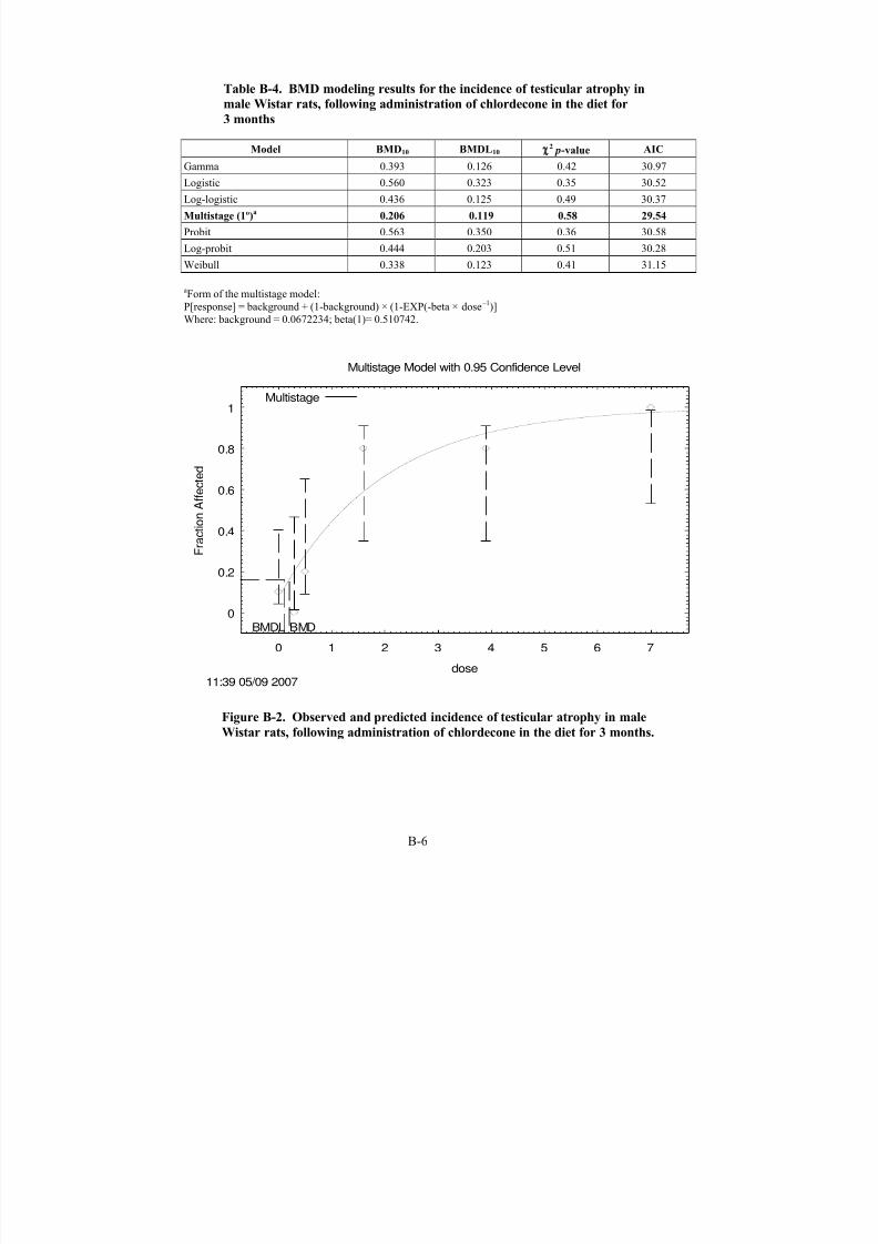
8/7/2019 United States Environmental Protect
http://slidepdf.com/reader/full/united-states-environmental-protect 151/183

8/7/2019 United States Environmental Protect
http://slidepdf.com/reader/full/united-states-environmental-protect 152/183

8/7/2019 United States Environmental Protect
http://slidepdf.com/reader/full/united-states-environmental-protect 153/183
Background 1 -0.41
Beta(1) -0.41 1
Parameter Estimates
Variable Estimate Std. Err.Background 0.0672234 0.22791
Beta(1) 0.510742 0.227823
Analysis of Deviance Table
Model Log(likelihood) Deviance Test DF P-valueFull model -10.7569
Fitted model -12.7712 4.02865 4 0.4021Reduced model -23.9018 26.2898 5 <.0001
AIC: 29.5424
Goodness of Fit
Dose Est._Prob. Expected Observed Size Chi^2 Res.-----------------------------------------------------------------------
i: 10.0000 0.0672 0.672 1 10 0.523
i: 20.3000 0.1997 0.999 0 5 -1.250
i: 30.5000 0.2774 1.387 1 5 -0.386i: 4
1.6000 0.5880 2.940 4 5 0.875i: 5
3.9000 0.8727 4.364 4 5 -0.655i: 6
7.0000 0.9739 4.869 5 5 1.027
Chi-square = 2.87 DF = 4 P-value = 0.5800
Benchmark Dose Computation
Specified effect = 0.1
Risk Type = Extra risk
Confidence level = 0.95
BMD = 0.206289
BMDL = 0.118596
B-8

8/7/2019 United States Environmental Protect
http://slidepdf.com/reader/full/united-states-environmental-protect 154/183
Liver Lesions (Fatty Changes and Hyperplasia) in Male and Female Rats Exposed to
Chlordecone in the Diet for 1–2 years
The Larson et al. (1979a) study did not include statistics for liver lesions. Statistical
analysis, performed for this review, of the frequency of liver lesions in each dose by sex
(Fisher’s exact test and Cochran-Armitage trend test) revealed that the incidence of liver lesionsin some of the exposure groups was statistically different from controls. An examination of liver
lesion incidence based on sex indicated (by Fisher’s exact test) no significant differences; the
incidence data for males and females were combined. The incidence data were used to fit the
dichotomous models available in the U.S. EPA BMDS version 1.3.2. The frequency of liver
lesions (fatty changes and hyperplasia) in both sexes combined was statistically different from
control at 0.5 and 1.6 mg/kg-day (see Table B-5). In addition, the Cochran-Armitage trend test
showed a statistically significant dose-response trend in the frequency of liver lesions (fatty
changes and hyperplasia) for both sexes combined.
Table B-5. Incidence of histopathologic liver lesions (fatty changes and
hyperplasia) in Wistar rats, following administration of chlordecone in the
diet for 1–2 years
Endpoint
Dose (mg/kg-day)
0 0.06 0.3 0.5 1.6
Liver lesionsa Male ratsFemale ratsBoth
1/222/343/56
1/111/132/24
2/62/174/23
3/4b 2/94/12b
6/21b
1/44/8b
aStatistically significant trend for increased incidence by Cochran-Armitage test.bStatistically significantly different from controls according to Fisher’s exact test performed for this review.
All models for dichotomous variables available in the U.S. EPA BMDS version 1.3.2
were fit to the data in Table B-5. All of the dichotomous models provided adequate fit to the
data based on the summary results reported in the BMDS output and a more detailed
examination of the graphs and chi-square goodness-of-fit statistics (summarized in Table B-6).
The gamma, multistage, and Weibull models yielded identical fits, and the lowest AIC
(i.e, 98.9) value. Thus, these models were selected to calculate a potential POD for the RfD,
based on the incidence data for liver lesions (fatty changes and hyperplasia) among rats. The
model-predicted a BMD10 associated with a 10% extra risk for liver lesions (fatty changes and
hyperplasia) of 0.23 mg/kg-day. The BMDL10of 0.14 mg/kg-day was considered a potential
POD for the RfD.
B-9
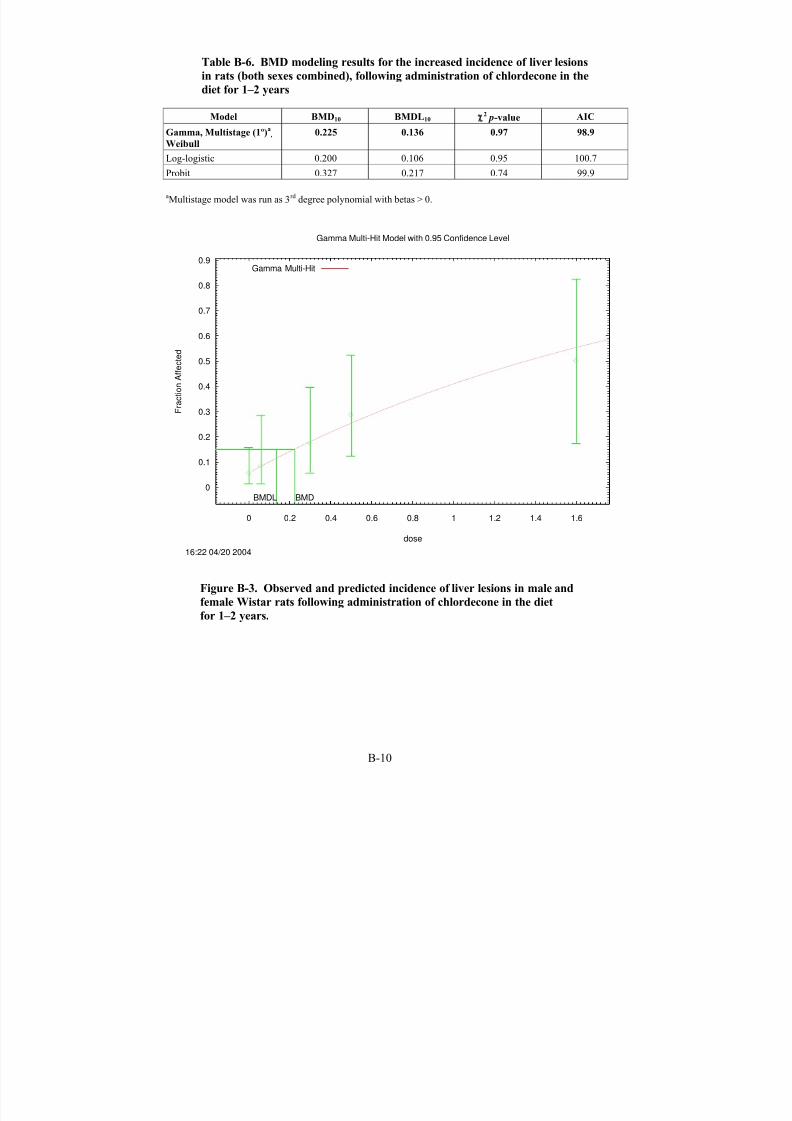
8/7/2019 United States Environmental Protect
http://slidepdf.com/reader/full/united-states-environmental-protect 155/183
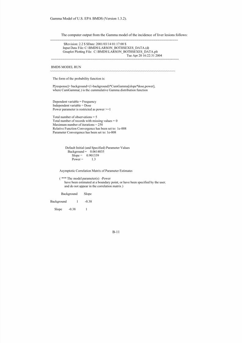
8/7/2019 United States Environmental Protect
http://slidepdf.com/reader/full/united-states-environmental-protect 156/183
Gamma Model of U.S. EPA BMDS (Version 1.3.2).
The computer output from the Gamma model of the incidence of liver lesions follows:
====================================================================$Revision: 2.2 $ $Date: 2001/03/14 01:17:00 $Input Data File: C:\BMDS\LARSON_BOTHSEXES_DATA.(d)Gnuplot Plotting File: C:\BMDS\LARSON_BOTHSEXES_DATA.plt
Tue Apr 20 16:22:31 2004====================================================================
BMDS MODEL RUN~~~~~~~~~~~~~~~~~~~~~~~~~~~~~~~~~~~~~~~~~~~~~~~~~~~~~~~~~~~~~~~~~~~~~
The form of the probability function is:
P[response]= background+(1-background)*CumGamma[slope*dose,power],where CumGamma(.) is the cummulative Gamma distribution function
Dependent variable = FrequencyIndependent variable = DosePower parameter is restricted as power >=1
Total number of observations = 5Total number of records with missing values = 0Maximum number of iterations = 250Relative Function Convergence has been set to: 1e-008Parameter Convergence has been set to: 1e-008
Default Initial (and Specified) Parameter ValuesBackground = 0.0614035
Slope = 0.901339Power = 1.3
Asymptotic Correlation Matrix of Parameter Estimates
( *** The model parameter(s) -Power have been estimated at a boundary point, or have been specified by the user,
and do not appear in the correlation matrix )
Background Slope
Background 1 -0.38
Slope -0.38 1
B-11
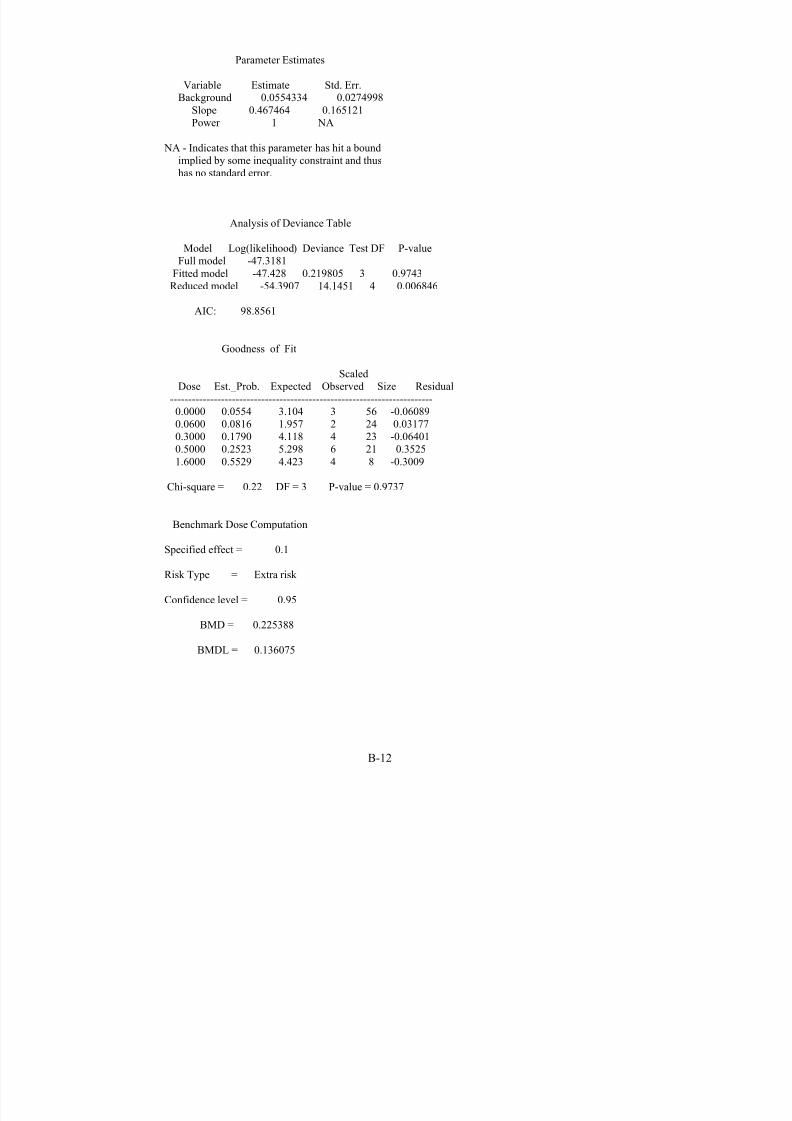
8/7/2019 United States Environmental Protect
http://slidepdf.com/reader/full/united-states-environmental-protect 157/183
Parameter Estimates
Variable Estimate Std. Err.Background 0.0554334 0.0274998
Slope 0.467464 0.165121Power 1 NA
NA - Indicates that this parameter has hit a boundimplied by some inequality constraint and thushas no standard error.
Analysis of Deviance Table
Model Log(likelihood) Deviance Test DF P-valueFull model -47.3181
Fitted model -47.428 0.219805 3 0.9743Reduced model -54.3907 14.1451 4 0.006846
AIC: 98.8561
Goodness of Fit
ScaledDose Est._Prob. Expected Observed Size Residual
------------------------------------------------------------------------0.0000 0.0554 3.104 3 56 -0.060890.0600 0.0816 1.957 2 24 0.031770.3000 0.1790 4.118 4 23 -0.06401
0.5000 0.2523 5.298 6 21 0.35251.6000 0.5529 4.423 4 8 -0.3009
Chi-square = 0.22 DF = 3 P-value = 0.9737
Benchmark Dose Computation
Specified effect = 0.1
Risk Type = Extra risk
Confidence level = 0.95
BMD = 0.225388
BMDL = 0.136075
B-12
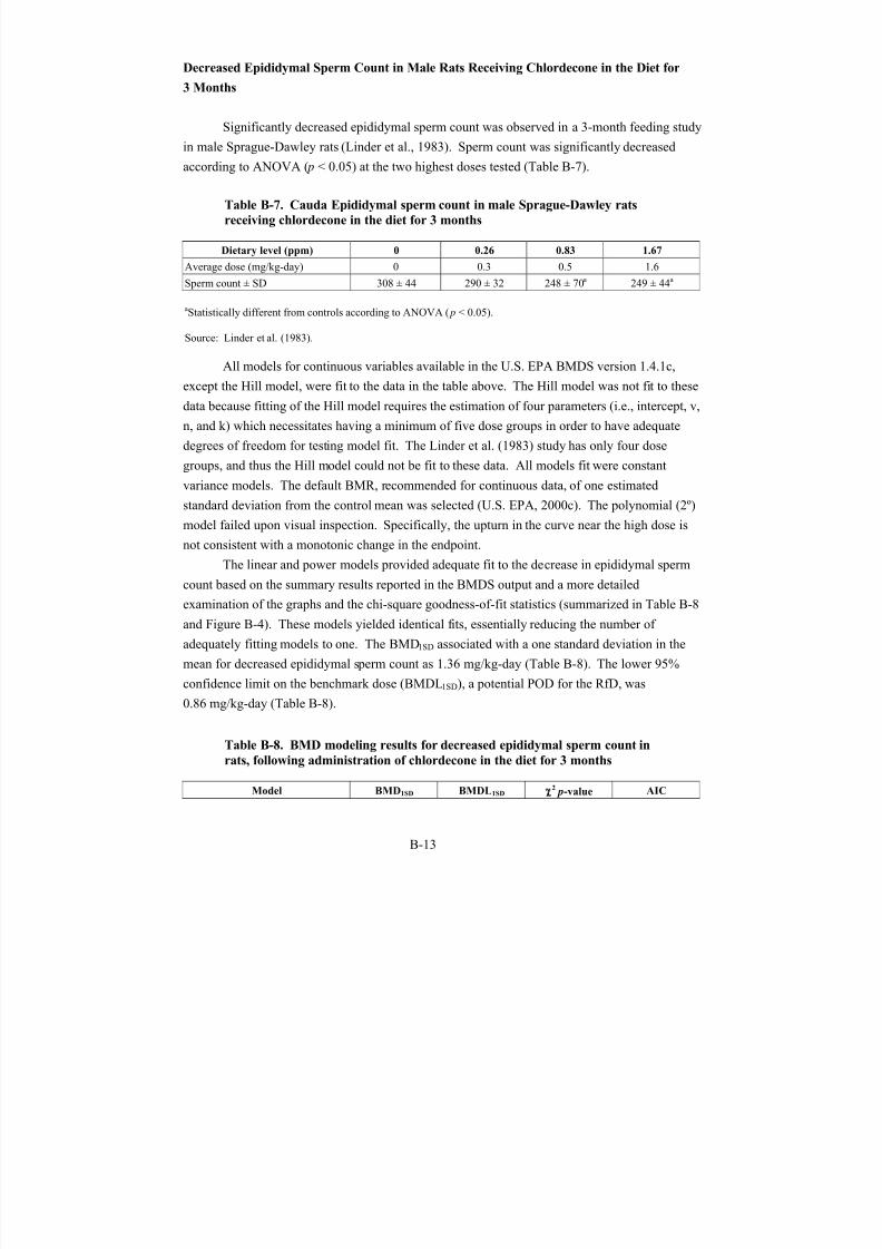
8/7/2019 United States Environmental Protect
http://slidepdf.com/reader/full/united-states-environmental-protect 158/183
Decreased Epididymal Sperm Count in Male Rats Receiving Chlordecone in the Diet for
3 Months
Significantly decreased epididymal sperm count was observed in a 3-month feeding study
in male Sprague-Dawley rats (Linder et al., 1983). Sperm count was significantly decreased
according to ANOVA (p < 0.05) at the two highest doses tested (Table B-7).
Table B-7. Cauda Epididymal sperm count in male Sprague-Dawley rats
receiving chlordecone in the diet for 3 months
Dietary level (ppm) 0 0.26 0.83 1.67
Average dose (mg/kg-day) 0 0.3 0.5 1.6
Sperm count ± SD 308 ± 44 248 ± 70a 249 ± 44a 290 ± 32
aStatistically different from controls according to ANOVA (p < 0.05).
Source: Linder et al. (1983).
All models for continuous variables available in the U.S. EPA BMDS version 1.4.1c,
except the Hill model, were fit to the data in the table above. The Hill model was not fit to these
data because fitting of the Hill model requires the estimation of four parameters (i.e., intercept, v,
n, and k) which necessitates having a minimum of five dose groups in order to have adequate
degrees of freedom for testing model fit. The Linder et al. (1983) study has only four dose
groups, and thus the Hill model could not be fit to these data. All models fit were constant
variance models. The default BMR, recommended for continuous data, of one estimated
standard deviation from the control mean was selected (U.S. EPA, 2000c). The polynomial (2º)model failed upon visual inspection. Specifically, the upturn in the curve near the high dose is
not consistent with a monotonic change in the endpoint.
The linear and power models provided adequate fit to the decrease in epididymal sperm
count based on the summary results reported in the BMDS output and a more detailed
examination of the graphs and the chi-square goodness-of-fit statistics (summarized in Table B-8
and Figure B-4). These models yielded identical fits, essentially reducing the number of
adequately fitting models to one. The BMD1SD associated with a one standard deviation in the
mean for decreased epididymal sperm count as 1.36 mg/kg-day (Table B-8). The lower 95%
confidence limit on the benchmark dose (BMDL1SD), a potential POD for the RfD, was
0.86 mg/kg-day (Table B-8).
Table B-8. BMD modeling results for decreased epididymal sperm count in
rats, following administration of chlordecone in the diet for 3 months
Model BMD1SD BMDL1SD χ2 p-value AIC
B-13
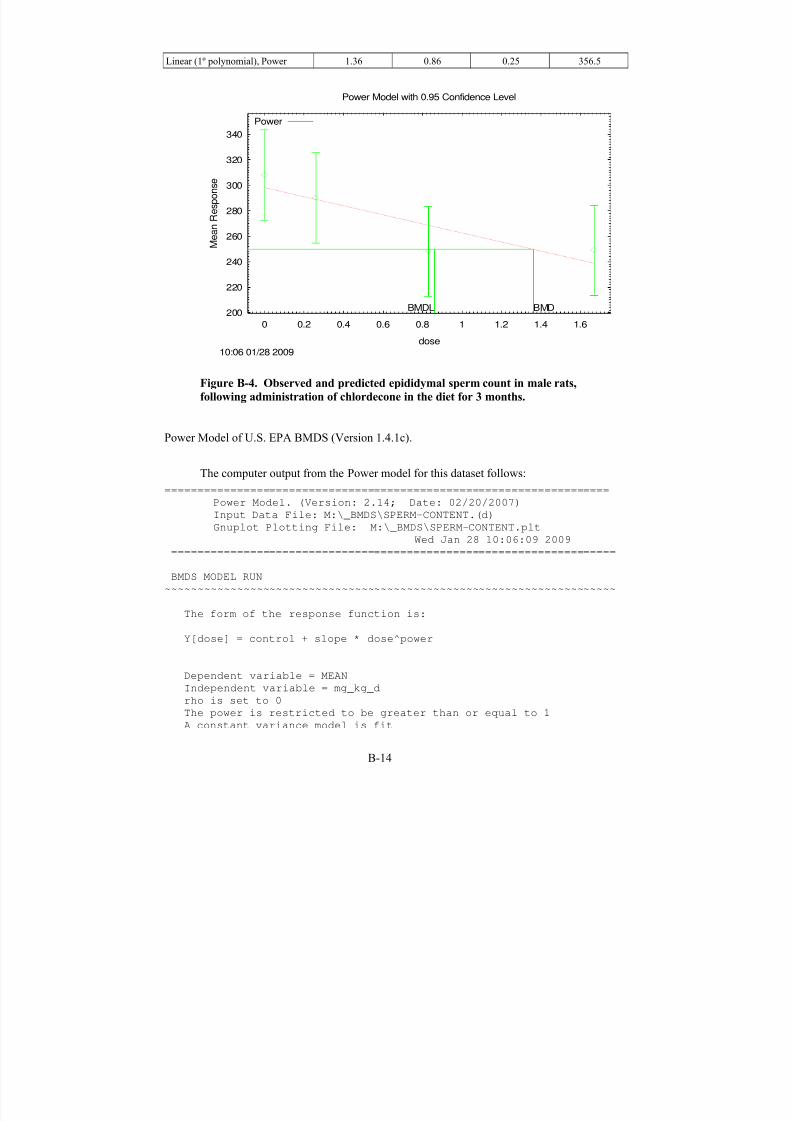
8/7/2019 United States Environmental Protect
http://slidepdf.com/reader/full/united-states-environmental-protect 159/183
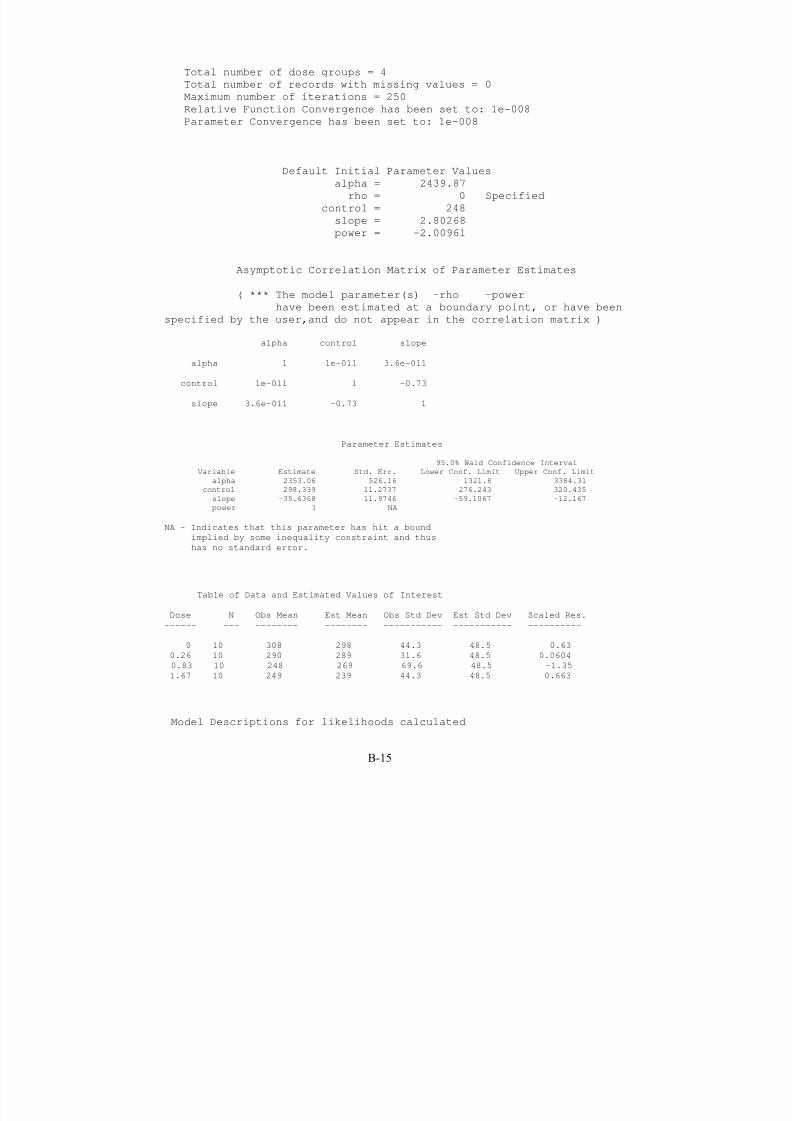
8/7/2019 United States Environmental Protect
http://slidepdf.com/reader/full/united-states-environmental-protect 160/183
Total number of dose groups = 4
Total number of records with missing values = 0
Maximum number of iterations = 250
Relative Function Convergence has been set to: 1e-008
Parameter Convergence has been set to: 1e-008
Default Initial Parameter Values
alpha = 2439.87
rho = 0 Specified
control = 248
slope = 2.80268
power = -2.00961
Asymptotic Correlation Matrix of Parameter Estimates
( *** The model parameter(s) -rho -power
have been estimated at a boundary point, or have been
specified by the user,and do not appear in the correlation matrix )
alpha control slope
alpha 1 1e-011 3.6e-011
control 1e-011 1 -0.73
slope 3.6e-011 -0.73 1
Parameter Estimates
95.0% Wald Confidence Interval
Variable Estimate Std. Err. Lower Conf. Limit Upper Conf. Limit
alpha 2353.06 526.16 1321.8 3384.31
control 298.339 11.2737 276.243 320.435
slope -35.6368 11.9746 -59.1067 -12.167
power 1 NA
NA - Indicates that this parameter has hit a bound
implied by some inequality constraint and thus
has no standard error.
Table of Data and Estimated Values of Interest
Dose N Obs Mean Est Mean Obs Std Dev Est Std Dev Scaled Res.
------ --- -------- -------- ----------- ----------- ----------
0 10 308 298 44.3 48.5 0.63
0.26 10 290 289 31.6 48.5 0.0604
0.83 10 248 269 69.6 48.5 -1.35
1.67 10 249 239 44.3 48.5 0.663
Model Descriptions for likelihoods calculated
B-15

8/7/2019 United States Environmental Protect
http://slidepdf.com/reader/full/united-states-environmental-protect 161/183

8/7/2019 United States Environmental Protect
http://slidepdf.com/reader/full/united-states-environmental-protect 162/183
B-17
different variance model
The p-value for Test 4 is greater than .1. The model chosen seems
to adequately describe the data
Benchmark Dose Computation
Specified effect = 1
Risk Type = Estimated standard deviations from the control
mean
Confidence level = 0.95
BMD = 1.36119
BMDL = 0.859849
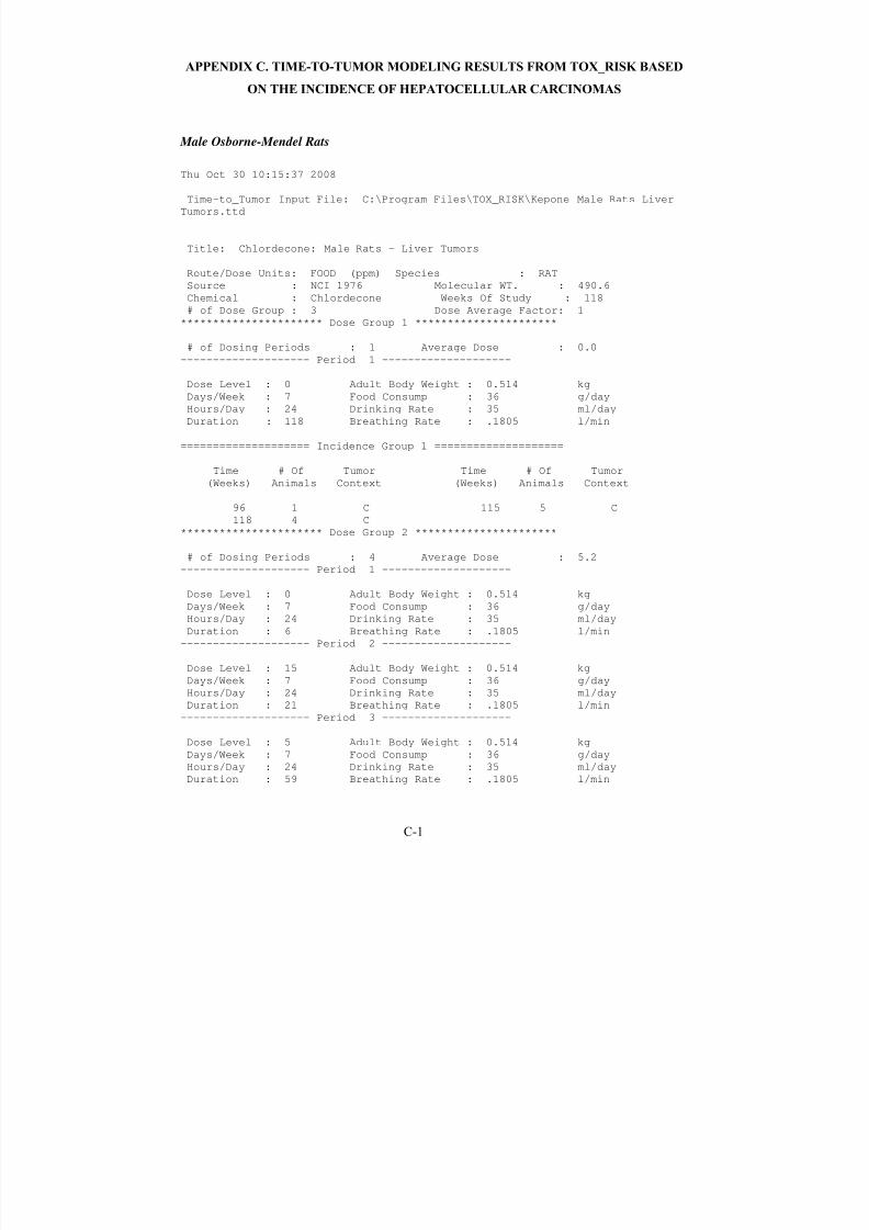
8/7/2019 United States Environmental Protect
http://slidepdf.com/reader/full/united-states-environmental-protect 163/183
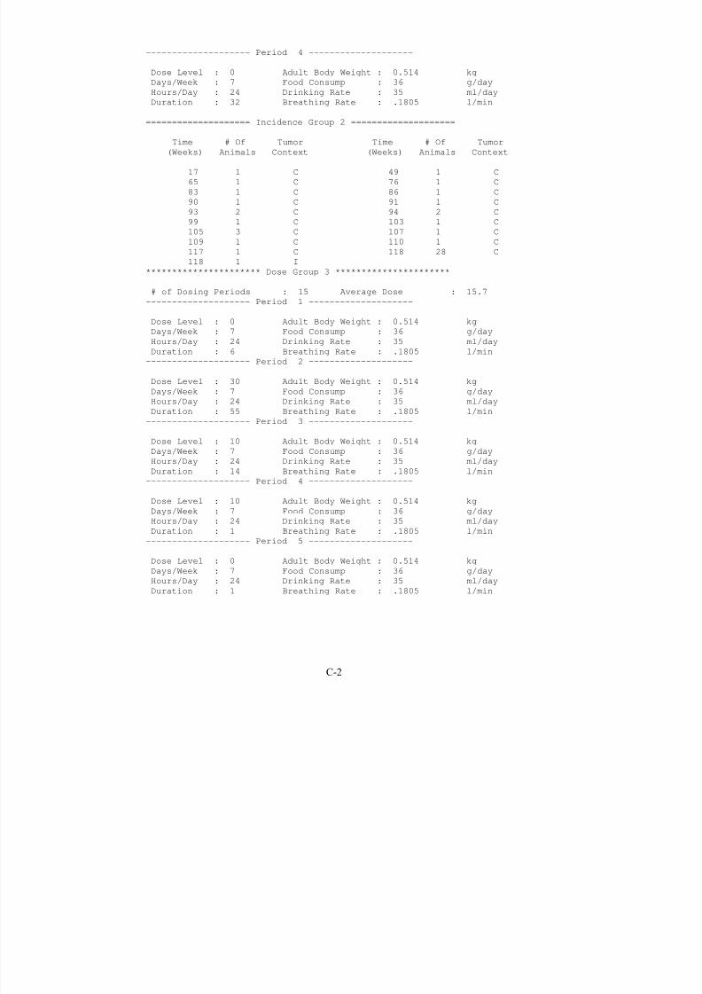
8/7/2019 United States Environmental Protect
http://slidepdf.com/reader/full/united-states-environmental-protect 164/183
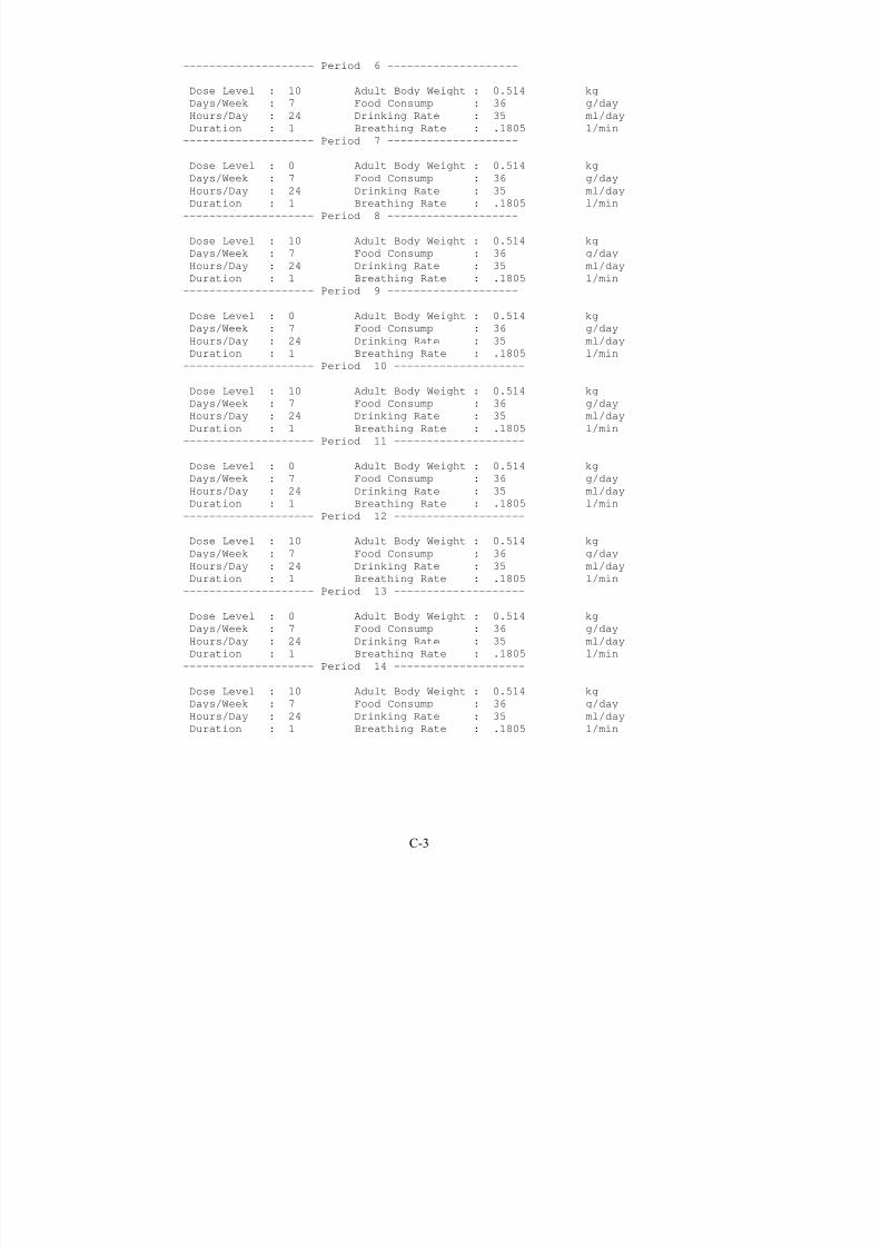
8/7/2019 United States Environmental Protect
http://slidepdf.com/reader/full/united-states-environmental-protect 165/183
-------------------- Period 6 --------------------
Dose Level : 10 Adult Body Weight : 0.514 kg
Days/Week : 7 Food Consump : 36 g/day
Hours/Day : 24 Drinking Rate : 35 ml/day
Duration : 1 Breathing Rate : .1805 l/min
-------------------- Period 7 --------------------
Dose Level : 0 Adult Body Weight : 0.514 kgDays/Week : 7 Food Consump : 36 g/day
Hours/Day : 24 Drinking Rate : 35 ml/day
Duration : 1 Breathing Rate : .1805 l/min
-------------------- Period 8 --------------------
Dose Level : 10 Adult Body Weight : 0.514 kg
Days/Week : 7 Food Consump : 36 g/day
Hours/Day : 24 Drinking Rate : 35 ml/day
Duration : 1 Breathing Rate : .1805 l/min
-------------------- Period 9 --------------------
Dose Level : 0 Adult Body Weight : 0.514 kg
Days/Week : 7 Food Consump : 36 g/day
Hours/Day : 24 Drinking Rate : 35 ml/dayDuration : 1 Breathing Rate : .1805 l/min
-------------------- Period 10 --------------------
Dose Level : 10 Adult Body Weight : 0.514 kg
Days/Week : 7 Food Consump : 36 g/day
Hours/Day : 24 Drinking Rate : 35 ml/day
Duration : 1 Breathing Rate : .1805 l/min
-------------------- Period 11 --------------------
Dose Level : 0 Adult Body Weight : 0.514 kg
Days/Week : 7 Food Consump : 36 g/day
Hours/Day : 24 Drinking Rate : 35 ml/day
Duration : 1 Breathing Rate : .1805 l/min-------------------- Period 12 --------------------
Dose Level : 10 Adult Body Weight : 0.514 kg
Days/Week : 7 Food Consump : 36 g/day
Hours/Day : 24 Drinking Rate : 35 ml/day
Duration : 1 Breathing Rate : .1805 l/min
-------------------- Period 13 --------------------
Dose Level : 0 Adult Body Weight : 0.514 kg
Days/Week : 7 Food Consump : 36 g/day
Hours/Day : 24 Drinking Rate : 35 ml/day
Duration : 1 Breathing Rate : .1805 l/min
-------------------- Period 14 --------------------
Dose Level : 10 Adult Body Weight : 0.514 kg
Days/Week : 7 Food Consump : 36 g/day
Hours/Day : 24 Drinking Rate : 35 ml/day
Duration : 1 Breathing Rate : .1805 l/min
C-3
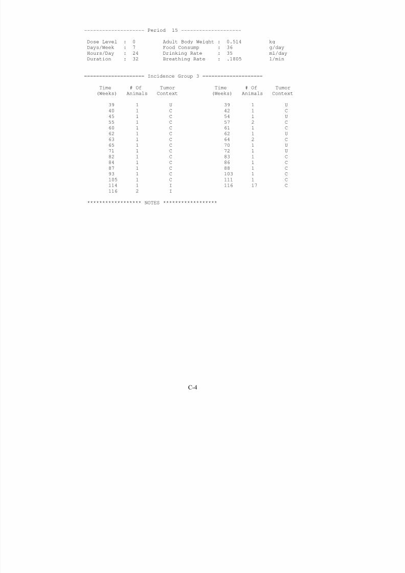
8/7/2019 United States Environmental Protect
http://slidepdf.com/reader/full/united-states-environmental-protect 166/183

8/7/2019 United States Environmental Protect
http://slidepdf.com/reader/full/united-states-environmental-protect 167/183
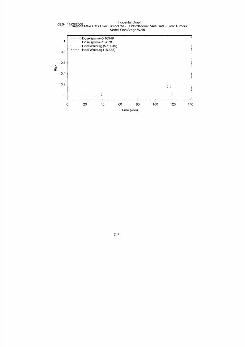
8/7/2019 United States Environmental Protect
http://slidepdf.com/reader/full/united-states-environmental-protect 168/183
0
0.2
0.4
0.6
0.8
1
0 20 40 60 80 100 120 140
Risk
Time (wks)
Incidental GraphKepone Male Rats Liver Tumors.ttd - Chlordecone: Male Rats - Liver Tumors
Model: One Stage Weib
09:54 11/05/2008
Dose (ppm)=5.16949Dose (ppm)=15.678
Hoel Walburg (5.16949)Hoel Walburg (15.678)
C-6
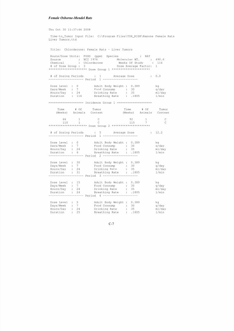
8/7/2019 United States Environmental Protect
http://slidepdf.com/reader/full/united-states-environmental-protect 169/183
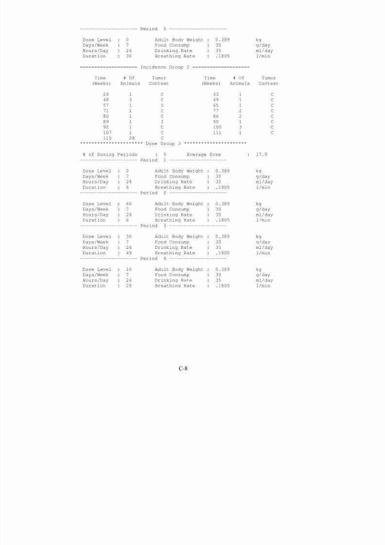
8/7/2019 United States Environmental Protect
http://slidepdf.com/reader/full/united-states-environmental-protect 170/183
-------------------- Period 5 --------------------
Dose Level : 0 Adult Body Weight : 0.389 kg
Days/Week : 7 Food Consump : 30 g/day
Hours/Day : 24 Drinking Rate : 35 ml/day
Duration : 30 Breathing Rate : .1805 l/min
==================== Incidence Group 2 ====================
Time # Of Tumor Time # Of Tumor
(Weeks) Animals Context (Weeks) Animals Context
29 1 C 33 1 C
48 3 C 49 1 C
57 1 U 65 1 C
71 1 C 77 2 C
80 1 C 86 2 C
89 1 I 90 1 C
92 1 C 105 3 C
107 1 C 111 1 C
115 28 C
********************** Dose Group 3 **********************
# of Dosing Periods : 5 Average Dose : 17.9
-------------------- Period 1 --------------------
Dose Level : 0 Adult Body Weight : 0.389 kg
Days/Week : 7 Food Consump : 30 g/day
Hours/Day : 24 Drinking Rate : 35 ml/day
Duration : 6 Breathing Rate : .1805 l/min
-------------------- Period 2 --------------------
Dose Level : 60 Adult Body Weight : 0.389 kg
Days/Week : 7 Food Consump : 30 g/day
Hours/Day : 24 Drinking Rate : 35 ml/day
Duration : 6 Breathing Rate : .1805 l/min-------------------- Period 3 --------------------
Dose Level : 30 Adult Body Weight : 0.389 kg
Days/Week : 7 Food Consump : 30 g/day
Hours/Day : 24 Drinking Rate : 35 ml/day
Duration : 49 Breathing Rate : .1805 l/min
-------------------- Period 4 --------------------
Dose Level : 10 Adult Body Weight : 0.389 kg
Days/Week : 7 Food Consump : 30 g/day
Hours/Day : 24 Drinking Rate : 35 ml/day
Duration : 25 Breathing Rate : .1805 l/min
C-8
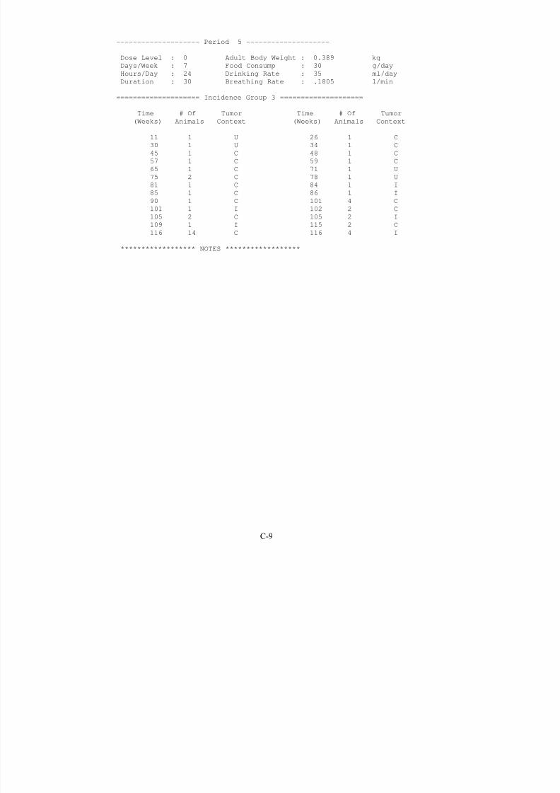
8/7/2019 United States Environmental Protect
http://slidepdf.com/reader/full/united-states-environmental-protect 171/183
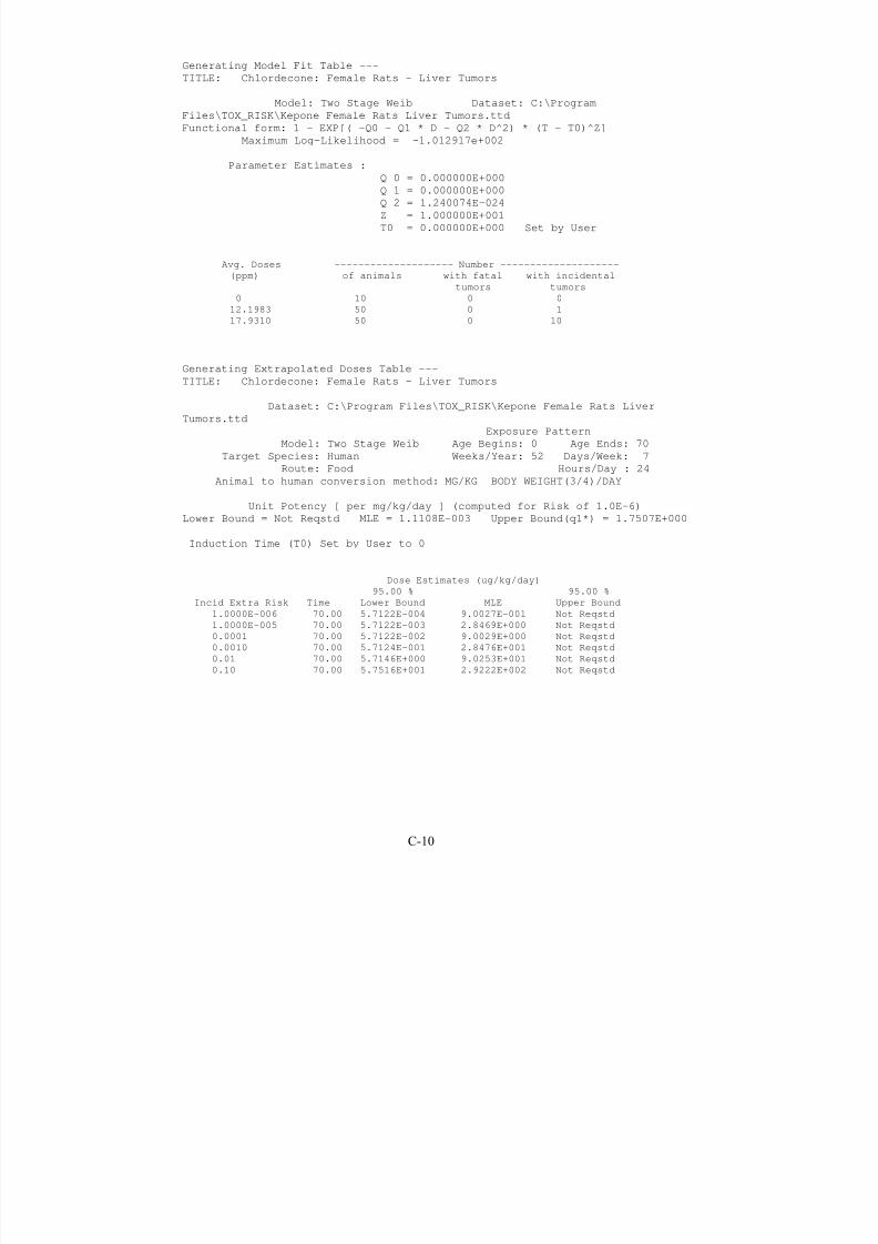
8/7/2019 United States Environmental Protect
http://slidepdf.com/reader/full/united-states-environmental-protect 172/183
Generating Model Fit Table ---
TITLE: Chlordecone: Female Rats - Liver Tumors
Model: Two Stage Weib Dataset: C:\Program
Files\TOX_RISK\Kepone Female Rats Liver Tumors.ttd
Functional form: 1 - EXP[( -Q0 - Q1 * D - Q2 * D^2) * (T - T0)^Z]
Maximum Log-Likelihood = -1.012917e+002
Parameter Estimates :Q 0 = 0.000000E+000
Q 1 = 0.000000E+000
Q 2 = 1.240074E-024
Z = 1.000000E+001
T0 = 0.000000E+000 Set by User
Avg. Doses -------------------- Number --------------------
(ppm) of animals with fatal with incidental
tumors tumors
0 10 0 0
12.1983 50 0 1
17.9310 50 0 10
Generating Extrapolated Doses Table ---
TITLE: Chlordecone: Female Rats - Liver Tumors
Dataset: C:\Program Files\TOX_RISK\Kepone Female Rats Liver
Tumors.ttd
Exposure Pattern
Model: Two Stage Weib Age Begins: 0 Age Ends: 70
Target Species: Human Weeks/Year: 52 Days/Week: 7
Route: Food Hours/Day : 24
Animal to human conversion method: MG/KG BODY WEIGHT(3/4)/DAY
Unit Potency [ per mg/kg/day ] (computed for Risk of 1.0E-6)Lower Bound = Not Reqstd MLE = 1.1108E-003 Upper Bound(q1*) = 1.7507E+000
Induction Time (T0) Set by User to 0
Dose Estimates (ug/kg/day)
95.00 % 95.00 %
Incid Extra Risk Time Lower Bound MLE Upper Bound
1.0000E-006 70.00 5.7122E-004 9.0027E-001 Not Reqstd
1.0000E-005 70.00 5.7122E-003 2.8469E+000 Not Reqstd
0.0001 70.00 5.7122E-002 9.0029E+000 Not Reqstd
0.0010 70.00 5.7124E-001 2.8476E+001 Not Reqstd
0.01 70.00 5.7146E+000 9.0253E+001 Not Reqstd
0.10 70.00 5.7516E+001 2.9222E+002 Not Reqstd
C-10
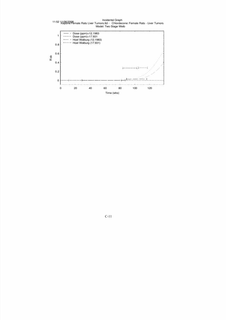
8/7/2019 United States Environmental Protect
http://slidepdf.com/reader/full/united-states-environmental-protect 173/183
0
0.2
0.4
0.6
0.8
1
0 20 40 60 80 100 120
Risk
Time (wks)
Incidental GraphKepone Female Rats Liver Tumors.ttd - Chlordecone: Female Rats - Liver Tumors
Model: Two Stage Weib
11:52 11/06/2008
Dose (ppm)=12.1983Dose (ppm)=17.931
Hoel Walburg (12.1983)
Hoel Walburg (17.931)
C-11
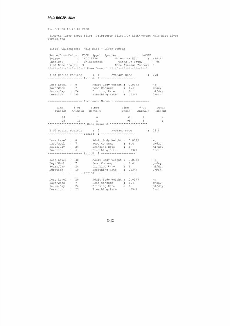
8/7/2019 United States Environmental Protect
http://slidepdf.com/reader/full/united-states-environmental-protect 174/183
Male B6C3F1 Mice
Tue Oct 28 15:20:02 2008
Time-to_Tumor Input File: C:\Program Files\TOX_RISK\Kepone Male Mice Liver
Tumors.ttd
Title: Chlordecone: Male Mice - Liver Tumors
Route/Dose Units: FOOD (ppm) Species : MOUSE
Source : NCI 1976 Molecular WT. : 490.6
Chemical : Chlordecone Weeks Of Study : 95
# of Dose Group : 3 Dose Average Factor: 1
********************** Dose Group 1 **********************
# of Dosing Periods : 1 Average Dose : 0.0
-------------------- Period 1 --------------------
Dose Level : 0 Adult Body Weight : 0.0373 kg
Days/Week : 7 Food Consump : 6.4 g/dayHours/Day : 24 Drinking Rate : 6 ml/day
Duration : 95 Breathing Rate : .0347 l/min
==================== Incidence Group 1 ====================
Time # Of Tumor Time # Of Tumor
(Weeks) Animals Context (Weeks) Animals Context
66 1 U 92 1 I
95 13 C 95 5 I
********************** Dose Group 2 **********************
# of Dosing Periods : 5 Average Dose : 16.8
-------------------- Period 1 --------------------
Dose Level : 0 Adult Body Weight : 0.0373 kg
Days/Week : 7 Food Consump : 6.4 g/day
Hours/Day : 24 Drinking Rate : 6 ml/day
Duration : 6 Breathing Rate : .0347 l/min
-------------------- Period 2 --------------------
Dose Level : 40 Adult Body Weight : 0.0373 kg
Days/Week : 7 Food Consump : 6.4 g/day
Hours/Day : 24 Drinking Rate : 6 ml/day
Duration : 19 Breathing Rate : .0347 l/min
-------------------- Period 3 --------------------
Dose Level : 20 Adult Body Weight : 0.0373 kg
Days/Week : 7 Food Consump : 6.4 g/day
Hours/Day : 24 Drinking Rate : 6 ml/day
Duration : 23 Breathing Rate : .0347 l/min
C-12

8/7/2019 United States Environmental Protect
http://slidepdf.com/reader/full/united-states-environmental-protect 175/183

8/7/2019 United States Environmental Protect
http://slidepdf.com/reader/full/united-states-environmental-protect 176/183
==================== Incidence Group 3 ====================
Time # Of Tumor Time # Of Tumor
(Weeks) Animals Context (Weeks) Animals Context
3 1 C 21 1 C
48 1 C 49 2 C
69 1 I 71 2 I
72 1 U 77 1 I79 1 C 80 3 I
83 6 I 84 2 I
85 1 I 88 1 I
94 2 I 95 24 I
****************** NOTES ******************
C-14
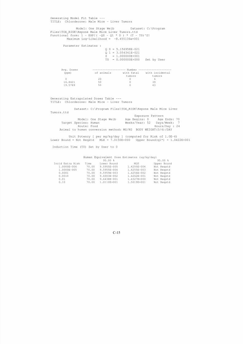
8/7/2019 United States Environmental Protect
http://slidepdf.com/reader/full/united-states-environmental-protect 177/183

8/7/2019 United States Environmental Protect
http://slidepdf.com/reader/full/united-states-environmental-protect 178/183
0
0.2
0.4
0.6
0.8
1
0 20 40 60 80 100
Risk
Time (wks)
Incidental GraphKepone Male Mice Liver Tumors.ttd - Chlordecone: Male Mice - Liver Tumors
Model: One Stage Weib
16:07 11/05/2008
Dose (ppm)=16.8421Dose (ppm)=19.5789
Hoel Walburg (16.8421)Hoel Walburg (19.5789)
C-16

8/7/2019 United States Environmental Protect
http://slidepdf.com/reader/full/united-states-environmental-protect 179/183
Female B6C3F1 Mice
Tue Oct 28 15:21:04 2008
Time-to_Tumor Input File: C:\Program Files\TOX_RISK\Kepone Female Mice
Liver Tumors.ttd
Title: Chlordecone: Female Mice - Liver Tumors
Route/Dose Units: FOOD (ppm) Species : MOUSE
Source : NCI 1976 Molecular WT. : 490.6
Chemical : Chlordecone Weeks Of Study : 95
# of Dose Group : 3 Dose Average Factor: 1
********************** Dose Group 1 **********************
# of Dosing Periods : 1 Average Dose : 0.0
-------------------- Period 1 --------------------
Dose Level : 0 Adult Body Weight : 0.0353 kg
Days/Week : 7 Food Consump : 6.1 g/dayHours/Day : 24 Drinking Rate : 6 ml/day
Duration : 95 Breathing Rate : .0347 l/min
==================== Incidence Group 1 ====================
Time # Of Tumor Time # Of Tumor
(Weeks) Animals Context (Weeks) Animals Context
93 1 C 95 9 C
********************** Dose Group 2 **********************
# of Dosing Periods : 5 Average Dose : 16.8
-------------------- Period 1 --------------------
Dose Level : 0 Adult Body Weight : 0.0353 kg
Days/Week : 7 Food Consump : 6.1 g/day
Hours/Day : 24 Drinking Rate : 6 ml/day
Duration : 6 Breathing Rate : .0347 l/min
-------------------- Period 2 --------------------
Dose Level : 40 Adult Body Weight : 0.0353 kg
Days/Week : 7 Food Consump : 6.1 g/day
Hours/Day : 24 Drinking Rate : 6 ml/day
Duration : 19 Breathing Rate : .0347 l/min
-------------------- Period 3 --------------------
Dose Level : 20 Adult Body Weight : 0.0353 kgDays/Week : 7 Food Consump : 6.1 g/day
Hours/Day : 24 Drinking Rate : 6 ml/day
Duration : 23 Breathing Rate : .0347 l/min
C-17
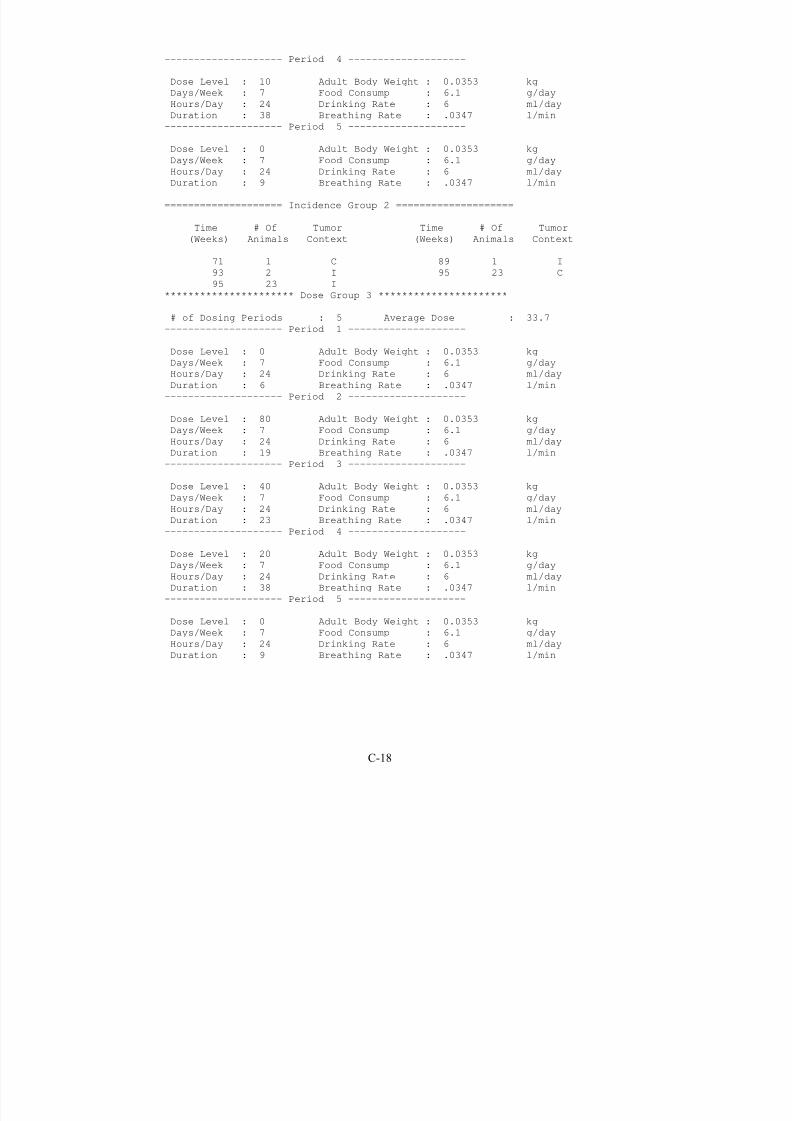
8/7/2019 United States Environmental Protect
http://slidepdf.com/reader/full/united-states-environmental-protect 180/183
-------------------- Period 4 --------------------
Dose Level : 10 Adult Body Weight : 0.0353 kg
Days/Week : 7 Food Consump : 6.1 g/day
Hours/Day : 24 Drinking Rate : 6 ml/day
Duration : 38 Breathing Rate : .0347 l/min
-------------------- Period 5 --------------------
Dose Level : 0 Adult Body Weight : 0.0353 kgDays/Week : 7 Food Consump : 6.1 g/day
Hours/Day : 24 Drinking Rate : 6 ml/day
Duration : 9 Breathing Rate : .0347 l/min
==================== Incidence Group 2 ====================
Time # Of Tumor Time # Of Tumor
(Weeks) Animals Context (Weeks) Animals Context
71 1 C 89 1 I
93 2 I 95 23 C
95 23 I
********************** Dose Group 3 **********************
# of Dosing Periods : 5 Average Dose : 33.7
-------------------- Period 1 --------------------
Dose Level : 0 Adult Body Weight : 0.0353 kg
Days/Week : 7 Food Consump : 6.1 g/day
Hours/Day : 24 Drinking Rate : 6 ml/day
Duration : 6 Breathing Rate : .0347 l/min
-------------------- Period 2 --------------------
Dose Level : 80 Adult Body Weight : 0.0353 kg
Days/Week : 7 Food Consump : 6.1 g/day
Hours/Day : 24 Drinking Rate : 6 ml/day
Duration : 19 Breathing Rate : .0347 l/min-------------------- Period 3 --------------------
Dose Level : 40 Adult Body Weight : 0.0353 kg
Days/Week : 7 Food Consump : 6.1 g/day
Hours/Day : 24 Drinking Rate : 6 ml/day
Duration : 23 Breathing Rate : .0347 l/min
-------------------- Period 4 --------------------
Dose Level : 20 Adult Body Weight : 0.0353 kg
Days/Week : 7 Food Consump : 6.1 g/day
Hours/Day : 24 Drinking Rate : 6 ml/day
Duration : 38 Breathing Rate : .0347 l/min
-------------------- Period 5 --------------------
Dose Level : 0 Adult Body Weight : 0.0353 kg
Days/Week : 7 Food Consump : 6.1 g/day
Hours/Day : 24 Drinking Rate : 6 ml/day
Duration : 9 Breathing Rate : .0347 l/min
C-18
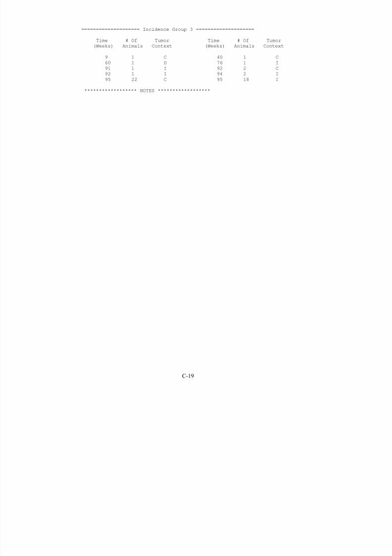
8/7/2019 United States Environmental Protect
http://slidepdf.com/reader/full/united-states-environmental-protect 181/183

8/7/2019 United States Environmental Protect
http://slidepdf.com/reader/full/united-states-environmental-protect 182/183
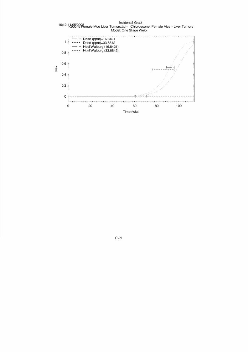
8/7/2019 United States Environmental Protect
http://slidepdf.com/reader/full/united-states-environmental-protect 183/183