uncoupling of gtp hydrolysis from eif6 release on the
TRANSCRIPT

Uncoupling of GTP hydrolysis from eIF6release on the ribosome causesShwachman-Diamond syndrome
Andrew J. Finch,1,2,10 Christine Hilcenko,1,2,10 Nicolas Basse,1,2 Lesley F. Drynan,1
Beatriz Goyenechea,1,2 Tobias F. Menne,1,2 Africa Gonzalez Fernandez,3 Paul Simpson,1,2
Clive S. D’Santos,4 Mark J. Arends,5 Jean Donadieu,6 Christine Bellanne-Chantelot,7
Michael Costanzo,8,9 Charles Boone,8,9 Andrew N. McKenzie,1 Stefan M.V. Freund,1
and Alan J. Warren1,2,11
1Medical Research Council Laboratory of Molecular Biology, Cambridge CB2 0QH, United Kingdom; 2Department ofHaematology, University of Cambridge, Cambridge CB2 0XY, United Kingdom; 3Immunology Department, BiomedicalResearch Center, University of Vigo, Vigo, Pontevedra 36310, Spain; 4Cancer Research UK, Cambridge Research Institute,Cambridge, CB2 0RE, United Kingdom; 5Pathology Department, University of Cambridge, Addenbrooke’s Hospital, CambridgeCB2 0QQ, United Kingdom; 6Service d’Hemato Oncologie Pediatrique, Registre des Neutropenies Congenitales, HopitalTrousseau, Paris F 75012, France; 7Department of Genetics, Hopital Pitie-Salpetriere, Universite Pierre et Marie Curie, Paris75651, France; 8Banting and Best Department of Medical Research, Terrence Donnelly Center for Cellular and BiomolecularResearch, University of Toronto, Toronto, Ontario M5S 3E1, Canada; 9Department of Molecular Genetics and Microbiology,Terrence Donnelly Center for Cellular and Biomolecular Research, University of Toronto, Toronto, Ontario M5S 3E1, Canada
Removal of the assembly factor eukaryotic initiation factor 6 (eIF6) is critical for late cytoplasmic maturationof 60S ribosomal subunits. In mammalian cells, the current model posits that eIF6 release is triggered followingphosphorylation of Ser 235 by activated protein kinase C. In contrast, genetic studies in yeast indicatea requirement for the ortholog of the SBDS (Shwachman-Bodian-Diamond syndrome) gene that is mutatedin the inherited leukemia predisposition disorder Shwachman-Diamond syndrome (SDS). Here, by isolating latecytoplasmic 60S ribosomal subunits from Sbds-deleted mice, we show that SBDS and the GTPase elongationfactor-like 1 (EFL1) directly catalyze eIF6 removal in mammalian cells by a mechanism that requires GTPbinding and hydrolysis by EFL1 but not phosphorylation of eIF6 Ser 235. Functional analysis of disease-associated missense variants reveals that the essential role of SBDS is to tightly couple GTP hydrolysis byEFL1 on the ribosome to eIF6 release. Furthermore, complementary NMR spectroscopic studies suggestunanticipated mechanistic parallels between this late step in 60S maturation and aspects of bacterial ribosomedisassembly. Our findings establish a direct role for SBDS and EFL1 in catalyzing the translational activationof ribosomes in all eukaryotes, and define SDS as a ribosomopathy caused by uncoupling GTP hydrolysis fromeIF6 release.
[Keywords: bone marrow failure syndromes; ribosome assembly; eIF6; human genetics; leukemia; ribosomopathy;NMR]
Supplemental material is available for this article.
Received January 6, 2011; revised version accepted March 15, 2011.
Shwachman-Diamond syndrome (SDS; OMIM 260400)is an autosomal recessive disorder characterized by bonemarrow dysfunction with a striking cumulative risk ofprogression to myelodysplastic syndrome (MDS) and acutemyeloid leukemia (AML) (Donadieu et al. 2005) that iscaused by mutations in the essential, highly conserved
SBDS (Shwachman-Bodian-Diamond syndrome) gene(Boocock et al. 2003), whose specific function remainsunknown. Genetic studies in Saccharomyces cerevisiaeindicate that the SBDS ortholog Sdo1 and the GTPaseelongation factor-like 1 (Efl1), a structural homolog ofelongation factor G (EF-G), function in a pathway torelease and recycle the essential nucleolar factor Tif6(mammalian eukaryotic initiation factor 6 [eIF6]) fromlate cytoplasmic pre-60S ribosomal subunits (Menneet al. 2007). Tif6 is critical for the biogenesis and nuclearexport of pre-60S subunits (Basu et al. 2001), and acts as
10These authors contributed equally to this work.11Corresponding author.E-MAIL [email protected]; FAX 01223-412-178.Article is online at http://www.genesdev.org/cgi/doi/10.1101/gad.623011.
GENES & DEVELOPMENT 25:917–929 � 2011 by Cold Spring Harbor Laboratory Press ISSN 0890-9369/11; www.genesdev.org 917
Cold Spring Harbor Laboratory Press on January 16, 2022 - Published by genesdev.cshlp.orgDownloaded from

a ribosomal anti-association factor by physically blockingintersubunit bridge formation (Gartmann et al. 2010).Therefore, dissociation of Tif6 from nascent 60S ribo-somes is essential to allow the assembly of activelytranslating 80S subunits (Ceci et al. 2003).
Multiple gain-of-function TIF6 alleles suppress the pre-60S nuclear export defect and cytoplasmic mislocaliza-tion of Tif6 observed in cells deleted for SDO1 (sdo1D)(Menne et al. 2007) or EFL1 (efl1D) (Becam et al. 2001;Senger et al. 2001) by reducing the affinity of Tif6 for theribosome, suggesting that the release of Tif6 may bedirectly catalyzed by the concerted action of Sdo1 andEfl1. However, direct biochemical evidence supportingthis notion is lacking. Indeed, Efl1 alone was reported todissociate 60S–Tif6 complexes in vitro in the presenceof GTP (Senger et al. 2001). Although cosedimentationof human SBDS with free cytoplasmic 60S ribosomalsubunits in sucrose gradients (Ganapathi et al. 2007)would be consistent with a conserved role for SBDS in60S subunit maturation, the current model in mamma-lian cells posits that eIF6 removal is triggered followingphosphorylation of Ser 235 by protein kinase C (PKC) andRACK1 (receptor for activated protein C) (Ceci et al.2003). Furthermore, diverse alternate functions for SBDSin mammalian cells have been suggested, includingmitotic spindle stabilization (Austin et al. 2008), chemo-taxis (Wessels et al. 2006), Fas ligand-induced apoptosis(Rujkijyanont et al. 2008), cellular stress responses (Ballet al. 2009), and Rac2-mediated monocyte migration(Leung et al. 2010). Thus, despite the prior genetic studiesin yeast, the mechanism of eIF6 release in mammaliancells is controversial, biochemical evidence supportingdirect catalysis of eIF6 release by SBDS and EFL1 ineukaryotic cells is currently lacking, and the specificfunction of the SBDS protein, its mode of action, and themolecular mechanism of the cooperative interactionwith EFL1 remain obscure.
To resolve these issues, we solved a high-resolutionNMR structure of the human SBDS protein and bio-chemically reconstituted eIF6 release for the first timeex vivo using genetically stalled pre-60S subunits isolatedfrom Sbds-deleted mice. We demonstrate that humanSBDS and EFL1 cooperate to directly catalyze eIF6 re-moval from mammalian pre-60S subunits by a mecha-nism that requires GTP binding and hydrolysis by EFL1but not eIF6 phosphorylation on Ser 235. We reveal thatthe essential role of the SBDS protein is to tightly couplethe activation of GTP hydrolysis on the ribosome andeIF6 release, and identify a conserved residue (Lys151)mutated in SDS that is required for the cooperativeinteraction with EFL1. Furthermore, complementaryNMR studies suggest unanticipated mechanistic parallelsbetween this late step in 60S ribosome biogenesis andaspects of bacterial ribosome disassembly. By elucidatingthe conserved mechanism of eIF6 release in eukaryotes,this study provides compelling evidence that SDS is aribosomopathy caused by uncoupling GTP hydrolysis byEFL1 from eIF6 release on the ribosome. Our data supportthe evaluation of strategies that promote eIF6 removal forSDS therapy.
Results
Histopathological abnormalities in Sbds-deleted liver
To test the hypothesis that SBDS and EFL1 cooperate todirectly trigger the release of eIF6 from pre-60S ribosomalsubunits, we generated a floxed Sbds allele (Sbdsfl) bygene targeting in mouse embryonic stem cells (Fig. 1A,B;Supplemental Fig. S1), designing the targeting constructsuch that Cre-mediated recombination of the loxP elementswould remove Sbds exon 2. The resulting chimeras werecrossed with wild-type C57Bl/6 mice to produce floxedheterozygous Sbdsfl/+ animals (Fig. 1C). Sbdsfl/+ mice werecrossed with a germline deleter transgenic strain (Schwenket al. 1995) to generate mice that were heterozygous for adeleted Sbds allele (Sbds-/+). Excision of exon 2 was con-firmed by PCR of tail DNA (Fig. 1D).
As homozygous exon 2 deletion was embryonic-lethal(data not shown), Sbdsfl/flTg:Mx1-cre mice were bred withSbdsfl/-Tg:Mx1-cre mice to conditionally delete the Sbdsgene. Following induction of Cre recombinase by admin-istration of poly(I:C) for 2 wk, PCR for Sbds transcripts(data not shown) and immunoblotting for the Sbds pro-tein indicated Sbds deletion of 100% in the livers ofSbdsfl/flTg:Mx1-cre and Sbdsfl/-Tg:Mx1-cre mice (Fig. 1E).No recombination was observed in control mice lackingthe Tg:Mx1-cre transgene or in mice injected with vehiclealone (data not shown).
Prominent histological abnormalities were observed inthe livers of Sbds-deleted mice (Fig. 1F, panels I–IX). Com-pared with undeleted controls (Fig. 1F, panel I), Sbds-deleted livers showed disordered architecture in zone 2between the portal triads and central veins (Fig. 1F, panelII), with a range of degenerative hepatocyte appearances,including hydropic cytoplasmic swelling (Fig. 1F, panel III),nuclear cavitation, degenerative nuclear change, necrosis,and apoptosis (Fig. 1F, panels IV,V). Many zone 2 hepato-cytes showed striking nucleolar abnormalities, includingenlarged, ring-shaped nucleoli with eosinophilic centers(Fig. 1F, panel VI) and multiple enlarged eosinophilicnucleoli of varying sizes and shapes (Fig. 1F, panel VII).There were also scattered subcapsular areas of hepato-cyte necrosis with an associated acute inflammatoryreaction (Fig. 1F, panels VIII,IX).
Sbds deletion causes a ribosomal subunit-joiningdefect
We postulated that the striking histological abnormali-ties in Sbds-deleted livers might be a consequence of aprimary defect in 60S ribosomal subunit maturation. Totest this hypothesis, we examined the abundance of cyto-plasmic ribosomal subunits, monosomes, and polysomes inliver cell extracts by performing sucrose gradient centri-fugation (Fig. 2A). Compared with undeleted controls,Sbds-deleted extracts showed accumulation of free cy-toplasmic 40S and 60S subunits and halfmer ribosomes(representing 43S initiation complexes that are stalled atthe AUG start codon awaiting binding of 60S subunits),with no significant difference in the ratio of 60S to 40Ssubunits (Supplemental Fig. S2). These data indicate that
Finch et al.
918 GENES & DEVELOPMENT
Cold Spring Harbor Laboratory Press on January 16, 2022 - Published by genesdev.cshlp.orgDownloaded from

Sbds deletion causes a ribosomal subunit-joining defectin mammalian cells in vivo.
Nucleolar eIF6 is not limiting for 60S subunitbiogenesis in Sbds-deleted cells
The absence of a ribosomal subunit imbalance followingSbds deletion in mouse livers contrasts with the under-accumulation of 60S ribosomal subunits in sdo1D yeastcells where 60S subunit deficiency is secondary to thedelayed preribosomal RNA (rRNA) processing and de-
fective nuclear export of pre-60S subunits caused byimpaired nucleolar recycling of Tif6 (Menne et al. 2007).We hypothesized that nuclear export of pre-60S subunitsmight be maintained in Sbds-deleted liver because eIF6is not limiting in the nucleolus. To test this, liver cellswere fractionated into cytoplasmic, soluble, and insolu-ble nuclear extracts and immunoblotted to detect eIF6. InSbds-deleted liver cells, eIF6 accumulates in the cyto-plasmic, nuclear, and nucleolar fractions (Fig. 2B). Thus,in contrast with sdo1D yeast cells where Tif6 is markedlyredistributed from the nucleolus to the cytoplasm, the
Figure 1. Histopathological abnormalitiesin Sbds-deleted mouse livers. (A) Schematicof the targeted Sbds allele on mouse chro-mosome 5. Exons I–V are shown in red, loxPsites are shown in yellow, and the neomycincassette is shown in green. (B) Representa-tive filter hybridization analysis of neomy-cin-resistant embryonic stem cell clones.Genomic DNA was digested with BamH1and hybridized to the 59 probe (SupplementalFig. S1). The 15-kb- and 11-kb bands repre-sent wild-type Sbds+ (lanes 1,3) and targetedSbdsfl (lane 2) alleles, respectively. (C) Rep-resentative filter hybridization for genotyp-ing (Geno) of genomic tail DNA from micecarrying wild-type (+) and targeted (fl) Sbds
alleles. DNA was digested with BamH1 andhybridized with the 59 probe. (D) PCR geno-typing of tail DNA to detect wild-type (+) andfloxed (fl) (panel I) or exon 2 deleted (�) (panelII) Sbds alleles. The sizes of the PCR prod-ucts are indicated. (E) Sbds protein is absentin Sbds-deleted mouse livers. The Sbds ge-notype and the absence (�Cre) or presence(+Cre) of the pMx1-cre transgene are indi-cated. Liver extracts were immunoblotted tovisualize Sbds and Rps14. (F) Histopatholog-ical abnormalities in Sbds-deleted mouselivers. (Panels I–IX) H&E sections of liversfrom representative Sbdsfl/- mice after 4 wkof treatment with poly(I:C) in the absence(�Cre) or presence (+Cre) of the pMx1-cre
transgene demonstrating normal zone 2(503) (panel I); disordered architecture inzone 2 between the portal triads and centralveins (503) (panel II); hydropic cytoplasmicswelling of hepatocytes in zone 2 (4003)(panel III); hepatocytes showing apparentcavitation of the nucleus (thin arrow), de-generative nuclear change (thick arrow), andapoptotic cells (asterisks) (4003) (panel IV);a swollen hepatocyte containing an enlargedabnormal nucleus (arrow) (4003) (panel V);an enlarged, ring-shaped nucleolus with aneosinophilic center (arrow) (4003) (panel VI);multiple enlarged eosinophilic nucleoli (ar-row) (4003) (panel VII); the area of necrosisadjacent to the liver capsule with surround-ing neutrophil infiltrate (asterisk) (1003)(panel VIII); and the necrotic area showingkaryolysis of hepatocytes (4003) (panel IX).See also Supplemental Figure S1.
Molecular mechanism of eIF6 release
GENES & DEVELOPMENT 919
Cold Spring Harbor Laboratory Press on January 16, 2022 - Published by genesdev.cshlp.orgDownloaded from

level of eIF6 is not limiting in the nucleolus of Sbds-deleted cells, thereby maintaining 60S subunit biogenesisand export.
Accumulation of stalled late cytoplasmic pre-60Sribosomes in Sbds-deleted cells
We hypothesized that the subunit-joining defect in Sbds-deleted cells might be a consequence of failure to releaseone or more late trans-acting ribosome assembly factorsfrom the 60S intersubunit interface, and anticipated thatsuch factors would accumulate in the cytoplasm in theabsence of Sbds. Indeed, compared with undeleted con-trols, we observed significant cytoplasmic accumulationof the late pre-60S assembly factors eIF6, Nmd3, and Lsg1in Sbds-deleted extracts relative to Gapdh (Fig. 2B). Ad-ditionally, we observed cytoplasmic accumulation of theGTPase Efl1 (NP_780526) but not the early nucleolarGTPase Nog1 or the nuclear protein nucleophosmin(Npm). Furthermore, Ebp1, the mammalian ortholog ofthe yeast 60S biogenesis factor Arx1, did not appear toaccumulate or show redistribution upon Sbds deletion.The Sbds protein itself was clearly cytoplasmic, with nodetectable signal in the nucleolar fraction (marked byNog1).
In light of the ribosomal subunit-joining defect in theabsence of Sbds, we hypothesized that the cytoplasmicaccumulation of late pre-60S assembly factors was mostlikely due to their retention on late pre-60S particles. Wetherefore tested whether eIF6, Nmd3, and Lsg1 cofraction-ated with cytoplasmic pre-60S ribosomes by sucrose
gradient sedimentation analysis and immunoblotting ofliver cell extracts (Fig. 2C). The absorbance profile of thesucrose gradients at 254 nm revealed the distribution of40S and 60S subunits, 80S monosomes, and polysomes,while antiserum to Rpl28 revealed the 60S subunits (alsohighlighted by antiserum to the stalk protein P0 and anti-Ebp1) and Rps14 antiserum detected the 40S subunits(data not shown). The assembly factors eIF6, Nmd3, andLsg1 cosedimented with cytoplasmic pre-60S ribosomalsubunits and did not accumulate significantly at the topof the sucrose gradient on sedimentation analysis of Sbds-deleted liver cell extracts. The Sbds protein cofraction-ated with pre-60S subunits in undeleted extracts, witha minor portion in the free fraction at the top of thegradient, but was undetectable in Sbds-deleted extracts,as expected. These data indicate that deletion of Sbdscauses a ribosomal subunit-joining defect associated withthe cytoplasmic accumulation of pre-60S particles carry-ing the ribosome assembly factors eIF6, Nmd3, and Lsg1.
As both Tif6 and Nmd3 bind to the intersubunitinterface of the 60S subunit in yeast (Gartmann et al.2010; Sengupta et al. 2010), we considered the most likelycause of the subunit-joining defect in Sbds-deleted cellsto be due to a failure to release eIF6 and Nmd3 from theintersubunit interface of late pre-60S subunits.
Genome-wide arrays reveal extensive overlapin the genetic interactions between SDO1 and EFL1
We postulated that eIF6 rather than Nmd3 might be thedirect target of SBDS and EFL1. To provide further support
Figure 2. 60S subunit maturation defect in Sbds-deleted mouse livers. (A) Polysome profiles of liver cell extracts from undeleted(�Cre) or Sbds-deleted (+Cre) mice. Halfmer ribosomes are indicated with arrows. (B) Cytoplasmic accumulation of late pre-60Sassembly factors in Sbds-deleted cells. Subcellular fractions from undeleted (�Cre) or Sbds-deleted (+Cre) liver cells wereimmunoblotted to visualize the indicated factors. Rps14 and Rpl28 reveal the 40S and 60S ribosomal subunits, respectively. Gapdhis a cytoplasmic marker, Npm is a nuclear marker, and histone H3 is a chromatin marker. (C) Cytoplasmic fraction; (N) soluble nuclearfraction; (I) insoluble nuclear fraction containing nucleoli and chromatin. (C) Cosedimentation of eIF6, Nmd3, and Lsg1 with pre-60Ssubunits in Sbds-deleted cells. Extracts from undeleted (�Cre) or Sbds-deleted (+Cre) liver cells as above were fractionated by sucrosegradient sedimentation and immunoblotted to visualize the indicated factors. P0 reveals the 60S ribosomal stalk, and thesedimentation positions of the 40S, 60S, and 80S ribosomal particles are indicated. See also Supplemental Figure S2.
Finch et al.
920 GENES & DEVELOPMENT
Cold Spring Harbor Laboratory Press on January 16, 2022 - Published by genesdev.cshlp.orgDownloaded from

for this hypothesis, we turned to S. cerevisiae, as we haddemonstrated previously that the fitness defect of yeastcells deleted for SDO1 (sdo1D), EFL1 (efl1D), or both(sdo1D efl1D) is rescued by a plasmid carrying the do-minant TIF6-V192F suppressor allele (Menne et al. 2007).Strikingly, 11 spontaneous suppressor yeast strains thathad bypassed the fitness defect of the sdo1D efl1D double-deletion mutant harbored point mutations in the TIF6gene (N9Y, N9D, V15I, V15D, R61G, Q75R, L101I, L101V,A103S, A103T, and A103V).
We next screened for synthetic sick/lethal interactionsin a genome-wide array of two-factor crosses using sdo1D
and efl1D query strains (harboring a dominant gain-of-function TIF6 suppressor allele) and the set of ;5000viable deletion alleles. This analysis revealed an over-lapping set of 36 identical genetic interactions sharedbetween SDO1 and EFL1 (accounting for ;50% of theSDO1 genetic interactions identified, P ; 0) (Supplemen-tal Fig. S3; Supplemental Table S1). Taken together, thesegenetic data strongly support the hypothesis that Sdo1
and Efl1 function in a common pathway upstream of Tif6,raising the possibility that Tif6 is the direct target of Sdo1and Efl1 in yeast, and that, by implication, eIF6 is thedirect target of SBDS and EFL1 in mammalian cells.
Human SBDS and EFL1 are necessary and sufficientto catalyze eIF6 release
We set out to obtain direct evidence in mammalian cellsthat the release of eIF6 from pre-60S ribosomal subunits isdirectly catalyzed by the cooperative interaction betweenhuman SBDS and EFL1. We biochemically reconstitutedan ex vivo eIF6 release assay by adding recombinant hu-man SBDS and EFL1 to eIF6-loaded pre-60S subunitsisolated from Sbds-deleted mouse liver. A schematicoverview of the eIF6 release assay is shown in Figure 3A.Endogenous murine Sbds and Efl1 were not detected onthe isolated pre-60S subunits by immunoblotting (datanot shown). As shown in Figure 3B, dissociation of eIF6was triggered by human SBDS and EFL1 and the nucleotide
Figure 3. SBDS and EFL1 cooperate to directlycatalyze eIF6 release. (A) Schematic of eIF6 releaseassay. Pre-60S subunits were isolated from Sbds-deleted liver cell extracts by sucrose gradient sedi-mentation, incubated with recombinant release fac-tors, and pelleted through a 30% (w/v) sucrosecushion. Immunoblotting revealed the distributionof eIF6 in the supernatant (‘‘free’’) and pellet (‘‘bound’’).(B) SBDS and EFL1 are jointly required for GTP-dependent release of eIF6 from purified pre-60S sub-units. Indicated combinations of recombinant humanSBDS and EFL1 were incubated with pre-60S subunitsin the presence of GTP. eIF6, Nmd3, and Ebp1 werevisualized by immunoblotting. A minus sign (�) in-dicates no added factor. (C) Nucleotide dependence ofeIF6 release by SBDS and EFL1. Recombinant humanSBDS and EFL1 were incubated with pre-60S subunitsin the presence of the indicated guanine nucleotides.eIF6 and Ebp1 were visualized by immunoblotting.(D) Ser 235 is not required for GTP-dependent eIF6release by SBDS and EFL1 in vitro. (Left panel)Purified RRL 60S subunits (preloaded with eIF61-225)were incubated with the indicated combinations ofrecombinant human SBDS and EFL1 and GTP. (Right
panel) RRL 60S subunits were incubated with SBDSand EFL1 and the indicated nucleotides. EDTA wasused as a positive control for eIF6 release. eIF6 andRpl28 were visualized by immunoblotting. See alsoSupplemental Figures S3 and S4 and SupplementalTables S1 and S2.
Molecular mechanism of eIF6 release
GENES & DEVELOPMENT 921
Cold Spring Harbor Laboratory Press on January 16, 2022 - Published by genesdev.cshlp.orgDownloaded from
triphosphate GTP, but not the addition of SBDS, EFL1, orGTP alone. In the absence of added nucleotide triphos-phate or the presence of GDP, SBDS and EFL1 failed totrigger eIF6 removal, and there was only marginal releasein the presence of the nonhydrolysable GTP analogGDPNP (Fig. 3C). ATP was inactive in the assay (datanot shown). Furthermore, two catalytically inactive EFL1mutants (T33A and H96A) were defective for eIF6 disso-ciation (data not shown). Consistent with the hypothesisthat eIF6 removal is specifically triggered by SBDS/EFL1,there was no detectable dissociation of Ebp1 or Nmd3(Fig. 3B). Taken together, these data indicate that SBDSand EFL1 act in concert to directly catalyze eIF6 releasefrom murine pre-60S particles by a mechanism thatrequires both GTP binding and GTP hydrolysis by EFL1.Furthermore, as EFL1 is highly conserved, we took advan-tage of a genetic complementation assay in yeast to de-termine whether GTP hydrolysis is critical for its functionin vivo. Indeed, wild-type but not mutant EFL1 allelescarrying mutations in key conserved catalytic residuescomplemented the growth defect of efl1D yeast cells invivo (Supplemental Fig. S4), indicating that GTP bindingand hydrolysis are critical for EFL1 function both in vitroand in vivo.
Catalysis of eIF6 removal is independentof Ser 235 phosphorylation
A distinct mechanism of eIF6 release by RACK1 and PKChas been proposed in mammalian cells involving eIF6phosphorylation on S235 (Ceci et al. 2003). To assesswhether S235 phosphorylation is required for eIF6 re-moval in vitro, we prepared salt-washed 60S ribosomalsubunits from rabbit reticulocyte lysate (RRL) andassayed the ability of SBDS and EFL1 to release preboundrecombinant eIF6 protein truncated after residue N225(eIF61-225) and therefore excluding S235. Indeed, GTP-dependent release of eIF61-225, but not ribosomal proteinRpl28, was promoted when eIF61-225-bound 60S subunitswere incubated with both SBDS and EFL1 (Fig. 3D). Asa positive control, EDTA promoted factor-independenteIF6 release. Thus, eIF6 that lacks residue S235 can bespecifically released from mature 60S subunits by SBDSand EFL1 in vitro in the absence of exogenous 40S sub-units, RACK1, or PKC. We therefore conclude that SBDSand EFL1 are necessary and sufficient for GTP-dependentrelease of eIF6 from 60S subunits in vitro.
To identify specific eIF6 phosphopeptides in mamma-lian cells, we subjected eIF6-enriched pre-60S particlesisolated from Sbds-deleted mice to analysis by massspectrometry. Although we identified eIF6 phosphopep-tides containing phosphates at residues S174, S175, T165,and S166 and unambiguously identified multiple trypticpeptides containing S235, we found no evidence for invivo phosphorylation of this residue (Supplemental TableS2). Thus, phosphorylation of eIF6 on S235 is neitherpresent in vivo nor required for eIF6 release ex vivo (Fig.3B,C) or in vitro (Fig. 3D). These data do not supporta requirement for S235 phosphorylation for eIF6 release inmammalian cells.
High-resolution solution structure of human SBDS andthe impact of disease mutations
We next set out to determine the specific role of the SBDSprotein in the mechanism of eIF6 release. As a prerequi-site for this, we solved a high-resolution solution NMRstructure of the human SBDS protein (Fig. 4A–E; Supple-mental Table S3). The secondary structure elements andoverall fold of the human and archaeal SBDS orthologs(AF0491) (Shammas et al. 2005) are identical, despite only22% amino acid sequence identity and 42% amino acidsimilarity (Supplemental Fig. S3A–D), strongly support-ing an evolutionarily conserved function. The humanSBDS protein has a three-domain architecture: domain I,also called the FYSH domain (residues S2–S96); domain II(residues D97–A170); and domain III (residues H171–E250). The N (residues S2–V15) and C (residues N238–E250) termini are unstructured. The N-terminal domainconsists of a twisted five-stranded anti-parallel b sheetwith four a helices positioned on one face of the sheet.The central domain consists of a three-helical, right-handed twisted bundle, while the C-terminal domainhas a typical ferredoxin-like fold. Importantly, helix a5(residues D97–D117) is well defined in our structure, incontrast with a recent study in which 11 of these residues(D97–M107) were unassigned and therefore were as-sumed to be unstructured (de Oliveira et al. 2010).
We directly assessed the impact of SDS-associatedmutations using 15N,1H heteronuclear single-quantumcorrelation (HSQC) spectroscopy. Chemical shift pertur-bation caused by a mutation can be interpreted in termsof local or global changes in protein conformation, as15N,1H HSQC spectra provide a fingerprint of the localenvironment of all amide resonances. The 29 amino acidstargeted by missense mutations in SDS patients (Supple-mental Table S4) predominantly map to domains I and II(Fig. 4A). Overlays of the 15N,1H HSQC spectra for wild-type SBDS and three representative SDS-associated dis-ease mutants (C84R, R126T, and K151N) are shown inFigure 4F, and similar overlays for an additional 25 SBDSmutants are displayed in Supplemental Figure S2, A–D.Based on these data, we subdivided SDS-associated mis-sense mutations into two categories: Class A mutationsaffect SBDS protein stability or fold, and class B muta-tions alter surface epitopes without affecting the SBDSprotein stability or fold (Supplemental Table S5).
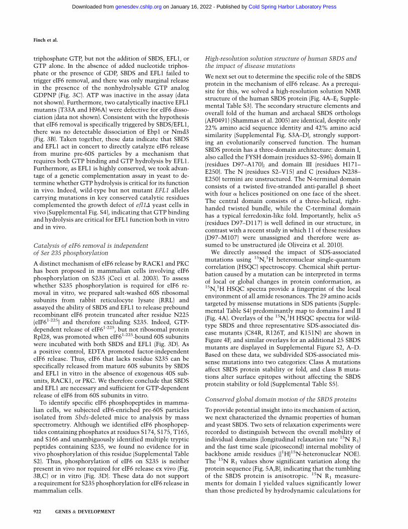
Conserved global domain motion of the SBDS proteins
To provide potential insight into its mechanism of action,we next characterized the dynamic properties of humanand yeast SBDS. Two sets of relaxation experiments wererecorded to distinguish between the overall mobility ofindividual domains (longitudinal relaxation rate 15N R1)and the fast time scale (picosecond) internal mobility ofbackbone amide residues ({1H}15N-heteronuclear NOE).The 15N R1 values show significant variation along theprotein sequence (Fig. 5A,B), indicating that the tumblingof the SBDS protein is anisotropic. 15N R1 measure-ments for domain I yielded values significantly lowerthan those predicted by hydrodynamic calculations for
Finch et al.
922 GENES & DEVELOPMENT
Cold Spring Harbor Laboratory Press on January 16, 2022 - Published by genesdev.cshlp.orgDownloaded from

an independently mobile domain, but were higher thanthe values anticipated for a completely rigid module. Thesedata indicate that domain I tumbles largely, but notcompletely, independently of domains II and III (Figure 5B).
Interestingly, the transition point or hinge betweenregions of the SBDS molecule with differing global motionis centered within the N terminus of helix a5 (residueT102), which has a high degree of internal flexibility(Fig. 5C,D), suggesting that it may convert rapidly be-tween a fully folded and partially unfolded conforma-tion. Strikingly, comparison of the backbone relaxationmeasurements between humans and yeast reveals thatthe dynamic properties of the SBDS proteins are highlyconserved (Fig. 5A,C), although the transition point(residue Q94) within the yeast protein is shifted eightresidues toward the N terminus. These data indicate thatdomain I of SBDS can sample a variety of positions andpotentially propagate conformational change. We consid-
ered the dynamic mobility of SBDS to be reminiscent ofthe interdomain motion of bacterial ribosome-recyclingfactor (RRF) that is critical for the cooperative interactionwith EF-G in the disassembly of post-termination ribo-somes (Yoshida et al. 2003; Gao et al. 2007; Savelsberghet al. 2009), raising the possibility that the key role ofSBDS might be to tightly couple GTP-dependent confor-mational change in EFL1 (an EF-G homolog) to the releaseof eIF6.
SBDS stimulates 60S-dependent GTP hydrolysisby EFL1
To address this hypothesis, we first asked whether SBDSmight function as an adaptor that recruits EFL1 to thepre-60S subunit. However, we found that EFL1 and SBDScan bind independently to 60S ribosomal subunits in vitro,with no evidence of significant binding cooperativity in
Figure 4. Solution structure of human SBDS andimpact of SDS-associated mutations. (A) Ribbonrepresentation of the lowest-energy human SBDSNMR structure surrounded by the solvent-accessi-ble surface (radius probe 1.4 A), prepared using theprogram PyMOL (http://www.pymol.org). Domain Iis colored red, domain II is colored yellow, domain IIIis colored green, and loops are colored gray. Theindicated SDS-associated mutations modify surfaceepitopes (blue spheres) or protein stability (pinkspheres). (B–D) Overlay of the backbone atoms ofthe 20 lowest-energy structures from domain I (A16–T89) (A), domain II (D97–K164) (B), and domain III(H171–L237) (C). The 20 conformers were overlaidusing Clusterpose (Diamond 1995). (E) Representa-tion of the electrostatic surface potential of thehuman SBDS protein, calculated by the programAPBS (Baker et al. 2001) and colored using a linearcolor ramp from �15 kT (red) to +15 kT (blue). TheSDS-associated mutation K151N is indicated. (F)Overlays of the 1H,15N HSQC spectra for wild-type(blue) and three SDS-associated mutants (C84R,R126T, and K151N) (red). Arrows indicate peaksvisible in the wild-type spectrum but not in theC84R mutant. See also Supplemental Figures S5 andS6 and Supplemental Tables S3–S5.
Molecular mechanism of eIF6 release
GENES & DEVELOPMENT 923
Cold Spring Harbor Laboratory Press on January 16, 2022 - Published by genesdev.cshlp.orgDownloaded from
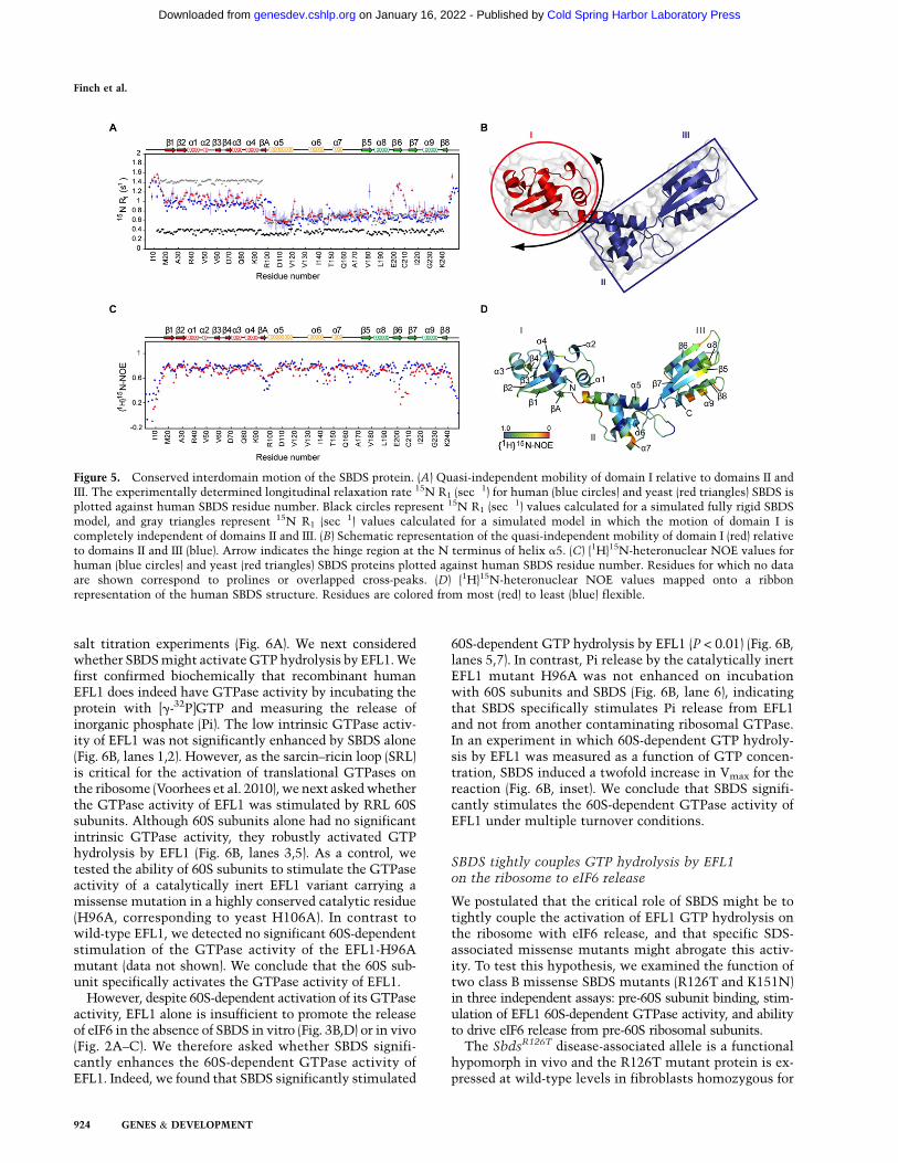
salt titration experiments (Fig. 6A). We next consideredwhether SBDS might activate GTP hydrolysis by EFL1. Wefirst confirmed biochemically that recombinant humanEFL1 does indeed have GTPase activity by incubating theprotein with [g-32P]GTP and measuring the release ofinorganic phosphate (Pi). The low intrinsic GTPase activ-ity of EFL1 was not significantly enhanced by SBDS alone(Fig. 6B, lanes 1,2). However, as the sarcin–ricin loop (SRL)is critical for the activation of translational GTPases onthe ribosome (Voorhees et al. 2010), we next asked whetherthe GTPase activity of EFL1 was stimulated by RRL 60Ssubunits. Although 60S subunits alone had no significantintrinsic GTPase activity, they robustly activated GTPhydrolysis by EFL1 (Fig. 6B, lanes 3,5). As a control, wetested the ability of 60S subunits to stimulate the GTPaseactivity of a catalytically inert EFL1 variant carrying amissense mutation in a highly conserved catalytic residue(H96A, corresponding to yeast H106A). In contrast towild-type EFL1, we detected no significant 60S-dependentstimulation of the GTPase activity of the EFL1-H96Amutant (data not shown). We conclude that the 60S sub-unit specifically activates the GTPase activity of EFL1.
However, despite 60S-dependent activation of its GTPaseactivity, EFL1 alone is insufficient to promote the releaseof eIF6 in the absence of SBDS in vitro (Fig. 3B,D) or in vivo(Fig. 2A–C). We therefore asked whether SBDS signifi-cantly enhances the 60S-dependent GTPase activity ofEFL1. Indeed, we found that SBDS significantly stimulated
60S-dependent GTP hydrolysis by EFL1 (P < 0.01) (Fig. 6B,lanes 5,7). In contrast, Pi release by the catalytically inertEFL1 mutant H96A was not enhanced on incubationwith 60S subunits and SBDS (Fig. 6B, lane 6), indicatingthat SBDS specifically stimulates Pi release from EFL1and not from another contaminating ribosomal GTPase.In an experiment in which 60S-dependent GTP hydroly-sis by EFL1 was measured as a function of GTP concen-tration, SBDS induced a twofold increase in Vmax for thereaction (Fig. 6B, inset). We conclude that SBDS signifi-cantly stimulates the 60S-dependent GTPase activity ofEFL1 under multiple turnover conditions.
SBDS tightly couples GTP hydrolysis by EFL1on the ribosome to eIF6 release
We postulated that the critical role of SBDS might be totightly couple the activation of EFL1 GTP hydrolysis onthe ribosome with eIF6 release, and that specific SDS-associated missense mutants might abrogate this activ-ity. To test this hypothesis, we examined the function oftwo class B missense SBDS mutants (R126T and K151N)in three independent assays: pre-60S subunit binding, stim-ulation of EFL1 60S-dependent GTPase activity, and abilityto drive eIF6 release from pre-60S ribosomal subunits.
The SbdsR126T disease-associated allele is a functionalhypomorph in vivo and the R126T mutant protein is ex-pressed at wild-type levels in fibroblasts homozygous for
Figure 5. Conserved interdomain motion of the SBDS protein. (A) Quasi-independent mobility of domain I relative to domains II andIII. The experimentally determined longitudinal relaxation rate 15N R1 (sec�1) for human (blue circles) and yeast (red triangles) SBDS isplotted against human SBDS residue number. Black circles represent 15N R1 (sec�1) values calculated for a simulated fully rigid SBDSmodel, and gray triangles represent 15N R1 (sec�1) values calculated for a simulated model in which the motion of domain I iscompletely independent of domains II and III. (B) Schematic representation of the quasi-independent mobility of domain I (red) relativeto domains II and III (blue). Arrow indicates the hinge region at the N terminus of helix a5. (C) {1H}15N-heteronuclear NOE values forhuman (blue circles) and yeast (red triangles) SBDS proteins plotted against human SBDS residue number. Residues for which no dataare shown correspond to prolines or overlapped cross-peaks. (D) {1H}15N-heteronuclear NOE values mapped onto a ribbonrepresentation of the human SBDS structure. Residues are colored from most (red) to least (blue) flexible.
Finch et al.
924 GENES & DEVELOPMENT
Cold Spring Harbor Laboratory Press on January 16, 2022 - Published by genesdev.cshlp.orgDownloaded from
the SbdsR126T allele (Ball et al. 2009), indicating that it isfunctionally defective. Residue R126 lies on the surface ofthe a5–a6 loop at the interface between domains II and III(Fig. 4A). The 15N,1H HSQC spectrum of the R126Tmutant displayed only local chemical shift perturbation
of residues within loops a5–a6 and a7–b6 (Fig. 4F),indicating that it is has the same fold as wild-type SBDS.Although the R126T mutant bound efficiently to pre-60Ssubunits (Supplemental Fig. S7), it did not significantlyenhance the 60S-dependent GTPase activity of EFL1compared with wild-type SBDS (Fig. 6B, lanes 5,7,8).Furthermore, R126T was strongly defective comparedwith wild-type SBDS in triggering eIF6 release in concertwith EFL1 and GTP (Fig. 6C). These data are consistentwith the hypothesis that the primary defect in SDS isimpaired release of eIF6 from pre-60S ribosomes.
Residue K151 is conserved between yeast and humanSBDS and localizes to a basic patch on helix a7 on thesurface of domain II (Fig. 4A; Supplemental Fig. S5). The15N,1H HSQC spectrum of the K151N mutant displayedno significant chemical shift perturbation compared withwild-type SBDS (Fig. 4F). Although K151N bound effi-ciently to pre-60S ribosomal subunits (Supplemental Fig.S7), it was strongly defective compared with wild-typeSBDS in triggering eIF6 dissociation with EFL1 and GTP(Fig. 6C). However, in contrast to R126T, the K151Nmutant significantly stimulated the 60S-dependentGTPase activity of EFL1 (P < 0.01), and the degree ofstimulation was not significantly different comparedwith wild type (Fig. 6B, lane 9). Thus, the K151N mutantis defective in eIF6 release despite significantly stimulat-ing 60S-dependent GTP hydrolysis by EFL1.
Finally, we took advantage of the conservation of residueK151 in yeast SDO1 to examine the effect of the K151Nmutation on SDO1 function in vivo by genetic comple-mentation. As the sole source of SDO1, the sdo1-K151Nallele failed to complement the fitness defect of sdo1D
yeast cells despite appropriate expression (data not shown),indicating that residue K151 is critical for SDO1 func-tion in vivo. Thus, we conclude that, although SBDS cansignificantly stimulate the 60S-dependent GTPase activityof EFL1 in multiple turnover conditions, its essential roleis to tightly couple the activation of EFL1 GTP hydrolysison the ribosome to eIF6 release.
Discussion
Our study describes the establishment of an ex vivosystem that reconstitutes a late cytoplasmic step in thematuration of pre-60S ribosomal subunits. This systemallows us to conclude that (1) human SBDS is an essentialcofactor for the EFL1 GTPase, and together they co-operate to directly catalyze the release of eIF6 frommammalian pre-60S ribosomal subunits; (2) the mecha-nism of eIF6 release requires GTP binding and hydrolysisby EFL1, but is independent of eIF6 phosphorylation onSer 235; (3) SBDS stimulates 60S-dependent GTP hydro-lysis by EFL1, but its essential role is to tightly couplethe activation of EFL1 GTP hydrolysis on the ribosometo eIF6 release; and (4) a conserved lysine (K151) that ismutated in SDS and maps to the central three-helicalbundle of the SBDS protein is required for cooperativitywith EFL1. Taken together with the comprehensivehigh-resolution NMR structural and dynamic analysisof both human and yeast SBDS proteins reported here,
Figure 6. SBDS tightly couples activation of EFL1 GTP hydro-lysis to eIF6 release. (A) SBDS and EFL1 bind independently to60S subunits. SBDS and EFL1 were bound to RRL 60S subunitsover the indicated range of KCl concentrations and pelletedthrough 30% (w/v) sucrose cushions. Bound SBDS, EFL1, andRpl28 were visualized by immunoblotting. (B) SBDS stimulates60S-dependent GTP hydrolysis by EFL1. 60S-dependent GTPaseactivity of human wild-type EFL1 or a catalytically inactivemutant (H96A) was measured in the presence of wild-type orvariant (R126T and K151N) SBDS. The experiment was repeatedthree times and the average values with SD are presented. (Inset)Dose response curve for 60S-dependent EFL1 catalytic activityas a function of GTP concentration in the presence (black line)or absence (red line) of human SBDS. Measurements wereperformed in duplicate. (C) SDS-associated SBDS variants aredefective in eIF6 release. Pre-60S subunits were incubated withGTP, EFL1, and either wild-type or variant (R126T or K151N)SBDS proteins. Following sucrose pelleting, ‘‘bound’’ and ‘‘free’’fractions were immunoblotted to visualize eIF6 and Ebp1. Seealso Supplemental Figure S7.
Molecular mechanism of eIF6 release
GENES & DEVELOPMENT 925
Cold Spring Harbor Laboratory Press on January 16, 2022 - Published by genesdev.cshlp.orgDownloaded from
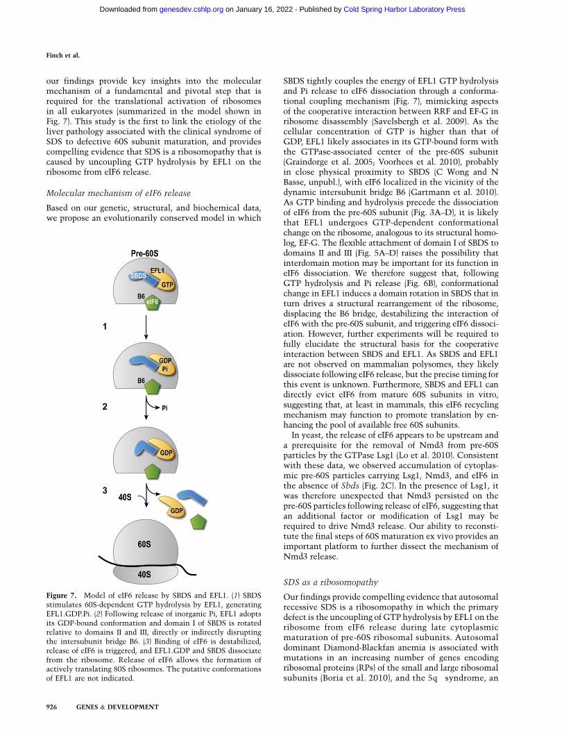
our findings provide key insights into the molecularmechanism of a fundamental and pivotal step that isrequired for the translational activation of ribosomesin all eukaryotes (summarized in the model shown inFig. 7). This study is the first to link the etiology of theliver pathology associated with the clinical syndrome ofSDS to defective 60S subunit maturation, and providescompelling evidence that SDS is a ribosomopathy that iscaused by uncoupling GTP hydrolysis by EFL1 on theribosome from eIF6 release.
Molecular mechanism of eIF6 release
Based on our genetic, structural, and biochemical data,we propose an evolutionarily conserved model in which
SBDS tightly couples the energy of EFL1 GTP hydrolysisand Pi release to eIF6 dissociation through a conforma-tional coupling mechanism (Fig. 7), mimicking aspectsof the cooperative interaction between RRF and EF-G inribosome disassembly (Savelsbergh et al. 2009). As thecellular concentration of GTP is higher than that ofGDP, EFL1 likely associates in its GTP-bound form withthe GTPase-associated center of the pre-60S subunit(Graindorge et al. 2005; Voorhees et al. 2010), probablyin close physical proximity to SBDS (C Wong and NBasse, unpubl.), with eIF6 localized in the vicinity of thedynamic intersubunit bridge B6 (Gartmann et al. 2010).As GTP binding and hydrolysis precede the dissociationof eIF6 from the pre-60S subunit (Fig. 3A–D), it is likelythat EFL1 undergoes GTP-dependent conformationalchange on the ribosome, analogous to its structural homo-log, EF-G. The flexible attachment of domain I of SBDS todomains II and III (Fig. 5A–D) raises the possibility thatinterdomain motion may be important for its function ineIF6 dissociation. We therefore suggest that, followingGTP hydrolysis and Pi release (Fig. 6B), conformationalchange in EFL1 induces a domain rotation in SBDS that inturn drives a structural rearrangement of the ribosome,displacing the B6 bridge, destabilizing the interaction ofeIF6 with the pre-60S subunit, and triggering eIF6 dissoci-ation. However, further experiments will be required tofully elucidate the structural basis for the cooperativeinteraction between SBDS and EFL1. As SBDS and EFL1are not observed on mammalian polysomes, they likelydissociate following eIF6 release, but the precise timing forthis event is unknown. Furthermore, SBDS and EFL1 candirectly evict eIF6 from mature 60S subunits in vitro,suggesting that, at least in mammals, this eIF6 recyclingmechanism may function to promote translation by en-hancing the pool of available free 60S subunits.
In yeast, the release of eIF6 appears to be upstream anda prerequisite for the removal of Nmd3 from pre-60Sparticles by the GTPase Lsg1 (Lo et al. 2010). Consistentwith these data, we observed accumulation of cytoplas-mic pre-60S particles carrying Lsg1, Nmd3, and eIF6 inthe absence of Sbds (Fig. 2C). In the presence of Lsg1, itwas therefore unexpected that Nmd3 persisted on thepre-60S particles following release of eIF6, suggesting thatan additional factor or modification of Lsg1 may berequired to drive Nmd3 release. Our ability to reconsti-tute the final steps of 60S maturation ex vivo provides animportant platform to further dissect the mechanism ofNmd3 release.
SDS as a ribosomopathy
Our findings provide compelling evidence that autosomalrecessive SDS is a ribosomopathy in which the primarydefect is the uncoupling of GTP hydrolysis by EFL1 on theribosome from eIF6 release during late cytoplasmicmaturation of pre-60S ribosomal subunits. Autosomaldominant Diamond-Blackfan anemia is associated withmutations in an increasing number of genes encodingribosomal proteins (RPs) of the small and large ribosomalsubunits (Boria et al. 2010), and the 5q� syndrome, an
Figure 7. Model of eIF6 release by SBDS and EFL1. (1) SBDSstimulates 60S-dependent GTP hydrolysis by EFL1, generatingEFL1.GDP.Pi. (2) Following release of inorganic Pi, EFL1 adoptsits GDP-bound conformation and domain I of SBDS is rotatedrelative to domains II and III, directly or indirectly disruptingthe intersubunit bridge B6. (3) Binding of eIF6 is destabilized,release of eIF6 is triggered, and EFL1.GDP and SBDS dissociatefrom the ribosome. Release of eIF6 allows the formation ofactively translating 80S ribosomes. The putative conformationsof EFL1 are not indicated.
Finch et al.
926 GENES & DEVELOPMENT
Cold Spring Harbor Laboratory Press on January 16, 2022 - Published by genesdev.cshlp.orgDownloaded from

acquired subtype of MDS, is linked to haploinsufficiencyof the RPS14 gene (Ebert et al. 2008; Barlow et al. 2010a).Thus, the primary defect underlying SDS is distinct fromthe other ribosomopathies associated with hematologi-cal disease. We suggest that the diverse alternate func-tions proposed for SBDS in mammalian cells (Wesselset al. 2006; Austin et al. 2008; Rujkijyanont et al. 2008; Ballet al. 2009; Leung et al. 2010) are downstream secondaryconsequences of the primary defect in 60S ribosomal sub-unit maturation.
How do we explain the SDS phenotype? It seems likelyto reflect, at least in part, the sensitivities of specific celltypes to cellular stress responses during development thatresult from defective ribosome biogenesis. Indeed, recentin vivo data have established the RP–Mdm2 interactionas a p53 stress signaling pathway that is activated by aber-rant ribosome biogenesis and is essential to protect againstoncogenic c-MYC-induced tumorigenesis (Macias et al.2010). Furthermore, the p53 dependence of the pathologiesassociated with defective ribosome biogenesis is supportedby genetic data in a number of animal models (Barlow et al.2010b).
The dramatic disruption of nucleolar architecture inSbds-deleted livers is consistent with the hypothesis thatnucleolar stress may contribute to the observed hepato-cyte defects following complete deletion of Sbds. How-ever, as SDS is a consequence of partial, not complete,loss of SBDS function, further work will be required toclarify the importance of nucleolar stress responses to theclinical SDS phenotype. Nevertheless, as abnormal liverfunction tests and hepatomegaly are recognized featuresof SDS at an early age (Toiviainen-Salo et al. 2009), thisstudy directly links the etiology of a pathological abnor-mality associated with the clinical syndrome of SDS todefective 60S subunit maturation and a ribosomal subunit-joining defect.
Our findings demonstrate the feasibility and power ofbiochemically recapitulating late maturation of mamma-lian ribosomal subunits ex vivo to elucidate the underly-ing molecular mechanisms. Finally, this study stronglysupports the hypothesis that small molecules that mimicthe effects of TIF6 suppressor mutations in yeast mayhave utility in the treatment of SDS and possibly otherforms of bone marrow failure.
Materials and methods
Protein expression and purification
Recombinant human eIF6 (NP_852133) and SBDS (NP_057122)were expressed and purified from Escherichia coli, and humanEFL1 (NP_078856) was expressed and purified from S. cerevisiae.Recombinant proteins were purified by Ni-NTA affinity and gelfiltration chromatography. See the Supplemental Material fordetailed protocols.
NMR spectroscopy
NMR spectra were assigned and the solution structure wasdetermined using standard techniques (Wuthrich 1986; Bax1994).
Mouse gene targeting
The mouse genomic Sbds locus was PCR-amplified from mouse129 DNA. The targeting vector pMCTV1 was generated suchthat exposure to Cre recombinase results in deletion of exon 2and the selection cassette. All animal experiments were under-taken with the approval of the UK Home Office.
Sucrose density gradients
Frozen liver (typically 30 mg) was homogenized with a pestle indetergent lysis buffer A (10 mM Tris-HCl at pH 7.4, 10 mMNaCl, 1.5 mM MgCl2, 0.5% [v/v] Triton X-100, 0.5% [w/v]deoxycholate, 1% [v/v] Tween 20, 100 mg/mL cycloheximide)with complete EDTA-free protease inhibitors (Roche) and 0.5U/mL RNase inhibitor (Promega) and incubated for 10 min onice. Lysates were cleared in a microfuge. Equal amounts (typi-cally 10–20 A254 U) were applied to a 10%–50% (w/v) sucrosegradient in 30 mL of buffer B (10 mM Tris-HCl at pH 7.4, 75 mMKCl, 1.5 mM MgCl2) and centrifuged (Beckmann SW32 rotor) at32,000 rpm for 2 h and 46 min at 4°C. For ribosome profiles, liverlysates were prepared in buffer A (without MgCl2 or cyclohex-imide) in the presence of 5 mM EDTA; MgCl2 was removed fromthe sucrose gradient buffer B.
Samples were unloaded using a Brandel gradient fractionator,the polysome profiles were detected using a UV monitor (UV-1,Pharmacia) at A254, and 1-mL fractions were collected. Theelectronic outputs of the UV-1 monitor and fraction collectorwere fed into a Labjack U3-LV data acquisition device with aLJTick-InAmp preamplifier. Proteins were precipitated from su-crose gradient fractions with 10% (v/v) trichloroacetic acid (TCA),separated on SDS-PAGE gels and transferred to PVDF membranesfor immunoblotting.
Preparation of liver extracts and immunoblotting
Whole-liver extracts were prepared by homogenizing 10 mg ofliver in RIPA buffer C (20 mM HEPES at pH 7.4, 20 mMb-glycerophosphate, 10 mM NaF, 0.5 mM EDTA, 0.5 mM EGTA,0.2 M NaCl, 1% [v/v] Nonidet P-40, 0.5% [w/v] sodium deoxy-cholate, 0.1% [w/v] SDS) with complete EDTA-free proteaseinhibitors (Roche) using a pellet pestle (Sigma) and leaving for10 min on ice with occasional vortexing. Samples were clearedin a microfuge and normalized for protein concentration usinga BCA protein assay kit (Pierce). Samples were fractionated usingNE-PER nuclear and cytoplasmic extraction reagents (Pierce)according to the manufacturer’s instructions. All fractions werenormalized against the protein concentration of the cytoplasmicfractions.
eIF6 release assay
The reaction mixture (100 mL) contained 50 mL of sucrosegradient-purified 60S subunits (;0.5–1 pmol); 5 pmol of SBDS;5 pmol of EFL1; 100 mM purified GTP, GDP, or GDPNP (Sigma)in buffer B with complete EDTA-free protease inhibitors (Roche);and 0.5 U/mL RNasin (Promega). GDP and GTP were purified asin Wilden et al. (2006).
Alternatively, 5 pmol of 60S subunits was purified from RRL(Pisarev et al. 2007) and incubated with an excess of recombinanteIF61-225 for 20 min on ice, and free eIF6 was removed usinga 100-kDa Microcon centrifugal filter unit (Sigma). The eIF6-loaded RRL was then used as a substrate in the eIF6 release assay.Reaction mixtures were incubated for 10 min at 37°C, layeredonto a 150-mL 30% (w/v) sucrose cushion in buffer B, andcentrifuged (Beckmann TLA120.1 rotor) at 120,000 rpm for 30
Molecular mechanism of eIF6 release
GENES & DEVELOPMENT 927
Cold Spring Harbor Laboratory Press on January 16, 2022 - Published by genesdev.cshlp.orgDownloaded from
min at 4°C. Two-hundred microliters of supernatant (‘‘free’’)was removed, and the remaining 50 mL was resuspended(‘‘bound’’) in 150 mL of buffer B. Samples were TCA-precipitatedfor immunoblotting.
RRL 60S-binding assay
The reaction mixture (100 mL) contained 5 pmol of 60S subunitspurified from RRL (Pisarev et al. 2007) with 25 pmol each ofSBDS and EFL1 in buffer B. Additional KCl was added wherespecified. Samples were subsequently processed as above for eIF6release.
60S-dependent GTP hydrolysis by EFL1
The reaction mixture (50 mL) contained 5 nmol of [g-32P]GTP (6Ci/mmol; Perkin-Elmer), 5 pmol of RRL 60S subunits, 6.3 pmol ofSBDS, and 40 pmol of EFL1 in buffer D (50 mM Tris at pH 7.4, 2mM MgCl2, 150 mM NaCl, 2 mM DTT). The reaction mixturewas incubated for 30 min at 37°C. An aliquot (40 mL) of thereaction mix was added to 360 mL of chilled charcoal slurry(5% [v/v] Norit in 50 mM NaH2PO4, at pH 3.0) and centrifugedat 13,000 rpm for 10 min. The amount of radioactivity in 100 mLof supernatant was quantified by liquid scintillation counting.
Yeast strains and plasmids
S. cerevisiae strains used in this study are listed in SupplementalTable S6 and primers are listed in Supplemental Table S7.Deletion strains were generated by homologous recombinationusing appropriate PCR products to transform strain Y5538. Thecoding sequence for wild-type EFL1 was PCR-amplified fromyeast genomic DNA and cloned into pYCT/C2 to generate plasmidpEFL1. Plasmids pEFL1-T33A, pEFL1-D102A, pEFL1-H106A, andpEFL1-H106I, pEFL1-D159A, pEFL1-W240E were generated byQuickChange site-directed mutagenesis (Stratagene).
Suppressor clone isolation
Suppressor clone isolation was performed as described (Menneet al. 2007).
Synthetic genetic array analysis and randomsporulation assays
SGA and random sporulation analysis was performed asdescribed (Tong and Boone 2006; Baryshnikova et al. 2010;Costanzo et al. 2010). MATa efl1DTNatMX4 TIF6-R61G andMATa sdo1DTNatMX4 TIF6-R61G query strains harboring SGAmarkers and haploid-specific reporters were crossed to an arraycontaining the set of ;5000 viable deletion mutant strains.Nourseothricin was obtained from Werner BioAgents.
Peptide separation, mass spectrometry, and database analysis
For peptide separation, mass spectrometry, and database analysisused in this study, see the Supplemental Material.
Antibodies
For antibodies used in this study, see the Supplemental Material.
Statistical analysis
Student’s t-test was used to determine significant differences. P <
0.01 was considered significant.
Accession numbers
Coordinates for the human SBDS structure have been depositedin the Protein Data Bank (ID 2L9N), and the 1H, 15N, and 13Cchemical shifts have been deposited in the BioMagResBankdatabase (accession code 17479).
Acknowledgments
We thank the European NMR Large-Scale Facility (Utrecht,Holland) and E. Reynaud (University College Dublin) for Lsg1antibodies; M. Tchorzewski (Maria Curie-Skodowska Univer-sity) for P0 antisera; J. Sale for critical comments on the manu-script; A. Newman for strain BCY123; R. Mittal for assistancewith GTPase assays; Mark Allen, Mark Bycroft, and TrevorRutherford for critical advice and assistance with NMR spec-troscopy; M. Schwarz (St. Mary’s Hospital, Manchester) forcommunicating unpublished data; the LMB animal facility staff;and L. Easton and P. Lukavsky for RRL ribosomes. Leukemia andLymphoma Research, the Association for International CancerResearch, the Sylvia Aitkin Trust, the Medical Research Coun-cil, the MDS Foundation, the Leukemia and Lymphoma Societyof America, Shwachman-Diamond Support UK, Ted’s Gang, theTesni Parry Memorial Fund, and the Cambridge NIHR Bio-medical Research Center supported this work. M.J.A. is sup-ported by Cancer Research UK. J.D. and C.B.-C. are supportedby the Institut de Veille Sanitaire, Inserm, the AssociationLaurette Fugain, and the Association Sportive de Saint QuentinFallavier. C.B. is supported by the Canadian Institutes of HealthResearch (CIHR) (MOP-102629), the National Institutes ofHealth (NIH) (1R01HG005853-01), and the Ontario ResearchFund (ORF-GL2) (GL2-01-22).
References
Austin KM, Gupta ML, Coats SA, Tulpule A, Mostoslavsky G,Balazs AB, Mulligan RC, Daley G, Pellman D, Shimamura A.2008. Mitotic spindle destabilization and genomic instabilityin Shwachman-Diamond syndrome. J Clin Invest 118: 1511–1518.
Baker NA, Sept D, Joseph S, Holst MJ, McCammon JA. 2001.Electrostatics of nanosystems: application to microtubulesand the ribosome. Proc Natl Acad Sci 98: 10037–10041.
Ball HL, Zhang B, Riches JJ, Gandhi R, Li J, Rommens JM, MyersJS. 2009. Shwachman-Bodian Diamond syndrome is a multi-functional protein implicated in cellular stress responses.Hum Mol Genet 18: 3684–3695.
Barlow JL, Drynan LF, Hewett DR, Holmes LR, Lorenzo-AbaldeS, Lane AL, Jolin HE, Pannell R, Middleton AJ, Wong SH,et al. 2010a. A p53-dependent mechanism underlies macro-cytic anemia in a mouse model of human 5q� syndrome. Nat
Med 16: 59–66.Barlow JL, Drynan LF, Trim NL, Erber WN, Warren AJ, McKenzie
AN. 2010b. New insights into 5q� syndrome as a ribosomop-athy. Cell Cycle 9: 4286–4293.
Baryshnikova A, Costanzo M, Kim Y, Ding H, Koh J, Toufighi K,Youn JY, Ou J, San Luis BJ, Bandyopadhyay S, et al. 2010.Quantitative analysis of fitness and genetic interactions inyeast on a genome scale. Nat Methods 7: 1017–1024.
Basu U, Si K, Warner JR, Maitra U. 2001. The Saccharomycescerevisiae TIF6 gene encoding translation initiation factor 6is required for 60S ribosomal subunit biogenesis. Mol Cell
Biol 21: 1453–1462.Bax A. 1994. Multidimensional nuclear-magnetic-resonance
methods for protein studies. Curr Opin Struct Biol 4: 738–744.
Finch et al.
928 GENES & DEVELOPMENT
Cold Spring Harbor Laboratory Press on January 16, 2022 - Published by genesdev.cshlp.orgDownloaded from

Becam AM, Nasr F, Racki WJ, Zagulski M, Herbert CJ. 2001.Ria1p (Ynl163c), a protein similar to elongation factors 2, isinvolved in the biogenesis of the 60S subunit of the ribosomein Saccharomyces cerevisiae. Mol Genet Genomics 266:454–462.
Boocock GR, Morrison JA, Popovic M, Richards N, Ellis L, DuriePR, Rommens JM. 2003. Mutations in SBDS are associatedwith Shwachman-Diamond syndrome. Nat Genet 33: 97–101.
Boria I, Garelli E, Gazda HT, Aspesi A, Quarello P, Pavesi E,Ferrante D, Meerpohl JJ, Kartal M, Da Costa L, et al. 2010.The ribosomal basis of Diamond-Blackfan anemia: mutationand database update. Hum Mutat 31: 1269–1279.
Ceci M, Gaviraghi C, Gorrini C, Sala LA, Offenhauser N,Marchisio PC, Biffo S. 2003. Release of eIF6 (p27BBP) fromthe 60S subunit allows 80S ribosome assembly. Nature 426:579–584.
Costanzo M, Baryshnikova A, Bellay J, Kim Y, Spear ED, SevierCS, Ding H, Koh JL, Toufighi K, Mostafavi S, et al. 2010. Thegenetic landscape of a cell. Science 327: 425–431.
de Oliveira JF, Sforca ML, Blumenschein TM, Goldfeder MB,Guimaraes BG, Oliveira CC, Zanchin NI, Zeri AC. 2010.Structure, dynamics, and RNA interaction analysis of thehuman SBDS protein. J Mol Biol 396: 1053–1069.
Diamond R. 1995. Coordinate-based cluster analysis. Acta
Crystallogr D Biol Crystallogr 51: 127–135.Donadieu J, Leblanc T, Bader Meunier B, Barkaoui M, Fenneteau
O, Bertrand Y, Maier-Redelsperger M, Micheau M, StephanJL, Phillipe N, et al. 2005. Analysis of risk factors formyelodysplasias, leukemias and death from infection amongpatients with congenital neutropenia. Experience of theFrench Severe Chronic Neutropenia Study Group. Haema-
tologica 90: 45–53.Ebert BL, Pretz J, Bosco J, Chang CY, Tamayo P, Galili N, Raza
A, Root DE, Attar E, Ellis SR, et al. 2008. Identification ofRPS14 as a 5q� syndrome gene by RNA interference screen.Nature 451: 335–339.
Ganapathi KA, Austin KM, Lee CS, Dias A, Malsch MM, ReedR, Shimamura A. 2007. The human Shwachman-Diamondsyndrome protein, SBDS, associates with ribosomal RNA.Blood 110: 1458–1465.
Gao N, Zavialov AV, Ehrenberg M, Frank J. 2007. Specificinteraction between EF-G and RRF and its implication forGTP-dependent ribosome splitting into subunits. J Mol Biol
374: 1345–1358.Gartmann M, Blau M, Armache JP, Mielke T, Topf M, Beckmann
R. 2010. Mechanism of eIF6-mediated inhibition of ribosomalsubunit joining. J Biol Chem 285: 14848–14851.
Graindorge JS, Rousselle JC, Senger B, Lenormand P, Namane A,Lacroute F, Fasiolo F. 2005. Deletion of EFL1 results inheterogeneity of the 60 S GTPase-associated rRNA confor-mation. J Mol Biol 352: 355–369.
Leung R, Cuddy K, Wang Y, Rommens J, Glogauer M. 2010. Sbdsis required for Rac2-mediated monocyte migration andsignaling downstream of RANK during osteoclastogenesis.Blood 117: 2044–2053.
Lo KY, Li Z, Bussiere C, Bresson S, Marcotte EM, Johnson AW.2010. Defining the pathway of cytoplasmic maturation of the60S ribosomal subunit. Mol Cell 39: 196–208.
Macias E, Jin A, Deisenroth C, Bhat K, Mao H, Lindstrom MS,Zhang Y. 2010. An ARF-independent c-MYC-activated tu-mor suppression pathway mediated by ribosomal protein-Mdm2 Interaction. Cancer Cell 18: 231–243.
Menne TF, Goyenechea B, Sanchez-Puig N, Wong CC, TonkinLM, Ancliff PJ, Brost RL, Costanzo M, Boone C, Warren AJ.2007. The Shwachman-Bodian-Diamond syndrome protein
mediates translational activation of ribosomes in yeast. Nat
Genet 39: 486–495.Pisarev AV, Unbehaun A, Hellen CU, Pestova TV. 2007. Assem-
bly and analysis of eukaryotic translation initiation com-plexes. Methods Enzymol 430: 147–177.
Rujkijyanont P, Watanabe K, Ambekar C, Wang H, Schimmer A,Beyene J, Dror Y. 2008. SBDS-deficient cells undergo accel-erated apoptosis through the Fas-pathway. Haematologica
93: 363–371.Savelsbergh A, Rodnina MV, Wintermeyer W. 2009. Distinct
functions of elongation factor G in ribosome recycling andtranslocation. RNA 15: 772–780.
Schwenk F, Baron U, Rajewsky K. 1995. A cre-transgenic mousestrain for the ubiquitous deletion of loxP-flanked genesegments including deletion in germ cells. Nucleic Acids
Res 23: 5080–5081.Senger B, Lafontaine DL, Graindorge JS, Gadal O, Camasses A,
Sanni A, Garnier JM, Breitenbach M, Hurt E, Fasiolo F. 2001.The nucle(ol)ar Tif6p and Efl1p are required for a latecytoplasmic step of ribosome synthesis. Mol Cell 8: 1363–1373.
Sengupta J, Bussiere C, Pallesen J, West M, Johnson AW, Frank J.2010. Characterization of the nuclear export adaptor proteinNmd3 in association with the 60S ribosomal subunit. J CellBiol 189: 1079–1086.
Shammas C, Menne TF, Hilcenko C, Michell SR, Goyenechea B,Boocock GR, Durie PR, Rommens JM, Warren AJ. 2005.Structural and mutational analysis of the SBDS proteinfamily. Insight into the leukemia-associated Shwachman-Diamond syndrome. J Biol Chem 280: 19221–19229.
Toiviainen-Salo S, Durie PR, Numminen K, Heikkil P, MarttinenE, Savilahti E, Makitie O. 2009. The natural history ofShwachman-Diamond syndrome-associated liver disease fromchildhood to adulthood. J Pediatr 155: 807–811.e2. doi:10.1016/j.jpeds.2009.06.047.
Tong AH, Boone C. 2006. Synthetic genetic array analysis inSaccharomyces cerevisiae. Methods Mol Biol 313: 171–192.
Voorhees RM, Schmeing TM, Kelley AC, Ramakrishnan V.2010. The mechanism for activation of GTP hydrolysis onthe ribosome. Science 330: 835–838.
Wessels D, Srikantha T, Yi S, Kuhl S, Aravind L, Soll DR. 2006.The Shwachman-Bodian-Diamond syndrome gene encodesan RNA-binding protein that localizes to the pseudopod ofDictyostelium amoebae during chemotaxis. J Cell Sci 119:370–379.
Wilden B, Savelsbergh A, Rodnina MV, Wintermeyer W. 2006.Role and timing of GTP binding and hydrolysis duringEF-G-dependent tRNA translocation on the ribosome. Proc
Natl Acad Sci 103: 13670–13675.Wuthrich K. 1986. NMR of proteins and nucleic acid. John
Wiley & Sons, Inc., New York.Yoshida T, Oka S, Uchiyama S, Nakano H, Kawasaki T, Ohkubo
T, Kobayashi Y. 2003. Characteristic domain motion in theribosome recycling factor revealed by 15N NMR relaxationexperiments and molecular dynamics simulations. Biochem-
istry 42: 4101–4107.
Molecular mechanism of eIF6 release
GENES & DEVELOPMENT 929
Cold Spring Harbor Laboratory Press on January 16, 2022 - Published by genesdev.cshlp.orgDownloaded from

10.1101/gad.623011Access the most recent version at doi: 25:2011, Genes Dev.
Andrew J. Finch, Christine Hilcenko, Nicolas Basse, et al. causes Shwachman-Diamond syndromeUncoupling of GTP hydrolysis from eIF6 release on the ribosome
Material
Supplemental
http://genesdev.cshlp.org/content/suppl/2011/04/25/25.9.917.DC1
Related Content
Genes Dev. May , 2011 25: 898-900
Arlen W. Johnson and Steve R. Ellisribosomopathy?Of blood, bones, and ribosomes: is Swachman-Diamond syndrome a
References
http://genesdev.cshlp.org/content/25/9/917.full.html#related-urls
Articles cited in:
http://genesdev.cshlp.org/content/25/9/917.full.html#ref-list-1This article cites 38 articles, 13 of which can be accessed free at:
License
ServiceEmail Alerting
click here.right corner of the article or
Receive free email alerts when new articles cite this article - sign up in the box at the top
Copyright © 2011 by Cold Spring Harbor Laboratory Press
Cold Spring Harbor Laboratory Press on January 16, 2022 - Published by genesdev.cshlp.orgDownloaded from