unconventional use of intense pulsed light
TRANSCRIPT
Clinical StudyUnconventional Use of Intense Pulsed Light
D. Piccolo,1,2 D. Di Marcantonio,3 G. Crisman,4 G. Cannarozzo,2 M. Sannino,2
A. Chiricozzi,3,5 and S. Chimenti3
1 Department of Dermatology, University of L’Aquila, Via Vetoio, Coppito 2, 67100 L’Aquila, Italy2 Italian Society of Laser Dermatology (SILD), Via Nicolo dall’Arca 7, 70121 Bari, Italy3 Department of Dermatology, University of Rome, Tor Vergata, Italy4Department of Dermatology, University of Bologna, Italy5 Laboratory for Investigative Dermatology, The Rockefeller University, New York City, USA
Correspondence should be addressed to D. Piccolo; [email protected]
Received 6 February 2014; Revised 15 June 2014; Accepted 16 June 2014; Published 3 September 2014
Academic Editor: Silvia Moretti
Copyright © 2014 D. Piccolo et al. This is an open access article distributed under the Creative Commons Attribution License,which permits unrestricted use, distribution, and reproduction in any medium, provided the original work is properly cited.
According to the literature, intense pulsed light (IPL) represents a versatile tool in the treatment of some dermatological conditions(i.e., pigmentation disorders, hair removal, and acne), due to its wide range of wavelengths. The authors herein report on 58unconventional but effective uses of IPL in several cutaneous diseases, such as rosacea (10 cases), port-wine stain (PWS) (10 cases),disseminated porokeratosis (10 cases), pilonidal cyst (3 cases), seborrheic keratosis (10 cases), hypertrophic scar (5 cases) and keloidscar (5 cases), Becker’s nevus (2 cases), hidradenitis suppurativa (2 cases), and sarcoidosis (1 case). Our results should suggest thatIPL could represent a valid therapeutic support and option by providing excellent outcomes and low side effects, even thoughit should be underlined that the use and the effectiveness of IPL are strongly related to the operator’s experience (acquired byattempting at least one specific course on the use of IPL and one-year experience in a specialized centre). Moreover, the daily use ofthese devices will surely increase clinical experience and provide new information, thus enhancing long-term results and improvingIPL effectiveness.
1. Introduction
First introduced in 1990s, intense pulsed light (IPL) wasobtained by U.S. Food and Drug Administration (FDA)authorization in 1995 for the treatment of lower-limb telang-iectasias.
This polychromatic, noncoherent, and broad-spectrumpulsed light source (xenon lamp) is able to emit light ofa wavelength between 390 nm and 1200 nm [1]. Its basicprinciple consists in the absorption of photons by exoge-nous or endogenous chromophores within the skin; thistransfer of energy to the target structures generates heatand subsequent destruction of the target through a processcalled selective photothermolysis. The wavelength should beselected in dependence of the absorption peak of the targetchromophore and the pulse duration should last less than thethermal relaxation time. This limits the diffusion of heat anddamage to surrounding structures.
The main chromophores of the skin, such as haemo-globin, melanin, and water, have a broad absorption spec-trum. Through the use of a filter, available from 500 nm to755 nm, it is possible to select the wavelengths suitable forthe established treatment.The IPL’s pulse durationmay be setwithin a relatively wide range between 1 and 100milliseconds,depending on the selected device. In addition, a wide rangeof treatment parameters, including pulse sequence and pulsedelay time, can be customized, thus giving users greaterversatility and accuracy [1].
Versatility represents a significant advantage for expe-rienced dermatologists, but it could be a serious limit fornonexperienced physicians and for nonmedical staff sincean erroneous selection of the setting can cause serious sideeffects.
In daily practice, the application of a gel is necessary, aswell as direct contact between the handpiece and the skin,although this hinders the local immediate response.
Hindawi Publishing CorporationBioMed Research InternationalVolume 2014, Article ID 618206, 10 pageshttp://dx.doi.org/10.1155/2014/618206
2 BioMed Research International
Table 1: Clinical data.
Number of patients Gender Mean age (range)Rosacea 10 5 M, 5 F 51.6 (38–62)Port wine stain 10 7 M, 3 F 52.1 (8–52)Disseminated Porokeratosis 10 2 M, 8 F 58.3 (41–70)Pilonidal cyst 3 3 M 24.6 (18–34)Seborrheic keratosis 10 6 M, 4 F 61.7 (35–83)Hypertrophic scar/keloids 5/5 3 M, 2 F/2 M, 3 F 30.2 (21–37)/34.8 (27–43)Becker’s nevus 2 2 M 29 (26–32)Hidradenitis suppurativa 2 1 M, 1 F 32 (26–38)Sarcoidosis 1 1 F 26Total 58 32M, 26F 42.7 (8–83)
The combination of wavelength, pulse duration, delay,and fluence allows the use of IPL devices in the treatmentof several dermatological conditions, such as acne vulgaris,pigmentation disorders, vascular lesions, hirsutism, photo-damaged skin, scars and birthmarks, and melasma [2–4].
The authors herein suggest many unconventional uses ofIPL in the treatment of different dermatological conditions,such as rosacea (10 cases), port-wine stain (PWS) (10 cases),disseminated porokeratosis (10 cases), pilonidal cyst (3 cases),seborrheic keratosis (10 cases), hypertrophic scar (5 cases)and keloid scar (5 cases), Becker’s nevus (2 cases), hidradeni-tis suppurativa (2 cases), and sarcoidosis (1 case).
Acne rosacea or rosacea is a chronic dermatitis ofunknown aetiology, characterized by erythema, telangiec-tasias, papules and pustules [5, 6].
Port-wine stain is a common congenital vascular malfor-mation occurring in up to 25% of infants [7–10].
Disseminated porokeratosis is a localized alteration of ker-atinization. Clinically, one or more atrophic mainly asymp-tomatic and sometimesmildly itching plaques surrounded byan hyperkeratotic border (histologically defined as a cornoidlamella) are observed due to a rapid proliferation of atypicalkeratinocytes [11–14].
Pilonidal cyst, also known as pilonidal sinus or sacrococ-cygeal cyst (due to its frequent onset in this area), is a cystcontaining hair and skin debris [15–19].
Seborrheic keratosis is a benign skin lesion of the epider-mis, mainly localized on seborrheic areas, in particular, theface and trunk. The most common clinical presentation isa lesion with warty or squamous crusted surface of variablesize, coloured yellow-brown or dark-brown with blackishspecks, with soft consistency [20, 21].
Hypertrophic scars and keloids are a serious physical andpsychological dermatological condition for patients. Despitethe several studies performed on metabolisms and treatmentof wounds and scars, the exact pathogenesis of keloidsand hypertrophic scars remains unknown and this makestherapies even more complicated [22].
Becker’s nevus is a mostly male-predominant birthmarkhyperpigmentation, presenting with a unilateral (rarely bilat-eral), benign hypermelanotic patch usually sited on the shoul-der, chest, or lower back. Grouped brown spots with a bizarreborder are the common presentation, with hypertrichosis inhalf of the cases [23, 24].
Hidradenitis suppurativa is a common disease, alsoknown as acne inversa, which leads to a chronic relapsingsuppurative inflammation of regions where apocrine glandsoccur, that is, axilla, inguinal folds, perineum, genitalia,and periareolar region. Several predisposing, triggering, andetiologic factors have been encountered (androgenic dys-function, obesity, etc.); thus, authors agree that aetiology isstill unclear. Commonly, the follicles into which the apocrineglands open are plugged by keratin and infections, mainlycaused by anaerobic organisms which develop the followingstasis and cause cysts that are extremely painful to palpation[25–27].
Sarcoidosis is both a systemic and a dermatologic syn-drome of unknown etiology which can affect the skin as wellthe lymph nodes and viscera. The lesions can be single ormultiple and can range from macules to large plaques andnodules. Cutaneous involvement is referred to in up to 25%of patients with systemic sarcoidosis. Plaques,maculopapulareruptions, subcutaneous nodules, and lupus pernio can beobserved as well as cutaneous manifestations [28–33].
2. Material and Methods
58 consecutive patients (32 males and 26 females, mean age42.7—range 8–83) presenting with nine different dermato-logical disorders were treated with IPL as an unconventionalapproach (Table 1). The aim of the study was to verify theefficacy of IPL by comparing the obtained results with resultsachieved through conventional treatment options (accordingto the literature) using either clinical or dermoscopic picturesbefore and after each session. Notably, dermoscopy con-ducted before treatment confirmed its usefulness in confirm-ing diagnosis and in highlighting specific characteristics ofeach condition, such as number and calibre of blood vessels,distribution of pigment, and presence of crusts or hairs; thus,it also represents a valid method for outcome assessments[34]. An IPL device (DekaM.E.L.A. Srl, Calenzano, Florence,Italy) with two different handpieces for 500 nm and 550 nmfilters was used and set according to the skin type and clinicalcharacteristics of each patient. Dermoscopic images weremade in all cases before, immediately after and at distancefrom each treatment using a special lens for dermoscopy(DermLite Foto, 3GEN LLC, San Juan Capistrano, CA, USA)connected to a digital camera (Canon PowerShot A360).

BioMed Research International 3
Table 2: IPL setting for each off-label dermatological disease treated.
Filters Number of pulses Pulse duration Delay Fluence Number of sessionsRosacea erythematotelangiectaticcomponent 500 2 5–10msec 10msec 12–16 J/cm2 Up to 4
Rosacea papulopustular component 550 2 5–10msec 10msec 10–12 J/cm2 Up to 5
Port wine stain 500 2 5–10msec 10msec 13–16 J/cm2 Up to 5
Disseminated porokeratosis 550 2 5–10msec 10msec 10–12 J/cm2 Up to 4
Pilonidal cyst 550 3 5msec 20msec 7–9 J/cm2 Up to 3
Seborrheic keratosis 550 2 5–10msec 10msec 10–12 J/cm2 2
Hypertrophic scar and keloidpigmented component 550 2 5–10msec 10msec 10–12 J/cm2 Up to 5
Hypertrophic scar and keloidvascular component 500 2 5–10msec 10msec 14–17 J/cm2 Up to 6
Becker’s nevushair removal 550 2-3 5msec 10–20msec 7–9 J/cm2 Up to 4
Becker’s nevuspigmented component 550 2 5–10msec 10msec 9–12 J/cm2 Up to 5
Hidradenitis suppurativa hairremoval 550 2-3 5msec 10–20msec 7–9 J/cm2 Up to 4
Hidradenitis suppurativainflammatory component 550 2 5–10msec 10msec 8–10 J/cm2 Up to 5
Sarcoidosis 500 2 5–10msec 10msec 12–16 J/cm2 Up to 4
A soothing cream, a gentle cleansing, and a photoprotec-tion (SPF50) solution were prescribed to each patient aftereach session.
In the following, the authors describe the IPL schemetreatments and the results obtained for each dermatologicalcondition. Each patient has been informed that at least twosessions up to six sessions, with intervals of approximately20–30 days, are needed to gain significant results.
Rosacea. Ten patients (5 females and 5 males) aged between38 and 62 years (average age 51.6 years) with Fitzpatrickphototype II-III presented with rosacea, 6 with an erythema-totelangiectatic form, 3 with papules and pustules, and onlyone with rhinophyma.
The telangiectatic component was treated with the500 nm handpiece, while the papulopustular component wassubsequently treated with the 550 nm handpiece (Table 2).
Port-Wine Stain. Ten patients (7 males and 3 females) agedbetween 8 and 52 years (average age 22.1 years) with Fitz-patrick phototype II-III were treated for the presence of aPWS. Lesions were sited on the malar part of the face (3cases), on the nose (2 cases), on the glabella (1 case), on theupper lip (1 case), on the forehead (1 case), on the posteriorpart of the neck (1 case), and on the posterior upper-rightlimb (1 case), respectively. Table 2 shows the IPL setting usedin these cases.
Disseminated Porokeratosis. Ten patients (8 females and 2males) aged between 41 and 70 years (average age 58.3years) with Fitzpatrick phototypes II–IV were treated forthe presence of multiple disseminated, atrophic, and slightlyitchy plaques with a hyperkeratotic border. The lesions weremainly located on the lower extremities (50%), on the upperextremities (40%), and on the back (10%). Protocol shown onTable 2 has been successfully applied to these patients.
Pilonidal Cyst.Three patients (3males) 18, 22, and 34 years old(average age 24.6 years) presented with a recurrent, inflamed,sore, and swollen cyst localized in the sacrococcygeal region.The lesion of the oldest patient had already been surgicallytreated. IPL action on hair follicles is well known andwe thus suggested the use of this device with the aim ofdestroying hairs encapsulated within the cyst and hairs in thesurrounding area. The anti-inflammatory properties of IPLproved to be effective in reducing the risk of recurrence. Wedecided to treat the lesion according to the protocol shownon Table 2.
Seborrheic Keratosis. Ten patients (6 males and 4 females)aged between 35 and 83 years (average age 61.7 years) withFitzpatrick phototypes I–III were treated for the presenceof multiple disseminated small seborrheic keratoses sitedon the face (30%), on the chest (25%), and on the back(45%).
4 BioMed Research International
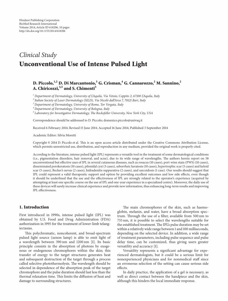
Figure 1: Rosacea: significant results with a significant reduction in vessel number and size and a complete disappearance of papules havebeen achieved after 4 IPL sessions.
All lesions were treated at intervals of 15–20 days for atotal of 4 sessions per case according to the protocol shownin Table 2.
Dermoscopic images were obtained for each case before(also for diagnostic purpose), immediately after, and at adistance from each treatment using the same equipmentdescribed above.
Hypertrophic Scars and Keloids. Ten patients, 5 presentingwith hypertrophic scars (3 males and 2 females aged between21 and 37 years, average age 30.2 years) and 5 presentingwith keloids (3 females and 2 males aged between 27 and 43years, average age 34.8 years), were treated with both 500 nm(vascular component) and 550 nm (pigmented component)wavelength handpieces. The first sessions with the 550 nmhandpiece were carried out for the pigmented componentwhere present. Whereupon, successive treatments with the500 nm handpiece have been made for treating the vascularcomponent (Table 2).
At least 30 days of rest are required before the subsequentsession and a few months are needed to obtain very positiveresults.
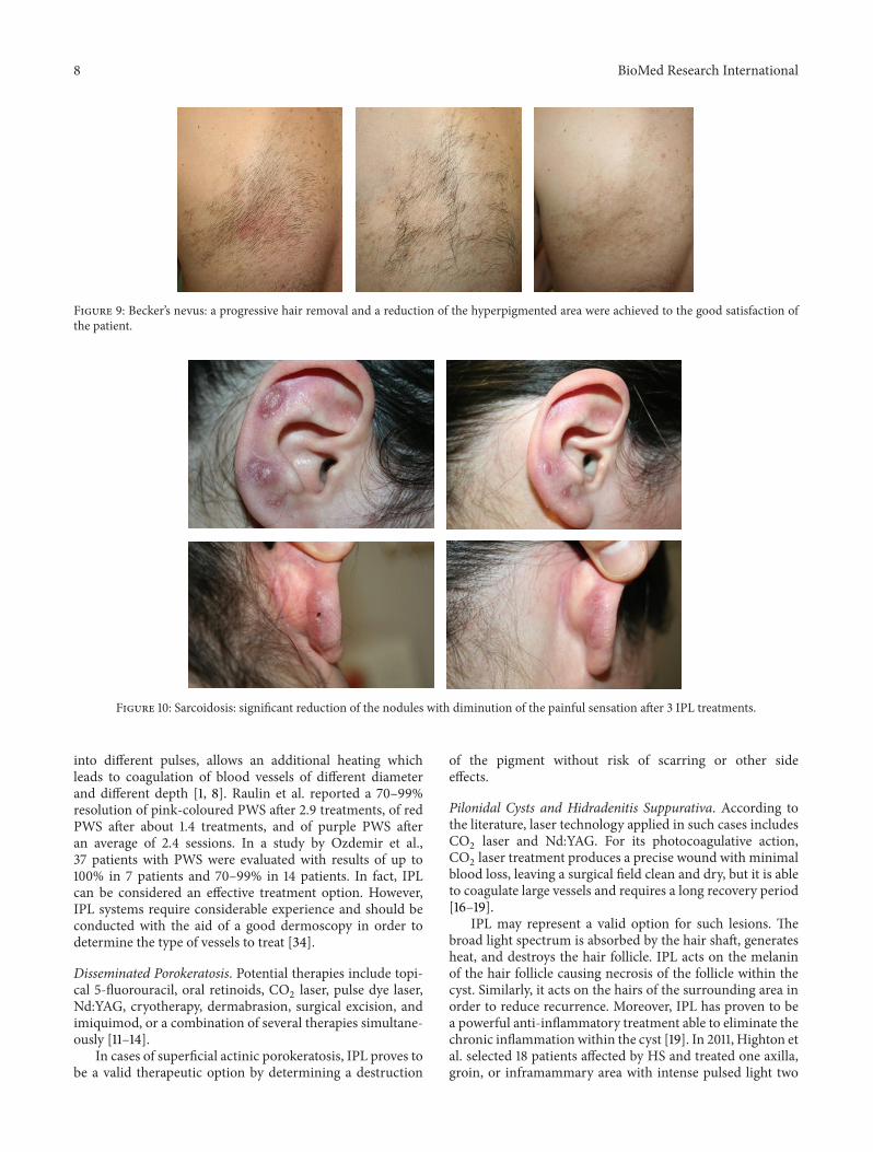
Becker’s Nevus. A 32-year-old man presented with Becker’snevus sited on his left shoulder blade. Clinically, a hypertri-chotic brown patch with irregular edges of 12 cm × 9 cm insize was observed. Successively, a 26-year-old man presentedwith Becker’s nevus without hypertrichosis of 8,5 cm × 8 cmin size and sited on his upper-right chest. In the first case, wedecided to first use a 550nm wavelength handpiece with theaim of removing the hair components (Table 2).
After four sessions of IPL at intervals of 40 days, weperformed two additional sessions with the aim of treatingthe hyperpigmented component (Table 2). Only the protocolshown in Table 2 was applied to the second patient since thehypertrichotic component was not present.
Hidradenitis Suppurativa. One 38-year-old man, previouslytreated in a surgical way (clinical stage II (Hurley’s stag-ing system) and sartorius score of 36), and one 26-year-old woman presented with hidradenitis suppurativa of theaxillary region, bilateral (clinical stage I (Hurley’s stagingsystem) and sartorius score of 24).
After four sessions of IPL at intervals of 15–20 days,we performed two additional sessions with 2 pulses of 5ms
and 10ms separated by a delay of 10ms and a fluence of9 J/cm2 with the aim of treating the inflammatory component(Table 2).
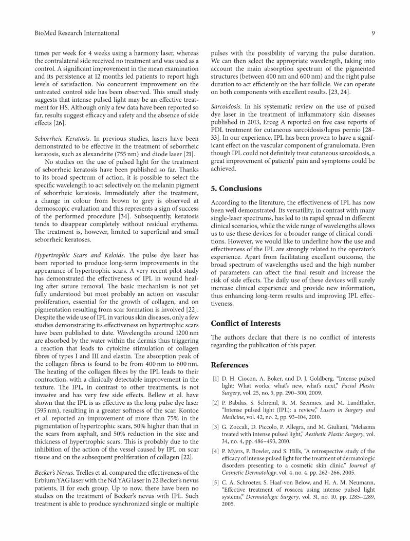
Sarcoidosis. A 26-year-old female presented with threepainful, firm, and vascularized nodules sited on the anteriorand posterior parts of the pinna and on the helix. Throughhistopathological examination, a diagnosis of sarcoidosis wasposed. The patient had already undergone intralesional cor-ticosteroid therapy without results. Thus, we suggested usingthe IPL device with the aim of hitting the very prominent(especially on dermoscopic evaluation) vascular componentwithin the lesions.
3. Results
In this study, we obtained good outcomes for all the treatedpatients, who were affected by different dermatological con-ditions. Our results are summarized as follows.
Rosacea. Patients required from 2 to 5 sessions, at intervalsof approximately 20–30 days, to gain significant results, eventhough amoderate reduction in vessel number and size and apartial disappearance of papules were observed subsequent tothe second session (Figure 1). A 12-month follow-up revealedthe complete absence of recurrences and the persistence ofthe achieved outcomes in 7 of 10 patients (70%) whereas theother 3 patients required a new treatment within the year forthe slight relapse of the papulopustular component.
Port-Wine Stain. The results were already visible after the endof the first session. Dermoscopy performed before treatmenthighlighted the number, calibre, and depth of the targetvessels. Superficial vessels were hit with greater accuracyby IPL and dermoscopic examinations revealed a change invessel colour from red to blue immediately after treatment.In cases of high numbers of vessels, erosions and crusts canfollow treatment sessions for several days. The number of thetreatments required to gain significant results depended onthe depth and site of the PWS.
Three out of 10 patients (30%) obtained excellent results(disappearance of PWS), 6 of 10 (60%) obtained good results(disappearance of almost 70% of treated vessels), and onlyone (10%) obtained amoderate result (disappearance of about30% of the lesion) (Figure 2).The obtained results, confirmed

BioMed Research International 5
Figure 2: Port-wine stain: after 4 IPL sessions, the patient gained excellent results.
Figure 3: Disseminated porokeratosis: after 4 treatments, an important reduction of the hyperkeratotic edge and a reduction in the intensityof melanin have been observed.
by dermoscopy, were stable after a follow-up period rangingfrom 1 to 3 years.
Disseminated Porokeratosis. All treated patients showed inter-esting results, despite the fact that the histology confirmed thepersistence of cornoid lamella. In fact, one patient who hadshown significant improvements after four sessions presentedat the follow-up visit with an important reduction of thehyperkeratotic edge and a reduction in the intensity ofmelanin (Figure 3); a punch biopsy was performed and thehistopathologic examination revealed the persistence of acornoid lamella.
Pilonidal Cyst. A complete resolution was achieved by thethird session (80 days after the first visit) in 3 patients treated.(Figure 4) After a follow-up period of 5 years, for the firstpatient treated, and one year, for the other two, no recurrencehas been observed.
Seborrheic Keratosis. Superficial and small seborrheic ker-atoses responded well to IPL, whereas larger and/or deeperlesions may require a CO
2laser or other treatment. Der-
moscopy is useful either to confirm diagnosis or to demon-strate a change in lesion colour from brown to grey immedi-ately after treatment, thus predicting a good response to thetreatment. Seborrheic keratosis was usually resolved with amild inflammation and a complete recovery within 30 daysafter an average of two treatments (Figure 5).
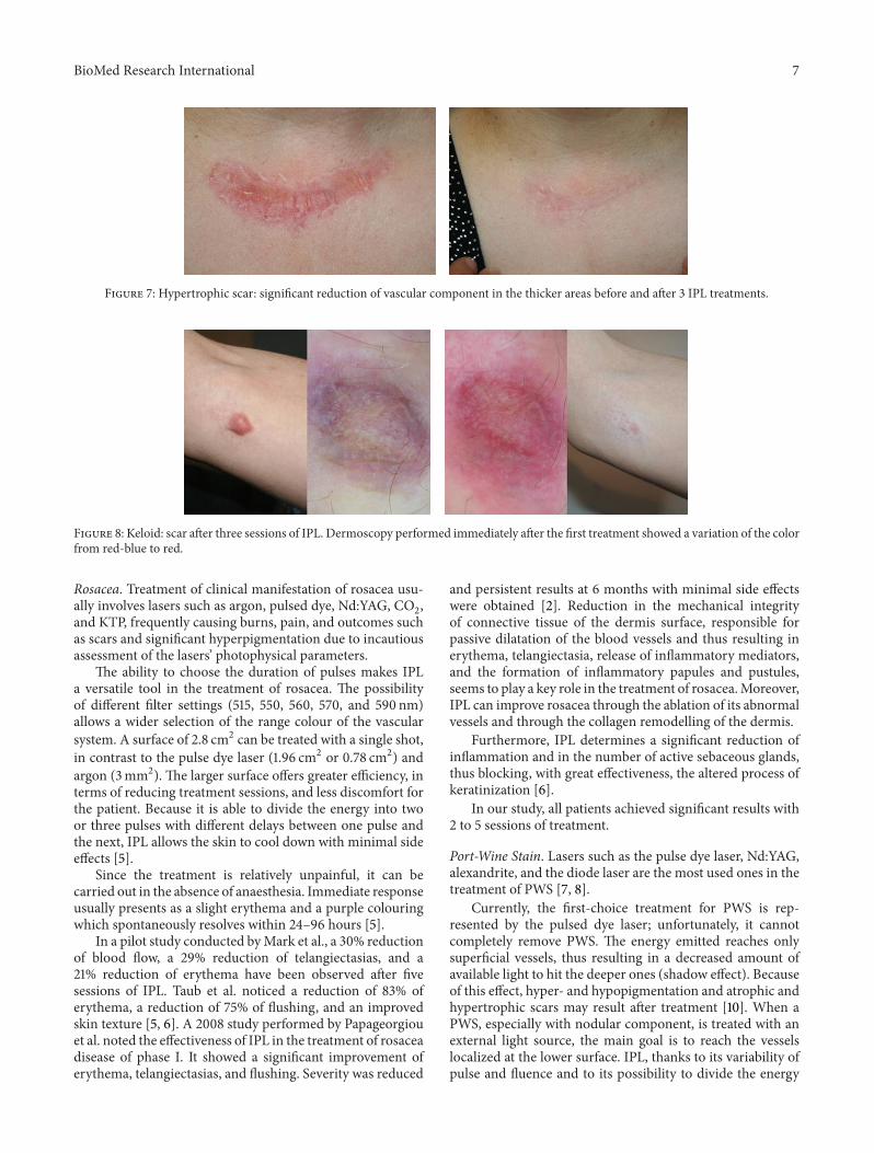
Hypertrophic Scars and Keloids. Dermoscopic images reve-aled a significant reduction of vascular component in thethicker areas. Scars flattened and became smaller after threesessions. (Figures 6 and 7) All in all, good results wereachieved, even though lengthy treatment (several months) isneeded. The obtained results were stable during the follow-up. In one out of 5 cases of keloids, the lesion has resumed itsgrowth phase.
Becker’s Nevus. A progressive hair removal and a reductionof the hyperpigmented area were achieved to the goodsatisfaction of both patients (Figure 8).
Hidradenitis Suppurativa. At the end of the suggested schemeprotocol, hidradenitis suppurativa was completely removedin both its inflammatory and painful components; hairremoval was also achieved (Figure 9).
Sarcoidosis. A significant reduction of the vascular compo-nent and in the consistency of the lesions was achieved, thusleading to pain disappearance (Figure 10).
4. Discussion
In this study, we report on our good results achieved withalmost all 58 patients affected by different dermatologicalconditions. With the aim of providing more exhaustivedetails, we will briefly discuss each condition separately.

6 BioMed Research International
Figure 4: Pilonidal cyst: a complete resolution was achieved by the third session.
Figure 5: Hidradenitis suppurativa: complete resolution of pustular-papules progressive hair removal after 3 IPL treatments, bilaterally.Clinical stage I (Hurley’s staging) and sartorius score of 24.
(a)
(b)Figure 6: Seborrheic keratosis: (a) significant reduction of multiple seborrheic keratoses of the face after 2 IPL sessions, (b) seborrheickeratoses of the back disappeared after 2 IPL sessions.
BioMed Research International 7
Figure 7: Hypertrophic scar: significant reduction of vascular component in the thicker areas before and after 3 IPL treatments.
Figure 8: Keloid: scar after three sessions of IPL. Dermoscopy performed immediately after the first treatment showed a variation of the colorfrom red-blue to red.
Rosacea. Treatment of clinical manifestation of rosacea usu-ally involves lasers such as argon, pulsed dye, Nd:YAG, CO
2,
and KTP, frequently causing burns, pain, and outcomes suchas scars and significant hyperpigmentation due to incautiousassessment of the lasers’ photophysical parameters.
The ability to choose the duration of pulses makes IPLa versatile tool in the treatment of rosacea. The possibilityof different filter settings (515, 550, 560, 570, and 590 nm)allows a wider selection of the range colour of the vascularsystem. A surface of 2.8 cm2 can be treated with a single shot,in contrast to the pulse dye laser (1.96 cm2 or 0.78 cm2) andargon (3mm2). The larger surface offers greater efficiency, interms of reducing treatment sessions, and less discomfort forthe patient. Because it is able to divide the energy into twoor three pulses with different delays between one pulse andthe next, IPL allows the skin to cool down with minimal sideeffects [5].
Since the treatment is relatively unpainful, it can becarried out in the absence of anaesthesia. Immediate responseusually presents as a slight erythema and a purple colouringwhich spontaneously resolves within 24–96 hours [5].
In a pilot study conducted byMark et al., a 30% reductionof blood flow, a 29% reduction of telangiectasias, and a21% reduction of erythema have been observed after fivesessions of IPL. Taub et al. noticed a reduction of 83% oferythema, a reduction of 75% of flushing, and an improvedskin texture [5, 6]. A 2008 study performed by Papageorgiouet al. noted the effectiveness of IPL in the treatment of rosaceadisease of phase I. It showed a significant improvement oferythema, telangiectasias, and flushing. Severity was reduced
and persistent results at 6 months with minimal side effectswere obtained [2]. Reduction in the mechanical integrityof connective tissue of the dermis surface, responsible forpassive dilatation of the blood vessels and thus resulting inerythema, telangiectasia, release of inflammatory mediators,and the formation of inflammatory papules and pustules,seems to play a key role in the treatment of rosacea.Moreover,IPL can improve rosacea through the ablation of its abnormalvessels and through the collagen remodelling of the dermis.
Furthermore, IPL determines a significant reduction ofinflammation and in the number of active sebaceous glands,thus blocking, with great effectiveness, the altered process ofkeratinization [6].
In our study, all patients achieved significant results with2 to 5 sessions of treatment.
Port-Wine Stain. Lasers such as the pulse dye laser, Nd:YAG,alexandrite, and the diode laser are the most used ones in thetreatment of PWS [7, 8].
Currently, the first-choice treatment for PWS is rep-resented by the pulsed dye laser; unfortunately, it cannotcompletely remove PWS. The energy emitted reaches onlysuperficial vessels, thus resulting in a decreased amount ofavailable light to hit the deeper ones (shadow effect). Becauseof this effect, hyper- and hypopigmentation and atrophic andhypertrophic scars may result after treatment [10]. When aPWS, especially with nodular component, is treated with anexternal light source, the main goal is to reach the vesselslocalized at the lower surface. IPL, thanks to its variability ofpulse and fluence and to its possibility to divide the energy
8 BioMed Research International
Figure 9: Becker’s nevus: a progressive hair removal and a reduction of the hyperpigmented area were achieved to the good satisfaction ofthe patient.
Figure 10: Sarcoidosis: significant reduction of the nodules with diminution of the painful sensation after 3 IPL treatments.
into different pulses, allows an additional heating whichleads to coagulation of blood vessels of different diameterand different depth [1, 8]. Raulin et al. reported a 70–99%resolution of pink-coloured PWS after 2.9 treatments, of redPWS after about 1.4 treatments, and of purple PWS afteran average of 2.4 sessions. In a study by Ozdemir et al.,37 patients with PWS were evaluated with results of up to100% in 7 patients and 70–99% in 14 patients. In fact, IPLcan be considered an effective treatment option. However,IPL systems require considerable experience and should beconducted with the aid of a good dermoscopy in order todetermine the type of vessels to treat [34].
Disseminated Porokeratosis. Potential therapies include topi-cal 5-fluorouracil, oral retinoids, CO
2laser, pulse dye laser,
Nd:YAG, cryotherapy, dermabrasion, surgical excision, andimiquimod, or a combination of several therapies simultane-ously [11–14].
In cases of superficial actinic porokeratosis, IPL proves tobe a valid therapeutic option by determining a destruction
of the pigment without risk of scarring or other sideeffects.
Pilonidal Cysts and Hidradenitis Suppurativa. According tothe literature, laser technology applied in such cases includesCO2laser and Nd:YAG. For its photocoagulative action,
CO2laser treatment produces a precise wound with minimal
blood loss, leaving a surgical field clean and dry, but it is ableto coagulate large vessels and requires a long recovery period[16–19].
IPL may represent a valid option for such lesions. Thebroad light spectrum is absorbed by the hair shaft, generatesheat, and destroys the hair follicle. IPL acts on the melaninof the hair follicle causing necrosis of the follicle within thecyst. Similarly, it acts on the hairs of the surrounding area inorder to reduce recurrence. Moreover, IPL has proven to bea powerful anti-inflammatory treatment able to eliminate thechronic inflammationwithin the cyst [19]. In 2011, Highton etal. selected 18 patients affected by HS and treated one axilla,groin, or inframammary area with intense pulsed light two
BioMed Research International 9
times per week for 4 weeks using a harmony laser, whereasthe contralateral side received no treatment and was used as acontrol. A significant improvement in the mean examinationand its persistence at 12 months led patients to report highlevels of satisfaction. No concurrent improvement on theuntreated control side has been observed. This small studysuggests that intense pulsed light may be an effective treat-ment for HS. Although only a few data have been reported sofar, results suggest efficacy and safety and the absence of sideeffects [26].
Seborrheic Keratosis. In previous studies, lasers have beendemonstrated to be effective in the treatment of seborrheickeratosis, such as alexandrite (755 nm) and diode laser [21].
No studies on the use of pulsed light for the treatmentof seborrheic keratosis have been published so far. Thanksto its broad spectrum of action, it is possible to select thespecific wavelength to act selectively on the melanin pigmentof seborrheic keratosis. Immediately after the treatment,a change in colour from brown to grey is observed atdermoscopic evaluation and this represents a sign of successof the performed procedure [34]. Subsequently, keratosistends to disappear completely without residual erythema.The treatment is, however, limited to superficial and smallseborrheic keratoses.
Hypertrophic Scars and Keloids. The pulse dye laser hasbeen reported to produce long-term improvements in theappearance of hypertrophic scars. A very recent pilot studyhas demonstrated the effectiveness of IPL in wound heal-ing after suture removal. The basic mechanism is not yetfully understood but most probably an action on vascularproliferation, essential for the growth of collagen, and onpigmentation resulting from scar formation is involved [22].Despite thewide use of IPL in various skin diseases, only a fewstudies demonstrating its effectiveness on hypertrophic scarshave been published to date. Wavelengths around 1200 nmare absorbed by the water within the dermis thus triggeringa reaction that leads to cytokine stimulation of collagenfibres of types I and III and elastin. The absorption peak ofthe collagen fibres is found to be from 400 nm to 600 nm.The heating of the collagen fibres by the IPL leads to theircontraction, with a clinically detectable improvement in thetexture. The IPL, in contrast to other treatments, is notinvasive and has very few side effects. Bellew et al. haveshown that the IPL is as effective as the long pulse dye laser(595 nm), resulting in a greater softness of the scar. Kontoeet al. reported an improvement of more than 75% in thepigmentation of hypertrophic scars, 50% higher than that inthe scars from asphalt, and 50% reduction in the size andthickness of hypertrophic scars. This is probably due to theinhibition of the action of the vessel caused by IPL on scartissue and on the subsequent proliferation of collagen [22].
Becker’s Nevus. Trelles et al. compared the effectiveness of theErbium:YAG laser with theNd:YAG laser in 22 Becker’s nevuspatients, 11 for each group. Up to now, there have been nostudies on the treatment of Becker’s nevus with IPL. Suchtreatment is able to produce synchronized single or multiple
pulses with the possibility of varying the pulse duration.We can then select the appropriate wavelength, taking intoaccount the main absorption spectrum of the pigmentedstructures (between 400 nm and 600 nm) and the right pulseduration to act efficiently on the hair follicle. We can operateon both components with excellent results. [23, 24].
Sarcoidosis. In his systematic review on the use of pulseddye laser in the treatment of inflammatory skin diseasespublished in 2013, Erceg A reported on five case reports ofPDL treatment for cutaneous sarcoidosis/lupus pernio [28–33]. In our experience, IPL has been proven to have a signif-icant effect on the vascular component of granulomata. Eventhough IPL could not definitely treat cutaneous sarcoidosis, agreat improvement of patients’ pain and symptoms could beachieved.
5. Conclusions
According to the literature, the effectiveness of IPL has nowbeen well demonstrated. Its versatility, in contrast with manysingle-laser spectrums, has led to its rapid spread in differentclinical scenarios, while the wide range of wavelengths allowsus to use these devices for a broader range of clinical condi-tions. However, we would like to underline how the use andeffectiveness of the IPL are strongly related to the operator’sexperience. Apart from facilitating excellent outcome, thebroad spectrum of wavelengths used and the high numberof parameters can affect the final result and increase therisk of side effects. The daily use of these devices will surelyincrease clinical experience and provide new information,thus enhancing long-term results and improving IPL effec-tiveness.
Conflict of Interests
The authors declare that there is no conflict of interestsregarding the publication of this paper.
References
[1] D. H. Ciocon, A. Boker, and D. J. Goldberg, “Intense pulsedlight: What works, what’s new, what’s next,” Facial PlasticSurgery, vol. 25, no. 5, pp. 290–300, 2009.
[2] P. Babilas, S. Schreml, R. M. Szeimies, and M. Landthaler,“Intense pulsed light (IPL): a review,” Lasers in Surgery andMedicine, vol. 42, no. 2, pp. 93–104, 2010.
[3] G. Zoccali, D. Piccolo, P. Allegra, and M. Giuliani, “Melasmatreated with intense pulsed light,” Aesthetic Plastic Surgery, vol.34, no. 4, pp. 486–493, 2010.
[4] P. Myers, P. Bowler, and S. Hills, “A retrospective study of theefficacy of intense pulsed light for the treatment of dermatologicdisorders presenting to a cosmetic skin clinic,” Journal ofCosmetic Dermatology, vol. 4, no. 4, pp. 262–266, 2005.
[5] C. A. Schroeter, S. Haaf-von Below, and H. A. M. Neumann,“Effective treatment of rosacea using intense pulsed lightsystems,” Dermatologic Surgery, vol. 31, no. 10, pp. 1285–1289,2005.
10 BioMed Research International
[6] P. Papageorgiou, W. Clayton, S. Norwood, S. Chopra, and M.Rustin, “Treatment of rosacea with intense pulsed light: signif-icant improvement and long-lasting results,” British Journal ofDermatology, vol. 159, no. 3, pp. 628–632, 2008.
[7] G. Li, T. Lin, Q.Wu, Z. Zhou, andM. H. Gold, “Clinical analysisof port wine stains treated by intense pulsed light,” Journal ofCosmetic and Laser Therapy, vol. 12, no. 1, pp. 2–6, 2010.
[8] A. J. Burns and J. A. Navarro, “Role of laser therapy in pediatricpatients,” Plastic and Reconstructive Surgery, vol. 124, no. 1,supplement, pp. 82e–92e, 2009.
[9] M. Ozdemir, B. Engin, and I. Mevlitoglu, “Treatment of facialport-wine stains with intense pulsed light: a prospective study,”Journal of Cosmetic Dermatology, vol. 7, no. 2, pp. 127–131, 2008.
[10] A. Sevila, E.Nagore, R. Botella-Estrada et al., “Videomicroscopyof venular malformations (port-wine stain type): Prediction ofresponse to pulsed dye laser,” Pediatric Dermatology, vol. 21, no.5, pp. 589–596, 2004.
[11] J. Levitt, J. J. Emer, and P. O. Emanuel, “Treatment of poroker-atosis of Mibelli with combined use of photodynamic therapyand fluorouracil cream,” Archives of Dermatology, vol. 146, no.4, pp. 371–373, 2010.
[12] R. Hartman, R. Mandal, M. Sanchez, and J. A. Stein, “Poroker-atosis plantaris, palmaris, et disseminata,” Dermatology OnlineJournal, vol. 16, no. 11, article 22, 2010.
[13] S. Venkatarajan, T. M. Leleux, D. Yang, T. Rosen, and I.Orengo, “Porokeratosis of Mibelli: successful treatment with 5percent topical imiquimod and topical 5 percent 5-fluorouracil,”Dermatology Online Journal, vol. 16, no. 12, article 10, 2010.
[14] M. Itoh and H. Nakagawa, “Successful treatment of dissemi-nated superficial actinic porokeratosis with Q-switched rubylaser,” Journal of Dermatology, vol. 34, no. 12, pp. 816–820, 2007.
[15] E. A. Badawy and M. N. Kanawati, “Effect of hair removal byNd:YAG laser on the recurrence of pilonidal sinus,” Journal ofthe European Academy of Dermatology and Venereology, vol. 23,no. 8, pp. 883–886, 2009.
[16] Y. Oram, F. Kahraman, Y. Karincaoglu, and E. Koyuncu,“Evaluation of 60 patients with pilonidal sinus treated with laserepilation after surgery,” Dermatologic Surgery, vol. 36, no. 1, pp.88–91, 2010.
[17] A. V. Benedetto and A. T. Lewis, “Pilonidal sinus disease treatedby depilation using an 800 nm diode laser and review of theliterature,” Dermatologic Surgery, vol. 31, no. 5, pp. 587–591,2005.
[18] N. S. Sadick and J. Yee-Levin, “Laser and light treatments forpilonidal cysts,” Cutis, vol. 78, no. 2, pp. 125–128, 2006.
[19] V. Jain and A. Jain, “Use of lasers for the management of refrac-tory cases of hidradenitis suppurativa and pilonidal sinus,”Journal of Cutaneous andAesthetic Surgery, vol. 5, no. 3, pp. 190–192, 2012.
[20] D. Mehrabi and R. T. Brodell, “Use of the alexandrite laser fortreatment of seborrheic keratoses,” Dermatologic Surgery, vol.28, no. 5, pp. 437–439, 2002.
[21] G. R. Culbertson, “532-nm diode laser treatment of seborrheickeratoses with color enhancement,” Dermatologic Surgery, vol.34, no. 4, pp. 525–527, 2008.
[22] O. O. Erol, A. Gurlek, G. Agaoglu, E. Topcuoglu, and H. Oz,“Treatment of hypertrophic scars and keloids using intensepulsed light (IPL),” Aesthetic Plastic Surgery, vol. 32, no. 6, pp.902–909, 2008.
[23] R. Alhusayen, N. Kanigsberg, and R. Jackson, “Becker nevus onthe lower limb: case report and review of the literature,” Journalof CutaneousMedicine and Surgery, vol. 12, no. 1, pp. 31–34, 2008.
[24] C. Raulin, M. P. Schonermark, B. Greve, and S. Werner, “Q-switched ruby laser treatment of tattoos and benign pigmentedskin lesions: a critical review,” Annals of Plastic Surgery, vol. 41,no. 5, pp. 555–565, 1998.
[25] K. M. Mitchell and D. E. Beck, “Hidradenitis suppurativa,”Surgical Clinics of North America, vol. 82, no. 6, pp. 1187–1197,2002.
[26] L. Highton, W. Chan, N. Khwaja, and J. K. G. Laitung, “Treat-ment of hidradenitis suppurativa with intense pulsed light: aprospective study,” Plastic and Reconstructive Surgery, vol. 128,no. 2, pp. 459–465, 2011.
[27] Y. Endo, A. Tamura, O. Ishikawa, and Y. Miyachi, “Perianalhidradenitis suppurativa: early surgical treatment gives goodresults in chronic or recurrent cases,” British Journal of Derma-tology, vol. 139, no. 5, pp. 906–910, 1998.
[28] A. Erceg, E. M. J. G. de Jong, P. C. M. van de Kerkhof, and M.M. B. Seyger, “The efficacy of pulsed dye laser treatment forinflammatory skin diseases: a systematic review,” Journal of theAmerican Academy of Dermatology, vol. 69, no. 4, pp. 609.e8–615.e8, 2013.
[29] M. M. Goodman and K. Alpern, “Treatment of lupus perniowith the flashlamp pulsed dye laser,” Lasers in Surgery andMedicine, vol. 12, no. 5, pp. 549–551, 1992.
[30] S. Cliff, R. H. Felix, L. Singh, and C. C. Harland, “The successfultreatment of lupus pernio with the flashlamp pulsed dye laser,”Journal of Cosmetic and Laser Therapy, vol. 1, no. 1, pp. 49–52,1999.
[31] R. D. Holzmann, S. Astner, T. Forschner, and G. Sterry, “Scarsarcoidosis in a child: case report of successful treatment withthe pulsed dye laser,” Dermatologic Surgery, vol. 34, no. 3, pp.393–396, 2008.
[32] S. Roos, C. Raulin, H. Ockenfels, and S. Karsai, “Successfultreatment of cutaneous sarcoidosis lesions with the flashlamppumped pulsed dye laser,” Dermatologic Surgery, vol. 35, no. 7,pp. 1139–1140, 2009.
[33] M. Ekback and L. Molin, “Effective laser treatment in a case oflupus pernio,” Acta Dermato-Venereologica, vol. 85, no. 6, pp.521–522, 2005.
[34] D. Piccolo, The Usefulness of Dermoscopy in Laser and IntensePulsed Light Treatments, Remo Sandron, Florence, Italy, 2012.