types of multifocal iols - domedics ag · pdf filetypes of multifocal iols ... have one focal...
TRANSCRIPT


Types of Multifocal IOLs
Refractive IOLs• are designed with several refractive optical
zones on the intraocular lens. These zones provide various focal points, allowing for an improvement in distance, intermediate, and near vision.
Diffractive IOLs
• Use diffractive optic principle.• they have diffractive steps on the intraocular
lens and they distribute incoming light rays into two principal foci
• Different technologies (diffractive –refractive; diffractive apodized)

Types of Multifocal IOLs
Segmental Multifocal IOL• Combines an aspheric, asymmetric distance
vision zone with sector-shaped near vision zone of 3.00D
• Two specific surfaces with different radii – one main surface and one embedded surface, to create two defined focal points
Accommodative IOL• So-called “accommodative” intraocular lenses
have one focal point only. Their working principle is based upon a focal shift.

Multifocal IOLs
Diffractive
Full OpticDiffractive
Diffractive centralzone/refractive
peripheryRefractive
„Accomodative“
Medicontur approach to Multifocal IOLs

• „Fresnel principle“ – The Fresnel lens is a type of lens originally developed by French physicist Augustin-Jean Fresnel for lighthouses (1820)
• Fresnel lens, in general, is a thin refractivelens
• Fresnel lens is constructed by removing the „non-refractive part” of the lens
• The size of the lens fragments are not necessarily in the order of lambda
• Fresnel lens – refractive monofocal
Diffractive Optical principles
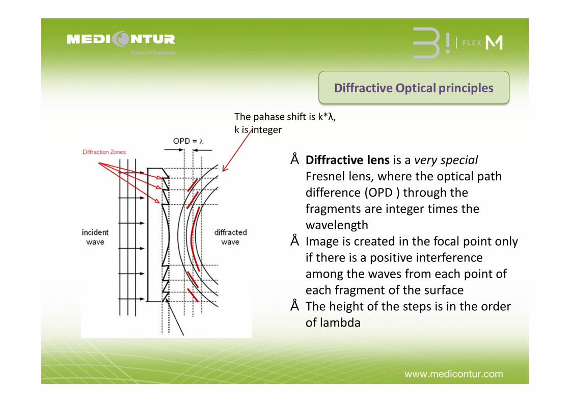
• Diffractive lens is a very special Fresnel lens, where the optical path difference (OPD ) through the fragments are integer times the wavelength
• Image is created in the focal point only if there is a positive interference among the waves from each point of each fragment of the surface
• The height of the steps is in the order of lambda
The pahase shift is k*λ, k is integer

+
Refractive• base power
Diffractive• added power
Bifocal:• base power• base + added power
• The light intensity is shared between refractive and diffractive focus because
• The height of the steps is not exactly k*λ(n2-n1) (intentionally and due to manufacturing precision)
• The diffractive structure is optimal for a specific lambda, and not optimal for other wavelengths
n2
n1

• Diffraction refers to various phenomena which occur when a wave encounters an obstacle.
• = bending of waves around small obstacles and the spreading out of waves past small openings
• As the waves spread out into each other, constructive and destructive interference occurs
Diffractive Optical principles

• Diffractive Bifocal Lens = refractive lens + diffractive microstructure• Diffraction effect is caused by a micro-structured surface profile of the lens• The diffracted light contributes to the near focus and the undiffracted light to
the far focus1st-order Zero ordernear focus far focus

The steps width determines the addition power
The steps height determines the energy repartition between far and near vision
Diffractive Steps

The steps height determinesthe energy repartition
between far and near vision
Diffractive Steps
The same step hights over the whole optic surface = the same energy repartition between far/near vision whatever the pupil diameter

APODIZATION
Decreasing steps height from the center to the periphery = variable repertition of the energy between far and near vision respected pupil diameter= APODIZATION

Multifocal Hydrophilic Aspheric Diffractive Apodized IOLTo restore near, intermediate and distance vision
• manufactured by lathe-milling process
• polish free technology for perfect Sharp Edge
• aspheric biconvex optic with neutral approach – to maintain depth of field
• combination of apodizeddiffractive and refractive zones
• apodized diffractive part– to improve image quality– to minimize visual
disturbances

Bi Flex M • diffractive–refractive design concept
to provide improved control energy distribution
• 7 diffractive discontinuities, or steps, that have been incorporated in the anterior surface of the acrylic optic to provide the diffractive added power
• central diffractive region directs light into near power
• apodized diffractive optic design • apodization improves image quality
by optimizing light energy delivered to the retina by distributing the appropriate amounts of light to near and distant focal points, regardless of lighting situation.

Apodization
• Gradual reduction or blending of the diffractive step heigs
• Apodization describes a change in a property of the lens or its function from center to periphery in a radial fashion.
• The apodization property of the Bi Flex M is defined by the gradual reduction in diffractive step heights from center to periphery.
The term apodization derives from the Greek “cutting off the feet”.
1.4 microns
2.2 microns

Neuron cells 4-100 micronsRods 15 micronsCones 50-55 microns (at night) Reticulocytes 8 microns
PRECISION
Diffractive steps1.4 – 2.2 microns

Bi Flex M Technology
Central 3.1 mmapodized diffractive
structure
Step heights decreaseperipherally
from 2.2 – 1.4 microns
+3.5D at lens planeequaling +2.7D atspectacle plane
Outer refractive zone

AnteriorApodizedOptic
6 mm
Aspheric Optic(neutral approach)
6 mm
BiconvexOptic
13mm

Light energy balanceDistance/Near = 60/40
• Near tasks (reading) – smallpupil– Good light– Accommodative reflex
• Distance – dominant activities – larger pupil– Less light– Without accommodative
reflex
Rela
tive
Ener
gy
Pupil Diameter (mm)
Near Vision Distance Vision

Assessment of multifocal optical quality
• The modulation transfer function is a measure of the transfer of modulation (or contrast) from the subject to the image.
• The spatial frequency is a measure of how often sinusoidal components of the structure repeat per unit of distance (lines/mm).
• Modulation Transfer Function (MTF)

Assessment of multifocal optical quality
• MTF means how faithfully the lens reproduces detail from the object to the image produced by the lens.
Modulation Tranfer Function
Transmitted contrast 100%
Transmitted contrast 50%
Transmitted contrast 2%
MTF = 1
MTF = 0.5
MTF = 0.02

Through focus curve - Bi Flex M
Tested at 50 cyc/mm (according ISO norms)

Bi Flex M • The advantage of the Bi Flex M is that
the apodized diffractive portion turns into a pure refractive lens in the periphery, and that tends to suppress halos and glare
• 2 primary focal points– one at distance and the other at
near– the near point is equivalent to
approximately a +2.7 D added power in the spectacle plane.
• The base lens provides the distance power using its refractive shape

Bi Flex M ReStor Tecnis M Flex Acrilisa PY-60MV iSert
Diffractive x x x x
Apodized x x
Aspheric x x x x x x
Pupil Independ.
x x x x
*MICS x x
**SE 360⁰ x x
Toric Option
coming soon x x x x
ABBE number
58 37 57 58 58 42
Chromatic abberation
Low High Low Low Low High
**SE – sharp edge; *MICS – suitable for „sub 2“ Microincision surgery
Bi Flex M & Competitors

Major assets for Medicontur Bi Flex M
1. Material2. Design3. Optic4. Precision

MATERIAL ( BENZ 25)optimally combining hydrophobic and hydrophilic monomers
n°1
- HEMA, highly hydrophilic monomerPure HEMA = 38% water content>> BIOCOMPATIBILITY
- EOEMA, hydrophobic monomerApproximately 2% water content>> ELASTICITY>> SOFTNESS>> SHAPE MEMORY
EOEMA
36% HEMA
64%
A hydrophobichydrophilic
25% copolymer

A long chain acts as a « molecular eyelash »
to prevent cell adhesion and protein deposit
Our raw material : low adhesion for a low PCO rate
• Anti-PCO behaviour

• LOW Chromatic Abberation • ABBE number: 58
Excellent optical performance
• Uneven focusing of an optical system whichcauses wavelengths of light to have different focalpoints thus decreasing optical performance
WHAT IS CHROMATIC ABBERATION?
Do you know the ABBE number?
The Abbe number - defined by the German physicist Ernst Abbe -is a measure of a transparent material's dispersion in relation to the refractive index . The HIGHER Abbe number the LOWER chromatic aberration

A unique & patented designwith a “Dolphin zone” and double haptics specific design
n°2
13mm 9mm
for REPRODUCIBLE CENTRATION OF THE IOLfor IMMEDIATE & SYMMETRIC UNFOLDING

A unique & patented designWith 180° total contact angle with capsular bag equator
n°2
Medicontur Bi-Flex Average contact angle: 88.8°
Competitor 1Average contact Angle: 69°
Experimental simulator with a diameter of 9 mm
Competitor 2 Average contact Angle: 64.4°

Measurement of radii of Square Edge (SE) at optic-haptic junction by Scanning Electron Microscopy. The IOLs with discontinuity of SE in optic-haptic junction are not included
The smaller the radius,
The more effectivethe square edge effect
60
35
25 25 2520 10
*Comp = Competitor
DESIGN - 360 ⁰Square Edge

Comp 325 microns
Comp 260 microns
Comp 135 microns
Bi Flex 677ABY10 microns
DESIGN – 360⁰ Square EdgeComparison of Square Edge of different IOLs at Optic – Haptic Junction

Comp 5 Comp 6Comp 4
No Squere Edge at all at
Optic-Haptic Junctions
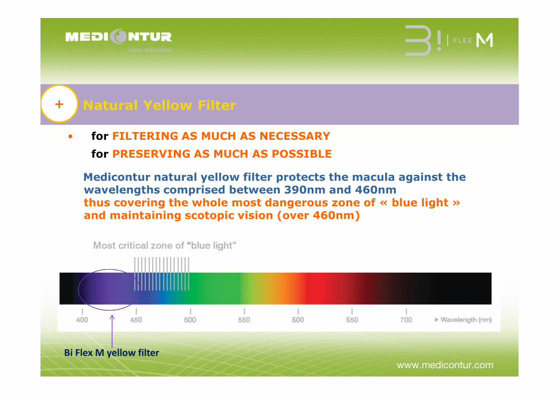
Natural Yellow Filter+
• for FILTERING AS MUCH AS NECESSARY for PRESERVING AS MUCH AS POSSIBLE
Medicontur natural yellow filter protects the macula against the wavelengths comprised between 390nm and 460nmthus covering the whole most dangerous zone of « blue light » and maintaining scotopic vision (over 460nm)
Bi Flex M yellow filter

MEDICONTURNatural
Yellow filter
Simulation of vision without and with yellow filter+
IOLWith NON natural
Yellow filter
IOLWith NO
Yellow filter

Bi Flex M characteristicsSUMMARY
To meet surgeons and their patientsrequirements
MATERIAL
BENZ 25 material (USA)
v A combination of two monomers (HEMA 34% & OEMA 64%)
v Low water content
v Low refractive index (RI)
v Preventing cells adhesion and protein deposits
Ø High ABBE Number (58)
ü Natural Yellow filter (390 – 460 nm)
v BIOCOMPATIBILITY
v ELASTICv SOFTv SHAPE MEMORY
v Better optical performance
v PCO prevention
Ø Low chromatic abberation – excellent optical performance
ü Blocking the most dangerous light for macula ü Preserving scotopic visionü Preventing a large shift in color in a „bluish“
direction after cataract surgery
OPTIC
v Apodized Diffractive – Refractive design
v Aspheric (neutral approach)
vMade with high Precision
vOptimizing light energy distributionv Improving the image qualityv Supresing halos and glares
vmaintaining depth of field
v Precise and stable refractive results
DESIGN • 360° square edge (r=10 microns)
Ø Double loop haptic
• PCO prevention
Ø Improved axial, radial and rotational stabilityØ 180⁰ contact angleØ Easy implantation through micro incision
MICS

Conclusion
Bi Flex M and its´ strenghts Bi Flex M
37
• Diffractive – refractive optic• Apodized diffractive central part• Refractive outer zone• High Precision• Suitable for MICS (incision size sub 2)• Material
• BENZ 25• Low chromatic abberation• Natural Yellow filter (390-460 nm)• PCO prevention• Refractive Index 1.46
• Design• PCO prevention• 360⁰ Square Edge (r = 10microns) • material• Rotation stability - 180⁰ contact angle

Bi Flex MMultifocal Apodized Diffractive Aspheric
Hydrophylic Acrylic IOLOptic diameter 6.0 mmOverall diameter 13.0 mm
Diffractive zone Between radius 1 mm - 3.1 mm
Diffractive discontinuity 7
Characteristics of steps Gradually decreasing 2.2 – 1.4 microns
Addition +3.5 D
Optic
Aspheric, Biconvex Apodized Diffractive
Optic
Abbe number 58

Bi Flex M
Multifocal Apodized Diffractive IOL
MTF / light energy distribution
Dependance of Pupille Diameter
Refractive index 1.46
Estimated A -const 118.0
injector MedJet MB
Estimated incision size 1.8-2.2 mm
PCO prevention 360⁰ square edge
Natural Yellow Filter (covalent bound)
390-460nm
Temperature (20-25⁰)
Shelf Life 5 years

Dr Assouline’s Results
Presented at the SFO (Société Française d’Ophtalmologie) May 2013
-French National Congress

Dr Assouline (Paris, France)
Defocus CurvePseudophake solutions for intermediary visionMedicontur 677MY (n=9), Lentis M+ (n=19) Finevision (n=16) ATLisa-Trifocal (n=3) ATLisa-Bifocal (n=38) Restor (n=6)

Binocular Defocus CurveMix & MatchLentis + Medicontur 677MY (n=6), Lentis M+ Finevision (n=11) ATLisa-Trifocal + Finevision (n=3)
Dr Assouline (Paris, France)
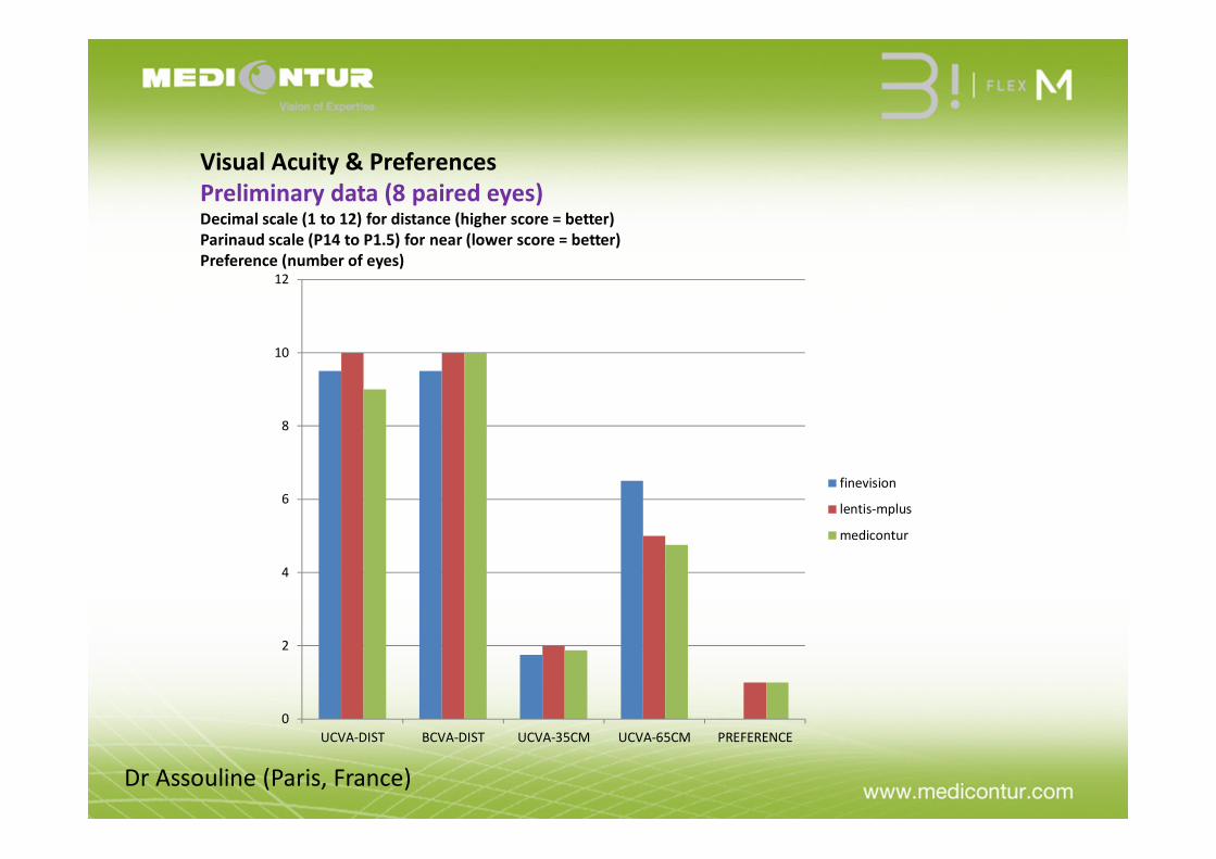
Visual Acuity & PreferencesPreliminary data (8 paired eyes)Decimal scale (1 to 12) for distance (higher score = better)Parinaud scale (P14 to P1.5) for near (lower score = better)Preference (number of eyes)
0
2
4
6
8
10
12
UCVA-DIST BCVA-DIST UCVA-35CM UCVA-65CM PREFERENCE
finevision
lentis-mplus
medicontur
Dr Assouline (Paris, France)