type a aortic dissection presenting with inferior st-elevation … · type a aortic dissection...
TRANSCRIPT
Type A Aortic Dissection Presenting with Inferior
ST-Elevation Myocardial Infarction
Bao-Tzung Wu, Chun-Yi Li and Ying-Tsung Chen
Type A aortic dissection with concurrent ST-elevation myocardial infarction (STEMI) is relatively rare. However, it
can be potentially fatal and easily misdiagnosed as STEMI alone. Misdiagnosis will lead to inappropriate administration
of anticoagulant and thrombolytic therapy and delayed surgical repair of the aorta. In patients with STEMI, short
reperfusion time is associated with improved survival, and minimizing the door-to-balloon time is the goal of
therapy worldwide. However, signs critical for differential diagnosis may be overlooked in the rush to primary
percutaneous coronary intervention. When a patient is encountered who presents with chest pain and ST
elevation on electrocardiogram, STEMI should not be the only diagnosis considered. By using bedside available
information, detailed history taking and focused physical examination, it is possible to avoid a mistaken diagnosis.
Here we report a case of Stanford type A aortic dissection with STEMI that was initially misdiagnosed as sole acute
inferior wall myocardial infarction. Patient mortality may have resulted from delayed diagnosis and surgical
treatment.
Key Words: Acute myocardial infarction � Aortic dissection
INTRODUCTION
Aortic dissection can present with a wide range of
manifestations, and its diagnosis can be challenging for
primary care emergency physicians.1
Approximately
one-fourth of patients with acute aortic syndrome pre-
sent with an electrocardiographic pattern resembling
that of acute coronary syndrome. Patients with an elec-
trocardiogram (ECG) suggestive of myocardial ischemia
are easily misdiagnosed,2
and the possibility of mis-
diagnosis is even higher in patients with concurrent
ST-elevation myocardial infarction (STEMI).3
Here we
present a case with Stanford type A aortic dissection
and STEMI who failed to survive because he was initially
misdiagnosed, leading to delayed diagnosis and surgical
treatment.
CASE REPORT
A 70-year-old man with a history of uncontrolled
hypertension presented to our emergency department
with acute onset of chest pain. The pain radiated to the
patient’s back and was associated with cold sweating.
The ECG showed ST elevation in the inferior leads with
reciprocal ST depression in leads I and aVL (Figure 1A).
Hypotension was noted and treated with a vasopressor.
Acute STEMI of the inferior wall with shock was im-
pressed. A chest radiograph revealed mediastinal wi-
dening, a prominent aortic knob, and left pleural effu-
sion (Figure 2A). Approximately 5 min later, significant
resolution of ST segment elevation was observed on a
right-sided ECG (Figure 1B). Hypotension persisted and
the patient was sent for primary percutaneous coronary
Acta Cardiol Sin 2014;30:248�252 248
Caes Report Acta Cardiol Sin 2014;30:248�252
Received: May 13, 2013 Accepted: October 4, 2013
Division of Cardiology, Department of Internal Medicine, Tungs’
Taichung Metro Harbor Hospital, Taichung, Taiwan.
Address correspondence and reprint requests to: Dr. Bao-Tzung Wu,
Section of Cardiology, Tung’s Taichung Metro Harbor Hospital, No.
699, Sec. 8, Taiwan Blvd., Wuqi Dist., Taichung City 43503, Taiwan.
Tel: 886-4-2658-1919 ext. 73090; Fax: 886-4-2658-2193; E-mail:

249 Acta Cardiol Sin 2014;30:248�252
Type A Aortic Dissection Presenting with STEMI
Figure 1. (A) An initial ECG showing ST elevation in the inferior leads with reciprocal ST depression in leads I and aVL. (B) Resolution of ST elevation
on a right-sided electrocardiogram. (C) New ST depression in the anterior leads after primary percutaneous coronary intervention.
A
B
C
intervention (PCI). Coronary angiography revealed a le-
sion causing 50% stenosis in the middle left anterior de-
scending artery and dynamic stenosis of the proximal
right coronary artery (RCA) (Figure 2B). The culprit le-
sion in the proximal RCA was quite different from those
responsible for most cases of STEMI. The luminal sur-
face was relatively smooth, and there was no dissection
plaque or thrombus. The RCA was almost totally occluded
in the systolic phase, whereas flow improved in the dia-
stolic phase (Figure 2C). The stenosis could not be re-
lieved by vasodilator injection or angioplasty. A stent
was inserted into the proximal RCA and Thrombolysis in
Myocardial Infarction 3 grade flow was established. The
door-to-balloon time was 79 min. Resistance was en-
countered while pushing the wire up the abdominal
aorta during the procedure; therefore, abdominal
aortography was performed after PCI. Aortography re-
vealed a large abdominal aortic aneurysm (Figure 2D,
black arrow), which explained the resistance encoun-
tered.
After PCI, the observed shock worsened. A follow-
up ECG revealed complete resolution of ST elevation in
the inferior leads; however, new ST depression had ap-
peared in the anterior leads, suggesting anterior is-
chemia (Figure 1C). Heart echocardiography revealed
hypokinesis of the inferior wall with normal anterior
wall motion. In addition, a dilated aortic root with mild
to moderate aortic regurgitation and mild pericardial
effusion was also noted. Clinical information was re-
viewed again. A history of chest pain and hypertension,
mediastinal widening and a prominent aortic knob on
chest radiograph, an unusual coronary lesion and echo-
cadiographic findings of aortic regurgitation and peri-
cardial effusion all suggested aortic dissection. A dis-
section plane across the abdominal aneurysm was noted
on the abdominal aortogram after careful review (Figure
2D, white arrow). Enhanced chest computed tomo-
graphy (CT) showed a dissection plane extending from
the aortic root to the left common iliac artery and
across the abdominal aneurysm (Figure 2E). The false
lumen perforated into the region of the aortic root, with
formation of a periaortic hematoma that compressed
the stented proximal RCA (Figure 2F). A final diagnosis
of Stanford type A aortic dissection with RCA involve-
ment and resultant acute inferior myocardial infarction
was made. The time from arrival at the emergency de-
partment to definitive diagnosis was 16 h. The patient
was transferred for emergency surgery, but he could not
be weaned from cardiopulmonary bypass and died in
the operating room.
DISCUSSION
Acute myocardial infarction due to extension of an
acute Stanford type A aortic dissection is a rare but
devastating event, occurring in approximately 3% pa-
tients with aortic dissection.4
The RCA is involved in
most patients with concurrent infarction. Differentiating
acute Stanford A aortic dissection with coronary mal-
Acta Cardiol Sin 2014;30:248�252 250
Bao-Tzung Wu et al.
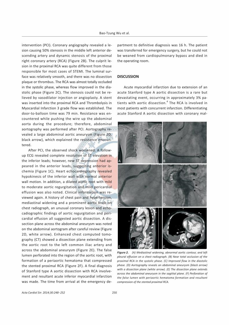
Figure 2. (A) Mediastinal widening, abnormal aortic contour, and left
pleural effusion on a chest radiograph. (B) Near total occlusion of the
proximal RCA in the systolic phase. (C) Improved flow in the diastolic
phase. (D) Aortography reveals an abdominal aneurysm (black arrow)
with a dissection plane (white arrow). (E) The dissection plane extends
across the abdominal aneurysm in the sagittal plane. (F) Perforation of
the false lumen with periaortic hematoma formation and resultant
compression of the stented proximal RCA.
A B
C D
E F

perfusion from true acute myocardial infarction is chal-
lenging for emergency physicians, and misdiagnosis is
very common in patients with ST segment elevation on
initial ECGs.3,5
Inappropriate treatment with antiplate-
let, antithrombin, and thrombolytic agents can cause
catastrophic bleeding.
In patients with STEMI, timely reperfusion improves
the clinical outcome. The guidelines for STEMI recom-
mended a door-to-balloon time of < 90 min and most
medical personnel are under constant pressure to achi-
eve this goal. The guidelines also contained the follow-
ing recommendations to facilitate the differentiation of
STEMI from aortic dissection:6
“Evaluation of the pa-
tient’s complaints should focus on chest discomfort, as-
sociated symptoms, sex and age-related differences in
presentation, hypertension, diabetes mellitus, and the
possibility of aortic dissection”, “Patients with STEMI
should have a portable chest X-ray, but this should not
delay implementation of reperfusion therapy (unless a
potential contraindication is suspected, such as aortic
dissection)”, and “Imaging studies such as a high-quality
portable chest X-ray, transthoracic and/or transeso-
phageal echocardiography, and contrast chest CT scan
or magnetic resonance imaging scan should be used for
differentiating STEMI from aortic dissection in patients
in whom this distinction is initially unclear”. However,
under pressure to achieve short reperfusion times, im-
portant clinical clues for differential diagnosis may be
overlooked.
Aortic dissection has been frequently misdiagnosed
as acute coronary syndrome. Acute coronary syndrome
has the diagnostic advantages of the ECG and cardiac
enzymes testing, allowing risk stratification and emer-
gent treatment. Aortic dissection has no equivalent
diagnostic tests that are rapidly available. The Interna-
tional Registry of Acute Aortic Dissection Substudy on
Biomarkers (IRAD-Bio) study evaluated the diagnostic
performance of D-dimer testing in a study population of
patients with suspected aortic dissection.7
At the cutoff
level of 500 ng/mL versus control, sensitivity and speci-
ficity were 96.6% and 46.6%, respectively. When the
cutoff level was increased to 1600 ng/mL, D-dimer may
be a useful tool in differential diagnosis among aortic
dissection and myocardial infarction, angina or other
ischemia heart diseases within the first 6 h. D-dimer, al-
though imperfect due to its low specificity, is currently
the only commercially available test that can be used for
the biochemical diagnosis of aortic dissection.
In the absence of a rapid, accurate, and readily
available diagnostic test, the current diagnosis of aortic
dissection requires comprehensive interpretation of
available information. In 2010, the American Heart
Association released guidelines for the diagnosis and
management of patients with thoracic aortic disease,
which introduced a risk assessment tool focusing on
specific high-risk predisposing conditions, pain features,
and physical examination findings. This aortic dissection
detection risk score is highly sensitive (95.7%) for the
detection of acute aortic dissection when applied to the
patients diagnosed with aortic dissection in the IRAD
database.8
The results from this study suggested that
the aortic dissection detection risk score, with the use
of only information that is available at bedside, offers
adequate sensitivity to capture the vast majority of pa-
tients presenting with acute aortic dissection.
In our patient, there were several indications of aor-
tic dissection. First, detailed characterization of chest
pain in the emergency department may have provided
clues to aortic dissection. Typically, aortic dissection
presents with severe tearing chest pain that instantly
reaches its maximal intensity. The qualitative nature of
the patient’s chest pain was not clarified. Second, the
patient had a history of uncontrolled hypertension, and
chest radiography had revealed mediastinal widening,
an abnormal aortic contour, and left pleural effusion,
which should have prompted further imaging studies.
Third, a murmur from aortic regurgitation or pulse de-
ficit could have been detected on physical examination.
Fourth, resistance encountered during catheterization is
a sign of possible structural damage to the aorta or its
branches. Although an abdominal aortogram was ob-
tained, a dissection plane was overlooked. Fifth, the cul-
prit vessel was not completely occluded and the lesion
characteristics were quite different from those of con-
ventional STEMI. The significant difference between
central (aortic root) and peripheral (right femoral ar-
tery) blood pressure (around 50 mmHg) was also ig-
nored during PCI.
In emergency departments, the number of patients
treated for acute coronary syndrome is greater than
that of patients treated for aortic dissection. Differenti-
ating acute coronary syndrome from type A aortic dis-
251 Acta Cardiol Sin 2014;30:248�252
Type A Aortic Dissection Presenting with STEMI
section with concomitant coronary malperfusion is chal-
lenging, because of demands for minimal door-to-
balloon time. Our findings from this case suggested that
acute aortic dissection should always be considered as a
differential diagnosis in patients presenting with symp-
toms suggesting acute coronary syndrome. If there is a
high index of suspicion, it is wise to consider further
imaging studies as the recommendation of the above-
mentioned guidelines. Despite the importance of a
short reperfusion time, achieving this goal should not
compromise diagnostic accuracy.
REFERENCES
1. Hsieh TH, Tsai LM, Tsai MZ. Characteristics of and atypical pre-
sentations in patients with acute aortic dissection � A single
center experience. Acta Cardiol Sin 2011;27:238-43.
2. Harris KM, Strauss CE, Eagle KA, et al. Correlates of delayed
recognition and treatment of acute type A aortic dissection: The
International Registry of Acute Aortic Syndrome (IRAD). Circu-
lation 2011;124:1911-8.
3. Luo JL, Wu CK, Lin YH, et al. Type A aortic dissection manifesting
as acute myocardial infarction: still a lesion to learn. Acta Cardiol
2009;64:499-504.
4. Hagan PG, Nienaber CA, Isselbacher EM, et al. The International
Registry of Acute Aortic Dissection (IRAD): new insights into an
old disease. JAMA 2000;283:897-903.
5. Hansen MS, Nogareda GJ, Hutchison SJ. Frequency of and in-
appropriate treatment of misdiagnosis of acute aortic dissec-
tion. Am J Cardiol 2007;99:852-6.
6. Antman EM, Anbe DT, Armstrong PT, et al. ACC/AHA guidelines
for the management of patients with ST-elevation myocardial
infarction-executive summary. A report of the American College
of Cardiology/American Heart Association Task Force on Practice
Guidelines (Writing Committee to Revise the 1999 Guidelines for
the Management of Patients with Acute Myocardial Infarction).
J Am Coll Cardiol 2004;44:671-719.
7. Suzuki T, Distante A, Zizza A, et al. Diagnosis of acute aortic dis-
section by D-dimer: the International Registry of Acute Aortic
Dissection Substudy on Biomarkers (IRAD-Bio) experience. Cir-
culation 2009;119:2702-7.
8. Rogers AM, Hermann LK, Booher AM, et al. Sensitivity of the
aortic dissection detection risk score, a novel guideline-based
tool for identification of acute aortic dissection at initial presen-
tation: results from the International Registry of Acute Aortic
Dissection. Circulation 2011;123:2213-8.
Acta Cardiol Sin 2014;30:248�252 252
Bao-Tzung Wu et al.