tympanometry - international hearing societykatz, handbook of clinical audiology fig 13.18 stimulus...
TRANSCRIPT
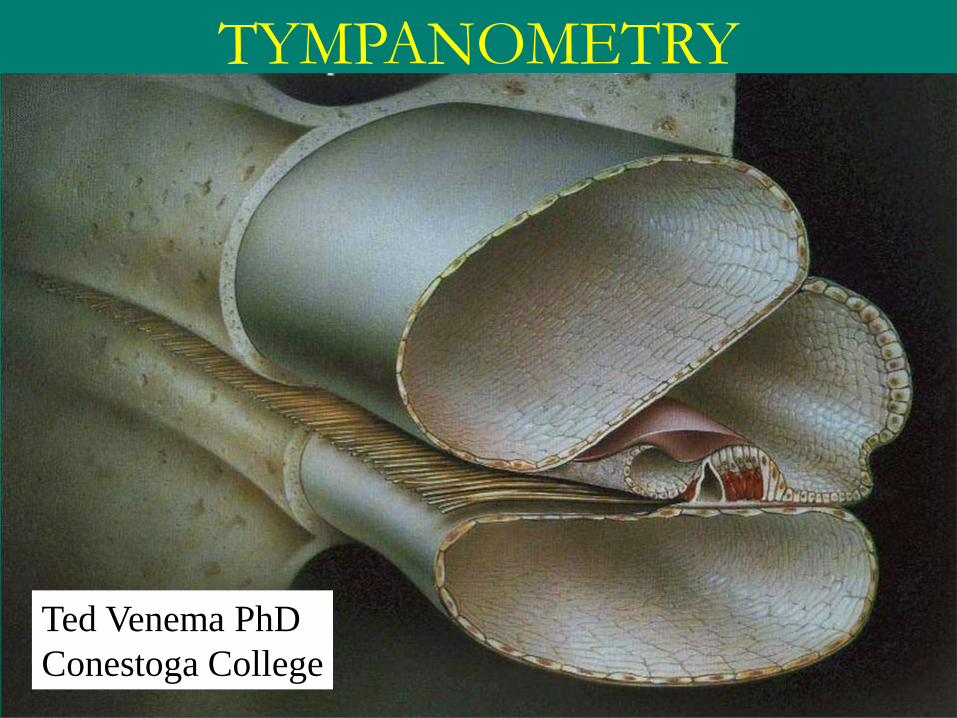
TYMPANOMETRY
Ted Venema PhD
Conestoga College

Why Do We Have a Middle Ear?

1. Size
of TM
Compared To
Size
of Stapes
To Increase Sound Pressure
So airborne sound can penetrate fluid-filled Cochlea
How?
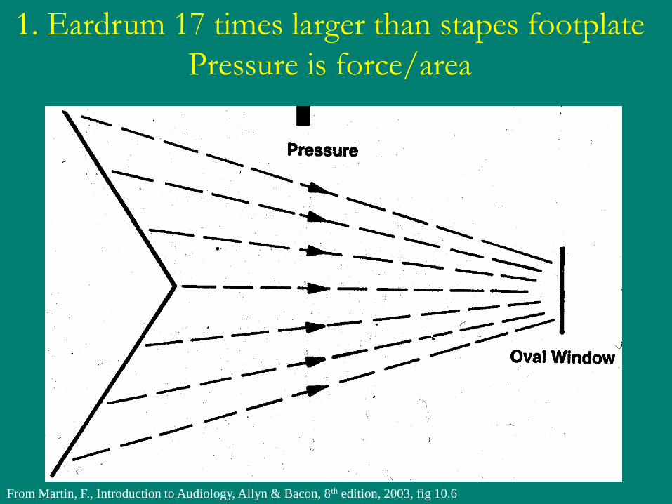
From Martin, F., Introduction to Audiology, Allyn & Bacon, 8th edition, 2003, fig 10.6
1. Eardrum 17 times larger than stapes footplate
Pressure is force/area


2. Then there’s this leverage thing…
An axis runs
right through
here
ie. ossicular chain
pivots on
this axis
Malleus is
lightly longer
than incus
1.3:1

The Fulcrum:
Head of Malleus & Short Process of Incus
An axis runs
right through
here
Note:
Malleus is 1.3 times
longer than
Incus long process

Middle
Ear
Ossicles
From Yost, WA
Fundamentals of
Hearing
(1994)

From Martin, F., Introduction to Audiology, Allyn & Bacon, 8th edition, 2003, fig 10.6
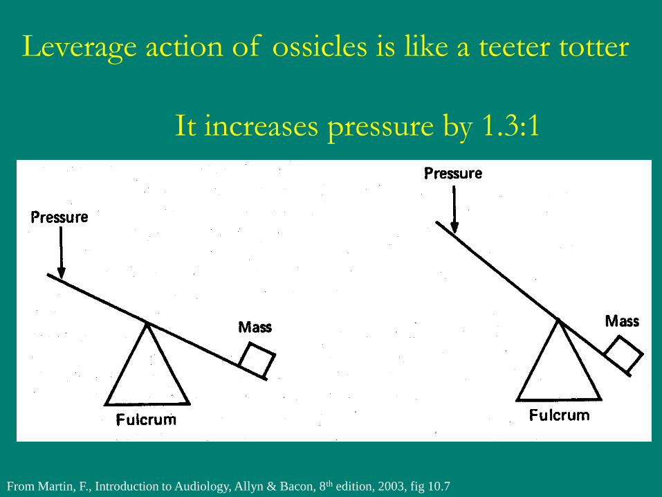
From Martin, F., Introduction to Audiology, Allyn & Bacon, 8th edition, 2003, fig 10.7
Leverage action of ossicles is like a teeter totter
It increases pressure by 1.3:1

3. Buckling Action of TM
Due to its conical shape,
the TM “buckles”
as it vibrates…This
doubles the movement
of the Malleus
Annulus
Annulus
Umbo
TM at rest
Buckling
action

SO, IN SUMMARY
The middle ear provides a sound pressure increase of 44:1
1. Eardrum – stapes size: 17:1
2. Ossicles leverage action: 1.3:1
3. Eardrum buckling action: 2:1
17
1.3
X 2
44

0 10 100 1000 10,000 100,000 1,000,000
120
100
80
60
40
20
0
Pressure increase
dB
SPL
44:1
33dB
Middle Ear Adds 33dB to Incoming Sound
It has to, Because the Cochlea is Filled with Fluid!
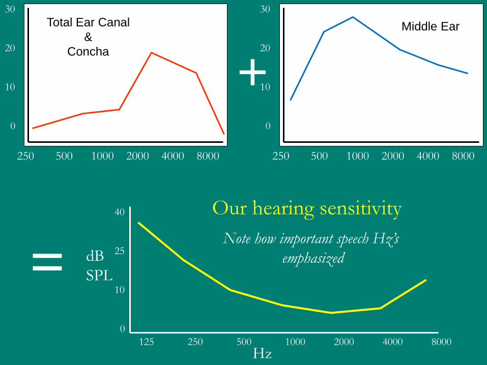
+
=
Hz
dB
SPL
125 250 500 1000 2000 4000 8000
Our hearing sensitivity 40
25
10
0
Note how important speech Hz’s
emphasized
250 500 1000 2000 4000 8000
30
20
10
0
Total Ear Canal
&
Concha
250 500 1000 2000 4000 8000
30
20
10
0
Middle Ear
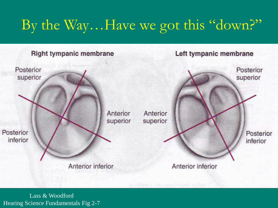
Lass & Woodford
Hearing Science Fundamentals Fig 2-7
By the Way…Have we got this “down?”

Otitis Media & Kids
Their Eustachian Tubes Are More Horizontal


Normal
Right
Tympanic
Membrane

Serous
Otitis
Media

Acute
Bulging
Otitis
Media

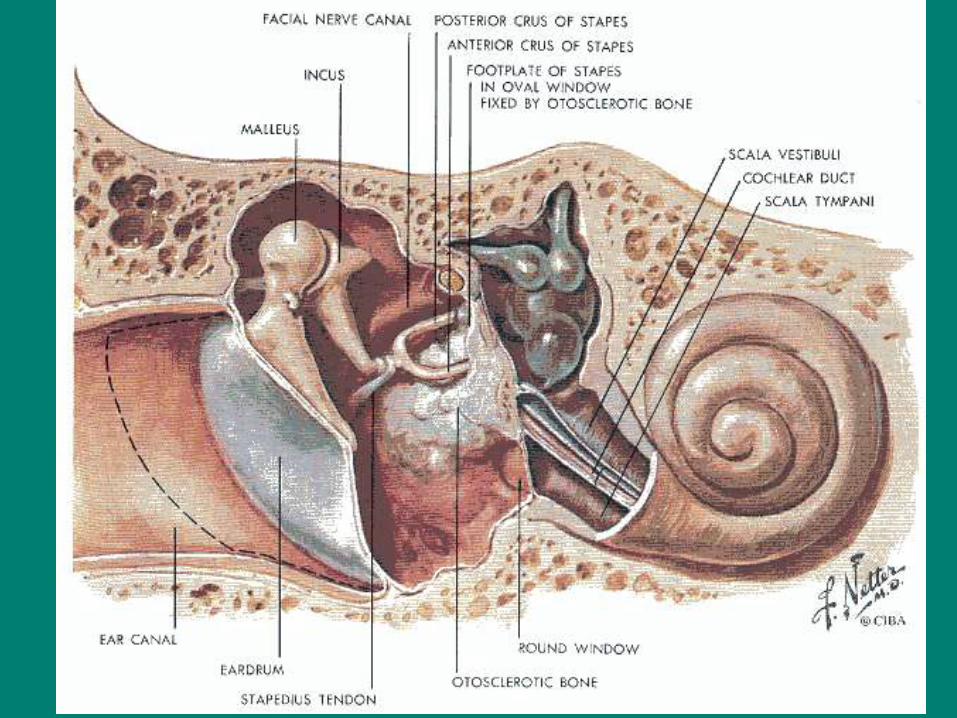
Retracted TM
Note stapes
in background

From: Northern & Downs, Hearing in Children 4th ed, Williams & Wilkins 1991

PE
(pressure
equalizing)
Tube


Key Concept of Tympanometry:
For the Middle
Ear to be most
Efficient…
Air pressure must be
even on both
sides of TM
Lass & Woodford
Hearing Science Fundamentals Fig 2-8
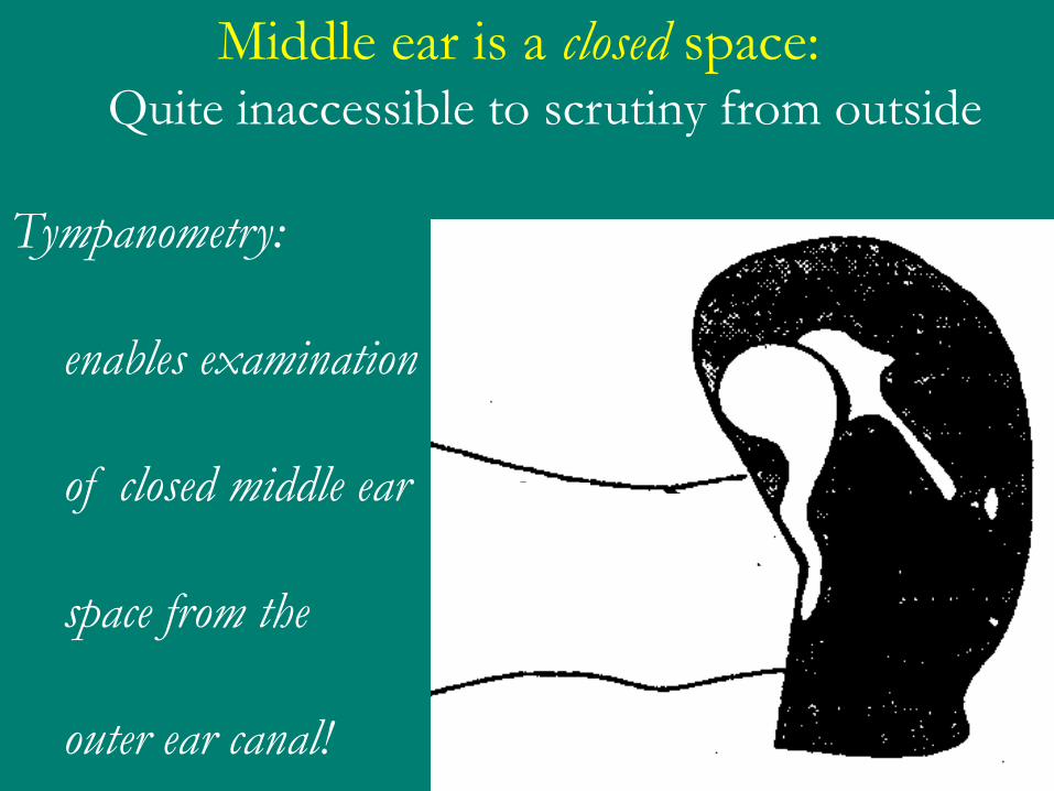
Middle ear is a closed space: Quite inaccessible to scrutiny from outside
Tympanometry:
enables examination
of closed middle ear
space from the
outer ear canal!

Here’s How:
Tympanometry Involves Impedance
•Middle ear “Impedance”is comprised of 3 things:
•Mass
Resonates with Low Hz’s
•Stiffness
Resonates with High Hz’s
•Resistance
Like simple friction; equal for all Hz’s in any object


Impedance & the Middle Ear:
•Mass
Middle ear ossicles
•Stiffness
Load of fluid pressure from inner ear on stapes footplate
•Resistance
Ligaments of middle ear ossicles
•In short:
Middle ear is stiffness dominated system!

When Air Pressure is equal on both sides of TM,
This creates least Stiffness; most Compliance
Tympanometry
is really a test
of middle ear
efficiency
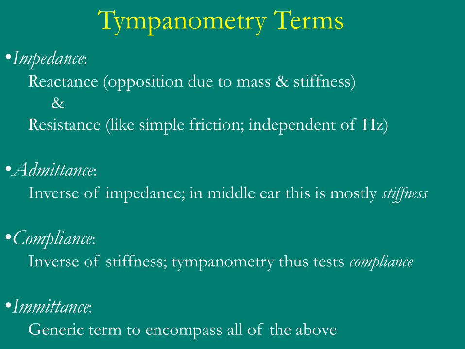
Tympanometry Terms
•Impedance: Reactance (opposition due to mass & stiffness)
&
Resistance (like simple friction; independent of Hz)
•Admittance: Inverse of impedance; in middle ear this is mostly stiffness
•Compliance: Inverse of stiffness; tympanometry thus tests compliance
•Immittance: Generic term to encompass all of the above
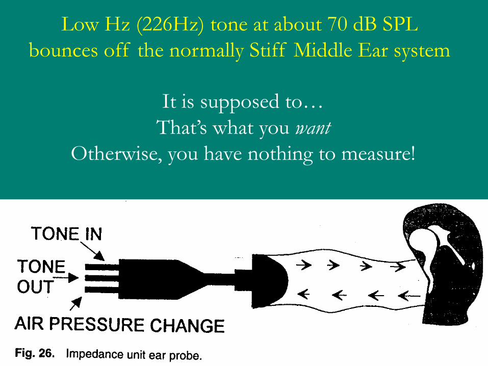
Low Hz (226Hz) tone at about 70 dB SPL
bounces off the normally Stiff Middle Ear system
It is supposed to…
That’s what you want
Otherwise, you have nothing to measure!
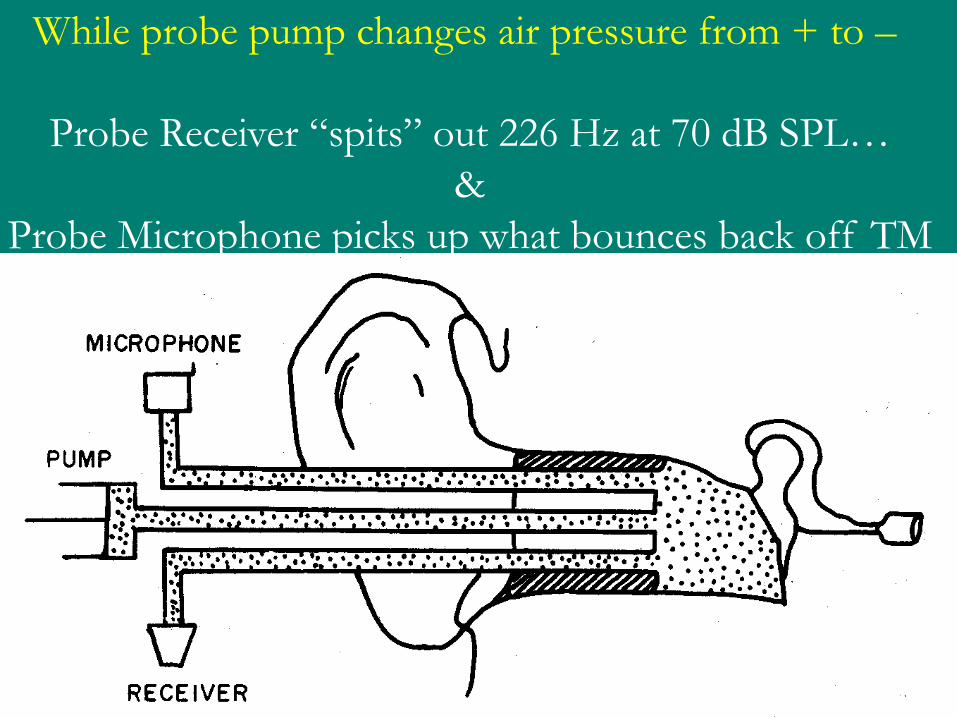
While probe pump changes air pressure from + to –
Probe Receiver “spits” out 226 Hz at 70 dB SPL…
&
Probe Microphone picks up what bounces back off TM

Hah!

Why the 226 Hz tone? Why not 250 Hz?
True compliance at this Hz
is equal to an enclosed volume of air
Admittance is 1 mmho when measured in 1 cc cavity
also, one can then obtain PV of ear canal in cc’s

From Clinical Impedance Audiometry 2nd ed Jerger, Northern
The tympanogram shows SPL change in ear canal
as air pressure
in ear canal
is changed

Less SPL at the probe tip in ear canal means…
more SPL
is going through
the middle ear!

Tympanometry (Impedance)
Normally consists of Four Tests
1. Tympanogram
2. Static Compliance
3. Physical Volume of EAM
4. Acoustic Reflexes
From Clinical Impedance Audiometry 2nd ed
Jerger, Northern
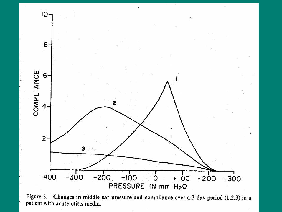
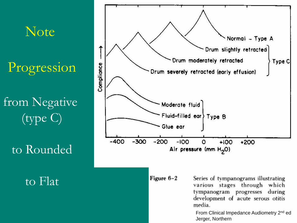
Note
Progression
from Negative
(type C)
to Rounded
to Flat

From Clinical Impedance Audiometry 2nd ed Jerger, Northern
Static Compliance According to the description below…
Isn’t it just the height of the Tympanogram?

From Clinical Impedance Audiometry 2nd ed Jerger, Northern

From Clinical Impedance Audiometry 2nd ed Jerger, Northern
Note the unusually high static compliance

From Clinical Impedance Audiometry 2nd ed Jerger, Northern
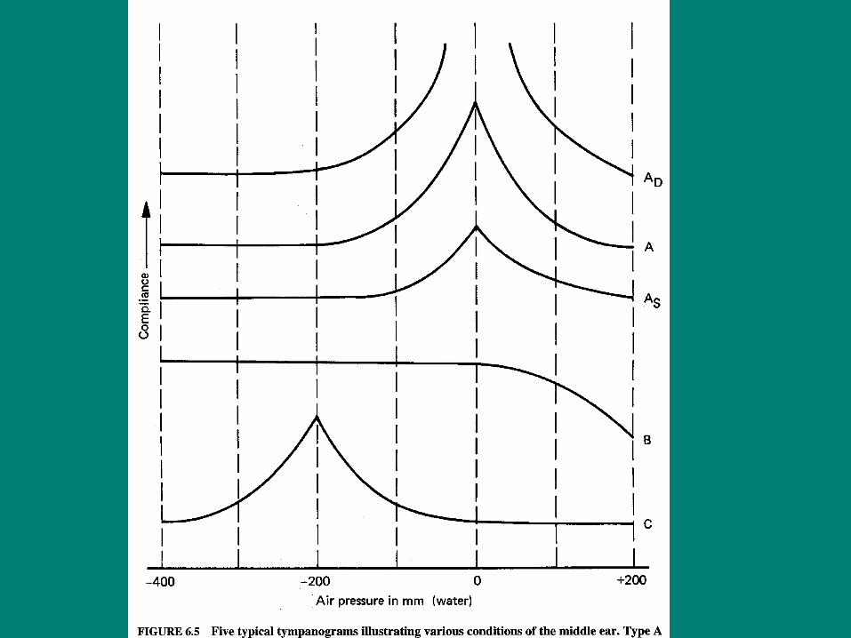
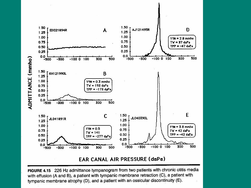
Physical Volume of ear canal Normally is between 1.0 to 1.5 cc
•True Type B tympanogram has normal PV
•If Type B with tiny PV, then probe tip is against EAM wall
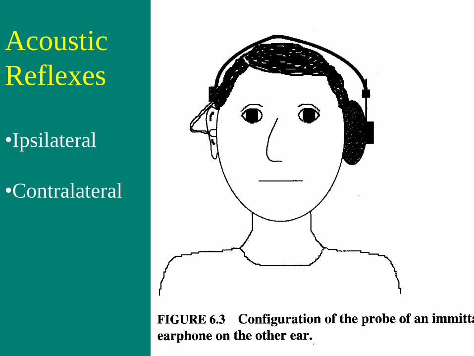
Acoustic
Reflexes
•Ipsilateral
•Contralateral
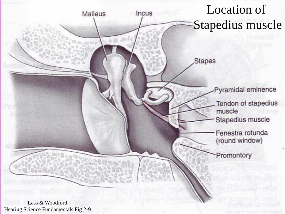
Location of
Stapedius muscle
Lass & Woodford
Hearing Science Fundamentals Fig 2-9
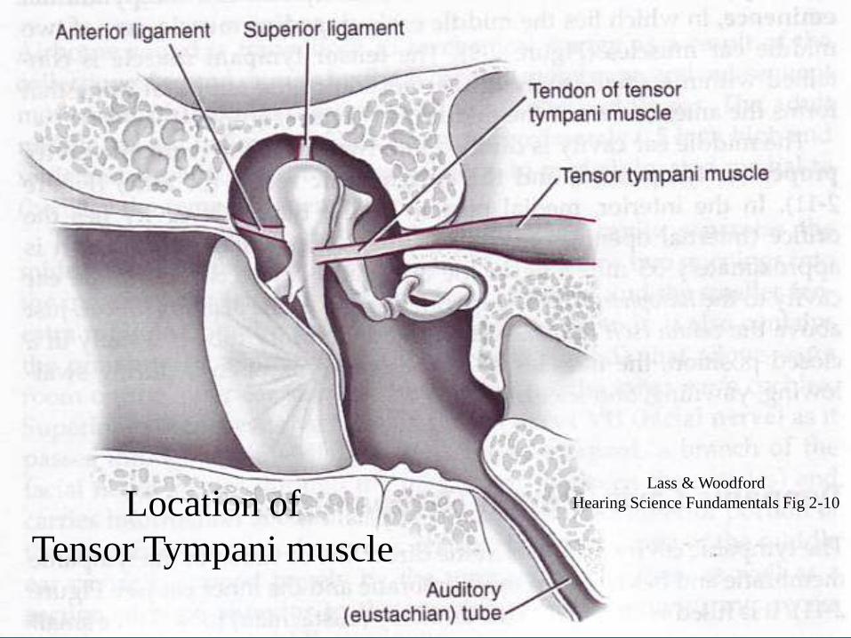
Location of
Tensor Tympani muscle
Lass & Woodford
Hearing Science Fundamentals Fig 2-10

V nerve
to Tensor Tympani
VII nerve
to Stapedius
V nerve
to Tensor Tympani
VII nerve
to Stapedius
Acoustic Reflex Arc
From Martin, F.
Introduction to Audiology
Allyn & Bacon
8th edition, 2003
Note:
if one ear gets
the loud sound,
the AR occurs
in both ears

Another view of the Acoustic Reflex Arc
This ear is
getting the stimulus
and has an AR
This ear did not get
the stimulus but still
has an AR

Question:
Why do we really have acoustic reflexes??
Musiek
&
Rintelmann
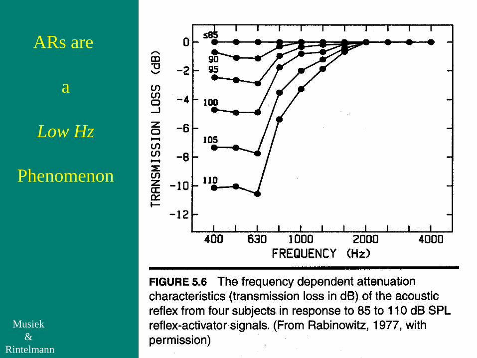
ARs are
a
Low Hz
Phenomenon

Answer:
Not to protect against NIHL
• one of the fallicies we are commonly lead to believe
ARs occur especially while we speak
• they actually kick in about 50 msec before we talk
Note ARs strongest for low Hz’s (500, 1000Hz)
• because our voices are louder for vowels
ARs reduce upward spread of masking
• allow us to hear high Hz’s better while talking
• all mammals have them

ARs & Speech Discrimination
AR deals with IHCs
AR arc: IHCs send afferent information to VIII nerve
IHCs are critical part of AR arc
OAEs deal with OHCs
Cochlea: OHCs receive efferent information
OHCs not involved w/ AR arc
Two people with same moderate SNHL
may have very different SD
One with good SD probably has ARs at reduced SLs
one with poor SD probably has absent ARs

Hair Cells:
A Closer Look
Inner hair cells
Outer hair cells
Picture from Australian Hearing
Hear & Now, issue 4, 1998

Normal
Inner
&
Outer
Hair Cells
Fig 1-7, Venema, T.
Compression for
Clinicians 2nd edition,
Thomson Delmar
Learning 2006

Damaged
Hair
Cells
(mostly
outer)
Fig 1-8, Venema, T.
Compression for
Clinicians 2nd edition,
Thomson Delmar
Learning 2006

With ARs, you are still using Tympanometry
measure how much 226 Hz tone is bouncing off TM
Tympanograms change middle ear compliance by air pressures
ARs change middle ear compliance by loud sounds!
ARs are done at Static Compliance
at the air pressure yielding highest tympanogram
The Acoustic Reflex & Tympanometry
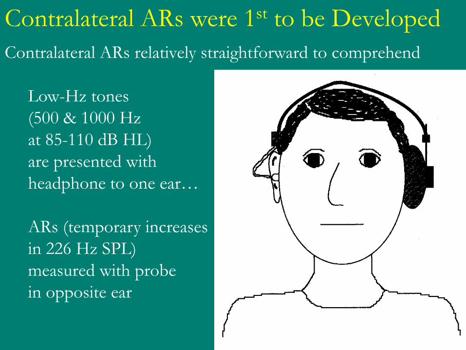
Contralateral ARs relatively straightforward to comprehend
Low-Hz tones
(500 & 1000 Hz
at 85-110 dB HL)
are presented with
headphone to one ear…
ARs (temporary increases
in 226 Hz SPL)
measured with probe
in opposite ear
Contralateral ARs were 1st to be Developed

Loud 500 or 1000Hz tone stimuli & 226 Hz tone to measure AR
are all in same ear canal at same time!
Temporary increases in 226 Hz SPL) are now measured
with probe in opposite ear
Ipsilateral ARs are harder to Comprehend
Probe & Headphone
226 Hz tone
to measure AR
500 or 1000 Hz
AR stimulus tone
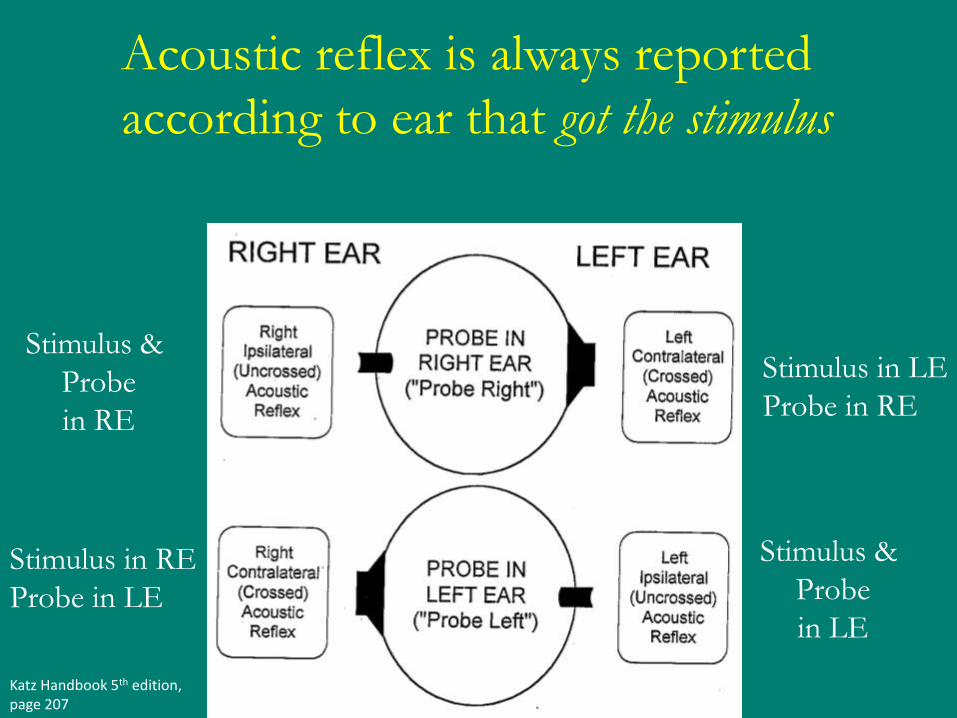
Katz Handbook 5th edition, page 207
Stimulus &
Probe
in RE
Stimulus in LE
Probe in RE
Stimulus in RE
Probe in LE
Stimulus &
Probe
in LE
Acoustic reflex is always reported
according to ear that got the stimulus

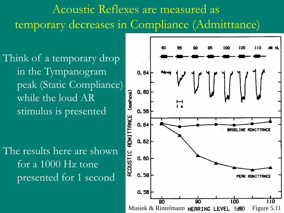
Figure 5.11 Musiek & Rintelmann
Acoustic Reflexes are measured as
temporary decreases in Compliance (Admitttance)
Think of a temporary drop
in the Tympanogram
peak (Static Compliance)
while the loud AR
stimulus is presented
The results here are shown
for a 1000 Hz tone
presented for 1 second

ARs Reported in Sensation Level (SL)
ARs tested in dB HL (tympanometer calibrated in dB HL)
ARs normally found between 80-100 dB HL
ARs reported in dB SL (always relative to one’s own thresholds)
ARs can be tested at 500, 1000, 2000, or 4000 Hz
Normal & mild-moderate SNHL
may have ARs at similar dB HL
but at very different SLs
Eg: normal HL & flat 50 dB SNHL may have ARs at 100 dB HL
but normal HL has ARs at 100 dB SL
the SNHL has ARs at 50 dB SL

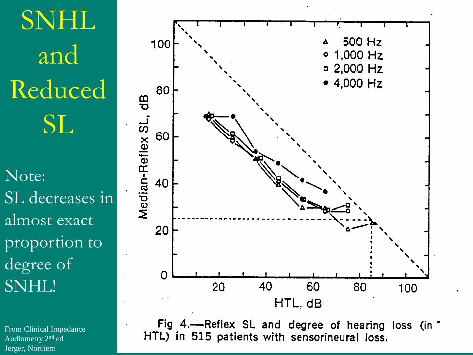
Note:
The SL for ARs
tends to decrease
linearly as the
degree of SNHL
increases (up to
about 60 dB HL
ARs
and
SNHL
From Clinical Impedance
Audiometry 2nd ed
Jerger, Northern
Note:
SL decreases in
almost exact
proportion to
degree of
SNHL!
SNHL
and
Reduced
SL

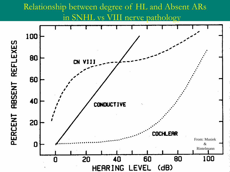
Absent ARs
and
SNHL
Note:
Absence of ARs
increases dramatically
with SNHL of
60 dB HL or more

Bilateral SNHL & ARs
Katz, Handbook of
Clinical Audiology

Unilateral SNHL & ARs
Contralateral & Ipsilateral AR are normal for both ears
Katz, Handbook of
Clinical Audiology fig 13.18

Unilateral Conductive HL
Conductive HL tends to obliterate ARs
Contralateral ARs with stimulus in good ear:
mechanical middle ear problems in bad ear prevent AR
Contralateral ARs with stimulus in bad ear:
HL in bad ear prevents intensity sufficient to cause AR in opposite ear
Result: Only good ear Ipsilateral AR present

Conductive HL tends to obliterate ARs
Bilateral Conductive HL
Contralateral & Ipsilateral ARs all absent

Katz, Handbook of Clinical Audiology

Katz, Handbook of Clinical Audiology
From: Musiek
&
Rintelmann
Relationship between degree of HL and Absent ARs
in SNHL vs VIII nerve pathology

Katz, Handbook of Clinical Audiology fig 13.18
Stimulus Tone
No Decay
No Decay b/c AR
reduced by <50%
over 10 sec
AR Decay b/c AR
Reduced by 50%
over 10 sec
Acoustic Reflex Decay & VIII nerve pathology