twice nobel prize winner twice nobel prize winner frederick sanger hard work is paid in form of...
TRANSCRIPT

Twice Nobel Prize WinnerTwice Nobel Prize Winner
FREDERICK SANGERFREDERICK SANGERHARD WORK IS PAID IN FORM HARD WORK IS PAID IN FORM
OF AWARDSOF AWARDS
Prasanna KhandavilliPrasanna Khandavilli

Curiosity is the key for Scientific Curiosity is the key for Scientific DiscoveryDiscovery

Frederick SangerFrederick Sanger
"for his work on the structure of "for his work on the structure of proteins, especially that of insulinproteins, especially that of insulin””
The Nobel Prize in Chemistry 1958The Nobel Prize in Chemistry 1958

The Nobel Prize in Chemistry 1980The Nobel Prize in Chemistry 1980
“for their contributions concerning the determination of base sequences in nucleic acids” Walter Gilbert Frederick Sanger

Frederick SangerFrederick Sanger
Born: August 13, 1918Born: August 13, 1918
Place of Birth: Rendcombe, Gloucestershire, Place of Birth: Rendcombe, Gloucestershire, EnglandEngland
Residence: U.S.A./Great BritainResidence: U.S.A./Great Britain
Affiliation: MRC Laboratory of Molecular Affiliation: MRC Laboratory of Molecular Biology, CambridgeBiology, Cambridge

Basic Principles of Protein Basic Principles of Protein ChemistryChemistry
Proteins - Amino Acid residuesProteins - Amino Acid residues
Physical and Biological Properties-Physical and Biological Properties-
Arrangement of the Amino Acid residuesArrangement of the Amino Acid residues

Bergmann and NiemannBergmann and Niemann
Periodic arrangement of Amino AcidsPeriodic arrangement of Amino Acids
Pure protein – A random mixture of similar Pure protein – A random mixture of similar
individualsindividuals

ChibnallChibnall
Studies on Insulin:Studies on Insulin:
Simpler compositionSimpler composition Tryptophan and Methionine absentTryptophan and Methionine absent Accurate analysisAccurate analysis

Van Slyke ProcedureVan Slyke Procedure
High content of free High content of free αα-amino groups-amino groups Short Polypeptide chainsShort Polypeptide chains
Jensen & Evans:Jensen & Evans:
Phenylalanine at the end of one of the chainsPhenylalanine at the end of one of the chains
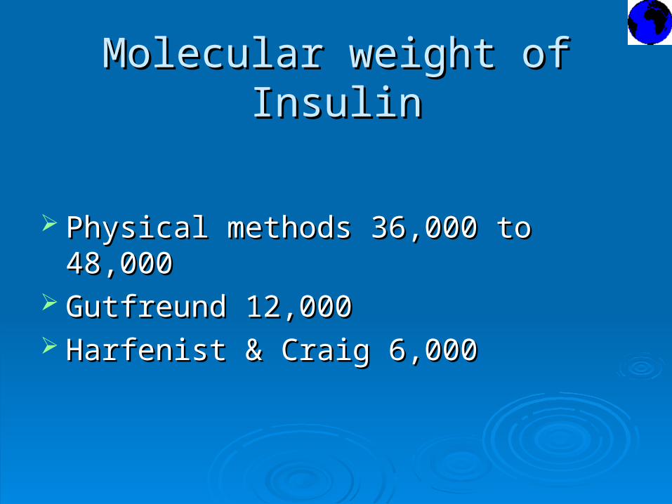
Molecular weight of InsulinMolecular weight of Insulin
Physical methods 36,000 to 48,000Physical methods 36,000 to 48,000 Gutfreund 12,000Gutfreund 12,000 Harfenist & Craig 6,000Harfenist & Craig 6,000

Dinitrophenyl (DNP) methodDinitrophenyl (DNP) method
1:2:4 flourodinitrobenzene (FDNB)1:2:4 flourodinitrobenzene (FDNB)
*Alkaline conditions*Alkaline conditions
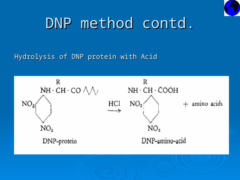
DNP method contd.DNP method contd.
Hydrolysis of DNP protein with AcidHydrolysis of DNP protein with Acid

DNP method contd.DNP method contd.
Extraction with EtherExtraction with Ether
Fractionation (Partition Chromatography)Fractionation (Partition Chromatography)
Comparison of Chromatographic rates (Silica-gel Comparison of Chromatographic rates (Silica-gel Chromatography or Paper Chromatography)Chromatography or Paper Chromatography)
Identification and Estimation CalorimetricallyIdentification and Estimation Calorimetrically

DNP labeling of InsulinDNP labeling of Insulin
Three yellow DNP-derivativesThree yellow DNP-derivatives
εε-DNP-lysine (not extracted with Ether)-DNP-lysine (not extracted with Ether) DNP-phenylalanineDNP-phenylalanine DNP-glycineDNP-glycine

Edman phenyl isothiocyanate Edman phenyl isothiocyanate methodmethod
Standard method for studying N-terminal Standard method for studying N-terminal residuesresidues

Disulphide bridgesDisulphide bridges
Cystine residuesCystine residues
Reduction to –SH derivatives Reduction to –SH derivatives
Polymerization gave insoluble productsPolymerization gave insoluble products
How to break these Disulfide bridges?How to break these Disulfide bridges?

Oxidation with Performic AcidOxidation with Performic Acid

Precipitation of Oxidized InsulinPrecipitation of Oxidized Insulin
Fraction A :Fraction A : N-terminal residue GlycineN-terminal residue Glycine AcidicAcidic Simpler composition (Lys, Arg, His, Phe, Simpler composition (Lys, Arg, His, Phe, Thr, Pro were absent)Thr, Pro were absent)
Fraction B:Fraction B: N-terminal residue PhenylalanineN-terminal residue Phenylalanine Basic Amino acidsBasic Amino acids
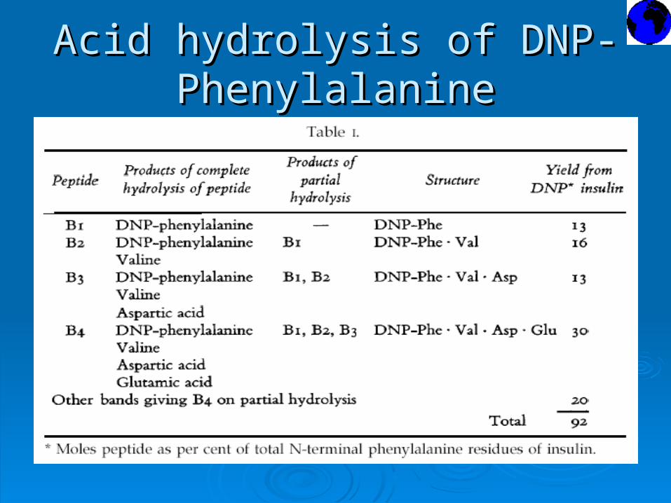
Acid hydrolysis of DNP-Acid hydrolysis of DNP-PhenylalaninePhenylalanine

ConclusionsConclusions
Position of residuesPosition of residues
Only two types of chainsOnly two types of chains
Molecular weight 12,000 Molecular weight 12,000

FractionationFractionation
Paper Chromatography for Fractionation ofPaper Chromatography for Fractionation of
small peptidessmall peptides
Consden, Gordon, Martin & Synge worked onConsden, Gordon, Martin & Synge worked on
pentapeptide Gramicidin-Spentapeptide Gramicidin-S

Fraction B studiesFraction B studies
Ionophoresis, Ion-exchange Chromatography,Ionophoresis, Ion-exchange Chromatography,
Adsorption on Charcoal Adsorption on Charcoal
5-20 peptides5-20 peptides
Paper ChromatographyPaper Chromatography
Analysis of the constituent Amino AcidsAnalysis of the constituent Amino Acids
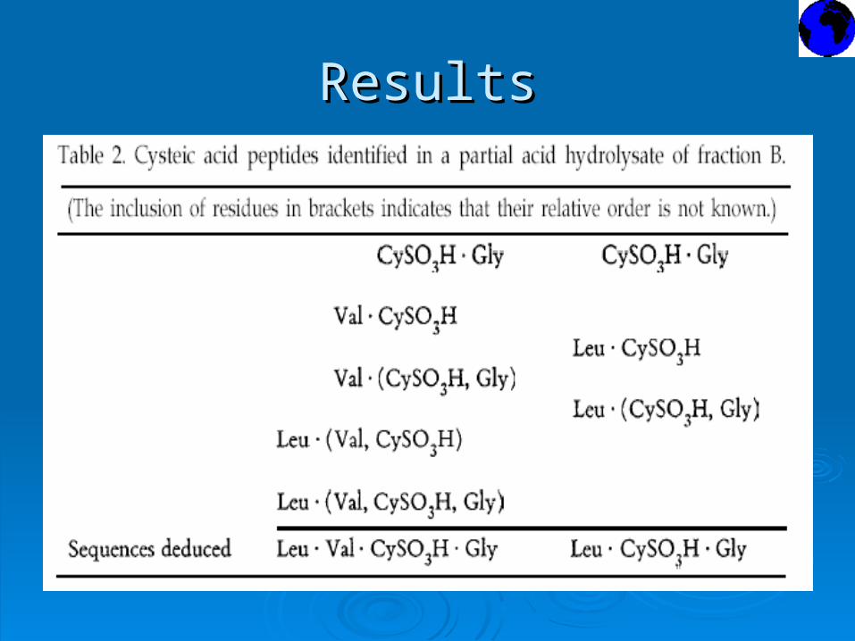
ResultsResults

ConclusionsConclusions
Five sequences present in Phenylalanine ChainFive sequences present in Phenylalanine Chain

ProblemsProblems
How the 5 sequences are joined ?How the 5 sequences are joined ?
Hurdles in solving this mystery:Hurdles in solving this mystery:
Technical difficulty in fractionating peptides Technical difficulty in fractionating peptides with non-polar residues (Tyr & Leu)with non-polar residues (Tyr & Leu)
Acid lability of the bonds involving Serine and Acid lability of the bonds involving Serine and ThreonineThreonine

Solution is………Solution is………
Enzymatic Hydrolysis:Enzymatic Hydrolysis:
Use of Proteolytic enzymes Use of Proteolytic enzymes
More specific than acid hydrolysisMore specific than acid hydrolysis

Proteolytic EnzymesProteolytic Enzymes
Pepsin – Peptide Bp3 fragmentPepsin – Peptide Bp3 fragment
Phe (CySO,H, Asp, Glu, Ser, Gly, Val, Leu, His)Phe (CySO,H, Asp, Glu, Ser, Gly, Val, Leu, His)
Trypsin, Chymotrypsin studiesTrypsin, Chymotrypsin studies

Fraction A studiesFraction A studies
Problems in applying fraction B studies to Problems in applying fraction B studies to fraction A:fraction A:
Few residues that occur only once Few residues that occur only once
Less susceptible to enzymatic hydrolysisLess susceptible to enzymatic hydrolysis
Water soluble peptides- difficult to fractionate Water soluble peptides- difficult to fractionate on paper chromatographyon paper chromatography

Paper IonophoresisPaper Ionophoresis
pH 2.75pH 2.75
-COOH groups uncharged-COOH groups uncharged
-SO-SO33H groups negative chargeH groups negative charge
-NH-NH22 groups positive charge groups positive charge
pH 3.5pH 3.5
-COOH groups charged-COOH groups charged

Results of Paper IonophoresisResults of Paper Ionophoresis
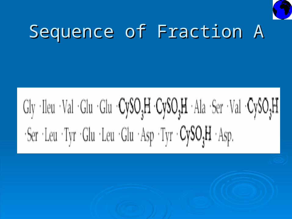
Sequence of Fraction ASequence of Fraction A

Acid HydrolysisAcid Hydrolysis
Ammonia produced from Amide groups on Ammonia produced from Amide groups on
Aspartic and Glutamic acid residuesAspartic and Glutamic acid residues
Position of Amide groups:Position of Amide groups:
Ionophoretic ratesIonophoretic rates
Amide contents of peptidesAmide contents of peptides

Arrangement of Disulphide bridgesArrangement of Disulphide bridges
Assumptions and hypothesis:Assumptions and hypothesis:
Harfenist & Craig Mol Wt 6000Harfenist & Craig Mol Wt 6000
Two chains with three disulphide bridges:Two chains with three disulphide bridges:
Two bridges connecting the two chainsTwo bridges connecting the two chains
One intrachain bridge in fraction AOne intrachain bridge in fraction A

Disulphide interchange reactionDisulphide interchange reaction

Disulphide interchange reactionDisulphide interchange reaction Contd. Contd.
Two types of disulphide interchange reactionsTwo types of disulphide interchange reactions In neutral & alkaline solution catalyzed by In neutral & alkaline solution catalyzed by
– –SH compoundsSH compounds

Enzymic HydrolysisEnzymic Hydrolysis
Chymotrypsin actionChymotrypsin action
-CySO-CySO33H.AspNHH.AspNH
-Leu.Val. CySO-Leu.Val. CySO33H.Gly.Glu.Arg.Gly.Phe.PheH.Gly.Glu.Arg.Gly.Phe.Phe

Cystine peptide structureCystine peptide structure

The Structure of InsulinThe Structure of Insulin

Sequenced Insulin supports Protein Sequenced Insulin supports Protein chemistry theorieschemistry theories
Hofmeister & Fischer – Classical peptide Hofmeister & Fischer – Classical peptide hypothesishypothesis
No evidence of periodicityNo evidence of periodicity
Random orderRandom order
Unique & most significant orderUnique & most significant order

Insulin from different speciesInsulin from different species

Determination of Nucleotide Determination of Nucleotide SequencesSequences
Smallest DNA molecule - Bacteriophage Smallest DNA molecule - Bacteriophage φφX174 – 5,000 nucleotidesX174 – 5,000 nucleotides
tRNA - 75 nucleotidestRNA - 75 nucleotides

Fractionation of Fractionation of 3232P-labelled P-labelled oligonucleotidesoligonucleotides
G.G.Brownlee and B.G.Barrell method:G.G.Brownlee and B.G.Barrell method:
Partial degradation by enzymesPartial degradation by enzymes Separation of smaller productsSeparation of smaller products Determination of sequenceDetermination of sequence Applied to RNA sequencesApplied to RNA sequences

DisadvantagesDisadvantages
Slow and tediousSlow and tedious Requires successive digestions and Requires successive digestions and
fractionationsfractionations Not easy to apply to larger DNA moleculesNot easy to apply to larger DNA molecules

Copying ProceduresCopying Procedures
C.Weissmann: Bacteriophage QC.Weissmann: Bacteriophage Qββ -Q-Qββ Replicase – Complementary copy Replicase – Complementary copy -Pulse-labeling with radio actively labeled-Pulse-labeling with radio actively labeled nucleotidesnucleotides
DNA Polymerase substitutes ReplicaseDNA Polymerase substitutes Replicase -Primer, Triphosphates containing -Primer, Triphosphates containing 3232P in P in αα
position - Sangerposition - Sanger

Copying ProcedureCopying Procedure

Primer SourcePrimer Source
Synthetic OligonucleotidesSynthetic Oligonucleotides
Restriction enzymes Restriction enzymes

Copying procedureCopying procedure
ResultsResults Short specific regions of labeled DNA were Short specific regions of labeled DNA were
obtainedobtained
Unable to obtain individual residues for Unable to obtain individual residues for sequencingsequencing
How to obtain individual How to obtain individual nucleotide residues?nucleotide residues?
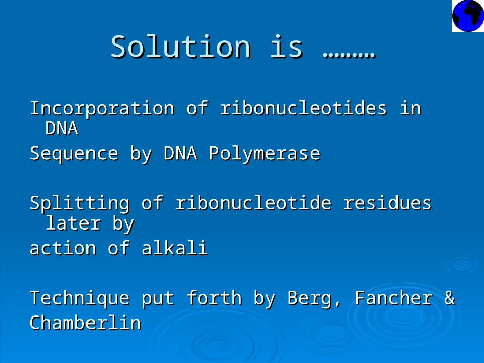
Solution is ………Solution is ………
Incorporation of ribonucleotides in DNA Incorporation of ribonucleotides in DNA Sequence by DNA PolymeraseSequence by DNA Polymerase
Splitting of ribonucleotide residues later by Splitting of ribonucleotide residues later by action of alkaliaction of alkali
Technique put forth by Berg, Fancher & Technique put forth by Berg, Fancher & ChamberlinChamberlin

The ‘Plus and Minus’ methodThe ‘Plus and Minus’ method
αα[[3232P]-dNTP labeling and sequence specific P]-dNTP labeling and sequence specific
terminationtermination
J.E.Donelson - Ionophoresis of products on J.E.Donelson - Ionophoresis of products on
acrylamide gelsacrylamide gels

The Dideoxy methodThe Dideoxy method
Quicker and more accurateQuicker and more accurate
φφX174X174 Bacteriophage G4Bacteriophage G4 Mammalian mitochondrial DNAMammalian mitochondrial DNA

Dideoxynucleoside triphosphatesDideoxynucleoside triphosphates
Lack 3’ hydroxyl groupLack 3’ hydroxyl group Incorporated into growing DNA chain by Incorporated into growing DNA chain by
DNA polymeraseDNA polymerase Chain terminating analoguesChain terminating analogues

Dideoxy nucleotide triphosphateDideoxy nucleotide triphosphate

Chain Termination with ddNTPChain Termination with ddNTP

Chain-Terminating MethodChain-Terminating Method

Autoradiograph Autoradiograph DNA sequencing gel DNA sequencing gel

Chain terminating methodChain terminating method
Problem: Problem: Requires single Requires single
stranded DNA as templatestranded DNA as template
SolutionSolutionA.J.H.Smith Exonuclease IIIA.J.H.Smith Exonuclease III
Fragments cloned in Fragments cloned in
plasmid vectors and Humanplasmid vectors and Human
mitochondrial DNAmitochondrial DNA
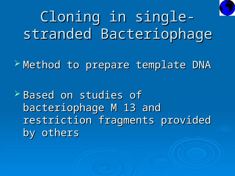
Cloning in single-stranded Cloning in single-stranded BacteriophageBacteriophage
Method to prepare template DNA Method to prepare template DNA
Based on studies of bacteriophage M 13 and Based on studies of bacteriophage M 13 and restriction fragments provided by othersrestriction fragments provided by others

CloningCloning
Gronenborn & Messing – M13 BacteriophageGronenborn & Messing – M13 Bacteriophage
Insert of Insert of ββ-galactosidase gene with an EcoRI -galactosidase gene with an EcoRI restriction enzyme site in itrestriction enzyme site in it
Heidccker 96-nucleotide long restriction Heidccker 96-nucleotide long restriction fragment from M13 vector flanking EcoRI sitefragment from M13 vector flanking EcoRI site

CloningCloning

AdvantagesAdvantages
Same primer on all clonesSame primer on all clones Very efficient and rapid method of Very efficient and rapid method of
fractionating fractionating Each clone represents progeny of a single Each clone represents progeny of a single
molecule and is therefore puremolecule and is therefore pure No theoretical limit to the size of DNA that No theoretical limit to the size of DNA that
could be sequencedcould be sequenced
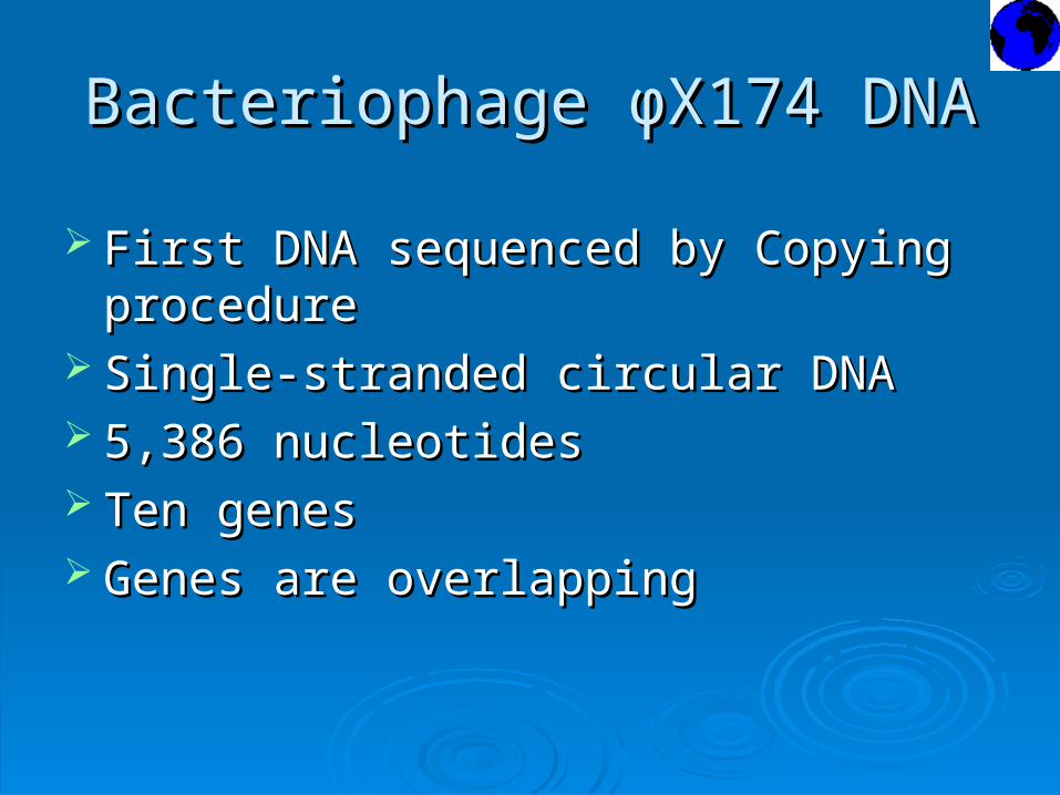
Bacteriophage Bacteriophage φφX174 DNAX174 DNA
First DNA sequenced by Copying procedureFirst DNA sequenced by Copying procedure Single-stranded circular DNASingle-stranded circular DNA 5,386 nucleotides 5,386 nucleotides Ten genesTen genes Genes are overlappingGenes are overlapping

Gene Map Gene Map

Reading FramesReading Frames

Mammalian mitochondrial DNAMammalian mitochondrial DNA
Two ribosomal RNAs (rRNAs)Two ribosomal RNAs (rRNAs) 22-23 transfer RNAs (tRNAs)22-23 transfer RNAs (tRNAs) 10-13 inner mitochondrial membrane proteins 10-13 inner mitochondrial membrane proteins
Transcription and translation machinery of Transcription and translation machinery of
mitochondria is different from other biologicalmitochondria is different from other biological
systemssystems

The genetic code in mitochondriaThe genetic code in mitochondria
Steffans & Buse - Sequence of Subunit II of Steffans & Buse - Sequence of Subunit II of
Cytochrome Oxidase (COII) from bovine Cytochrome Oxidase (COII) from bovine
mitochondriamitochondria
Barrel, Bankier & Drouin – DNA sequence for Barrel, Bankier & Drouin – DNA sequence for
protein homologous to the above amino acid protein homologous to the above amino acid
sequence in human beingssequence in human beings

FindingsFindings
TGA - Tryptophan (not termination codon)TGA - Tryptophan (not termination codon) ATA – Methionine (not isoleucine)ATA – Methionine (not isoleucine)
Is it Species variation (?)Is it Species variation (?)
Young & Anderson-isolated bovine mtDNA Young & Anderson-isolated bovine mtDNA
- Confirmed Uniqueness of mtDNA- Confirmed Uniqueness of mtDNA

mtDNA Genetic CodemtDNA Genetic Code


Transfer RNAsTransfer RNAs
Cytoplasmic tRNAs:Cytoplasmic tRNAs:
Clover-leaf modelClover-leaf model
Invariable featuresInvariable features Mammalian mt-tRNA:Mammalian mt-tRNA:
Invariable features missingInvariable features missing
Serine tRNA lacks loop of cloverleaf structureSerine tRNA lacks loop of cloverleaf structure

Cytoplasmic Transfer RNAsCytoplasmic Transfer RNAs
Wobble effect forming Family boxesWobble effect forming Family boxes

Mitochondrial Transfer RNAsMitochondrial Transfer RNAs
22 tRNA genes in Mammalian mtDNA 22 tRNA genes in Mammalian mtDNA
For all family boxes-For all family boxes-
Only one which had a T in the positionOnly one which had a T in the position
corresponding to the third position of the corresponding to the third position of the codoncodon
One tRNA-Recognizes all codons in a familyOne tRNA-Recognizes all codons in a family
boxbox

Distribution of Protein genesDistribution of Protein genes
Cytochrome oxidaseCytochrome oxidase
ATPase complexATPase complex
Cytochrome bCytochrome b
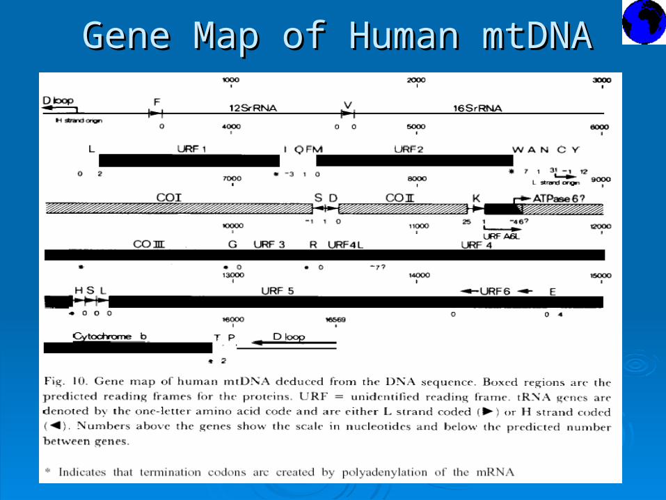
Gene Map of Human mtDNAGene Map of Human mtDNA

Mitochondrial DNA ConclusionsMitochondrial DNA Conclusions
Very compact structureVery compact structure
Reading frames coding for proteins and rRNA Reading frames coding for proteins and rRNA genes are flanked by tRNA genesgenes are flanked by tRNA genes
Simple model for transcriptionSimple model for transcription

TRENDS AND PROGRESS TRENDS AND PROGRESS
IN IN
SEQUENCING FIELDSEQUENCING FIELD

TrendsTrends
19741974 Conventional Sequencing MethodConventional Sequencing Method Sanger, Sanger, Maxam & GilbertMaxam & Gilbert
19861986 A regiment of scientists and technicians – A regiment of scientists and technicians – Caltech and Applied Biosystems Inc.,invented Caltech and Applied Biosystems Inc.,invented the the Automated DNA Fluorescence Sequencer. Automated DNA Fluorescence Sequencer.

TrendsTrends
Craig Venter's Craig Venter's Sequencing MethodSequencing Method
In 1991, working withIn 1991, working with Nobel laureate Hamilton Nobel laureate Hamilton
Smith, Venter's genomicSmith, Venter's genomic research project (TIGR) research project (TIGR)
created a new sequencing created a new sequencing process coined ‘shotgun process coined ‘shotgun technique’.technique’.
““Trend Setter” & “Gene Hunter”Trend Setter” & “Gene Hunter” Dr. Craig VenterDr. Craig Venter
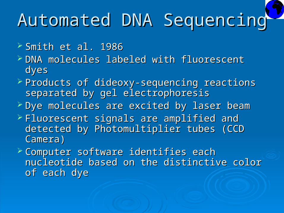
Automated DNA SequencingAutomated DNA Sequencing
Smith et al. 1986Smith et al. 1986 DNA molecules labeled with fluorescent dyesDNA molecules labeled with fluorescent dyes Products of dideoxy-sequencing reactions Products of dideoxy-sequencing reactions
separated by gel electrophoresisseparated by gel electrophoresis Dye molecules are excited by laser beamDye molecules are excited by laser beam Fluorescent signals are amplified and detected Fluorescent signals are amplified and detected
by Photomultiplier tubes (CCD Camera)by Photomultiplier tubes (CCD Camera) Computer software identifies each nucleotide Computer software identifies each nucleotide
based on the distinctive color of each dyebased on the distinctive color of each dye

Automated Sequencing (Contd)Automated Sequencing (Contd)

Automated Sequencing (Contd)Automated Sequencing (Contd)

Genome ProjectsGenome Projects 1999 “Celera genomics”– Rockville, Maryland
Drosophila genome
2000 Completed Human Genome Project http:// www.genome.gov/
2002 Mouse Genome Project www.informatics.jax.org/

Human Genome ProjectHuman Genome Project The Human Genome ProjectThe Human Genome Project
Started in 1988, Public Domain Started in 1988, Public Domain Collaborative work between Celera Genomics Collaborative work between Celera Genomics and NIHand NIH
Accomplishments:Accomplishments: Identify all the approximately 35,000 genes in Identify all the approximately 35,000 genes in
human DNA human DNA Determine the sequences of the 3 billion Determine the sequences of the 3 billion
chemical bases that make up human DNA chemical bases that make up human DNA (completed July 2000)(completed July 2000)

Other Genome DatabasesOther Genome Databases A lot of Organism specific databases at NCBI A lot of Organism specific databases at NCBI
Allows for Comparative Genomics studies Allows for Comparative Genomics studies
Phylogenetic Analysis studiesPhylogenetic Analysis studies
Gene Annotation and Identification issuesGene Annotation and Identification issues
Drug therapy and Gene Therapy- Cystic Drug therapy and Gene Therapy- Cystic Fibrosis etc.Fibrosis etc.
DNA VaccinesDNA Vaccines

Insulin and BiotechnologyInsulin and Biotechnology
1978: Genentech, Inc. - Genetic engineering 1978: Genentech, Inc. - Genetic engineering techniques used to produce human insulin in techniques used to produce human insulin in E.E. colicoli
19831983: : Genetech, Inc. licensedGenetech, Inc. licensed Eli LilyEli Lily to make to make insulininsulin

Insulin Production in E.coliInsulin Production in E.coli

3D STRUCTURE OF INSULIN3D STRUCTURE OF INSULIN

Insulin TrendsInsulin Trends
Insulin was first isolated from the pancreas Insulin was first isolated from the pancreas of cows and pigs in the early 1920s of cows and pigs in the early 1920s
In 1978, a synthetic version of the human In 1978, a synthetic version of the human insulin gene was constructed and inserted insulin gene was constructed and inserted into the bacterium Eschericia coli, in the into the bacterium Eschericia coli, in the laboratory of Herbert Boyer at the laboratory of Herbert Boyer at the University of California at San Francisco University of California at San Francisco

Insulin Trends in MedicineInsulin Trends in Medicine Recombinant human insulin was developed Recombinant human insulin was developed
by Boyer's fledgling company, Genentech, in by Boyer's fledgling company, Genentech, in October of 1982, the first product of modern October of 1982, the first product of modern biotechnology biotechnology
HumulinHumulin
Various modes of delivering Insulin to the Various modes of delivering Insulin to the TissueTissue
Less Adverse reactions, More strict glucose Less Adverse reactions, More strict glucose control in diabeticscontrol in diabetics

ReferencesReferences
Nobel e-MuseumNobel e-Museum The Nobel Prize Internet ArchiveThe Nobel Prize Internet Archive Britannica Nobel Prizes, Guide to the Nobel PrizesBritannica Nobel Prizes, Guide to the Nobel Prizes Michigan State University, Department of ChemistryMichigan State University, Department of Chemistry Science DailyScience Daily http://www.geocities.com/jdelaney25/FrederickSahttp://www.geocities.com/jdelaney25/FrederickSa
nger.htmlnger.html The wellcome Trust Sanger InstituteThe wellcome Trust Sanger Institute

Questions and SuggestionsQuestions and Suggestions
Our View changes our WorldOur View changes our World