tuberculosis pathogenesis and transmission in response to old caseous pneumonia and produce...
TRANSCRIPT

Tuberculosis Pathogenesis and Transmission
Pamela B. Hackert, MD, JD, MPH
Objectives• Identify why the paradigm shift identifying the caseating
granuloma as the characteristic lesion of all TB occurred with the introduction of effective antibiotics
• Understand how using a three act model can better identify the actual pathology that is occurring in tuberculosis
• Review questions that can be addressed only by using the new paradigm
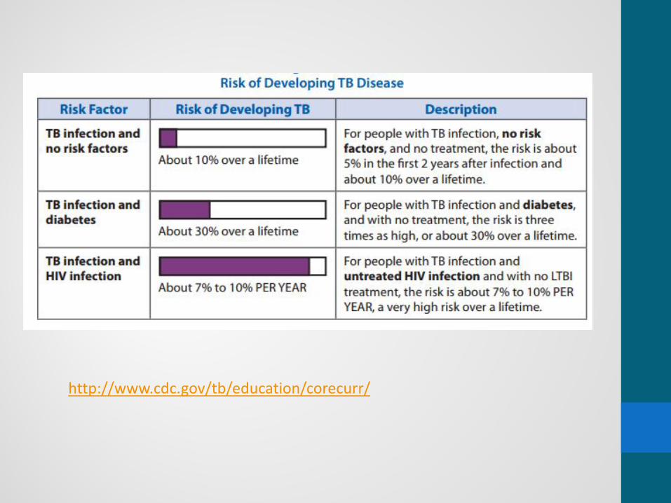
• Very briefly look at diabetes as a risk factor for progression to active disease

Looking at TB Pathogenesis With a Traditional Eye

Looking at TB Pathogenesis With a Traditional Eye

Looking at TB Pathogenesis With a Traditional Eye

Unanswered Questions For Traditional Paradigm of Pathogenesis of Tuberculosis
• What protects most adults from disease following infection?
• Why are immunocompetent young adults especially susceptible to disease and death?
• Why does recovery from disease fail to produce immunity, but actually produces increased susceptibility to recurrent disease?
• Why have vaccines that prevent disseminated TB in children, failed to protect adults from pulmonary TB?
• Why does post-primary TB localize in the upper lobes of the lungs?
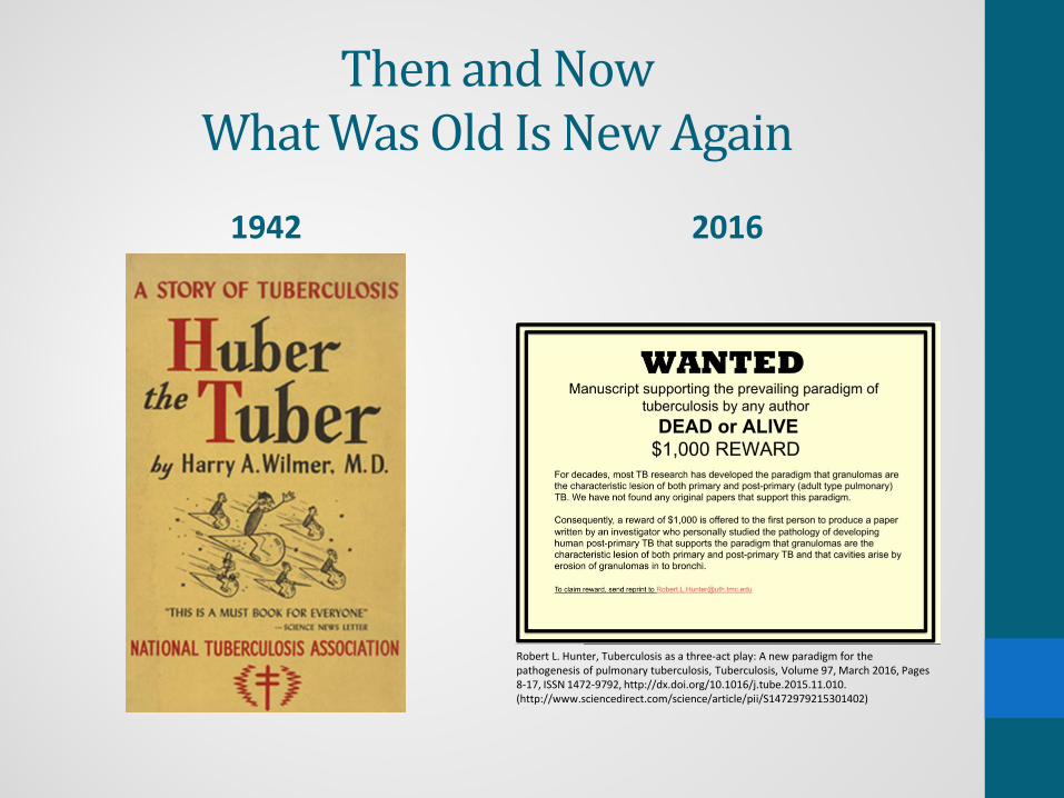
Then and NowWhat Was Old Is New Again
1942 2016
Robert L. Hunter, Tuberculosis as a three-act play: A new paradigm for the pathogenesis of pulmonary tuberculosis, Tuberculosis, Volume 97, March 2016, Pages 8-17, ISSN 1472-9792, http://dx.doi.org/10.1016/j.tube.2015.11.010.(http://www.sciencedirect.com/science/article/pii/S1472979215301402)

What Were They Thinking?(and how did they get off track?)
• Antibiotics has reduced the number of cases seen by pathologists of post primary tuberculosis
• Pre-antibiotic era investigators consistently described post primary TB as an exudative reaction• A tuberculous lipid pneumonia of foamy alveolar
macrophages• Undergoes caseation necrosis and fragmentation
to produce cavities
• Granulomas in post primary disease arise only in response to old caseous pneumonia and produce fibrosis, NOT cavities
• Concept that cavities arise from caseating granulomas arose from M.bovis studies• M.bovis does not produce post primary
tuberculosis in any species• Produces an aggressive primary TB that can
develop small cavities by erosion of caseating granulomas
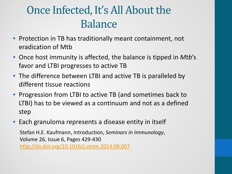
Once Infected, It’s All About the Balance
• Protection in TB has traditionally meant containment, not eradication of Mtb
• Once host immunity is affected, the balance is tipped in Mtb’s favor and LTBI progresses to active TB
• The difference between LTBI and active TB is paralleled by different tissue reactions
• Progression from LTBI to active TB (and sometimes back to LTBI) has to be viewed as a continuum and not as a defined step
• Each granuloma represents a disease entity in itselfStefan H.E. Kaufmann, Introduction, Seminars in Immunology, Volume 26, Issue 6, Pages 429-430http://dx.doi.org/10.1016/j.smim.2014.09.007
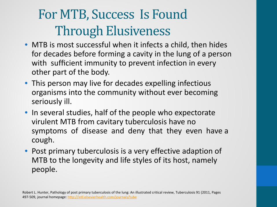
For MTB, Success Is Found Through Elusiveness
• MTB is most successful when it infects a child, then hides for decades before forming a cavity in the lung of a person with sufficient immunity to prevent infection in every other part of the body.
• This person may live for decades expelling infectious organisms into the community without ever becoming seriously ill.
• In several studies, half of the people who expectorate virulent MTB from cavitary tuberculosis have no symptoms of disease and deny that they even have a cough.
• Post primary tuberculosis is a very effective adaption of MTB to the longevity and life styles of its host, namely people.
Robert L. Hunter, Pathology of post primary tuberculosis of the lung: An illustrated critical review, Tuberculosis 91 (2011, Pages 497-509, journal homepage: http://intl.elsevierhealth.com/journals/tube

Pathology of Human TB
Hunter 2016
“Old” Paradigm Thinking
The current paradigm of the pathogenesis of TB considers TB to be a one act play in which the caseating granuloma modulated by cell mediated immunity (CMI) is the characteristic lesion of all TB. While this is an appropriate model for M. bovis and primary TB, it fails to recognize the existence of obstructive lobular pneumonia that initiates and drives all of post-primary TB.

The Three Distinct Stages Hypothesized
Hunter, 2016

Act IThe War of Attrition
• MTB try to multiply while the host attempts to contain them within granulomas
• With no or little immunity, there is greater lymphatic or hematogenous spread
• Control is through cell mediated immunity

Act II-The Sneak Attack• Act II Post-primary bronchogenic
TB begins asymptomatically in the apices of the lung, at some distance from the site initial infection
• It is part of latent TB since there are no clinical symptoms
• Few numbers of MTB in modified alveolar macrophages drive accumulation of host lipids and mycobacterial antigens in an isolated section of lung in preparation for a sudden necrotizing reaction sufficient to produce a cavity
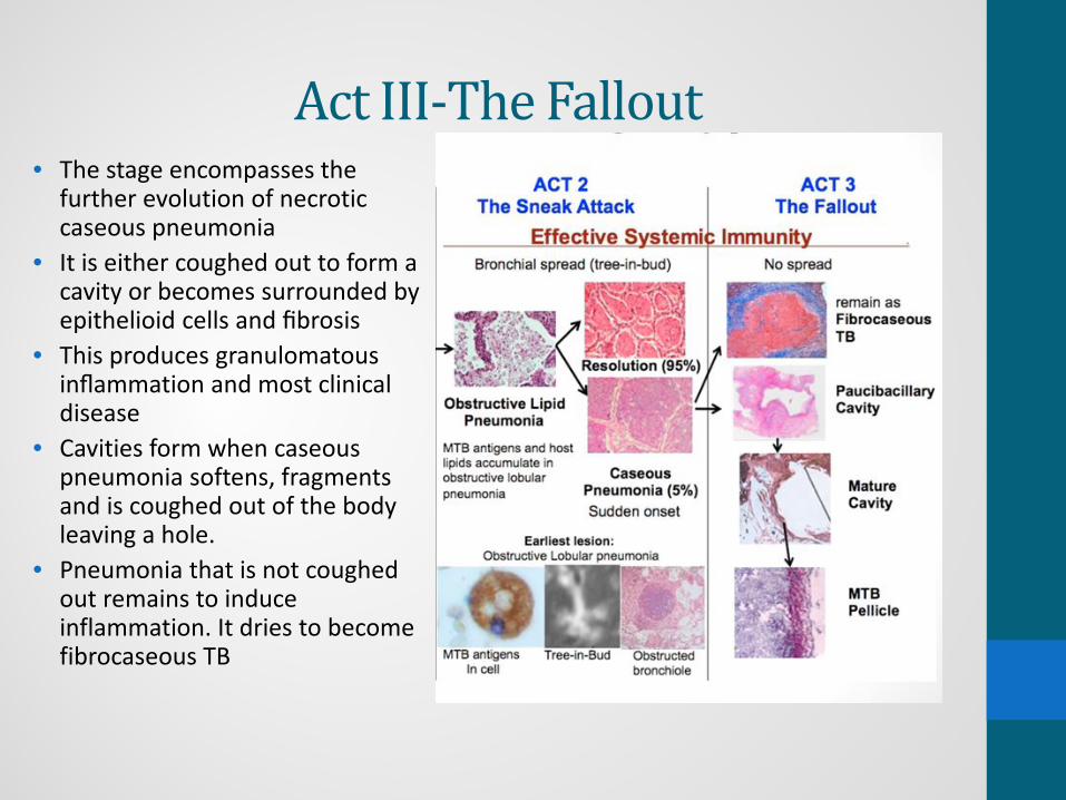
Act III-The Fallout• The stage encompasses the
further evolution of necrotic caseous pneumonia
• It is either coughed out to form a cavity or becomes surrounded by epithelioid cells and fibrosis
• This produces granulomatous inflammation and most clinical disease
• Cavities form when caseous pneumonia softens, fragments and is coughed out of the body leaving a hole.
• Pneumonia that is not coughed out remains to induce inflammation. It dries to become fibrocaseous TB
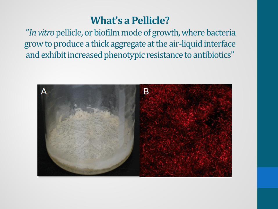
What’s a Pellicle? ”In vitro pellicle, or biofilm mode of growth, where bacteria grow to produce a thick aggregate at the air-liquid interface and exhibit increased phenotypic resistance to antibiotics”

Why does post-primary TB localize in the upper lobes of the lungs?
Hunter 2016

What protects most adults from disease following infection?
Hashtag throwback
Usually used when someone wants to post an old photo on
Instagram and expects reactions such as “awe so cute” or “that
was so great”
Why are immunocompetent young adults especially susceptible to disease and death?

How can multiple pulmonary lesions in a single lung act independently as if the others did not
exist?

Why does recovery from post-primary TB NOT produce immunity?

Why have vaccines that prevent disseminated TB in children, failed to protect adults from
pulmonary TB?

https://www.theguardian.com/science/2016/feb/23/rats-who-sniff-out-tubersulosis?CMP=share_btn_link

TB Laboratory Testing &Case Studies
April 8, 2016Angie Schooley, MT
James Sunstrum, M.D.
Objectives
• Review the cascade of laboratory tests a clinician may order to diagnose TB disease
• Integrate molecular assays with culture results• Demonstrate the proper use of TB diagnostic tests using 3 sample cases of TB disease (easy, medium & difficult)
4/5/2016 2

Disclosures
• None
4/5/2016 3

What do all the words mean? 😵
◦ Prevent Disease ◦ Promote Wellness ◦ Improve Quality of Life ◦
NAA Amplification MGIT MTD
PCR Pyrosequencing Molecular mutation HPLC MALDI‐Tof Gene Xpert
MDDR NAAT Genotyping
WGS 16 S Sequencing
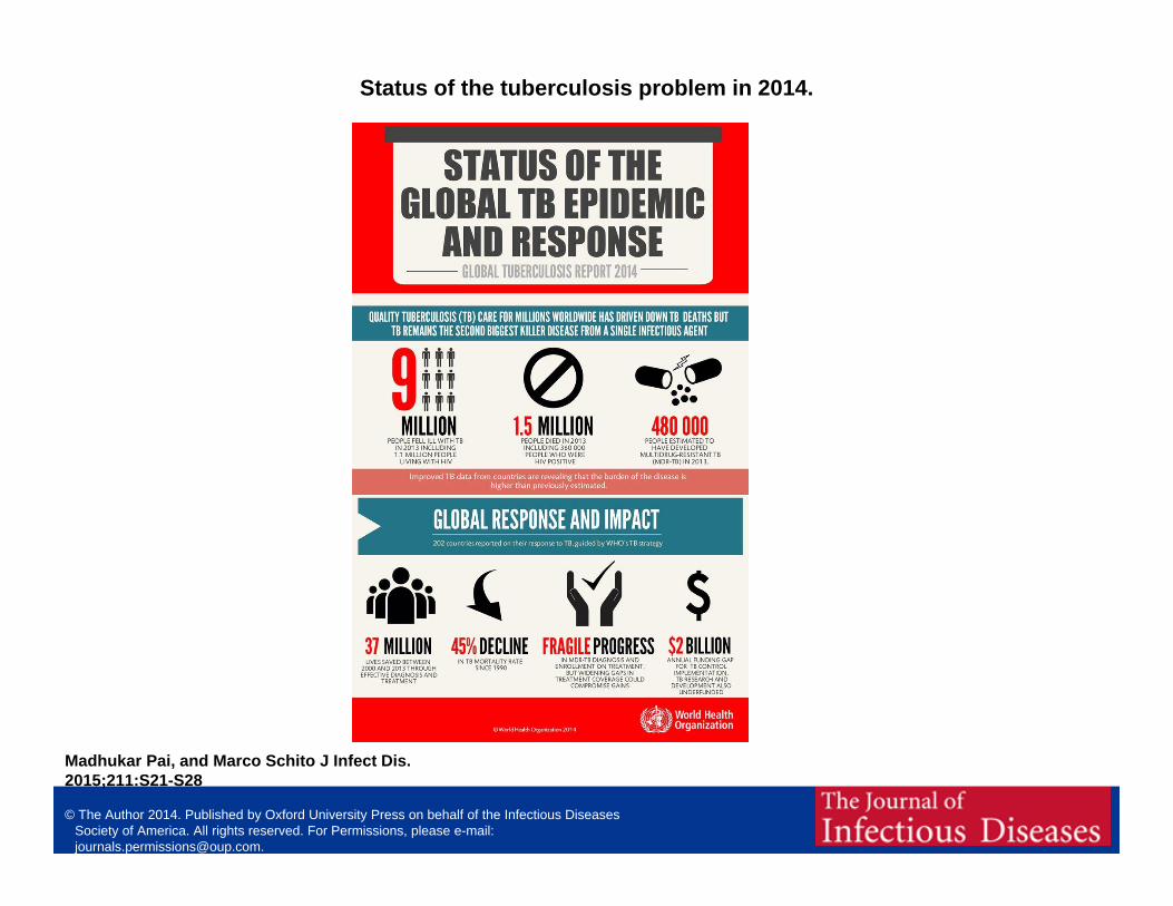
Status of the tuberculosis problem in 2014.
Madhukar Pai, and Marco Schito J Infect Dis. 2015;211:S21-S28
© The Author 2014. Published by Oxford University Press on behalf of the Infectious Diseases Society of America. All rights reserved. For Permissions, please e-mail: [email protected].

Does this patient have TB disease?
CLINICAL CLUES• Cough > 2 weeks• Fever > 2weeks• Exposure to TB• Chronic immune
suppression• Endemic country• Abnormal physical exam
LABORATORY TESTS• PPD• IGRA• Sputum studies: AFB CulturesMolecular studies• X‐rays• Biopsies
4/5/2016 6

Recommended diagnostic options for pulmonary TB
• See the bugs [AFB microscopy]
• Multiply the bugs [NAATs]
• Grow the bugs [cultures]
• Courtesy of Prof. Madhukar Pai, MD, PhD Mayo TB Center Webinar March 2016
4/5/2016 7

Mycobacterial ExaminationMycobacterial examination has 6 stages:
1. Proper specimen collection
2. Examination of acid‐fast bacilli (AFB) smears
3. Direct identification (NAAT‐nucleic acid amplification test)
4. Specimen culturing and final identification
5. Drug susceptibility testing
6. TB genotyping
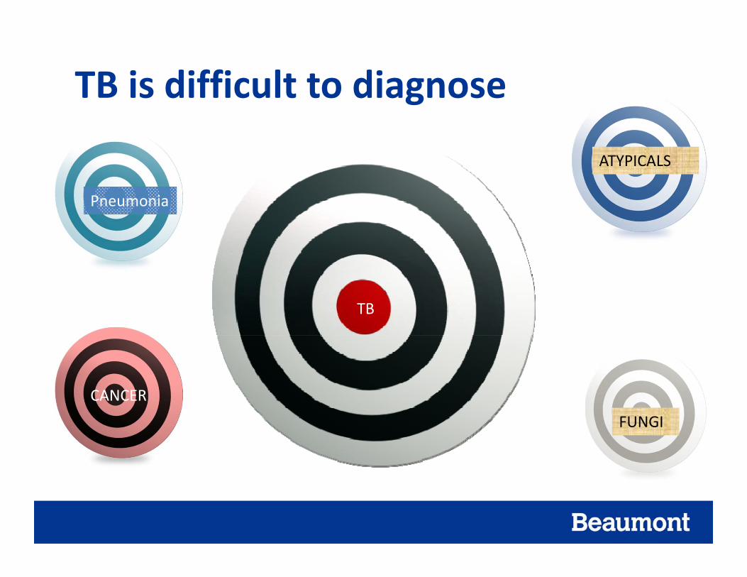
TB is difficult to diagnose
ATYPICALS
FUNGI
TB
Pneumonia
CANCER
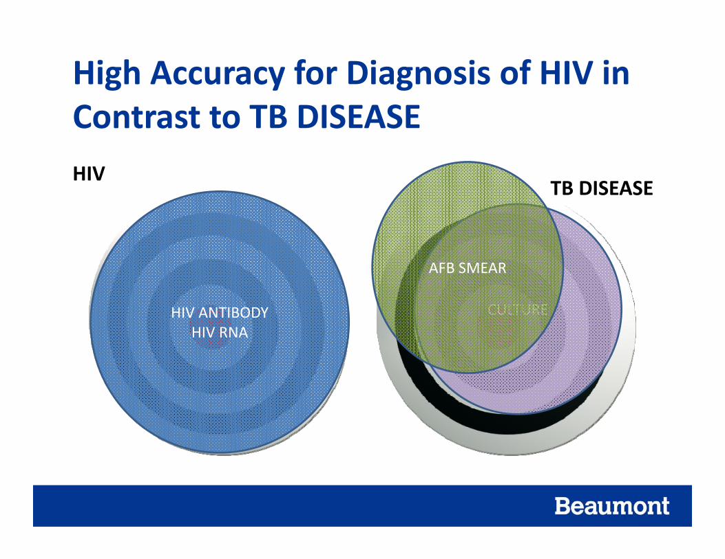
High Accuracy for Diagnosis of HIV in Contrast to TB DISEASEHIV
TB DISEASE
HIV ANTIBODYHIV RNA
CULTURE
AFB SMEAR

Studies Michigan 2015 pulmonary TB cases….N= 87Test % POSITIVE COMMENT
AFB smear 44% Negative smear does not rule out TB
NAAT on AFB+ smear 84% May be performed on AFB smear negative sputums
AFB culture confirms M. tb 78% Gold standard, not always positive
IGRA 89% May be negative even with positive cultures!
4/5/2016 11

Specimen Sources
• Sputum (primary)• Pulmonary aspiration (secondary)• Body fluids (CSF, pleural, peritoneal, etc)• Tissue biopsy• Blood• Urine• Gastric aspirate • Stool (special request)• Other

Sputum and AFB smears
“See the bugs”

Specimen CollectionPulmonary Specimen (sputum)
• Early morning specimens = highest yield of AFB
• Collect at least three consecutive specimens at 8‐24 hr intervals (at least 1 early morning specimen)
• Recommended volume for testing is 5‐10 ml, less may compromise recovery of AFB
• Infection control precautions during specimen collection
• If patient cannot produce sputum by coughing, consider other methods: sputum induction, bronchoscopy, or gastric aspiration
• All persons suspected of TB disease should have sputum cultured

Specimen Collection • Collect in sterile, leak proof containers
• Seal with tape
• Refrigerate specimen to reduce overgrowth of contaminating bacteria during transit to lab
• Deliver specimen to TB lab within 24 hrs
• Always include patient name on both test request form and the specimen container

Acid‐fast Bacilli (AFB) smear• Least sensitive of all AFB Tests (20‐75% positivity)
• Requires 10,000 AFB/ml to be positive
• Positive slide does not differentiate TB from atypical mycobacteria (i.e. M. avium)
• Reported within 24 hours of receiving the specimen in the laboratory

Fluorescent AFB Smear Using Auramine‐O Staining
• Very sensitive, takes minutes to read
• Not all that is fluorescent is AFB (need a careful eye)
• Chemical fluorescence, not an immune stain or Direct Fluorescent Antibody
• Can be confirmed with Ziehl‐Neelson (ZN) smear

Nucleic Acid Amplification (NAA) or PCR
“Multiply the bugs”

New CDC Guidelines of Use of NAA MMWR January 16, 2009
• “NAA testing should be performed on at least one respiratory specimen from each patient with signs and symptoms of pulmonary TB for whom a diagnosis of TB is being considered but has not yet been established, and for whom the test result would alter case management or TB control activities.”
• NAAT should be performed on all new AFB+ sputum specimens
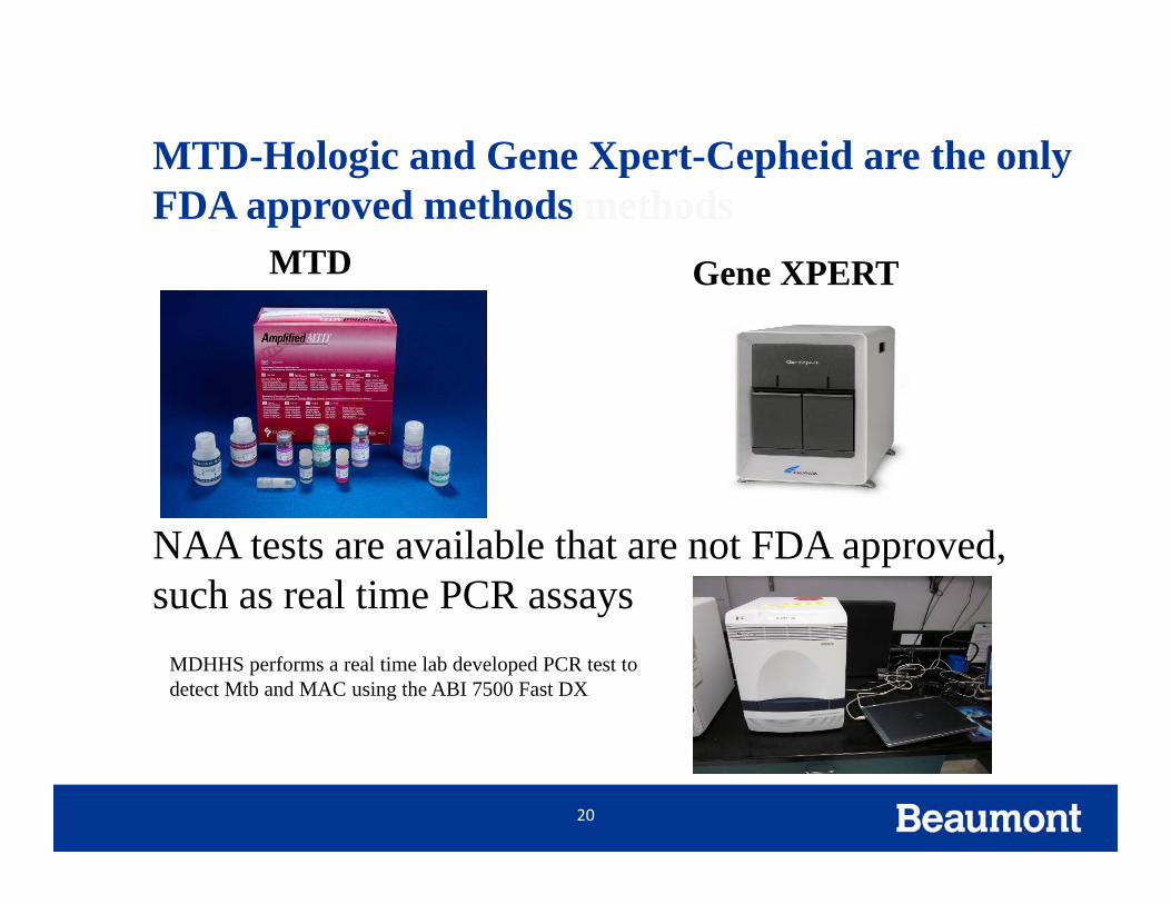
MTD-Hologic and Gene Xpert-Cepheid are the only FDA approved methods methods
MTD Gene XPERT
20
NAA tests are available that are not FDA approved, such as real time PCR assays
MDHHS performs a real time lab developed PCR test to detect Mtb and MAC using the ABI 7500 Fast DX

GenExpert Assay Procedure for the MTB/RIF Test.
Boehme CC et al. N Engl J Med 2010;363:1005‐1015.

AFB Cultures
“Grow the bugs”

AFB Culture Test• More sensitive than AFB smear
• 10 AFB/ml can produce a positive result, whereas AFB smear needs 10,000 AFB/ml
• Culture may be AFB positive even if smear was negative for AFB

Tests Performed on Growth in Mycobacteria Culture• Accuprobe DNA test (not amplified)• HPLC (high performance liquid chromatography)
• MALDI‐TOF• Biochemical Identification Confirmation• Drug Susceptibility

Susceptibility Testing of M. tuberculosisWhen to test
• All new M. tb isolates • Repeat after 90 days of therapy, if specimens continue to produce M. tb
• Relapse or failed therapy

Additional Molecular Tests for TB

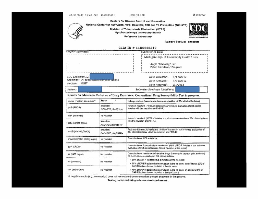
CDC – Molecular Detection of TB Drug Resistance (MDDR)
• Rapid testing for DNA mutations associated with drug resistance
• NAAT (+) sputum specimens or culture isolates (prior approval)
• Must meet the following criteria:– Known Rifampin resistance– Known MDR– High risk of Rifampin resistance or MDR‐TB– High profile patient (e.g. daycare worker, nurse)– Mixed or non‐viable culture– Drug Adverse reaction (e.g. Rifampin allergy)
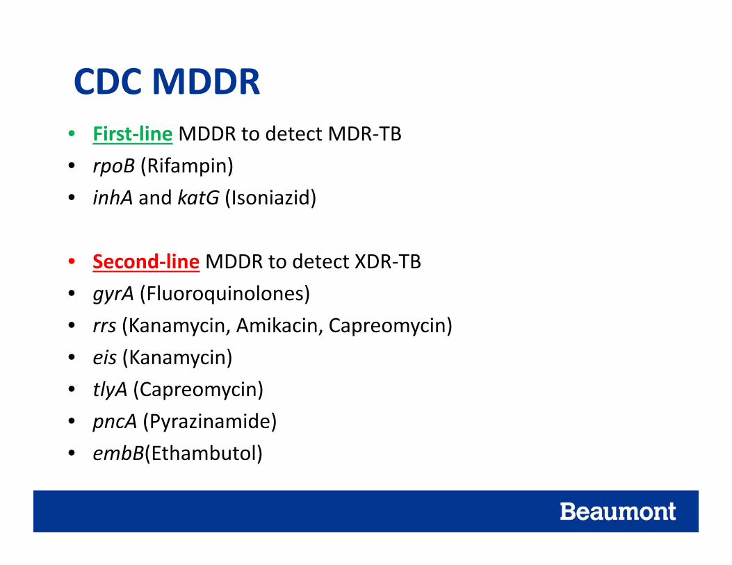
CDC MDDR• First‐lineMDDR to detect MDR‐TB• rpoB (Rifampin)• inhA and katG (Isoniazid)
• Second‐lineMDDR to detect XDR‐TB• gyrA (Fluoroquinolones)• rrs (Kanamycin, Amikacin, Capreomycin)• eis (Kanamycin)• tlyA (Capreomycin)• pncA (Pyrazinamide) • embB(Ethambutol)
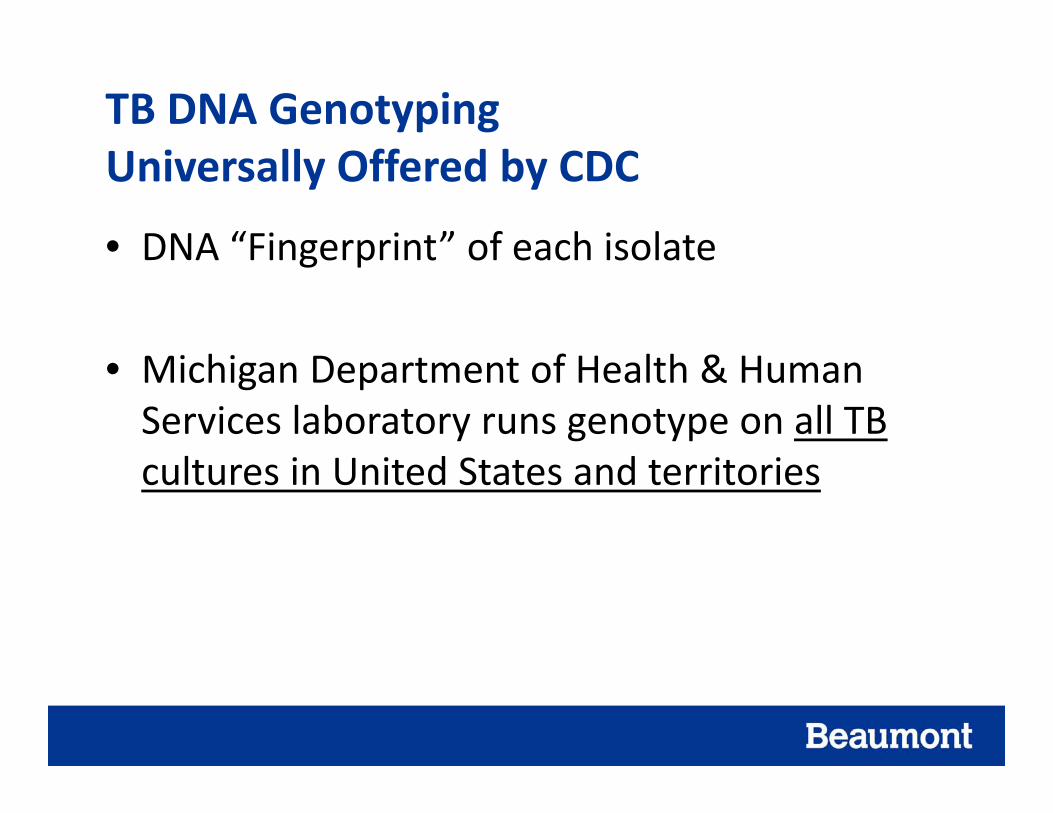
TB DNA Genotyping Universally Offered by CDC
• DNA “Fingerprint” of each isolate
• Michigan Department of Health & Human Services laboratory runs genotype on all TB cultures in United States and territories
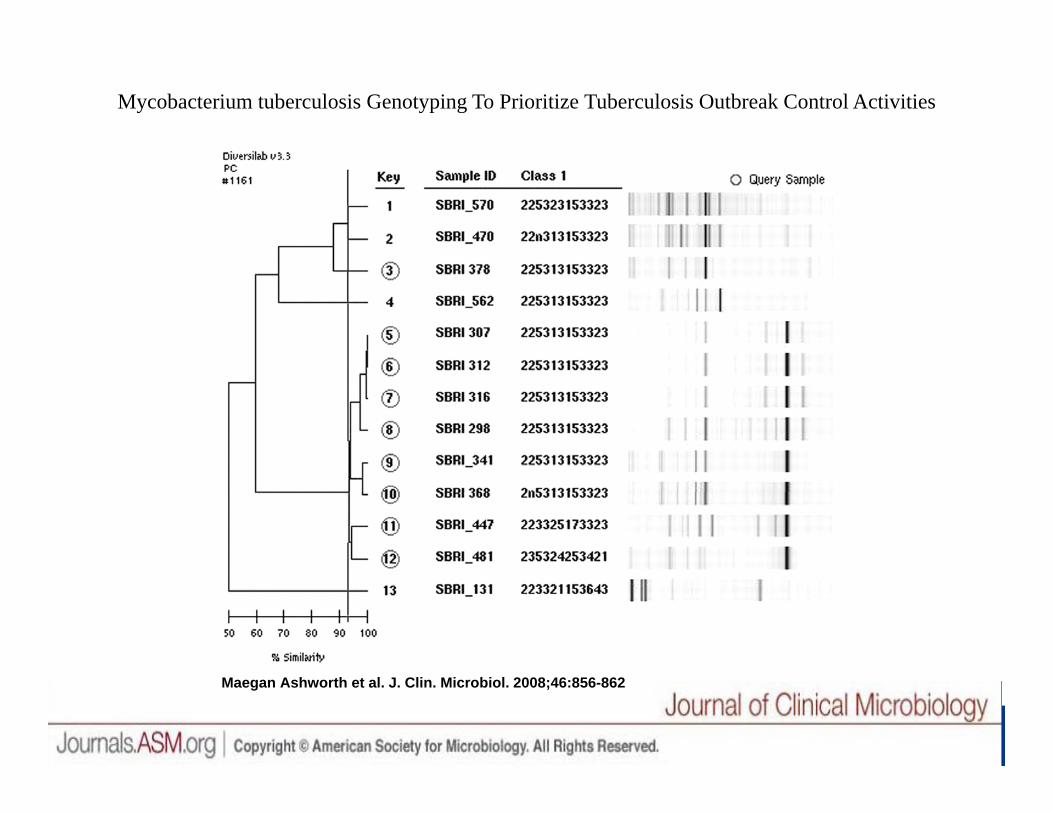
Mycobacterium tuberculosis Genotyping To Prioritize Tuberculosis Outbreak Control Activities
Maegan Ashworth et al. J. Clin. Microbiol. 2008;46:856-862

Demographics of Selected Genotype Clusters in Southeast Michigan, 2008 – 2012
PCR00012 (MI_0002) n = 58
PCR00291 (MI_0008) n = 48
PCR04678 (MI_0047) n =23
Race 63% African‐American16% White
97% African‐American 100% African‐American
Ethnicity 11% Hispanic 3% Hispanic 0% Hispanic
Homeless 37% 44% 27%
Alcohol 32% 35% 27%
Drug 42% 29% 46%
Incarceration 0% 6% 9%
HIV positive 16% 15% 0%
MDR 0% 6% 0%
* All clusters were majority 45 – 64 yrs of age; male and US-born.
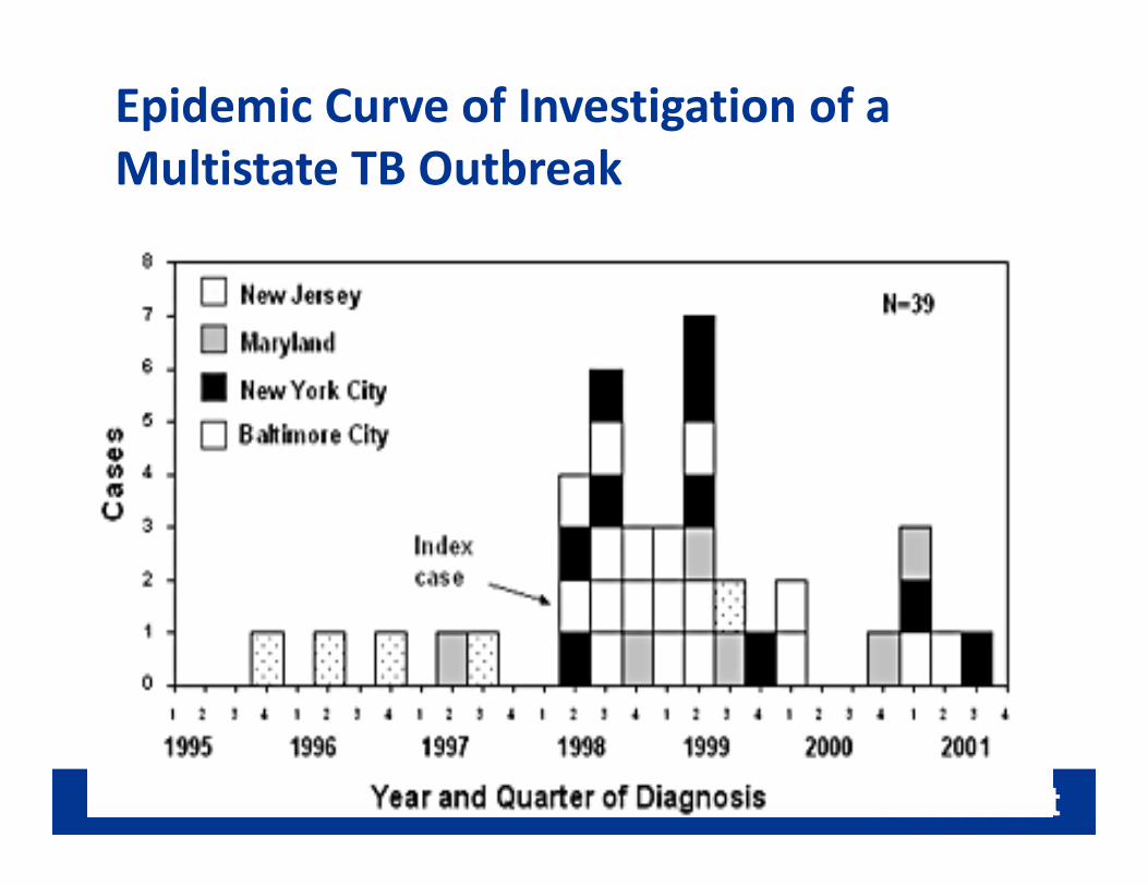
Epidemic Curve of Investigation of a Multistate TB Outbreak
3 Sample Cases

Date of download: 8/22/2012 Copyright © 2012 American Medical Association. All rights reserved.
From: Current Approaches to Tuberculosis in the United States
JAMA. 2012;308(3):283-289. doi:10.1001/jama.2012.7505
Admission chest radiograph showing bilateral lung infiltrates with prominence in the right upper lobe and lingula of the left lung.
Figure Legend:
Case #1 EASY

1TB suspected
2SputumPPD/IGRA
3AFB smear positive
4PPD 15 mm
5NAAT positive
6INH, RIF, PZA, EMB
7 8 9
10 11 12AFB in brothDNA probe+
13 14 15 16
17 18 19 20 21 22Drug susceptibility
23
24 25 26DNA genotype
27 28 29 30
4/5/2016 35
APRIL 2016 “EASY” CASE

57 yr male• Routine cultures negative
• No improvement• Bronchoscopy AFB smear negative
• HIV +• CD4 478 cells/mm3
#2 case MEDIUM
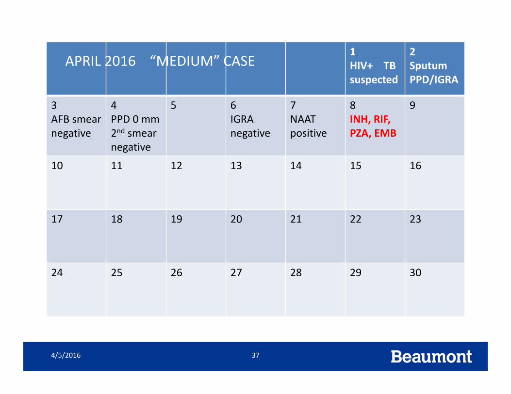
1HIV+ TB suspected
2SputumPPD/IGRA
3AFB smear negative
4PPD 0 mm2nd smear negative
5 6IGRA negative
7NAAT positive
8INH, RIF, PZA, EMB
9
10 11 12 13 14 15 16
17 18 19 20 21 22 23
24 25 26 27 28 29 30
4/5/2016 37
APRIL 2016 “MEDIUM” CASE

Case #3Difficult
• Patient from Africa• History of 3 prior episodes of pulmonary TB
• Coughing, sick again

#3 case MDR suspect
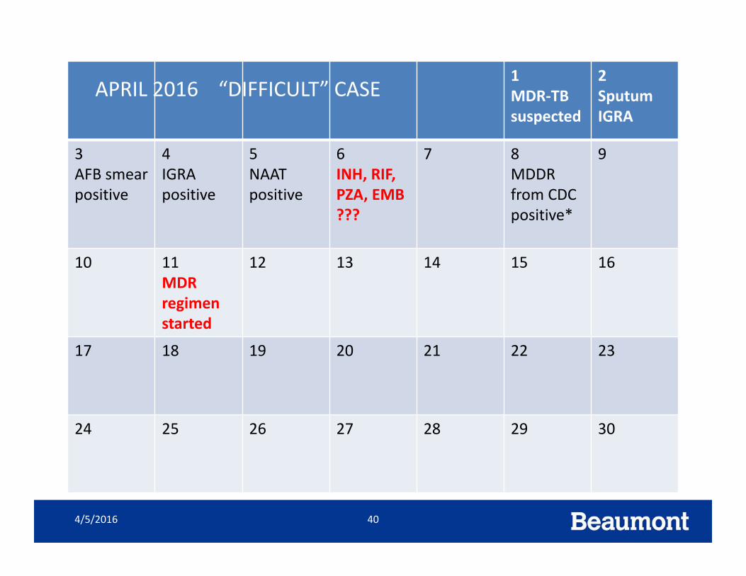
1MDR‐TB suspected
2SputumIGRA
3AFB smear positive
4IGRApositive
5NAAT positive
6INH, RIF, PZA, EMB ???
7 8MDDR from CDC positive*
9
10 11MDR regimen started
12 13 14 15 16
17 18 19 20 21 22 23
24 25 26 27 28 29 30
4/5/2016 40
APRIL 2016 “DIFFICULT” CASE

MDCH Lab Confirmation of 2nd Line Drugs
INH RRifampin RPZA REthambutol ROfloxacin S
Ethionamide RStreptomycin SKanamycin SAmikacin SCapreomycin SCycloserine SPAS S

IN CONCLUSION
• See the bugs [AFB microscopy]
• Multiply the bugs [NAATs]
• Grow the bugs [cultures]
• Kill the bugs
4/5/2016 43