tropism of streptococcus species.. plos one, 11(10...
TRANSCRIPT

http://www.diva-portal.org
This is the published version of a paper published in PLoS ONE.
Citation for the original published paper (version of record):
Spoerry, C., Hessle, P., Lewis, M J., Paton, L., Woof, J M. et al. (2016)Novel IgG-Degrading Enzymes of the IgdE Protease Family Link Substrate Specificity to HostTropism of Streptococcus Species..PLoS ONE, 11(10)https://doi.org/10.1371/journal.pone.0164809
Access to the published version may require subscription.
N.B. When citing this work, cite the original published paper.
Permanent link to this version:http://urn.kb.se/resolve?urn=urn:nbn:se:umu:diva-129369

RESEARCH ARTICLE
Novel IgG-Degrading Enzymes of the IgdE
Protease Family Link Substrate Specificity to
Host Tropism of Streptococcus Species
Christian Spoerry1, Pontus Hessle1, Melanie J. Lewis2, Lois Paton2, Jenny M. Woof2,
Ulrich von Pawel-Rammingen1*
1 Department of Molecular Biology and UmeåCentre for Microbial Research, UmeåUniversity, Umeå,
Sweden, 2 Cell Signalling and Immunology, School of Life Sciences, University of Dundee, Wellcome Trust
Building, Dundee, United Kingdom
Abstract
Recently we have discovered an IgG degrading enzyme of the endemic pig pathogen S.
suis designated IgdE that is highly specific for porcine IgG. This protease is the founding
member of a novel cysteine protease family assigned C113 in the MEROPS peptidase
database. Bioinformatical analyses revealed putative members of the IgdE protease family
in eight other Streptococcus species. The genes of the putative IgdE family proteases of S.
agalactiae, S. porcinus, S. pseudoporcinus and S. equi subsp. zooepidemicus were cloned
for production of recombinant protein into expression vectors. Recombinant proteins of all
four IgdE family proteases were proteolytically active against IgG of the respective Strepto-
coccus species hosts, but not against IgG from other tested species or other classes of
immunoglobulins, thereby linking the substrate specificity to the known host tropism. The
novel IgdE family proteases of S. agalactiae, S. pseudoporcinus and S. equi showed IgG
subtype specificity, i.e. IgdE from S. agalactiae and S. pseudoporcinus cleaved human
IgG1, while IgdE from S. equi was subtype specific for equine IgG7. Porcine IgG subtype
specificities of the IgdE family proteases of S. porcinus and S. pseudoporcinus remain to
be determined. Cleavage of porcine IgG by IgdE of S. pseudoporcinus is suggested to be
an evolutionary remaining activity reflecting ancestry of the human pathogen to the porcine
pathogen S. porcinus. The IgG subtype specificity of bacterial proteases indicates the spe-
cial importance of these IgG subtypes in counteracting infection or colonization and oppor-
tunistic streptococci neutralize such antibodies through expression of IgdE family
proteases as putative immune evasion factors. We suggest that IgdE family proteases
might be valid vaccine targets against streptococci of both human and veterinary medical
concerns and could also be of therapeutic as well as biotechnological use.
PLOS ONE | DOI:10.1371/journal.pone.0164809 October 17, 2016 1 / 20
a11111
OPENACCESS
Citation: Spoerry C, Hessle P, Lewis MJ, Paton L,
Woof JM, von Pawel-Rammingen U (2016) Novel
IgG-Degrading Enzymes of the IgdE Protease
Family Link Substrate Specificity to Host Tropism
of Streptococcus Species. PLoS ONE 11(10):
e0164809. doi:10.1371/journal.pone.0164809
Editor: Adam Lesner, Uniwersytet Gdanski,
POLAND
Received: June 24, 2016
Accepted: September 30, 2016
Published: October 17, 2016
Copyright: © 2016 Spoerry et al. This is an open
access article distributed under the terms of the
Creative Commons Attribution License, which
permits unrestricted use, distribution, and
reproduction in any medium, provided the original
author and source are credited.
Data Availability Statement: All relevant data are
within the paper.
Funding: Financial support was provided by Carl
Tryggers foundation (CTS13-514) (www.
carltryggersstiftelse.se/index.php?sida=rules&n=3)
and UmeåUniversity insamlingsstiftelse (2.1.12-
1605-14) (www.umu.se) to UPR, Wellcome Trust
(074863) to JW, with support of Kempestiftelsen
(CS) and the Medical research School Scotland
(LP). The funders had no role in study design, data
collection and analysis, decision to publish, or
preparation of the manuscript.

Introduction
Gram-positive bacteria of the genus Streptococcus are highly intertwinedwith humans and ani-mals as commensal, opportunistic and pathogenic bacteria. Often streptococci show pro-nounced host tropism, but these bacteria can also cause zoonotic or anthroponotic infectionsin more uncommon hosts [1].
Starting with the observation of a Immunoglobulin (Ig) G degrading activity in culturesupernatants of the important endemic pig pathogen Streptococcus (S.) suis, we identifiedrecently a novel IgG degrading enzyme designated IgdE [2]. Through inhibitor screening, in sil-ico modeling and mutational studies of the potential catalytic triad residues, IgdE was assignedto be a cysteine protease. This protease does not have homology to any protease previouslydescribed and is thereby the founding member of a novel cysteine protease family designatedC113 within the CA clan in the MEROPS peptidase database (https://merops.sanger.ac.uk/)[3]. IgdE of S. suis is highly specific for porcine IgG and no other substrate has been identified.Immune evasion from Ig mediated immune defense seems to be of special importance for bac-teria of the Streptococcus genus. Several Ig degrading enzymes of streptococci have been identi-fied, such as the IgG specific proteases, IdeS of S. pyogenes [4], IdeZ of S. equi subsp.zooepidemicus and IdeE of S. equi subsp. equi [5]. In addition IgA-specific proteases of S. pneu-moniae, S. oralis, S. sanguis and S.mitis have been described [6, 7] and recently, we describedan IgM specific protease in S. suis, designated IdeSsuis [8]. Most of these proteases cleave theheavy chain of Ig molecules in the hinge region, thereby impairing all effector functions of Igexcept neutralization.
IgG is the major antibody in serum of most higher organisms [9, 10]. Human IgG is catego-rized into four different subclasses; IgG1, IgG2, IgG3 and IgG4, with abundance in the sameorder [11]. These subclasses have subtle variations in structure resulting in different effectormechanisms. IgG1 and IgG3 are the prevalent Ig classes in humans able to cross the placentalbarrier to protect the fetus and newborn by passive immunization [12]. Every IgG subclass hasan individual FcγR-binding profile with IgG1 and IgG3 binding efficiently to most FcγR, whileIgG2 and IgG4 have a reduced affinity for some FcγR [13]. IgG1 and IgG3 activate the classicalcomplement pathway efficiently compared to IgG2 and IgG4 [14–16]. Consequently IgG1 andIgG3 are more involved in complement activation, phagocyte binding, sensitization of mastcells and sensitization of natural killer cells for killing, while all four subclasses are equallyinvolved in neutralization, opsonization and extravascular diffusion [17].
Porcine IgG is divided into six subclasses [18]. These subclasses are considered to be bio-chemically inseparable. Thus the different IgG subtypes and their proposed properties are onlypredicted by sequence analyses [19]. Equine IgG is divided into seven subclasses [20]. Experi-mental studies with recombinant equine IgG subclasses revealed that IgG1, IgG3, IgG4 andIgG7 are the most potent activators of the classical complement pathway via C1q binding andelicit also a strong respiratory burst from equine peripheral blood leukocytes [21].
In this study, we employed rigorous homology searches to identify several homologues ofIgdE of S. suis in other Streptococcus species as putative IgdE family proteases and comparedthem by phylogenetic analysis. The igdE genes of S. agalactiae, S. porcinus, S. pseudoporcinusand S. equi subsp. zooepidemicus were cloned for expression and purification of recombinantprotein followed by screening for potential substrates of these putative proteases.
S. agalactiae, also known as Group B Streptococcus, is commonly found as a commensal incattle and humans [22, 23], but is also able to cause mastitis in cows [24] and genitourinaryinfections, neonatal sepsis, CNS infections and endocarditis in humans [25].
S. porcinus is a bacterium most commonly found in the respiratory tract of pigs [26] andhas been associated with lymphadenitis [27] and still birth [28]. S. pseudoporcinus has recently
Novel Streptococcal Members of the IgG Degrading Protease Family IgdE
PLOS ONE | DOI:10.1371/journal.pone.0164809 October 17, 2016 2 / 20
Competing Interests: We have read the journal’s
policy and the authors of this manuscript have the
following competing interests: A patent application
[New streptococcal proteases, 1630021-2] for IgdE
proteases has been filed. CS and UPR are listed as
inventors in this application. This does not alter our
adherence to PLOS ONE policies on sharing data
and materials.

been distinguished from S. porcinus as a separate species [29] and has been shown to be anemerging and common organism colonizing the genitourinary tract of women [30].
S. equi subsp. zooepidemicus, a commensal and opportunistic pathogen of horses as well asother mammals, can cause severe zoonotic infections in humans, such as sepsis, meningitis andendocarditis [31]. In horses S. equi subsp. zooepidemicus can cause several different patholo-gies, including respiratory tract infections, uteritis and wound infections. The cause of thehighly contagious upper respiratory tract disease strangles, S. equi subsp. equi, is believed to bea clonal descendent of an ancestral strain of S. equi subsp. zooepidemicus [32, 33].
All putative IgdE family proteases tested in this study showed enzymatic activity and sub-strate specificity for IgG of specific hosts. IgdE of S. agalactiae was specific for human IgG1.IgdE of S. porcinus was specific for porcine IgG, while IgdE of S. equi subsp. zooepidemicus wasspecific for equine IgG7. IgdE of S. pseudoporcinus degraded both human IgG1 as well as por-cine IgG thereby being the only identified IgdE family protease with multiple substrates. Thesubstrate specificities, in regard of host species IgG, of these novel members of the IgdE prote-ase family correlate well with the known host tropism of the respective Streptococcus species.The IgG subclass specificities of these proteases might implicate special importance of thesespecific IgG subtypes in immune defense against these Streptococcus species during certainstages of infection which the bacteria might counteract through expression of IgG subtype spe-cific proteases.
The IgdE proteases of S. agalactiae, S. porcinus, S. pseudoporcinus and S. equi subsp. zooepi-demicus are designated IgdEagalactiae, IgdEporcinus, IgdEpseudoporcinus and IgdEequi in this study.
Materials and Methods
Computational identification of novel IgdE protease family members
within Streptococci
Coding sequences of all available Streptococcus genomes were downloaded from NCBI (ftp://ftp.ncbi.nlm.nih.gov/genomes/Bacteria/ on Aug-21-2015) and from PATRIC (ftp://ftp.patricbrc.org/patric2/ on Aug-25-2015). As a reference sequence for an IgdE protease theRefSeq sequence WP_014636499.1 of S. suis was used. The N-terminal signal peptide and theC-terminal region only present in sequences from S. suis were removed, leaving amino acids38–520, hereafter called IgdE_domain.
The IgdE_domain was used as query sequence in blastp searches (E-value cutoff 1 to keepall possible proteases) against the NCBI sequences as well as the PATRIC sequences.Sequences not containing the catalytic cysteine were removed from further consideration.Many of the sequences found are annotated as S-layer proteins or as containing an S-layerhomology domain W. These are often present in two or more copies in the same genome,and have an SxC or GxC motif in the catalytic site instead of the AxC motif found in aa 300–302 of the original IgdE sequence of S. suis. In order to distinguish these sequences, whichare not members of the IgdE protease family, all sequences lacking the AxC motif were alsoremoved.
The obtained hits were in turn used as query sequences against the same databases, usingthe same parameters. From the list of matched sequences those that in the second round had amatch overlapping with the region matched in the first round, when the IgdE_domain wasused as query, were chosen. Sequencesmatching to the Transglutcore model with an E-value ofat most 1e-6 or sequences not containing the catalytic AxC motif were excluded. The remain-ing sequences were trimmed at both ends to contain only the parts matching the IgdE_domainsequence. In cases where this resulted in identical sequences only one copy was kept.
Novel Streptococcal Members of the IgG Degrading Protease Family IgdE
PLOS ONE | DOI:10.1371/journal.pone.0164809 October 17, 2016 3 / 20

Phylogenetic analysis
Clustal Omega version 1.2.1 (http://www.ebi.ac.uk/Tools/msa/clustalo/) [34] was used to gener-ate a multiple sequence alignment of the sequences obtained above using default settings. Todetermine the best fitting amino acid substitution model we used ProtTest version 3.4 [35]. AJones-Taylor-Thornton (JTT)model with a gamma distribution, a proportion of invariablesites, and observedamino acid frequencies was the best model, and therefore used to construct amaximum likelihood (ML) tree with PhyML version 20131022 [36]. To assess the significanceof phylogenetic grouping a bootstrap analysis with 100 repetitions was performed. The tree wasrooted using an out-group consisting of two non-streptococcal protein sequences homologousto the IgdE_domain (WP_029500965.1; WP_016310821.1). These sequences were trimmed atboth ends to contain only the parts matching the IgdE_domain sequence. Encoded proteins lackdescribed functions. The phylogenetic tree was visualizedwith iTOL (http://itol.embl.de/) [37].
Bacterial strains and growth conditions
Escherichia coli strains were cultured in Lysogeny Broth (LB) or Lysogeny Agar (LA) under aer-obic conditions at 30°C or 37°C. When appropriate, 50 μg/ml kanamycin or 25 μg/ml chloram-phenicol was added.
Cloning of IgdE homologues for recombinant expression
Genes of the IgdE homologues lacking the signal peptide were amplified from chromosomalDNA of S. porcinus strain DSM20725 (kindly received from Christoph G. Bauns, College ofVeterinary Medicine, University Leipzig, Leipzig, Germany), S. pseudoporcinus strain LQ940-04T (ATCC), S. agalactiae strain CCUG420 (kindly received from Åsa Gylfe, Department ofClinical Microbiology, Umeå University, Umeå, Sweden) and S. equi subsp. zooepidemicusstrain 203 (kindly received from National Veterinary Institute, Uppsala, Sweden) as templatesusing primer pairs designated in Table 1. PCR fragments were cloned into pET_ZZ_1a afterdigestion with restriction enzymes (all Thermo Scientific) denoted in primer names. Thecloned plasmids were verified by sequencing and transformed into E. coli BL21 (DE3) pLysSfor recombinant expression of the proteins.
Expression and purification of recombinant IgdE homologues
E. coli BL21(DE3) pLysS isolates carrying pET_ZZ_1a igdEporcinus, igdEpseudoporcinus, igdEagalactiae
or igdEequi were grown to OD600 0.5 at 30°C. Protein expression was induced with 0.5 mMIPTG for 5h at 30°C. Cells were lysed for crude soluble extracts by BugBuster HT ProteinExtractionReagent (Novagen) according to manufacture protocol or lysed by sonication in 20mM sodium phosphate, 0.5M NaCl, 40 mM imidazole, pH 7.4 prior to further purification.The His-ZZ-tagged proteins were purified on HisTrap FF (GE Healthcare) using standard pro-tocols. The tag was removed by enzymatic cleavage by Tev-protease for 20h at 4°C followed bya second round of purification on HisTrap FF (GE Healthcare). The flow through, containinguntagged recombinant protein, was collected and buffer exchanged against PBS. Protein con-centrations were determined by Nanodrop A280 measurements at appropriate dilutions. Incase of IgdEequi no great overexpression was achieved and no purification attempt wasconducted.
Screening for Ig-degrading activities of recombinant IgdE homologues
If not stated otherwise, all reactions were carried out at 37°C for 16h in PBS. 20 μg/ml purifiedrecombinant proteins or 5% crude soluble extracts of E. coli expressing the igdE constructs
Novel Streptococcal Members of the IgG Degrading Protease Family IgdE
PLOS ONE | DOI:10.1371/journal.pone.0164809 October 17, 2016 4 / 20

were incubated with 0.5 mg/ml porcine, human, bovine, horse, goat, and mouse IgG (allSigma), 0.25 mg/ml human IgG1 kappa, IgG2 kappa, IgG3 kappa, IgG4 kappa, IgA and IgM(all Sigma), 0.09 mg/ml purified recombinant horse IgG1, IgG2, IgG3, IgG4, IgG5, IgG6 andIgG7 expressed in Chinese hamster ovary cells or FreeStyle 293-F cells (according to [21]) and1% human plasma, porcine plasma (kindly received from Christoph G. Baums, College of Vet-erinaryMedicine, University Leipzig, Leipzig, Germany) and equine serum (Sigma). Reactionsamples were analyzed using SDS-PAGE or Western Blot analyses. Experiments were repeatedat least two times and representative analyses are shown.
SDS-PAGE and Western Blot analysis
Samples for SDS-PAGE were prepared with reducing sample buffer and heated to 95°C for 5min. 12% SDS-PAGE was either stained with Coomassie blue (Sigma), Coomassie Fluor™Orange Protein Gel Stain (Invitrogen) or blotted to Hybond-P PVDF membrane (GE Health-care) for Western Blot analyses. Membranes were blocked with 5% dry milk powder in 0.1%PBS-Tween, followed by incubation with horse-radish peroxidase conjugated primary antibod-ies or unconjugated primary antibody (according to Table 2). Membranes were thoroughlywashed with 0.1% PBS-Tween and development with Amersham ECL SelectWestern blottingdetection reagent (GE Healthcare) according to manufacturer’s instruction and chemilumines-cent signal was captured by LAS4000 imaging system (Fujifilm). Prestained protein ladderswere pictured with the same system by standard epi-white illumination.
N-terminal Edman sequencing
IgG degradation reactions were separated by SDS-PAGE as previously explained and trans-ferred to PVDF blotting membrane (GE Health Care) by semi-dry blotting with blotting bufferconsisting of 50 mM Sodiumborate and 20% Methanol. The membrane was stained with
Table 1. Used primers for cloning of igdE genes.
Locus tag aa primers
igdEporcinus STRPO_RS07810 34–527 IgdEporcinus-frw_NcoI GTACCCATGGCTGTTCTTGCGAGAGAAAATAG
IgdEporcinus-rev_Acc65I GTACGGTACCTTAGTTACCTGCATTCTTTGTTTC
igdEpseudoporcinus STRPS_RS03610 38–535 IgdEpseudoporcinus-
frw_Eco31I
GTACGGTCTCCCATGAGAGAAAATGAAAACGTAAGACAATTAC
IgdEpseudoporcinus-
rev_Acc65I
GTACGGTACCTTACTGTGCATGCTTTGTTGTTG
igdEagalactiae MSA_19930 37–623 IgdEagalactiae-frw_BspHI GTACTCATGAATCAAAATAATATTCAAGAAACT
IgdEagalactiae-rev_Acc65I GTACGGTACCTAATTCGTGTTCGTTTCTC
igdEequi M837_01916 1-517(no signal
peptide)
IgdEequi-frw_NcoI GTACCCATGGAAGCATGGAAGCATG
IgdEequi-rev_Acc65I GTACGGTACCTTATTGATTAGCGCTTTCACATTG
doi:10.1371/journal.pone.0164809.t001
Table 2. Used antibodies and dilutions for Western Blot analysis.
Name Manufacturer Dilution
Goat anti-pig IgG-HRP Thermo Scientific (PA1-28685) 1:300000
Goat anti-pig IgM-HRP Thermo Scientific (PA1-84622) 1:300000
Goat anti-pig IgA-HRP Thermo Scientific (PA1-84625) 1:150000
Rabbit anti-horse IgG-HRP Abcam (ab6921) 1:30’000
Goat anti-horse IgM-HRP Abcam (ab112879) 1:15’000
Rabbit anti-horse IgA-HRP Abcam (ab112871) 1:15’000
doi:10.1371/journal.pone.0164809.t002
Novel Streptococcal Members of the IgG Degrading Protease Family IgdE
PLOS ONE | DOI:10.1371/journal.pone.0164809 October 17, 2016 5 / 20

Ponceau S 0.5% (Sigma) 1% acetic acid in MQ water and destained with MQ water. The mem-brane was quickly dried, where after the degradation product was neatly cut out. N-terminalEdman sequencing of the degradation product was performed by Proteome Factory (Berlin,Germany). BLAST homology searches were used to identify the position of the obtainedsequence in the IgG molecule.
Results
Putative IgdE family proteases are spread among several
Streptococcus species
To identify novel IgdE family proteases, streptococcal genomes were searched for sequencesencoding homologues of the IgdE_domain. The IgdE_domain was defined as amino acids 38–520 of the RefSeq sequence WP_014636499.1 of S. suis 05ZYH33, thereby excluding the N-ter-minal signal peptide and the C-terminal region only present in sequences from S. suis. Onlysequences that, when used as query in a second round of homology searches, had an overlapwith the region matched in the first round and carried the conserved catalytic cysteine residuewere kept for further analysis (Table 3).
After homology searches and filtering steps, 55 unique sequences were identified represent-ing putative IgdE family proteases, 23 from S. suis, 18 from S. agalactiae, five from S. dysgalac-tiae, three from S. equi subsp. zooepidemicus and one each from S. porcinus, S. pseudoporcinus,S. canis and S. castoreus respectively. Two sequences from the same genome of S. merionis wereidentified. All sequences from S. agalactiae are very similar in the defined IgdE_domain region,often only differing in one or a few amino acid positions.
To illustrate the relationship of these 55 sequences a phylogenetic tree was inferred by maxi-mum likelihood based on the JTT model of sequence evolution (Fig 1). The IgdE_domainregion sequences of S. canis and S. castoreus showed similarity to the IgdE_domain regionsequences of S. dysgalactiae. The two IgdE_domain sequences of S. merionis grouped together,but were still distinct from each other. IgdE_domain region sequences of all S. agalactiaestrains grouped close together, as did sequences of S. equi and S. dysgalactiae, while thesequences of S. suis were more diverse. Also the two sequences obtained from S. porcinus andS. pseudoporcinus grouped close together.
Locations of igdE genes within Streptococcus genomes
Each gene encoding a putative IgdE protease was localized in the respective genome as well asgenes flanking the igdE gene (Table 4). Location of the igdE genes was generally conserved
Table 3. Number of identified putative IgdE family protease sequences and searched genomes.
Streptococcus
species
identified putative IgdE family protease
sequences
searched
genomes
percentage of genomes encoding putative IgdE family
protease sequences [%]
S. agalactiae 171 309 55
S. dysgalactiae 13 13 100
S. equi 6 13 46
S. suis 79 98 81
S. porcinus 1 1 100
S. pseudoporcinus 2 2 100
S. canis 1 1 100
S. castoreus 1 1 100
S. merionis 2 1 100
doi:10.1371/journal.pone.0164809.t003
Novel Streptococcal Members of the IgG Degrading Protease Family IgdE
PLOS ONE | DOI:10.1371/journal.pone.0164809 October 17, 2016 6 / 20

within species, although two out of 18 S. agalactiae strains and one out of 23 S. suis strains haddeviant neighboring genes. Location and neighboring genes were conserved between the spe-cies S. castoreus, S. canis and S. dysgalactiae. For sequences retrieved from genome drafts thelocations could not be determined or could only be approximated.
IgdE family proteases are highly specific for IgG of different host species
The igdE genes from one representative strain of S. agalactiae, S. porcinus, S. pseudoporcinusand S. equi subsp. zooepidemicus (corresponding to the sequences marked in bold in Fig 1)were cloned into expression vectors in E. coli. The encoded proteins were over-expressed andrecombinant protein was used for substrate screening by overnight incubation with potentialsubstrates prior to analyses by SDS-PAGE and western blots.
Recombinant IgdEagalactiae was able to degrade human IgG and a diagnostic cleavage productof 32 kDa appeared, when rIgdEagalactiae was incubated with human IgG, but not when incu-bated with porcine, bovine, equine, caprine or murine IgG (Fig 2A). Similar to that rIgdEporcinus
was only able to degrade porcine IgG; the diagnostic cleavage product of 32 kDa appeared onlywhen incubated with porcine IgG, but not when incubated with human, bovine, equine, caprineor murine IgG (Fig 2B). Recombinant IgdEpseudoporcinus had in contrast dual substrate specificitytowards human and porcine IgG, again characterized through appearance of diagnostic cleavageproducts of 32 kDa, while no degradation of bovine, equine, caprine or murine IgG could beobserved (Fig 2C). Recombinant IgdEequi possessed degrading activity against equine IgG, whileno degradation of human, porcine, bovine, caprine or murine IgG could be observed (Fig 2D).
IgG specificity of these novel members of the IgdE protease family was investigated by incu-bation of recombinant protein with porcine and equine serum, respectively. These cleavagereactions were analyzed by anti-IgG, anti-IgM and anti-IgA western blots. RecombinantIgdEporcinus degraded porcine serum IgG, but not IgM or IgA (Fig 3A). A similar observationwas made when serum was incubated with rIgdEpseudoporcinus (Fig 3B), which also was also spe-cific for IgG. Recombinant IgdEequi cleaved equine serum IgG, but not IgM or IgA (Fig 3C).
Specificity of the IgdE proteases from S. agalactiae and S. pseudoporcinus for human IgG incomparison to human IgM and IgA was tested through incubation of recombinant proteinwith purified Ig (all Sigma) and analyzed by reducing SDS-PAGE. The findings showed thatrIgdEagalactiae (Fig 4A) and rIgdEpseudoporcinus (Fig 4B) were specific for IgG and cleaved humanIgG, but not human IgM or IgA.
IgG subtype specificity of IgdE proteases of S. agalactiae, S.
pseudoporcinus and S. equi subsp. zooepidemicus
Given the shown specificity of IgdE proteases for IgG of specific hosts, we were interested inIgG subtype specificity of these proteases. Recombinant IgdEagalactiae (Fig 5A) andrIgdEpseudoporcinus (Fig 5B) were therefore incubated with human IgG1, IgG2, IgG3 and IgG4from myeloma source prior to reducing SDS-PAGE analysis. Both rIgdEagalactiae andrIgdEpseudoporcinus were strictly IgG1 specific, and no degradation of human IgG2, IgG3 andIgG4 was observed.The equine IgG subtype specificity of rIgdEequi was tested by incubation ofprotease preparations with purified recombinant equine IgG1, IgG2, IgG3, IgG4, IgG5, IgG6and IgG7 (Fig 5C). Strikingly, pronounced subtype specificitywas also observed for rIgdEequi
and of all equine IgG subtypes only recombinant equine IgG7 was cleaved.
Cleavage sites of IgdE family proteases
To determine the exact cleavage site of the IgdE proteases within the respective substrate IgGmolecules, the 32 kDa cleavage products generated by IgdEagalactiae, IgdEporcinus,
Novel Streptococcal Members of the IgG Degrading Protease Family IgdE
PLOS ONE | DOI:10.1371/journal.pone.0164809 October 17, 2016 7 / 20

Fig 1. Phylogenetic tree of identified putative IgdE proteases. Phylogenic analysis of the IgdE_domain of 55 identified
putative IgdE proteases. The maximum likelihood (ML) tree was constructed using a Jones-Taylor-Thornton (JTT) model with
PhyML. Bootstrap values greater than or equal to 80% are shown. Putative IgdE protease sequences that corresponded to the
genes cloned for expression of recombinant protein are marked in bold. Tree scale is given as average number of
substitutions per site.
doi:10.1371/journal.pone.0164809.g001
Novel Streptococcal Members of the IgG Degrading Protease Family IgdE
PLOS ONE | DOI:10.1371/journal.pone.0164809 October 17, 2016 8 / 20

IgdEpseudoporcinus, and IgdEequi were subjected to N-terminal Edman sequencing. All obtainedsequences corresponded to the hinge region of the respective IgG molecules (Fig 6A).IgdEagalactiae and IgdEpseudoporcinus cleave both human IgG1, but cleavage sites are not identical,being located two residues apart from each other. IgdEagalactiae cleaves the human IgG1 heavychain two residues N-terminal of the putative homodimer disulfide bond cysteine residues;while all other cleavage sites of IgdE family proteases are located directly adjacent to the puta-tive homodimer disulfide bond cysteine residues in IgG hinge regions. The cleavage sites iden-tified in porcine IgG generated through cleavage with both IgdEporcinus and IgdEpseudoporcinus
were identical and found in porcine IgG2, IgG4 and IgG6 (Fig 6B). Similar sequences can, how-ever, also be found in IgG1 and IgG5. The sequence obtained by N-terminal Edman sequencingof the cleavage product of equine IgG generated by IgdEequi corresponded to the hinge regionof equine IgG7. The amino acid sequences analogous to those adjacent to the cleavage site inequine IgG7 are different in all the other horse IgG subtypes, providing a rationale for theobserved subtype specificity.
Discussion
Based on the identification of the founding member of a novel cysteine protease family, IgdE ofS. suis [2], we identified several putative IgdE family proteases through homology searcheswithin the genus Streptococcus. Locations of the genes of these putative proteases were con-servedwithin, and to certain degree also between, different Streptococcus species suggestingthat igdE genes are part of the core genome and not part of mobile elements (Table 4). Onesequence of S. suis and two of S. agalactiae had, however, different neighboring genes than theother 22 sequences of S. suis and 16 of S. agalactiae, respectively. Putative IgdE family proteasesequences were however only found in 55% of S. agalactiae, 46% of S. equi and 81% of S. suisgenomes (Table 3). This could be due to the real absence of an igdE gene in some strains ofthese species or due to pore, incomplete or missanotated coding sequences of these genomes.
Table 4. Location of igdE genes within the genomes of the respective Streptococcus species and flanking genes.
Species Flanking genes Location (nucleotide
number)
S. agalactiae (16) Between "4-Hydroxy-2-oxoglutarate aldolase (EC 4.1.3.16)" and "Nitroreductase family protein" ~ 1790000–1890000
S. agalactiae (1) Between "Voltage-gated chloride channel family protein" and "Transcriptional regulator, MarR
family"
?
S. agalactiae (1) Between "4-Hydroxy-2-oxoglutarate aldolase (EC 4.1.3.16)" and "sensor histidine kinase" ?
S. castoreus (1) Between "UDP-N-acetylmuramoylalanine—D-glutamate ligase" and "GTP-binding protein TypA/
BipA"
~ 1440000–1540000
S. canis (1)
S. dysgalactiae (5)
S. equi subsp.
zooepidemicus (3)
Between "S-adenosylmethionine:tRNA ribosyltransferase-isomerase" and "Manganese superoxide
dismutase "
~657000
~1210000
~1500000
S. merionis (1) Between "SatD" and "tRNA:m(5)U-54 MTase gid" ?
S. merionis (1) Between "Transmembrane component MtsC of energizing module of methionine-regulated ECF
transporter" and "putative toxic anion resistance protein"
?
S. porcinus (1) Between "Glutathione S-transferase, omega" and "Two-component system response regulator" ?
S. pseudoporcinus (1)
S. suis (22) Between "Protein export cytoplasm protein SecA ATPase RNA helicase" and "Fructokinase" ~ 1600000–1860000
S. suis (1) Between "Choline binding protein A" and "Uridine kinase" ?
doi:10.1371/journal.pone.0164809.t004
Novel Streptococcal Members of the IgG Degrading Protease Family IgdE
PLOS ONE | DOI:10.1371/journal.pone.0164809 October 17, 2016 9 / 20

Fig 2. IgG host species specificity of IgdE family proteases. 0.5 mg/ml human, porcine, equine, bovine and murine IgG were incubated for 16h at
37˚C with (A) 20 μg/ml purified rIgdEagalactiae, (B) 20 μg/ml purified rIgdEporcinus, (C) 5% soluble fraction of E. coli cells expressing rIgdEpseudoporcinus, (D)
5% soluble fraction of E. coli cells expressing rIgdEequi. PBS (A and B) or 5% soluble fraction of E. coli cells without recombinant construct (C and D)
were used as negative controls (-). Reactions were analyzed by Coomassie blue SDS-PAGE under reducing conditions. Images of different SDS-PAGE
run in parallel have been assembled into one figure. The diagnostic 32 kDa IgG cleavage product (*) appeared when rIgdEagalactiae was incubated with
human IgG, rIgdEporcinus with porcine IgG, rIgdEpseudoporcinus with human IgG and porcine IgG, and rIgdEequi with equine IgG.
doi:10.1371/journal.pone.0164809.g002
Novel Streptococcal Members of the IgG Degrading Protease Family IgdE
PLOS ONE | DOI:10.1371/journal.pone.0164809 October 17, 2016 10 / 20

Fig 3. IgdE family proteases are specific for IgG compared to IgM and IgA. 2% porcine plasma was
incubated with (+) or without (-) 20 μg/ml purified rIgdEporcinus (A) or 5% soluble fraction of E. coli cells
expressing rIgdEpseudoporcinus (B) respectively for 16h at 37˚C. 2% equine serum was incubated with 5%
soluble fraction of E. coli cells expressing rIgdEequi (C) for 16h at 37˚C. The reactions were analyzed by anti-
porcine or anti-equine IgG, IgM and IgA Western blots under reducing conditions. Only degradation products
of IgG (*) could be observed.
doi:10.1371/journal.pone.0164809.g003
Novel Streptococcal Members of the IgG Degrading Protease Family IgdE
PLOS ONE | DOI:10.1371/journal.pone.0164809 October 17, 2016 11 / 20

All investigated IgdE family proteases except IgdEpseudoporcinus, showed only specificitytowards IgG of one host species. The substrate specificity of these IgdE family proteases corre-lates well with the known host tropism of the respective Streptococcus species. However,IgdEagalactiae does not cleave bovine IgG, despite S. agalactiae being the cause of mastitis in cat-tle besides being an important human pathogen. The observed substrate preference for humanIgG1 might reflect that most human invasive S. agalactiae isolates represent distinct subtypesfrom bovine isolates, as it has been suggested in a temporally and geographically matched iso-late characterization study [40]. IgdEpseudoporcinus showed double specificity for both humanIgG1 and porcine IgG (Fig 2). Since S. pseudoporcinus is closely related to the pig pathogen S.porcinus, the specificity for porcine IgG is not that surprising. It is, however, astonishing thatIgdEpseudoporcinus, being a human pathogen, has evolved the ability to cleave human IgG1,despite the close relationship to IgdEporcinus that does not possess this ability. Thus, it seemsadvantageous for S. pseudoporcinus to have a human IgG1 degrading protease, underlining theimportance of IgG1 in immune responses towards bacterial pathogens. Mutational studies of
Fig 4. IgdEagalactiae and IgdEpseudoporcinus are specific for human IgG compared to IgM and IgA. 0.5
mg/ml human IgG, IgM and IgA were incubated for 16h at 37˚C with (+) or without (-) 20 μg/ml purified
rIgdEagalactiae (A) or 5% soluble fraction of E. coli cells expressing rIgdEpseudoporcinus (B). Reactions were
analyzed by SDS-PAGE under reducing conditions. SDS-PAGE was stained with Coomassie blue. Order of
lanes within SDS-PAGE was adjusted.
doi:10.1371/journal.pone.0164809.g004
Novel Streptococcal Members of the IgG Degrading Protease Family IgdE
PLOS ONE | DOI:10.1371/journal.pone.0164809 October 17, 2016 12 / 20
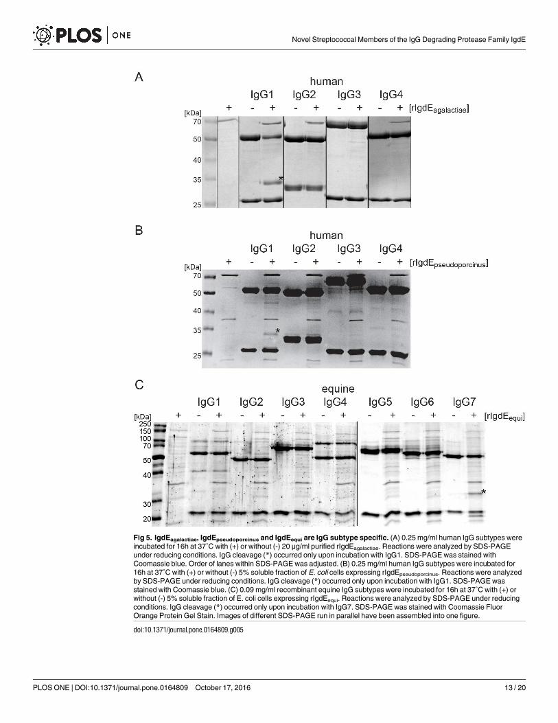
Fig 5. IgdEagalactiae, IgdEpseudoporcinus and IgdEequi are IgG subtype specific. (A) 0.25 mg/ml human IgG subtypes were
incubated for 16h at 37˚C with (+) or without (-) 20 μg/ml purified rIgdEagalactiae. Reactions were analyzed by SDS-PAGE
under reducing conditions. IgG cleavage (*) occurred only upon incubation with IgG1. SDS-PAGE was stained with
Coomassie blue. Order of lanes within SDS-PAGE was adjusted. (B) 0.25 mg/ml human IgG subtypes were incubated for
16h at 37˚C with (+) or without (-) 5% soluble fraction of E. coli cells expressing rIgdEpseudoporcinus. Reactions were analyzed
by SDS-PAGE under reducing conditions. IgG cleavage (*) occurred only upon incubation with IgG1. SDS-PAGE was
stained with Coomassie blue. (C) 0.09 mg/ml recombinant equine IgG subtypes were incubated for 16h at 37˚C with (+) or
without (-) 5% soluble fraction of E. coli cells expressing rIgdEequi. Reactions were analyzed by SDS-PAGE under reducing
conditions. IgG cleavage (*) occurred only upon incubation with IgG7. SDS-PAGE was stained with Coomassie Fluor
Orange Protein Gel Stain. Images of different SDS-PAGE run in parallel have been assembled into one figure.
doi:10.1371/journal.pone.0164809.g005
Novel Streptococcal Members of the IgG Degrading Protease Family IgdE
PLOS ONE | DOI:10.1371/journal.pone.0164809 October 17, 2016 13 / 20

Fig 6. IgdE family proteases cleave IgG in the hinge region. (A) The cleavage sites within IgG molecules were determined through N-terminal
Edman sequencing of the 32 kDa IgG cleavage products generated by IgdE family proteases. The identified aa sequences (bold) were found in
the hinge regions of the respective IgG heavy chains. Homodimer disulfide bond cysteine residues are underlined. 10 aa N- and C-terminal from
the identified cleavage site (scissor symbol) of the respective IgG heavy chain are shown. Porcine IgG4a was chosen as a representative for
porcine IgG. (B) Sequences of the hinge regions and adjacent parts of the CH1 and CH2 domains of human, porcine and equine IgG subtypes
Novel Streptococcal Members of the IgG Degrading Protease Family IgdE
PLOS ONE | DOI:10.1371/journal.pone.0164809 October 17, 2016 14 / 20

IgdEpseudoporcinus and IgdEporcinus might be able to dissect the residues that mediate this sub-strate specificity.
We suggest that substrate specificity of IgdE family proteases contribute to the host tropismof some Streptococcus species. Co-evolution of streptococcal opportunistic pathogens and theirhost could be reflected on a molecular level by co-evolution of IgdE family proteases and theirsubstrate IgG heavy chain molecules. This is highly reminiscent of the co-evolution describedbetween IgA and bacterial proteins targeting IgA, such as IgA-binding proteins and IgA-spe-cific proteases, where reiterative episodes of natural selection are predicted to have shaped theinteractions between the IgA and the bacterial proteins, reflecting an ‘arms race’ [41, 42].
The subtype specificity of the IgdE family proteases of S. agalactiae and S. pseudoporcinustowards human IgG1 and the IgdE family protease of S. equi subsp. zooepidemicus towardsequine IgG7 is striking and surprising (Fig 5). The evolutionary benefit for streptococci to pos-sess such IgG subtype specific proteases compared to proteases with broader specificity is atthe first glance puzzling. Cleavage of these IgG subtypes might, however, be sufficient to over-come key immune defense mechanisms in certain niches. For example, S. agalactiae is a com-mon cause of invasive neonatal infections in humans [43] and human IgG1 is, along withIgG3, the major human Ig transported across the placental barrier [44]. Thereby cleavage ofhuman IgG1 might be sufficient to evade the Ig mediated immune response in the newbornhuman host. Moreover, targeted disruption of IgG7 function by S. equi in the horse is likely tosignificantly comprise IgG-mediated protection, given that IgG7 is one of the predominantsubclasses in equine serum [45]. Due to the high diversity in the hinge region of different IgGsubtypes [17, 46] it might also be difficult to evolve IgG degrading proteases with broader spec-ificity. Since some IgG subtypes, for example human IgG4, are believed to mediate tolerance[47, 48] it might even be beneficial for an opportunistic pathogen to carry proteases incapableof cleaving these IgG subtypes. Investigations on the potential role of IgdEagalactiae in immuneevasion in the neonatal and adult host are currently ongoing in our group.
IgdEequi is the third IgG degrading protease of S. equi subsp. zooepidemicus described, besideIdeZ1 [5] and IdeZ2 [49]. IgdEequi is highly specific for equine IgG7, while both IdeZ1 andIdeZ2 have broader specificity, cleaving IgG of several host species. The abundance of genesencoding IgG degrading proteases in S. equi subsp. zooepidemicus implicates a special impor-tance of an IgG cleaving phenotype of this species. These Ig-degrading proteases might be regu-lated by different gene regulation systems and thereby expressed during different stages ofinfection or colonization.
All IgdE family proteases recognize IgG as substrates although the amino acid sequences atthe cleavage sites in the respective hinge regions are quite diverse (Fig 6). Therefore preferencefor IgG as substrates of IgdE family proteases might not only be conferred by the actual cleav-age site, but also by motifs lying adjacent to it or within the Fc fragment or F(ab) fragment.Indeed, this possibility mirrors that observedwith certain human IgA1 proteases in that resi-dues within the Fc region of IgA have been shown to be essential for recognition of humanIgA1 as a substrate for cleavage [50, 51]. Further parallels with IgA1 protease cleavage of IgA1hinge and the cleavage of IgG hinges by IgdE family proteases describedhere can be noted. Dif-ferent IgA1 proteases are known to cleave at different specific peptide bonds in the IgA1 hingesequence, and evidence suggests that for cleavage to occur each protease has a requirement forthe Fab and Fc regions to be separated by a particular number of amino acids, presumably toallow appropriate access and orientation of the protease [52]. Possibly similar spatial
were aligned using T-COFFEE (Version_8.93) [38] to illustrate hinge region diversity. Alignment reliability assessed by TCS [39] is color coded
(blue to red).
doi:10.1371/journal.pone.0164809.g006
Novel Streptococcal Members of the IgG Degrading Protease Family IgdE
PLOS ONE | DOI:10.1371/journal.pone.0164809 October 17, 2016 15 / 20

considerations may impact on the ability of IgdE family members to cleave their respective sub-strates, and may provide a further explanation for their exquisite specificity for particular IgGsubtypes.
IgdEagalactiae has the same cleavage site in human IgG1 as papain [53]. However comparedto papain, IgdEagalactiae is highly specific for human IgG1 and has only one distinct cleavage sitewithin the heavy chain. Interestingly, this cleavage site is not shared by IgdEpseudoporcinus thatinstead cleaves the heavy chain two residues closer to the C-terminus, just N-terminal of theputative homodimer disulfide bond cysteine residue. Differential cleavage sites implicate thattargeting IgG1 has evolved independently in these two proteases, highlighting the importancefor the bacteria to counteract IgG1. The cleavage sites of IgdEporcinus, IgdEpseudoporcinus andIgdEequi have a CPxCP motif just C-terminal of the cleavage site in common. This motif can,however, also be found in many IgG heavy chain molecules that are not substrates of any inves-tigated IgdE family protease, supporting the idea that substrate specificity is determined by fea-tures others than cleavage site sequences.
Secreted Ig degrading proteases have been shown to be protective antigens in experimentalvaccine and infection studies with S. suis in pigs [54] and S. equi in horses [55]. The describedIgdE proteases might therefore be suitable vaccine targets. Given the homology of these prote-ases, especially in regions close to the active site, vaccination might even give cross protectionagainst several Streptococcus species. This would especially be desirable in the cases of S. agalac-tiae and S. pseudoporcinus in humans and S. suis and S. porcinus in pigs. Antibodies elicited bysuch vaccines might both neutralize the proteolytic function of these potential immune evasionfactors and potentially mediate antibody dependent cell cytotoxicity against the streptococcalpathogen.
Lastly IgdE family proteases with pronounced species and subtype specificitymight also beof biotechnological or therapeutical use, i.e. similar to what has been proven for the IgGdegrading enzyme of S. pyogenes IdeS [56–61].
Acknowledgments
Christoph G. Baums (College of Veterinary Medicine, University Leipzig, Leipzig, Germany) isacknowledged for kindly providing porcine plasma and S. porcinus and Åsa Gylfe (Departmentof Clinical Microbiology, Umeå University, Umeå, Sweden) for S. agalactiae. Parts of this workwere planned and performed by the Umeå Protein Expertise Platform. Support by BILS (Bioin-formatics Infrastructure for Life Sciences) is gratefully acknowledged.
Author Contributions
Conceptualization:CS UPR.
Formal analysis:CS.
Funding acquisition:UPR JW.
Investigation: CS PH ML LP.
Methodology:CS.
Project administration:UPR.
Resources:UPR JW.
Supervision:UPR JW.
Validation: CS PH.
Novel Streptococcal Members of the IgG Degrading Protease Family IgdE
PLOS ONE | DOI:10.1371/journal.pone.0164809 October 17, 2016 16 / 20

Visualization: CS.
Writing – original draft:CS UPR.
Writing – review& editing:CS UPR JW.
References1. Facklam R. What Happened to the Streptococci: Overview of Taxonomic and Nomenclature Changes.
Clin Microbiol Rev. 152002. p. 613–30. doi: 10.1128/CMR.15.4.613-630.2002 PMID: 12364372
2. Spoerry C, Seele J, Valentin-Weigand P, Baums CG, von Pawel-Rammingen U. Identification and
Characterization of IgdE, a Novel IgG-degrading Protease of Streptococcus suis with Unique Specific-
ity for Porcine IgG. J Biol Chem. 2016; 291(15):7915–25. doi: 10.1074/jbc.M115.711440 PMID:
26861873; PubMed Central PMCID: PMCPMC4824999.
3. Rawlings ND, Waller M, Barrett AJ, Bateman A. MEROPS: the database of proteolytic enzymes, their
substrates and inhibitors. Nucleic Acids Res. 2014; 42(Database issue):D503–9. Epub 2013/10/26.
doi: 10.1093/nar/gkt953 PMID: 24157837; PubMed Central PMCID: PMCPMC3964991.
4. von Pawel-Rammingen U, Johansson BP, Bjorck L. IdeS, a novel streptococcal cysteine proteinase
with unique specificity for immunoglobulin G. Embo j. 2002; 21(7):1607–15. Epub 2002/04/03. doi: 10.
1093/emboj/21.7.1607 PMID: 11927545; PubMed Central PMCID: PMCPMC125946.
5. Lannergard J, Guss B. IdeE, an IgG-endopeptidase of Streptococcus equi ssp. equi. FEMS Microbiol
Lett. 2006; 262(2):230–5. Epub 2006/08/23. doi: 10.1111/j.1574-6968.2006.00404.x PMID:
16923080.
6. Kilian M, Mestecky J, Schrohenloher RE. Pathogenic species of the genus Haemophilus and Strepto-
coccus pneumoniae produce immunoglobulin A1 protease. Infect Immun. 1979; 26(1):143–9. Epub
1979/10/01. 40878; PubMed Central PMCID: PMCPMC414586. PMID: 40878
7. Reinholdt J, Tomana M, Mortensen SB, Kilian M. Molecular aspects of immunoglobulin A1 degradation
by oral streptococci. Infect Immun. 1990; 58(5):1186–94. PMID: 2182537; PubMed Central PMCID:
PMCPMC258608.
8. Seele J, Singpiel A, Spoerry C, von Pawel-Rammingen U, Valentin-Weigand P, Baums CG. Identifica-
tion of a novel host-specific IgM protease in Streptococcus suis. J Bacteriol. 195. United States2013.
p. 930–40. doi: 10.1128/JB.01875-12 PMID: 23243300
9. Markowska-Daniel I, Pomorska-Mol M, Pejsak Z. Dynamic changes of immunoglobulin concentrations
in pig colostrum and serum around parturition. Pol J Vet Sci. 2010; 13(1):21–7. PMID: 21077427.
10. Panda S, Ding JL. Natural antibodies bridge innate and adaptive immunity. J Immunol. 2015; 194
(1):13–20. doi: 10.4049/jimmunol.1400844 PMID: 25527792.
11. Schur PH. IgG subclasses. A historical perspective. Monogr Allergy. 1988; 23:1–11. PMID: 3290655.
12. Simister NE. Placental transport of immunoglobulin G. Vaccine. 2003; 21(24):3365–9. Epub 2003/07/
10. PMID: 12850341.
13. Bruhns P, Iannascoli B, England P, Mancardi DA, Fernandez N, Jorieux S, et al. Specificity and affinity
of human Fcgamma receptors and their polymorphic variants for human IgG subclasses. Blood. 2009;
113(16):3716–25. doi: 10.1182/blood-2008-09-179754 PMID: 19018092.
14. Bindon CI, Hale G, Bruggemann M, Waldmann H. Human monoclonal IgG isotypes differ in comple-
ment activating function at the level of C4 as well as C1q. J Exp Med. 1988; 168(1):127–42. PMID:
3260935; PubMed Central PMCID: PMCPMC2188986.
15. Schumaker VN, Calcott MA, Spiegelberg HL, Muller-Eberhard HJ. Ultracentifuge studies of the binding
of IgG of different subclasses to the Clq subunit of the first component of complement. Biochemistry.
1976; 15(23):5175–81. PMID: 990273.
16. Tao MH, Smith RI, Morrison SL. Structural features of human immunoglobulin G that determine iso-
type-specific differences in complement activation. J Exp Med. 1993; 178(2):661–7. PMID: 8340761;
PubMed Central PMCID: PMCPMC2191116.
17. Vidarsson G, Dekkers G, Rispens T. IgG subclasses and allotypes: from structure to effector functions.
Front Immunol. 2014; 5:520. doi: 10.3389/fimmu.2014.00520 PMID: 25368619; PubMed Central
PMCID: PMCPMC4202688.
18. Butler JE, Wertz N. Antibody repertoire development in fetal and neonatal piglets. XVII. IgG subclass
transcription revisited with emphasis on new IgG3. J Immunol. 2006; 177(8):5480–9. PMID:
17015734.
Novel Streptococcal Members of the IgG Degrading Protease Family IgdE
PLOS ONE | DOI:10.1371/journal.pone.0164809 October 17, 2016 17 / 20

19. Butler JE, Wertz N, Deschacht N, Kacskovics I. Porcine IgG: structure, genetics, and evolution. Immu-
nogenetics. 2009; 61(3):209–30. Epub 2008/12/03. doi: 10.1007/s00251-008-0336-9 PMID:
19048248.
20. Wagner B, Miller DC, Lear TL, Antczak DF. The complete map of the Ig heavy chain constant gene
region reveals evidence for seven IgG isotypes and for IgD in the horse. J Immunol. 2004; 173
(5):3230–42. PMID: 15322185.
21. Lewis MJ, Wagner B, Woof JM. The different effector function capabilities of the seven equine IgG sub-
classes have implications for vaccine strategies. Mol Immunol. 2008; 45(3):818–27. Epub 2007/08/03.
doi: 10.1016/j.molimm.2007.06.158 PMID: 17669496; PubMed Central PMCID: PMCPMC2075531.
22. Bliss SJ, Manning SD, Tallman P, Baker CJ, Pearlman MD, Marrs CF, et al. Group B Streptococcus
colonization in male and nonpregnant female university students: a cross-sectional prevalence study.
Clin Infect Dis. 2002; 34(2):184–90. Epub 2001/12/12. doi: 10.1086/338258 PMID: 11740706.
23. Ippolito DL, James WA, Tinnemore D, Huang RR, Dehart MJ, Williams J, et al. Group B streptococcus
serotype prevalence in reproductive-age women at a tertiary care military medical center relative to
global serotype distribution. BMC Infect Dis. 2010; 10:336. doi: 10.1186/1471-2334-10-336 PMID:
21106080; PubMed Central PMCID: PMCPMC3004907.
24. Yang Y, Liu Y, Ding Y, Yi L, Ma Z, Fan H, et al. Molecular characterization of Streptococcus agalactiae
isolated from bovine mastitis in Eastern China. PLoS One. 2013; 8(7):e67755. doi: 10.1371/journal.
pone.0067755 PMID: 23874442; PubMed Central PMCID: PMCPMC3707890.
25. Sunkara B, Bheemreddy S, Lorber B, Lephart PR, Hayakawa K, Sobel JD, et al. Group B Streptococ-
cus infections in non-pregnant adults: the role of immunosuppression. Int J Infect Dis. 2012; 16(3):
e182–6. doi: 10.1016/j.ijid.2011.11.008 PMID: 22236484.
26. O’Sullivan T, Friendship R, Blackwell T, Pearl D, McEwen B, Carman S, et al. Microbiological identifi-
cation and analysis of swine tonsils collected from carcasses at slaughter. Can J Vet Res. 2011; 75
(2):106–11. PMID: 21731180; PubMed Central PMCID: PMCPMC3062919.
27. Hajtos I, Glavits R, Makrai L, Hallgatone IS, Jochman J. Occurrence of porcine purulent lymphadenitis
caused by Streptococcus porcinus in Hungary. Magyar Allatorvosok Lapja. 2002; 124(3):161–8.
WOS:000174545500005.
28. Lammler C, Bahr KH. Characterisation of Streptococcus porcinus serogroup P isolated from an
aborted fetus of a pig. Medical Science Research. 1996; 24(3):177–8. WOS:A1996UF45700014.
29. Bekal S, Gaudreau C, Laurence RA, Simoneau E, Raynal L. Streptococcus pseudoporcinus sp nov., a
novel species isolated from the genitourinary tract of women. Journal of Clinical Microbiology. 2006; 44
(7):2584–6. doi: 10.1128/jcm.02707-05. WOS:000239157400045. PMID: 16825387
30. Stoner KA, Rabe LK, Austin MN, Meyn LA, Hillier SL. Incidence and epidemiology of Streptococcus
pseudoporcinus in the genital tract. J Clin Microbiol. 2011; 49(3):883–6. Epub 2010/12/31. doi: 10.
1128/jcm.01965-10 PMID: 21191057; PubMed Central PMCID: PMCPMC3067687.
31. Pelkonen S, Lindahl SB, Suomala P, Karhukorpi J, Vuorinen S, Koivula I, et al. Transmission of Strep-
tococcus equi subspecies zooepidemicus infection from horses to humans. Emerg Infect Dis. 2013; 19
(7):1041–8. doi: 10.3201/121365 PMID: 23777752; PubMed Central PMCID: PMCPMC3713971.
32. Timoney JF. The pathogenic equine streptococci. Vet Res. 2004; 35(4):397–409. doi: 10.1051/
vetres:2004025 PMID: 15236673
33. Jorm LR, Love DN, Bailey GD, McKay GM, Briscoe DA. Genetic structure of populations of beta-hae-
molytic Lancefield group C streptococci from horses and their association with disease. Res Vet Sci.
1994; 57(3):292–9. Epub 1994/11/01. PMID: 7871247.
34. Sievers F, Wilm A, Dineen D, Gibson TJ, Karplus K, Li W, et al. Fast, scalable generation of high-qual-
ity protein multiple sequence alignments using Clustal Omega. Molecular Systems Biology. 2011; 7(1).
doi: 10.1038/msb.2011.75 PMID: 21988835
35. Darriba D, Taboada GL, Doallo R, Posada D. ProtTest 3: fast selection of best-fit models of protein
evolution. Bioinformatics. 2011; 27(8):1164–5. Epub 2011/02/22. doi: 10.1093/bioinformatics/btr088
PMID: 21335321.
36. Guindon S, Gascuel O. A simple, fast, and accurate algorithm to estimate large phylogenies by maxi-
mum likelihood. Syst Biol. 2003; 52(5):696–704. Epub 2003/10/08. PMID: 14530136.
37. Letunic I, Bork P. Interactive Tree Of Life (iTOL): an online tool for phylogenetic tree display and anno-
tation. Bioinformatics. 2007; 23(1):127–8. Epub 2006/10/20. doi: 10.1093/bioinformatics/btl529 PMID:
17050570.
38. Notredame C, Higgins DG, Heringa J. T-Coffee: A novel method for fast and accurate multiple
sequence alignment. J Mol Biol. 2000; 302(1):205–17. doi: 10.1006/jmbi.2000.4042 PMID: 10964570.
Novel Streptococcal Members of the IgG Degrading Protease Family IgdE
PLOS ONE | DOI:10.1371/journal.pone.0164809 October 17, 2016 18 / 20

39. Chang JM, Di Tommaso P, Notredame C. TCS: a new multiple sequence alignment reliability measure
to estimate alignment accuracy and improve phylogenetic tree reconstruction. Mol Biol Evol. 2014; 31
(6):1625–37. doi: 10.1093/molbev/msu117 PMID: 24694831.
40. Sukhnanand S, Dogan B, Ayodele MO, Zadoks RN, Craver MP, Dumas NB, et al. Molecular subtyping
and characterization of bovine and human Streptococcus agalactiae isolates. J Clin Microbiol. 2005;
43(3):1177–86. doi: 10.1128/JCM.43.3.1177-1186.2005 PMID: 15750080; PubMed Central PMCID:
PMCPMC1081236.
41. Abi-Rached L, Dorighi K, Norman PJ, Yawata M, Parham P. Episodes of natural selection shaped the
interactions of IgA-Fc with FcalphaRI and bacterial decoy proteins. J Immunol. 2007; 178(12):7943–
54. PMID: 17548632.
42. Pinheiro A, Woof JM, Abi-Rached L, Parham P, Esteves PJ. Computational analyses of an evolution-
ary arms race between mammalian immunity mediated by immunoglobulin A and its subversion by
bacterial pathogens. PLoS One. 2013; 8(9):e73934. doi: 10.1371/journal.pone.0073934 PMID:
24019941; PubMed Central PMCID: PMCPMC3760800.
43. Bekker V, Bijlsma MW, van de Beek D, Kuijpers TW, van der Ende A. Incidence of invasive group B
streptococcal disease and pathogen genotype distribution in newborn babies in the Netherlands over
25 years: a nationwide surveillance study. Lancet Infect Dis. 2014; 14(11):1083–9. Epub 2014/12/03.
doi: 10.1016/s1473-3099(14)70919-3 PMID: 25444407.
44. Simister NE, Story CM, Chen HL, Hunt JS. An IgG-transporting Fc receptor expressed in the syncytio-
trophoblast of human placenta. Eur J Immunol. 1996; 26(7):1527–31. doi: 10.1002/eji.1830260718
PMID: 8766556.
45. Sheoran AS, Timoney JF, Holmes MA, Karzenski SS, Crisman MV. Immunoglobulin isotypes in sera
and nasal mucosal secretions and their neonatal transfer and distribution in horses. Am J Vet Res.
2000; 61(9):1099–105. Epub 2000/09/08. PMID: 10976743.
46. Ellison JW, Berson BJ, Hood LE. The nucleotide sequence of a human immnnoglobulin Cγl gene.
Nucleic Acids Research. 1982; 10(13):4071–9. doi: 10.1093/nar/10.13.4071 PMID: 6287432
47. Aalberse RC, Stapel SO, Schuurman J, Rispens T. Immunoglobulin G4: an odd antibody. Clin Exp
Allergy. 2009; 39(4):469–77. Epub 2009/02/19. doi: 10.1111/j.1365-2222.2009.03207.x PMID:
19222496.
48. Jutel M, Akdis CA. Immunological mechanisms of allergen-specific immunotherapy. Allergy. 2011; 66
(6):725–32. Epub 2011/04/07. doi: 10.1111/j.1398-9995.2011.02589.x PMID: 21466562.
49. Hulting G, Flock M, Frykberg L, Lannergård J, Flock JI, Guss B. Two novel IgG endopeptidases of
Streptococcus equi. FEMS Microbiol Lett. 2009; 298(1):44–50. doi: 10.1111/j.1574-6968.2009.01698.
x PMID: 19659725.
50. Chintalacharuvu KR, Chuang PD, Dragoman A, Fernandez CZ, Qiu J, Plaut AG, et al. Cleavage of the
human immunoglobulin A1 (IgA1) hinge region by IgA1 proteases requires structures in the Fc region
of IgA. Infect Immun. 2003; 71(5):2563–70. PMID: 12704129; PubMed Central PMCID:
PMCPMC153282. doi: 10.1128/IAI.71.5.2563-2570.2003
51. Senior BW, Woof JM. Sites in the CH3 domain of human IgA1 that influence sensitivity to bacterial
IgA1 proteases. J Immunol. 2006; 177(6):3913–9. PMID: 16951354.
52. Senior BW, Woof JM. The influences of hinge length and composition on the susceptibility of human
IgA to cleavage by diverse bacterial IgA1 proteases. J Immunol. 2005; 174(12):7792–9. PMID:
15944283.
53. Wang AC, Wang IY. Cleavage sites of human IgG1 immunoglobulin by papain. Immunochemistry.
1977; 14(3):197–200. Epub 1977/03/01. PMID: 863464.
54. Seele J, Hillermann LM, Beineke A, Seitz M, von Pawel-Rammingen U, Valentin-Weigand P, et al. The
immunoglobulin M-degrading enzyme of Streptococcus suis, IdeSsuis, is a highly protective antigen
against serotype 2. Vaccine. 2015; 33(19):2207–12. doi: 10.1016/j.vaccine.2015.03.047 PMID:
25825330.
55. Guss B, Flock M, Frykberg L, Waller AS, Robinson C, Smith KC, et al. Getting to grips with strangles:
an effective multi-component recombinant vaccine for the protection of horses from Streptococcus
equi infection. PLoS Pathog. 2009; 5(9):e1000584. doi: 10.1371/journal.ppat.1000584 PMID:
19763180; PubMed Central PMCID: PMCPMC2736577.
56. Johansson BP, Shannon O, Bjorck L. IdeS: a bacterial proteolytic enzyme with therapeutic potential.
PLoS One. 2008; 3(2):e1692. doi: 10.1371/journal.pone.0001692 PMID: 18301769; PubMed Central
PMCID: PMCPMC2253494.
57. Winstedt L, Jarnum S, Nordahl EA, Olsson A, Runstrom A, Bockermann R, et al. Complete Removal of
Extracellular IgG Antibodies in a Randomized Dose-Escalation Phase I Study with the Bacterial
Enzyme IdeS—A Novel Therapeutic Opportunity. PLoS One. 2015; 10(7):e0132011. doi: 10.1371/
journal.pone.0132011 PMID: 26177518; PubMed Central PMCID: PMCPMC4503742.
Novel Streptococcal Members of the IgG Degrading Protease Family IgdE
PLOS ONE | DOI:10.1371/journal.pone.0164809 October 17, 2016 19 / 20

58. Yang R, Otten MA, Hellmark T, Collin M, Bjorck L, Zhao MH, et al. Successful treatment of experimen-
tal glomerulonephritis with IdeS and EndoS, IgG-degrading streptococcal enzymes. Nephrol Dial
Transplant. 2010; 25(8):2479–86. doi: 10.1093/ndt/gfq115 PMID: 20219834.
59. Takahashi R, Yuki N. Streptococcal IdeS: therapeutic potential for Guillain-Barre syndrome. Sci Rep.
2015; 5:10809. doi: 10.1038/srep10809 PMID: 26194472; PubMed Central PMCID:
PMCPMC4508529.
60. An Y, Zhang Y, Mueller HM, Shameem M, Chen X. A new tool for monoclonal antibody analysis: appli-
cation of IdeS proteolysis in IgG domain-specific characterization. MAbs. 2014; 6(4):879–93. doi: 10.
4161/mabs.28762 PMID: 24927271; PubMed Central PMCID: PMCPMC4171023.
61. Nandakumar KS, Johansson BP, Bjorck L, Holmdahl R. Blocking of experimental arthritis by cleavage
of IgG antibodies in vivo. Arthritis Rheum. 2007; 56(10):3253–60. doi: 10.1002/art.22930 PMID:
17907170.
Novel Streptococcal Members of the IgG Degrading Protease Family IgdE
PLOS ONE | DOI:10.1371/journal.pone.0164809 October 17, 2016 20 / 20