progression and preserve genome integrity. (2016) plos...
TRANSCRIPT

http://www.diva-portal.org
This is the published version of a paper published in PLoS Genetics.
Citation for the original published paper (version of record):
McDonald, K R., Guise, A J., Pourbozorgi-Langroudi, P., Cristea, I M., Zakian, V A. et al.(2016)Pfh1 Is an Accessory Replicative Helicase that Interacts with the Replisome to Facilitate ForkProgression and Preserve Genome Integrity.PLoS Genetics, 12(9): e1006238http://dx.doi.org/10.1371/journal.pgen.1006238
Access to the published version may require subscription.
N.B. When citing this work, cite the original published paper.
Permanent link to this version:http://urn.kb.se/resolve?urn=urn:nbn:se:umu:diva-125854

RESEARCH ARTICLE
Pfh1 Is an Accessory Replicative Helicase thatInteracts with the Replisome to FacilitateFork Progression and Preserve GenomeIntegrityKarin R. McDonald1, Amanda J. Guise1, Parham Pourbozorgi-Langroudi2, IleanaM. Cristea1, Virginia A. Zakian1, John A. Capra3*, Nasim Sabouri2*
1 Department of Molecular Biology, Princeton University, Princeton, New Jersey, United States of America,2 Department of Medical Biochemistry and Biophysics, UmeåUniversity, Umeå, Sweden, 3 Department ofBiological Sciences, Vanderbilt Genetics Institute, and Center for Structural Biology, Vanderbilt University,Nashville, Tennessee, United States of America
* [email protected] (JAC); [email protected] (NS)
AbstractReplicative DNA helicases expose the two strands of the double helix to the replication appa-
ratus, but accessory helicases are often needed to help forksmove past naturally occurring
hard-to-replicate sites, such as tightly bound proteins, RNA/DNA hybrids, and DNA secondary
structures. Although the Schizosaccharomyces pombe 5’-to-3’DNA helicase Pfh1 is known to
promote fork progression, its genomic targets, dynamics, and mechanisms of action are
largely unknown. Here we address these questions by integrating genome-wide identification
of Pfh1 binding sites, comprehensive analysis of the effects of Pfh1 depletion on replication
and DNA damage, and proteomic analysis of Pfh1 interaction partners by immunoaffinity puri-
fication mass spectrometry. Of the 621 high confidence Pfh1-binding sites in wild type cells,
about 40%were sites of fork slowing (asmarked by high DNA polymerase occupancy) and/or
DNA damage (as marked by high levels of phosphorylated H2A). The replication and integrity
of tRNA and 5S rRNA genes, highly transcribed RNA polymerase II genes, and nucleosome
depleted regions were particularly Pfh1-dependent. The association of Pfh1 with genomic
integrity at highly transcribed genes was S phase dependent, and thus unlikely to be an artifact
of high transcription rates. Although Pfh1 affected replication and suppressed DNA damage at
discrete sites throughout the genome, Pfh1 and the replicative DNA polymerase bound to sim-
ilar extents to both Pfh1-dependent and independent sites, suggesting that Pfh1 is proximal to
the replication machinery during S phase. Consistent with this interpretation, Pfh1 co-purified
with many key replisome components, including the hexameric MCM helicase, replicative
DNA polymerases, RPA, and the processivity clamp PCNA in an S phase dependent manner.
Thus, we conclude that Pfh1 is an accessory DNA helicase that interacts with the replisome
and promotes replication and suppresses DNA damage at hard-to-replicate sites. These data
provide insight into mechanisms by which this evolutionarily conserved helicase helps pre-
serve genome integrity.
PLOS Genetics | DOI:10.1371/journal.pgen.1006238 September 9, 2016 1 / 29
a11111
OPEN ACCESS
Citation: McDonald KR, Guise AJ, Pourbozorgi-Langroudi P, Cristea IM, Zakian VA, Capra JA, et al.(2016) Pfh1 Is an Accessory Replicative Helicase thatInteracts with the Replisome to Facilitate ForkProgression and Preserve Genome Integrity. PLoSGenet 12(9): e1006238. doi:10.1371/journal.pgen.1006238
Editor: Francisco Antequera, CSIC/Universidad deSalamanca, SPAIN
Received: January 4, 2016
Accepted: August 11, 2016
Published: September 9, 2016
Copyright: © 2016 McDonald et al. This is an openaccess article distributed under the terms of theCreative Commons Attribution License, which permitsunrestricted use, distribution, and reproduction in anymedium, provided the original author and source arecredited.
Data Availability Statement: Sequencing data areavailable at GEO, data set GSE59178.
Funding:Work in the Zakian lab was supported byNIH grant GM26938 (to VAZ), The Wenner-GrenFoundations (to NS), Swedish Society for MedicalResearch (to NS), and the New Jersey Commissionon Cancer Research (to KRM). Work in the Sabourilab was supported by the Swedish Research Council,The Wenner-Gren Foundations, and The KempeFoundations (JCK-1325). Work in the Cristea lab wassupported by NIH grants DA026192 and GM114141.

Author Summary
Progression of the DNA replication machinery is challenged in every S phase by activetranscription, tightly bound protein complexes, and formation of stable DNA secondarystructures. Using genome-wide analyses, we show that the evolutionarily conserved fissionyeast Pfh1 DNA helicase promotes fork progression and suppresses DNA damage at natu-ral sites of fork pausing, which occur at “hard-to-replicate” sites. Our data suggest thatPfh1 interacts with the replication apparatus. First, mass spectrometry revealed that Pfh1interacts with many components of the replication machinery. Second, Pfh1 and the lead-ing strand DNA polymerase occupy many common regions genome-wide, not only hard-to-replicate sites, but also sites whose replication is not Pfh1-dependent. The humangenome encodes a Pfh1 homolog, hPIF1, and contains all of the same hard-to-replicatefeatures that make fission yeast DNA replication dependent upon Pfh1. Thus, human cellslikely also require replicative accessory DNA helicases to facilitate replication at hard-to-replicate sites, and hPIF1 is a good candidate for this role.
IntroductionFaithful and efficient replication of the genome is essential in every cell cycle, yet there aremany naturally occurring obstacles that impede fork progression. These sites include stableprotein complexes, DNA secondary structures, and ongoing transcription, each of which canchallenge replication fork progression. Failure to circumvent these obstacles can cause DNAdouble strand breaks (DSBs) that impair genome integrity and increase the risk of cancer andother disorders that are associated with genome instability. As many of the proteins involvedin DNA replication are highly conserved, model organisms such as S. pombe provide geneti-cally tractable systems to identify and characterize genes with important roles in genome pres-ervation whose human orthologs might have similar functions.
DNA replication is accomplished by the multi-subunit replisome, a complex that is assem-bled at and moves bi-directionally away from replication origins. Replicative helicases, such asthe Escherichia coli DnaB and the eukaryotic hexameric MCM complex, are required tounwind the double helix to allow DNA polymerases access to the replication template. In addi-tion, accessory helicases, such as the E. coli rep, dinG and UvrD proteins help the polymerasemaneuver past protein complexes, RNA transcripts, and other naturally occurring impedi-ments [1–5]. In Bacillus subtilis the essential accessory DNA helicase PcrA promotes forkmovement through transcribed genes [6, 7]. E. coli rep physically interacts with the replicativeDnaB helicase to bypass protein complexes on DNA [8].
The best-studied eukaryotic accessory DNA helicases are the two budding yeast enzymes,ScRrm3 and ScPif1, which are both members of the Pif1 family [9]. Although these two heli-cases have largely non-overlapping functions, they both promote progression past naturallyoccurring hard-to-replicate sites. ScRrm3 acts at over 1000 sites, including RNA polymerase IIItranscribed genes, the replication fork barrier (RFB) within ribosomal DNA (rDNA), inactivereplication origins, silencers, telomeres, centromeres, and converged replication forks [10–14].The diverse ScRrm3-sensitive sites have the common feature of being assembled into stableprotein complexes. Removal of these proteins in trans or mutation of their binding sites in cisrelieves the requirement for ScRrm3 at the affected site. In its absence, forks tend to stall andbreak at ScRrm3-sensitive sites. Rather than being recruited to its sites of action, ScRrm3moves with the replisome through both ScRrm3-sensitive and insensitive sites [15] and
Pfh1 Is an Accessory Replicative DNA Helicase
PLOS Genetics | DOI:10.1371/journal.pgen.1006238 September 9, 2016 2 / 29
AJG was supported by an NSF graduate researchfellowship. JAC was supported by institutional fundsfrom Vanderbilt University. The funders had no role instudy design, data collection and analysis, decision topublish, or preparation of the manuscript.
Competing Interests: The authors have declaredthat no competing interests exist.

interacts with leading strand DNA polymerase ε and PCNA [15, 16], suggesting that it is areplisome component.
ScPif1 also promotes fork progression, but so far this activity has been observed only atputative G-quadruplex (G4) structures [17–21]. G4 DNA is a stable, four-stranded second-ary structure held together by non-canonical G-G base pairs [reviewed in 22]. In cells lack-ing ScPif1, DNA replication slows and DNA damage is detected at many G4 motifs. Currentevidence suggests that ScPif1 is recruited to G4 motifs after their replication [18], and theabundance of ScPif1, unlike that of ScRrm3, is cell cycle regulated, peaking in late S phase[15, 23]. Thus, unlike ScRrm3, ScPif1 probably does not move with the leading strand DNApolymerase. ScRrm3 is a backup helicase for ScPif1 at G4 motifs [17]. As ScRrm3 and ScPif1are both associated with stalled replication forks [24], ScPif1 might be a backup for ScRrm3at some of its targets, such as RNA polymerase III transcribed genes. ScPif1 actions are notlimited to its being an accessory DNA helicase, as it also inhibits telomerase by displacing itfrom DNA ends [25, 26], promotes formation of long flap Okazaki fragments [27, 28], andis needed for the stable maintenance of mitochondrial DNA [29] and break-induced replica-tion [30–33].
Unlike budding yeast, most eukaryotes, including fission yeast and humans, encode a singlePif1 family helicase. While neither ScRrm3 nor ScPif1 is essential, the fission yeast Pfh1 DNAhelicase is required for maintenance of both the mitochondrial and nuclear genomes [34]. Pfh1also affects nuclear DNA repair: it localizes to DNA damage foci upon exogenous DNA dam-age, and its absence results in spontaneous DNA damage foci [34].
In earlier work, we and others used two-dimensional gels to show that Pfh1, like ScRrm3promotes fork progression through specific stable protein complexes, including RNA polymer-ase II and III transcribed genes, silencers, converged replication forks [35, 36], and telomeres[37]. In addition, microscopic studies show that Pfh1 suppresses formation of ultrafine ana-phase bridges that arise at incompletely replicated regions, such as Lac repressor bound LacOarrays [38], supporting the idea that it is needed to complete DNA replication. Like ScPif1,Pfh1 binds to and suppresses DNA damage at G4 motifs [39]. Also, ScPif1 and Pfh1 bothunwind G4 structures in vitro [17, 40–43].
In this paper, we address two fundamental questions about Pfh1 function: where doesPfh1 act along the genome and how is it recruited to its sites of action? Earlier studiesfocused on Pfh1’s role in replication of a few examples of hard-to-replicate sites [35, 37, 39].Here we used chromatin immunoprecipitation combined with genome-wide deep sequenc-ing (ChIP-seq) to study the full range of Pfh1-sensitive sites. This analysis revealed hun-dreds of sites of Pfh1 binding where replication slows and DNA damage occurs, especially inthe absence of Pfh1. These Pfh1-sensitive sites included all previously identified hard-to-replicate sites as well as novel sites, such as nucleosome depleted regions (NDR). Second, weassayed binding and fork progression to determine if Pfh1 is nearby the replisome during Sphase or, if it is recruited to its sites of action. These analyses revealed that Pfh1 and theleading strand DNA polymerase bind both Pfh1-sensitive and Pfh1-insensitive sites to asimilar extent. Likewise, mass spectrometry (MS) found that Pfh1 interacts with many keyreplisome components. Together these data argue that Pfh1 is not just recruited to its sitesof action, but that it is in proximity to the replisome during DNA synthesis and functions asan accessory DNA helicase at all known classes of hard-to-replicate sites. These resultsinform our understanding of Pif1 helicases in higher eukaryotes, such as humans, which likeS. pombe, encode only one Pif1 family helicase. Given that the helicase domains of Pfh1 andhPIF1 are related (36% sequence identity) [25], hPIF1 may have similar functions in pre-serving genome integrity.
Pfh1 Is an Accessory Replicative DNA Helicase
PLOS Genetics | DOI:10.1371/journal.pgen.1006238 September 9, 2016 3 / 29
Results
Many sites in the S. pombe genome display high Pfh1 occupancyWe analyzed 621 previously identified Pfh1 binding sites across the S. pombe genome fromChIP-seq on cycling WT cells (S1 Table) [39]. Given the low coverage of rDNA repeats andtelomeres in the S. pombe annotated genome, the rest of this paper considers only non-telo-meric, non-rDNA Pfh1 binding sites.
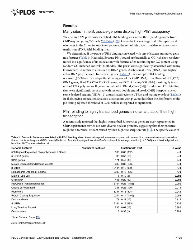
We determined if the peaks of Pfh1 binding correlated with any of sixteen annotated geno-mic features (Table 1; Methods). Because Pfh1 bound preferentially to GC-rich sites, we deter-mined the significance of its association with features after accounting for GC content usingrandom GC-matched controls (Methods). Pfh1 peaks were significantly associated with manyknown hard-to-replicate sites, such as tRNA genes, 5S ribosomal RNA (rRNA), and highlyactive RNA polymerase II transcribed genes (Table 1). For example, Pfh1 bindingoccurred� 300 base pairs (bp), the shearing size of the ChIP DNA, from 80 out of 171 (47%)tRNA genes, 18 of 33 (55%) 5S rRNA genes, and 302 of the top 500 (60%) most highly tran-scribed RNA polymerase II genes (as defined in Rhind, Chen (44)). In addition, Pfh1 bindingsites were significantly associated with meiotic double strand break (DSB) hotspots, nucleo-some depleted regions (NDRs), 3’ untranslated regions (UTRs), and mating type loci (Table 1).In all following association analyses, associations with p-values less than the Bonferroni multi-ple testing adjusted threshold of 0.003 will be interpreted as significant.
Pfh1 binding to highly transcribed genes is not an artifact of their hightranscriptionA recent study reported that highly transcribed S. cerevisiae genes are over-represented inChIP experiments carried out with diverse nuclear proteins, suggesting that their presencemight be a technical artifact caused by their high transcription rate [45]. The specific cause of
Table 1. Genomic features associated with Pfh1 binding sites. Association p-values were computed with an empirical permutation-based procedurethat accounts for length and GC content (Methods). Associations significant after Bonferroni multiple testing correction (p < 0.003) are in bold. All p-valuesless than 10−50 are reported as�0.
Genome Features Number of Features Fraction with Pfh1 p-value
500 Highest Expressed RNA polymerase II Genes 500 0.60 (302) � 0
5S rRNA genes 33 0.55 (18) � 0
tRNA genes 171 0.47 (80) � 0
Meiotic Double Strand Break Hotspots 288 0.37 (106) � 0
3’ UTRs 5144 0.18 (939) � 0
Nucleosome Depleted Regions 2300 0.18 (406) � 0
Mating Type Loci 5 0.40 (2) 0.002
G4 motifs* 446 0.20 (90) 0.002
RNA Pol II Transcribed Genes 5144 0.23 (1188) 0.005
Origins of Replication 741 0.23 (170) 0.014
Promoters 3237 0.16 (504) 0.042
Protein Coding Sequence 10244 0.14 (1459) 0.093
Dubious Genes 71 0.21 (15) 0.112
5’ UTRs 5144 0.13 (693) 0.139
Long Terminal Repeat 236 0.07 (16) 0.982
Centromeres 3 0.33 (1) 0.999
* from Sabouri, Capra [39].
doi:10.1371/journal.pgen.1006238.t001
Pfh1 Is an Accessory Replicative DNA Helicase
PLOS Genetics | DOI:10.1371/journal.pgen.1006238 September 9, 2016 4 / 29

the “hyper-ChIPability” of these regions has not been resolved. It has been proposed that,because highly transcribed genes are more accessible during the pull-down, they may be morelikely to interact with beads or antibodies during the IP, and therefore be subject to nonspecificprecipitation by the antibody.
To ensure that the association with highly transcribed RNA polymerase II genes was notdue to this artifact, we used ChIP combined with quantitative PCR (qPCR) to examine Pfh1association to four highly transcribed genes, hsp90+, tdh1+, adh1+, and hta1+, which are amongthe top 500 most highly transcribed genes and were all Pfh1-associated sites in the genome-wide analysis. Transcription of hsp90+, tdh1+, adh1+ occurs throughout the cell cycle, whilehta1+ transcription peaks in S phase [46]. In fission yeast, the G2 phase comprises about 75%of the cell cycle, so the majority of cells in an asynchronous culture are in G2 phase, and mostgenes are transcribed in this interval [47]. Thus, if the association of Pfh1 with highly tran-scribed genes was non-specific, it should occur to a similar extent in asynchronous andG2-arrested cells for hsp90+, tdh1+, adh1+, but not hta1+.
We performed ChIP-qPCR experiments in both asynchronous and G2 arrested cells in atemperature sensitive cdc25-22 strain that expressed Pfh1-13Myc. Pfh1 was significantly associ-ated to all four genes in asynchronous cells grown at 25°C, compared to an untagged controlstrain (S1 Fig), which validated the peaks found in the ChIP-seq data. To arrest the cells in G2phase, cells growing logarithmically at 25°C were shifted to 37°C for 4h.
Consistent with the expectation in the absence of bias, high Pfh1 binding to all four of thehighly transcribed genes varied across the cell cycle; it was approximately four times higher inasynchronous compared to G2 arrested cells in the ChIP-qPCR assay (p� 0.016; Fig 1). Incontrast, Pfh1 binding to the much less frequently transcribed ade6+ gene was not significantlydifferent in asynchronous versus G2 arrested cells (p = 0.14, Fig 1). Thus, the ChIP-qPCR
Fig 1. Pfh1 is enriched at highly transcribed genes in asynchronous cells compared to G2 arrested cells.Samples from asynchronous or G2 arrested cells expressing Pfh1-13Myc were chromatin immunoprecipitatedusing an anti-Myc antibody. The immunoprecipitated DNA was analyzed using quantitative PCR with primersspecific for four highly transcribed genes, hsp90+, tdh1+, adh1+, hta1+, and a low/medium transcribed control gene,ade6+. Pfh1 association is shown as immunoprecipitated DNA divided by input DNA. Data are means of threeindependent replicates. Error bars are the standard deviation. The p-value was determined by two-tailed Student’st-test. An “*” indicates significant (p < 0.05), and “ns” indicates a non-significant difference between asynchronouscells compared to G2 arrested cells.
doi:10.1371/journal.pgen.1006238.g001
Pfh1 Is an Accessory Replicative DNA Helicase
PLOS Genetics | DOI:10.1371/journal.pgen.1006238 September 9, 2016 5 / 29

experiment confirmed the high Pfh1 binding to these highly transcribed genes seen by ChIP-seq and established that this binding is not an artifact of the ChIP.
Replisome pausing is increased genome-wide at hard-to-replicate sitesin Pfh1-depleted cellsTo identify genomic sites that require Pfh1 for their timely replication, we analyzed genome-wide occupancy of Cdc20, the catalytic subunit of the leading strand DNA polymerase ԑ [48] inWT and Pfh1-depleted cells. As previously reported [39], there are 485 peaks of high Cdc20occupancy in WT cells and 517 in Pfh1-depleted cells (S1 Table). Although all genomic sitesare Cdc20-associated when they are being replicated, high DNA polymerase occupancy corre-lates with replication fork slowing in both S. pombe and S. cerevisiae [39, 49]. Most of the highCdc20 occupancy sites in WT cells were also found in Pfh1-depleted cells (390/485, 80%) andvice versa (389/517, 75%) (Fig 2A).
In an earlier study, we used these data to demonstrate that many G4 motifs bind Pfh1 andthat fork slowing and DNA breakage is more frequent at G4 motifs than at other G-rich regionsin Pfh1-depleted cells (S2 Fig) [39]. Here we extend this analysis from G4 motifs to the rest ofthe genome. This analysis showed that in addition to G4 motifs, tRNA genes, 5S rRNA genes,NDRs, replication origins, RNA polymerase II promoters, RNA polymerase II transcribedgenes, and meiotic DSB hotspots were significantly enriched among high Cdc20 occupancysites in both WT and Pfh1-depleted cells (Table 2 and S2 Table; p< 0.001). The sets of highCdc20 occupancy were identical in the two conditions except that dubious genes were enrichedin Pfh1-depleted but not in WT cells. Despite this similarity, the evidence for elevated Cdc20occupancy was significantly greater in Pfh1-depleted cells (p� 0, Wilcoxon signed-rank test,p-values< 10−50 are reported as�0; Fig 2B). This pattern held for all genomic features tested(Fig 2C–2F; S3 Table). For example, 69% of Cdc20 peaks near NDRs (p = 2.4x10⁻⁵; Fig 2C) and62% of peaks near highly transcribed RNA polymerase II genes (p� 0; Fig 2D) were signifi-cantly stronger in Pfh1-depleted cells. This effect was particularly striking at tRNA (p� 0; Fig2E) and 5S rRNA (p = 1.4x10⁻⁶; Fig 2F) genes, where over 97% of the peaks showed evidence ofsignificantly elevated Cdc20 occupancy when cells were Pfh1-depleted compared to WT cells.These findings argue that these genomic features, all of which had significant Pfh1 occupancyin WT cells (Table 1), were particularly dependent on Pfh1 for timely replication.
To compare the Pfh1-dependent effects in more detail, we analyzed the genomic featuresassociated with the 95 peaks of high Cdc20 binding unique to WT cells versus those associatedwith the 128 peaks of high Cdc20 binding that were found only in Pfh1-depleted cells. No fea-tures were enriched among the unique WT peaks. In sharp contrast, 5S rRNA and tRNAgenes, meiotic DSB hotspots, NDRs, and dubious genes were all significantly enriched amongstthe Cdc20 peaks unique to Pfh1-depleted cells (S2 Table).
Taken together, our results show that several classes of genomic features, especially RNApolymerase III transcribed genes, depend on Pfh1 for normal fork progression. In cases wheresites were hard to replicate even in WT cells, fork pausing at these sites was significantly morepronounced in Pfh1-depleted cells.
Pfh1 protects the genome from DNA damageIn virtually all eukaryotes, including S. pombe, phosphorylation of H2A (γ-H2A) marks sites ofDNA damage, typically DSBs [50]. To determine if the site-specific increases in replicationpausing detected in Pfh1-depleted cells were associated with DNA damage, we analyzed peaksfrom previous ChIP-seq experiments using anti-γ-H2A antibodies in WT or Pfh1-depleted
Pfh1 Is an Accessory Replicative DNA Helicase
PLOS Genetics | DOI:10.1371/journal.pgen.1006238 September 9, 2016 6 / 29

Fig 2. Fork pausing asmarked by Cdc20 occupancy is increased in the absence of Pfh1. (A) Venndiagram showing the overlap of genome-wide Cdc20 peaks in the presence and absence of Pfh1. The twonumbers in the intersection are the number of WT peaks that overlap a Pfh1-depleted peak and vice versa.(B) Scatter plot comparing Cdc20 peak strength (-10*log10(p-value)) in WT and Pfh1-depleted cells. Eachpoint represents a genomic region with a Cdc20 occupancy peak in at least one context. If a peak was notpresent in a context, it is plotted at 0 on the corresponding axis. The number in the bottom right of each plotgives the percentage of peaks stronger in Pfh1-depleted cells. When all peaks were considered, 64% ofpeaks were significantly stronger in Pfh1-depleted cells (p� 0, Wilcoxon signed-rank test). Likewise, peaks
Pfh1 Is an Accessory Replicative DNA Helicase
PLOS Genetics | DOI:10.1371/journal.pgen.1006238 September 9, 2016 7 / 29
cells (S1 Table) [39]. We quantified the genomic distribution and Pfh1-dependence of theγ-H2A peaks with the same methods used for Cdc20.
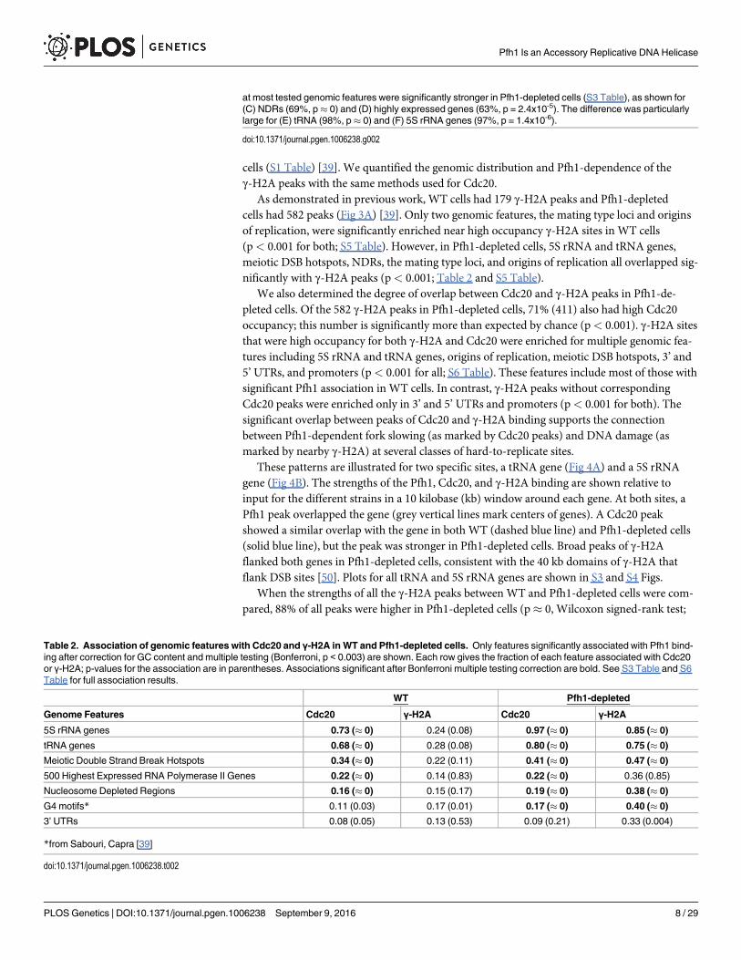
As demonstrated in previous work, WT cells had 179 γ-H2A peaks and Pfh1-depletedcells had 582 peaks (Fig 3A) [39]. Only two genomic features, the mating type loci and originsof replication, were significantly enriched near high occupancy γ-H2A sites in WT cells(p< 0.001 for both; S5 Table). However, in Pfh1-depleted cells, 5S rRNA and tRNA genes,meiotic DSB hotspots, NDRs, the mating type loci, and origins of replication all overlapped sig-nificantly with γ-H2A peaks (p< 0.001; Table 2 and S5 Table).
We also determined the degree of overlap between Cdc20 and γ-H2A peaks in Pfh1-de-pleted cells. Of the 582 γ-H2A peaks in Pfh1-depleted cells, 71% (411) also had high Cdc20occupancy; this number is significantly more than expected by chance (p< 0.001). γ-H2A sitesthat were high occupancy for both γ-H2A and Cdc20 were enriched for multiple genomic fea-tures including 5S rRNA and tRNA genes, origins of replication, meiotic DSB hotspots, 3’ and5’ UTRs, and promoters (p< 0.001 for all; S6 Table). These features include most of those withsignificant Pfh1 association in WT cells. In contrast, γ-H2A peaks without correspondingCdc20 peaks were enriched only in 3’ and 5’ UTRs and promoters (p< 0.001 for both). Thesignificant overlap between peaks of Cdc20 and γ-H2A binding supports the connectionbetween Pfh1-dependent fork slowing (as marked by Cdc20 peaks) and DNA damage (asmarked by nearby γ-H2A) at several classes of hard-to-replicate sites.
These patterns are illustrated for two specific sites, a tRNA gene (Fig 4A) and a 5S rRNAgene (Fig 4B). The strengths of the Pfh1, Cdc20, and γ-H2A binding are shown relative toinput for the different strains in a 10 kilobase (kb) window around each gene. At both sites, aPfh1 peak overlapped the gene (grey vertical lines mark centers of genes). A Cdc20 peakshowed a similar overlap with the gene in both WT (dashed blue line) and Pfh1-depleted cells(solid blue line), but the peak was stronger in Pfh1-depleted cells. Broad peaks of γ-H2Aflanked both genes in Pfh1-depleted cells, consistent with the 40 kb domains of γ-H2A thatflank DSB sites [50]. Plots for all tRNA and 5S rRNA genes are shown in S3 and S4 Figs.
When the strengths of all the γ-H2A peaks between WT and Pfh1-depleted cells were com-pared, 88% of all peaks were higher in Pfh1-depleted cells (p� 0, Wilcoxon signed-rank test;
at most tested genomic features were significantly stronger in Pfh1-depleted cells (S3 Table), as shown for(C) NDRs (69%, p� 0) and (D) highly expressed genes (63%, p = 2.4x10⁻⁵). The difference was particularlylarge for (E) tRNA (98%, p� 0) and (F) 5S rRNA genes (97%, p = 1.4x10⁻⁶).
doi:10.1371/journal.pgen.1006238.g002
Table 2. Association of genomic features with Cdc20 and γ-H2A in WT and Pfh1-depleted cells. Only features significantly associated with Pfh1 bind-ing after correction for GC content and multiple testing (Bonferroni, p < 0.003) are shown. Each row gives the fraction of each feature associated with Cdc20or γ-H2A; p-values for the association are in parentheses. Associations significant after Bonferroni multiple testing correction are bold. See S3 Table and S6Table for full association results.
WT Pfh1-depleted
Genome Features Cdc20 γ-H2A Cdc20 γ-H2A
5S rRNA genes 0.73 (� 0) 0.24 (0.08) 0.97 (� 0) 0.85 (� 0)
tRNA genes 0.68 (� 0) 0.28 (0.08) 0.80 (� 0) 0.75 (� 0)
Meiotic Double Strand Break Hotspots 0.34 (� 0) 0.22 (0.11) 0.41 (� 0) 0.47 (� 0)
500 Highest Expressed RNA Polymerase II Genes 0.22 (� 0) 0.14 (0.83) 0.22 (� 0) 0.36 (0.85)
Nucleosome Depleted Regions 0.16 (� 0) 0.15 (0.17) 0.19 (� 0) 0.38 (� 0)
G4 motifs* 0.11 (0.03) 0.17 (0.01) 0.17 (� 0) 0.40 (� 0)
3’ UTRs 0.08 (0.05) 0.13 (0.53) 0.09 (0.21) 0.33 (0.004)
*from Sabouri, Capra [39]
doi:10.1371/journal.pgen.1006238.t002
Pfh1 Is an Accessory Replicative DNA Helicase
PLOS Genetics | DOI:10.1371/journal.pgen.1006238 September 9, 2016 8 / 29

Fig 3. DNA damage as marked by phosphorylated histone H2A (γ-H2A) levels is increased in theabsence of Pfh1. (A) Venn diagram showing the overlap of genome-wide γ-H2A occupancy peaks in thepresence and absence of Pfh1. The two numbers in the intersection are the number of WT peaks thatoverlap a Pfh1-depleted peak and vice versa. (B) Scatter plot comparing γ-H2A peak strength in WT andPfh1-depleted cells. The layout is the same as in Fig 2B. Overall, γ-H2A peaks were significantly stronger inPfh1-depleted cells (88%, p� 0, Wilcoxon signed-rank test). Peaks near most tested genomic features weresignificantly stronger in Pfh1-depleted cells (S3 Table), e.g., (C) NDRs (88%, p� 0) and (D) highly expressedgenes (88%, p� 0). γ-H2A peaks associated with (E) tRNA (99%, p� 0) and (F) 5S rRNA genes (100%, p�0) were almost universally stronger in Pfh1-depleted cells.
doi:10.1371/journal.pgen.1006238.g003
Pfh1 Is an Accessory Replicative DNA Helicase
PLOS Genetics | DOI:10.1371/journal.pgen.1006238 September 9, 2016 9 / 29

Fig 3B). This pattern held for the subsets of γ-H2A peaks associated with nearly all classes ofgenomic features (Fig 3C–3F; S3 Table). However, as seen for Cdc20, the magnitude of the dif-ference was strongest for the RNA polymerase III transcribed genes: 190 of 192 γ-H2A peaksnear tRNA genes (p� 0, Fig 3E) and all 67 γ-H2A 5S rRNA peaks (p� 0, Fig 3F) were greaterin Pfh1-depleted cells.
During S phase, Pfh1 associates with Pfh1-sensitive andPfh1-insensitive sites throughout the genomeOur data indicate that Pfh1 promotes replication and suppresses DNA damage at many dis-crete sites in the genome. We considered two models to explain this pattern of Pfh1 action.First, Pfh1 could act by binding nearby the replisome and mitigating replication obstacles asthe replisome moves past Pfh1-sensitive sites. Alternatively, Pfh1 could be recruited only tosites that are hard to replicate or to stalled replication forks. In that case, Pfh1 would have lowor no binding to sites that are Pfh1-insensitive. To distinguish between the two possibilities, weused ChIP-qPCR to monitor association of Pfh1 and Cdc20 in synchronized cells. For theseexperiments, we used a cdc25-22 strain that expressed Pfh1-13Myc inserted at the leu1+ locusunder the control of the pfh1+ promoter (the endogenous pfh1+ locus was unaltered). Thisstrain also expressed Cdc20-3HA from its endogenous location.
Cells were arrested in G2 phase by incubation at the non-permissive temperature (37°C)and then released at permissive temperature (25°C). The position in the cell cycle and the qual-ity of the synchrony were determined by FACS analysis (Fig 5A). To determine association andmovement of the replication fork, samples were taken throughout one synchronous cell cycle.At each time point, we examined association of Pfh1-13Myc and Cdc20-3HA to three originsof replication and their adjacent regions. We examined binding to the efficient ars3002 and a
Fig 4. ChIP-seq signal surrounding two representative RNA-PolIII transcribed genes: a threonine tRNA gene(A) and a 5S rRNA gene (B). Each plot displays the smoothed base 2 logarithm of the ChIP-seq reads mapping to eachposition in the experimental context over the corresponding input only read count. The raw signal was smoothed byconvolution with a 1 kb Hanning window. The coordinates on the x-axis give the genomic location of the gene (graybox). These examples highlight a common pattern for tRNA and 5S rRNA genes: a peak of Pfh1 occupancy centered onthe gene (black), with overlapping Cdc20 binding that is increased in the absence of Pfh1 (blue), and elevated γ-H2A inthe regions flanking the genes in Pfh1-depleted cells (red). Plots for all tRNA and 5S rRNA genes are given in S3 andS4 Figs.
doi:10.1371/journal.pgen.1006238.g004
Pfh1 Is an Accessory Replicative DNA Helicase
PLOS Genetics | DOI:10.1371/journal.pgen.1006238 September 9, 2016 10 / 29

Fig 5. Pfh1 and DNA Pol ε both bind Pfh1-sensitive and insensitive sites. (A) FACS analysis of cell synchrony. (B)Schematic of the regions examined by qPCR. Open circles represent origins of replication. Boxes mark the position of
Pfh1 Is an Accessory Replicative DNA Helicase
PLOS Genetics | DOI:10.1371/journal.pgen.1006238 September 9, 2016 11 / 29
region18 kb away from ars3002 (ars3002_18kb), ars2004 and a region 30 kb from ars2004(ars2004_30kb), and ars3005 and a region 26 kb from ars3005 (ars3005_26kb) (Fig 5B).
If Pfh1 were nearby or associated with the replisome, Pfh1 and Cdc20 would have similartemporal patterns of binding to the three origins and their adjacent sites. If Pfh1 were recruitedonly to hard-to-replicate sites, then Cdc20 and Pfh1 would have different binding patterns. Infact, under the second model, Pfh1 should not bind to any of these sites, as none of them werePfh1-dependent in the whole genome analysis. Consistent with the first model, Cdc20 andPfh1 displayed similar association patterns at all three origins and their adjacent regions (Fig5C–5E). We first examined the binding to ars3002 and its adjacent region ars3002_18kb (Fig5C). Although Cdc20 bound in early S phase to ars3002, earlier than Pfh1, the peak binding forCdc20 and Pfh1 was reached at 95 min (Fig 5C). Both proteins had their start of binding toars3002_18kb at 80 min after release from G2 phase and their peak binding at 95 min (Fig 5C).Thus, Pfh1 and Cdc20 bound to the Pfh1-insensitive site located 18 kb downstream of ars3002similarly. However, while clear movement of Cdc20 was detected in this region, movementfrom this origin to the downstream regions was not visible for Pfh1.
Next, we examined the binding of Pfh1 and Cdc20 to the four other Pfh1-insensitive sitesars2004, ars2004_30kb, ars3005, and ars3005_26kb. Pfh1 bound to all these four regions, simi-larly to Cdc20 (Fig 5D and 5E). However, movement of neither Cdc20 nor Pfh1 was detectedat any of these origins to their adjacent sites.
Because the dynamics of Pfh1 and the replisome were not clear from the above experiments,we further investigated the binding pattern of Pfh1 and Cdc20 at five other regions, includingboth Pfh1-sensitive and Pfh1-insensitve sites (Fig 5F). We tested two regions that were not ori-gins of replication on chromosome II: one Pfh1-sensitive (Chr II_nonars_1236154), and aPfh1-insensitive site 36 kb away (Chr II_nonars_1272741). Finally, we examined two Pfh1-sen-sitive tRNAs (tRNAGLU.05 and tRNAASN.05) and one Pfh1-insensitive site 6 kb away fromtRNAASN.05 (tRNAASN.05_6kb).
At all five regions, both Pfh1 and Cdc20 had peak binding at 95 min after release from theG2 arrest, which by FACS analysis is mid-S phase (Fig 5). To determine if Pfh1 was recruitedonly to Pfh1-sensitive sites, we calculated the ratio of Pfh1 to Cdc20 binding at Pfh1-sensitiveand -insensitive sites (IP/input of Pfh1 divided by IP/input for Cdc20) at the peak of replicationfor all sites (Fig 5F). If Pfh1 were recruited solely to Pfh1-sensitive sites, the ratio of Pfh1/Cdc20 would be higher at Pfh1-sensitive sites compared to Pfh1-insensitive sites. If Pfh1 werein proximity to the replisome, the ratios should be similar at the two classes of sites. Indeed, thePfh1/Cdc20 ratio was on average 1.1 and 0.9 for Pfh1-sensitive and Pfh1-insensitive sites,respectively (Fig 5F). Thus, Pfh1 binds similarly to both Pfh1-sensitive and -insensitive sites.These data suggest that Pfh1 is near the replisome during S phase, rather than being recruitedto its sites of action at Pfh1-sensitive sites. However, we cannot rule out the possibility that
primer-pairs for qPCR that detect ars3002, ars3002_18kb, ars2004, ars2004_30kb, ars3005, and ars3005_26kb for ChIP-qPCR experiments. (C-E) Samples from each time point of the synchronized Cdc20-3HA Pfh1-13Myc cdc25-22 strainwere immunoprecipitated with either anti-HA (left) or anti-Myc (right) antibody. The DNA was analyzed with qPCR usingprimer pairs for (C) ars3002 and ars3002_18kb, (D) ars2004 and ars2004_30kb, and (E) ars3005 and ars3005_26kb.Although cells appeared synchronous by FACS analysis, and Pfh1 and Cdc20 showed similar temporal patterns with peakbinding at 95 min, we did not detect progression of the replisome from the ars2004 and ars3005 origins to their adjacentregions (ars2004_30kb and ars3005_26kb). This is likely due to the documented heterogeneity of origin usage in differentcells in the same population [66] and to inefficient origins. The experiments were performed at least two times and thegraphs show one representative biological replicate. (F) The ratio of Pfh1 IP/input to Cdc20 IP/input at peak binding afterG2 release for eleven regions. Samples from Cdc20-3HA Pfh1-13Myc cdc25-22 synchronized cells wereimmunoprecipitated with either anti-HA or anti-Myc antibody and analyzed by qPCR. The experiments were repeated twotimes and the reported ratio is the average of two biological replicates; the standard deviation is given in parentheses. Thenames of adjacent regions are in the same color. Y: yes, N: no.
doi:10.1371/journal.pgen.1006238.g005
Pfh1 Is an Accessory Replicative DNA Helicase
PLOS Genetics | DOI:10.1371/journal.pgen.1006238 September 9, 2016 12 / 29

additional Pfh1 molecules are recruited to some or even all Pfh1-sensitive sites upon replisomepausing.
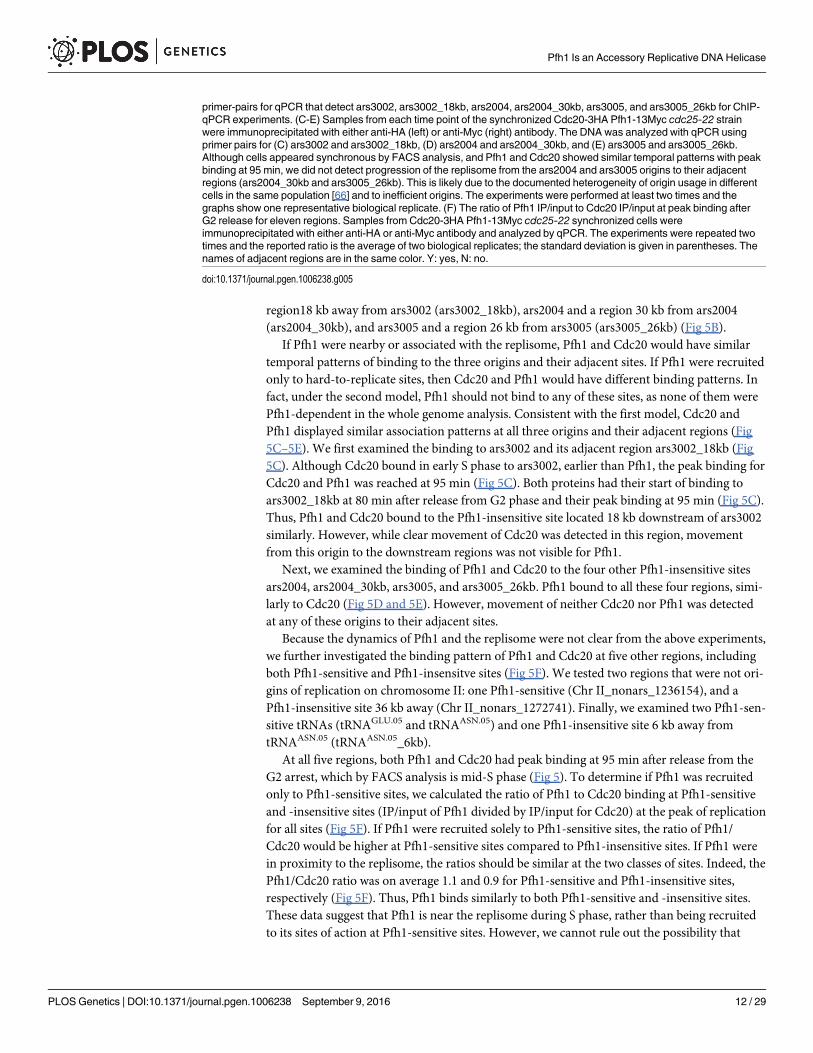
Mass spectrometry identifies Pfh1 interacting proteinsIf Pfh1 maintains proximity to the replisome, as suggested by its pattern of binding to chromo-somal DNA (Fig 5), then Pfh1 should be associated in vivo with known replisome subunits. Toaddress this possibility, we used immunoaffinity purification mass spectrometry (IP-MS) toidentify the Pfh1 protein interactions in S phase cells (Fig 6A). In these experiments, Pfh1 wasexpressed under its endogenous promoter as a GFP fusion (Pfh1-GFP), allowing immunoaffi-nity isolation of Pfh1 and its associated proteins through the GFP tag [51]. Cells expressing aSV40 nuclear localization signal-GFP fusion (NLS-GFP) were used as a negative control fornon-specific association of proteins to GFP. Two biological replicates of both Pfh1-GFP andNLS-GFP were isolated in parallel from S phase cells (Fig 6A) using anti-GFP antibodies(Fig 6B).
Following mass spectrometry analysis of Pfh1-GFP and NLS-GFP immunoisolates, theinteraction specificity of individual co-isolating proteins was assessed using the SAINT (signifi-cance analysis of interactome) algorithm [52]. SAINT determines confidence scores (rangingfrom 0 to 1) for protein-protein interactions based on the spectrum count distributionsobtained from bait (Pfh1-GFP) isolations relative to the negative control (NLS-GFP). Highconfidence Pfh1 interactions were defined as proteins having a SAINT score� 0.80, a thresh-old used previously to identify functional protein interactions [53, 54]. By this metric, therewere 50 high confidence Pfh1 protein associations that comprise the Pfh1 S phase interactome(Table 3 and S7 Table). Although five of the Pfh1 interacting proteins are uncharacterized,there is functional data for 45 of the 50 proteins. Table 3 lists these proteins.
We also assessed the relative abundance of individual Pfh1 interacting proteins within theinteraction network by calculating the normalized spectrum abundance factor (NSAF) for eachprotein relative to its proteome abundance value (PAX) [55]. NSAF values provide a measureof protein abundance by accounting for factors such as protein length and sample complexitythat can influence the number of spectra acquired for a given protein within a sample. Normal-izing NSAF values to PAX values, as described in [56], provides insight into proteins and func-tional protein classes that are enriched in the Pfh1 isolation relative to their abundances in thecellular proteome. These data are presented in Fig 6C, which also categorizes interacting pro-teins by function.
Pfh1 interacts with multiple replisome componentsThe replisome is the multi-protein complex that is present at the replication fork as it movesthrough the chromosome. Multiple replisome components interacted with Pfh1 with highspecificity (SAINT score� 0.80; Fig 6C and Table 3). These proteins were: (1) five of the sixsubunits of the replicative helicase, the MCM complex (Mcm2 and Mcm4-7); (2) catalytic sub-units of two of the three replicative polymerases (DNA Pol1 from DNA polymerase α; DNAPol2/Cdc20, from DNA polymerase ε); (3) Pol12, the β subunit of DNA Pol1; (4) proliferatingcell nuclear antigen (PCNA, Pcn1), a processivity factor that encircles and slides along theDNA; (5) the three subunits of the single-strand binding replication factor A (RPA, Ssb1, 2 and3); (6) the Dna2 helicase-nuclease that is required for Okazaki fragment maturation; and (7)the two subunits of the FACT complex (facilitates chromatin transcription), Pob3 and Spt1,which facilitates nucleosome remodeling during both transcription and DNA replication. Theassociation of FACT subunits with Pfh1 suggests that FACT and Pfh1 might act synergisticallyto promote replication through hard-to-replicate sites. Four mismatch repair (MMR) proteins,
Pfh1 Is an Accessory Replicative DNA Helicase
PLOS Genetics | DOI:10.1371/journal.pgen.1006238 September 9, 2016 13 / 29
Fig 6. Identification of Pfh1 protein interactions during S phase. (A) Experimental design for parallel immunoaffinity purificationsof Pfh1-GFP and NLS-GFP from cells harvested at S phase of the cell cycle and relative quantification of Pfh1 specific interactionsby MS. As determined by FACS analysis, cdc25-22 cells were collected at the start of S phase, 84 minutes after G2 phase arrest. (B)Immunoaffinity purifications of GFP or Pfh1-GFP from S phase resolved by SDS-PAGE gel with the target and associated proteinsvisualized by Coomassie Blue staining. (C) Interaction network of high confidence Pfh1 protein associations during S phase, as
Pfh1 Is an Accessory Replicative DNA Helicase
PLOS Genetics | DOI:10.1371/journal.pgen.1006238 September 9, 2016 14 / 29
Msh1, 2, 3 and 6, were also Pfh1-associated. The Msh2/6 and Msh2/3 heterodimers interactdirectly with DNA for the recognition of base pair mismatches. Because MMR and DNA repli-cation are strongly coupled in budding yeast, MMR proteins are proposed to track with thereplisome and hence can also be considered replisome components [57, 58].
Additional replisome components were present in the Pfh1-GFP isolations but did not meetour SAINT score criterion. These proteins were: (1) the sixth Mcm subunit (Mcm3; SAINTscore, 0.68); (2) the catalytic subunit of the lagging strand DNA polymerase; Pol3 (SAINTscore, 0.15); (3) Dpb2, the second largest subunit of Pol ε (SAINT score 0.31); (4) Pri1 andPri2, the primase subunits that function together with DNA polymerase α to synthesize theprimers on the leading and lagging strand (SAINT scores of 0.65 and 0.33, respectively); and(5) Mcl1, the S. cerevisiae Ctf4 homologue that interacts with DNA polymerase α (SAINTscore 0.70). While SAINT scores point to high confidence interactions, being based on detectedprotein spectral counts, they are influenced by sample complexity and the dynamic range ofthe co-isolated proteins, and thereby weighted towards large and abundant proteins, and stableinteractions. Pfh1-associated replisome components with lower SAINT scores may be smallerproteins, have lower cellular abundances, and/or form transient interactions [59].
We performed two additional experiments to confirm the association of Pfh1 with the repli-some. First, we isolated Pfh1 from asynchronous cells both in the presence and absence ofDNase (S7 Table and S5 and S6 Figs). Because this experiment was performed with an asyn-chronous population, only a subset of the proteins that interacted with Pfh1 in S phase wasdetected (S6 Fig), even without DNase treatment. Of the 19 replisome/replication proteins thatpassed our stringent SAINT score threshold, ten were found in the DNase untreated sample,and nine of these retained their Pfh1 association after DNase treatment: Ssb1 and 2, Msh2,Mcm4, 5, and 6, Cdc20 (Pol2), Pol12, and Spt16. Because these interactions were not DNA-dependent, they are likely due to protein-protein interactions. Second, we isolated Pfh1 and itsassociated proteins from G2 arrested cells. We detected eight Pfh1-associated replication/repli-some proteins in G2, and all eight were detected with fewer spectral counts in G2 extracts thanin S phase extracts. The remaining eleven were not detected at all as Pfh1-interacting proteinsin G2 phase (Table 3; S7 Table). Thus, as expected for a replisome component, Pfh1 associationwith known replisome subunits was either lost or diminished in G2 phase.
Together, these results show that Pfh1 associated in vivo with numerous replisome proteins,and that replisome and replication-related proteins represent a substantial subset of specificPfh1 interactions (19 out of 50 proteins (38%) with SAINT score of�0.8) (Table 3; in bold; Fig6C). Almost all of these associations were S phase-limited or S phase-enriched as well as DNAindependent.
Pfh1 interacts with multiple mitochondrial and repair proteinsPfh1 is a multi-functional protein: in addition to its role in nuclear DNA replication, it pro-motes DNA repair and is essential for maintenance of mitochondrial (mt) DNA [34]. Consis-tent with Pfh1 having mt function, 8 of the 50 high confidence Pfh1 interaction proteins havemt annotations (Fig 6C; mt proteins are underlined in Table 3). This subset includes severalproteins implicated in mtDNA replication, such as (1) Rim1, the mt single-strand DNA bind-ing protein (MS analyses reveal that ScPif1 is also ScRim1-associated; [60]), (2) Rpo1, the
identified by MS and assessed for specificity of binding by SAINT (p� 0.80, n = 2). Nodes represent individual proteins interactingwith Pfh1; node color represents relative enrichment compared to their abundance in the proteome (NSAF/PAX); and edgesrepresent known interactions curated by the STRING database. Replisome components are highlighted by the blue dotted line.
doi:10.1371/journal.pgen.1006238.g006
Pfh1 Is an Accessory Replicative DNA Helicase
PLOS Genetics | DOI:10.1371/journal.pgen.1006238 September 9, 2016 15 / 29

Table 3. Pfh1-GFP interacting proteins. Pfh1-associated proteins during S phase (SAINT score� 0.80) presented in alphabetical order. Names in boldare replisome components or replication related; italics indicate repair/recombination proteins; underlining indicates mitochondrial proteins. Functional anno-tations are from PomBase [68].
Protein Accession number Function SAINT score
Abp1 P49777 ARS-binding protein, CENP-B homologue, less abundant in G2 0.83
Coq3 O74421 Mitochondrial hexaprenyldihydroxybenzoate methyltransferase 0.86
Dna2 Q9URU2 ATP-dependent helicase-nuclease, processes Okazaki fragments 0.95
Fbh1 Q9USU3 F-box DNA helicase, modulates homologous recombination 0.97
Irc3 Q1MTR1 Putative mitochondrial ATP-dependent helicase 1.00
Mcm2 P40377 Subunit of MCM replicative DNA helicase 0.89
Mcm4 P29458 Subunit of MCM replicative DNA helicase 0.92
Mcm5 P41389 Subunit of MCM replicative DNA helicase 0.90
Mcm6 P49731 Subunit of MCM replicative DNA helicase 0.83
Mcm7 O75001 Subunit of MCM replicative DNA helicase 0.94
Mfh1 Q9UT23 FANCM-like DNA helicase, repairs inter-strand crosslinks, promotes meiotic recombination 0.99
Mgm101 O14354 Mitochondrial genome maintenance protein 0.87
Msh1 O13921 DNAmismatch repair protein 0.98
Msh2 O74773 DNAmismatch repair protein 0.99
Msh3 P26359 DNAmismatch repair protein 0.99
Msh6 O74502 DNAmismatch repair protein 0.99
Pcn1 Q03392 Subunit of homotrimeric PCNA (Proliferating cell nuclear antigen) 0.90
Pku70 O94395 Subunit of Ku heterodimer 1.00
Pku80 Q9HGM8 Subunit of Ku heterodimer 1.00
Pob3 O94529 FACT complex subunit, remodels histones 0.86
Pol1 P28040 Catalytic subunit of DNA polymerase alpha 0.82
Pol12 O74946 Subunit B of DNA polymerase alpha 0.84
Pol2 P87154 Catalytic subunit of DNA polymerase epsilon; also called Cdc20 0.99
Rad22 P36592 DNA repair and recombination protein, homologue budding yeast Rad52 0.83
Rad3 Q02099 ATR-like checkpoint protein kinase 0.98
Rim1 O14087 Mitochondrial single-stranded DNA-binding protein 0.99
Rmi1 Q10160 Subunit of Rqh1/Top3 complex, suppresses DNA damage 0.89
Rpc1 O94666 Subunit of RNA polymerase III 0.98
Rpc2 Q10233 Subunit of RNA polymerase III 0.83
Rpc25 O94285 Subunit of RNA polymerase III 0.82
Rpc3 Q9C106 Subunit of RNA polymerase III 0.86
Rpc37 O74883 Subunit of RNA polymerase III 0.83
Rpc4 O74857 Subunit of RNA polymerase III 0.89
Rpo41 O13993 Mitochondrial RNA polymerase, priming mtDNA replication 0.89
Rqh1 Q09811 RecQ family DNA helicase subunit of Rqh1/Top3 complex, suppresses DNA damage 1.00
Sdh1 Q9UTJ7 Mitochondrial protein, probable succinate dehydrogenase flavoprotein subunit 0.83
SPAC167.05 P87132 Largely uncharacterized but implicated in meiotic chromosome segregation 0.84
SPAC1F5.11c Q8TFH4 Tra2 subunit of NuA4 complex phosphatidylinositol pseudokinase 0.99
SPAC31G5.19 O14114 Uncharacterized AAA domain-containing protein 1.00
SPAPB17E12.10c Q8TFH4 Mitochondrial protein, affects RNA processing 0.99
SPBC3B8.08 O59716 Uncharacterized protein 0.90
SPCC553.01c O74939 Meiotic chromosome segregation protein Dbl2 0.99
Spt16 O94267 FACT complex subunit, remodels histones 0.86
Ssb1 Q92372 Replication factor A protein 1, RPA subunit 1.00
Ssb2 Q92373 Replication factor A protein 2, RPA subunit 0.99
(Continued)
Pfh1 Is an Accessory Replicative DNA Helicase
PLOS Genetics | DOI:10.1371/journal.pgen.1006238 September 9, 2016 16 / 29

mtRNA polymerase that is thought to prime mtDNA replication, and (3) Mgm101, which isrequired for maintenance of mtDNA by an unknown mechanism.
Consistent with the reported DNA repair functions of Pfh1 [34], we observed multiplerepair proteins among the high confidence Pfh1 interactions (Table 3, italics), includingRad22, the S. pombe homolog of budding yeast Rad52, which is required for homologousrecombination [61], Rad3, the ATR-like checkpoint kinase [62], and both subunits of the non-homologous end joining Ku complex, pKu70 and 80. In addition, Rqh1 (homolog of humanBLM) DNA helicase and its two interacting partners, the topoisomerase Top3 and Rmi1, werePfh1-associated. This highly conserved heterotrimeric complex has multiple functions, but isbest known for suppressing DNA damage at hard-to-replicate sites, such as converged forks[63] and/or collapsed replication forks—functions relevant to those of Pfh1.
Finally, six subunits of the 26 subunit RNA polymerase III complex were Pfh1-associatedwith high confidence (Rpc1, 2, 3, 4, 25 and 37), while four others were Pfh1-associated but hadSAINT scores<0.8 (Rpc6, 0.71; Rpc9 and 10, 0.30; Rpc19, 0.76) [64]. This finding is probablyrelated to RNA polymerase III transcribed genes being among the most potent replicationimpediments in Pfh1-depleted cells (Figs 2–4; see Discussion).
Discussion
Pfh1 acts at diverse sites genome-wideWe used genome-wide assays to determine sites where replication and genome integrity arePfh1-dependent. The most striking aspect of these data is the strong dependence of RNA poly-merase III transcribed genes on Pfh1. 2D gel analyses showed previously that replication of fiveof five tested tRNA genes is Pfh1-dependent, and this dependence is seen regardless of whetherreplication is co-directional or opposite to the direction of transcription through the gene [35].Here we show that close to 50% (80/171) of the tRNA genes bound Pfh1 (Table 1). Moreover,nearly all of the tRNA genes that bound Pfh1 were sites of fork pausing and DNA damage inboth WT and Pfh1-depleted cells (Table 2; high Cdc20 binding: WT: 74/80, Pfh1-depleted: 80/80: high γ-H2A: WT: 25/80, Pfh1-depleted: 69/80). However, both Cdc20 (Fig 2E) and γ-H2Abinding (Fig 3E) were significantly higher at virtually all (99%) tRNA genes in Pfh1-depletedcompared to WT cells. Likewise, genome-wide analyses revealed that 55% (18/32) of 5S rRNAgenes bound Pfh1 (Table 1), and again the majority of these genes were sites of fork pausing(Table 2; WT: 16/18, Pfh1-depleted: 18/18) and/or DNA damage (WT: 5/18, Pfh1-depleted:14/18), and both features were significantly higher in Pfh1-depleted compared to WT cells(Figs 2F and 3F). The independent MS analysis also supports the importance of Pfh1 at RNApolymerase III transcribed genes, as Pfh1 interacted with multiple subunits of RNA polymeraseIII (Fig 6C; Table 3). The association of these subunits with Pfh1 is consistent with a modelwhere Pfh1 promotes replication past genes by displacing these proteins from DNA.
In addition to RNA polymerase III genes, 60% of the 500 most highly expressed protein-coding genes over a range of growth conditions were bound by Pfh1 (Table 1), and 48% were
Table 3. (Continued)
Protein Accession number Function SAINT score
Ssb3 Q92374 Replication factor A protein 3, RPA subunit 0.87
Tea3 O14248 Tip elongation aberrant protein 3, cell polarity 0.94
Top3 O60126 DNA topoisomerase 3, interacts with Rqh1 helicase, suppresses DNA damage 0.98
Tsf1 Q9HGL5 Mitochondrial translation elongation factor 0.85
Ubr11 O13731 E3 ubiquitin-protein ligase ubr11, affects chromosome stability, perhaps affects kinetochore 0.97
doi:10.1371/journal.pgen.1006238.t003
Pfh1 Is an Accessory Replicative DNA Helicase
PLOS Genetics | DOI:10.1371/journal.pgen.1006238 September 9, 2016 17 / 29
sites of fork slowing and/or DNA damage in Pfh1-depeleted cells (Figs 2D and 3D; S2 and S3Tables). The likelihood of Pfh1 association was significantly greater for highly expressed genesthan expected from other RNA polymerase II expressed genes (p�0; hypergeometric test) asonly 23% of all genes were Pfh1 associated.
A recent paper suggested that association of proteins with highly expressed genes in S. cere-visiaemight be an artifact of the ChIP assay [45]. This interpretation is unlikely for our S.pombe analyses as a non-ChIP method, 2D gel analysis of replication intermediates, alsoshowed that replication of three of three tested highly expressed RNA polymerase II tran-scribed genes is Pfh1-dependent [35]. Also, the peak strength at the highly transcribed geneswas elevated in Pfh1-depleted cells compared to WT cells, suggesting that the association is nota ChIP artifact. Moreover, the genome-wide approach in this paper showed that high associa-tion of Pfh1 to highly transcribed genes was S phase specific (Fig 1), even though transcriptionof the genes is not S phase-limited. In addition, Pfh1, Cdc20, and γ-H2A all associate with ribo-somal DNA (rDNA), probably the most highly transcribed region in all organisms, but highbinding of all three does not occur over the 18 or 28S coding regions [39]. Also, the patterns ofγ-H2A occupancy—low at the gene site and highest in flanking regions—are inconsistent withtranscription artifacts (Fig 4), because the artifactual enrichment was observed to be highacross the gene body. Thus, high binding of anti-Myc (used for Pfh1-13Myc), anti-HA (usedfor Cdc20-3HA), and anti-γ-H2A to highly transcribed genes is unlikely an artifact of theirhigh transcription.
Fork pausing at highly transcribed genes as marked by high Pol2 occupancy is also demon-strated in budding yeast [49]. In contrast to S. pombe, where Pfh1 depletion increased forkpausing, pausing at these sites is not affected in the absence of ScRrm3 [49] or ScPif1 [18].However, cells with a double deletion have not been tested, so it is possible that ScRrm3 andScPif1 have overlapping functions in promoting replication past these genes.
Replication of multiple classes of genomic features is Pfh1-dependent. In addition to highlytranscribed RNA polymerase II and III genes, this study identified several novel sites of Pfh1association, such as NDRs, 3’UTRs, and preferred meiotic DSB sites. In previous work, weshowed that Pfh1 promotes fork movement past G4 motifs [39]. We analyzed whether thenovel Pfh1 associations could be explained by overlap with G4 motifs or other Pfh1-associatedfeatures (S7 Fig). For example, NDRs often overlapped other significantly Pfh1-associated fea-tures, such as highly transcribed genes (32%), but none were sufficient to explain the associa-tion completely. Given their open state, it is likely that nucleosome-free regions are enrichedfor tightly bound proteins, other interactions, or formation of stable secondary structures thatpause replication. The association with meiotic DSB hotspots in mitotically growing cells islargely driven by overlap with other elements that challenge replication; 92% (265 of 289 DSB)overlap another hard-to-replicate site identified in this study, most notably highly expressedgenes and 3’UTRs.
As noted above, several lines of evidence point to RNA polymerase III genes as the mostPfh1-dependent set of sites. Significant, but relatively smaller, fractions of the other Pfh1-asso-ciated features ultimately produce fork stalling and DNA damage in the absence of Pfh1(Table 2). However, given the greater length of RNA polymerase II transcribed genes, if somecause fork stalling throughout their entire length, they may have a greater impact on fork pro-gression genome-wide than the shorter tRNA genes.
The comprehensive analysis of sites of Pfh1 activity provided here demonstrates that, inaddition to G4 motifs [39], there are many classes of hard-to-replicate sites that depend onPfh1 for their proper replication. Many of these other sites, in particular RNA polymerase IIItranscribed genes, exhibit even stronger Pfh1-dependent effects than G4 motifs (Tables 1 and 2
Pfh1 Is an Accessory Replicative DNA Helicase
PLOS Genetics | DOI:10.1371/journal.pgen.1006238 September 9, 2016 18 / 29

and S3 Table). Altogether, Pfh1 supports DNA replication at thousands of diverse sites acrossthe genome.
Pfh1 is an accessory helicase that maintains proximity to the replisomeAlthough accessory helicases are well studied in bacteria, it is not clear for any of the bacterialenzymes if they are recruited to their sites of action or if they move with the replisome throughthe genome. Here, we present several lines of evidence that support the association of Pfh1with the replisome in S. pombe. First, using a strain in which Pfh1 and Cdc20 were both epitopetagged, Pfh1 had strong binding to three different origins during S phase (Fig 5), even thoughthese origins were not a peak of Pfh1 binding in asynchronous cells. Pfh1 was also bound toadjacent regions of the origins, although neither of these sites were Pfh1-dependent sites. Thetemporal patterns of binding to the sites adjacent to the origins were similar to those of Cdc20in the same cells; however, we could not detect a movement of Pfh1 from an origin to its adja-cent region, as we detected for Cdc20 at ars3002. This may be due to the role of DNA polymer-ase ԑ in replication initiation [65], technical challenges, and/or stochastic origin usage indifferent cells in the synchronized population [66]. If Pfh1 were recruited only to Pfh1-depen-dent sites, we would have expected to see low binding to these Pfh1-independent sites com-pared to Cdc20. Second, the levels of Pfh1 and Cdc20 binding to five other sites were similar,regardless of whether the site was Pfh1-dependent or independent. This again argues against amodel in which Pfh1 is only recruited and associated with sites that require it for their normalreplication. Third, genome wide analyses show that many sites of high Cdc20 binding overlapwith Pfh1 peaks (230 of the 485 high Cdc20 binding sites are also sites of high Pfh1 binding).These data are most consistent with Pfh1 being in close proximity to the replisome throughoutthe genome.
Our MS analyses also provide independent evidence in support of the hypothesis that Pfh1maintains proximity to the replisome and is likely a replisome component. During S phase,Pfh1 was associated with many replisomal proteins, including the MCM replicative helicase,subunits of the replicative polymerases, the processivity clamp PCNA, RPA, and four MMRproteins (Fig 6C). The association of Pfh1 with replisome proteins was either S phase limitedor S phase enhanced and almost all of these associations were DNase-resistant. While it is pos-sible that Pfh1 would have interactions with replisome subunits if it were recruited after repli-some pausing, in the context of the evidence presented above, we conclude that Pfh1’s strongassociations with replication proteins are likely due to protein-protein interactions, as expectedif it is a replisome subunit.
Pfh1 interacts with the FACT complexPfh1 also strongly associated with Spt16 and Pob3 (homologs of human SPT16 and SSRP1),two subunits of the heterodimeric evolutionarily conserved chromatin remodeling FACT com-plex (Table 3). In budding yeast, FACT promotes replication at replication conflicts past tran-scribed regions, and when FACT is depleted, ScRrm3 occupancy increases at highlytranscribed RNA polymerase II and III genes [67]. Also, R-loop formation is elevated in FACTdepleted cells, suggesting that the FACT complex resolves R-loop-mediated transcription-rep-lication conflicts. The S phase association of FACT with Pfh1 suggests that the two cooperateto promote replication through these genes in S. pombe as they do in budding yeast. Pfh1 mayfacilitate replication at these genes by removing R-loops, as does budding yeast ScPif1 [27, 41].In combination with its biochemical activities, the enrichment of Pfh1 at 3’UTRs may alsoreflect a role in resolving R-loop-mediated transcription-replication conflicts.
Pfh1 Is an Accessory Replicative DNA Helicase
PLOS Genetics | DOI:10.1371/journal.pgen.1006238 September 9, 2016 19 / 29
SummaryThe genomes of all organisms are littered with hard-to-replicate sites. Accessory helicases pro-mote the movement of the replisome past these natural impediments. Although multiple bacte-rial accessory helicases have been characterized, much less is known about accessory helicasesin eukaryotes. Our genomic and proteomic analyses, in combination with previous work, showthat Pfh1 promotes replication and suppresses DNA damage at hundreds of diverse, naturallyoccurring hard-to-replicate sites. The pattern of binding of Pfh1 through the genome com-bined with its association with many replisome components argues that it is in close proximitywith the replisome to help it maneuver past these obstacles. For many of the Pfh1-sensitivesites (e.g., tRNA and other highly transcribed genes), replication slowed at these sites even inWT cells, but usually did not result in significant DNA damage. However, when Pfh1 wasdepleted, fork slowing intensified and DNA damage increased dramatically at hard-to-replicatesites. In budding yeast, the two Pif1 family helicases, ScPif1 and ScRrm3, collaborate to pro-mote fork progression past replication impediments. Our results establish the importance ofthis helicase family in S. pombe, a eukaryotic organism that is deeply diverged from S. cerevisiaeand shares many genomic characteristics in common with higher eukaryotes. As the replica-tion-impeding obstacles found in budding and fission yeasts are ubiquitous across genomes ofother organisms, accessory helicases are likely to be required in all organisms, even though heli-cases that act at most of these sites have not been identified in higher eukaryotes. Thus, we pro-pose that Pif1 family helicases present in multicellular eukaryotes also act as accessory helicasesto promote fork progression and preserve genome stability.
Materials and Methods
ChIP analysisChIP-qPCR were performed in cdc25-22 leu1-32::PJK148-Pfh1-13MYC-kanmx6 cdc20::cdc20-3HA-kanmx6 cells (S8 Table). Asynchronous samples were grown at 25°C. The G2 phase cellswere arrested at 37°C and collected after 4h arrest. To perform ChIP-qPCR in synchronizedcells, cells were arrested at 37°C for 4h and released at permissive temperature at 25°C. Samplesfrom the cell synchrony were collected for FACS analysis and ChIP-qPCR at 0, 20, 40, 60, 65,70, 75, 80, 85, 90, 95, 115, 140, and 165 min. The ChIP experiments were performed asdescribed previously [35]. Briefly, cells were cross-linked in 1% formaldehyde at 25°C for 5min. The chromatin was sheared to an average of ~300 bp with a Covaris E220 system. G2phase and asynchronous cells were immunoprecipitated with anti-Myc antibody (ClontechCat. nr 631206). The synchronous cells were divided (3/4 of sample for Pfh1-Myc and 1/4 ofsample for Cdc20-HA) and immunoprecipitated with either anti-Myc antibody or anti-HAantibody (Santa Cruz Biotechnology Cat. nr Sc7392x). Both immunoprecipitated DNA and thecorresponding input amount for each sample were purified and quantified by real-time PCR,using the primer pairs described in S9 Table. At least two biological replicates were performedfor each synchronous ChIP analysis. To calculate the average ratio of the peak binding time inFig 5F, the IP/Input of two biological replicates were used. The time for peak binding (highestbinding) in replicate 1 was 95 min and 70 min for replicate 2. The difference between the high-est peak binding for the two synchronies was due to different start times of S phase after G2release, which was detected by FACS analysis.
Flow cytometric analysis (FACS)S. pombe cells were collected in 165 mM EDTA, 0.1% sodium azide 70% EtOH. Cells were pel-leted, washed in 100% EtOH, and stored at 4°C. In preparation for FACS analysis,
Pfh1 Is an Accessory Replicative DNA Helicase
PLOS Genetics | DOI:10.1371/journal.pgen.1006238 September 9, 2016 20 / 29
approximately 2 x 10⁶ cells were washed in 3 ml of 50 mMNa citrate, pH 7.2, and incubatedovernight at 37°C in 0.5 ml 50 mMNa citrate plus 0.1 mg/ml RNaseA. Following sonication,cells were incubated in 1 μM Sytox Green (Molecular Probes) at room temperature for 30 min-utes. Cells were analyzed using a FACScan single laser fixed-alignment benchtop analyzer.
Genomic annotation enrichment analysisWe tested for enrichment between the genomic locations of the ChIP-seq peaks with sixteensets of genomic annotations (Table 1). The ChIP-seq data for Pfh1, Cdc20, and γ-H2A are avail-able in GEO data set GSE59178 [39]. All peaks analyzed are available in S1 Table. We took thelocation of genes, coding sequences, essential genes, dubious genes, 3’ and 5’UTRs, promoters,tRNAs, centromeres, 5S rRNAs, long terminal repeats, and the mating type loci from PomBase[68]. We also considered the locations of meiotic DSB hotspots [69], NDRs [70] using podbat[71], origins of replication [72], the 500 most highly expressed genes from expression data col-lected across several growth conditions [44], and G4 motifs [39]. For Pfh1 and Cdc20 sites,regions within 300 bp were considered associated, and for γ-H2A peaks, we considered a win-dow of 5 kb, since DNA damage results in elevated phosphorylation in a window of roughlythis size around the break [50]. Overlaps between sets of genomic locations were calculatedusing BEDTools [73].
To determine if the observed association between two sets of genomic features, such as Pfh1binding peaks and tRNA genes, was significantly greater than expected, we followed our previ-ously described procedure for generating GC-content-aware empirical p-values [18, 39, 74]. Inbrief, we compared the observed number of overlaps to the number obtained when scramblingthe peak locations 1,000 times in a manner that preserved the length, chromosome, and GCcontent of the regions. The number of randomized sets of peaks that obtain as many or moreoverlaps with the annotation (e.g., tRNA) as the actual peaks is the empirical p-value. If no ran-dom sets meet the level of overlap with the actual peaks, then the p-value is reportedas< 0.001. We accounted for the testing of each set of ChIP-seq peaks with multiple genomicfeatures using the Bonferroni correction.
Immunoaffinity purification (IP)Strains expressing Pfh1-GFP were previously described (S8 Table) [34]. Briefly, the pJK148-in-tegrating vector was used to express Pfh1-GFP from the leu1 locus using the endogenous Pfh1promoter. The control IP strain expressed GFP-NLS from the leu1 locus under the control ofthe P3nmt promoter. The GFP-NLS construct was generated in pJK148 using the plasmidpFA6a-kanMX6-P3nmt1-GFP tagging construct as a PCR template with the addition of twoSV40 nuclear localization signals introduced by PCR primers.
For cell synchronization, cdc25-22 strains were grown to early mid-log (0.5 x 10⁷ cells/ml) atthe permissive temperature of 25°C. The cells were collected by filtration and shifted to 37°Cfor G2 arrest. After 4 hours of incubation at 37°C, the media was quickly cooled (2 minutes byswirling in an ice bath) to 25°C for synchronized growth. Cells harvested at the G2 time pointwere collected at the end of the 4 hour 37°C incubation. Cells harvested at the S phase timepoint were collected at 84 minutes, corresponding to the start of replication. Cell cycle progres-sion and the timing of DNA replication were confirmed by FACS analysis. Strains expressingeither Pfh1-GFP (yKM333) or GFP alone (yKM346) from the S. pombe leu1-32 locus in a strainbackground containing the cdc25-22mutant were confirmed to progress normally through thecell cycle by FACS analysis.
Two liters of S. pombe cells were either synchronized or grown to mid-log and harvested bycentrifugation at 4°C for 10 minutes at 4,000 rpm (2,704 x g). Cell pellets were resuspended in
Pfh1 Is an Accessory Replicative DNA Helicase
PLOS Genetics | DOI:10.1371/journal.pgen.1006238 September 9, 2016 21 / 29

freezing buffer (20 mM Na-HEPES, 1.2% polyvinylpyrrolidone (W/V), pH 7.4 containing aprotease inhibitor cocktail (v/v 1/100) (Sigma) and frozen as cell droplets in liquid nitrogen[75]. Frozen cell droplets were cryogenically ground using a Retsch MM301 Mixer Mill (20cycles x 2.5 min at 30 Hz) (Retsch, Newtown, PA) to achieve greater than 85% cell lysis, asassessed using light microscopy. Approximately 12 grams of ground, frozen cells were resus-pended in lysis buffer (100mMHepes KOH, pH 7.9, 300mM potassium acetate, 10mMmagne-sium acetate, 10% glycerol, 0.1% NP-40, 2mM EDTA, 2mM B-glycerophosphate, 50mM NaF,10mM NaVO4, 1mMDTT, protease inhibitor cocktail (Roche) in a ratio of 5ml of lysis bufferper 1 gram of cells. Cells were gradually added to the lysis buffer with continuous mixing toavoid cell clumps. Lysis buffer conditions of varying salt concentrations (50–900 mM potas-sium acetate) were optimized for efficiency of Pfh1-GFP purification. Cell lysate was homoge-nized using a PT 10–35 Poyltron (Kinematica) for 3 sets of 10 seconds (30 seconds total) with1 minute on ice in between each set. Insoluble material from the cell lysate was removed bycentrifugation at 8000 rpm (9265 x g) for 10 minutes at 4°C. For immunoaffinity purificationof Pfh1-GFP, the cell lysate supernatant was incubated for 30 minutes at 4°C with approxi-mately 20 mg of M-270 epoxy magnetic beads (Invitrogen Dynal) conjugated with 50 μg of in-house developed rabbit polyclonal anti-GFP [51]. Following incubation, the beads were col-lected and washed six times with 1ml of lysis buffer. Proteins were eluted from the beads byincubation with 40 μl of 1x LDS sample buffer (Life Technologies) by shaking for 10 minutes atroom temperature, followed by 10 minutes at 70°C. Eluted proteins were alkylated with 50 mMchloroacetamide for 30 minutes at room temperature in the dark.
To determine if DNAmediates the interactions of Pfh1-GFP, chromosomal DNA of the celllysate was incubated with an excess of DNaseI (640 U/g of cell or ~70 ug/ml, 30 min. at 4°C)during the IP experiment immediately before the addition of conjugated beads (S6 Fig). DNa-seI digestion was assessed by precipitation of DNA from the cell lysate before and after DNaseItreatment and was visualized on an agarose gel by ethidium bromide staining (S6B Fig). Lowmolecular weight DNA was observed in samples of cell lysate taken before and after DNaseItreatment (S6B Fig, lanes 1 and 2), suggesting that chromosomal DNA was degraded duringearlier steps of the IP experiment before the addition of DNaseI. Enzymatic activity of DNaseIwas not affected in the cell lysate, as demonstrated by the digestion of plasmid DNA (pDNA)that was added to a sample of the cell lysate prior to DNaseI digestion (S6B Fig, lanes 3 and 4).
Mass spectrometry analysisFollowing immunoaffinity purifications (n = 2) of Pfh1-GFP or NLS-GFP from S phase cells,the isolated protein complexes were separated by gel electrophoresis. Samples were digested in-gel with trypsin and peptides were extracted from gel pieces using 0.5% formic acid. Peptideswere concentrated by vacuum centrifugation to approximately 12 μl. 4uL of the sample wasinjected for nanoscale liquid chromatography tandem mass spectrometry (nLC-MS/MS) anal-ysis on a Dionex Ultimate 3000 RSLC directly coupled to an LTQ-Orbitrap Velos with (ETD(ThermoFisher Scientific) instrument. Data were automatically acquired with MS2 fragmenta-tion of the top 20 most intense precursor ions by collision-induced dissociation (CID). Param-eters for data processing were also followed as described previously [56]. Briefly, raw filescontaining MS2 data were extracted by Proteome Discoverer (version 1.3; Thermo Scientific)and uploaded to SEQUEST (version 1.20) and searched against a compiled database of theyeast protein sequences from S. cervisiae and S. pombe. Post-search validation of the SEQUESTdata was conducted by an X! Tandem algorithm in Scaffold (version Scaffold_3_00_04; Prote-ome Software) using the following filter selections to reduce peptide and protein global false
Pfh1 Is an Accessory Replicative DNA Helicase
PLOS Genetics | DOI:10.1371/journal.pgen.1006238 September 9, 2016 22 / 29

discovery rate to< 1%: 99% protein confidence, 95% peptide confidence, and a minimum oftwo unique peptides per protein.
Assessing specificity of interactionsProtein interactions were accessed for specificity and enrichment (Pfh1-GFP vs. NLS-GFP con-trol) using the SAINT (significance analysis of interactome) algorithm [52]. A SAINT confi-dence score cutoff of 0.80 was selected to retain high confidence Pfh1 interactions.
Building protein interaction networksThe protein interaction partners of Pfh1 were placed in the context of known protein interac-tion data from STRING (v9.1, medium confidence level, text mining = OFF) [76] and visual-ized using Cytoscape [77]. Within Cytoscape, nodes represent proteins that interact with Pfh1,and edges represent previously reported interactions among proteins in the network. To deter-mine enrichment of protein interactions within the network relative to the background prote-ome, NSAF (normalized spectrum abundance factor) values were calculated to take intoaccount protein length and the total number of spectra present in each individual IP experi-ment. NSAF values were normalized to proteome abundance (PAX) values for S phase S.pombe (pax-db.org), and the NSAF/PAX ratio was mapped onto each node (node color).
Supporting InformationS1 Fig. Pfh1 is bound to all DNA regions tested. Samples from asynchronous cells eitherexpressing Pfh1-13Myc or an untagged control were chromatin immunoprecipitated using ananti-Myc antibody. Data for Pfh1-13Myc were also shown in Fig 1. The immunoprecipitatedDNA was quantified using quantitative PCR with primers specific for hsp90+, tdh1+, adh1+,hta1+, and ade6+. Data are shown as immunoprecipitated DNA divided by input DNA. Datarepresent the mean of three independent replicates and error bars are standard deviation. Thep-value was determined by two-tailed Student’s t-test and “�” indicates p< 0.05.(PDF)
S2 Fig. Fork pausing and DNA damage are increased at G4 motifs in the absence of Pfh1.(A) Scatter plot comparing Cdc20 peak strength (-10�log10(p-value)) at G4 motifs in WT andPfh1-depleted cells. Each point represents a genomic region with a Cdc20 occupancy peak in atleast one context. If a peak was not present in a context, it is plotted at 0 on the correspondingaxis. The number in the bottom right of each plot gives the percentage of peaks stronger inPfh1-depleted cells. (B) Scatter plot comparing γ-H2A peak strength at G4 motifs in WT andPfh1-depleted cells. The layout is the same as in part (A).(PDF)
S3 Fig. Pfh1, Cdc20, and γ-H2A signals surrounding all tRNA genes.Details are as in Fig 3.(PDF)
S4 Fig. Pfh1, Cdc20, and γ-H2A signals surrounding all 5S rRNA genes. Details are as in Fig3.(PDF)
S5 Fig. Interaction of Pfh1-GFP with DNA replication proteins Pol2 and the Mcm helicasecomplex observed in asynchronous cells. Immunoaffinity-purification of Pfh1-GFP fromasynchronous cells. Proteins were resolved by SDS-PAGE and visualized by Coomassie stain.Peptides of identified proteins were confirmed by MS with nanoLC LTQ Orbitrap CID analy-ses. Pfh1 interaction was observed with proteins involved in DNA replication and repair such
Pfh1 Is an Accessory Replicative DNA Helicase
PLOS Genetics | DOI:10.1371/journal.pgen.1006238 September 9, 2016 23 / 29

as the replicative DNA polymerase Pol2, members of the replicative Mcm helicase complex,and the Ku70/Ku80 heterodimer required for DNA repair and telomere maintenance ineukaryotic cells.(PDF)
S6 Fig. Immunoaffinity-purification of Pfh1-GFP is not affected by DNaseI treatment ofthe cell lysate. (A) Proteins were resolved by SDS-PAGE and visualized by Coomassie stain.(B) Ethidium bromide stained agarose gel of precipitated DNA from an aliquot of the cell lysatebefore and after DNAseI treatment (lanes 1 and 2) and with the addition of plasmid DNA(lanes 3 and 4) as a control for DNase I activity in the experimental lysis buffer.(PDF)
S7 Fig. (A) Fraction of Pfh1-bound features of a given type that overlap Pfh1-boundinstances of each other tested feature type. (B) Fraction of Pfh1-sensitive (in terms of Cdc20)features of a given type that overlap Pfh1-bound instances of each other tested feature type. Ineach panel, the fraction is given in terms of the y-axis feature. For example, the 0.09 in the sec-ond entry in the first row of (A), indicates that 9% of the Pfh1-bound 3’ UTRs overlap aPfh1-bound 5’UTR.(PDF)
S1 Table. Genome-wide Peak Locations for Pfh1, Cdc20 in WT, Cdc20 in Pfh1-depletedcells, γ-H2A in WT, and γ-H2A in Pfh1-depleted Cells.(XLSX)
S2 Table. Genomic Features Associated with Cdc20 Occupied Regions in WT and Pfh1-de-pleted Cells.(XLSX)
S3 Table. Cdc20 and γ-H2A Peaks Are Stronger in Pfh1-depleted Cells than WT Cells.(XLSX)
S4 Table. Analysis of Pfh1-binding at sites that are sensitive to the depletion of Pfh1.(XLSX)
S5 Table. Genomic Features Associated with γ-H2A Occupied Regions in WT and Pfh1-de-pleted Cells.(XLSX)
S6 Table. Genomic Features Associated with shared and unique Cdc20 and γ-H2A Occu-pied Regions.(XLSX)
S7 Table. Proteins Interacting with Pfh1 in either S or G2 Phase Identified by Immunoaffi-nity Purification Mass Spectrometry.(XLSX)
S8 Table. S. pombe strains used in this study.(DOCX)
S9 Table. Oligonucleotides used for qPCR experiments.(DOCX)
Pfh1 Is an Accessory Replicative DNA Helicase
PLOS Genetics | DOI:10.1371/journal.pgen.1006238 September 9, 2016 24 / 29

AcknowledgmentsWe thank C. Redon for the kind gift of the γ-H2A antibody and K. Labib for his input on theMS data.
Author Contributions
Conceived and designed the experiments: KRM AJG PP IMC VAZ JAC NS.
Performed the experiments: KRM AJG PP JAC NS.
Analyzed the data: KRM AJG PP IMC VAZ JAC NS.
Contributed reagents/materials/analysis tools: IMC VAZ JAC NS.
Wrote the paper: KRM AJG IMC VAZ JAC NS.
References1. Guy CP, Atkinson J, Gupta MK, Mahdi AA, Gwynn EJ, Rudolph CJ, et al. Rep provides a second motor
at the replisome to promote duplication of protein-bound DNA. Molecular cell. 2009; 36(4):654–66. doi:10.1016/j.molcel.2009.11.009 PMID: 19941825; PubMed Central PMCID: PMC2807033.
2. Boubakri H, de Septenville AL, Viguera E, Michel B. The helicases DinG, Rep and UvrD cooperate topromote replication across transcription units in vivo. The EMBO journal. 2010; 29(1):145–57. doi: 10.1038/emboj.2009.308 PMID: 19851282; PubMed Central PMCID: PMC2770101.
3. Atkinson J, Gupta MK, Rudolph CJ, Bell H, Lloyd RG, McGlynn P. Localization of an accessory heli-case at the replisome is critical in sustaining efficient genome duplication. Nucleic acids research.2011; 39(3):949–57. doi: 10.1093/nar/gkq889 PMID: 20923786; PubMed Central PMCID:PMC3035471.
4. Bidnenko V, Lestini R, Michel B. The Escherichia coli UvrD helicase is essential for Tus removal duringrecombination-dependent replication restart from Ter sites. Molecular microbiology. 2006; 62(2):382–96. doi: 10.1111/j.1365-2958.2006.05382.x PMID: 17020578.
5. Gupta MK, Guy CP, Yeeles JT, Atkinson J, Bell H, Lloyd RG, et al. Protein-DNA complexes are the pri-mary sources of replication fork pausing in Escherichia coli. Proceedings of the National Academy ofSciences of the United States of America. 2013; 110(18):7252–7. doi: 10.1073/pnas.1303890110PMID: 23589869; PubMed Central PMCID: PMC3645559.
6. Merrikh CN, Brewer BJ, Merrikh H. The B. subtilis Accessory Helicase PcrA Facilitates DNA Replicationthrough Transcription Units. PLoS genetics. 2015; 11(6):e1005289. doi: 10.1371/journal.pgen.1005289 PMID: 26070154; PubMed Central PMCID: PMC4466434.
7. Fagerburg MV, Schauer GD, Thickman KR, Bianco PR, Khan SA, Leuba SH, et al. PcrA-mediated dis-ruption of RecA nucleoprotein filaments—essential role of the ATPase activity of RecA. Nucleic acidsresearch. 2012; 40(17):8416–24. doi: 10.1093/nar/gks641 PMID: 22743269; PubMed Central PMCID:PMC3458574.
8. Atkinson J, Gupta MK, McGlynn P. Interaction of Rep and DnaB on DNA. Nucleic acids research. 2011;39(4):1351–9. doi: 10.1093/nar/gkq975 PMID: 20959294; PubMed Central PMCID: PMC3045612.
9. Bochman ML, Sabouri N, Zakian VA. Unwinding the functions of the Pif1 family helicases. DNA repair.2010; 9(3):237–49. doi: 10.1016/j.dnarep.2010.01.008 PMID: 20097624; PubMed Central PMCID:PMC2853725.
10. Fachinetti D, Bermejo R, Cocito A, Minardi S, Katou Y, Kanoh Y, et al. Replication termination ateukaryotic chromosomes is mediated by Top2 and occurs at genomic loci containing pausing ele-ments. Molecular cell. 2010; 39(4):595–605. doi: 10.1016/j.molcel.2010.07.024 PMID: 20797631;PubMed Central PMCID: PMC3041477.
11. Ivessa AS, Lenzmeier BA, Bessler JB, Goudsouzian LK, Schnakenberg SL, Zakian VA. The Saccharo-myces cerevisiae helicase Rrm3p facilitates replication past nonhistone protein-DNA complexes.Molecular cell. 2003; 12(6):1525–36. PMID: 14690605.
12. Ivessa AS, Zhou JQ, Schulz VP, Monson EK, Zakian VA. Saccharomyces Rrm3p, a 5' to 3' DNA heli-case that promotes replication fork progression through telomeric and subtelomeric DNA. Genes &development. 2002; 16(11):1383–96. doi: 10.1101/gad.982902 PMID: 12050116; PubMed CentralPMCID: PMC186315.
Pfh1 Is an Accessory Replicative DNA Helicase
PLOS Genetics | DOI:10.1371/journal.pgen.1006238 September 9, 2016 25 / 29
13. Ivessa AS, Zhou JQ, Zakian VA. The Saccharomyces Pif1p DNA helicase and the highly relatedRrm3p have opposite effects on replication fork progression in ribosomal DNA. Cell. 2000; 100(4):479–89. PMID: 10693764.
14. Torres JZ, Bessler JB, Zakian VA. Local chromatin structure at the ribosomal DNA causes replicationfork pausing and genome instability in the absence of the S. cerevisiae DNA helicase Rrm3p. Genes &development. 2004; 18(5):498–503. doi: 10.1101/gad.1154704 PMID: 15037547; PubMed CentralPMCID: PMC374232.
15. Azvolinsky A, Dunaway S, Torres JZ, Bessler JB, Zakian VA. The S. cerevisiae Rrm3p DNA helicasemoves with the replication fork and affects replication of all yeast chromosomes. Genes & development.2006; 20(22):3104–16. doi: 10.1101/gad.1478906 PMID: 17114583; PubMed Central PMCID:PMC1635146.
16. Schmidt KH, Derry KL, Kolodner RD. Saccharomyces cerevisiae RRM3, a 5' to 3' DNA helicase, physi-cally interacts with proliferating cell nuclear antigen. The Journal of biological chemistry. 2002; 277(47):45331–7. doi: 10.1074/jbc.M207263200 PMID: 12239216.
17. Paeschke K, Bochman ML, Garcia PD, Cejka P, Friedman KL, Kowalczykowski SC, et al. Pif1 familyhelicases suppress genome instability at G-quadruplex motifs. Nature. 2013; 497(7450):458–62. doi:10.1038/nature12149 PMID: 23657261; PubMed Central PMCID: PMC3680789.
18. Paeschke K, Capra JA, Zakian VA. DNA replication through G-quadruplex motifs is promoted by theSaccharomyces cerevisiae Pif1 DNA helicase. Cell. 2011; 145(5):678–91. doi: 10.1016/j.cell.2011.04.015 PMID: 21620135; PubMed Central PMCID: PMC3129610.
19. Ribeyre C, Lopes J, Boule JB, Piazza A, Guedin A, Zakian VA, et al. The yeast Pif1 helicase preventsgenomic instability caused by G-quadruplex-forming CEB1 sequences in vivo. PLoS genetics. 2009; 5(5):e1000475. doi: 10.1371/journal.pgen.1000475 PMID: 19424434; PubMed Central PMCID:PMC2673046.
20. Piazza A, Serero A, Boule JB, Legoix-Ne P, Lopes J, Nicolas A. Stimulation of gross chromosomal rear-rangements by the human CEB1 and CEB25 minisatellites in Saccharomyces cerevisiae depends onG-quadruplexes or Cdc13. PLoS genetics. 2012; 8(11):e1003033. doi: 10.1371/journal.pgen.1003033PMID: 23133402; PubMed Central PMCID: PMC3486850.
21. Lopes J, Piazza A, Bermejo R, Kriegsman B, Colosio A, Teulade-Fichou MP, et al. G-quadruplex-induced instability during leading-strand replication. The EMBO journal. 2011; 30(19):4033–46. doi: 10.1038/emboj.2011.316 PMID: 21873979; PubMed Central PMCID: PMC3209785.
22. Bochman ML, Paeschke K, Zakian VA. DNA secondary structures: stability and function of G-quadru-plex structures. Nature reviews Genetics. 2012; 13(11):770–80. doi: 10.1038/nrg3296 PMID:23032257; PubMed Central PMCID: PMC3725559.
23. Vega LR, Phillips JA, Thornton BR, Benanti JA, Onigbanjo MT, Toczyski DP, et al. Sensitivity of yeaststrains with long G-tails to levels of telomere-bound telomerase. PLoS genetics. 2007; 3(6):e105. doi:10.1371/journal.pgen.0030105 PMID: 17590086; PubMed Central PMCID: PMC1892048.
24. Rossi SE, Ajazi A, CarotenutoW, Foiani M, Giannattasio M. Rad53-Mediated Regulation of Rrm3 andPif1 DNA Helicases Contributes to Prevention of Aberrant Fork Transitions under Replication Stress.Cell reports. 2015; 13(1):80–92. doi: 10.1016/j.celrep.2015.08.073 PMID: 26411679; PubMed CentralPMCID: PMC4597105.
25. Zhou J, Monson EK, Teng SC, Schulz VP, Zakian VA. Pif1p helicase, a catalytic inhibitor of telomerasein yeast. Science. 2000; 289(5480):771–4. PMID: 10926538.
26. Boule JB, Vega LR, Zakian VA. The yeast Pif1p helicase removes telomerase from telomeric DNA.Nature. 2005; 438(7064):57–61. doi: 10.1038/nature04091 PMID: 16121131.
27. Boule JB, Zakian VA. The yeast Pif1p DNA helicase preferentially unwinds RNA DNA substrates.Nucleic acids research. 2007; 35(17):5809–18. doi: 10.1093/nar/gkm613 PMID: 17720711; PubMedCentral PMCID: PMC2034482.
28. Pike JE, Burgers PM, Campbell JL, Bambara RA. Pif1 helicase lengthens some Okazaki fragmentflaps necessitating Dna2 nuclease/helicase action in the two-nuclease processing pathway. The Jour-nal of biological chemistry. 2009; 284(37):25170–80. doi: 10.1074/jbc.M109.023325 PMID: 19605347;PubMed Central PMCID: PMC2757220.
29. Foury F, Kolodynski J. pif mutation blocks recombination between mitochondrial rho+ and rho-genomes having tandemly arrayed repeat units in Saccharomyces cerevisiae. Proceedings of theNational Academy of Sciences of the United States of America. 1983; 80(17):5345–9. PMID: 6310571;PubMed Central PMCID: PMC384252.
30. Saini N, Ramakrishnan S, Elango R, Ayyar S, Zhang Y, Deem A, et al. Migrating bubble during break-induced replication drives conservative DNA synthesis. Nature. 2013; 502(7471):389–92. doi: 10.1038/nature12584 PMID: 24025772; PubMed Central PMCID: PMC3804423.
Pfh1 Is an Accessory Replicative DNA Helicase
PLOS Genetics | DOI:10.1371/journal.pgen.1006238 September 9, 2016 26 / 29

31. Wilson MA, Kwon Y, Xu Y, ChungWH, Chi P, Niu H, et al. Pif1 helicase and Poldelta promote recombi-nation-coupled DNA synthesis via bubble migration. Nature. 2013; 502(7471):393–6. doi: 10.1038/nature12585 PMID: 24025768; PubMed Central PMCID: PMC3915060.
32. Stafa A, Donnianni RA, Timashev LA, Lam AF, Symington LS. Template switching during break-induced replication is promoted by the Mph1 helicase in Saccharomyces cerevisiae. Genetics. 2014;196(4):1017–28. doi: 10.1534/genetics.114.162297 PMID: 24496010; PubMed Central PMCID:PMC3982708.
33. Vasianovich Y, Harrington LA, Makovets S. Break-induced replication requires DNA damage-inducedphosphorylation of Pif1 and leads to telomere lengthening. PLoS genetics. 2014; 10(10):e1004679.doi: 10.1371/journal.pgen.1004679 PMID: 25329304; PubMed Central PMCID: PMC4199488.
34. Pinter SF, Aubert SD, Zakian VA. The Schizosaccharomyces pombe Pfh1p DNA helicase is essentialfor the maintenance of nuclear and mitochondrial DNA. Molecular and cellular biology. 2008; 28(21):6594–608. doi: 10.1128/MCB.00191-08 PMID: 18725402; PubMed Central PMCID:PMC2573227.
35. Sabouri N, McDonald KR, Webb CJ, Cristea IM, Zakian VA. DNA replication through hard-to-replicatesites, including both highly transcribed RNA Pol II and Pol III genes, requires the S. pombe Pfh1 heli-case. Genes & development. 2012; 26(6):581–93. doi: 10.1101/gad.184697.111 PMID: 22426534;PubMed Central PMCID: PMC3315119.
36. Steinacher R, Osman F, Dalgaard JZ, Lorenz A, Whitby MC. The DNA helicase Pfh1 promotes forkmerging at replication termination sites to ensure genome stability. Genes & development. 2012; 26(6):594–602. doi: 10.1101/gad.184663.111 PMID: 22426535; PubMed Central PMCID: PMC3315120.
37. McDonald KR, Sabouri N, Webb CJ, Zakian VA. The Pif1 family helicase Pfh1 facilitates telomere repli-cation and has an RPA-dependent role during telomere lengthening. DNA repair. 2014. doi: 10.1016/j.dnarep.2014.09.008 PMID: 25303777.
38. Sofueva S, Osman F, Lorenz A, Steinacher R, Castagnetti S, Ledesma J, et al. Ultrafine anaphase brid-ges, broken DNA and illegitimate recombination induced by a replication fork barrier. Nucleic acidsresearch. 2011; 39(15):6568–84. doi: 10.1093/nar/gkr340 PMID: 21576223; PubMed Central PMCID:PMC3159475.
39. Sabouri N, Capra JA, Zakian VA. The essential Schizosaccharomyces pombe Pfh1 DNA helicase pro-motes fork movement past G-quadruplex motifs to prevent DNA damage. BMC biology. 2014; 12(1):101. doi: 10.1186/s12915-014-0101-5 PMID: 25471935; PubMed Central PMCID: PMC4275981.
40. Wallgren M, Mohammad JB, Yan KP, Pourbozorgi-Langroudi P, Ebrahimi M, Sabouri N. G-rich telo-meric and ribosomal DNA sequences from the fission yeast genome form stable G-quadruplex DNAstructures in vitro and are unwound by the Pfh1 DNA helicase. Nucleic acids research. 2016. doi: 10.1093/nar/gkw349 PMID: 27185885.
41. Zhou R, Zhang J, Bochman ML, Zakian VA, Ha T. Periodic DNA patrolling underlies diverse functionsof Pif1 on R-loops and G-rich DNA. eLife. 2014; 3:e02190. doi: 10.7554/eLife.02190 PMID: 24843019;PubMed Central PMCID: PMC3999857.
42. Byrd AK, Raney KD. A parallel quadruplex DNA is bound tightly but unfolded slowly by pif1 helicase.The Journal of biological chemistry. 2015; 290(10):6482–94. doi: 10.1074/jbc.M114.630749 PMID:25589786; PubMed Central PMCID: PMC4358282.
43. Mendoza O, Gueddouda NM, Boule JB, Bourdoncle A, Mergny JL. A fluorescence-based helicaseassay: application to the screening of G-quadruplex ligands. Nucleic acids research. 2015; 43(11):e71.doi: 10.1093/nar/gkv193 PMID: 25765657; PubMed Central PMCID: PMC4477640.
44. Rhind N, Chen Z, Yassour M, Thompson DA, Haas BJ, Habib N, et al. Comparative functional geno-mics of the fission yeasts. Science. 2011; 332(6032):930–6. doi: 10.1126/science.1203357 PMID:21511999; PubMed Central PMCID: PMC3131103.
45. Teytelman L, Thurtle DM, Rine J, van Oudenaarden A. Highly expressed loci are vulnerable to mislead-ing ChIP localization of multiple unrelated proteins. Proceedings of the National Academy of Sciencesof the United States of America. 2013; 110(46):18602–7. doi: 10.1073/pnas.1316064110 PMID:24173036; PubMed Central PMCID: PMC3831989.
46. Santos A, Wernersson R, Jensen LJ. Cyclebase 3.0: a multi-organism database on cell-cycle regula-tion and phenotypes. Nucleic acids research. 2015; 43(Database issue):D1140–4. doi: 10.1093/nar/gku1092 PMID: 25378319; PubMed Central PMCID: PMC4383920.
47. Hoffman CS, Wood V, Fantes PA. An Ancient Yeast for Young Geneticists: A Primer on the Schizosac-charomyces pombe Model System. Genetics. 2015; 201(2):403–23. doi: 10.1534/genetics.115.181503PMID: 26447128; PubMed Central PMCID: PMC4596657.
48. Miyabe I, Kunkel TA, Carr AM. The major roles of DNA polymerases epsilon and delta at the eukaryoticreplication fork are evolutionarily conserved. PLoS genetics. 2011; 7(12):e1002407. doi: 10.1371/journal.pgen.1002407 PMID: 22144917; PubMed Central PMCID: PMC3228825.
Pfh1 Is an Accessory Replicative DNA Helicase
PLOS Genetics | DOI:10.1371/journal.pgen.1006238 September 9, 2016 27 / 29

49. Azvolinsky A, Giresi PG, Lieb JD, Zakian VA. Highly transcribed RNA polymerase II genes are impedi-ments to replication fork progression in Saccharomyces cerevisiae. Molecular cell. 2009; 34(6):722–34. doi: 10.1016/j.molcel.2009.05.022 PMID: 19560424; PubMed Central PMCID: PMC2728070.
50. Rozenzhak S, Mejia-Ramirez E, Williams JS, Schaffer L, Hammond JA, Head SR, et al. Rad3 deco-rates critical chromosomal domains with gammaH2A to protect genome integrity during S-Phase in fis-sion yeast. PLoS genetics. 2010; 6(7):e1001032. doi: 10.1371/journal.pgen.1001032 PMID:20661445; PubMed Central PMCID: PMC2908685.
51. Cristea IM, Williams R, Chait BT, Rout MP. Fluorescent proteins as proteomic probes. Molecular & cel-lular proteomics: MCP. 2005; 4(12):1933–41. doi: 10.1074/mcp.M500227-MCP200 PMID: 16155292.
52. Choi H, Larsen B, Lin ZY, Breitkreutz A, Mellacheruvu D, Fermin D, et al. SAINT: probabilistic scoringof affinity purification-mass spectrometry data. Nat Methods. 2011; 8(1):70–3. Epub 2010/12/07. doi:10.1038/nmeth.1541 PMID: 21131968; PubMed Central PMCID: PMC3064265.
53. Diner BA, Li T, Greco TM, CrowMS, Fuesler JA, Wang J, et al. The functional interactome of PYHINimmune regulators reveals IFIX is a sensor of viral DNA. Molecular systems biology. 2015; 11(1):787.doi: 10.15252/msb.20145808 PMID: 25665578; PubMed Central PMCID: PMC4358659.
54. Lin KW, McDonald KR, Guise AJ, Chan A, Cristea IM, Zakian VA. Proteomics of yeast telomerase iden-tified Cdc48-Npl4-Ufd1 and Ufd4 as regulators of Est1 and telomere length. Nature communications.2015; 6:8290. doi: 10.1038/ncomms9290 PMID: 26365526; PubMed Central PMCID: PMC4579843.
55. Zybailov B, Mosley AL, Sardiu ME, ColemanMK, Florens L, Washburn MP. Statistical analysis of mem-brane proteome expression changes in Saccharomyces cerevisiae. Journal of proteome research.2006; 5(9):2339–47. doi: 10.1021/pr060161n PMID: 16944946.
56. Tsai YC, Greco TM, Boonmee A, Miteva Y, Cristea IM. Functional proteomics establishes the interac-tion of SIRT7 with chromatin remodeling complexes and expands its role in regulation of RNA polymer-ase I transcription. Molecular & cellular proteomics: MCP. 2012; 11(5):60–76. doi: 10.1074/mcp.A111.015156 PMID: 22586326; PubMed Central PMCID: PMC3418843.
57. Hombauer H, Srivatsan A, PutnamCD, Kolodner RD. Mismatch repair, but not heteroduplex rejection,is temporally coupled to DNA replication. Science. 2011; 334(6063):1713–6. doi: 10.1126/science.1210770 PMID: 22194578; PubMed Central PMCID: PMC3806717.
58. Haye JE, Gammie AE. The Eukaryotic Mismatch Recognition Complexes Track with the Replisomeduring DNA Synthesis. PLoS genetics. 2015; 11(12):e1005719. doi: 10.1371/journal.pgen.1005719PMID: 26684201; PubMed Central PMCID: PMCPMC4684283.
59. Joshi P, Greco TM, Guise AJ, Luo Y, Yu F, Nesvizhskii AI, et al. The functional interactome landscapeof the human histone deacetylase family. Molecular systems biology. 2013; 9:672. doi: 10.1038/msb.2013.26 PMID: 23752268; PubMed Central PMCID: PMC3964310.
60. Ramanagoudr-Bhojappa R, Blair LP, Tackett AJ, Raney KD. Physical and functional interactionbetween yeast Pif1 helicase and Rim1 single-stranded DNA binding protein. Nucleic acids research.2013; 41(2):1029–46. doi: 10.1093/nar/gks1088 PMID: 23175612; PubMed Central PMCID:PMC3553982.
61. Ostermann K, Lorentz A, Schmidt H. The fission yeast rad22 gene, having a function in mating-typeswitching and repair of DNA damages, encodes a protein homolog to Rad52 of Saccharomyces cerevi-siae. Nucleic acids research. 1993; 21(25):5940–4. PMID: 8290356; PubMed Central PMCID:PMC310478.
62. Bentley NJ, Holtzman DA, Flaggs G, Keegan KS, DeMaggio A, Ford JC, et al. The Schizosaccharo-myces pombe rad3 checkpoint gene. The EMBO journal. 1996; 15(23):6641–51. PMID: 8978690;PubMed Central PMCID: PMC452488.
63. Hickson ID, Mankouri HW. Processing of homologous recombination repair intermediates by the Sgs1-Top3-Rmi1 and Mus81-Mms4 complexes. Cell cycle. 2011; 10(18):3078–85. PMID: 21876385.
64. Huang Y, Maraia RJ. Comparison of the RNA polymerase III transcription machinery in Schizosacchar-omyces pombe, Saccharomyces cerevisiae and human. Nucleic acids research. 2001; 29(13):2675–90. PMID: 11433012; PubMed Central PMCID: PMC55761.
65. D'Urso G, Nurse P. Schizosaccharomyces pombe cdc20+ encodes DNA polymerase epsilon and isrequired for chromosomal replication but not for the S phase checkpoint. Proceedings of the NationalAcademy of Sciences of the United States of America. 1997; 94(23):12491–6. PMID: 9356477;PubMed Central PMCID: PMC25014.
66. Patel PK, Arcangioli B, Baker SP, Bensimon A, Rhind N. DNA replication origins fire stochastically infission yeast. Molecular biology of the cell. 2006; 17(1):308–16. doi: 10.1091/mbc.E05-07-0657 PMID:16251353; PubMed Central PMCID: PMCPMC1345668.
67. Herrera-Moyano E, Mergui X, Garcia-Rubio ML, Barroso S, Aguilera A. The yeast and human FACTchromatin-reorganizing complexes solve R-loop-mediated transcription-replication conflicts. Genes &
Pfh1 Is an Accessory Replicative DNA Helicase
PLOS Genetics | DOI:10.1371/journal.pgen.1006238 September 9, 2016 28 / 29

development. 2014; 28(7):735–48. doi: 10.1101/gad.234070.113 PMID: 24636987; PubMed CentralPMCID: PMC4015491.
68. Wood V, Harris MA, McDowall MD, Rutherford K, Vaughan BW, Staines DM, et al. PomBase: a com-prehensive online resource for fission yeast. Nucleic acids research. 2012; 40(Database issue):D695–9. doi: 10.1093/nar/gkr853 PMID: 22039153; PubMed Central PMCID: PMC3245111.
69. Fowler KR, Gutierrez-Velasco S, Martin-Castellanos C, Smith GR. Protein determinants of meioticDNA break hot spots. Molecular cell. 2013; 49(5):983–96. doi: 10.1016/j.molcel.2013.01.008 PMID:23395004; PubMed Central PMCID: PMC3595357.
70. Lantermann AB, Straub T, Stralfors A, Yuan GC, Ekwall K, Korber P. Schizosaccharomyces pombegenome-wide nucleosome mapping reveals positioning mechanisms distinct from those of Saccharo-myces cerevisiae. Nature structural & molecular biology. 2010; 17(2):251–7. doi: 10.1038/nsmb.1741PMID: 20118936.
71. Sadeghi L, Bonilla C, Stralfors A, Ekwall K, Svensson JP. Podbat: a novel genomic tool reveals Swr1-independent H2A.Z incorporation at gene coding sequences through epigenetic meta-analysis. PLoScomputational biology. 2011; 7(8):e1002163. doi: 10.1371/journal.pcbi.1002163 PMID: 21901086;PubMed Central PMCID: PMC3161910.
72. Siow CC, Nieduszynska SR, Muller CA, Nieduszynski CA. OriDB, the DNA replication origin databaseupdated and extended. Nucleic acids research. 2012; 40(Database issue):D682–6. doi: 10.1093/nar/gkr1091 PMID: 22121216; PubMed Central PMCID: PMC3245157.
73. Quinlan AR, Hall IM. BEDTools: a flexible suite of utilities for comparing genomic features. Bioinformat-ics. 2010; 26(6):841–2. doi: 10.1093/bioinformatics/btq033 PMID: 20110278; PubMed Central PMCID:PMC2832824.
74. Capra JA, Paeschke K, Singh M, Zakian VA. G-quadruplex DNA sequences are evolutionarily con-served and associated with distinct genomic features in Saccharomyces cerevisiae. PLoS computa-tional biology. 2010; 6(7):e1000861. doi: 10.1371/journal.pcbi.1000861 PMID: 20676380; PubMedCentral PMCID: PMC2908698.
75. Cristea IM, Chait BT. Affinity purification of protein complexes. Cold Spring Harb Protoc. 2011;2011:pdb. prot5611.
76. Franceschini A, Szklarczyk D, Frankild S, Kuhn M, Simonovic M, Roth A, et al. STRING v9.1: protein-protein interaction networks, with increased coverage and integration. Nucleic acids research. 2013; 41(Database issue):D808–15. doi: 10.1093/nar/gks1094 PMID: 23203871; PubMed Central PMCID:PMC3531103.
77. Smoot ME, Ono K, Ruscheinski J, Wang PL, Ideker T. Cytoscape 2.8: new features for data integrationand network visualization. Bioinformatics. 2011; 27(3):431–2. Epub 2010/12/15. doi: 10.1093/bioinformatics/btq675 PMID: 21149340; PubMed Central PMCID: PMC3031041.
Pfh1 Is an Accessory Replicative DNA Helicase
PLOS Genetics | DOI:10.1371/journal.pgen.1006238 September 9, 2016 29 / 29