trkb expression and phospho-erk activation by brain-derived neurotrophic factor in rat spinothalamic...
TRANSCRIPT

TrkB Expression and Phospho-ERKActivation by Brain-DerivedNeurotrophic Factor in Rat
Spinothalamic Tract Neurons
SARAH E. SLACK, JOHN GRIST, QING MAC, STEPHEN B. MCMAHON,
AND SOPHIE PEZET*
The London Pain Consortium, King’s College London, Neurorestoration, Center for AgeRelated Diseases, London SE1 1UL, United Kingdom
ABSTRACTBrain-derived neurotrophic factor (BDNF) is a neurotrophin implicated in the phenom-
ena of synaptic plasticity in the adult. It is found in terminals of nociceptive primaryafferents. Following a pain-related stimulus, it is released in the spinal cord, where itactivates its high-affinity receptor TrkB, leading to the phosphorylation of the mitogen-activated protein kinase (MAPK) extracellular signal-regulated kinase (ERK). A large body ofevidence suggests that BDNF has a positive neuromodulatory effect on glutamate transmis-sion in the spinal cord. However, none of these studies examined anatomically whetherprojection neurons known to be involved in transmission of nociceptive inputs expressBDNF‘s receptor. Because the spinothalamic tract (STT) is a well-characterized pathway forits role in the transfer and integration of sensory and nociceptive informations, this study inrats aimed to 1) determine whether neurons of the STT pathway express the TrkB receptor,2) establish the rostrocaudal and laminar distribution of STT-TrkB neurons in the wholespinal cord, and 3) test the potential functionality of TrkB expression in these cells byinvestigating the ability of BDNF to activate the MAP kinase ERK. Using tract tracingcoupled to immunofluorescent labeling for TrkB, we observed that in all levels of the spinalcord most STT neurons were immunoreactive for TrkB. Furthermore, microinjections ofBDNF into the spinal cord or release of endogenous BDNF by intraplantar injection ofcapsaicin activated ERK phosphorylation in TrkB-containing STT neurons. These datasuggest an important role for BDNF in nociception as an activator of spinothalamic projectionneurons. J. Comp. Neurol. 489:59–68, 2005. © 2005 Wiley-Liss, Inc.
Indexing terms: thalamus; supraspinal; plasticity; pain; nociception
Noxious information is encoded in peripheral tissues byspecialized peripheral neurons, termed nociceptors. Thecentral terminals of these primary afferent neurons ter-minate in the dorsal horn of the spinal cord. Increasedactivity in nociception due to the application of an acutenoxious stimulus or following a peripheral inflammationleads to the arrival of volleys of action potentials in thespinal dorsal horn. This pain-related information is trans-mitted and integrated by second-order projection neuronsin the spinal cord that convey the information to differentparts of the brain (for review, see Besson and Chaouch,1987; Guilbaud et al., 1994; Craig and Dostrovsky, 1999).The projection pathways implicated in the process of no-ciceptive informations are the spinosolitary tract (Men-etrey and Basbaum, 1987), the spinoreticular tract (Men-
etrey et al., 1980, 1983), the spinomesencephalic tract(Menetrey et al., 1982), the spino(trigemino)pontoamyg-daloid pathway (Cechetto et al., 1985), the spinohypotha-lamic pathway (Burstein et al., 1990a), and the spinotha-lamic tract (STT), which terminates in various nuclei of
Grant sponsor: the Wellcome Trust.*Correspondence to: Sophie Pezet, Neurorestoration, Centre for Age
Related Diseases, Wolfson Wing, King’s College London SE1 1UL, UK.E-mail: [email protected]
Received 4 October 2004; Revised 22 November 2004; Accepted 14 March2005
DOI 10.1002/cne.20606Published online in Wiley InterScience (www.interscience.wiley.com).
THE JOURNAL OF COMPARATIVE NEUROLOGY 489:59–68 (2005)
© 2005 WILEY-LISS, INC.
the thalamus (Peschanski et al., 1980; Guilbaud et al.,1980).
The thalamus has classically been regarded as the keyrelay structure for the supraspinal integration and trans-fer of nociceptive information (Guilbaud et al., 1987ab).Spinothalamic projection neurons that relay nociceptiveinformation from the spinal cord are located in laminae Iand II outer, the dorsal horn deep layers (IV–VI), andintermediate parts of the spinal gray matter (base of thedorsal horn and laminae VII and VIII). Axons of the spi-nothalamic neurons decussate in the spinal cord to ascendto the thalamic nuclei in the lateral part, consisting of theventrobasal zone (VB; the ventroposterolateral [VPL] andventroposteromedial [VPM] regions), the posterior nuclei(Po), the nucleus centralis lateralis, and the nucleus sub-medius, where they terminate by synapsing on third-orderneurons that project to various forebrain areas (for review,see Besson and Chaouch, 1987).
The neurotrophin brain-derived neurotrophic factor(BDNF) is a trophic factor implicated in the survival ofcertain neuronal populations during development. In theadult, it is an important mediator of synaptic plasticity inthe hippocampus and the spinal cord (Pezet et al., 2002b;Malcangio and Lessmann, 2003). In naı̈ve animals, BDNFis synthesized by a population of sensory neurons thatcontain the nerve growth factor (NGF) receptor TrkA(Zhou and Rush, 1996). Its expression is increased in thecell body of these neurons in the dorsal root ganglion(DRG), following NGF treatment (Michael et al., 1997),peripheral inflammation (Mannion et al., 1999), or a pe-ripheral nerve injury (Ernfors et al., 1993). At the termi-nals of these neurons, i.e., in the spinal dorsal horn, BDNFis localized in varicosities in the superficial and deep lam-inae of the spinal cord (Ernfors et al., 1990; Zhou andRush, 1996 ; Yan et al., 1997b; Walker et al., 2001), co-packaged with calcitonin gene-related peptide (CGRP) indense-core vesicles. BDNF is released from the primaryafferent fibers following electrical stimulation at C fiberstrength or noxious stimulation with capsaicin (Lever etal., 2001). BDNF‘s high-affinity receptor TrkB is locatedon neurons in the spinal dorsal horn (Zhou et al., 1993;Yan et al., 1997a), where it is also upregulated followingchronic inflammation (Mannion et al., 1999).
Several behavioral studies have revealed a pronocicep-tive role for BDNF in the spinal cord. Local delivery ofBDNF to the L5 DRG induced mechanical allodynia (Zhouet al., 2000). Intrathecally delivered BDNF or TrkB anti-sense oligonucleotides attenuated carageenan-induced hy-peralgesia (Groth and Aanonsen, 2002). Intrathecal deliv-ery of the BDNF-sequestering molecule TrkB-IgG reducedformalin-, carrageenin-, and nerve injury-induced hyper-algesia (Kerr et al., 1999). In addition, the use of a BDNF-sequestering antibody reduced the foot-withdrawal re-sponses induced by mechanical and thermal stimulationsin models of neuropathic pain (Zhou et al., 2000; Yajima etal., 2002).
Application of a peripheral stimulus has been shown toinduce the release of BDNF in the cord and the subse-quent activation of TrkB (Pezet et al., 2002a). The activa-tion of TrkB leads to activation of the downstreammitogen-activated protein kinase (MAPK) extracellularsignal-regulated kinase (ERK) in the dorsal horn (Pezet etal., 2002a), an event known to be important in the devel-opment of hyperalgesia (Ji et al., 1999). BDNF is thoughtto induce its neuromodulatory role via activation of ERK,
which subsequently modulates glutamatergic neurotrans-mission (Kerr et al., 1999; Slack and Thompson, 2002;Garraway et al., 2003; Slack et al., 2004). As TrkB-immunoreactive neurons have been poorly characterized,our study aimed to 1) determine whether neurons of thespinothalamic pathway express the TrkB receptor, 2) de-termine the rostrocaudal and laminar distribution ofthese neurons, and 3) investigate the potential activationof ERK by BDNF in these STT cells.
MATERIALS AND METHODS
All reagents were purchased from Sigma-Aldrich (Poole,UK) unless otherwise stated. All experiments were per-formed in accordance with institutional and Home Officeregulations.
Stereotaxic injections of FG in thethalamus: tracing of STT neurons
Wistar rats (225–250 g, Harlan, Bicester, UK, n � 18)were anesthetized with medetomidine (250 mg/kg) andketamine (Parke-Davis, Gwent, UK, 60 mg/kg i.p.) andplaced in a stereotaxic frame. Access to the left thalamuswas acquired through small incisions in the skull. Twelveinjections of 2% Fluoro-Gold (FG) (0.1 �l each) were per-formed unilaterally into the left ventrobasal complex ofthe thalamus using a 1-�l Hamilton syringe (Carnforth,UK) mounted on a micromanipulator. Three anteroposte-rior coordinates were used, with each having two lateralpoints and two heights, according to Burstein et al.(1990b) and using coordinates from the atlas of Paxinosand Watson (1986) as follows: Bregma: AP �3.8 mm, �3.0mm, �2.3 mm; H: 6.5 mm, 5.5 mm; L: 2.5 mm and 1.5 mmfrom Bregma 0. Following placement of the syringe ineach coordinate, the animal was left for 5 minutes to allowequilibration, and then the injection was made over a5-minute period to allow slow penetration of the tissue.The syringe was removed slowly after a 5-minute period tominimize leakage of FG along the tract. The wounds werethen surgically closed and the animals were left to recover.
Intraspinal injection of BDNF
One week after the injection of FG into the left thala-mus, 14 adult rats were anesthetized with urethane (1.5g/kg, i.p), and a 1-cm laminectomy was performed at thelumbar level. Animals received either 1 �l of saline (n � 7)or BDNF (100 ng/�l, n � 7), injected from a glass micropi-pette (50-�m tip diameter) with the tip located at a depthof 500 �m into the right dorsal horn of the cord (laminaeI–V). Animals were then perfused with paraformaldehyde/picric acid (4% w/v paraformaldehyde, 15% saturatedpicric acid in 0.1 M. phosphate buffer pH 7.4) 10 minutesafter the injections.
Induction of ERK by intraplantar capsaicin
Eight adult rats received stereotaxic injections of FG(see above) in the right thalamus in order to trace STTneurons. Ten days later, animals were anesthetized withurethane (1.5 g/kg, i.p) and then received 50 �g capsaicin(8-methyl-N-vanillyl-6-noneamide; Sigma, injected in 50�l into the plantar surface of paws, dissolved in 10%ethanol, 10% Tween 80 in 0.9 % NaCl) either in the lefthindpaw (n � 4) or forepaw (n � 4). Two minutes after theinjection, animals were transcardially perfused with 100
60 S.E. SLACK ET AL.

ml heparinized saline (0.9% w/v NaCl) followed by 400 mlof 4% w/v paraformaldehyde in 0.1 M. phosphate buffer(PB), pH 7.4 (PFA), 15% of a saturated solution of picricacid. The spinal cords were postfixed in the same fixativeovernight and cryoprotected overnight in 20%w/v sucrosein 0.1 M PB at 4°C.
Immunohistochemistry
Rats were deeply anesthetized with pentobarbital (140mg/kg) and transcardially perfused with 100 ml heparin-ized saline (0.9% w/v NaCl) followed by 400 ml of 4% w/vparaformaldehyde in 0.1 M. phosphate buffer, pH 7.4(PFA). p-ERK staining required the addition of picric acidwith the PFA (see above). The brains and whole spinalcords dissected were cryoprotected in 20%w/v sucrose in0.1 M PB for 24 hours at 4°C. The spinal cords were cutinto five portions, embedded in OCT (BDH, Poole, UK),and then frozen over liquid nitrogen; the brains werefrozen in isopentane at �40°C. Transverse sections ofspinal cord and brain were cut serially (20 �m thickness)on a cryostat, and every third section was kept in PBSsolution (free-floating method) or mounted onto Super-frost slides (BDH; every second section for the lumbarportion of cords). A small cut was made to identify theright side of the ventral cord segment and the left side ofthe brain to permit orientation.
One in six of all sections was processed for TrkB immu-nostaining. Sections were washed in phosphate-bufferedsaline solution 0.01 M (PBS), and then endogenous perox-idase activity was blocked with 0.3% hydrogen peroxide inPBS (10 minutes). Following several washes in PBS, sec-tions were blocked with 10% v/v normal goat serum madein PBS-Triton 0.3% v/v (PBST) for 1 hour. They were thenstained for TrkB protein by incubation overnight withrabbit anti-TrkB antibody 1:3,000 (gift from Prof. R.A.Rush, Flinders University, Australia). The followingmorning, after several PBS washes, the sections wereincubated with biotinylated goat anti rabbit-IgG 1:400(Jackson ImmunoResearch, West Grove, PA) for 1 hour.After washing with PBS, the sections were incubated instreptavidin-horseradish peroxidase (HRP; 1:400 in PBS,45 minutes), washed in PBS, and incubated in fluoresceintyramide reagent for 10 minutes (1:400 in amplificationbuffer provided by the manufacturer; TSA FluoresceinSystem, PerkinElmer, Oak Brook, IL). Finally, free-floating sections were mounted onto Superfrost plus slides(BDH). In terms of the specificity of the anti-TrkB anti-body used, a previous study had established that preab-sorption with immunizing peptide did not produce anystaining. In addition, Western blot analysis showed thatthis antibody recognizes specifically the full-length andtruncated forms of TrkB (Zhou et al., 1993).
For TrkB-NeuN double labeling, sections were immuno-stained for TrkB as described above, then washed in PBS,and incubated overnight with mouse anti-NeuN (Ref#MAB 377, Chemicon, Hampshire, UK; 1:500, in PBST0.3% v/v). This antibody is raised against neuronal nuclei(clone A60) and recognizes two to three bands in molecularweights 46–48 kDa. The following day they were washedin PBS, incubated in goat anti-mouse Cy3-conjugated an-tibody (Molecular Probes, Eugene, OR; 2 hours, 1:500),and then washed and mounted onto Superfrost slides inVectashield medium (Vector, Burlingame, CA).
For p-ERK-TrkB double immunolabeling, following theTrkB immunolabeling (described as above), slides were
washed in PBS, blocked in normal goat serum 10% v/v for1 hour, and incubated overnight in mouse anti-phosphorylated-ERK1/ERK2 (recognizing sites Thr 202and Tyr 204 of ERK1/2, Ref # 9106, E10 monoclonal anti-body, New England Biolabs, Beverly, MA, 1:400). Afterseveral washes in PBS, sections were incubated in goatanti-mouse antibody conjugated to Cy3 (Molecular Probes;1:1,000) for 2 hours. Finally slides were washed andmounted with Vectashield medium (Vector). The singlep-ERK staining (Fig. 7) was performed in the same way,omitting the steps for TrkB staining. The anti-p-ERKantibody recognizes the phosphorylation site only of ERK1/2, recognizing on Western blots two bands at 42 and 44kDa (Pezet et al., 2002a). Control experiments showedthat omitting the primary antibody did not produce anystaining. In addition, pretreatment of sections or PVDFmembranes of Western blots with calf intestinal phospha-tase induce a lack of p-ERK staining, suggesting that thisantibody only recognizes the phosphorylated form of theprotein (Pezet et al., 2002a).
For FG-BDNF double labeling, free-floating sectionswere incubated in 0.3% hydrogen peroxide in PBST for 10minutes, washed extensively, and then incubated for 1hour in normal goat serum 10% v/v. Sections were thenincubated in rabbit anti-BDNF (a gift from Amgen, Thou-sand Oaks, CA; 1:2,000, 38 hours). Sections were thenwashed and incubated in biotinylated goat anti-rabbit (1:400, 90 minutes; Jackson ImmunoResearch), washed inPBS followed by incubation in avidin-biotin complex (Vec-tor; 1:5 in PBS, 30 minutes), and incubated in biotinylatedtyramide (1:75, 8 minutes in amplification buffer; PerkinElmer). Finally, after several PBS washes, sections wereincubated in extra-avidin-fluorescein isothiocyanate(FITC; Sigma; 2 hours, 1:500) and mounted onto Super-frost Plus slides (BDH) in Vectashield medium (Vector).
To maintain consistency, control and treated sectionswere immunostained simultaneously. Omission of the pri-mary antibody or omission of any stage in the protocol didnot result in labeling. All antibodies and sera were dilutedin 0.01 M PBS, 0.1% w/v sodium azide, and 0.2% v/v TritonX-100, and incubation of slides was performed at roomtemperature.
Sections were viewed under an Axioplan 2 Imagingmicroscope (Imaging Associates) fitted with 10�, 20�,and 40� Plan-Neofluor objectives (Zeiss, Germany) andimages of the dorsal horn were taken by using an Axio-Cam Hrm digital camera (Zeiss, Welwyn Garden City,UK) and AxioVision software (Imaging Associates, Bices-ter, UK).
Quantification
Proportion of STT neurons that contain TrkB. Sec-tions were first examined by using darkfield microscopy todetermine the segmental level according to Molander etal. (1984, 1989).
Twenty sections of each segment of cord were chosen atrandom, and the number of FG-labeled profiles with andwithout TrkB double labeling was counted. For each ani-mal, data from sections of adjacent levels (C1–C4, C5–C8,T1–T9, T10–T13, L1–L6, and S1–S4) were pooled. Eachcount was sorted into laminae groups, as follows: I–II (forsimplicity as very few lamina II STT cells were observed),III–IV, V–VI, VII–VIII, IX–X, and dorsolateral funiculus.The percentage of double-labeled neurons over totalFluoro-Gold-labeled profiles was calculated. (The total
61TrkB DISTRIBUTION AND ACTIVATION IN STT NEURONS

population of FG-containing profiles is 100%.) Labeledneurons in the lateral cervical nucleus (LCN) werecounted as STT profiles, as they project to the thalamusand have been counted as STT neurons in previous studiesof the STT (Burstein et al., 1990b).
Proportion of STT neurons that are p-ERK positive
and contain TrkB. At least 40 sections were countedper animal to provide a good proportion of STT cells. Ineach section the left (saline treated) and right side (BDNFtreated) were quantified separately as follows: 1) the num-ber of phospho-ERK-positive only neurons (which had noTrkB or FG labeling), 2) the number of phospho-ERK-positive and TrkB-positive (but no FG) neurons, and 3) thenumber of FG- and phospho-ERK-positive neurons (but noTrkB) neurons.
Statistical analysis
A Mann-Whitney rank sum test was used, employingSigma stat software. A value of P � 0.05 was consideredsignificant and a value of P � 0.001 highly significant.
RESULTS
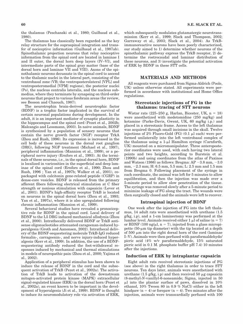
As previously reported, TrkB immunostaining was ob-served throughout the spinal cord, in all spinal (cervical,thoracic, lumbar, and sacral) segments analyzed. In allsegments, strong labeling was observed in motorneurons(Fig. 1B), whereas the immunolabeling was weaker in thedorsal horn (Fig. 1A,C). In the dorsal horn, TrkB immu-nolabeling was observed in the superficial dorsal horn(laminar I and II) and in the neck of the dorsal horn(laminae V and VI; Fig. 1A,C), areas that contain largepopulations of nociceptive neurons. Labeling was also re-corded in the nucleus proprius (laminae III and IV), whichis considered to be non-nociceptive. Immunoreactivity wasalso detected in the ventromedial gray matter, includinglaminae VII, VIII, and X, as well as in lamina IX (Fig. 1B).Labeling was confined to neuronal cells, as revealed by thecolocalization of TrkB and the neuronal marker NeuN(Fig. 1C–E). TrkB labeling was found on cell bodies, axons,and dendrites, as described previously in other parts ofthe rat central nervous system (CNS; Zhou et al., 1993;Yan et al., 1997a).
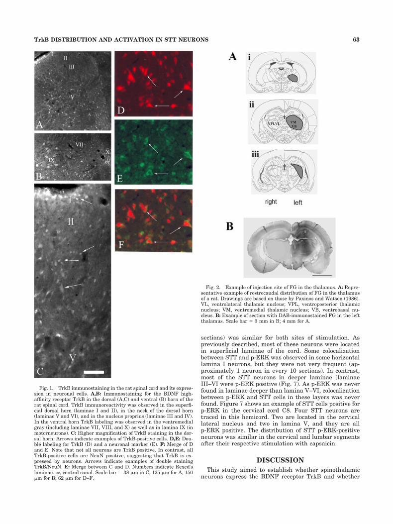
The whole thalamus was labeled with FG in all the ani-mals included in this experiment. The ventrobasal zone (VB;ventroposterolateral [VPL] and ventroposteromedial [VPM]regions; Fig. 2A,B) was labeled more strongly than otherparts of the thalamus. FG was only observed in the thalamusand in a part of the hippocampus (due to the spread of thetracer during removal of syringe).
In most sections of cervical, thoracic, lumbar, and sacralcord, spinothalamic projection neurons (Fluoro-Gold la-beled) were observed. Grains of retrogradely transportedFluoro-Gold accumulated in cell bodies as well as in den-drites (Fig. 3). Many of the labeled cells could be typed aspyramidal, multipolar, or fusiform (Fig. 3B). However,some cells had atypical morphology. A large proportion ofFG-labeled neurons was found in the superficial dorsalhorn (mostly in lamina I and very rarely in lamina II),particularly in the upper cervical regions of the cord. STTcells were also located deeper in the dorsal horn in lami-nae V, VII, and VIII in all segments of the cord, especiallyin the lumbar enlargement (Figs. 4, 5A). Finally, in half ofthe animals a small proportion of STT cells was found inarea X and in the lateral spinal nucleus of the dorsal
funiculus (Figs. 4, solid and open circles, 5A). Most of theretrogradely labeled neurons were found contralateral tothe injection site; small but significant numbers were alsolocated ipsilaterally, especially in upper cervical segments(Fig. 4). The overall distribution of labeled neurons in thegray matter was thus similar to that previously described(Burstein et al., 1990b; Marshall et al., 1996).
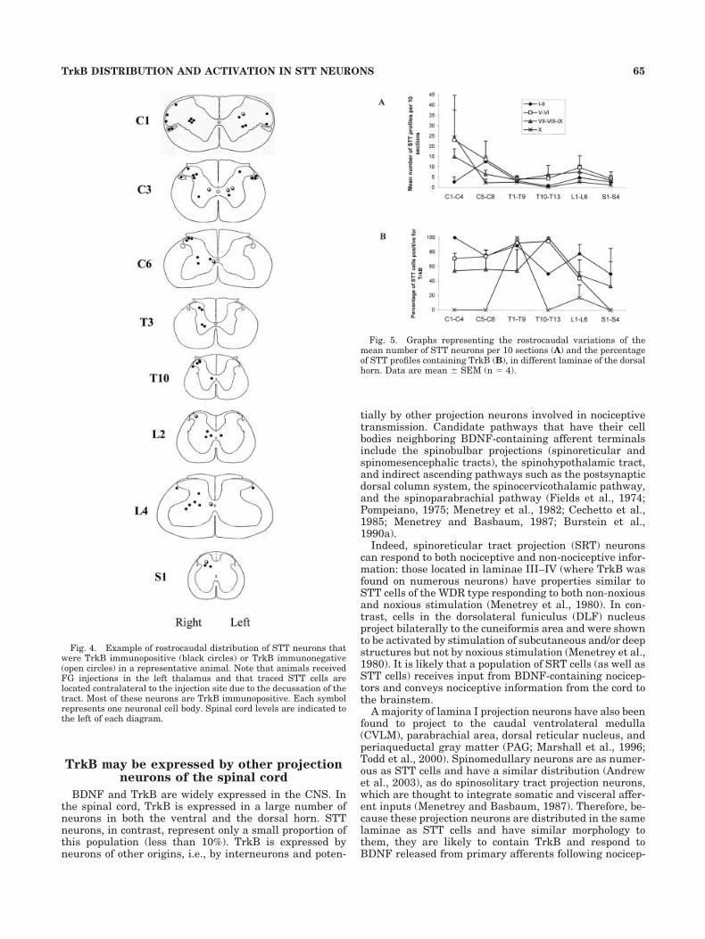
Figure 5A shows the rostrocaudal distribution of themean number (� SEM) of STT labeled cells per laminae(in laminae I–II, V–VI, VII–IX, and X). Figure 5B presentsthe average percentage of STT cells positive for TrkB. Itshows that at all levels of the spinal cord (cervical, tho-racic, lumbar, and sacral), a large proportion of STT neu-rons (50–100%) in laminae I–II, V–VI, and VII–IX wereTrkB immunopositive. As mentioned in the section above,many of the dually labeled cells could be typed as pyrami-dal, multipolar, or fusiform, but some cells had atypicalmorphology, and hence quantification of STT neurons im-munopositive for TrkB pooled all three types. In contrast,in area X, almost no STT cells were TrkB positive in allcord level examined, except in T1–T9, (Fig. 5B). In T1–T9,where the average number of STT cells in lamine X wasless than 5 for 10 sections, all these neurons were positivefor TrkB (Fig. 5A,B).
These results suggest that (with the exception of area X)most neurons of the spinothalamic tract express BDNF‘shigh-affinity receptor TrkB. Immunofluorescent stainingfor BDNF showed that in the superficial laminae of thespinal cord, FG-positive neurons are located in the vicinityof BDNF-immunoreactive terminals, suggesting that notonly do STT neurons express BDNF‘s receptor but alsothat these neurons are located in the vicinity of the trophicfactor. This suggests that physiological BDNF releasefrom terminals could act on STT neurons in superficiallaminae (Fig. 6A).
ERK has previously been shown to be activated in spi-nal neurons specifically in relation to noxious stimulations(Ji et al., 1999), and we have shown that such activation ispartly due to TrkB activation by BDNF (Pezet et al.,2002b; Lever et al., 2004), N-methyl-D-aspartate (NMDA)receptor activation, and metabolic glutamatergic receptoractivation, but not via AMPA or NK1 receptors (Lever etal., 2004). In order to show that TrkB receptors expressedby spinothalamic neurons could be functionally activatedby BDNF, we microinjected BDNF into the spinal cord ofrats that had received prior STT tracing with FG. Wemicroinjected the molecule rather than applying a noxiousstimulus (which would induce its release) due to the topo-graphical discrepancy between TrkB expression and thedistribution of BDNF-immunoreactive terminals.
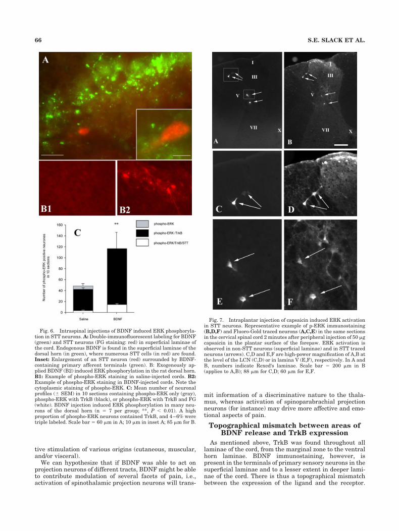
The BDNF injection significantly induced ERK phos-phorylation in numerous neurons of the dorsal horn (Fig.6B): 116 � 30 phospho-ERK neurons were labeled per 10sections in BDNF-treated cords compared with 48 � 4phospho-ERK-labeled neurons per 10 sections in saline-treated cords (n � 6–8, P � 0.01). In BDNF-injectedanimals the majority (99.6 � 0.2%) of phospho-ERK neu-rons contained TrkB, as revealed by double labeling,whereas approximately 4.5% of phospho-ERK cells con-tained both TrkB and FG.
Injection of capsaicin (50 �g) into the plantar surface ofthe left hindpaw or forepaw induced activation of ERK inmany neurons of the dorsal horn in spinal cord levelsL3–L5 and C5–C8, respectively. The distribution and theamplitude of p-ERK activation (up to 15–20 neurons per
62 S.E. SLACK ET AL.
sections) was similar for both sites of stimulation. Aspreviously described, most of these neurons were locatedin superficial laminae of the cord. Some colocalizationbetween STT and p-ERK was observed in some horizontallamina I neurons, but they were not very frequent (ap-proximately 1 neuron in every 10 sections). In contrast,most of the STT neurons in deeper laminae (laminaeIII–VI were p-ERK positive (Fig. 7). As p-ERK was neverfound in laminae deeper than lamina V–VI, colocalizationbetween p-ERK and STT cells in these layers was neverfound. Figure 7 shows an example of STT cells positive forp-ERK in the cervical cord C8. Four STT neurons aretraced in this hemicord. Two are located in the cervicallateral nucleus and two in lamina V, and they are allp-ERK positive. The distribution of STT p-ERK-positiveneurons was similar in the cervical and lumbar segmentsafter their respective stimulation with capsaicin.
DISCUSSION
This study aimed to establish whether spinothalamicneurons express the BDNF receptor TrkB and whether
Fig. 1. TrkB immunostaining in the rat spinal cord and its expres-sion in neuronal cells. A,B: Immunostaining for the BDNF high-affinity receptor TrkB in the dorsal (A,C) and ventral (B) horn of therat spinal cord. TrkB immunoreactivity was observed in the superfi-cial dorsal horn (laminae I and II), in the neck of the dorsal horn(laminae V and VI), and in the nucleus proprius (laminae III and IV).In the ventral horn TrkB labeling was observed in the ventromedialgray (including laminae VII, VIII, and X) as well as in lamina IX (inmotorneurons). C: Higher magnification of TrkB staining in the dor-sal horn. Arrows indicate examples of TrkB-positive cells. D,E: Dou-ble labeling for TrkB (D) and a neuronal marker (E). F: Merge of Dand E. Note that not all neurons are TrkB positive. In contrast, allTrkB-positive cells are NeuN positive, suggesting that TrkB is ex-pressed by neurons. Arrows indicate examples of double stainingTrkB/NeuN. E: Merge between C and D. Numbers indicate Rexed‘slaminae. cc, central canal. Scale bar � 38 �m in C; 125 �m for A; 150�m for B; 62 �m for D–F.
Fig. 2. Example of injection site of FG in the thalamus. A: Repre-sentative example of rostrocaudal distribution of FG in the thalamusof a rat. Drawings are based on those by Paxinos and Watson (1986).VL, ventrolateral thalamic nucleus; VPL, ventroposterior thalamicnucleus; VM, ventromedial thalamic nucleus; VB, ventrobasal nu-cleus. B: Example of section with DAB-immunostained FG in the leftthalamus. Scale bar � 3 mm in B; 4 mm for A.
63TrkB DISTRIBUTION AND ACTIVATION IN STT NEURONS

this population of neurons could be activated by BDNF.We observed that the majority of neurons of the STTpathway indeed express TrkB, and that this receptor isfunctional, as it could be activated by exogenous BDNF,which via phosphorylation mechanisms activated MAPKERK. These results support growing evidence demonstrat-ing that BDNF has a pronociceptive neuromodulatory rolein the spinal cord and suggest that it may act on neuronsof the STT pathway.
TrkB is present in most spinothalamictract cells
Previous studies have highlighted the role of STT cellsin the transmission of nociceptive information to the thal-amus in rats and cats (Willis and Coggeshall, 1991; Guil-baud et al., 1994). Cell bodies of these neurons are locatedin laminae I and IV–VI and are somatotopically organizedin rat and monkey (Willis and Coggeshall, 1991). Neuronsin lamina I usually have extremely small receptive fields(RFs), in contrast to neurons of laminae V–VII, whichhave expanded and complex RFs that receive additionalproprioceptive inputs. Lamina I STT neurons are high-threshold nociceptive-specific neurons that respond toheat/pinch/cold. However, STT neurons located in laminaIV–V or VII–VIII are wide dynamic range (WDR) neuronsthat respond to nociceptive and innocuous stimuli.
By using combined retrograde tracing and immunoflu-orescent staining, this study analyzed the proportion ofSTT pathways that express TrkB. The distribution of STTcells in this study (traced with FG) was consistent withthat described by Burstein et al (1990b). Colocalization ofFG with TrkB showed that BDNF‘s receptor is found in alltypes of STT neurons: in fusiform, multipolar lamina ISTT cells (nociceptive), in pyramidal STT cells (non-nociceptive), and in STT cells with no distinguishablemorphology (possibly a mixture of WDR nociceptive STTcells and lamina IV–V and VII–VIII non-nociceptive STTcells). Increasing evidence suggests that when BDNF isendogenously released from primary afferents, it acts as apronociceptive neuromodulator in the spinal cord (Pezet etal., 2002b). TrkB expression by STT neurons suggests thatBDNF may activate these neurons. Our observation ofERK activation in STT neurons induced by injection ofBDNF or natural release of BDNF following noxious stim-ulation supports this hypothesis and suggests that patho-physiologically released BDNF may activate this subpopu-lation of neurons, therefore having a pro-nociceptive roleon TrkB-expressing STT cells.
Fig. 3. Spinothalamic projection neurons in the spinal cord andtheir colocalization with TrkB. A,B: Photomicrographs showing typi-cal examples of FG-traced spinothalamic neurons in the spinal dorsalhorn. A: STT neuron located in lamina I (dashed arrows) and laminaV (solid arrow). The dotted line shows the border between gray andwhite matter. B: Enlarged photomicrograph of a lamina I neuronshowing the typical morphology of arborization in a horizontal planewith dendrites spreading in laminae I and IIo. C,D: Example of STTFG traced neuron immunopositive for TrkB. The solid arrow indicatesan STT traced neuron positive for TrkB, and the dashed arrow indi-cates a non-STT TrkB-positive neuron. Scale bar � 100 �m in A; 50�m in B,C (applies to C,D).
64 S.E. SLACK ET AL.
TrkB may be expressed by other projectionneurons of the spinal cord
BDNF and TrkB are widely expressed in the CNS. Inthe spinal cord, TrkB is expressed in a large number ofneurons in both the ventral and the dorsal horn. STTneurons, in contrast, represent only a small proportion ofthis population (less than 10%). TrkB is expressed byneurons of other origins, i.e., by interneurons and poten-
tially by other projection neurons involved in nociceptivetransmission. Candidate pathways that have their cellbodies neighboring BDNF-containing afferent terminalsinclude the spinobulbar projections (spinoreticular andspinomesencephalic tracts), the spinohypothalamic tract,and indirect ascending pathways such as the postsynapticdorsal column system, the spinocervicothalamic pathway,and the spinoparabrachial pathway (Fields et al., 1974;Pompeiano, 1975; Menetrey et al., 1982; Cechetto et al.,1985; Menetrey and Basbaum, 1987; Burstein et al.,1990a).
Indeed, spinoreticular tract projection (SRT) neuronscan respond to both nociceptive and non-nociceptive infor-mation: those located in laminae III–IV (where TrkB wasfound on numerous neurons) have properties similar toSTT cells of the WDR type responding to both non-noxiousand noxious stimulation (Menetrey et al., 1980). In con-trast, cells in the dorsolateral funiculus (DLF) nucleusproject bilaterally to the cuneiformis area and were shownto be activated by stimulation of subcutaneous and/or deepstructures but not by noxious stimulation (Menetrey et al.,1980). It is likely that a population of SRT cells (as well asSTT cells) receives input from BDNF-containing nocicep-tors and conveys nociceptive information from the cord tothe brainstem.
A majority of lamina I projection neurons have also beenfound to project to the caudal ventrolateral medulla(CVLM), parabrachial area, dorsal reticular nucleus, andperiaqueductal gray matter (PAG; Marshall et al., 1996;Todd et al., 2000). Spinomedullary neurons are as numer-ous as STT cells and have a similar distribution (Andrewet al., 2003), as do spinosolitary tract projection neurons,which are thought to integrate somatic and visceral affer-ent inputs (Menetrey and Basbaum, 1987). Therefore, be-cause these projection neurons are distributed in the samelaminae as STT cells and have similar morphology tothem, they are likely to contain TrkB and respond toBDNF released from primary afferents following nocicep-
Fig. 4. Example of rostrocaudal distribution of STT neurons thatwere TrkB immunopositive (black circles) or TrkB immunonegative(open circles) in a representative animal. Note that animals receivedFG injections in the left thalamus and that traced STT cells arelocated contralateral to the injection site due to the decussation of thetract. Most of these neurons are TrkB immunopositive. Each symbolrepresents one neuronal cell body. Spinal cord levels are indicated tothe left of each diagram.
Fig. 5. Graphs representing the rostrocaudal variations of themean number of STT neurons per 10 sections (A) and the percentageof STT profiles containing TrkB (B), in different laminae of the dorsalhorn. Data are mean � SEM (n � 4).
65TrkB DISTRIBUTION AND ACTIVATION IN STT NEURONS
tive stimulation of various origins (cutaneous, muscular,and/or visceral).
We can hypothesize that if BDNF was able to act onprojection neurons of different tracts, BDNF might be ableto contribute modulation of several facets of pain, i.e.,activation of spinothalamic projection neurons will trans-
mit information of a discriminative nature to the thala-mus, whereas activation of spinoparabrachial projectionneurons (for instance) may drive more affective and emo-tional aspects of pain.
Topographical mismatch between areas ofBDNF release and TrkB expression
As mentioned above, TrkB was found throughout alllaminae of the cord, from the marginal zone to the ventralhorn laminae. BDNF immunostaining, however, ispresent in the terminals of primary sensory neurons in thesuperficial laminae and to a lesser extent in deeper lami-nae of the cord. There is thus a topographical mismatchbetween the expression of the ligand and the receptor.
Fig. 6. Intraspinal injections of BDNF induced ERK phosphoryla-tion in STT neurons. A: Double-immunofluorescent labeling for BDNF(green) and STT neurons (FG staining: red) in superficial laminae ofthe cord. Endogenous BDNF is found in the superficial laminae of thedorsal horn (in green), where numerous STT cells (in red) are found.Inset: Enlargement of an STT neuron (red) surrounded by BDNF-containing primary afferent terminals (green). B: Exogenously ap-plied BDNF (B2) induced ERK phosphorylation in the rat dorsal horn.B1: Example of phospho-ERK staining in saline-injected cords. B2:Example of phospho-ERK staining in BDNF-injected cords. Note thecytoplasmic staining of phospho-ERK. C: Mean number of neuronalprofiles (� SEM) in 10 sections containing phospho-ERK only (gray),phospho-ERK with TrkB (black), or phospho-ERK with TrkB and FG(white). BDNF injection induced ERK phosphorylation in many neu-rons of the dorsal horn (n � 7 per group; **, P � 0.01). A highproportion of phospho-ERK neurons contained TrkB, and 4–6% weretriple labeled. Scale bar � 60 �m in A; 10 �m in inset A; 85 �m for B.
Fig. 7. Intraplantar injection of capsaicin induced ERK activationin STT neurons. Representative example of p-ERK immunostaining(B,D,F) and Fluoro-Gold traced neurons (A,C,E) in the same sectionsin the cervical spinal cord 2 minutes after peripheral injection of 50 �gcapsaicin in the plantar surface of the forepaw. ERK activation isobserved in non-STT neurons (superficial laminae) and in STT tracedneurons (arrows). C,D and E,F are high-power magnification of A,B atthe level of the LCN (C,D) or in lamina V (E,F), respectively. In A andB, numbers indicate Rexed‘s laminae. Scale bar � 200 �m in B(applies to A,B); 88 �m for C,D; 60 �m for E,F.
66 S.E. SLACK ET AL.

This mismatch may be functionally overcome in severalways: 1) BDNF may diffuse to deeper laminae (a theoryproposed in the case of SP and the NK1 receptor; Liu et al.,1994), 2) dendrites of neurons of deep laminae may reachthe superficial laminae and hence respond to BDNF re-leased in the dorsal horn, which is likely to be the case forlamina V neurons (of a WDR nature); and 3) For certainneuronal populations, such as motorneurons, BDNF isretrogradely transported from their peripheral targets(Thoenen, 1995).
Functional significance of TrkB expressionon STT neurons
Our data, together with previous reports, suggest thatfollowing a peripheral noxious stimulus BDNF, containedin terminals of a subset of DRG neurons (Michael et al.,1997; Luo et al., 2001), will be released in the spinal cordin an activity-dependent manner (Lever et al., 2001) andwill activate TrkB (Pezet et al., 2002a) on postsynapticSTT neurons. Following the binding of BDNF to TrkB onthose neurons, the receptor is activated by autophosphor-ylation mechanisms, allowing an intracellular signalingcascade that leads to a positive modulation of the gluta-mate receptor NMDA, through phosphorylation of its NR1subunit (Di Luca et al., 2001; Slack and Thompson, 2002;Slack et al., 2004) and possibly NR2 subunit, hence exac-erbating the glutamatergic transmission (Kerr et al.,1999; Heppenstall and Lewin, 2001; Arvanian and Men-dell, 2001; Groth and Aanonsen, 2002), thereby increasingthe response of postsynaptic neurons. The ultimate func-tional consequence of this biochemical activation is in-creased neuronal activation of the spinothalamic neuronsand hence increased transmission of pain-related signals.Our observations are consistent with previous observa-tions reporting an increased activation of NR1 subunit inneurons of the spinothalamic pathway following acutenoxious stimulation (Zou et al., 2000).
We report that exogenous application of BDNF leads toTrkB and ERK activation by phosphorylation in spinotha-lamic neurons. It is likely that downstream effects of thisactivation is the activation by phosphorylation ofstimulus-inducible transcription factors such as calcium/cyclic AMP response element binding protein (CREB)and/or the induction of expression of immediate earlygenes, such as c-fos. Their activation following noxiousstimulation or BDNF application is well documented(Hunt et al., 1987; Abbadie and Besson, 1993, 1994;Soyguder et al., 1994; Abbadie et al., 1994; Honore et al.,1995; Presley et al., 1990; Kerr et al., 1999). Consistentwith this hypothesis, c-fos-containing ascending tract neu-rons make up 6–8% of spinal neurons that express c-fosfollowing noxious stimulation (Menetrey et al., 1989).These changes are likely to lead to phenotypic changesand long-term functional changes in these projection STTneurons.
ACKNOWLEDGMENTS
The authors thank Professor R.A. Rush (Flinders Uni-versity, Australia) for his generous gift of anti-TrkB anti-body and Thomas Bishop for critical reading of the article.
LITERATURE CITED
Abbadie C, Besson JM. 1993. Effects of morphine and naloxone on basaland evoked Fos-like immunoreactivity in lumbar spinal cord neurons ofarthritic rats. Pain 52:29–39.
Abbadie C, Besson JM. 1994. Chronic treatments with aspirin or acetamin-ophen reduce both the development of polyarthritis and Fos-like im-munoreactivity in rat lumbar spinal cord. Pain 57:45–54.
Abbadie C, Honore P, Besson JM. 1994. Intense cold noxious stimulation ofthe rat hindpaw induces c-fos expression in lumbar spinal cord neu-rons. Neuroscience 59:457–468.
Andrew D, Krout KE, Craig AD. 2003. Differentiation of lamina I spi-nomedullary and spinothalamic neurons in the cat. J Comp Neurol458:257–271.
Arvanian VL, Mendell LM. 2001. Acute modulation of synaptic transmis-sion to motorneurons by BDNF in the neonatal rat spinal cord. EurJ Neurosci 14:1800–1808.
Besson JM, Chaouch A. 1987. Peripheral and spinal mechanisms of noci-ception. Physiol Rev 67:67–186.
Burstein R, Cliffer KD, Giesler GJ Jr. 1990a. Cells of origin of the spino-hypothalamic tract in the rat. J Comp Neurol 291:329–344.
Burstein R, Dado RJ, Giesler GJ Jr. 1990b. The cells of origin of thespinothalamic tract of the rat: a quantitative reexamination. Brain Res511:329–337.
Cechetto DF, Standaert DG, Saper CB. 1985. Spinal and trigeminal dorsalhorn projections to the parabrachial nucleus in the rat. J Comp Neurol240:153–160.
Craig AD, Dostrovsky JO. 1999. Medulla to thalamus. In: Wall PD, Mel-zack R, editors. The textbook of pain. London: Churchill Livingstone.
Di Luca M, Gardoni F, Finardi A, Pagliardini S, Cattabeni F, Battaglia G,Missale C. 2001. NMDA receptor subunits are phosphorylated by acti-vation of neurotrophin receptors in PSD of rat spinal cord. Neuroreport12:1301–1305.
Ernfors P, Wetmore C, Olson L, Persson H. 1990. Identification of cells inrat brain and peripheral tissues expressing mRNA for members of thenerve growth factor family. Neuron 5:511–526.
Ernfors P, Rosario CM, Merlio JP, Grant G, Aldskogius H, Persson H.1993. Expression of messenger-RNAs for neurotrophin receptors in thedorsal-root ganglion and spinal-cord during development and followingperipheral or central axotomy. Mol Brain Res 17:217–226.
Fields HL, Anderson SD, Wagner GM. 1974. The spinoreticular tract: analternate pathway mediating pain. Trans Am Neurol Assoc 99:211–213.
Garraway SM, Petruska JC, Mendell LM. 2003. BDNF sensitizes theresponse of lamina II neurons to high threshold primary afferent in-puts. Eur J Neurosci 18:2467–2476.
Groth R, Aanonsen L. 2002. Spinal brain-derived neurotrophic factor(BDNF) produces hyperalgesia in normal mice while antisense directedagainst either BDNF or trkB, prevent inflammation-induced hyperal-gesia. Pain 100:171–181.
Guilbaud G, Benoist JM, Neil A, Kayser V, Gautron M. 1987a. Neuronalresponse thresholds to and encoding of thermal stimuli duringcarrageenin-hyperalgesic-inflammation in the ventro-basal thalamusof the rat. Exp Brain Res 66:421–431.
Guilbaud G, Neil A, Benoist JM, Kayser V, Gautron M. 1987b. Thresholdsand encoding of neuronal responses to mechanical stimuli in theventro-basal thalamus during carrageenin-induced hyperalgesic in-flammation in the rat. Exp Brain Res 68:311–318.
Guilbaud G, Peschanski M, Gautron M, Binder D. 1980. Neurones respond-ing to noxious stimulation in VB complex and caudal adjacent regionsin the thalamus of the rat. Pain 8:303–318.
Guilbaud G, Bernard JF, Besson JM. 1994. Brain areas involved in noci-ception and pain. In: Wall PD, Melzack R, editors. Textbook of pain.Edinburgh: Churchill-Livingstone. p 113–128.
Heppenstall PA, Lewin GR. 2001. BDNF but not NT-4 is required fornormal flexion reflex plasticity and function. Proc Natl Acad Sci U S A98:8107–8112.
Honore P, Chapman V, Buritova J, Besson JM. 1995. Reduction of carra-geenin oedema and the associated c-Fos expression in the rat lumbarspinal cord by nitric oxide synthase inhibitor. Br J Pharmacol 114:77–84.
Hunt SP, Pini A, Evan G. 1987. Induction of c-fos-like protein in spinal cordneurons following sensory stimulation. Nature 328:632–634.
Ji RR, Baba H, Brenner GJ, Woolf CJ. 1999. Nociceptive-specific activation
67TrkB DISTRIBUTION AND ACTIVATION IN STT NEURONS
of ERK in spinal neurons contributes to pain hypersensitivity. NatNeurosci 2:1114–1119.
Kerr BJ, Bradbury EJ, Bennett DL, Trivedi PM, Dassan P, French J,Shelton DB, McMahon SB, Thompson SW. 1999. Brain-derived neuro-trophic factor modulates nociceptive sensory inputs and NMDA-evokedresponses in the rat spinal cord. J Neurosci 19:5138–5148.
Lever IJ, Bradbury EJ, Cunningham JR, Adelson DW, Jones MG, McMa-hon SB, Marvizon JC, Malcangio M. 2001. Brain-derived neurotrophicfactor is released in the dorsal horn by distinctive patterns of afferentfiber stimulation. J Neurosci 21:4469–4477.
Liu H, Brown JL, Jasmin L, Maggio JE, Vigna SR, Mantyh PW, BasbaumAI. 1994. Synaptic relationship between substance P and the substanceP receptor: light and electron microscopic characterization of the mis-match between neuropeptides and their receptors. Proc Natl Acad SciU S A 91:1009–1013.
Luo XG, Rush RA, Zhou XF. 2001. Ultrastructural localization of brain-derived neurotrophic factor in rat primary sensory neurons. NeurosciRes 39:377–384.
Malcangio M, Lessmann V. 2003. A common thread for pain and memorysynapses? Brain-derived neurotrophic factor and trkB receptors.Trends Pharmacol Sci 24:116–121.
Mannion RJ, Costigan M, Decosterd I, Amaya F, Ma QP, Holstege JC, JiRR, Acheson A, Lindsay RM, Wilkinson GA, Woolf CJ. 1999. Neuro-trophins: peripherally and centrally acting modulators of tactilestimulus-induced inflammatory pain hypersensitivity. Proc Natl AcadSci U S A 96:9385–9390.
Marshall GE, Shehab SA, Spike RC, Todd AJ. 1996. Neurokinin-1 recep-tors on lumbar spinothalamic neurons in the rat. Neuroscience 72:255–263.
Menetrey D, Basbaum AI. 1987. Spinal and trigeminal projections to thenucleus of the solitary tract: a possible substrate for somatovisceraland viscerovisceral reflex activation. J Comp Neurol 255:439–450.
Menetrey D, Chaouch A, Besson JM. 1980. Location and properties ofdorsal horn neurons at origin of spinoreticular tract in lumbar enlarge-ment of the rat. J Neurophysiol 44:862–877.
Menetrey D, Chaouch A, Binder D, Besson JM. 1982. The origin of thespinomesencephalic tract in the rat: an anatomical study using theretrograde transport of horseradish peroxidase. J Comp Neurol 206:193–207.
Menetrey D, Roudier F, Besson JM. 1983. Spinal neurons reaching thelateral reticular nucleus as studied in the rat by retrograde transportof horseradish peroxidase. J Comp Neurol 220:439–452.
Menetrey D, Gannon A, Levine JD, Basbaum AI. 1989. Expression of c-fosprotein in interneurons and projection neurons of the rat spinal cord inresponse to noxious somatic, articular, and visceral stimulation.J Comp Neurol 285:177–195.
Michael GJ, Averill S, Nitkunan A, Rattray M, Bennett DL, Yan Q, Priest-ley JV. 1997. Nerve growth factor treatment increases brain-derivedneurotrophic factor selectively in TrkA-expressing dorsal root ganglioncells and in their central terminations within the spinal cord. J Neu-rosci 17:8476–8490.
Molander C, Xu Q, Grant G. 1984. The cytoarchitectonic organization ofthe spinal cord in the rat. I. The lower thoracic and lumbosacral cord.J Comp Neurol 230:133–141.
Molander C, Xu Q, Rivero-Melian C, Grant G. 1989. Cytoarchitectonicorganization of the spinal cord in the rat: II. The cervical and upperthoracic cord. J Comp Neurol 289:375–385.
Paxinos G, Watson C. 1986. The rat brain in stereotaxic coordinates. NewYork: Academic Press.
Peschanski M, Guilbaud G, Gautron M, Besson JM. 1980. Encoding ofnoxious heat messages in neurons of the ventrobasal thalamic complexof the rat. Brain Res 197:401–413.
Pezet S, Malcangio M, Lever IJ, Perkinton MS, Thompson SW, WilliamsRJ, McMahon SB. 2002a. Noxious stimulation induces Trk receptorand downstream ERK phosphorylation in spinal dorsal horn. Mol CellNeurosci 21:684–695.
Pezet S, Malcangio M, McMahon SB. 2002b. BDNF: a neuromodulator innociceptive pathways? Brain Res Brain Res Rev 40:240–249.
Pompeiano O. 1975. Macular input to neurons of the spinoreticulocerebel-lar pathway. Brain Res 95:351–368.
Presley RW, Menetrey D, Levine JD, Basbaum AI. 1990. Systemic mor-phine suppresses noxious stimulus-evoked Fos protein-like immunore-activity in the rat spinal cord. J Neurosci 10:323–335.
Slack SE, Thompson SW. 2002. Brain-derived neurotrophic factor inducesNMDA receptor 1 phosphorylation in rat spinal cord. Neuroreport13:1967–1970.
Slack SE, Pezet S, McMahon SB, Thompson SW, Malcangio M. 2004.Brain-derived neurotrophic factor induces NMDA receptor subunit onephosphorylation via ERK and PKC in the spinal cord. Eur J Neurosci20:1769–1778.
Soyguder Z, Schmidt HH, Morris R. 1994. Postnatal development of nitricoxide synthase type 1 expression in the lumbar spinal cord of the rat:a comparison with the induction of c-fos in response to peripheralapplication of mustard oil. Neurosci Lett 180:188–192.
Thoenen H. 1995. Neurotrophins and neuronal plasticity. Science 270:593–598.
Todd AJ, McGill MM, Shehab SA. 2000. Neurokinin 1 receptor expressionby neurons in laminae I, III and IV of the rat spinal dorsal horn thatproject to the brainstem. Eur J Neurosci 12:689–700.
Walker SM, Mitchell VA, White DM, Rush RA, Duggan AW. 2001. Releaseof immunoreactive brain-derived neurotrophic factor in the spinal cordof the rat following sciatic nerve transection. Brain Res 899:240–247.
Willis WD, Coggeshall RE. 1991. Sensory mechanisms of the spinal cord.New York: Plenum Press.
Yajima Y, Narita M, Narita M, Matsumoto N, Suzuki T. 2002. Involvementof a spinal brain-derived neurotrophic factor/full-length TrkB pathwayin the development of nerve injury-induced thermal hyperalgesia inmice. Brain Res 958:338–346.
Yan Q, Radeke MJ, Matheson CR, Talvenheimo J, Welcher AA, FeinsteinSC. 1997a. Immunocytochemical localization of TrkB in the centralnervous system of the adult rat. J Comp Neurol 378:135–157.
Yan Q, Rosenfeld RD, Matheson CR, Hawkins N, Lopez OT, Bennett L,Welcher AA. 1997b. Expression of brain-derived neurotrophic factorprotein in the adult rat central nervous system. Neuroscience 78:431–448.
Zhou XF, Parada LF, Soppet D, Rush RA. 1993. Distribution of trkBtyrosine kinase immunoreactivity in the rat central nervous system.Brain Res 622:63–70.
Zhou XF, Deng YS, Xian CJ, Zhong JH. 2000. Neurotrophins from dorsalroot ganglia trigger allodynia after spinal nerve injury in rats. EurJ Neurosci 12:100–105.
Zhou XF, Rush RA. 1996. Endogenous brain-derived neurotrophic factor isanterogradely transported in primary sensory neurons. Neuroscience74:945–953.
Zou X, Lin Q, Willis WD. 2000. Enhanced phosphorylation of NMDAreceptor 1 subunits in spinal cord dorsal horn and spinothalamic tractneurons after intradermal injection of capsaicin in rats. J Neurosci20:6989–6997.
68 S.E. SLACK ET AL.