treating your ankle arthritis - fasohio.comfasohio.com/totalanklejoint/wright_total_ankle.pdf ·...
TRANSCRIPT

Treating Your
Ankle Arthritis
with the
INBONE®
Total Ankle System

3 Glossary
9 Anatomy of the Ankle
11 Diseases of the Ankle
12 What Is the INBONE® Total Ankle System?
14 Things You Should Know
16 What is PROPHECY®?
18 What Are the Alternatives?
21 What Are the Benefits?
23 What Are the Risks?
24 Patients Speak Out
26 Frequently Asked Questions
Table of Contents
Achilles Tendon – The Achilles tendon is the tendon which connects the three strongest flexor muscles of the leg to the foot. It is a tendon which connects the two heads of the gastrocnemius muscle and the deeper soleus muscle to the calcaneus, or heel bone. The tendon can be felt in the back of the ankle and is just under the skin. This tendon is a common source of pain in runners and other athletes. Achilles tendonitis, tendonosis and rupture are some of the common problems encountered with this structure.
Ankle Instability – Chronic, repetitive sprains of the ankle. This can be due to an injury that never healed properly but can also be due to weak ankle ligaments or a heel that tilts inward (varus heel).
Ankle Joint – The joint between the foot and the lower leg. It allows the foot to dorsiflex (move upward) and plantarflex (move downward). It is made up of the two bones of the lower leg (tibia & fibula) and the ankle bone (talus). There are ligaments that hold the joint together on the inside (deltoid) and outside (lateral ankle ligament complex).
Arthritis – Arthritis typically refers to the wearing away of joint surfaces. Arthritis falls into one of three categories: Osteoarthritis is primary arthritis of the joint and may be related to family history. Traumatic arthritis is arthritis that develops after injury to a joint. Inflammatory arthritis occurs when a disease affecting the patient causes the cartilage to wear away. Treatment is dependent on the cause and extent of the arthritis and may include medication, bracing, physical therapy or surgery.
Calcaneus – The heel bone. Two joints are present: the subtalar joint which allows motion with the talus (ankle) bone which allows inversion and eversion of
3
Glossary
the foot with the leg; and the calcaneocuboid joint has a complicated biomechanical function that controls flexibility of the foot and controls the arch of the foot. The Achilles tendon attaches to the back of the calcaneus, and the plantar fascia also attaches to the bottom of the calcaneus.
Cartilage – Cartilage is a living tissue that lines our joints. It is a matrix of proteins and collagen that is tough, absorbs shock and is very smooth. Healthy cartilage can, and often does, last our whole life without problems. Disease of the cartilage or trauma can cause the cartilage cells to die. Unlike most tissues in our body, joint cartilage cells do not reproduce themselves once our skeletons are fully grown. Basic science and clinical research has led to recent innovations in cartilage transplantation and growth.
Congenital Vertical Talus (CVT) – A condition where the foot is malformed at birth. The ankle bone (talus) is not formed correctly and this condition frequently requires surgery. The foot may have a reversed arch or “rocker bottom” deformity where the middle of the foot sits lower than the heel and toes. This condition requires treatment by an orthopaedic surgeon.
Cuboid – The midfoot bone on the outside of the foot. This bone lies between the calcaneus and metatarsals. This bone may be crushed (“Nutcracker fracture”) when the midfoot is injured.
Eversion – Twisting out, away from the midline of the body.
Extensor digitorum longus – A foot extensor is a muscle which raises the toes or ankle. The extensor digitorum longus stabilizes the toes against the ground in push-off and propulsion.
4

Extensor hallucis longus – The extensor hallucis longus helps to stablize the first metatarasophalaneal joint (where the toes meet the foot) and forefoot during push-off.
Fibula – The most prominent bone on the outer side of the ankle that also extends to the knee.
Fracture – A fractured bone is one that has cracked or broken. Bones are comprised primarily of calcium and are quite hard. A crack usually occurs as a result of an injury. In cases of abnormal bone structure, a fracture can occur after a very minor injury. Overuse can cause a “stress” fracture. Displacement refers to the amount the two broken pieces have moved from each other. In non-displaced fractures the pieces of bone haven’t separated at all. Displaced fractures have some separation between the broken pieces. Some bones can heal properly even with a lot of displacement, but some fractured bones require surgery for even a small amount of displacement. Evaluation and treatment by an orthopaedic surgeon is necessary.
Inflammatory arthritis – Inflammatory arthritis occurs when a disease affecting the patient causes the cartilage to die off. Treatment is dependent on the cause and extent of the arthritis and may entail medication, bracing or surgery.
Inversion – Twisting in, towards the midline of the body.
Lateral malleolus – The end of the fibula, the most prominent bone on the outside of the ankle.
Ligament – A band of tissue that connects one bone to another, typically to support a joint. Ligaments are made primarily of collagen. Injury to a ligament is referred to as a sprain.
Medial malleolus – The most prominent bone on the inner side of the ankle.
5
Navicular – A “boat” shaped bone in the midfoot. Two joints are present: the talonavicular joint which has a complicated biomechanical function that controls flexibility of the foot and controls the arch of the foot; and the naviculocuneiform joint which can be injured in midfoot injuries and can contribute to flatfoot deformity.
Osteoarthritis – Osteoarthritis is primary arthritis of the joint and may be related to family history.
Peroneal tendon – The peroneal tendons are behind the outside bone of the ankle (the fibula). These two tendons move the foot outwards in a direction called eversion. They balance the ankle and the back of the foot and prevent the foot from turning inwards repetitively. The peroneal tendons are susceptible to injury as the ankle turns, rolls or becomes sprained because they are not as strong as the muscles and tendons on the inside of the ankle.
Posterior tibial tendon – The posterior tibial tendon and other supportive ligaments help to maintain the arch of the foot. This tendon goes behind the ankle and around the medial malleolus (a bone inside the ankle).
Rheumatoid arthritis – One of the inflammatory arthritis diseases. This is an autoimmune disorder where the patient’s immune system malfunctions and attacks the cartilage and tendons.
Sprain – An injury causing tearing of a ligament. Sprains vary in severity and can range from a partial tearing of the ligament to a complete rupture.
Sprained ankle – A rupture of one or more of the ligaments that surround the ankle.
Talus – The ankle bone. This bone sits within the ankle “Mortise” or hinge which is made up of the two leg bones, the tibia and fibula. Three joints are present: the ankle, which allows the up and down motion of
6
the foot with the leg; the subtalar joint which allows “inversion” and “eversion” of the foot with the leg; and the talonavicular joint which has a complicated biomechanical function that controls flexibility of the foot and the arch of the foot. The talus has no muscular attachments and is mostly covered with cartilage, which makes injuries to the talus difficult to heal.
Tendon – A tendon is a structure in the body that connects muscle to bone. As the muscle contracts, it pulls on the tendon which moves the bone. Tendons are made mostly of collagen. Inflammation of a tendon is called tendonitis. Tendons can tear or rupture if they are pulled too hard by the muscle, or if they degenerate.
Tendonitis – Inflammation of a tendon. Most cases of tendonitis are caused by some type of injury, overuse or a mechanical abnormality in the foot or ankle. Treatment depends on the specific tendon involved, the extent of involvement and the length of time the symptoms have been present.
Tendonosis – A later stage of tendonitis where the tendon starts to fray and tear.
Tibia (shinbone) – The large bone of the leg that extends from the knee to the ankle.
Tibialis anterior tendon – The function of the tibialis anterior is to move the ankle upwards. It stabilizes the foot in the latter part of the stance phase of gait and extends the foot at the beginning and middle portions of the swing phase of gait.
Valgus – Tilted “outward” or away from the midline of the body.
Varus – Tilted “inward” or towards the midline of the body.
7
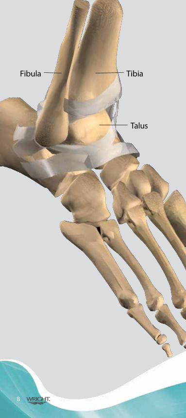
Fibula Tibia
Talus
8

Anatomy of the Ankle
The human ankle is a joint that acts much like a hinge. The joint is formed by the union of three bones. The ankle bone is the talus. The top of the talus fits inside a socket that is formed by the lower end of the tibia, often called the shinbone, and the fibula, the small bone of the lower leg. The bottom of the talus sits on the heel bone, called the calcaneus. As one of the most flexible, free-moving joints in the body, the normal ankle can move forwards, from side-to-side, and twist.
Like other free-moving joints, the ankle contains cartilage which absorbs shock. It is held together with ligaments – straps of tough tissue, which help prevent the joint from dislocating. Full function of the ankle joint depends on the successful coordination of many interrelated parts including bones, muscles, tendons, ligaments, and nerves. In patients with arthritis, one of the most common reasons for ankle replacement surgery, the cartilage in the joint has worn down, resulting in bone-on-bone contact, causing pain and limited activity.
9
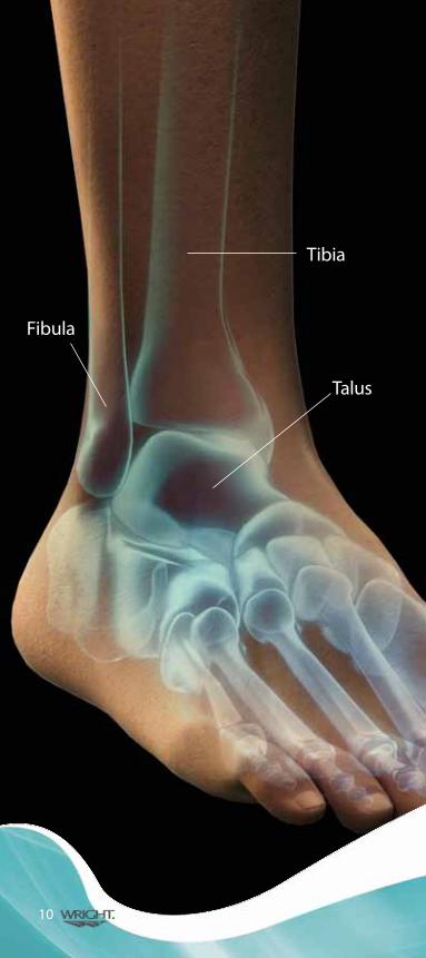
Fibula
Tibia
Talus
10
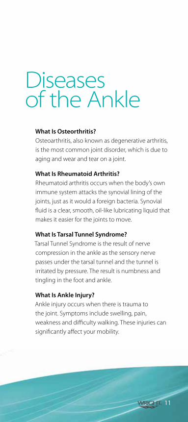
What Is Osteorthritis? Osteoarthritis, also known as degenerative arthritis, is the most common joint disorder, which is due to aging and wear and tear on a joint.
What Is Rheumatoid Arthritis? Rheumatoid arthritis occurs when the body’s own immune system attacks the synovial lining of the joints, just as it would a foreign bacteria. Synovial fluid is a clear, smooth, oil-like lubricating liquid that makes it easier for the joints to move.
What Is Tarsal Tunnel Syndrome? Tarsal Tunnel Syndrome is the result of nerve compression in the ankle as the sensory nerve passes under the tarsal tunnel and the tunnel is irritated by pressure. The result is numbness and tingling in the foot and ankle.
What Is Ankle Injury? Ankle injury occurs when there is trauma to the joint. Symptoms include swelling, pain, weakness and difficulty walking. These injuries can significantly affect your mobility.
Diseases of the Ankle
11

The INBONE® Total Ankle System consists of a talar dome with a modular talar stem, a tibial tray, a four- component tibial stem assembly that attaches to the tibial tray, and an ultra-high molecular weight polyethylene insert (UHMWPE). All components are available in varying sizes to best match your anatomy.
What is the INBONE® Total Ankle System?
INBONE® Talar Dome
INBONE® UHWMPE Insert
INBONE®Talar Stem
INBONE® Tibial Stems
INBONE® Tibial Tray
12
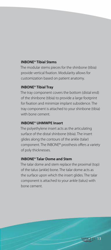
INBONE® Tibial StemsThe modular stems pieces for the shinbone (tibia) provide vertical fixation. Modularity allows for customization based on patient anatomy.
INBONE® Tibial TrayThe tray component covers the bottom (distal end) of the shinbone (tibia) to provide a large footprint for fixation and minimize implant subsidence. The tray component is attached to your shinbone (tibia) with bone cement.
INBONE® UHMWPE InsertThe polyethylene insert acts as the articulating surface of the distal shinbone (tibia). The insert glides along the contours of the ankle (talar) component. The INBONE® prosthesis offers a variety of poly thicknesses.
INBONE® Talar Dome and StemThe talar dome and stem replace the proximal (top) of the talus (ankle) bone. The talar dome acts as the surface upon which the insert glides. The talar component is attached to your ankle (talus) with bone cement.
13

Things You Should Know
What is the purpose of the INBONE® Total Ankle System? (Indications for use)The INBONE® Total Ankle is intended to give a patient limited mobility by reducing pain, restoring alignment and replacing the flexion and extension movement in the ankle joint.
The INBONE® Total Ankle is indicated for patients with ankle joints damaged by severe rheumatoid, post-traumatic, or degenerative arthritis. The INBONE® Total Ankle is additionally indicated for patients with a failed previous ankle surgery.
CAUTION: The ankle prosthesis is intended for cement use only.
14
When should the INBONE® Total Ankle not be used? (Contraindications)You should not receive the INBONE® Total Ankle if:• Youhaveaninfectionofthebodyorbone• Excessivebonelossattheanklejoint;• Steroiduse;• Infectionattheanklesiteorinfectionsat
distant sites that could migrate to the ankle;• Sepsis;• Muscularatrophy;• Dementia;• Poorbloodsupplyintheanklejoint;• Skeletallyimmaturepatients(patientisless
than 21 years of age at the time of surgery);• Caseswherethereisinadequate
neuromuscular status, poor bone stock, poor skin coverage around the joint which would make the procedure unjustifiable;
• Neuropathicjoints;• HepatitisorHIVinfection;• Excessiveloadsascausedbyactivityorpatient
weight;• Femaleofchildbearingage,forwhoma
negative pregnancy test is not obtained; and,• Neurologicalormusculoskeletaldiseasethat
may adversely affect gait or weight bearing.
15
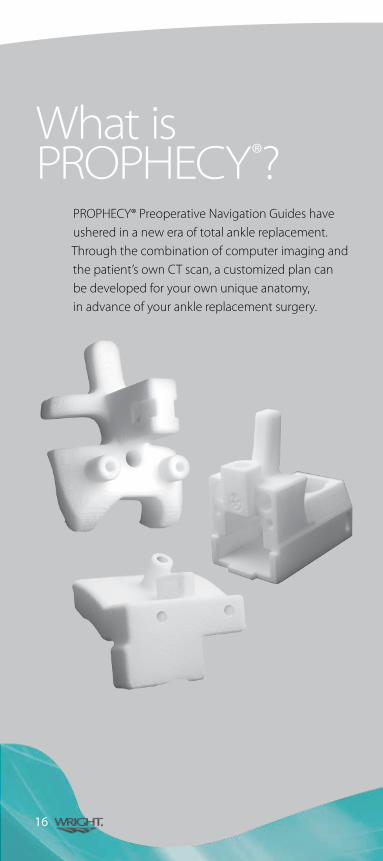
PROPHECY® Preoperative Navigation Guides have ushered in a new era of total ankle replacement. Through the combination of computer imaging and the patient’s own CT scan, a customized plan can be developed for your own unique anatomy, in advance of your ankle replacement surgery.
What isPROPHECY®?
16

What are the benefits of preoperative navigation?PROPHECY® guides provide the alignment accuracy of the traditional INBONE® Total Ankle System while reducing surgical steps. Patients may benefit from the following: • Patient-specificinstrumentation• Crucialanatomiclandmarksidentified
before your surgery• Lessradiationexposurefromx-rays
during surgery
PROPHECY® Preoperative Plan
Postoperative X-ray
17
There are both surgical and non-surgical alternatives to ankle replacement surgery. First line treatments for arthritis of the ankle are non-surgical methods. Several of the non-surgical methods provide relief because they limit motion, thus decreasing the irritation of the arthritic joint. One such method is the use of shoe inserts (orthotics), such as pads and arch supports. The use of an ankle brace or a cane can also help to take pressure and stress off the arthritic joint. An ankle-foot orthrosis (AFO), or a custom-made shoe with a stiff sole and a rocker bottom, can also work by decreasing motion thru the ankle joint. Direct injection of medication into the arthritic joint can give up to several months of pain relief. It is important to note that weight control is also an important method of decreasing the stress on the ankle. Although none of these treatments can reverse or cure the deteriorated cartilage, they can provide improved function with decreased pain.
If the non-surgical treatments don’t adequately reduce your pain, surgical options can be pursued. The specific surgery that is right for you depends on the extent and pattern of cartilage damage and level of pain associated with the ankle. Alternatives to ankle replacement include: debridement, allograft (cadaver) arthroplasty, distraction arthroplasty, and arthrodesis (fusion).
What Are the Alternatives?
18
A debridement is essentially “cleaning up the ankle joint” and can be done arthroscopically or through open techniques. This procedure involves the removal of inflamed synovial tissue (joint lining), loose cartilage fragments and osteophytes (bone spurs). In ankles that still have a reasonable amount of normal cartilage remaining, this method can give relief of pain for several months to years. Another surgical procedure is an allograft, where cadaver donated cartilage and bone can be transplanted into your ankle in order to replace focal areas of damaged cartilage. Distraction arthroplasty typically involves a debridement of the ankle followed by application of a wire frame thru the bones above and below the ankle. This apparatus holds the ankle stiff and slightly separated, in order to allow some cartilage healing to occur. For ankles that have diffuse cartilage loss, arthrodesis (fusion) is a procedure where your orthopaedic surgeon takes out the remaining cartilage and uses screws and other metal “hardware” to stabilize the joint to allow the bone to heal together and eliminate motion and pain.
19

20

Your surgeon has decided that you will benefit from ankle replacement surgery. The benefits may include the relief of pain and return of function to the ankle. When thinking about the benefits of the INBONE® Total Ankle System, you should compare the possible risks and benefits of the INBONE® Total Ankle System to the risks and benefits of ankle fusion.
Total Ankle Replacement Versus Ankle FusionThe INBONE® Total Ankle System is an ankle replacement device. With an ankle replacement device, the surgeon covers your distal tibia with a metal tibal tray, your talus with a metal talar dome, and a poly block is placed in between both
components. With an ankle fusion an orthopedic surgeon or doctor of podiatric medicine
determines which type of fixation is most appropriate for each patient. In external fixation, surgical pins are fixed inside the leg and ankle bones to keep the bones in place, and an outer metal rod and pins
hold the bones in place until they heal. More commonly, in an internal
fixation approach, the cartilage at the ankle joint is removed, and the ankle and leg bones are compressed with internal
plates and screws so that the bones fuse.
What Are theBenefits?
Traditional TTC Fusion
INBONE® II Total Ankle Replacement
21

22

What Are the Risks?The risks and complications associated with the INBONE® Total Ankle System include:
• Excessivebleeding
• Damageofbloodvesselsmayoccurduetosurgery
• Delayedwouldhealing
• Suddendropinbloodpressureduringsurgeryduetotheuse of bone cement or anesthesia
• Temporaryorpermanentnervedamage
• Allergicreactionduetoanesthesia,medication,ordevicematerial
• Allergicreactiontotheimplant’smaterials.Asthepartsrub against each other, metal ions are released into the body, which may cause an allergy. There are no known medical consequences of these ions at this time, however, studies are ongoing
• Infection,whichcanleadtoremovalofthedevice
• Devicelooseningfromsurroundingbone
• Increasedanklepainand/orreducedfunction
• Hardeningofthetissue(calcifications)orbonypointsaround the devices
• Devicerelatednoisesuchas,clickingpopping,squeakingor grinding
• Overuseofthedevicefromtoomuchweightoractivitymay cause the device to fail prematurely
• Prematurewearorbreakageoftheimplant
• Bonebreakageduetoosteoporosisoraccidents(trauma)
• Damagetothebonesandtissue(tissuenecrosis,pseudotumor) near the ankle joint, including loss of the surrounding bone (osteolysis) or staining of the ankle joint due to wearing of the metal parts overtime
• Pseudotumor;and
• Chronicinflammationresponseduetometalsensitivity(AsepticLymphocyteDominantVasculitisAssociatedLesion–AVAL)
23
Of course, individual results vary and only your physician can determine what is the best course of treatment for you. However, here’s what some recipients of Wright’s INBONE® implant are saying . . .
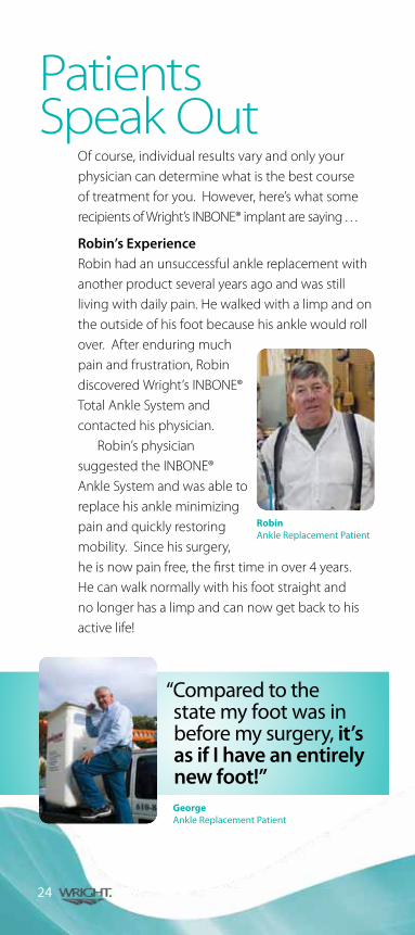
Robin’s ExperienceRobin had an unsuccessful ankle replacement with another product several years ago and was still living with daily pain. He walked with a limp and on the outside of his foot because his ankle would roll over. After enduring much pain and frustration, Robin discovered Wright’s INBONE® Total Ankle System and contacted his physician.
Robin’s physician suggested the INBONE® Ankle System and was able to replace his ankle minimizing pain and quickly restoring mobility. Since his surgery, he is now pain free, the first time in over 4 years. He can walk normally with his foot straight and no longer has a limp and can now get back to his active life!
RobinAnkle Replacement Patient
Patients Speak Out
24
“ Compared to the state my foot was in before my surgery, it’s as if I have an entirely new foot!”GeorgeAnkle Replacement Patient

Jeanne’s ExperienceJeanne, age 62, underwent ankle replacement and correction of tarsal coalition, an abnormal connection that develops between two bones in the back of the foot. After continued years of pain and facing amputation, Jeanne began to research other options and discovered a new solution for ankle pain.
Jeanne’s physician suggested she receive Wright’s INBONE® Total Ankle System and was able to implant an artificial ankle minimizing loss of bone and restoring mobility. At eight weeks, she was weight-bearing and experienced no pain or swelling!
In October 2007, Jeanne received her second ankle implant for the opposite ankle and recovery was faster than with her first replacement. Jeanne was out of her cast and walking after only six weeks. Six months later, Jeanne traveled to China for three weeks. With her new ankles, she successfully climbed the long and steep cobblestone pathway to see the Great Wall. “The Great Wall was my own personal Mt. Everest,” said Jeanne. “For the first time in my life, I have normal mobility and look forward to each day,“ she said. “This experience is profoundly life changing.”
“ I enjoyed the most active and pain-free period in the last 20 years. I’m looking forward to a full and active life for the first time ever.”
25
JeanneAnkle Replacement Patient
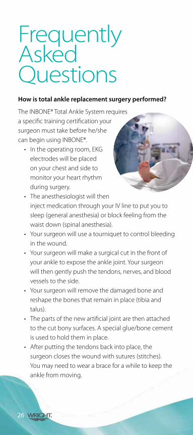
How is total ankle replacement surgery performed?
The INBONE® Total Ankle System requires a specific training certification your surgeonmusttakebeforehe/shecan begin using INBONE®.• Intheoperatingroom,EKG
electrodes will be placed on your chest and side to monitor your heart rhythm during surgery.
• TheanesthesiologistwilltheninjectmedicationthroughyourIVlinetoputyoutosleep (general anesthesia) or block feeling from the waist down (spinal anesthesia).
• Yoursurgeonwilluseatourniquettocontrolbleedingin the wound.
• Yoursurgeonwillmakeasurgicalcutinthefrontofyour ankle to expose the ankle joint. Your surgeon will then gently push the tendons, nerves, and blood vessels to the side.
• Yoursurgeonwillremovethedamagedboneandreshape the bones that remain in place (tibia and talus).
• Thepartsofthenewartificialjointarethenattachedtothecutbonysurfaces.Aspecialglue/bonecementis used to hold them in place.
• Afterputtingthetendonsbackintoplace,thesurgeon closes the wound with sutures (stitches). You may need to wear a brace for a while to keep the ankle from moving.
26
Frequently Asked Questions
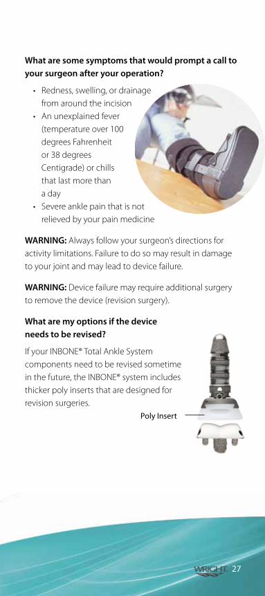
What are some symptoms that would prompt a call to your surgeon after your operation?
• Redness,swelling,ordrainagefrom around the incision
• Anunexplainedfever(temperature over 100 degrees Fahrenheit or 38 degrees Centigrade) or chills that last more than a day
• Severeanklepainthatisnotrelieved by your pain medicine
WARNING: Always follow your surgeon’s directions for activity limitations. Failure to do so may result in damage to your joint and may lead to device failure.
WARNING: Device failure may require additional surgery to remove the device (revision surgery).
What are my options if the device needs to be revised?
If your INBONE® Total Ankle System components need to be revised sometime in the future, the INBONE® system includes thicker poly inserts that are designed for revision surgeries.
27
Poly Insert

Every patient is different, and individual results will vary. There are risks and recovery times associated with surgery. Consult your doctor to determine if ankle replacement surgery is right for you.
You can ask your orthopaedic surgeon or doctor of podiatric medicine about total ankle replacement, or visit: www.wmt.com for more information.
Wright Medical Technology, Inc.5677 Airline RoadArlington, TN USA 38002901.867.9971 800.238.7117 www.wmt.com
Wright Medical EMEAHoogoorddreef 51101 BA AmsterdamThe Netherlands011.31.20.545.0100www.wmt-emea.com
©2012 Wright Medical Technology, Inc.
All Rights Reserved.
FA382-708 R1112