treacher collins syndrome tcof1 protein … collins syndrome tcof1 protein cooperates with nbs1 in...
TRANSCRIPT

Treacher Collins syndrome TCOF1 protein cooperateswith NBS1 in the DNA damage responseAlberto Cicciaa,b,c,1, Jen-Wei Huangc, Lior Izhara,b, Mathew E. Sowad, J. Wade Harperd, and Stephen J. Elledgea,b,1
aDepartment of Genetics and bHoward Hughes Medical Institute, Division of Genetics, Brigham and Women’s Hospital, Harvard University Medical School,Boston, MA 02115; cDepartment of Genetics and Development, Herbert Irving Comprehensive Cancer Center, Columbia University Medical Center,New York, NY 10032; and dDepartment of Cell Biology, Harvard University Medical School, Boston, MA 02115
Contributed by Stephen J. Elledge, November 26, 2014 (sent for review November 8, 2014)
The signal transduction pathway of the DNA damage response(DDR) is activated to maintain genomic integrity following DNAdamage. The DDR promotes genomic integrity by regulating alarge network of cellular activities that range from DNA replica-tion and repair to transcription, RNA splicing, and metabolism. Inthis study we define an interaction between the DDR factor NBS1and TCOF1, a nucleolar protein that regulates ribosomal DNA(rDNA) transcription and is mutated in Treacher Collins syndrome.We show that NBS1 relocalizes to nucleoli after DNA damage ina manner dependent on TCOF1 and on casein kinase II and ATM,which are known to modify TCOF1 by phosphorylation. Moreover,we identify a putative ATM phosphorylation site that is requiredfor NBS1 relocalization to nucleoli in response to DNA damage.Last, we report that TCOF1 promotes cellular resistance to DNAdamaging agents. Collectively, our findings identify TCOF1 asa DDR factor that could cooperate with ATM and NBS1 to suppressinappropriate rDNA transcription and maintain genomic integrityafter DNA damage.
DNA damage response | nucleolus | protein phosphorylation | ATM | CK2
The faithful conservation of genomic information is an es-sential process for cell survival and for preventing malignant
transformation (1). To maintain genomic integrity, DNA hasto be protected from damage either spontaneously induced orgenerated by environmental sources, including ionizing radiationor chemical agents. The DNA damage response (DDR) is a sig-nal transduction network that is activated to maintain genomicintegrity after DNA damage (1, 2). A principal component of theDDR is the ATM kinase, which is primarily activated by thepresence of DNA double-strand breaks (DSBs).DSBs are deleterious DNA lesions that can lead to cell death
if unresolved. DSBs are fixed either by joining the two DNA endstogether by nonhomologous end joining (NHEJ) or by homology-directed repair mediated by homologous recombination (HR) (3).The regulation of DSB end-processing represents a key step in thechoice between NHEJ and HR. Whereas NHEJ occurs withminimal end-processing, extensive resection of DNA ends andformation of single-stranded DNA regions is required for theinitiation of HR (4, 5).NBS1 is a critical component of the heterotrimeric MRE11-
RAD50-NBS1 (MRN) complex, which plays a central role in therepair of DSBs through the activation of the DDR and the ini-tiation of HR. After binding and stabilizing DSB ends, the MRNcomplex recruits ATM and the mediator protein MDC1 to thebreak site through their direct interaction with NBS1. MDC1subsequently associates with the phosphorylated histone variantH2AX (γH2AX) locally to amplify the ATM signaling cascade atDSBs (6–9). Direct interaction with NBS1 also promotes therecruitment of the DNA repair factor CtIP to DSB ends by theMRN complex, where it promotes end resection to initiate HR(10). The importance of NBS1 to the maintenance of genomicintegrity is further highlighted by the predisposition to growthdefects, craniofacial abnormalities, and B-cell lymphomas ofpatients with Nijmegen breakage syndrome, who carry biallelic
mutations in NBS1 (2). In addition, mutations in the subunits ofthe MRN complex have also been linked to familial breastcancer (1).To fully understand how NBS1 prevents genetic disorders and
cancer, it is important to have a comprehensive view of themultiple functions of NBS1 and define all NBS1-associatedfactors. Here we identify TCOF1, a nucleolar protein that reg-ulates ribosomal RNA transcription and is mutated in the cra-niofacial syndrome Treacher Collins, as an interactor of NBS1.We show that NBS1 colocalizes with TCOF1 in the nucleolustransiently after DNA damage in a manner dependent onTCOF1 and on ATM and casein kinase II (CK2), which arekinases known to phosphorylate TCOF1. Our experimentsidentify TCOF1 as a DDR factor that cooperates with NBS1 inthe DNA damage response.
ResultsTCOF1 Is an NBS1 Interactor. To characterize in detail the mecha-nisms by which NBS1 operates in the DDR, we sought to identifyfactors associated with NBS1 in human cells. To this end, weexpressed in human embryonic kidney HEK 293T-REx cellsa cDNA coding for human NBS1 fused to an HA tag and thenperformed anti-HA immunoprecipitation of NBS1 proteincomplexes following treatment with ionizing radiation (IR).Protein complexes were then identified by mass spectrometryand further analyzed using CompPASS software, which assignsto each protein a normalized weighted D (NWD) score de-pendent on protein abundance, frequency, and reproducibility ofthe interactions (11). In addition to known NBS1 interactors,such as the ATR kinase and the DDR mediator MDC1, we were
Significance
The DNA damage response (DDR) maintains genomic integrityfollowing DNA damage to prevent cancer and developmentaldisorders. The DDR operates in part through controlling local-ization of factors to chromatin. Here, we detail an interactionbetween the DDR protein NBS1 and TCOF1, a nucleolar proteinmutated in Treacher Collins syndrome that regulates ribosomalDNA transcription. We show that NBS1 relocalizes to nucleoliafter DNA damage in a manner dependent on TCOF1 and in-dependent on the NBS1-associated protein MRE11. This processis regulated by casein kinase II and ATM, two protein kinasesthat phosphorylate TCOF1 and promote NBS1 nucleolar lo-calization. Our work identifies TCOF1 as a DDR factor thatcooperates with NBS1 to preserve genomic stability afterDNA damage.
Author contributions: A.C., J.-W.H., L.I., M.E.S., and S.J.E. designed research; A.C., J.-W.H.,L.I., and M.E.S. performed research; A.C., M.E.S., and S.J.E. analyzed data; and A.C. andS.J.E. wrote the paper.
The authors declare no conflict of interest.1To whom correspondence may be addressed. Email: [email protected] [email protected].
This article contains supporting information online at www.pnas.org/lookup/suppl/doi:10.1073/pnas.1422488112/-/DCSupplemental.
www.pnas.org/cgi/doi/10.1073/pnas.1422488112 PNAS | December 30, 2014 | vol. 111 | no. 52 | 18631–18636
CELL
BIOLO
GY

able to identify the TCOF1 protein (also known as Treacle) asa potential component of the NBS1 protein complex (Fig. 1).The association between NBS1 and TCOF1 was confirmed fol-lowing immunoprecipitation of HA-tagged TCOF1 proteincomplexes from HEK 293T-REx cells after IR (Fig. 1B). TCOF1encodes a nucleolar protein that is mutated in Treacher Collinssyndrome (TCS), an autosomal dominant disorder that causeshypoplasia of the facial bones, hearing loss, and cleft palate in 1in 50,000 children (12). TCOF1 interacts with RNA polymerase Iand the transcription factor UBF to promote ribosomal DNA(rDNA) transcription (13). Consistent with this role, we identi-fied subunits of RNA polymerase I (POLR1A, B and E) ascomponents of TCOF1 complexes (Fig. 1B).
NBS1 Localizes to Nucleoli After DNA Damage in a TCOF1-DependentManner. To determine whether NBS1 colocalizes with TCOF1after DNA damage, human osteosarcoma U2OS cells weretransfected with HA-tagged NBS1 and stained with an antibodyagainst the HA tag. As shown in Fig. 2 and Fig. S1A, we observedthat, following treatments with IR or with the cross-linking agentcisplatin, a portion of the NBS1 protein pool relocates into thenucleolus, where it colocalizes with TCOF1. NBS1 localizestransiently to the nucleolus between 5 and 30 min after IR ina TCOF1-dependent manner, as indicated by the abrogation of
NBS1 nucleolar staining after siRNA-dependent depletion ofTCOF1 (Fig. 2 C and D). Similar data were also obtained forendogenous NBS1 expressed in U2OS cells (Fig. S2A). UnlikeNBS1, MRE11 did not exhibit localization to nucleoli after DNAdamage, suggesting that the nucleolar localization of NBS1 isMRE11-independent (Fig. S1B). Furthermore, other DDR com-ponents, such as MDC1 and 53BP1, did not colocalize withTCOF1 in nucleoli after IR treatment, as shown in Fig. S2B.
NBS1 Localization to Nucleoli After IR Is Dependent on Casein KinaseII and ATM Kinase Activity. Previous experiments have shown thatthe association between NBS1 and many of its interactors, suchas MDC1 and CtIP, is mediated by NBS1’s FHA domain, whichbinds to protein motifs phosphorylated by CK2 (14–16). TCOF1harbors 10 highly repetitive regions, known as treacle repeats,containing conserved CK2 sites and is a known CK2 substrate(17, 18). Consistent with these observations, we identified a CK2subunit CSNK2B as part of a TCOF1 complex (Fig. 1B). It hasbeen previously shown that CK2 is required for the associationbetween NBS1 and MDC1 (15, 16). To test whether CK2 is re-quired for the recruitment of NBS1 to the nucleolus after IR, wetreated cells with siRNAs against the CK2 subunits α and α′before irradiation (Fig. 3A). Following this treatment, weobserved that the recruitment of NBS1 to the nucleolus was
A Treacle repeats
TCOF1 1 1488
NBS1
Tot Pep NWD Symbol
38 4.49 NBS1
12 2.47 ATR
62 1.81 RAD50
29 0.83 MRE11
6 0.5 MDC1
12 0.32 TCOF1
TCOF1
Tot Pep NWD Symbol
9 3.46 POLR1E
57 2.91 POLR1A
14 1.73 POLR1B
220 1.46 TCOF1
2 1.09 CSNK2B
2 0.54 NBS1
B
Fig. 1. Characterization of the protein complexes of NBS1 and TCOF1. (A) Schematic representation of TCOF1 protein. Treacle repeats are indicated in red.(B) Lists of NBS1 or TCOF1 interactors identified after mass spectrometry and CompPASS analyses of NBS1 or TCOF1 protein complexes isolated from HEK293T-REx cells after IR. The total number of peptides (Tot Pep) and the NWD score are indicated as in ref. 11.
A HA-NBS1 TCOF1
Untreated
IR
MERGE B
CHA-NBS1 TCOF1 MERGE
ControlsiRNA+ IR
TCOF1siRNA+ IR
ED
TCOF1
GAPDH
TCO
F1 s
iRN
A #1
TCO
F1 s
iRN
A #2
Con
trol
siR
NA
Control s
iRNA
TCOF1 siR
NA #1
TCOF1 siR
NA #20
20
40
60
80
100+ IR
Cel
lsw
ithnu
cleo
larN
BS1
( %)
Untreate
d IR
Cel
ls w
ith n
ucle
olar
NB
S1(%
)
0
20
40
60
80
100
Fig. 2. NBS1 nucleolar localization after DNA damage. (A) Representative pictures of U2OS cells expressing HA-NBS1 stained with antibodies against the HAtag (green) or TCOF1 (red) with or without IR treatment (10 Gy). Images of cells with merged green and red signals are indicated. (B) Quantification of thepercentage of U2OS cells expressing HA-NBS1 that exhibit nucleolar staining of NBS1 with or without IR treatment (10 Gy) as in A. (C) Representative picturesof U2OS cells expressing HA-NBS1 stained as in A treated with control or TCOF1 siRNAs with IR treatment (10 Gy). Images of cells with merged green and redsignals are indicated. (D) Western blotting showing the levels of TCOF1 knockdown obtained with two TCOF1 siRNAs. (E) Quantification of the percentage ofU2OS cells expressing HA-NBS1 that exhibit nucleolar staining of NBS1 after treatment with TCOF1 siRNAs and IR.
18632 | www.pnas.org/cgi/doi/10.1073/pnas.1422488112 Ciccia et al.

strongly impaired, suggesting that CK2 is required for the nu-cleolar localization of NBS1 after DNA damage (Fig. 3 A and B).TCOF1 has previously been shown to be phosphorylated by
the ATM kinase in response to IR (19). To test whether ATMkinase activity could regulate the recruitment of NSB1 to thenucleolus, U2OS cells were incubated with an ATM kinase in-hibitor during and after IR. As indicated in Fig. 3, treatment withthe ATM kinase inhibitor KU-55933 abrogated the recruitmentof NBS1 to the nucleolus after IR, indicating that ATM activity isrequired for the colocalization of NBS1 with TCOF1 in thenucleolus after DNA damage (Fig. 3 C and D).
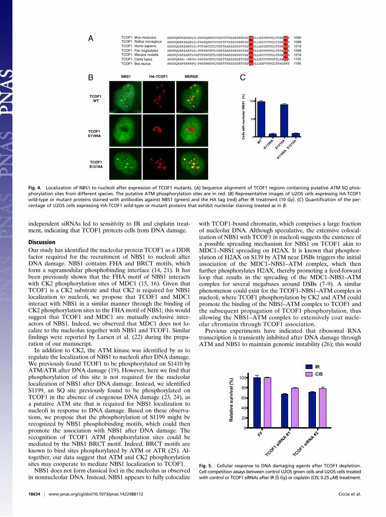
TCOF1 S1199 Is Required for the Nucleolar Localization of TCOF1 AfterIR. ATM and ATR kinases phosphorylate their substrates on SQor TQ motifs (1). We have previously shown that TCOF1 isphosphorylated by ATM/ATR kinases on an SQ site locatedat amino acids 1410–1411 of TCOF1 (19). To determine thefunction of this phosphorylation event, we mutated serine 1410to alanine. Following expression of wild-type and S1410A mutantHA-TCOF1, we observed that the S1410A mutant was pro-ficient for nucleolar localization of NBS1 after IR (Fig. S3).The C-terminal region of TCOF1 contains two additional SQsites at amino acids 1199–1200 and 1216–1217 that are highlyconserved among mammals (Fig. 4A). To examine the role ofthese SQ sites, we generated HA-TCOF1 SQ to AQ mutantsand expressed them in U2OS cells. As shown in Fig. 4 B and C,the TCOF1 S1199A mutant displayed defective localization ofNBS1 to nucleoli after IR, whereas the TCOF1 S1216A mutantexhibited a phenotype similar to wild-type TCOF1 in this as-say. These experiments suggest that ATM could potentially
phosphorylate TCOF1 to recruit or retain NBS1 at nucleolar sitesafter DNA damage.
A Small Fraction of TCOF1 Is Recruited to Sites of DNA Damage AfterLaser Microirradiation. TCOF1 is predominantly localized in thenucleolus. To determine whether TCOF1 localization could beaffected by DNA damage, we conducted UV laser micro-irradiation on U2OS cells expressing HA-TCOF1. Interestingly,we observed that a small fraction of TCOF1 localized to theDNA damage sites generated by the UV laser outside of thenucleolus (Fig. S4A).TCOF1 has at least six different isoforms in mammalian cells.
To determine whether the localization of TCOF1 to DNAdamage sites differs among TCOF1 isoforms, we expressed in U2OScells the TCOF1 isoform c [National Center for Biotechnology In-formation (NCBI) reference sequence NM_001008657.2], whichdisplays pan-nuclear localization due to the absence of the nucleolarsignal located at the C terminus of the protein. As shown in Fig.S4B, the TCOF1 isoform c exhibited a very clear recruitment toDNA damage sites induced by laser stripes. These observationssuggest that TCOF1 could also have a role in the DNA damageresponse outside of the nucleolus.
TCOF1 Protects Cells from DNA Damage. Many proteins that asso-ciate with NBS1 are known to protect cells from DNA damage.To test whether TCOF1 is required for survival in response toDNA damage, GFP-labeled U2OS cells treated with control orTCOF1 siRNAs were mixed with unlabeled U2OS cells. Therelative survival of green vs. unlabeled U2OS cells after IR (5 Gy)or cisplatin (0.25 μM) treatment was then measured by FACScytometry (20). As shown in Fig. 5, depletion of TCOF1 with two
A HA-NBS1 TCOF1 MERGE
ControlsiRNA+ IR
CK2α + CK2α’siRNA+ IR
B
Control s
iRNA
0
20
40
60
80+ IR
Cel
lsw
ithnu
cleo
larN
BS 1
(%)
CK2a +C
K2a’ s
iRNA
IR +ATMi
IR
HA-NBS1 TCOF1 MERGEC D
IR
IR + ATMi
0
20
40
60
80
100
Cel
lsw
ithnu
cleo
larN
BS1
(%)
Fig. 3. NBS1 localization to nucleoli after CK2 depletion or ATM inhibition. (A) Representative images of U2OS cells expressing HA-NBS1 treated with controlor siRNAs targeting the CK2 subunits α and α′ (CK2α and CK2α′) and stained as in Fig. 2 after IR treatment (10 Gy). (B) Quantification of the percentage ofU2OS cells expressing HA-NBS1 that exhibit nucleolar staining treated as in A. (C) Representative images of U2OS cells expressing HA-NBS1 with or withoutATM inhibitor (10 μM) and stained as in Fig. 2 after IR treatment (10 Gy). (D) Quantification of the percentage of U2OS cells expressing HA-NBS1 that exhibitnucleolar staining treated as in C.
Ciccia et al. PNAS | December 30, 2014 | vol. 111 | no. 52 | 18633
CELL
BIOLO
GY
independent siRNAs led to sensitivity to IR and cisplatin treat-ment, indicating that TCOF1 protects cells from DNA damage.
DiscussionOur study has identified the nucleolar protein TCOF1 as a DDRfactor required for the recruitment of NBS1 to nucleoli afterDNA damage. NBS1 contains FHA and BRCT motifs, whichform a supramodular phosphobinding interface (14, 21). It hasbeen previously shown that the FHA motif of NBS1 interactswith CK2 phosphorylation sites of MDC1 (15, 16). Given thatTCOF1 is a CK2 substrate and that CK2 is required for NBS1localization to nucleoli, we propose that TCOF1 and MDC1interact with NBS1 in a similar manner through the binding ofCK2 phosphorylation sites to the FHA motif of NBS1; this wouldsuggest that TCOF1 and MDC1 are mutually exclusive inter-actors of NBS1. Indeed, we observed that MDC1 does not lo-calize to the nucleolus together with NBS1 and TCOF1. Similarfindings were reported by Larsen et al. (22) during the prepa-ration of our manuscript.In addition to CK2, the ATM kinase was identified by us to
regulate the localization of NBS1 to nucleoli after DNA damage.We previously found TCOF1 to be phosphorylated on S1410 byATM/ATR after DNA damage (19). However, here we find thatphosphorylation of this site is not required for the nucleolarlocalization of NBS1 after DNA damage. Instead, we identifiedS1199, an SQ site previously found to be phosphorylated onTCOF1 in the absence of exogenous DNA damage (23, 24), asa putative ATM site that is required for NBS1 localization tonucleoli in response to DNA damage. Based on these observa-tions, we propose that the phosphorylation of S1199 might berecognized by NBS1 phosphobinding motifs, which could thenpromote the association with NBS1 after DNA damage. Therecognition of TCOF1 ATM phosphorylation sites could bemediated by the NBS1 BRCT motif. Indeed, BRCT motifs areknown to bind sites phosphorylated by ATM or ATR (25). Al-together, our data suggest that ATM and CK2 phosphorylationsites may cooperate to mediate NBS1 localization to TCOF1.NBS1 does not form classical foci in the nucleolus as observed
in nonnucleolar DNA. Instead, NBS1 appears to fully colocalize
with TCOF1-bound chromatin, which comprises a large fractionof nucleolar DNA. Although speculative, the extensive colocal-ization of NBS1 with TCOF1 in nucleoli suggests the existence ofa possible spreading mechanism for NBS1 on TCOF1 akin toMDC1–NBS1 spreading on H2AX. It is known that phosphor-ylation of H2AX on S139 by ATM near DSBs triggers the initialassociation of the MDC1–NBS1–ATM complex, which thenfurther phosphorylates H2AX, thereby promoting a feed-forwardloop that results in the spreading of the MDC1–NBS1–ATMcomplex for several megabases around DSBs (7–9). A similarphenomenon could exist for the TCOF1–NBS1–ATM complex innucleoli, where TCOF1 phosphorylation by CK2 and ATM couldpromote the binding of the NBS1–ATM complex to TCOF1 andthe subsequent propagation of TCOF1 phosphorylation, thusallowing the NBS1–ATM complex to extensively coat nucle-olar chromatin through TCOF1 association.Previous experiments have indicated that ribosomal RNA
transcription is transiently inhibited after DNA damage throughATM and NBS1 to maintain genomic instability (26); this would
CNBS1 HA-TCOF1
TCOF1 WT
TCOF1S1199A
MERGE
A
TCOF1S1216A
WT
S1199
A
S1216
A
S1199
A S12
16A
0
50
100
Cel
lsw
it hnu
cleo
larN
BS1
(%)
B
Fig. 4. Localization of NBS1 to nucleoli after expression of TCOF1 mutants. (A) Sequence alignment of TCOF1 regions containing putative ATM SQ phos-phorylation sites from different species. The putative ATM phosphorylation sites are in red. (B) Representative images of U2OS cells expressing HA-TCOF1wild-type or mutant proteins stained with antibodies against NBS1 (green) and the HA tag (red) after IR treatment (10 Gy). (C) Quantification of the per-centage of U2OS cells expressing HA-TCOF1 wild-type or mutant proteins that exhibit nucleolar staining treated as in B.
FF
TCOF1 siR
NA #1
TCOF1 siR
NA #20
20
40
60
80
100
IRCIS
Rel
ativ
esu
rviv
al (%
)
Fig. 5. Cellular response to DNA damaging agents after TCOF1 depletion.Cell competition assays between control U2OS green cells and U2OS cells treatedwith control or TCOF1 siRNAs after IR (5 Gy) or cisplatin (CIS; 0.25 μM) treatment.
18634 | www.pnas.org/cgi/doi/10.1073/pnas.1422488112 Ciccia et al.

facilitate the repair of DNA lesions by preventing the collisionbetween the transcriptional and DNA repair machineries. Basedon our observation that TCOF1 is required for the ATM-dependent recruitment of NBS1 to nucleoli after DNA damage,we suggest that TCOF1 could be a component of the ATM- andNBS1-dependent pathway that blocks ribosomal DNA transcrip-tion after DNA damage, as suggested also by Larsen et al. (22). Itwill be critical in the future to determine whether defective lo-calization of NBS1 to nucleoli results in increased instability ofribosomal DNA repeats.The craniofacial abnormalities caused by TCOF1 mutations
have been shown to depend on hyperactivation of p53-dependentapoptotic processes in neural crest progenitors during craniofacialdevelopment (27). The observation that mutations in RNA poly-merase I can cause TCS suggests that the increased cell deathobserved in TCS patients could result from impaired rDNAtranscription and consequent dysfunctional ribosomal biogenesis.However, excessive p53 activation in TCS may alternatively beconsistent with increased and/or unresolved DNA damage in TCScells due to defective regulation of RNA polymerase I transcrip-tion after DNA damage or to potential functions for TCOF1 innonnucleolar DNA repair. It will therefore be important to testthe possibility that defective DNA repair may contribute to TCSpathophysiology. Similar to TCS, the Nijmegen breakage syn-drome displays craniofacial abnormalities (1). Future experimentswill be needed to determine how defects in NBS1 nucleolar lo-calization contribute to the craniofacial abnormalities displayedby Nijmegen breakage syndrome patients.Together, our experiments have identified TCOF1 as a DDR
factor that recruits NBS1 to nucleoli after DNA damage andhave suggested a potential role of alterations of the DNAdamage response in the pathogenesis of TCS.
Materials and MethodsAntibodies. Rabbit polyclonal anti-TCOF1 (1:1,000; Proteintech, 11003-1-AP1),anti-GAPDH (1:2,000; Santa Cruz, sc-25778), and mouse monoclonal anti-HA(1:1,000; Covance, HA.11) antibodies were used in Western blot experiments.
DNA Clones. The TCOF1 clone corresponding to NCBI NM_001135243.1 wascloned by Gateway recombination into pENTRD-TOPO to generate pENTRD-TOPO-TCOF1. The Gateway pENTRD-TOPO-TCOF1-S1199A, pENTRD-TOPO-TCOF1-S1216A, and pENTRD-TOPO-TCOF1-S1410A were generated bysite-directed mutagenesis of pENTRD-TOPO-TCOF1. The TCOF1 isoform ccorresponding to NCBI NM_001008657.2, the NBS1 clone corresponding toNCBI AK312410 and the MRE11 clone corresponding to NCBI NM_005591were cloned by Gateway recombination into pDONR223. pENTRD-TOPO-TCOF1, pENTRD-TOPO-TCOF1-S1199A, pENTRD-TOPO-TCOF1-S1216A, pENTRD-TOPO-TCOF1-S1410A, pDONR223-NBS1, and pDONR223-MRE11 were usedin Gateway recombination reactions with pMSCV-FLAG-HA (28) to generatepMSCV-FLAG-HA-TCOF1; pMSCV-FLAG-HA-TCOF1-S1199A, pMSCV-FLAG-HA-TCOF1-S1216A, and pMSCV-FLAG-HA-TCOF1-S1410A; pMSCV-FLAG-HA-NBS1;
and pMSCV-FLAG-HA-MRE11. pDONR223-TCOF1 isoform c was recombinedwith the Gateway vector pHAGE-HA to generate pHAGE-HA-TCOF1 isoform c.
Cell Culture and RNAi. The human osteosarcoma cell line U2OS and humanembryonic kidney fibroblast cell line HEK 293T-REx were maintained inMcCoy’s or DMEM, respectively, supplemented with 10% (vol/vol) FBS. Sta-ble U2OS cell lines expressing HA-TCOF1; HA-TCOF1-S1199A, HA-TCOF1-S1216A, HA-TCOF1-S1410A; HA-NBS1; HA-MRE11; and HA-TCOF1 isoform cwere obtained after puromycin selection of cells infected with retroviruses orlentiviruses generated from the vectors pMSCV-FLAG-HA-TCOF1; pMSCV-FLAG-HA-TCOF1-S1199A, pMSCV-FLAG-HA-TCOF1-S1216A, and pMSCV-FLAG-HA-TCOF1-S1410A; pMSCV-FLAG-HA-NBS1; pMSCV-FLAG-HA-MRE11; and pHAGE-HA-TCOF1 isoform c. Stable HEK 293T-REx cells expressingHA-TCOF1 and HA-NBS1 were obtained after infection with retroviruses gen-erated from the pMSCV-FLAG-HA-TCOF1 and pMSCV-FLAG-HA-NBS1 vectors.
TCOF1 siRNAs (Invitrogen; Stealth siRNA HS110575 and HS110577), CK2αand CK2α′ siGenome siRNA pools (Dharmacon; M-003475-03-0005 andM-004752-00-0005) were used to transfect U2OS cells.
Immunofluorescence. Parental U2OS cells andU2OS cells expressing HA-TCOF1wild-type and mutant proteins, HA-TCOF1 isoform c, HA-NBS1, or HA-MRE11were stainedwith rabbit polyclonal anti-MDC1 (1:100; Abcam, ab11169), anti-NBS1 (1:100; Novus Biologicals, NB100-143), anti-TCOF1 (1:100; Proteintech,11003-1-AP1), anti-53BP1 (1:1,000; Bethyl Laboratories, A300-272A), ormouse monoclonal anti-HA (1:1,000; Covance, HA.11), anti-γH2AX (1:500;Millipore, JBW301), anti-TCOF1 (1:100; Santa Cruz Biotechnology, sc-374536)antibodies. For NBS1 localization experiments, U2OS cells expressing HA-NBS1 or HA-TCOF1 wild-type and mutant proteins were treated with 10 Gyirradiation with or without the ATM inhibitor KU-55933 (10 μM) and thenfixed with 4% formaldehyde 15 min after irradiation. Following permea-bilization with 0.2% Triton, U2OS cells were stained with the antibodiesindicated above. Similar experiments were conducted after cisplatin (1 μM)treatment for 6 h. The percentage of cells with NBS1 nucleolar localizationwas determined as the average value of three or more replicates. Micro-irradiation experiments were performed as previously described (29), andU2OS cells expressing HA-TCOF1 and HA-TCOF1 isoform c were fixed 5 minafter irradiation.
Protein Purification and Mass Spectrometry. Stable HEK 293T-REx cells weresubjected to doxycycline treatment to induce the expression of HA-TCOF1and HA-NBS1 and purification of HA-TCOF1 and HA-NBS1 protein complexesafter IR treatment (10 Gy) was conducted as previously described (29). Massspectrometry and CompPASS analysis was performed as reported (11, 29).
DNA Damage Sensitivity Assays. Cell competition assays were performed aspreviously described (28). In particular, U2OS cells were transfected withcontrol or TCOF1 siRNAs and then mixed with GFP expressing U2OS cells asreported (28). Cells were then treated with a single IR dose (5 Gy) or withcisplatin (0.25 μM) for 16 h, and the ratio of uncolored to GFP expressingU2OS cells was determined by flow cytometric analysis after 7 d.
ACKNOWLEDGMENTS.We thank members of the S.J.E. and A.C. laboratoriesfor comments and advice on this project. This work was supported byNational Institutes of Health Grants GM44664 (to S.J.E.) and AG011085 (toJ.-W.H.). S.J.E. is an Investigator with the Howard Hughes Medical Institute.
1. Ciccia A, Elledge SJ (2010) The DNA damage response: Making it safe to play with
knives. Mol Cell 40(2):179–204.2. Jackson SP, Bartek J (2009) The DNA-damage response in human biology and disease.
Nature 461(7267):1071–1078.3. West SC (2003) Molecular views of recombination proteins and their control. Nat Rev
Mol Cell Biol 4(6):435–445.4. Symington LS, Gautier J (2011) Double-strand break end resection and repair pathway
choice. Annu Rev Genet 45:247–271.5. Chapman JR, Taylor MR, Boulton SJ (2012) Playing the end game: DNA double-strand
break repair pathway choice. Mol Cell 47(4):497–510.6. Lukas J, Lukas C, Bartek J (2011) More than just a focus: The chromatin response to
DNA damage and its role in genome integrity maintenance. Nat Cell Biol 13(10):
1161–1169.7. Stewart GS, Wang B, Bignell CR, Taylor AM, Elledge SJ (2003) MDC1 is a mediator of
the mammalian DNA damage checkpoint. Nature 421(6926):961–966.8. Stucki M, et al. (2005) MDC1 directly binds phosphorylated histone H2AX to regulate
cellular responses to DNA double-strand breaks. Cell 123(7):1213–1226.9. Lou Z, et al. (2006) MDC1 maintains genomic stability by participating in the ampli-
fication of ATM-dependent DNA damage signals. Mol Cell 21(2):187–200.
10. Stracker TH, Petrini JH (2011) The MRE11 complex: Starting from the ends. Nat Rev
Mol Cell Biol 12(2):90–103.11. Sowa ME, Bennett EJ, Gygi SP, Harper JW (2009) Defining the human deubiquiti-
nating enzyme interaction landscape. Cell 138(2):389–403.12. Sakai D, Trainor PA (2009) Treacher Collins syndrome: Unmasking the role of Tcof1/
treacle. Int J Biochem Cell Biol 41(6):1229–1232.13. Valdez BC, Henning D, So RB, Dixon J, Dixon MJ (2004) The Treacher Collins syndrome
(TCOF1) gene product is involved in ribosomal DNA gene transcription by interacting
with upstream binding factor. Proc Natl Acad Sci USA 101(29):10709–10714.14. Williams RS, et al. (2009) Nbs1 flexibly tethers Ctp1 and Mre11-Rad50 to coordinate
DNA double-strand break processing and repair. Cell 139(1):87–99.15. Spycher C, et al. (2008) Constitutive phosphorylation of MDC1 physically links the
MRE11-RAD50-NBS1 complex to damaged chromatin. J Cell Biol 181(2):227–240.16. Melander F, et al. (2008) Phosphorylation of SDT repeats in the MDC1 N terminus
triggers retention of NBS1 at the DNA damage-modified chromatin. J Cell Biol 181(2):
213–226.17. Jones NC, Farlie PG, Minichiello J, Newgreen DF (1999) Detection of an appropriate
kinase activity in branchial arches I and II that coincides with peak expression of the
Treacher Collins syndrome gene product, treacle. Hum Mol Genet 8(12):2239–2245.
Ciccia et al. PNAS | December 30, 2014 | vol. 111 | no. 52 | 18635
CELL
BIOLO
GY

18. Wise CA, et al. (1997) TCOF1 gene encodes a putative nucleolar phosphoprotein thatexhibits mutations in Treacher Collins Syndrome throughout its coding region. ProcNatl Acad Sci USA 94(7):3110–3115.
19. Matsuoka S, et al. (2007) ATM and ATR substrate analysis reveals extensive proteinnetworks responsive to DNA damage. Science 316(5828):1160–1166.
20. Smogorzewska A, et al. (2007) Identification of the FANCI protein, a mono-ubiquitinated FANCD2 paralog required for DNA repair. Cell 129(2):289–301.
21. Lloyd J, et al. (2009) A supramodular FHA/BRCT-repeat architecture mediates Nbs1adaptor function in response to DNA damage. Cell 139(1):100–111.
22. Larsen DH, et al. (2014) The NBS1-Treacle complex controls ribosomal RNA tran-scription in response to DNA damage. Nat Cell Biol 16(8):792–803.
23. Courcelles M, et al. (2013) Phosphoproteome dynamics reveal novel ERK1/2 MAP ki-nase substrates with broad spectrum of functions. Mol Syst Biol 9:669.
24. Hsu PP, et al. (2011) The mTOR-regulated phosphoproteome reveals a mechanismof mTORC1-mediated inhibition of growth factor signaling. Science 332(6035):1317–1322.
25. Reinhardt HC, Yaffe MB (2013) Phospho-Ser/Thr-binding domains: Navigating the cellcycle and DNA damage response. Nat Rev Mol Cell Biol 14(9):563–580.
26. Kruhlak M, et al. (2007) The ATM repair pathway inhibits RNA polymerase I tran-scription in response to chromosome breaks. Nature 447(7145):730–734.
27. Jones NC, et al. (2008) Prevention of the neurocristopathy Treacher Collins syndromethrough inhibition of p53 function. Nat Med 14(2):125–133.
28. Ciccia A, et al. (2012) Polyubiquitinated PCNA recruits the ZRANB3 translocase tomaintain genomic integrity after replication stress. Mol Cell 47(3):396–409.
29. Ciccia A, et al. (2009) The SIOD disorder protein SMARCAL1 is an RPA-interactingprotein involved in replication fork restart. Genes Dev 23(20):2415–2425.
18636 | www.pnas.org/cgi/doi/10.1073/pnas.1422488112 Ciccia et al.