transgenic mice express human mpo 2463g/a alleles at
TRANSCRIPT

Transgenic mice express human MPO 2463G/A alleles
at atherosclerotic lesions, developing hyperlipidemia
and obesity in 2463G males
Lawrence W. Castellani,* James J. Chang,† Xuping Wang,* Aldons J. Lusis,* andWanda F. Reynolds1,†
Departments of Medicine, Microbiology, and Molecular Genetics,* University of California-Los Angeles,Los Angeles, CA 90095; and Sidney Kimmel Cancer Center,† San Diego, CA 92121
Abstract Myeloperoxidase (MPO) is an oxidant-generatingenzyme present in macrophages at atherosclerotic lesionsand implicated in coronary artery disease (CAD). Althoughmouse models are important for investigating the role ofMPO in atherosclerosis, neither mouse MPO nor its oxi-dation products are detected in lesions in murine models.To circumvent this problem, we generated transgenic miceexpressing two functionally different human MPO alleles,with either G or A at position 2463, and crossed these tothe LDL receptor-deficient (LDLR2/2) mouse. The 2463Gallele is linked to higher MPO expression and increasedCAD incidence in humans. Both MPO alleles were ex-pressed in a subset of lesions in high-fat-fed LDLR2/2mice,notably at necrotic lesions with cholesterol clefts. MPOG-expressing LDLR2/2 males (but not females) developedsignificantly higher serum cholesterol, triglycerides, andglucose, all correlating with increased weight gain/obesity,implicating MPO in lipid homeostasis. The MPOG- andMPOA-expressing LDLR2/2 males also exhibited signifi-cantly larger aortic lesions than control LDLR2/2 males.The human MPO transgenic model will facilitate studies ofMPO involvement in atherosclerosis and lipid homeosta-sis.—Castellani, L. W., J. J. Chang, X. Wang, A. J. Lusis, andW. F. Reynolds. Transgenic mice express human MPO2463G/A alleles at athrosclerotic lesions, developing hy-perlipidemia and obesity in2463Gmales. J. Lipid Res. 2006.47: 1366–1377.
Supplementary key words myeloperoxidase . atherosclerosis . hyper-lipidemia . cholesterol . triglycerides . macrophage
Atherosclerosis is a chronic inflammatory disease inwhich macrophages accumulate in the vascular wall. Themacrophages release oxidizing enzymes, including myelo-peroxidase (MPO), which oxidizes LDL particles, pro-ducing a more atherogenic form that is taken up moreeffectively by macrophages to create foam cells (1).
Mounting evidence implicates MPO in atherosclerosis.MPO catalyzes a reaction between chloride and hydrogenperoxide to generate hypochlorous acid, whereas it reactswith nitrite and nitric oxide to produce reactive nitrogenintermediates (2). MPO and its oxidant by-products arepresent in atherosclerotic lesions, colocalizing with foamcell macrophages (3, 4), and are especially abundant atsites of thrombosis (5). MPO preferentially oxidizes apo-lipoprotein A-I (apoA-I), the major protein component ofHDL, impairing ABCA1-mediated cholesterol efflux fromcells (6, 7). Circulating MPO levels are higher in patientswith coronary artery disease (CAD) (8, 9), whereas individ-uals with inherited MPO deficiencies have less cardiovas-cular disease (10). An MPO promoter polymorphism,2463 G/A, which alters expression levels (11, 12), has beenassociated with an increased incidence of CAD (13–15)and severity of atherosclerosis (16, 17). The 2463G allelehas been linked with higher MPO mRNA and protein ex-pression than the 2463A allele in human and transgenicmouse monocyte-macrophages (12).
Mouse models are important for investigations of themolecular pathways underlying atherosclerosis. To thatend, an MPO knockout mouse was generated, but it un-expectedly developed larger lesions in the LDL receptor-deficient (LDLR2/2) model (18, 19). Moreover, neithermouse MPO nor its specific product, 3-chlorotyrosine, wasdetected at lesions in LDLR2/2 mice. Because of the lackof mouse MPO gene expression at lesions, the LDLR2/2
mouse model cannot be used for analysis of MPO effects atthese sites. However, in transgenic mice expressing humanMPO, the enzyme is detected in lesions in the LDLR2/2
model (12, 19) and has been correlated with increasedlesion size (19).
The differential expression of mouse and human MPOgenes may be attributable in part to the 2463G/A poly-morphism in an upstream Alu element containing nuclear
Manuscript received 6 January 2006 and in revised form 15 February 2006 andin re-revised form 16 March 2006 and in re-re-revised form 25 April 2006.
Published, JLR Papers in Press, April 25, 2006.DOI 10.1194/jlr.M600005-JLR200
1To whom correspondence should be addressed.e-mail: [email protected]
Copyright D 2006 by the American Society for Biochemistry and Molecular Biology, Inc.
This article is available online at http://www.jlr.org1366 Journal of Lipid Research Volume 47, 2006
by guest, on March 6, 2018
ww
w.jlr.org
Dow
nloaded from

receptor response elements (11, 12). 2463G enhancesbinding by the SP1 transcription factor (11), whereas2463A promotes binding by estrogen receptor a (ERa)(12, 20). An overlapping site is recognized by peroxisomeproliferator-activated receptor g (PPARg) (12), a tran-scription factor that is expressed in foam cell macrophagesat atherosclerotic lesions and regulates genes involved ininflammatory responses, including CD36 scavenger recep-tor (21), liver X receptor (22), and MPO (12). PPARg li-gands markedly upregulate MPO expression (z20-fold) inmacrophage colony stimulating factor (MCSF)-treatedcells (12). Estrogen blocks the effects of PPARg, suggest-ing that ER binding sterically interferes with access byPPARg. The 2463A site is bound more effectively by ER,and estrogen blocks PPARg induction more effectively.Thus, competition between PPARg and ER may explainthe lower expression of the MPO 2463A allele, especiallyin females. Because Alu elements are primate-specific, themouse MPO gene lacks this PPARg binding site and is notupregulated by PPARg ligands (12). This lack of PPARgresponsiveness may explain the absence of mouse MPO atatherosclerotic lesions in the LDLR2/2 model.
To facilitate the investigation of MPO in atherosclerosisand other inflammatory diseases, we generated transgenicmice expressing the human MPO 2463G or 2463A alleleunder the control of native human promoter elements.The complex regulation by PPARg, including the block byestrogen, is replicated in transgenic MPOG and MPOAmice (12). In LDLR2/2 mice on an atherogenic diet, thehuman MPO 2463G transgene was found to be expressedin aortic lesions, colocalizing with PPARg (12). In thisstudy, we further examine the effects of the human MPO2463G and 2463A transgenes on serum lipids and lesionmorphology in LDLR2/2 mice. The MPO transgene wascorrelated with increases in serum cholesterol and tri-glycerides, weight gain/obesity, and increased lesion size.
METHODS
Animals
Transgenic mice carrying the human 2463G and 2463A al-leles were described previously (12, 23, 24). The mice weregenerated by microinjection of a 32 kb Bst11071 restrictionfragment directly into C57BL6/J eggs (12), avoiding the need forbackcrossing. To examine the effect of MPO expression in anatherosclerotic model, the mice were crossed to LDLR2/2 micebackcrossed for 10 generations onto the C57BL6/J background(Jackson Laboratories). Mice were housed five per cage, main-tained at 228C on a 12 h light/dark cycle, and provided withstandard rodent chow or a high-fat atherogenic Western diet(HFD; Harlan Teklad Laboratory; TD 88137) with 42% caloriesderived from fat (21.2% fat by weight, 0.2% cholesterol) forperiods of 9 or 20 weeks. Male and female mice at a mean age of12.4 weeks (60.7 SD) were fed a HFD for 9 weeks. Male andfemale mice at a mean age of 10.26 0.8 weeks were fed a HFD for20 weeks. The care of the mice, and all procedures used in thisstudy, were approved by our institutional animal care and usecommittee and complied with National Institutes of Health ani-mal care guidelines.
PCR analysis detected human MPO-flanking sequences in thetransgenics, extending at least 7 kb upstream and at least 4–11 kbdownstream for the MPOA and MPOG transgenes, respectively.The following PCR primer sets were used: z7,000 bp upstream(59-gtatctgaattcagtggaga-39 and 59-tttggatttcgctctttcag-39), z11 kbdownstream (59-gaatacgtaaagaactctca-39 and 59-cctatcagcaatgta-tgcat-39), and z4 kb downstream (59-gtgtggagggggagttct-39 and59-ccttggttttcaacagctca-39). The 2463G allele was excised from asequenced bacterial artificial chromosome clone (accession num-ber AC004687). The 2463A allele was from a DNA clone ob-tained by hybridization screening of a human bacterial artificialchromosome library (Research Genetics) (12).
To determine the sequence of the MPO mRNA producedby the transgenes, we isolated mRNA from bone marrow cellsusing the Trizol reagent and produced cDNA using oligo(dT)primers and the Omniscript cDNA kit (Qiagen). PCR primerswere chosen to amplify in 30 cycles the 2,568 bp cDNA as twoz1,300 bp fragments, and the fragments were subcloned into aplasmid vector for sequencing (Eton Biosciences, San Diego,CA). Sequences obtained were identical to the MPO cDNA se-quence reported on GenBank (accession number JO2694). Thetwo transgenic lines are identical at the relatively rare 2129G/Apolymorphism (A allelic frequency of 0.07) (25), both having themore common 2129G residue. Quantitative real-time PCR wasused to estimate copy number, using TaqMan MGB probes andprimers to compare the threshold cycle for a human MPO in-tronic sequence with a known mouse single-copy gene, apoB(Applied Biosystems; Sequences on Demand). Results wereconsistent with single-copy insertions for both the MPOG andMPOA transgenes.
Western blot analysis
Western analysis was carried out on homogenates of bonemarrow, brain, kidney, spleen, liver, and heart from controlC57BL6/J mice, mouse MPO knockout mice (18), and theMPOG and MPOA transgenics on the mouse MPO knockoutbackground. Cells or tissues were homogenized in SDS samplebuffer, and equal amounts of total protein were electrophoresedon 4–20% gradient denaturing SDS-PAGE gels and transferred topolyvinylidene difluoride membranes. Blots were blocked in 5%nonfat dry milk in Tris-buffered saline containing 1% Tween20 (TBS-T) for 1 h and incubated with polyclonal antibodies tohuman MPO (BioDesign International) overnight at 48C in TBS-T with 250 mM NaCl and 5% normal goat serum. Blots were thenwashed in TBS-T twice at 15 min intervals and incubated withhorseradish peroxidase-conjugated secondary antibody for30 min at room temperature with 5% milk. After two subsequentwashes in TBS-T, the blots were developed with ECL reagent(Amersham Biosciences).
Plasma lipid and glucose assays
Mice were fasted overnight, and blood was collected from theretro-orbital sinus under isoflurane anesthesia. Total cholesterol,HDL, triglycerides, and free fatty acid concentrations were deter-mined as described previously (26). HDL-cholesterol was quan-titated after precipitation of VLDL and LDL with heparin andmanganese chloride. An external control sample with knownanalyte concentration was run on each plate to ensure accuracy.Plasma glucose concentrations were determined in triplicateusing a kit from Sigma.
Atherosclerotic lesion analysis
The heart and proximal aorta were embedded in paraffin,and 5 mm sections were obtained over a 400 mm region spanning
Human MPO transgene effects in an LDLR2/2 model 1367
by guest, on March 6, 2018
ww
w.jlr.org
Dow
nloaded from

the aortic sinus and valve regions. Every eighth section wascollected and stained with Masson’s Trichrome. Digital imageswere obtained and analyzed with Adobe PhotoshopT. The lesionareas were outlined extending from the internal elastic lamina(IEL) to the luminal surface and converted to uniform blackcolor, and the number of black pixels was determined us-ing PhotoshopT.
Immunohistochemistry
Aortic arch sections were deparaffinized and heat-treated incitrate buffer for antigen retrieval according to standard proce-dures. Sections were incubated with 10% hydrogen peroxide for10 min, blocked with 10% goat normal serum for 12 h, incu-bated overnight with rabbit polyclonal antibody to human MPO(BioDesign International; 1:1,000) or rat monoclonal MOMA2antibody (Accurate Biochemicals; 1:1,000), followed by biotin-ylated secondary antibody and avidin-conjugated horseradishperoxidase (Vectastain ABC kit; Vector Laboratories, Burlin-game, CA), and detected with peroxidase chromogen kits (DABnickel, Vector SG, or 3-amino-9-ethylcarbazole; Vector Laborato-ries). Frozen sections of the aortic arch region were also used forOil Red O staining and some immunohistochemical analyses.Liver tissue was fixed in 4% paraformaldehyde, and free-floatingsections (35 mm) were obtained with a Leica VT1000S vibratingmicrotome. The sections were stained with antibodies to humanMPO (BioDesign International) and monoclonal rat anti-mouseCD68 (Hycult International).
Statistical analysis
Student’s t-test or ANOVA with Fisher’s protected least-squares differences was used to determine statistical significanceusing Statview software (SAS Institute, Inc., Cary, NC). The datawere determined to be appropriate for parametric analysis.All data are presented as means 6 SD. P , 0.05 was consid-ered significant.
RESULTS
The MPOG and MPOA transgenes produce MPO protein
Analysis of the expression patterns for the MPOG andMPOA transgenics has been reported previously (12). Thesequences of the transgenic MPO mRNAs were recentlydetermined and found to be identical to reported se-quences (GenBank). PCR analysis revealed at least 7 kb ofhuman MPO upstream sequence in both transgenics and4–11 kb of downstream sequences in the MPOA andMPOG transgenics. To determine that the human MPOtransgenics produce MPO protein, the mice were crossedonto the mouse MPO knockout background (18) to elimi-nate mouse MPO protein. Western analysis of bone mar-row extracts using an antibody generated against humanMPO (BioDesign International) did not detect the proteinin mouse MPO knockout bone marrow extract but diddetect both the heavy (59 kDa) and light (13.5 kDa) chainsin MPOG and MPOA transgenic bone marrow cells(Fig. 1A), indicating that the MPO precursor protein wascorrectly processed in the transgenic lines. Western anal-ysis of various tissues showed abundant levels of MPOprotein in bone marrow but relatively little to no signal innormal brain, heart, lung, liver, kidney, or spleen (Fig. 1B),consistent with the pattern of MPO expression observed inhuman tissues.
MPO is present in a subset of aortic lesions in theMPOG- and MPOA-expressing LDLR2/2 transgenics
Because macrophages are implicated in atheroscleroticlesions, and the human MPO gene is more inducible inmacrophages than is mouse MPO (12), we postulated thatthe human MPO transgenes would humanize the mouse
Fig. 1. The myeloperoxidase (MPO) G and A transgenes give rise to MPO protein. A: Western analysis wascarried out with bone marrow cell extracts from MPO knockout (ko), MPOG transgenics on the mouseknockout background (Gko), MPOA transgenics on the mouse MPO knockout background (Ako), and wild-type C57BL6/J (wt) and compared with purified humanMPO (Calbiochem). Both the MPO heavy and lightchains were detected in bone marrow extracts from Gko and Ako using polyclonal antibodies to humanMPO (BioDesign International). Protein levels were normalized to mouse b-actin. B: Western analysis oftissue extracts from MPOG or MPOA transgenics on the mouse MPO knockout background. Tissuesincluded bone marrow (BM), brain (Br), heart (Hrt), lung (Lu), liver (Liv), kidney (Kid), spleen (Spl), and,as a control, bone marrow from MPO knockout mice (KO). Arrows indicate MPO protein subunits, with b-actin used for normalization of protein concentration.
1368 Journal of Lipid Research Volume 47, 2006
by guest, on March 6, 2018
ww
w.jlr.org
Dow
nloaded from

atherosclerosis models with regard to MPO. Therefore, weinvestigated the effects of human MPO transgene expres-sion in the LDLR2/2 model. MPO transgenic mice on theLDLR2/2 background were fed a HFD for 9 or 20 weeksbefore analysis of sections throughout the proximal aorta.Figure 2A shows an early fatty streak lesion consisting ofa cluster of macrophages infiltrating the vascular wall,some with cytoplasmic vesicles characteristic of foam cells.Figure 2B shows a nearby section with strong immuno-staining for MPO within the macrophage cluster. Oil RedO staining shows neutral lipid in aortic valve lesions from afemale MPOG-expressing LDLR2/2 mouse (Fig. 2C) andstrong MPO immunostaining in one of three valve lesionsin a nearby section (Fig. 2D). These observations indicatedthat MPO is expressed in cells in a subset of early to in-termediate lesions in the MPOG-expressing LDLR2/2
model. The finding that MPO was not uniformly ex-pressed in all lesions suggested that the gene is induced inmonocyte-macrophages in response to agents at a subsetof lesions (12) or that the protein is retained in invadingmonocytes (5) as a result of such agents. MPO was alsodetected in lesions in the MPOA-expressing LDLR2/2 line
(Fig. 2E, F), indicating that MPO is expressed (or re-tained) in cells at lesions in both transgenic lines. MPOwas detected in lesions in both male and female mice;there was no obvious gender difference in the amountof MPO immunodetected in lesions in male or femaleMPOG- or MPOA-expressing LDLR2/2 mice.
MPO was abundant in invading monocyte-macrophagesat some advanced lesions, as shown in an example from amale MPOG-expressing LDLR2/2 mouse (Fig. 3). Mas-son’s Trichrome staining reveals a complex lesion ex-truded through the IEL into the underlying collagen-rich(blue) adventitia (Fig. 3A). In Fig. 3B, the same sectionwas immunostained for MPO (black) and the macrophagemarker MOMA2 (red). MOMA2 staining was detectedthroughout the lesion, marking the presence of macro-phages surrounding angular cholesterol clefts. Aggrega-tions of MPO-positive cells were most apparent at siteswhere the lesion erupted through the IEL into the ad-ventitia (Fig. 3B, C). MPO-positive cells infiltrating thecap and lesion were more easily seen in an unstainednearby section (Fig. 3D). Costaining for MPO (black) andMOMA2 (red) identified MPO-positive macrophages in
Fig. 2. Atherosclerosis in MPOG- and MPOA-expressingLDL receptor-deficient (LDLR2/2) mice fed a high-fatWestern diet (HFD). A: Paraffin sections of the aortic rootregion from a male MPOG-expressing LDLR2/2 mousewere stained with Masson’s Trichrome stain, showing anaggregation of macrophages in an early fatty streak lesion.B: Polyclonal antibodies to human MPO (BioDesign Inter-national) detect abundant MPO in a macrophage clusterin an adjacent section. C: A frozen section from the aorticvalve region of a female MPOG-expressing LDLR2/2
mouse was stained with Oil Red O to detect neutral lipids.D: An adjacent section shows strong MPO immunostain-ing in one of three lesions. E: MPO immunostaining wasalso detected in lesions in a female MPOA-expressingLDLR2/2 mouse (arrowheads); a higher magnificationimage is shown in F. Original objective magnifications areas follows: 203 (A, B, F), 63 (D), and 43 (C, E).
Human MPO transgene effects in an LDLR2/2 model 1369
by guest, on March 6, 2018
ww
w.jlr.org
Dow
nloaded from

the same region beneath the IEL (Fig. 3E). At highermagnification, punctate cytoplasmic MPO staining wasseen in macrophages beneath the IEL (Fig. 3F) and inmonocyte-macrophages infiltrating from the vessel lumen
(Fig. 3G). Consistent with previous reports (18, 19), anti-bodies to human MPO (BioDesign International) did notdetect mouse MPO protein at lesions in the LDLR2/2
model (data not shown).
Fig. 3. MPO is detected in macrophages at advanced lesions in MPOG-expressing LDLR2/2. Paraffinsections from MPOG-expressing LDLR2/2 proximal aorta were stained with Masson’s Trichrome (A) afterimmunostaining for MPO (B–G) and macrophage marker MOMA2 (B, C, E–G). A: Trichrome stainingshows a complex lesion erupting through the internal elastic lamina (IEL; red fibers) into the underlyingadventitia. Cholesterol clefts appear as clear angular areas. Collagen (blue) staining marks a fibrous cap atthe luminal surface. B: The same section was initially stained for MPO (black) and MOMA2 (red). C: Highermagnification showsMPO-positive cells associated with the invading lesion at the point of IEL breakthrough.D: MPO immunostaining of a nearby section shows MPO-positive cells associated with lesions extrudedunder the IEL and a few MPO-positive cells in the lesion proper and at the luminal surface. E: Highermagnification of the same section costained for MPO (black) and MOMA2 (red) shows colocalization in thearea under the IEL. F, G: Higher magnification shows punctate MPO staining in MOMA2-positivemacrophages in the sub-IEL area (F) and in macrophages at the luminal cap and infiltrating the lesion (G).Lu, lumen. Original objective magnifications are as follows: 103 (A–D), 203 (E), and 603 (F, G).
1370 Journal of Lipid Research Volume 47, 2006
by guest, on March 6, 2018
ww
w.jlr.org
Dow
nloaded from

Male MPOG-expressing LDLR2/2 transgenics develophigher serum cholesterol and triglycerides
Consistent with earlier findings (27), control LDLR2/2
males on the HFD for 20 weeks developed 2-fold highertriglyceride levels than female controls (420 6 144 vs.195 6 88 mg/dl; mean 6 SD) (Fig. 4A). Similarly, controlmales on the HFD for 9 weeks developed 1.7-fold highertriglyceride levels than females (216 6 104 vs. 126 6
67 mg/dl). The MPOG allele exacerbated this gender dif-ference. Nine week HFD-fed MPOG-expressing LDLR2/2
males had 2.3-fold higher triglyceride levels than con-trol LDLR2/2 males (502 6 186 vs. 216 6 104 mg/dl; P 5
0.0001). Female MPOG-expressing LDLR2/2 mice showeda 1.3-fold increase in triglycerides over control LDLR2/2
females, a difference that did not reach statistical signifi-cance (163 6 64 vs. 126 6 67 mg/dl). Thus, the MPOG al-lele exacerbated the increase in triglycerides in LDLR2/2
males, and there was less effect in females.In control LDLR2/2 mice on the HFD for 9 weeks, cho-
lesterol levels were slightly lower in males than in females(1,340 6 504 vs. 1,634 6 205 mg/dl). The MPOG trans-gene led to a 1.5-fold increase in cholesterol levels inMPOG-expressing LDLR2/2males compared with control
LDLR2/2 males (2,040 6 544 vs. 1,340 6 504 mg/dl; P 5
0.0005) (Fig. 4B). In contrast, there was no increase incholesterol levels in MPOG-expressing LDLR2/2 femalescompared with control females (1,669 6 491 vs. 1,634 6
205 mg/dl). Unesterified cholesterol was also higher inMPOG-expressing LDLR2/2 males than in control males(577 6 122 vs. 356 6 132 mg/dl; P 5 0.0005) (Table 1),whereas there was no effect in females attributable to theMPOG transgene (416 6 111 vs. 410 6 72 mg/dl). Thus,MPOG increased serum cholesterol in HFD-fed male, butnot female, LDLR2/2 mice.
Serum glucose levels were higher in MPOG-expressingLDLR2/2 males than in control males (2816 55 vs. 2066
43 mg/dl; P = 0.002) (Table 1). Glucose levels increased infemale MPOG-expressing LDLR2/2 mice compared withfemale controls, but this trend did not reach significance(231 6 42 vs. 206 6 32 mg/dl). Consistent with an earlierreport (27), there was no difference in glucose levels be-tween control LDLR2/2 males and females on the athero-genic diet (206 6 43 vs. 206 6 32 mg/dl).
Consistent results were obtained with mice on the HFDfor an extended period of 20 weeks. Cholesterol levelsremained significantly higher in MPOG-expressing
Fig. 4. MPOG-expressing LDLR2/2 males have increased cholesterol and triglycerides. Individual serum triglycerides (A) and cholesterol(B) values were determined for a total of 83 male and female MPOG-expressing LDLR2/2 and LDLR2/2 mice fed the HFD for either 9 or20 weeks. Mean values are indicated.
Human MPO transgene effects in an LDLR2/2 model 1371
by guest, on March 6, 2018
ww
w.jlr.org
Dow
nloaded from
LDLR2/2 males compared with control males (2,367 6
346 vs. 1,921 6 441 mg/dl; P 5 0.02) (Fig. 4B), althoughthe extended fat diet increased cholesterol levels in thecontrols, reducing the difference between MPOG-expres-sing LDLR2/2 and control males. Again, the MPOG trans-gene did not significantly affect cholesterol levels infemales (1,7086 405 vs. 1,6076 243mg/dl). Triglyceridesremained significantly higher in MPOG males comparedwith control males (753 6 424 vs. 420 6 144 mg/dl; P 5
0.037). Triglycerides were also higher in female MPOGcarriers, although this trend did not reach significance(269 6 123 vs. 195 6 88 mg/dl; P 5 0.2). These findingsindicate that the MPOG transgene promotes significantincreases in serum cholesterol, triglycerides, and glucosein males but has less effect in females.
MPOG-expressing LDLR2/2 males gain more weight thanLDLR2/2 males on the HFD
After 9 weeks on the HFD, the MPOG males gainedmore weight than control LDLR2/2males (15 vs. 8 g meanweight gain, MPOG-expressing LDLR2/2 vs. LDLR2/2;end weight, 40.56 2.7 vs. 336 3.5 g; P = 0.0001) (Fig. 5A).After 20 weeks on the HFD, the difference remained sig-nificant (46.4 6 3.3 vs. 38.8 6 5.5 g; P 5 0.0005). FemaleMPOG-expressing LDLR2/2 and control females gainedless weight than males (mean gain of 6.3 vs. 4.8 g, respec-tively). The MPOG-expressing LDLR2/2 females were notsignificantly heavier than LDLR2/2 females at the end ofthe 9 week period (27.5 6 3.8 vs. 26.1 6 2.9 g).
Cholesterol levels were also increased in MPOA-express-ing LDLR2/2 males (n 5 9) (Table 1), although thistrend did not reach statistical significance (1,5246 303 vs.1,340 6 504 mg/dl; P 5 0.23). However, MPOA malesgained more weight than LDLR2/2 control males (endweights, 36.1 6 3.8 vs. 33 6 3.5 g; P 5 0.04). As with theMPOG-expressing LDLR2/2 females, there was no signifi-cant increase in serum lipids (Table 1) or weight in MPOA-expressing LDLR2/2 females.
Aortic lesion size
To compare the size of atherosclerotic lesions in theMPOG-expressing LDLR2/2 and LDLR2/2 mice, serialsections were collected from a 400 mm region spanningthe aortic sinus and valve regions. Lesion area was de-termined with Adobe PhotoshopT by outlining the lesionsfrom the IEL to the luminal surface and converting to pixelnumber as described in Methods. The mean lesion areawas significantly larger in MPOG-expressing LDLR2/2
compared with LDLR2/2 males (182 6 54 vs. 118 6 53 3
103 mm2/section; P 5 0.004) (Fig. 5B). Lesions were alsosignificantly larger in MPOA-expressing LDLR 2/2 malescompared with LDLR2/2 males (166 6 47 3 103 mm2/section; P 5 0.02). Thus, both MPO transgenes increasedthe size of lesions in male HFD-fed LDLR2/2 mice.
In female HFD-fed LDLR2/2mice, the MPO transgenesdid not have a significant effect on lesion size. The meanlesion size was 135 6 19.6 3 103 mm2/section for MPOG-expressing LDLR2/2 females, 1206 43 103 mm2/sectionfor MPOA-expressing LDLR2/2 females (P 5 0.85), and124 6 6.5 3 103 mm2/section for LDLR2/2 females(three-way ANOVA; P 5 0.57 for comparison of MPOG-expressing LDLR2/2 with LDLR2/2 females, P 5 0.85 forcomparison of MPOA-expressing LDLR2/2 with LDLR2/2
females, and P5 0.46 for comparison of MPOA-expressingLDLR2/2 with MPOG-expressing LDLR2/2 females).
Regression analysis indicated a correlation between se-rum cholesterol levels and weight (R2 5 0.58; P , 0.0001)for LDLR2/2 males, including MPO transgenics and con-trol LDLR2/2 males (Fig. 5C). There was also a correla-tion between cholesterol levels and lesion size (R2 5 0.26;P 5 0.017) (Fig. 5D). These observations suggest thatthe increase in serum cholesterol in HFD-fed MPO-expres-sing LDLR2/2 males is related to the increase in weightand lesion size. Photographs of male MPOG-expressingLDLR2/2 and LDLR2/2 mice illustrate the difference inabdominal size (Fig. 6A). There was also a significant cor-relation between the amount of abdominal fat and bodyweight in HFD-fed LDLR2/2 males in a group including
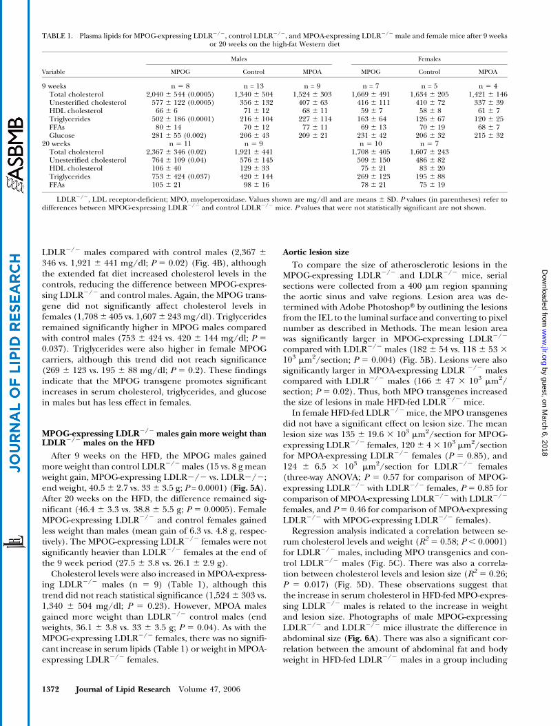
TABLE 1. Plasma lipids for MPOG-expressing LDLR2/2, control LDLR2/2, and MPOA-expressing LDLR2/2 male and female mice after 9 weeksor 20 weeks on the high-fat Western diet
Males Females
Variable MPOG Control MPOA MPOG Control MPOA
9 weeks n 5 8 n = 13 n = 9 n = 7 n = 5 n 5 4Total cholesterol 2,040 6 544 (0.0005) 1,340 6 504 1,524 6 303 1,669 6 491 1,634 6 205 1,421 6 146Unesterified cholesterol 577 6 122 (0.0005) 356 6 132 407 6 63 416 6 111 410 6 72 337 6 39HDL cholesterol 66 6 6 71 6 12 68 6 11 59 6 7 58 6 8 61 6 7Triglycerides 502 6 186 (0.0001) 216 6 104 227 6 114 163 6 64 126 6 67 120 6 25FFAs 80 6 14 70 6 12 77 6 11 69 6 13 70 6 19 68 6 7Glucose 281 6 55 (0.002) 206 6 43 209 6 21 231 6 42 206 6 32 215 6 32
20 weeks n 5 11 n 5 9 n 5 10 n 5 7Total cholesterol 2,367 6 346 (0.02) 1,921 6 441 1,708 6 405 1,607 6 243Unesterified cholesterol 764 6 109 (0.04) 576 6 145 509 6 150 486 6 82HDL cholesterol 106 6 40 129 6 33 75 6 21 83 6 20Triglycerides 753 6 424 (0.037) 420 6 144 269 6 123 195 6 88FFAs 105 6 21 98 6 16 78 6 21 75 6 19
LDLR2/2, LDL receptor-deficient; MPO, myeloperoxidase. Values shown are mg/dl and are means 6 SD. P values (in parentheses) refer todifferences between MPOG-expressing LDLR2/2 and control LDLR2/2 mice. P values that were not statistically significant are not shown.
1372 Journal of Lipid Research Volume 47, 2006
by guest, on March 6, 2018
ww
w.jlr.org
Dow
nloaded from

MPOG-expressing LDLR2/2, MPOA-expressing LDLR2/2,and LDLR2/2 males (R2 5 0.701; P 5 0.0004) (Fig. 6B).
HumanMPO is present in cells lining the hepatic vessels inHFD-fed MPOG-expressing LDLR2/2 mice
MPO has been reported to selectively oxidize apolipo-proteins, including apoA-I (6, 7), apoB (28), and apoE(29, 30). Such oxidation could influence protein-lipidassociations or the uptake of lipoprotein complexes byreceptors on hepatocytes or other cells, potentially con-tributing to the observed increases in serum cholesteroland triglycerides. We examined liver sections of MPOG-
expressing LDLR2/2 males fed the HFD and detectedabundant MPO immunostaining in cells lining hepaticvessels (Fig. 7C, D). This intense staining pattern was ob-served only in HFD-fed MPOG-expressing LDLR2/2
males; liver sections from a MPOG male with normalLDLR, fed a normal chow diet, showed relatively few MPO-positive cells surrounding hepatic vessels (Fig. 7A, B). Thisstaining pattern was not observed in HFD-fed MPOA-expressing LDLR2/2 liver sections (data not shown). TheMPO-positive cells did not colocalize with macrophagemarker CD68 (Fig. 7E, F), suggesting that these may notbe Kupffer macrophages. Another possible explanation is
Fig. 5. MPOG-expressing LDLR2/2 males exhibit increased body weight correlating with hyperlipidemia and increased size of aorticlesions. A: Individual weights (n 5 82) of male and female MPOG-expressing LDLR2/2 and LDLR2/2 mice fed the normal chow diet orthe HFD for 9 or 20 weeks. Means6 SD are shown. B: Mean aortic lesion size6 SD for male MPOG-expressing LDLR2/2 (n = 10), LDLR2/2
(n 5 19), or MPOA-expressing LDLR2/2 (n = 8) mice fed the HFD for 9 weeks. C, D: Regression plots show positive correlations betweenweight and serum cholesterol levels (C) and between lesion size and serum cholesterol levels (D) in LDLR2/2 males (including bothcontrol LDLR2/2 and MPO-expressing LDLR2/2 mice). ns, not significant.
Human MPO transgene effects in an LDLR2/2 model 1373
by guest, on March 6, 2018
ww
w.jlr.org
Dow
nloaded from

that serum MPO may be taken up by hepatocytes or othercells surrounding hepatic vessels.
DISCUSSION
These findings show that the human MPOG and MPOAtransgenes are expressed in cells that infiltrate atheroscle-rotic lesions in LDLR2/2 mice fed a HFD. The presenceof either the MPOG or MPOA transgene correlated withincreased lesion size in males. The higher expressingMPOG allele was linked to hyperlipidemia and weight gain
in males but not significantly in females. These observa-tions implicate MPO in atherosclerotic lesion formationand the modulation of serum lipid levels.
Immunohistological examination revealed that MPOwas not uniformly present at all lesions but was detected ina subset of early, intermediate, and advanced lesions,suggesting that agents released at particular sites mightactivate MPO gene expression in monocyte-macrophages(12). Alternatively, agents may preserve MPO protein ininfiltrating monocytes (5). A third alternative is that asubset of infiltrating macrophages expresses MPO, result-ing in a clonal, nonuniform pattern of expression.
Fig. 7. MPO is abundant in cells lining hepatic vesselsin high-fat-fed MPOG-expressing LDLR2/2 mice. MPOimmunostaining in liver sections is shown for MPOG-expressing LDLR2/2 transgenicmales on the normal chowdiet (A,B)or theHFD(C,D) for 9weeks.Adifferentpatternof immunostaining was obtained for the macrophagemarker CD68 in sections from high-fat-fed MPOG-expres-sing LDLR2/2 mice (E, F). Original objective magnifi-cations are as follows: 43 (A, C, E) and 103 (B, D, F).
Fig. 6. Correlation between body weight and amount ofabdominal fat in male MPOG-expressing LDLR2/2 mice.A: LDLR2/2 (37 g) and MPOG-expressing LDLR2/2
(46 g) males after 20 weeks on the HFD. B: Abdominal fatwas weighed and correlated with body weight in MPOG-expressing LDLR2/2 and LDLR2/2 males on the fat dietfor 9 weeks.
1374 Journal of Lipid Research Volume 47, 2006
by guest, on March 6, 2018
ww
w.jlr.org
Dow
nloaded from
MPO-positive macrophages infiltrate the lesions bothfrom the luminal surface and from the underlying ad-ventitia. The latter cells may derive from monocytes thatexit small arteries in the adventitia, or they may be tissuemacrophages that activate MPO expression in response toinflammatory signals. MPO was most abundant at lesionshaving cholesterol clefts, suggesting that expression maybe induced by oxidized lipids released by necrotic cells.Oxidized LDL (31) or nitrated fatty acids (32) provide li-gands for PPARg, a transcription factor that induces MPOexpression (12), especially in the presence of MCSF, whichis produced by endothelial cells in response to oxidizedLDL (33).
The higher expressing MPOG transgene correlated inmales with increases in serum cholesterol, triglycerides,and glucose, along with increased weight gain/obesity.The lower expressing MPOA transgene was also linked toincreased body weight, although to a lesser extent thanMPOG. This pattern of increased serum glucose, choles-terol, triglycerides, and obesity is suggestive of the meta-bolic syndrome, associated in humans with increasedsusceptibility to CAD or peripheral artery disease. Theseobservations suggest that MPO may be involved in modu-lating serum lipid levels. Earlier studies have suggestedthat MPO influences reverse cholesterol transport, theprocess by which excess cholesterol is transferred fromperipheral tissues to HDL for delivery to the liver forcatabolism and biliary secretion. ABCA1 facilitates choles-terol efflux from foam cell macrophages to lipid-free HDL,reducing or preventing atherosclerosis. MPO selectivelyoxidizes the apoA-I component of HDL, impairingABCA1-mediated cholesterol efflux from macrophages(6, 7). MPO also oxidizes apoB (28), a component of LDL,and apoE (29), a component of HDL and VLDL. MPOoxidation of apolipoprotein particles might impair inter-actions with receptors on hepatic cells, contributing tohyperlipidemia. The finding of high levels of MPO in cellslining hepatic vessels in HFD-fed LDLR2/2 mice seemsconsistent with this possibility. The source of the MPOin cells lining hepatic vessels is unclear, although se-rum MPO has been found to bind neutrophils throughCD11b/CD18 integrins (34) and to bind and transcytosevascular endothelial cells (35). Another possibility is thatMPO gene expression may be induced in hepatocytes inhyperlipidemic mice. Although MPO gene expression isgenerally thought to be myeloid-specific, there are recentreports of the presence of MPO in some reactive non-myeloid cells, including neurons (36) and astrocytes (37).
Further investigations will be necessary to determinewhich MPO-expressing cells are most relevant to the ob-served increases in serum lipids. The MPO present inmacrophages at a subset of atherosclerotic lesions mayhave little effect on serum lipid levels. MPO released bycirculating leukocytes may have greater impact as a resultof the oxidation of apolipoproteins or receptors, or theMPO present in hepatocytes may affect serum lipids byinfluencing the uptake or processing of cholesterol.
There was a gender difference in the impact of theMPOGtransgene in LDLR2/2 mice. Male MPOG-expressing
LDLR2/2 mice developed higher serum cholesterol, tri-glycerides, and glucose, correlating with increased bodyweight/obesity compared with LDLR2/2 males, yet the ef-fects in female MPOG-expressing LDLR2/2 mice did notreach statistical significance. This gender difference maystem from competition between ER and PPARg for bind-ing to adjacent sites in the MPO promoter (12). Estrogenblocks the induction of MPO expression by PPARg li-gands, suggesting that estrogen-bound ER enters the nu-cleus and binds the MPO promoter, blocking binding byPPARg. The MPO 2463A mutation creates a higher affin-ity ER binding site, correlating with greater estrogen inhi-bition of PPARg induction (12). This provides a possibleexplanation for the reduced impact of the MPOA allelein LDLR2/2 mice. In addition, higher estrogen levels infemales might explain the reduced impact of the MPOGor MPOA transgene in female LDLR2/2 mice. These ob-servations may have relevance to gender differencesobserved in some human association studies linking the2463G/A polymorphism to risk of chronic inflammatorydiseases, including Alzheimer’s disease (20, 38, 39), MPO-anti-nuclear cytoplasmic antibody vasculitis (40), and lungcancer (41), and periodontal disease (42).
The MPO transgenes may exacerbate some preexistinggender differences. An earlier study reported that maleLDLR2/2 mice develop higher triglycerides than femaleLDLR2/2 mice on a high-fat diet (27). The MPOGtransgene led to a further increase in triglycerides inmale LDLR2/2 mice, suggesting that the human MPOGgene exacerbates an existing predilection to hypertriglyc-eridemia in male LDLR2/2 mice. The MPOG transgenealso increased serum glucose in male LDLR2/2 animals,reminiscent of an earlier report of insulin resistance inmale (but not female) LDLR2/2 mice (27), again sug-gesting that MPOG exacerbates an existing predilection inmales toward high serum glucose. Further investigationswill be necessary to determine whether the2463G/A poly-morphism is associated with increased serum lipids inhumans, as reported in one study (43), and to reveal themechanisms underlying the effect of MPO on serum lipids.
Association studies have linked the 2463G/A polymor-phism to the risk of atherosclerosis/CAD. The higherexpressing MPOG allele has been associated with anincreased incidence of cardiovascular disease (14, 15), in-creased frequency of CAD events (13), more rapid pro-gression of atherosclerosis, and reduced coronary flowreserve (16), an indicator of endothelial dysfunction. Inother studies, the MPOA allele has been linked to a lessfavorable outcome after brain infarctions (38), largerfibrotic and calcified aortic lesions (17), and higher serumlipids (43). These findings suggest that MPO may have adual impact, either positive or negative, or that MPOAmaybe higher expressing than the MPOG allele in some cir-cumstances, as we reported previously for human periph-eral blood mononuclear cells or transgenic macrophagestreated with granulocyte-macrophage colony stimulatingfactor (GMCSF), estrogen, and PPARg ligands (12).
Prior studies have shown that the MPOG allele is, inmost circumstances, higher expressing than the MPOA
Human MPO transgene effects in an LDLR2/2 model 1375
by guest, on March 6, 2018
ww
w.jlr.org
Dow
nloaded from

allele. The relative expression levels of the MPOG andMPOA alleles, however, can differ dramatically dependingon the cell type and the presence of regulatory factors.Quantitative real-time PCR has shown that MPOG isseveral-fold higher expressing than MPOA in transgenicbone marrow cells and 9-fold higher expressing in MCSF-derived macrophages (12, 24). In the presence of thePPARg ligand rosiglitazone, MPOG was 294-fold higherexpressing than MPOA in MCSF-derived macrophages(12). In human monocyte-derived macrophages, the GGgenotype expressed 5- to 7-fold higher MPO mRNA levelsthan the GA/AA genotypes (12, 24). There were also sig-nificant differences between expression levels for theMPOG transgene and the mouse MPO gene. These wereexpressed at comparable levels in bone marrow cells,whereas in macrophages, MPOG was z17-fold higherexpressing than mouse MPO (12, 24). Thus, the MPOGallele is more susceptible to upregulation in macrophagesthan MPOA or the mouse MPO gene.
A recent related study found that overexpression ofhuman MPO in the LDLR2/2 model led to increasedaortic lesion size (19). In that study, bone marrow cellscarrying 50 copies of a human MPO cDNA driven by theVisna virus promoter were transplanted into lethally irra-diated female LDLR2/2 mice. Human MPO was detectedin lesions in HFD-fed mice and was associated with largerlesions, but no changes in cholesterol levels or weight weredetected. This finding is consistent with our observationsthat human MPO transgene expression did not lead toincreased cholesterol or weight in female LDLR2/2 mice.Further comparisons between these two studies are dif-ficult because of possible differences in regulated expres-sion by the human MPO promoter elements versus theVisna virus promoter. The MPOG and MPOA transgenicsare driven by extensive (4–11 kb) human MPO-flankingsequences, and the expression pattern is like that seen inhuman cells, with highest expression in bone marrowprecursors. The hyperlipidemia and weight gain observedin this study thus appears to be specially correlated withhuman MPO transgene expression and not seen withmouse MPO or with the Visna virus promoter-drivencDNA. The human-type MPO expression pattern may beattributable to the Alu-encoded PPARg binding site, whichis not present in mouse MPO or the Visna virus promoter.Both the Visna virus-driven transgenic model and theMPOG and MPOA transgenic models support the role ofMPO in atherosclerosis, and both approaches shouldprovide mechanistic insight in future studies.
There appears to be a single copy of either transgenein the MPOG or MPOA transgenic mouse, reducing thelikelihood that the observed effects are the result of ran-dom insertional events. In addition, the MPOG andMPOAtransgenics both led to significant increases in lesion sizeand body weight in HFD-fed LDLR2/2 mice, further ar-guing against these effects arising from random inser-tional events that might affect genes other than MPO.
In summary, the main findings of this study are thatthe human MPO transgenes are appropriately expressedin macrophages at atherosclerotic lesions in HFD-fed
LDLR2/2 mice and that expression correlates with in-creased lesion size. The higher expressing MPOG trans-gene is associated with hyperlipidemia and weight gain inmales, with no significant effect in females. Earlier studiessuggest a possible explanation for the gender and allelicdifferences, in that estrogen blocks the strong induction ofMPO expression by PPARg. The PPARg induction of theMPOA allele, with the stronger ER binding site, is moreeffectively blocked by estrogen. Mouse models provide animportant tool for the analysis of complex multigenicdiseases such as atherosclerosis; however, the lack ofmouse MPO expression at lesions has impeded studies ofMPO involvement. The MPO transgenics should facilitateinvestigations of the role of MPO in mouse models ofatherosclerosis and help decipher the mechanisms under-lying the gender-dependent effects on hyperlipidemiaand obesity.
This research was supported by grants from the National In-stitutes of Health: AG-17879 and AG-026539 (to W.F.R.) andHL-30568 (to A.J.L.).
REFERENCES
1. Podrez, E. A., M. Febbraio, N. Sheibani, D. Schmitt, R. L.Silverstein, D. P. Hajjar, P. A. Cohen, W. A. Frazier, H. F. Hoff,and S. L. Hazen. 2000. Macrophage scavenger receptor CD36 is themajor receptor for LDL modified by monocyte-generated reactivenitrogen species. J. Clin. Invest. 105: 1095–1108.
2. Eiserich, J. P., M. Hristova, C. E. Cross, A. D. Jones, B. A. Freeman,B. Halliwell, and A. van der Vliet. 1998. Formation of nitric oxide-derived inflammatory oxidants by myeloperoxidase in neutrophils.Nature. 391: 393–397.
3. Daugherty, A., J. L. Dunn, D. L. Rateri, and J. W. Heinecke. 1994.Myeloperoxidase, a catalyst for lipoprotein oxidation, is expressedin human atherosclerotic lesions. J. Clin. Invest. 94: 437–444.
4. Hazen, S. L., J. R. Crowley, D. M. Mueller, and J. W. Heinecke. 1997.Mass spectrometric quantification of 3-chlorotyrosine in humantissues with attomole sensitivity: a sensitive and specific marker formyeloperoxidase-catalyzed chlorination at sites of inflammation.Free Radic. Biol. Med. 23: 909–916.
5. Sugiyama, S., Y. Okada, G. K. Sukhova, R. Virmani, J. W. Heinecke,and P. Libby. 2001. Macrophage myeloperoxidase regulation bygranulocyte macrophage colony-stimulating factor in human ath-erosclerosis and implications in acute coronary syndromes. Am. J.Pathol. 158: 879–891.
6. Bergt, C., S. Pennathur, X. Fu, J. Byun, K. O’Brien, T. O.McDonald, P. Singh, G. M. Anantharamaiah, A. Chait, J. Brunzell,et al. 2004. The myeloperoxidase product hypochlorous acid oxi-dizes HDL in the human artery wall and impairs ABCA1-dependentcholesterol transport. Proc. Natl. Acad. Sci. USA. 101: 13032–13037.
7. Zheng, L., B. Nukuna, M. L. Brennan, M. Sun, M. Goormastic, M.Settle, D. Schmitt, X. Fu, L. Thomson, P. L. Fox, et al. 2004. Apo-lipoprotein A-I is a selective target for myeloperoxidase-catalyzedoxidation and functional impairment in subjects with cardiovascu-lar disease. J. Clin. Invest. 114: 529–541.
8. Zhang, R., M. L. Brennan, X. Fu, R. J. Aviles, G. L. Pearce, M. S.Penn, E. J. Topol, D. L. Sprecher, and S. L. Hazen. 2001. Associa-tion between myeloperoxidase levels and risk of coronary arterydisease. J. Am. Med. Assoc. 286: 2136–2142.
9. Brennan, M. L., M. S. Penn, F. Van Lente, V. Nambi, M. H.Shishehbor, R. J. Aviles, M. Goormastic, M. L. Pepoy, E. S.McErlean, E. J. Topol, et al. 2003. Prognostic value of myeloper-oxidase in patients with chest pain. N. Engl. J. Med. 349: 1595–1604.
10. Kutter, D., P. Devaquet, G. Vanderstocken, J. M. Paulus, V.Marchal, and A. Gothot. 2000. Consequences of total and subtotalmyeloperoxidase deficiency: risk or benefit? Acta Haematol. 104:10–15.
1376 Journal of Lipid Research Volume 47, 2006
by guest, on March 6, 2018
ww
w.jlr.org
Dow
nloaded from

11. Piedrafita, F. J., R. B. Molander, G. Vansant, E. A. Orlova, M. Pfahl,and W. F. Reynolds. 1996. An Alu element in the myeloperoxidasepromoter contains a composite SP1-thyroid hormone-retinoic acidresponse element. J. Biol. Chem. 271: 14412–14420.
12. Kumar, A. P., F. J. Piedrafita, and W. F. Reynolds. 2004. Peroxisomeproliferator-activated receptor gamma ligands regulate myeloper-oxidase expression in macrophages by an estrogen-dependentmechanism involving the2463GA promoter polymorphism. J. Biol.Chem. 279: 8300–8315.
13. Asselbergs, F. W., W. F. Reynolds, J. W. Cohen-Tervaert, G. A.Jessurun, and R. A. Tio. 2004. Myeloperoxidase polymorphismrelated to cardiovascular events in coronary artery disease. Am. J.Med. 116: 429–430.
14. Nikpoor, B., G. Turecki, C. Fournier, P. Theroux, and G. A.Rouleau. 2001. A functional myeloperoxidase polymorphic variantis associated with coronary artery disease in French-Canadians.Am. Heart J. 142: 336–339.
15. Pecoits-Filho, R., P. Stenvinkel, A. Marchlewska, O. Heimburger,P. Barany, C. M. Hoff, C. J. Holmes, M. Suliman, B. Lindholm,M. Schalling, et al. 2003. A functional variant of the myeloperox-idase gene is associated with cardiovascular disease in end-stagerenal disease patients. Kidney Int. Suppl. 84: 172–176.
16. Makela, R., R. Laaksonen, T. Janatuinen, R. Vesalainen, P. Nuutila,O. Jaakkola, J. Knuuti, and T. Lehtimaki. 2004. Myeloperoxidasegene variation and coronary flow reserve in young healthy men.J. Biomed. Sci. 11: 59–64.
17. Makela, R., P. J. Karhunen, T. A. Kunnas, E. Ilveskoski, O. A.Kajander, J. Mikkelsson, M. Perola, A. Penttila, and T. Lehtimaki.2003. Myeloperoxidase gene variation as a determinant of athero-sclerosis progression in the abdominal and thoracic aorta: an au-topsy study. Lab. Invest. 83: 919–925.
18. Brennan, M. L., M. M. Anderson, D. M. Shih, X. D. Qu, X. Wang,A. C. Mehta, L. L. Lim, W. Shi, S. L. Hazen, J. S. Jacob, et al. 2001.Increased atherosclerosis in myeloperoxidase-deficient mice. J.Clin. Invest. 107: 419–430.
19. McMillen, T. S., J. W. Heinecke, and R. C. LeBoeuf. 2005.Expression of human myeloperoxidase by macrophages promotesatherosclerosis in mice. Circulation. 111: 2798–2804.
20. Reynolds, W. F., M. Hiltunen, M. Pirskanen, A. Mannermaa, S.Helisalmi, M. Lehtovirta, I. Alafuzoff, and H. Soininen. 2000. MPOand APOEepsilon4 polymorphisms interact to increase risk for ADin Finnish males. Neurology. 55: 1284–1290.
21. Moore,K. J., E.D.Rosen,M.L.Fitzgerald, F.Randow,L.P.Andersson,D. Altshuler, D. S. Milstone, R. M.Mortensen, B. M. Spiegelman, andM. W. Freeman. 2001. The role of PPAR-gamma in macrophagedifferentiation and cholesterol uptake. Nat. Med. 7: 41–47.
22. Chawla, A., W. A. Boisvert, C. H. Lee, B. A. Laffitte, Y. Barak, S. B.Joseph, D. Liao, L. Nagy, P. A. Edwards, L. K. Curtiss, et al. 2001. APPAR gamma-LXR-ABCA1 pathway in macrophages is involved incholesterol efflux and atherogenesis. Mol. Cell. 7: 161–171.
23. Kumar, A. P., C. Ryan, V. Cordy, and W. F. Reynolds. 2005. Induc-ible nitric oxide synthase expression is inhibited by myeloperox-idase. Nitric Oxide. 13: 42–53.
24. Kumar, A. P., and W. F. Reynolds. 2005. Statins downregulatemyeloperoxidase gene expression in macrophages. Biochem. Bio-phys. Res. Commun. 331: 442–451.
25. Hoy, A., B. Leininger-Muller, O. Poirier, G. Siest, M. Gautier, A.Elbaz, P. Amarenco, and S. Visvikis. 2003. Myeloperoxidase poly-morphisms in brain infarction. Association with infarct size andfunctional outcome. Atherosclerosis. 167: 223–230.
26. Castellani, L. W., P. Gargalovic, M. Febbraio, S. Charugundla, M. L.Jien, and A. J. Lusis. 2004. Mechanisms mediating insulin resistancein transgenic mice overexpressing mouse apolipoprotein A-II.J. Lipid Res. 45: 2377–2387.
27. Li, A. C., K. K. Brown, M. J. Silvestre, T. M. Willson, W. Palinski,and C. K. Glass. 2000. Peroxisome proliferator-activated receptorgamma ligands inhibit development of atherosclerosis in LDLreceptor-deficient mice. J. Clin. Invest. 106: 523–531.
28. Carr, A. C., M. R. McCall, and B. Frei. 2000. Oxidation of LDL
by myeloperoxidase and reactive nitrogen species: reaction path-ways and antioxidant protection. Arterioscler. Thromb. Vasc. Biol. 20:1716–1723.
29. Jolivalt, C., B. Leininger-Muller, R. Drozdz, J. W. Naskalski, and G.Siest. 1996. Apolipoprotein E is highly susceptible to oxidation bymyeloperoxidase, an enzyme present in the brain. Neurosci. Lett.210: 61–64.
30. Jolivalt, C., B. Leininger-Muller, P. Bertrand, R. Herber, Y.Christen, and G. Siest. 2000. Differential oxidation of apolipopro-tein E isoforms and interaction with phospholipids. Free Radic. Biol.Med. 28: 129–140.
31. Nagy, L., P. Tontonoz, J. G. Alvarez, H. Chen, and R. M. Evans.1998. Oxidized LDL regulates macrophage gene expressionthrough ligand activation of PPARgamma. Cell. 93: 229–240.
32. Baker, P. R., Y. Lin, F. J. Schopfer, S. R. Woodcock, A. L. Groeger, C.Batthyany, S. Sweeney, M. H. Long, K. E. Iles, L. M. Baker, et al.2005. Fatty acid transduction of nitric oxide signaling: multiplenitrated unsaturated fatty acid derivatives exist in human blood andurine and serve as endogenous peroxisome proliferator-activatedreceptor ligands. J. Biol. Chem. 280: 42464–42475.
33. Shi, W., M. E. Haberland, M. L. Jien, D. M. Shih, and A. J. Lusis.2000. Endothelial responses to oxidized lipoproteins determinegenetic susceptibility to atherosclerosis in mice. Circulation. 102:75–81.
34. Lau, D., H. Mollnau, J. P. Eiserich, B. A. Freeman, A. Daiber, U. M.Gehling, J. Brummer, V. Rudolph, T. Munzel, T. Heitzer, et al.2005. Myeloperoxidase mediates neutrophil activation by associa-tion with CD11b/CD18 integrins. Proc. Natl. Acad. Sci. USA. 102:431–436.
35. Baldus, S., J. P. Eiserich, A. Mani, L. Castro, M. Figueroa, P.Chumley, W. Ma, A. Tousson, C. R. White, D. C. Bullard, et al. 2001.Endothelial transcytosis of myeloperoxidase confers specificity tovascular ECM proteins as targets of tyrosine nitration. J. Clin. Invest.108: 1759–1770.
36. Green, P. S., A. J. Mendez, J. S. Jacob, J. R. Crowley, W. Growdon,B. T. Hyman, and J. W. Heinecke. 2004. Neuronal expression ofmyeloperoxidase is increased in Alzheimer’s disease. J. Neurochem.90: 724–733.
37. Choi, D. K., S. Pennathur, C. Perier, K. Tieu, P. Teismann,D. C. Wu, V. Jackson-Lewis, M. Vila, J. P. Vonsattel, J. W. Heinecke,et al. 2005. Ablation of the inflammatory enzyme myeloperoxidasemitigates features of Parkinson’s disease in mice. J. Neurosci. 25:6594–6600.
38. Leininger-Muller, B., A. Hoy, B. Herbeth, M. Pfister, J. M. Serot, M.Stavljenic-Rukavina, L. Massana, P. Passmore, G. Siest, and S.Visvikis. 2003. Myeloperoxidase G2463A polymorphism andAlzheimer’s disease in the ApoEurope study. Neurosci. Lett. 349:95–98.
39. Reynolds, W. F., J. Rhees, D. Maciejewski, T. Paladino, H. Sieburg,R. A. Maki, and E. Masliah. 1999. Myeloperoxidase polymorphism isassociated with gender specific risk for Alzheimer’s disease. Exp.Neurol. 155: 31–41.
40. Reynolds, W. F., C. A. Stegeman, and J. W. Tervaert. 2002.2463 G/A myeloperoxidase promoter polymorphism is associated withclinical manifestations and the course of disease in MPO-ANCA-associated vasculitis. Clin. Immunol. 103: 154–160.
41. Schabath, M. B., M. R. Spitz, W. K. Hong, G. L. Delclos, W. F.Reynolds, G. B. Gunn, L. W. Whitehead, and X. Wu. 2002. Amyeloperoxidase polymorphism associated with reduced risk oflung cancer. Lung Cancer. 37: 35–40.
42. Meisel, P., T. Krause, I. Cascorbi, W. Schroeder, F. Herrmann, U.John, and T. Kocher. 2002. Gender and smoking-related riskreduction of periodontal disease with variant myeloperoxidasealleles. Genes Immun. 3: 102–106.
43. Hoy, A., D. Tregouet, B. Leininger-Muller, O. Poirier, M. Maurice,C. Sass, G. Siest, L. Tiret, and S. Visvikis. 2001. Serum myeloper-oxidase concentration in a healthy population: biological varia-tions, familial resemblance and new genetic polymorphisms. Eur.J. Hum. Genet. 9: 780–786.
Human MPO transgene effects in an LDLR2/2 model 1377
by guest, on March 6, 2018
ww
w.jlr.org
Dow
nloaded from