toward deep transcranial magnetic stimulation
TRANSCRIPT

Toward Deep Transcranial Magnetic Stimulation
Mai Lu*1, and Shoogo Ueno2
1Key Lab. of Opt-Electronic Technology and Intelligent Control of Ministry of Education, Lanzhou Jiaotong University, Lanzhou, 730070, Gansu Province, P. R. China.
2Department of Applied Quantum Physics, Graduate School of Engineering, Kyushu University, Fukuoka 812-8581, Japan.
Abstract Stimulation of deeper brain structures by transcranial magnetic stimulation (TMS) plays a role in the study of reward and motivation mechanisms, which may be beneficial in the treatment of several neurological and psychiatric disorders. This paper presents numerical simulation of deep transcranial magnetic stimulation (dTMS) by considering double cone, H-, Halo- and multiple concentric circular (MCC) coils. 3D distributions of the induced electric fields in realistic head models were calculated by impedance method and the results were compared with that of figure-of-eight (fo8) coil. Simulation results show that double cone and H-coils have significantly deep field penetration at the expense of induced higher and wider spread electrical fields in superficial cortical regions. The combination of Halo-coil with a conventional circular coil at the top of the head produce deeply penetrating electric field the same as double cone and H-coils, but the stimulation in superficial brain tissues are much lower. The MCC coils provide a flexible way to stimulate deep brain regions with improved focality. .
1. Introduction Deep transcranial magnetic stimulation (dTMS) is a technique of neuromodulation and neurostimulation based on the Faraday's principles of electromagnetic induction. dTMS is a modification of standard TMS [1-2], which have attracted considerable interests as an important tool for studying the functional organization of the human brain as well as a therapeutic tool to improve psychiatric disease. Standard TMS is applied with an electromagnetic coil called a round, or fo8 coil which is able to modulate cortical excitability up to a maximum depth of 1.5-2 cm from the scalp. To date, standard TMS has obtained greatest efficacy in the treatment of depression with a common target in the dorsolateral prefrontal cortex (DLPFC) [3].
Converging evidence suggests depression is unlikely to be a disease of a single brain region or neurotransmitter system. Rather, it is now generally viewed as a system level disorder affecting integrated pathways [4]. Many studies indicate that reward circuit is the focus in the study of depression. Medial prefrontal and orbitofrontal cortices and their connections to deeper brain sites are associated with reward processes and motivation [5]. The subgenual anterior cingulate cortex (SACC) is central to this network. Other emerging targets include the mesolimbic dopaminergic pathway consisting of the nucleus accumbens and the ventral tegmentum area, both interconnected with the dorsal and ventral lateral prefrontal cortices [6]. The SACC, the nucleus accumbens, and the inferior thalamic peduncle lie at depths of approximately 6 and 8 cm, respectively. The orbitofrontal, medial frontal cortices, and the frontal pole which have strong connectivity to anterior cingulate cortex lie at depths of 3 to 5 cm. There is a reason to assume that activation of these deeper prefrontal and limbic regions may increase the antidepressant effect. To stimulate deeper neuronal regions such as reward-related pathways directly, much higher stimulation intensities are needed, as the electric field decreases rapidly as a function of tissue depth. However, even if stimulation intensities could be highly increased at the source, the use of standard TMS coils at such high stimulation intensities does not allow safe stimulation and can lead to undesirable side effects. These limitations have led to the development of novel coil designs suitable for dTMS. In the past two decades, there are several coil configurations potentially suitable for dTMS: double cone, H-, and Halo-coils. The present study explores the effect of coil configurations on the stimulation depth and focality in deep brain regions.
2. Coil Designs for dTMS
The double cone coil [7] as shown in Figure 1(a) has a similar shape as the fo8 coil except that the two rings
create a fixed angle (100 degree) between them and their diameter is 110 mm, larger than that of standard fo8 coil. The
978-1-4673-5225-3/14/$31.00 ©2014 IEEE
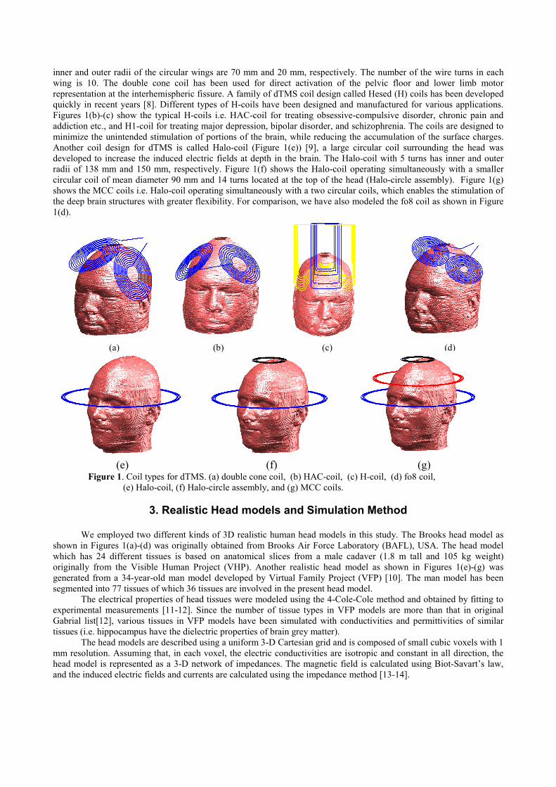
inner and outer radii of the circular wings are 70 mm and 20 mm, respectively. The number of the wire turns in each wing is 10. The double cone coil has been used for direct activation of the pelvic floor and lower limb motor representation at the interhemispheric fissure. A family of dTMS coil design called Hesed (H) coils has been developed quickly in recent years [8]. Different types of H-coils have been designed and manufactured for various applications. Figures 1(b)-(c) show the typical H-coils i.e. HAC-coil for treating obsessive-compulsive disorder, chronic pain and addiction etc., and H1-coil for treating major depression, bipolar disorder, and schizophrenia. The coils are designed to minimize the unintended stimulation of portions of the brain, while reducing the accumulation of the surface charges. Another coil design for dTMS is called Halo-coil (Figure 1(e)) [9], a large circular coil surrounding the head was developed to increase the induced electric fields at depth in the brain. The Halo-coil with 5 turns has inner and outer radii of 138 mm and 150 mm, respectively. Figure 1(f) shows the Halo-coil operating simultaneously with a smaller circular coil of mean diameter 90 mm and 14 turns located at the top of the head (Halo-circle assembly). Figure 1(g) shows the MCC coils i.e. Halo-coil operating simultaneously with a two circular coils, which enables the stimulation of the deep brain structures with greater flexibility. For comparison, we have also modeled the fo8 coil as shown in Figure 1(d).
(a) (b) (c) (d) (e) (f) (g) Figure 1. Coil types for dTMS. (a) double cone coil, (b) HAC-coil, (c) H-coil, (d) fo8 coil,
(e) Halo-coil, (f) Halo-circle assembly, and (g) MCC coils.
3. Realistic Head models and Simulation Method
We employed two different kinds of 3D realistic human head models in this study. The Brooks head model as shown in Figures 1(a)-(d) was originally obtained from Brooks Air Force Laboratory (BAFL), USA. The head model which has 24 different tissues is based on anatomical slices from a male cadaver (1.8 m tall and 105 kg weight) originally from the Visible Human Project (VHP). Another realistic head model as shown in Figures 1(e)-(g) was generated from a 34-year-old man model developed by Virtual Family Project (VFP) [10]. The man model has been segmented into 77 tissues of which 36 tissues are involved in the present head model.
The electrical properties of head tissues were modeled using the 4-Cole-Cole method and obtained by fitting to experimental measurements [11-12]. Since the number of tissue types in VFP models are more than that in original Gabrial list[12], various tissues in VFP models have been simulated with conductivities and permittivities of similar tissues (i.e. hippocampus have the dielectric properties of brain grey matter).
The head models are described using a uniform 3-D Cartesian grid and is composed of small cubic voxels with 1 mm resolution. Assuming that, in each voxel, the electric conductivities are isotropic and constant in all direction, the head model is represented as a 3-D network of impedances. The magnetic field is calculated using Biot-Savart’s law, and the induced electric fields and currents are calculated using the impedance method [13-14].
4. Results and Discussions
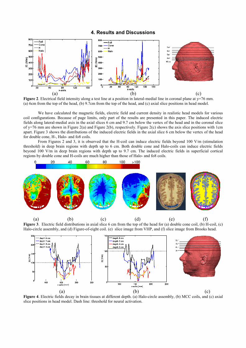
(a) (b) (c) Figure 2. Electrical field intensity along a test line at a position in lateral-medial line in coronal plane at y=76 mm. (a) 6cm from the top of the head, (b) 9.7cm from the top of the head, and (c) axial slice positions in head model.
We have calculated the magnetic fields, electric field and current density in realistic head models for various
coil configurations. Because of page limits, only part of the results are presented in this paper. The induced electric fields along lateral-medial axis in the axial slices 6 cm and 9.7 cm below the vertex of the head and in the coronal slice of y=76 mm are shown in Figure 2(a) and Figure 2(b), respectively. Figure 2(c) shows the axis slice positions with 1cm apart. Figure 3 shows the distributions of the induced electric fields in the axial slice 6 cm below the vertex of the head for double cone, H-, Halo- and fo8 coils.
From Figures 2 and 3, it is observed that the H-coil can induce electric fields beyond 100 V/m (stimulation threshold) in deep brain regions with depth up to 6 cm. Both double cone and Halo-coils can induce electric fields beyond 100 V/m in deep brain regions with depth up to 9.7 cm. The induced electric fields in superficial cortical regions by double cone and H-coils are much higher than those of Halo- and fo8 coils.
(a) (b) (c) (d) (e) (f)
Figure 3. Electric field distributions in axial slice 6 cm from the top of the head for (a) double cone coil, (b) H-coil, (c) Halo-circle assembly, and (d) Figure-of-eight coil. (e) slice image from VHP, and (f) slice image from Brooks head. (a) (b) (c) Figure 4. Electric fields decay in brain tissues at different depth. (a) Halo-circle assembly, (b) MCC coils, and (c) axial slice positions in head model. Dash line: threshold for neural activation.
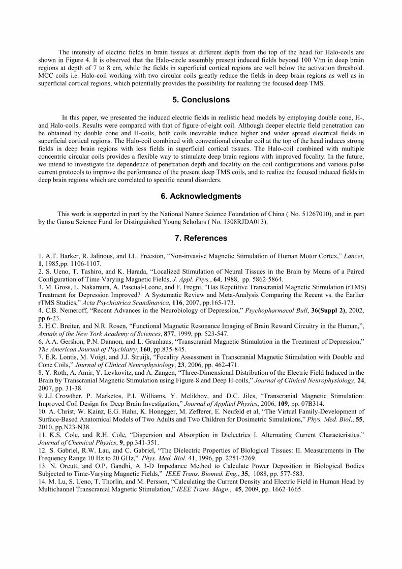
The intensity of electric fields in brain tissues at different depth from the top of the head for Halo-coils are shown in Figure 4. It is observed that the Halo-circle assembly present induced fields beyond 100 V/m in deep brain regions at depth of 7 to 8 cm, while the fields in superficial cortical regions are well below the activation threshold. MCC coils i.e. Halo-coil working with two circular coils greatly reduce the fields in deep brain regions as well as in superficial cortical regions, which potentially provides the possibility for realizing the focused deep TMS.
5. Conclusions
In this paper, we presented the induced electric fields in realistic head models by employing double cone, H-, and Halo-coils. Results were compared with that of figure-of-eight coil. Although deeper electric field penetration can be obtained by double cone and H-coils, both coils inevitable induce higher and wider spread electrical fields in superficial cortical regions. The Halo-coil combined with conventional circular coil at the top of the head induces strong fields in deep brain regions with less fields in superficial cortical tissues. The Halo-coil combined with multiple concentric circular coils provides a flexible way to stimulate deep brain regions with improved focality. In the future, we intend to investigate the dependence of penetration depth and focality on the coil configurations and various pulse current protocols to improve the performance of the present deep TMS coils, and to realize the focused induced fields in deep brain regions which are correlated to specific neural disorders.
6. Acknowledgments
This work is supported in part by the National Nature Science Foundation of China ( No. 51267010), and in part by the Gansu Science Fund for Distinguished Young Scholars ( No. 1308RJDA013).
7. References
1. A.T. Barker, R. Jalinous, and I.L. Freeston, “Non-invasive Magnetic Stimulation of Human Motor Cortex,” Lancet, 1, 1985,pp. 1106-1107. 2. S. Ueno, T. Tashiro, and K. Harada, “Localized Stimulation of Neural Tissues in the Brain by Means of a Paired Configuration of Time-Varying Magnetic Fields, J. Appl. Phys., 64, 1988, pp. 5862-5864. 3. M. Gross, L. Nakamura, A. Pascual-Leone, and F. Fregni, “Has Repetitive Transcranial Magnetic Stimulation (rTMS) Treatment for Depression Improved? A Systematic Review and Meta-Analysis Comparing the Recent vs. the Earlier rTMS Studies,” Acta Psychiatrica Scandinavica, 116, 2007, pp.165-173. 4. C.B. Nemeroff, “Recent Advances in the Neurobiology of Depression,” Psychopharmacol Bull, 36(Suppl 2), 2002, pp.6-23. 5. H.C. Breiter, and N.R. Rosen, “Functional Magnetic Resonance Imaging of Brain Reward Circuitry in the Human,”, Annals of the New York Academy of Sciences, 877, 1999, pp. 523-547. 6. A.A. Gershon, P.N. Dannon, and L. Grunhaus, “Transcranial Magnetic Stimulation in the Treatment of Depression,” The American Journal of Psychiatry, 160, pp.835-845. 7. E.R. Lontis, M. Voigt, and J.J. Struijk, “Focality Assessment in Transcranial Magnetic Stimulation with Double and Cone Coils,” Journal of Clinical Neurophysiology, 23, 2006, pp. 462-471. 8. Y. Roth, A. Amir, Y. Levkovitz, and A. Zangen, “Three-Dimensional Distribution of the Electric Field Induced in the Brain by Transcranial Magnetic Stimulation using Figure-8 and Deep H-coils,” Journal of Clinical Neurophysiology, 24, 2007, pp. 31-38. 9. J.J. Crowther, P. Marketos, P.I. Williams, Y. Melikhov, and D.C. Jiles, “Transcranial Magnetic Stimulation: Improved Coil Design for Deep Brain Investigation,” Journal of Applied Physics, 2006, 109, pp. 07B314. 10. A. Christ, W. Kainz, E.G. Hahn, K. Honegger, M. Zefferer, E. Neufeld et al, “The Virtual Family-Development of Surface-Based Anatomical Models of Two Adults and Two Children for Dosimetric Simulations,” Phys. Med. Biol., 55, 2010, pp.N23-N38. 11. K.S. Cole, and R.H. Cole, “Dispersion and Absorption in Dielectrics I. Alternating Current Characteristics.” Journal of Chemical Physics, 9, pp.341-351. 12. S. Gabriel, R.W. Lau, and C. Gabriel, “The Dielectric Properties of Biological Tissues: II. Measurements in The Frequency Range 10 Hz to 20 GHz,” Phys. Med. Biol. 41, 1996, pp. 2251-2269. 13. N. Orcutt, and O.P. Gandhi, A 3-D Impedance Method to Calculate Power Deposition in Biological Bodies Subjected to Time-Varying Magnetic Fields,” IEEE Trans. Biomed. Eng., 35, 1088, pp. 577-583. 14. M. Lu, S. Ueno, T. Thorlin, and M. Persson, “Calculating the Current Density and Electric Field in Human Head by Multichannel Transcranial Magnetic Stimulation,” IEEE Trans. Magn., 45, 2009, pp. 1662-1665.