effect of repeated transcranial magnetic stimulation
TRANSCRIPT
Keywords:
Epilepsy;
Kindling;
Transcranial magnetic
stimulation;
Action potentials;
Received:
7 Aug 2014
Accepted:
6 Jan 2015
*Correspondence to:
J. Mirnajafi-Zadeh
Tel:
021-82883865
Fax:
021-82884555
Email:
Original Article
Effect of repeated transcranial magnetic stimulation during epileptogenesis on spontaneous activity of hippocampal CA1 pyramidal neurons in rats
Amir Shojaei1, Saeed Semnanian1, Mahyar Janahmadi2, Homeira Moradi1, Seyed Mohammad Firoozabadi3,
Javad Mirnajafi-Zadeh1*
1. Department of Physiology, Faculty of Medical Sciences, Tarbiat Modares University, Tehran, Iran
2. Department of Physiology, Faculty of Medicine, Shahid Beheshti University of Medical Sciences, Tehran, Iran
3. Department of Medical Physics, Faculty of Medical Sciences, Tarbiat Modares University, Tehran, Iran
Introduction
Epilepsy is a chronic brain disorder in which a person
has repeated seizures over time. It affects millions of
people worldwide and has remained one of the most
common neurological conditions (Shorvon, 1996).
Despite many advances in epilepsy research, seizures
fail to come under control in many patients with
epilepsy (20 to 40% of the overall epileptic population)
(Leppik, 1992, Sander, 1993, Sillanpaa and Schmidt,
2006). Temporal lobe epilepsy is the most common
form of human epilepsy with intractable seizures
originating from the medial or lateral temporal lobe
Physiology and
Pharmacology
Physiol Pharmacol 19 (2015) 1-13 www.phypha.ir/ppj
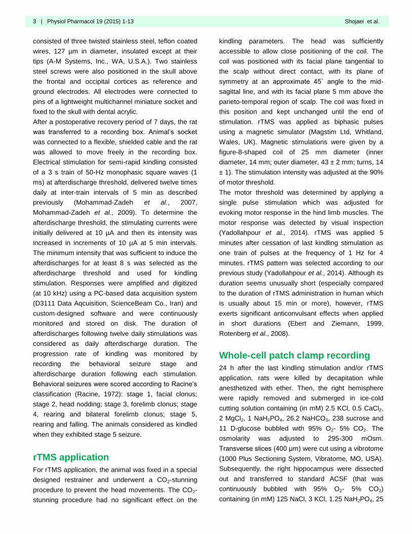
Abstract Introduction: Considering the antiepileptogenic effects of repeated transcranial magnetic
stimulation (rTMS), the effect of rTMS applied during amygdala kindling on spontaneous activity
of hippocampal CA1 pyramidal neurons was investigated.
Materials and Methods: A tripolar electrode was inserted in basolateral amygdala of Male Wistar
rats. After a recovery period, animals received daily kindling stimulations until they reached stage
5 seizure. In one group of animals, rTMS at frequency of 1 Hz were applied to hippocampus once
daily at 5 min after termination of kindling stimulations. 24 h after the last kindling stimulation,
spontaneous activity of CA1 pyramidal neurons of the hippocampus was investigated using whole
cell patch clamp technique.
Results: Kindling-induced seizures resulted in increment of spontaneous activity of hippocampal
CA1 neurons, but application of rTMS during amygdala kindling prevented it. Moreover, rTMS
administration inhibited the kindling-induced enhancement of afterdepolarization (ADP) amplitude
and action potential duration.
Conclusion: Results of this study suggest that rTMS exerts its anticonvulsant effect, in part,
through preventing the amygdala kindling-induced increase in spontaneous activity and
excitability of hippocampal CA1 pyramidal neurons.
Antiepileptogenic effect of rTMS in kindling Physiol Pharmacol 19 (2015) 1- 13 | 2
(Devinsky, 1991). Surgical removal of epileptogenic
tissue, particularly the hippocampus, often results in
pharmacologically controlled seizures after surgery
(Wiebe et al., 2001, Spencer, 2002, Spencer and
Burchiel, 2012), implying that the hippocampus plays a
crucial role in the drug-resistant temporal lobe
epilepsy.
The failure of current therapies in controlling the
refractory epileptic seizures highlights the need for
new, effective, and safe treatments. Transcranial
magnetic stimulation (TMS) is a novel technique for
noninvasive stimulation of brain through intact scalp
(Barker et al., 1985). It is a widely used method in
research of human brain physiology and a therapeutic
tool for some drug-resistant neuropsychiatric disorders
(Dayan et al., 2013).
It has been postulated that low frequency repetitive
TMS (rTMS; ≤2 Hz) decreases, while high frequency
rTMS (≥5 Hz) increases the cortical excitability
(Pascual-Leone et al., 1994, Chen et al., 1997, Tergau
et al., 1997, Kim et al., 2004). Furthermore, low
frequency rTMS has been shown to have diverse
degrees of seizure control in both epileptic patients
(Tergau et al., 1999, Menkes and Gruenthal, 2000,
Theodore et al., 2002, Misawa et al., 2005, Fregni et
al., 2006, Joo et al., 2007, Santiago-Rodriguez et al.,
2008) and in laboratory animals (Ebert and Ziemann,
1999, Fleischmann et al., 1999, Rotenberg et al.,
2008, Mongabadi et al., 2013). Focal epilepsies are
particularly ideal for the application of rTMS due to a
delimited zone of increased excitability (Santiago-
Rodriguez et al., 2008).
Considering the ability of rTMS as a potential
therapeutic manner for epileptic patients, it is
necessary to explore neuronal effects associated with
its anti-epileptic outcome. Understanding these effects
will also be useful in determining the probable side
effects following application of rTMS. It has been
suggested that long-term depression- or
depotentiation-like mechanisms may be responsible
for rTMS effects (Chen et al., 1997, Hallett, 2000,
Kobayashi and Pascual-Leone, 2003, Tokay et al.,
2009, Dayan et al., 2013).
Kindling is a widely studied animal model of temporal
lobe epilepsy in which repetitive focal electrical
stimulation of the brain induces progressive seizure
activity culminates in tonic-clonic convulsion (Sutula,
1990). Our recent study showed that application of
rTMS can prevent kindling-induced synaptic
potentiation (Yadollahpour et al., 2014). It has been
shown that synaptic potentiation is accompanied with
changes in electrophysiological properties of neurons
(Hallett et al., 1999, Wagner et al., 2009, Pell et al.,
2011). These changes can be observed in areas
involved in seizure generation or propagation of
amygdala kindling, including the hippocampus.
Previous studies showed that CA1 region of the
hippocampus is among the most important areas
involved in the propagation of amygdala kindling-
induced seizures (Dasheiff and McNamara, 1982,
Mirnajafi-Zadeh et al., 2002).
Regarding the inhibitory effect of rTMS on
epileptogenesis and considering the important role of
CA1 pyramidal neurons in spreading of the amygdala
kindling-induced seizures, in the present study, we
investigated whether application of rTMS during
amygdala kindling procedure can prevent the kindling-
induced changes in electrophysiological properties of
hippocampal CA1 pyramidal neurons during amygdala
kindling.
Materials and methods
Animals
Adult male Wistar rats (1 month old) obtained from
Pasteur institute of Iran were maintained in a colony
room kept at 23±2°C temperature on 12:12 light:dark
schedule (lights on at 7:00 A.M) and permitted free
access to food and water. All experiments were
performed in accordance with the ethical guidelines set
by the “Ethical Committee of Faculty of Medical
Sciences, Tarbiat Modares University”
Amygdala semi-rapid kindling
Animals underwent stereotaxical surgery under
ketamine/xylazine mixture (100/10 mg/kg, i.p.)
anesthesia and were chronically implanted with a
tripolar electrode in the basolateral amygdala of right
hemisphere (coordinates: A, 2.5 mm; L, 4.8 mm from
Bregma and 7.5 mm below dura) according to the atlas
of Paxinos and Watson (2006). The electrode
3 | Physiol Pharmacol 19 (2015) 1-13 Shojaei et al.
consisted of three twisted stainless steel, teflon coated
wires, 127 µm in diameter, insulated except at their
tips (A-M Systems, Inc., WA, U.S.A.). Two stainless
steel screws were also positioned in the skull above
the frontal and occipital cortices as reference and
ground electrodes. All electrodes were connected to
pins of a lightweight multichannel miniature socket and
fixed to the skull with dental acrylic.
After a postoperative recovery period of 7 days, the rat
was transferred to a recording box. Animal’s socket
was connected to a flexible, shielded cable and the rat
was allowed to move freely in the recording box.
Electrical stimulation for semi-rapid kindling consisted
of a 3 s train of 50-Hz monophasic square waves (1
ms) at afterdischarge threshold, delivered twelve times
daily at inter-train intervals of 5 min as described
previously (Mohammad-Zadeh et al., 2007,
Mohammad-Zadeh et al., 2009). To determine the
afterdischarge threshold, the stimulating currents were
initially delivered at 10 µA and then its intensity was
increased in increments of 10 µA at 5 min intervals.
The minimum intensity that was sufficient to induce the
afterdischarges for at least 8 s was selected as the
afterdischarge threshold and used for kindling
stimulation. Responses were amplified and digitized
(at 10 kHz) using a PC-based data acquisition system
(D3111 Data Aqcuisition, ScienceBeam Co., Iran) and
custom-designed software and were continuously
monitored and stored on disk. The duration of
afterdischarges following twelve daily stimulations was
considered as daily afterdischarge duration. The
progression rate of kindling was monitored by
recording the behavioral seizure stage and
afterdischarge duration following each stimulation.
Behavioral seizures were scored according to Racine’s
classification (Racine, 1972): stage 1, facial clonus;
stage 2, head nodding; stage 3, forelimb clonus; stage
4, rearing and bilateral forelimb clonus; stage 5,
rearing and falling. The animals considered as kindled
when they exhibited stage 5 seizure.
rTMS application
For rTMS application, the animal was fixed in a special
designed restrainer and underwent a CO2-stunning
procedure to prevent the head movements. The CO2-
stunning procedure had no significant effect on the
kindling parameters. The head was sufficiently
accessible to allow close positioning of the coil. The
coil was positioned with its facial plane tangential to
the scalp without direct contact, with its plane of
symmetry at an approximate 45˚ angle to the mid-
sagittal line, and with its facial plane 5 mm above the
parieto-temporal region of scalp. The coil was fixed in
this position and kept unchanged until the end of
stimulation. rTMS was applied as biphasic pulses
using a magnetic simulator (Magstim Ltd, Whitland,
Wales, UK). Magnetic stimulations were given by a
figure-8-shaped coil of 25 mm diameter (inner
diameter, 14 mm; outer diameter, 43 ± 2 mm; turns, 14
± 1). The stimulation intensity was adjusted at the 90%
of motor threshold.
The motor threshold was determined by applying a
single pulse stimulation which was adjusted for
evoking motor response in the hind limb muscles. The
motor response was detected by visual inspection
(Yadollahpour et al., 2014). rTMS was applied 5
minutes after cessation of last kindling stimulation as
one train of pulses at the frequency of 1 Hz for 4
minutes. rTMS pattern was selected according to our
previous study (Yadollahpour et al., 2014). Although its
duration seems unusually short (especially compared
to the duration of rTMS administration in human which
is usually about 15 min or more), however, rTMS
exerts significant anticonvulsant effects when applied
in short durations (Ebert and Ziemann, 1999,
Rotenberg et al., 2008).
Whole-cell patch clamp recording
24 h after the last kindling stimulation and/or rTMS
application, rats were killed by decapitation while
anesthetized with ether. Then, the right hemisphere
were rapidly removed and submerged in ice-cold
cutting solution containing (in mM) 2.5 KCl, 0.5 CaCl2,
2 MgCl2, 1 NaH2PO4, 26.2 NaHCO3, 238 sucrose and
11 D-glucose bubbled with 95% O2- 5% CO2. The
osmolarity was adjusted to 295-300 mOsm.
Transverse slices (400 μm) were cut using a vibrotome
(1000 Plus Sectioning System, Vibratome, MO, USA).
Subsequently, the right hippocampus were dissected
out and transferred to standard ACSF (that was
continuously bubbled with 95% O2- 5% CO2)
containing (in mM) 125 NaCl, 3 KCl, 1.25 NaH2PO4, 25
Antiepileptogenic effect of rTMS in kindling Physiol Pharmacol 19 (2015) 1- 13 | 4
NaHCO3, 10 D-Glucose, 2 CaCl2, 1.3 MgCl2 . The
osmolality was in the range of 290-300 mOsm and pH
was adjusted to 7.3-7.4 by NaOH 1 M. Slices were
then incubated for 1 h at 35°C and then stored at room
temperature (23–25°C) until they individually
transferred to a submerged recording chamber.
Recording chamber was mounted on a fixed-stage
upright microscope (Axioskop 2 FS MOT; Carl Zeiss,
Germany) and continually were perfused at 1.5-2.5
ml/min with standard ACSF at room temperature (23-
25°C). CA1 pyramidal neurons were visualized using
an IR-CCD camera (IR-1000, MTI, USA) with a 40x
water immersion objective lens. Neuronal somas were
selected for recording based on their relatively
pyramidal shape and smooth, low-contrast
appearance. Whole cell patch clamp recordings were
made under current clamp condition. Recording
microelectrodes (1.5 mm outer diameter, borosilicate
glass, GC150-11; Harvard Apparatus, UK) were pulled
with a horizontal puller (P-97, Sutter Instrument, USA)
and filled with intracellular solution containing (in mM):
115 K-gluconate, 20 KCl, 10 HEPES, 2 EGTA, 10
disodium-phosphocreatine, 2 MgATP and 0.3 NaGTP.
pH was adjusted to 7.25-7.30 and osmolality was in
the range of 285-290 mOsm. Electrode tip resistance
in the bath was typically 4 to 6 MΩ, and series
resistance ranged from 12 to 25 MΩ. Cells were
rejected if series resistance changes were more than
20% during experiment. Capacitance compensation
and bridge balance were carried out. Data were low-
pass filtered at 3 kHz and acquired at 10 kHz with a
Multiclamp 700B amplifier equipped with Digidata 1440
A/D converter (Molecular Devices, CA, USA). The
signal was recorded on a PC for offline analysis using
the Axon pClamp 10 acquisition software. After
establishment of a giga seal (more than 2 GΩ), the
whole-cell configuration was attained simply by
applying a brief suction.
To investigate the effect of daily rTMS treatment during
amygdala kindling on firing properties of CA1
pyramidal neurons, we recorded spontaneous activity
of CA1 pyramidal cells for 5 min in current clamp mode
(I=0) at least 5 min after achieving the whole cell
configuration.
Experimental groups
Following the postoperative recovery period, animals
were randomly allocated into four groups. In kindled
group (n=6), animals received daily kindling
stimulations until they achieved stage 5 seizure. In
kindled+rTMS (KrTMS) group (n=6), daily rTMS was
applied 5 min following termination of kindling
stimulations. The animals in rTMS group (n=3) were
treated with daily rTMS alone. The number of
stimulation days in KrTMS and rTMS groups was
equal to the mean stimulation days required to achieve
a stage 5 seizure in kindled group. Another two
groups, including sham-operated (n=3) and naive
(non-operated) (n=3) rats were also used. As, no
significant difference was found between the data of
these two groups, their data were pooled and
considered as control group (n=6). All of the measured
parameters in whole cell recordings were obtained
from slices derived from the above mentioned
numbers of animals.
Measured parameters
The occurrence of spontaneous action potentials and
their amplitude and half-width, amplitude of after
depolarization (ADP), maximum rise and decay slopes
were examined. Action potential amplitude was
determined as the peak voltage with respect to the
baseline of 10 ms before the peak of the action
potentials. Action potential half-width was computed as
the width of spike at one half of its maximum
amplitude. Maximum rise slope was measured as the
maximum slope of rising phase and maximum decay
slope was calculated as maximum incline of falling
phase of the spike. ADP amplitude was computed as
the peak voltage of ADP with respect to the baseline.
Single spikes were used for determining the action
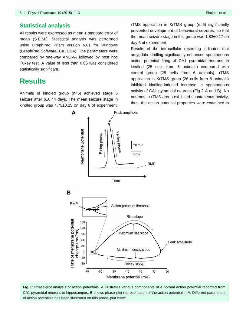
potential characteristics. We also utilized phase-plot
analysis to characterize action potential features
among various experimental groups. In phase-plot
analysis, the rate of change in membrane potential
with respect to time (dV/dt) is plotted as a function of
membrane potential. Various components of action
potentials are clearly reflected in phase-plot curve (Fig.
1).
5 | Physiol Pharmacol 19 (2015) 1-13 Shojaei et al.
Statistical analysis
All results were expressed as mean ± standard error of
mean (S.E.M.). Statistical analysis was performed
using GraphPad Prism version 6.01 for Windows
(GraphPad Software, Ca, USA). The parameters were
compared by one-way ANOVA followed by post hoc
Tukey test. A value of less than 0.05 was considered
statistically significant.
Results
Animals of kindled group (n=6) achieved stage 5
seizure after 6±0.44 days. The mean seizure stage in
kindled group was 4.75±0.25 on day 6 of experiment.
rTMS application in KrTMS group (n=6) significantly
prevented development of behavioral seizures, so that
the mean seizure stage in this group was 1.83±0.17 on
day 6 of experiment.
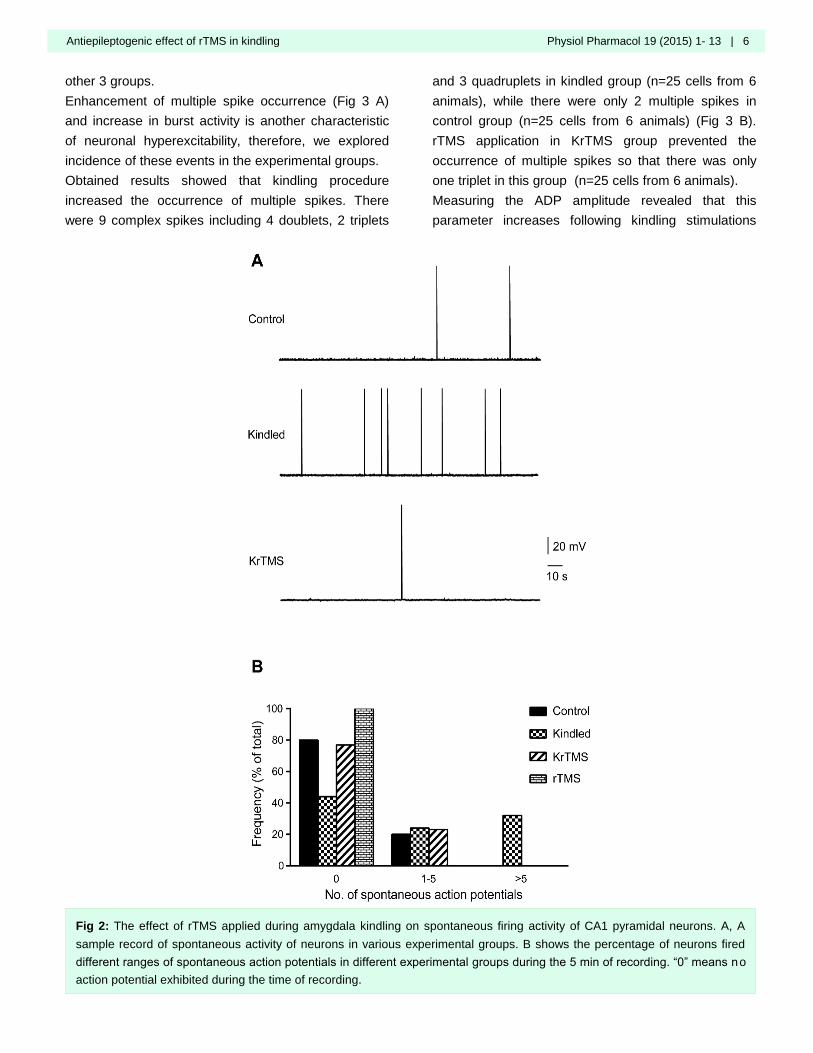
Results of the intracellular recording indicated that
amygdala kindling significantly enhances spontaneous
action potential firing of CA1 pyramidal neurons in
kindled (25 cells from 6 animals) compared with
control group (25 cells from 6 animals). rTMS
application in KrTMS group (26 cells from 6 animals)
inhibited kindling-induced increase in spontaneous
activity of CA1 pyramidal neurons (Fig 2 A and B). No
neurons in rTMS group exhibited spontaneous activity,
thus, the action potential properties were examined in
Fig 1: Phase-plot analysis of action potentials. A illustrates various components of a normal action potential recorded from
CA1 pyramidal neurons in hippocampus. B shows phase-plot representation of the action potential in A. Different parameters
of action potentials has been illustrated on this phase-plot curve.
Antiepileptogenic effect of rTMS in kindling Physiol Pharmacol 19 (2015) 1- 13 | 6
other 3 groups.
Enhancement of multiple spike occurrence (Fig 3 A)
and increase in burst activity is another characteristic
of neuronal hyperexcitability, therefore, we explored
incidence of these events in the experimental groups.
Obtained results showed that kindling procedure
increased the occurrence of multiple spikes. There
were 9 complex spikes including 4 doublets, 2 triplets
and 3 quadruplets in kindled group (n=25 cells from 6
animals), while there were only 2 multiple spikes in
control group (n=25 cells from 6 animals) (Fig 3 B).
rTMS application in KrTMS group prevented the
occurrence of multiple spikes so that there was only
one triplet in this group (n=25 cells from 6 animals).
Measuring the ADP amplitude revealed that this
parameter increases following kindling stimulations
Fig 2: The effect of rTMS applied during amygdala kindling on spontaneous firing activity of CA1 pyramidal neurons. A, A
sample record of spontaneous activity of neurons in various experimental groups. B shows the percentage of neurons fired
different ranges of spontaneous action potentials in different experimental groups during the 5 min of recording. “0” means no
action potential exhibited during the time of recording.

7 | Physiol Pharmacol 19 (2015) 1-13 Shojaei et al.
and there was significant variation in this parameter
among kindled (15.11±1.23 mV, 9 cells from 6
animals) and control (9.54±1.06 mV, 6 cells from 4
animals) groups (p<0.05, Fig 3 C and D). rTMS
administration in KrTMS group (5 cells from 4 animals)
significantly inhibited the kindling-induced increase in
this parameter (6.78±1.62 mV, p<0.01, Fig 3 C and D).
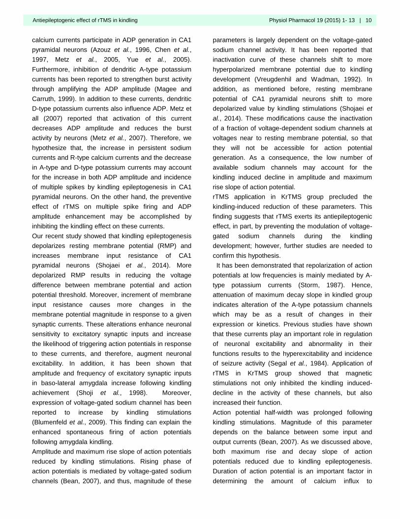
Amygdala kindling significantly decreased action
potential amplitude from 108.7±0.73 mV in control (n=5
cells from 4 animals) to 94.01±3.00 mV in kindled
group (n=11 cells from 6 animals) (Fig 4 A and C).
rTMS application significantly prevented the kindling-
induced reduction of this parameter in KrTMS group
(105.5±3.56 mV; 6 cells from 4 animals; p<0.05
compared to kindled group) (Fig 4 A and C).
Maximum rise slope of action potentials significantly
decreased in response to kindling stimulations in
kindled group (131.2±10.94 mV/ms, n=11 cells from 6
animals) compared to control group (203.3±14.81
mV/ms; 5 cells from 4 animals, p<0.01) (Fig 4 B and
D). rTMS treatment inhibited the decrease in this
parameter so that a significant difference was
observed between KrTMS (232.1±20.42 mV/ms, n=5
cells from 4 animals) and kindled group (p<0.001) (Fig
4 B and D).
A significant decrease was also observed in maximum
decay slope of action potentials in kindled
(-34.16±1.86 mV/ms, 10 cells from 6 animals)
compared to control (-43.34±3.20 mV/ms, 5 cells from
3 animals) group (p<0.05). rTMS application in KrTMS
group (-57.58±2.04, 6 cells from 4 animals) not only
significantly precluded kindling-induced reduction of
this parameter (p<0.001 compared to kindled), but also
increased this parameter compared to control group
(p<0.01, Fig 4 B and E).
As a result of kindling stimulations, action potential
half-width was significantly (p<0.01) prolonged in
neurons from kindled group (2.70±0.10 ms; 13 cells
Fig 3: Effects of rTMS application on incidence of multiple spike discharge and ADP amplitude during the kindling acquisition
in CA1 pyramidal neurons. A) Representative samples of duplet, triplet and quadruplet spikes recorded in this study. B)
Frequency of multiple spikes incidence among various experimental groups. C) Comparing samples of ADPs recorded in
different experimental groups. D) Quantitative comparison of ADP amplitude. *p<0.05 compared to control and ++ p<0.01
compared to kindled, mean±SEM.
Antiepileptogenic effect of rTMS in kindling Physiol Pharmacol 19 (2015) 1- 13 | 8
from 6 animals) compared to that of control group
(2.17±0.14 ms; 5 cells from 4 animals). This parameter
was significantly prevented to be lengthened in KrTMS
group (1.71±0.04 ms; 6 cells from 4 animals, p<0.001
compared to kindled group) (Fig 4 B and F).
As fig 5 indicates, phase plot representation of action potentials in kindled group showed distinct differences compared to that of control, reflecting the decrease in
Fig 4: Effects of rTMS treatment following kindling stimulations on properties of spontaneous action potentials in CA1
pyramidal neurons. A) Representative traces of action potentials recorded form various experimental groups. The dashed line
shows the spike amplitude of kindled group to simplify comparing the amplitude between different groups. B) Superimposed
typical traces of action potentials in CA1 pyramidal neurons. D-F show, respectively, quantitative comparison of amplitude,
maximum rise slope, maximum decay slope and half-width of action potentials in different groups. *p<0.05 and ** p<0.01
compared to control; +p<0.05 and +++p<0.001 compared to kindled; mean±SEM.

9 | Physiol Pharmacol 19 (2015) 1-13 Shojaei et al.
amplitude, maximum rise slope and maximum decay
slope of action potential following kindling stimulations.
Interestingly, action potential phase plot deflection in
KrTMS group was almost similar to that of the control
group, indicating application of rTMS prevented
kindling-induced changes in action potential dynamics.
Discussion
Results of the present study provided direct cellular
evidence that rTMS application prevents kindling-
induced changes in spontaneous activity of CA1
pyramidal neurons. These results are consistent with
our previous study which showed that administration of
low frequency rTMS during kindling process has an
inhibitory effect on epileptogenesis (Mongabadi et al.,
2013, Yadollahpour et al., 2014) accompanied with the
suppression of kindling-induced potentiation of
perforant path-evoked population spikes in the dentate
gyrus of awake rats (Yadollahpour et al., 2014).
Amygdala kindling enhanced the excitability of CA1
pyramidal neurons as evidenced by an increase in the
spontaneous action potential discharges. This
parameter is dependent on both neuronal intrinsic ionic
conductance and excitatory and/or inhibitory synaptic
inputs (Kaczmarek and Levitan, 1987, Urban et al.,
2012). rTMS application suppressed the excitatory
effect of kindling on spontaneous activity. This
inhibitory effect may be resulted from magnetic
stimulations impact on intrinsic ionic conductance of
CA1 pyramidal neurons or because of preventive
effect on kindling-induced changes of synaptic inputs
activity (Kaczmarek and Levitan, 1987), so that rTMS
reduced the neuronal excitability. Similar mechanisms
are likely involved in rTMS group resulting in no spike
firing.
Occurrence of multiple spikes was enhanced by
kindling stimulations. Although these spikes are
normally observed in spontaneous activity of CA1
pyramidal neurons and they have an important role in
learning and memory processes by hippocampus
(O'Keefe, 1976, Otto et al., 1991), but the increase in
frequency of these events is due to kindling-induced
hyperexcitability of neurons. Incidence of these events
in CA1 pyramidal neurons is attributed to ADP that
follows each spikes (Azouz et al., 1996). When the
amplitude of ADP is large enough to reach the action
potential threshold, it triggers spike(s) (Azouz et al.,
1996). These multiple spikes increase neurotransmitter
release from presynaptic terminals, and then amplify
output signals from the neurons (Borst and Sakmann,
1999). In the present study, ADP amplitude was
significantly increased by kindling epileptogenesis and
it can be considered as the main cause for the
increase in the number of multiple spikes. It has been
shown that persistent sodium currents and R-type
Fig 5: Comparing the phase-plot representative of action potentials in different experimental groups. As can be observed in
this figure, rTMS application almost prevented kindling-induced changes in action potential dynamics.
Antiepileptogenic effect of rTMS in kindling Physiol Pharmacol 19 (2015) 1- 13 | 10
calcium currents participate in ADP generation in CA1
pyramidal neurons (Azouz et al., 1996, Chen et al.,
1997, Metz et al., 2005, Yue et al., 2005).
Furthermore, inhibition of dendritic A-type potassium
currents has been reported to strengthen burst activity
through amplifying the ADP amplitude (Magee and
Carruth, 1999). In addition to these currents, dendritic
D-type potassium currents also influence ADP. Metz et
all (2007) reported that activation of this current
decreases ADP amplitude and reduces the burst
activity by neurons (Metz et al., 2007). Therefore, we
hypothesize that, the increase in persistent sodium
currents and R-type calcium currents and the decrease
in A-type and D-type potassium currents may account
for the increase in both ADP amplitude and incidence
of multiple spikes by kindling epileptogenesis in CA1
pyramidal neurons. On the other hand, the preventive
effect of rTMS on multiple spike firing and ADP
amplitude enhancement may be accomplished by
inhibiting the kindling effect on these currents.
Our recent study showed that kindling epileptogenesis
depolarizes resting membrane potential (RMP) and
increases membrane input resistance of CA1
pyramidal neurons (Shojaei et al., 2014). More
depolarized RMP results in reducing the voltage
difference between membrane potential and action
potential threshold. Moreover, increment of membrane
input resistance causes more changes in the
membrane potential magnitude in response to a given
synaptic currents. These alterations enhance neuronal
sensitivity to excitatory synaptic inputs and increase
the likelihood of triggering action potentials in response
to these currents, and therefore, augment neuronal
excitability. In addition, it has been shown that
amplitude and frequency of excitatory synaptic inputs
in baso-lateral amygdala increase following kindling
achievement (Shoji et al., 1998). Moreover,
expression of voltage-gated sodium channel has been
reported to increase by kindling stimulations
(Blumenfeld et al., 2009). This finding can explain the
enhanced spontaneous firing of action potentials
following amygdala kindling.
Amplitude and maximum rise slope of action potentials
reduced by kindling stimulations. Rising phase of
action potentials is mediated by voltage-gated sodium
channels (Bean, 2007), and thus, magnitude of these
parameters is largely dependent on the voltage-gated
sodium channel activity. It has been reported that
inactivation curve of these channels shift to more
hyperpolarized membrane potential due to kindling
development (Vreugdenhil and Wadman, 1992). In
addition, as mentioned before, resting membrane
potential of CA1 pyramidal neurons shift to more
depolarized value by kindling stimulations (Shojaei et
al., 2014). These modifications cause the inactivation
of a fraction of voltage-dependent sodium channels at
voltages near to resting membrane potential, so that
they will not be accessible for action potential
generation. As a consequence, the low number of
available sodium channels may account for the
kindling induced decline in amplitude and maximum
rise slope of action potential.
rTMS application in KrTMS group precluded the
kindling-induced reduction of these parameters. This
finding suggests that rTMS exerts its antiepileptogenic
effect, in part, by preventing the modulation of voltage-
gated sodium channels during the kindling
development; however, further studies are needed to
confirm this hypothesis.
It has been demonstrated that repolarization of action
potentials at low frequencies is mainly mediated by A-
type potassium currents (Storm, 1987). Hence,
attenuation of maximum decay slope in kindled group
indicates alteration of the A-type potassium channels
which may be as a result of changes in their
expression or kinetics. Previous studies have shown
that these currents play an important role in regulation
of neuronal excitability and abnormality in their
functions results to the hyperexcitability and incidence
of seizure activity (Segal et al., 1984). Application of
rTMS in KrTMS group showed that magnetic
stimulations not only inhibited the kindling induced-
decline in the activity of these channels, but also
increased their function.
Action potential half-width was prolonged following
kindling stimulations. Magnitude of this parameter
depends on the balance between some input and
output currents (Bean, 2007). As we discussed above,
both maximum rise and decay slope of action
potentials reduced due to kindling epileptogenesis.
Duration of action potential is an important factor in
determining the amount of calcium influx to
11 | Physiol Pharmacol 19 (2015) 1-13 Shojaei et al.
presynaptic terminal (Borst and Sakmann, 1999, Bean,
2007). It is believed that the 4th power of calcium
concentration in presynaptic terminal is translated to
the amount of neurotransmitter release (Borst and
Sakmann, 1999, Dayan et al., 2013). Broadening the
action potentials, especially at high frequencies such
as during seizure activity, heightens the calcium influx
to the presynaptic terminal and cause a several-fold
increase in neurotransmitter release (Borst and
Sakmann, 1999). These alterations can facilitate
propagation of amygdala-kindled seizures through the
hippocampus. Interestingly, rTMS application inhibited
the increase in action potential half-width following
kindling stimulations. This finding suggests that rTMS
treatment may exert its anti-epileptogenic effect by
prevention of broadening the action potentials. Results
of phase-plot analysis revealed that action potential
dynamics is altered due to propagation of seizures.
However, rTMS application prevents the kindling-
induced changes in characteristics of spikes, giving
them features that are close to normal action
potentials.
In summary, finding of the present study provide direct
electrophysiological evidences demonstrating rTMS
application prevents amygdala kindling-induced
changes in spontaneous activity of CA1 pyramidal
neurons. Results of this research are in agreement
with the studies which introduce rTMS as a therapeutic
tool for control of seizures.
Acknowledgments
This study is part of the PhD thesis of the first author
in physiology that has completely performed in
physiology department of Tarbiat Modares University.
This research was supported by grants from Iran
National Sciences Foundation (INSF) (Grant #
92040251).
Conflict of interest
All authors declared that there is no conflict of
interest.
References
Azouz R, Jensen MS, Yaari Y. Ionic basis of spike after-
depolarization and burst generation in adult rat
hippocampal CA1 pyramidal cells. J Physiol 1996; 492:
211-23.
Barker AT, Jalinous R, Freeston IL. Non-invasive magnetic
stimulation of human motor cortex. Lancet 1985; 1: 1106-7.
Bean BP. The action potential in mammalian central
neurons. Nat Rev Neurosci 2007; 8: 451-65.
Blumenfeld H, Lampert A, Klein JP, Mission J, Chen MC,
Rivera M, et al. Role of hippocampal sodium channel
Nav1.6 in kindling epileptogenesis. Epilepsia 2009; 50: 44-
55.
Borst JG, Sakmann B. Effect of changes in action potential
shape on calcium currents and transmitter release in a
calyx-type synapse of the rat auditory brainstem. Philos
Trans R Soc Lond B Biol Sci 1999; 354: 347-55.
Chen R, Classen J, Gerloff C, Celnik P, Wassermann EM,
Hallett M, et al. Depression of motor cortex excitability by
low-frequency transcranial magnetic stimulation. Neurology
1997; 48: 1398-403.
Dasheiff RM, McNamara JO. Intradentate colchicine retards
the development of amygdala kindling. Ann Neurol 1982;
11: 347-52.
Dayan E, Censor N, Buch ER, Sandrini M, Cohen LG.
Noninvasive brain stimulation: from physiology to network
dynamics and back. Nat Neurosci 2013; 16: 838-44.
Devinsky O. Interictal behavioral changes in epilepsy. In:
Devinsky O, Theodore WH, editors. Epilepsy and behavior.
New York: Wiley-Liss; 1991. p. 1–21.
Ebert U, Ziemann U. Altered seizure susceptibility after high-
frequency transcranial magnetic stimulation in rats.
Neurosci Lett 1999; 273: 155-8.
Fleischmann A, Hirschmann S, Dolberg OT, Dannon PN,
Grunhaus L. Chronic treatment with repetitive transcranial
magnetic stimulation inhibits seizure induction by
electroconvulsive shock in rats. Biol Psychiatry 1999; 45:
759-63.
Fregni F, Otachi PT, Do Valle A, Boggio PS, Thut G,
Rigonatti SP, et al. A randomized clinical trial of repetitive
transcranial magnetic stimulation in patients with refractory
epilepsy. Ann Neurol 2006; 60: 447-55.
Hallett M. Transcranial magnetic stimulation and the human
brain. Nature 2000; 406: 147-50.
Hallett M, Wassermann EM, Pascual-Leone A, Valls-Sole J.
Repetitive transcranial magnetic stimulation. The
International Federation of Clinical Neurophysiology.
Electroencephalogr Clin Neurophysiol Suppl 1999; 52:
105-13.
Joo EY, Han SJ, Chung SH, Cho JW, Seo DW, Hong SB.
Antiepileptic effects of low-frequency repetitive transcranial
magnetic stimulation by different stimulation durations and
locations. Clin Neurophysiol 2007; 118: 702-8.
Kaczmarek LK, Levitan IB. Neuromodulation: the
biochemical control of neuronal excitability. USA: Oxford
University Press; 1987.
Antiepileptogenic effect of rTMS in kindling Physiol Pharmacol 19 (2015) 1- 13 | 12
Kim YH, Park JW, Ko MH, Jang SH, Lee PK. Facilitative
effect of high frequency subthreshold repetitive transcranial
magnetic stimulation on complex sequential motor learning
in humans. Neurosci Lett 2004; 367: 181-5.
Kobayashi M, Pascual-Leone A. Transcranial magnetic
stimulation in neurology. Lancet Neurol 2003; 2: 145-56.
Leppik IE. Intractable epilepsy in adults. Epilepsy Res Suppl
1992; 5: 7-11.
Magee JC, Carruth M. Dendritic voltage-gated ion channels
regulate the action potential firing mode of hippocampal
CA1 pyramidal neurons. J Neurophysiol 1999; 82: 1895-
901.
Menkes DL, Gruenthal M. Slow-frequency repetitive
transcranial magnetic stimulation in a patient with focal
cortical dysplasia. Epilepsia 2000; 41: 240-2.
Metz AE, Jarsky T, Martina M, Spruston N. R-type calcium
channels contribute to afterdepolarization and bursting in
hippocampal CA1 pyramidal neurons. J Neurosci 2005; 25:
5763-73.
Metz AE, Spruston N, Martina M. Dendritic D-type potassium
currents inhibit the spike afterdepolarization in rat
hippocampal CA1 pyramidal neurons. J Physiol 2007; 581:
175-87.
Mirnajafi-Zadeh J, Mortazavi M, Fathollahi Y, Alasvand
Zarasvand M, Reza Palizvan M. Effect of transient
hippocampal inhibition on amygdaloid kindled seizures and
amygdaloid kindling rate. Brain Res 2002; 954: 220-6.
Misawa S, Kuwabara S, Shibuya K, Mamada K, Hattori T.
Low-frequency transcranial magnetic stimulation for
epilepsia partialis continua due to cortical dysplasia. J
Neurol Sci 2005; 234: 37-9.
Mohammad-Zadeh M, Mirnajafi-Zadeh J, Fathollahi Y, Javan
M, Ghorbani P, Sadegh M, et al. Effect of low frequency
stimulation of perforant path on kindling rate and synaptic
transmission in the dentate gyrus during kindling
acquisition in rats. Epilepsy Res 2007; 75: 154-61.
Mohammad-Zadeh M, Mirnajafi-Zadeh J, Fathollahi Y, Javan
M, Jahanshahi A, Noorbakhsh S, et al. The role of
adenosine A1 receptors in mediating the inhibitory effects
of low frequency stimulation of perforant path on kindling
acquisition in rats. Neuroscience 2009; 158: 1632-43.
Mongabadi S, Firoozabadi SM, Javan M, Shojaei A,
Mirnajafi-Zadeh J. Effect of different frequencies of
repetitive transcranial magnetic stimulation on acquisition
of chemical kindled seizures in rats. Neurol Sci 2013; 34:
1897-903.
O'Keefe J. Place units in the hippocampus of the freely
moving rat. Exp Neurol 1976; 51: 78-109.
Otto T, Eichenbaum H, Wible CG, Wiener SI.
Learning‐related patterns of CA1 spike trains parallel
stimulation parameters optimal for inducing hippocampal
long‐term potentiation. Hippocampus 1991; 1: 181-92.
Pascual-Leone A, Valls-Sole J, Wassermann EM, Hallett M.
Responses to rapid-rate transcranial magnetic stimulation
of the human motor cortex. Brain 1994; 117 ( Pt 4): 847-
58.
Paxinos G, Watson C. The rat brain in stereotaxic
coordinates. New York: Academic press; 2006.
Pell GS, Roth Y, Zangen A. Modulation of cortical excitability
induced by repetitive transcranial magnetic stimulation:
influence of timing and geometrical parameters and
underlying mechanisms. Prog Neurobiol 2011; 93: 59-98.
Racine RJ. Modification of seizure activity by electrical
stimulation. II. Motor seizure. Electroencephalogr Clin
Neurophysiol 1972; 32: 281-94.
Rotenberg A, Muller P, Birnbaum D, Harrington M, Riviello
JJ, Pascual-Leone A, et al. Seizure suppression by EEG-
guided repetitive transcranial magnetic stimulation in the
rat. Clin Neurophysiol 2008; 119: 2697-702.
Sander JW. Some aspects of prognosis in the epilepsies: a
review. Epilepsia 1993; 34: 1007-16.
Santiago-Rodriguez E, Cardenas-Morales L, Harmony T,
Fernandez-Bouzas A, Porras-Kattz E, Hernandez A.
Repetitive transcranial magnetic stimulation decreases the
number of seizures in patients with focal neocortical
epilepsy. Seizure 2008; 17: 677-83.
Segal M, Rogawski MA, Barker JL. A transient potassium
conductance regulates the excitability of cultured
hippocampal and spinal neurons. J Neurosci 1984; 4: 604-
9.
Shojaei A, Semnanian S, Janahmadi M, Moradi-Chameh H,
Firoozabadi SM, Mirnajafi-Zadeh J. Repeated transcranial
magnetic stimulation prevents kindling-induced changes in
electrophysiological properties of rat hippocampal CA1
pyramidal neurons. Neuroscience 2014; 280: 181-92.
Shoji Y, Tanaka E, Yamamoto S, Maeda H, Higashi H.
Mechanisms underlying the enhancement of excitatory
synaptic transmission in basolateral amygdala neurons of
the kindling rat. J Neurophysiol 1998; 80: 638-46.
Shorvon SD. The epidemiology and treatment of chronic and
refractory epilepsy. Epilepsia 1996; 37: S1-S3.
Sillanpaa M, Schmidt D. Natural history of treated childhood-
onset epilepsy: prospective, long-term population-based
study. Brain 2006; 129: 617-24.
Spencer D, Burchiel K. Selective
amygdalohippocampectomy. Epilepsy Res Treat 2012;
2012: 382095.
Spencer SS. When should temporal-lobe epilepsy be treated
surgically? Lancet Neurol 2002; 1: 375-82.
Storm JF. Action potential repolarization and a fast after-
hyperpolarization in rat hippocampal pyramidal cells. J
Physiol 1987; 385: 733-59.
Sutula TP. Experimental models of temporal lobe epilepsy:
new insights from the study of kindling and synaptic
reorganization. Epilepsia 1990; 31: S45-S54.
Tergau F, Naumann U, Paulus W, Steinhoff BJ. Low-
13 | Physiol Pharmacol 19 (2015) 1-13 Shojaei et al.
frequency repetitive transcranial magnetic stimulation
improves intractable epilepsy. Lancet 1999; 353: 2209.
Tergau F, Tormos J, Paulus W, Pascual-Leone A, Ziemann
U. Effects of repetitive transcranial magnetic stimulation
(rTMS) on cortico-spinal and cortico-cortical excitability.
Neurology 1997;48: A107.
Theodore WH, Hunter K, Chen R, Vega-Bermudez F,
Boroojerdi B, Reeves-Tyer P, et al. Transcranial magnetic
stimulation for the treatment of seizures: a controlled study.
Neurology 2002; 59: 560-2.
Tokay T, Holl N, Kirschstein T, Zschorlich V, Kohling R.
High-frequency magnetic stimulation induces long-term
potentiation in rat hippocampal slices. Neurosci Lett 2009;
461: 150-4.
Urban KR, Waterhouse BD, Gao WJ. Distinct age-dependent
effects of methylphenidate on developing and adult
prefrontal neurons. Biol Psychiatry 2012; 72: 880-8.
Vreugdenhil M, Wadman WJ. Enhancement of calcium
currents in rat hippocampal CA1 neurons induced by
kindling epileptogenesis. Neuroscience 1992; 49: 373-81.
Wagner T, Rushmore J, Eden U, Valero-Cabre A.
Biophysical foundations underlying TMS: setting the stage
for an effective use of neurostimulation in the cognitive
neurosciences. Cortex 2009; 45: 1025-34.
Wiebe S, Blume WT, Girvin JP, Eliasziw M, Effectiveness,
Efficiency of Surgery for Temporal Lobe Epilepsy Study G.
A randomized, controlled trial of surgery for temporal-lobe
epilepsy. N Engl J Med 2001; 345: 311-8.
Yadollahpour A, Firouzabadi SM, Shahpari M, Mirnajafi-
Zadeh J. Repetitive transcranial magnetic stimulation
decreases the kindling induced synaptic potentiation:
effects of frequency and coil shape. Epilepsy Res 2014;
108: 190-201.
Yue C, Remy S, Su H, Beck H, Yaari Y. Proximal persistent
Na+ channels drive spike afterdepolarizations and
associated bursting in adult CA1 pyramidal cells. J
Neurosci 2005; 25: 9704-20.