topic guide 2.4: dealing with microbial contaminants · 2013-10-31 · 2 unit 2: industrial...
TRANSCRIPT

1
Unit 2: Industrial Microbiology
What quality control measures do you think need to be in place in a commercial setting where microorganisms are used?
Quality control measures involve regular and routine testing for and subsequent removal of contaminant microorganisms that can reduce yield, cause spoilage or, if present in drinking water, harm humans. Sources of contamination include employees, air, dust and drinking water sources.
Humans carry microorganisms on their skin and in their intestines. Up to 1000 species of bacteria make up the skin flora. Some fungi also live on skin. The gut flora consists of up to 100 trillion bacteria. There are about 500 species of enteric bacteria but most people harbour about 30–40 species. There are also some gut-dwelling fungi and protozoa.
On successful completion of this topic you will: • be able to detect microbial contaminants and undertake quality control
procedures (LO4).
To achieve a Pass in this unit you will need to show that you can: • carry out selectively and safely standard and rapid methods of detection
and identification of microbial contaminants (4.1) • create a scheme of hazard analysis for two commercial applications of
microorganisms (4.2).
Dealing with microbial contaminants2.4

2
Unit 2: Industrial Microbiology
2.4: Dealing with microbial contaminants
1 Standard methods to detect and identify microbial contaminants
These methods involve Gram stain and observation under the microscope to ascertain shape, size and whether Gram positive or Gram negative, and culturing the bacteria on differential media or selective media. Certain chemicals in the media will only be metabolised by specific bacteria and their metabolic products react with an indicator in the media to give a characteristic positive result.
A range of biochemical tests may be carried out so that a profile of the enzymes present in the bacteria is obtained and this identifies the bacterium. Information can be checked in Bergey’s Manual of Systematic Bacteriology or identification can be made with the help of a computer program. This method is lengthy as the bacteria have to be cultured and completion and confirmation tests may also need to be carried out.
Tests for StaphylococciStaphylococci are part of the human skin flora and occur in sebaceous and moist areas of the skin. They are commensals in this habitat but can become pathogenic. S. aureus can cause food poisoning and septicaemia. S. aureus, S. epidermidis and S. saprophyticus are all mesophilic, Gram positive and non spore-forming.
They tolerate high salinity so can be cultured on Mannitol salt (7.5% NaCl) agar (MSA). All three grow in this agar but only S. aureus ferments the carbohydrate Mannitol and produces acid that changes the phenol red indicator in the medium to give a characteristic yellow zone around their golden yellow colonies (see Figure 2.4.1). The other two Staphylococci have white colonies.
These bacteria can also be cultured on Müeller-Hinton (contains a beef infusion) agar plates with a disc containing novobiocin. Only S. aureus and S. epidermidis are novobiocin-resistant and produce a zone of inhibition.
If they are cultured onto agar plates containing DNA, S. aureus will produce DNase. After incubation, this DNase activity can be determined by adding 0.1% toluidine blue to the surface of the agar. S. aureus colonies will have a rose pink halo.
S. aureus also produces coagulase and, when suspended in citrated blood plasma, causes a clot to form as fibrinogen changes to fibrin.
S. aureus also haemolyses red blood cells in blood agar.
Key termsGram stain: Differential stain involving staining with crystal violet, fixing with a mordant (a chemical that fixes a dye to a surface), washing out the stain with propanone and counterstaining with safranin. Gram positive bacteria retain the violet stain but Gram negatives do not and look pink when viewed under the microscope.
Differential media: Microbiological diagnostic growth media that contain chemicals that only certain types of microbes can metabolise, causing a visible change to the medium.
Selective media: Growth media for microorganisms that contain chemicals which inhibit the growth of many bacteria but promote the growth of other specific microorganisms.
Commensalism: Relationship between two organisms where one benefits but does not affect the other.
3
Unit 2: Industrial Microbiology
2.4: Dealing with microbial contaminants
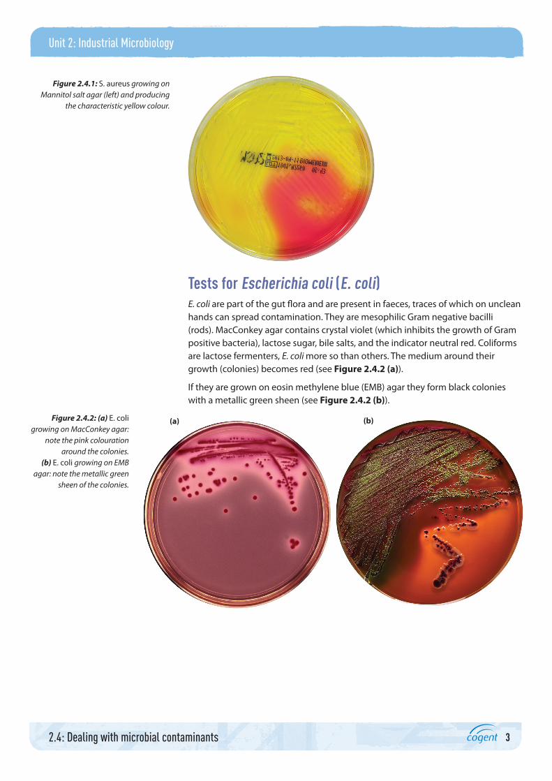
Tests for Escherichia coli (E. coli)E. coli are part of the gut flora and are present in faeces, traces of which on unclean hands can spread contamination. They are mesophilic Gram negative bacilli (rods). MacConkey agar contains crystal violet (which inhibits the growth of Gram positive bacteria), lactose sugar, bile salts, and the indicator neutral red. Coliforms are lactose fermenters, E. coli more so than others. The medium around their growth (colonies) becomes red (see Figure 2.4.2 (a)).
If they are grown on eosin methylene blue (EMB) agar they form black colonies with a metallic green sheen (see Figure 2.4.2 (b)).
Figure 2.4.1: S. aureus growing on Mannitol salt agar (left) and producing
the characteristic yellow colour.
Figure 2.4.2: (a) E. coli growing on MacConkey agar:
note the pink colouration around the colonies.
(b) E. coli growing on EMB agar: note the metallic green
sheen of the colonies.
(a) (b)

4
Unit 2: Industrial Microbiology
2.4: Dealing with microbial contaminants
Tests for MicrococcusThis genus of bacteria contains aerobic, usually immotile cocci (round bacteria) that occur in pairs or tetrads, have catalase enzyme, and are Gram variable or Gram positive. They do not produce gas from metabolism of carbohydrates but some can produce acid. They are widespread in soil, water and human skin so are present in dust. They may also contaminate beer. Most are mesophilic but some are psychrophilic and found in the Antarctic.
Colonies of M. luteus are yellow (see Figure 2.4.3 (a)) while those of M. roseus are red. Both are sensitive to novobiocin; M. luteus has oxidase, can hydrolyse gelatine and can use inorganic sources of nitrogen, such as ammonium dihydrogen phosphate. M. roseus can change nitrates to nitrite and produce acid from glucose. Micrococcus can tolerate quite high levels of salinity and do not form spores.
Figure 2.4.3 (b) shows stained cells of Micrococcus luteus, showing their round shape, and that they occur in groups of four (tetrads).
Tests for mycoplasmasMycoplasmas lack cell walls and are therefore resistant to penicillin but lyse when subjected to detergents. They are smaller than most bacteria, facultative or obligate anaerobes, pleomorphic – see Figure 2.4.4 (b) – (can have different shapes, often depending on their environment), mostly non-motile and need cholesterol to strengthen their cell membranes.
Figure 2.4.3: (a) Colonies of Micrococcus luteus.
(b) Direct stain of M. luteus.
(a)

5
Unit 2: Industrial Microbiology
2.4: Dealing with microbial contaminants
Most have urease enzyme. They are particularly problematic in laboratories where cell lines are used and some digest bacteria. However, culturing them is difficult and lengthy as they are slow-growing anaerobes.
Rapid testing kits for mycoplasmas
Rapid testing kits are available so that cell lines and culture media can be quickly tested and re-set up if contaminated. A DNA stain called fluorochrome (Hoechst 33258) binds specifically to DNA of cells in a culture and the mycoplasmas then show up as granules.
Mycoplasmal DNA can be identified by being extracted, augmented using the PCR and then run on an electrophoresis gel and stained.
Portfolio activity (4.1)You have cultures of S. aureus, S. epidermidis, E. coli, M. luteus and B. subtilis.
Make a plan of all the diagnostic tests you could carry out to distinguish between these organisms. Include Gram’s stain, use of differential media, colony examination, lactose fermentation and other biochemical tests (see Topic guide 2.4 Presentation for more information on biochemical tests).1 Write a plan and a list of apparatus and cultures you will need.2 Carry out the test and record your observations.3 Summarise your findings.4 Use the rapid test kits available to you to identify the bacteria.5 Evaluate the standard and rapid test methods.
Tests for moulds and yeastsMould fungi have thread-like hyphae and may form spores that can be transferred in dust and air. Yeasts are single-celled fungi. Both are eukaryotes. Some species of yeast live on human skin and hair. As contaminants of biotechnology processes, they will cause spoilage and some produce toxins.
Figure 2.4.4: (a) Colonies of Mycoplasma bacteria.
(b) Mycoplasma pneumoniae x 25 000. These bacteria
have no cell walls and have variable shape (pleomorphic).
(a) (b)

6
Unit 2: Industrial Microbiology
2.4: Dealing with microbial contaminants
They can be cultured in selective media and will survive lower pH limits than most bacteria. They can be stained and observed under a microscope. Petri dishes containing Sabouraud agar can be exposed to the air in the work environment and then incubated. Microscope slides can be made from the mycelium (hyphae) and the fungal morphology studied. Their cell walls contain chitin and they have membrane-bound organelles. Some can exist in the yeast form when in the environment and adopt the mycelial form when infecting an animal.
Activity: Fungal morphologyMake slides and examine the fungi you have available to you, for example Aspergillus, Saccharomyces and Mucor. Make annotated diagrams to show the structures present.
Expose some sterile Sabouraud agar plates to the air in your lab or outside for 30 minutes. Cover, label and incubate them at 30 °C for 5 days. Examine the plates and make microscope slides from them.
Can you identify the moulds on your plates?
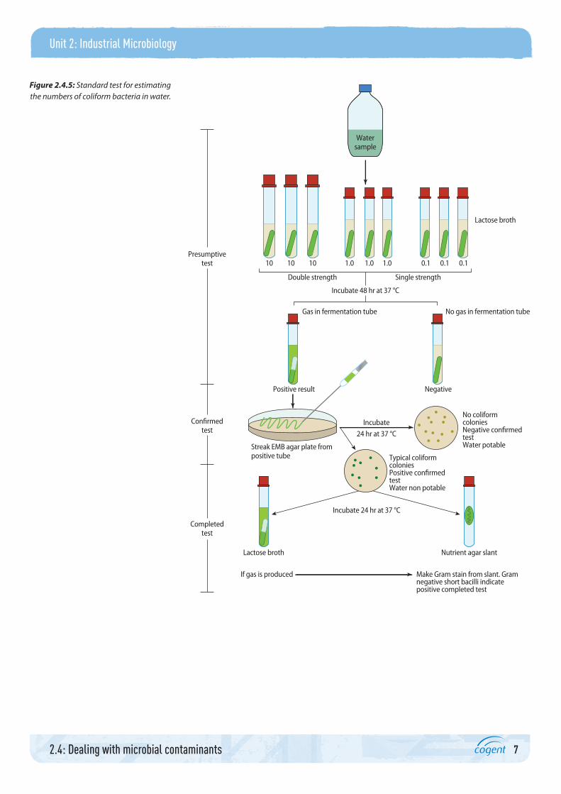
Testing water supplies for microorganismsAlthough most E. coli are harmless, their presence in water indicates possible faecal contamination and therefore the possible presence of cryptosporidia.
E. coli can be detected with the presumptive coliform test, followed by the confirmed and completion tests. This involves adding specific volumes of the water being tested to lactose broth containing Durham tubes to collect any gas produced during bacterial fermentation. The number of tubes showing positive acid and gas production are compared to tables of data to give the most probable number of coliforms per 100 ml. Liquid from positive result tubes is plated onto EMB agar where the presence of E. coli is confirmed by black colonies with a metallic green sheen. One of these colonies is added to lactose broth and one to an agar slant. If gas is produced, bacteria from the slant are stained and if they are Gram negative bacilli this completes the identification.
Figure 2.4.5 summarises the steps in the standard test for estimating the numbers of coliform bacteria in water.
7
Unit 2: Industrial Microbiology
2.4: Dealing with microbial contaminants
Watersample
10 10 10 1.0 1.0 1.0 0.1 0.1 0.1
NegativePositive result
Presumptivetest
Confirmedtest
Completedtest
Incubate 48 hr at 37 °C
Double strength Single strength
Gas in fermentation tube No gas in fermentation tube
Lactose broth
Streak EMB agar plate frompositive tube
Lactose broth Nutrient agar slant
Incubate 24 hr at 37 °C
If gas is produced Make Gram stain from slant. Gramnegative short bacilli indicatepositive completed test
Typical coliformcoloniesPositive confirmedtestWater non potable
No coliformcoloniesNegative confirmedtestWater potable
Incubate
24 hr at 37 °C
Figure 2.4.5: Standard test for estimating the numbers of coliform bacteria in water.

8
Unit 2: Industrial Microbiology
2.4: Dealing with microbial contaminants
Number of tubes giving positive reaction out of
MPN index per 100 ml
95% Confidence Index
3 of 10 ml each
3 of 1 ml each
3 of 0.1 ml each Lower Upper
0 0 1 3 <0.5 9
0 1 0 3 <0.5 13
1 0 0 4 <0.5 20
1 0 1 7 1 21
1 1 0 7 1 23
1 1 1 11 3 36
1 2 0 11 3 36
2 0 0 9 1 36
2 0 1 14 3 37
2 1 0 15 3 44
2 1 1 20 7 89
2 2 0 21 4 47
2 2 1 28 10 150
3 0 0 23 4 120
3 0 1 39 7 130
3 0 2 64 15 380
3 1 0 43 7 210
3 1 1 75 14 230
3 1 2 120 30 380
3 2 0 93 15 380
3 2 1 150 30 440
3 2 2 210 35 470
3 3 0 240 36 1300
3 3 1 460 71 2400
3 3 2 1100 150 4800
Portfolio activity (4.1, 2.5)Carry out the presumptive test, completed test and confirmed test and estimate the number of coliform bacteria in water from different sources. Use tap water, bottled drinking water and stream water.
Table 2.4.1: MPN determination from multiple tube test

9
Unit 2: Industrial Microbiology
2.4: Dealing with microbial contaminants
Cryptosporidia
Cryptosporidia are protozoa (eukaryotic) about 4–6 µm in diameter. Their oocysts (encapsulated eggs) can be stained and observed under a microscope but in a 100 ml sample there may not be enough to show up. Superchlorination (20 mg dm–3 chlorine for 7 hours) and filtering can remove the oocysts and prevent them from blocking pipes.
2 Rapid methodsRapid test kits for mycoplasmas have been discussed above. Other rapid method kits involve luciferase and bacterial RNA activity.
LuciferaseThis enzyme is involved in bioluminescence. It can be used to detect cellular ATP, and hence living cells, in what should be a sterile medium.
RNA activityWe have seen that bacteria can be identified according to the enzymes they have and the range of results for a series of biochemical tests. Enzymes are encoded by genes, their instructions acting via RNA. There are rapid test kits to identify the genes present from bacterial RNA activity, some of them detecting microbe specific tRNA. These tests can discriminate between dead and living cells and have results ready in about 3 hours, as opposed to days taken for incubating and culturing bacteria during the standard tests.
Some tests detect bacterial mRNA by isolating it, using a quantitative reverse transcription PCR, then using spectrophotometry to identify the mRNA encoding for specific genes. These tests are quantitative as they can relate to the bacterial cell numbers as well as the species of bacteria present.
3 Hazard analysisHazard analysis forms an integral part of total quality management systems deployed in any establishment using microorganisms or where microbial contamination could be a problem. See the Case study below for a real-life example of hazard analysis.
Activity: Schemes of hazard analysisChoose two commercial applications of using microorganisms.
For each one imagine that you are in charge of the quality management system and create a scheme of hazard analysis.

10
Unit 2: Industrial Microbiology
2.4: Dealing with microbial contaminants
Case study: Total Quality Management SystemMy name is Nadia Collins and I work in a large dairy. I oversee the Total Quality Management System and make sure that staff get appropriate training.
HACCP (Hazard Analysis Critical Control Point) is a management system involving analysing and controlling biological, physical, chemical and allergenic hazards. It began in the United States when NASA was developing food for astronauts to take into space. End of line product testing was deemed inappropriate as it would leave too little of the food to take. A system used during World War II for weapons manufacture was adopted and adapted instead. This involves identifying potential problems and preventing them from happening. This system is now internationally recognised and has extended from the food industry to cosmetics and pharmaceuticals.
There are seven principles to be followed:1 Conduct a hazard analysis – a hazard is any contaminant or property that can make the product unsafe.2 Identify critical control points – these are stages in the process where the hazard can be reduced or prevented/eliminated.3 Establish critical limits for each control point – maximum or minimum values to which each hazard should be controlled at each critical point.4 Establish critical control point monitoring requirements – this ensures that the process is under control at each critical control point.5 Establish corrective actions – these are to ensure that no unsafe products leave the premises.6 Establish procedures to ensure the HACCP is working as intended – this involves finding scientific evidence that the plan is working and so
validating it, and then verifying it. Regular routine testing for microbial contaminants is part of the verification procedure.7 Establish procedures for record keeping – all plants must have documents showing their HACCP plan and written records showing how their
critical control points have been monitored, their critical limits and how they have verified their system.
I am ultimately responsible for keeping the HACCP records in this dairy. Some in-house training is given to staff but I also send them on courses, which give them a qualification developed by the Chartered Institute of Environmental Health accredited by QCA.
During the early 1970s there were some cases of botulism poisoning from tinned meat, and pieces of glass were found in baby food. These cases led to international adoption of the HACCP system for controlling health and safety issues with products.
All employees here have to wear protective clothing including galoshes, cover their hair, not wear jewellery or make-up, walk through a trough of strong disinfectant when entering the work area, and must never eat while in the preparation areas. They should not come to work if they have any gastric complaints such as diarrhoea or vomiting, and after such an illness must be cleared by microbial screening of their faeces. The taps on all our sinks in the cloakrooms for hand washing are operated by a foot button or by sensing the presence of hands, so that employees do not touch the taps with their hands. Liquid soap is automatically dispensed and hot air dries the hands.
ChecklistIn this topic you should now be familiar with the following ideas:
equipment, workspaces and products have to be regularly screened for microbial contaminants
standard methods of screening are time-consuming as they involve culturing and incubating the microorganisms
rapid test kits have been developed that allow identification of the microbial contaminants in hours rather than days
there is an internationally recognised quality management system, HACCP, that companies using microorganisms have to put into practice.
Further readingWright, D. (2007) Human Physiology and Health (2nd edition), Oxford: Heinemann.

11
Unit 2: Industrial Microbiology
2.4: Dealing with microbial contaminants
AcknowledgementsThe publisher would like to thank the following for their kind permission to reproduce their photographs:
(Key: b-bottom; c-centre; l-left; r-right; t-top)
Corbis: Photoquest Ltd / Science Photo Library 1; Alamy: VEM / BSIP SA 4r, 5r; Getty Images: Guntars Grebezs / E+ 3br; Cytographics / Visuals Unlimited 3bl; Nathan Reading: 3t, 4l; Science Photo Library Ltd: BSIP, VEM 5l
All other images © Pearson Education
We are grateful to the following for permission to reproduce copyright material:
Pearson Education Inc. for the figure ‘Standard Method for Bacteriologica Water Analysis’ from Microbiology: A Laboratory Manual, 9th edition, by James Cappuccino and Natalie Sherman, p.324, copyright © 2011. Printed and electronically reproduced by permission of Pearson Education, Inc., Upper Saddle River, New Jersey
In some instances we have been unable to trace the owners of copyright material, and we would appreciate any information that would enable us to do so.