title on the stylohyoid bone of naumann's elephant...
TRANSCRIPT
Title On the Stylohyoid Bone of Naumann's Elephant (Elephasnaumanni MAKIYAMA) from Lake Nojiri
Author(s) Inuzuka, Norihisa; Hasegawa, Yoshikazu; Nogariya, Hiroshi;Kamei, Tadao
Citation Memoirs of the Faculty of Science, Kyoto University. Series ofgeology and mineralogy (1975), 41(1): 49-64
Issue Date 1975-01-31
URL http://hdl.handle.net/2433/186599
Right
Type Departmental Bulletin Paper
Textversion publisher
Kyoto University

MEMorRs oF THE FAcuLTy oF SclENcE, KyoTo UNIvERslTy, SERIEs oF GEoL. & MINERAL., Vol. XLI, No. 1 pp. 49-65, Pls. 5-7, Jan. 31, 1975
On the Stylohyoid
(ElePhas naumanni
Bone of Naumann's Elephant
MAKiyAMA) from Lake Nojiri
By
Norihisa INuzuKA, Yoshikazu HAsEGAwA*, Hiroshi NoGARiyA*' and Tadao KAMEi
(Received September 10, 1974)
Abstract
The find of the hyoid bone in fossil state has been very rarely reported hitherto. Recently, a part
of Proboscidean hyoid bone was unearthed from the bottom sediments at Lake Nojiri, centralJapan. That is identified as the stylohyoid bone of Naumann's eiephant (ElePhas naumanniMAKryAMA) which is commonly known from the Late Pleistocene deposits in Japan. Comparingthe present specimen with the hyoid bones of Asiatic, African and fossil elephants, it isdistinguishableinhaving some peculiar characteristios, viz. the presence of the angulus of the
postenor ramus etc..
IntrodUction
Since the time ofthe first excavation in 1962 it has been renowned that numerous
fossil bones of elephant and deer have been unearthed from the bottom sedimentsat Lake Nojiri in northern part of central Japan. Most of them were referable to
the fossil bones and teeth of Naumann's elephant (ElePhas naumanni MAKiyAMA)and those ofYabc's giant deer (Shinomegacereslabei SHiKAMA). As to the geological
age of the main fossil horizon, the Nojiriko Formation from which the fossil bones
were obtained is the deposits of the latest Pleistocene. Therefore, it may be sure
that those ,extinct animals were the inhabitants in the later halfofWUrmian. The
radio-carbon dating provides the range of those fossil animals to 35,OOO-16,OOO
years B.P.. (KAMEi and TARuNo, 1973). It was unique that, at the time of the fifth excavation of 1973, well-preserved
parts ofProboscidean hyoid bone were found from the I-C-9 grid at Tategahana, one
of the localities of the north-west shores of the lake. The materials were composed
of a pair ofthe stylohyoid bones.
It would be worth to note that Y. HAsEGAwA, one of the authors, first gave the
suggestion at the time of excavation that the present materials were referable to the
hyoid bone of Proboscidean fossils. Later, the members of the fossil vertebrate
research group of the Nojiri-ko Excavation Research Group have made examination
*
**
National Science Museum, TokyoCollege of Humanities and Sciences , Nihon University, Tokyo
50 Norihisa INuzuKA, Yoshikazu HAsEGAwA, Hiroshi NoGARiyA and Tadao KAMEi
carefu11y for those materials and have got a conclusion that they are the stylohyoid
bone of ElePhas naumanni MAKiyAMA. In fact, it was the first discovery of thehyoid bone of Proboscidean fossil in Japan. Furthermore, it may be true thatthe occurrence of such material as hyoid bone of fossil elephant has not frequently
been known in the world. In this regard, the object of this study was to describe
the material from Lake Nojiri and to study it in comparison with other hyoid bonesof living and some fossil elephants. But, as all of the specimens of the livings used
here belong to the materials of immature stage, it was impossible to examine them
from the view point of ontogeny. Therefore, the results of comparative study are
compelled to be insuMcient, and many problems left here untouched should besolved in future.
The authors are grateful to Dr. S. IJiRi for his interests in this work. Thanks
are given to Mr. T. FuJiTA of the Shinsha University, Mr. H. TARuNo of the Osaka
City Museum ofNatural History and Mr. M. GoTO of the Tokyo Medical and DentalUniversity for their helpful advice on the preparation of materials and photograph.
Thanks are also due to the members of the Nojiri-ko Excavation Research Groupwho gave the authors the opportunity and many conveniences during this work.
Description
a. Hyoid Bone of Elephant The hyoid bone is usually designated as the skeleton of the base of tongue and is
situated between the lowerjaw bone and the thyroid cartilage of the larynx. Thisbone consists of several small bones and serves special functions to connect theskull with the thyroid cartilage. In general, it is well-known that the shape of the
bone differs from each other in various kinds of animals. Among them, the hyoid
bone of the elephant is known in having peculiar shape dissimilar to other(FLowER, 1966). It can be divided into two parts as shown in Fig. IA. The onepart is composed of the basal hyoid bone (Basih7oideum) and the thyrohyoid bone
(Th2roh2oideum) which can be correspond respectively to the body (Corpzas) and the
greater horn (Cornu maijus) in the case of Homo (Fig. IB). The other part consists
mainly of the stylohyoid bone (Styloh"oideum) and the tympanohyoid bone (T2m-Panohloideum), both of which correspond to the styloid process (Processus st!loideus)
of Homo. To compare the present materials with those elements stated above, it is easy
to understand that the present materials of peculiar shape from Lake Nojiri are
referable to the stylohyoid bone of the elephant. The stylohyoid bone of theelephant is usually connected upwardly to the tempora! bone of the skull through the
intermediation of the tympanohyoid bone. It is characteristic that the bonebranches off ventrally in antero-posterior direction. Moreover, as the stylohyoidbone seems to be composed of three parts, the following nominations are given to
On the Sytlohyoid Bone ofNaumann's (Elephant EtePhas naumanni MAKiyAMA) from Lake Nojiri 51
FOSSA TEMPORALIS
eRBITA
".,
IA OS OCelPITALE x
.s., .
,,,.
iil,' X2,Ru,U?\,,i :U,S,
/./..,• ARCUS •1/.,
-/'
//.t/tG./:O //.MATIcy./.,,.
MAX:LLA
•• -•..
t 4. I, 't '{.'(gLyj')
"//
'coNnyLus
,OCCIPITALIS
".
iiPgi yLoHyo lll]EBu:MTA
/t/ i PORUSlili/ ll,lil,liill,,IR,l,,Flil:l':g25oSitJl'i"S2,ilii,,",il,l;,,,
IL
•CORNU MINUS
THYROHYOIDEww
OS INCISIVLM
A
BASIHYO:DEon
.,
CORPUS
4
/
/" CONDYLUS OCCIPITALIS
CORNU MAJUS
BFig. 1. Status of the hyoid bone in Elephas (A) and Homo (B).
those three parts tentatively in this paper. Namely, the inferior ramus is designated
to the branch which extends to the ventral side in the frontal position, the posterior
ramus is to the branch which protrudes posteriorly and the superior ramus is to the
dorsal side branch which has the articulation to the tympanohyoid bone.
The inferior ramus is generally longer than the posterior ramus. From thedistal end of the former the styloglossus muscle arises and raches to the both sides of
the tongue to serve function to the movement ofthe tongue. In the neighbourhoodof this place, the stylohyoid and stylopalatal ligaments arise, being in contact with
both the basal hyoid bone and the soft palate. The stylopharyngeal muscle, one of
the muscles which is active in the service to expand the pharynx, originates from the
posterior border in the middle of the inferior ramus.
The stylohyoid muscle arises from the anterior border near thejunction of the
inferior and the posterior rami and attaches to the basal hyoid bone, crossing over a
slip of the digastric muscle. According to SABAN (1968), however, in Asiaticelephant (ElePhas maximzas LiNNE) iacks such stylohyoid muscle.
The shape of the posterior ramus varies greatly from animals to animals. Inthe cases of Bos and Equtts, the parts which correspond to the posterior ramus of the
stylohyoid in Edephas are represented by a simple process called as the stylohyoid
angle or the muscular angle ofthe great cornu (SissoN, 1970). In the same manner,
this process has been named by some authors variously as the posterior process
(EALEs, 1926), talon du stylohyal or talon posterieur (GAsa, 1967). From thisposterior ramus, a part of the digastric muscle arises. It is well-known that both the
digastric and stylohyoid muscles stated above achieve importance in animals inconnection with mouth opening and swallowing.
52 Norihisa INuzuKA, Yoshikazu HAsEGAwA, Hiroshi NoGARiyA and Tadao KAMEi
b. StylohyoidBoneofNaumann'sElephant,ElephasnaumanniMAKIYAMA The stylohyoid bone of ElePhas naumanni MAKiyAMA has a distinctive feature,
unlike those of other elephants, in having a small erected process at the internalposterior border of the posterior ramus. For convenience to describe, this small
process is named tentatively as the "angulus of the posterior ramus."
As the specimens dealt with have been unearthed in the same place, it is highly
probable that they belong to a pair ofthe hyoid bone ofone same individual. Al-though those two bone materials differ slightly from each other in shape and size, a
result of carefu1 examination on the materials of the Iivings suggests that such differ-
ence is to be within a limit of variation in one individual. Nevertheless, one may
further propose some objections in dealing with them as the materials ofthe same one
individual, because they have still some differences respectively both in morphology
and in degree of preservation.
Left stylohyoid bone (coll. no. 5NC9-13, Fig. 2A; Fig. 3A, B; Pl. 5, Fig. 1: Pl.
6, Fig. 1; Pl. 7, Figs. 1, 3): The bone is more or less complete in preservation. It
looks like reversed Y figure in shape. As the distal portions of all rami turn about
c
SUPERIOR RA-"JS'
h
b---
c)---
INFERIOR
RAMUS
Q----T
a
f
e
.Q/-' //
i
ANGULUS OFPOSTERIOR RAMUS
All! ,a /! POSTERIOR
b
RAMUS
g
(>-
b
A
C)i
NNx
POSTERIOR-RAMUS
j
N
j
ANGVLU$ OF
o.Y
N
N
d isk
PO,STERIOR
c
i
INFERIOR
h
RAMVS
SUPERIOR RAMUS
o/aBRAMUS
Fig. 2. The stylohyoid bones of Elephas naumanni, showing the points formeasurements and the forms of cross sections of rami. A, Outer lateral
view of left stylohyoid bone; B, Idem of right stylohyoid bone.

On the Stylohyoid Bone ofNaumann's Elephant (ElePhas naumanni MAKiyAMA) from Lake Nojiri 53
.::J;.:.;.::.:.'
-:':----tti:-:'::
A B
Fig. 3. Diagrams to show the distribution of tuberosity in the left stylohyoid
bone ofElePhas naumanni. A, Outer lateral; B, Inner lateral.
slightly to the outside, the external surface of this bone seems to be somewhat concave
as awhole. The distal end ofthe inferior ramusis damagedin some extent, but the
distal extremities of both the posterior and the superior rami are rather wellpreserved. The rugged surfaces for attachment to the cartilarge bone are observed
in those portions. A plane-like portion is formed in the area where three rami meet,
but a certain ridge can be observed along the border from the superior ramus to the
angulus of the posterior ramus. The area of the tuberosity is shown by the stipplein Fig, 3.
The inferior ramus is much depressed and longest among three rami. Nearthe branching it is thick in fore-and-aft direction and thin transversely from side to
side. But the nearer the apex, the more it twists anticlockwise. Accordingly,as the twisting attains to make right-angle with the initial direction at the extremity,
the relation of the thickness observed tends to turn upside down. Though thecurvature is very small, the inferior ramus seems to bend slightly backward in
general. The ridges can be seen along anterior-external, anterior-internal andposterior-internal borders in the neighbourhood ofbranching, but near the extremity
ofthe inferior ramus, a ridge which runs along anterior-internal border tends to take
obtuse angle in cross section and shifts its position to the external side. Moreover,
a ridge along the posterior border becomes to join with that of the external border,and therefore, there can be seen only two, internai and external ridges at the distal
portion of the inferior ramus.

54 Norihisa INuzuKA, Yoshikazu HAsEGAwA, Hiroshi NoGARiyA and Tadao KAMEi
Consequently, the configuration of the rami in cross section varies in following
manner. In the neighbourhood ofthe branching, it resembles a compressed right-
angled triangle of which the base equates the internal surface. Near to the apicalpart, it tends to take obsolete or spindle form with a long axis extending from internal
to external side.
The posterior ramus is relatively short and thick. It tends to be twisted very
slightly by the angle of about 50 to the left. Except a slight outward bending near
the place ofbranching, it is rather straight as a whole. The ridges are observed on
both the anterior-internal and posterior borders stretching from the branchingarea to the extremity. As the external side ofthe ramus is formed to be convex, the
cross section of the ramus presents a semi-circular shape in general. The internal
side of the ramus is slightly concave at the middle part, owing to a distinct swelling
formed by anterior and posterior ridges.
The superior ramus is the shortest among those three rami but very thick from
inside to outside. No torsion and curvature can be observed in this superior ramus.
The ridge which corresponds to the extention from the inferior ramus runs along the
anterior-external border. That ridge is short but reaches virtually to the central
part of the posterior ramus. Furthermore, a wide but short sulcus develops con-spicuously between the ridge stated above and the other ridge which runs from the
anterior-internal border to the angulus of posterior ramus. The anterior-internal
border forms a marginal edge of the surface which is the attachment for the cartilage
bone. That sulcus runs on the hind surface of the ramus from upper internal to
lower external.
The superior ramus forms a triangle shape in the cross section of which thevertex faces to the external side. Itseems that every angles ofthe triangle are rather
rounded. The plane for articulation at the apical portion of the ramus is eliptical
in form and is perpendicular to the axis stretching from the extremity of the lower
anterior-external to the extremity of the upper interior-posterior of the ramus. A
kind of tuberculate structure can be seen on the external side of the ramus at the
place near the apex.
Right stylohyoid bone (coll. no. 5NC9-14, Fig. IB; Pl. 5, Fig.2; Pl.6, Fig,2;Pl. 7, Figs. 2, 4) : In comparison with the former material, the right stylohyoid bone is
rather ill preserved. Most ofthe inferior ramus and the apical portion ofthe superior
ramus have been lost. Owing to the weathering process, a fibrous texture hasbeen exposed on the surface and the tuberositas is rather obscure in this material.
A ridge is indicated in the area along the posterior border of the inferior ramus.
The internal side of the ramus presents conspicuous fiat border (see the cross section
shown in Fig. 2, B.).
The posterior ramus is distinctive in having two edges which are represented

On the Stylohyoid Bone ofNaumann's Elephant (ElePhas uaumanni MAKiyAMA) from Lake Nojiri 55
by the anterior and posterior margins. It twists to the right with angle ofabout 50,
but is nearly straight. Near the branching the ramus tends to turn about to the
external side. The cross section of the ramus exhibits a sort of bow shape of which
the convex side faces to the external side. Along the internal side a swelling portion
extends more than 45 mm in length from the angulus of the posterior ramus, whileat the middle of it a pit-like depression with the depth of about 1 mm is observed.
The superior ramus is stout and short without any torsion and turn. The ramus
is enclosed by distinct four planes, viz., internal, external, anterior and posterior
sides. All of the boundary areas made by those four planes are represented byweakly developed ridges. Therefore, the cross sections ofthe ramus are shown as
rounded trapezium forms with round edges.
'c. Stylohyoid Bone of Asiatic Elephant, Elephas maximus LINNE The materials observed belong to a part of immature feamle skeleton which is
kept in the National Science Museum, Tokyo. According to the observation onaccessible state of dental eruption, the teeth of the elephant are estimated to beearly Ml stage viz. about ten years old (HAsEGAwA, 1972). As it is evident that
those paired materials belong to one same individual, it was easy to examine the
variation of left and right stylohyoid bones within one individual. Besides them,
the tuberositas can be clearly observed in those materials, and therefore, they provide
some useful informations about the state ofmuscle attachment.
Left stylohyoid bone (Fig. 4 A, Fig. 5 A, C) : From posterior view, it is clear that
the posterior and superior rami are arranged in a straight line. On the other hand,
the inferior ramus tends to turn externally. The distribution of the tuberositas is
indicated in Fig. 5 A, C.
The inferior ramus turns in a certain extent to the left near the point imediately
below the branching oframi. Due to this torsion, the rotation oframus is given to
continue to the distal portion with very slight torsion angle. Just below the area
of bifurcation, the ramus bends rather strongly to the ventral side. The crosssection of the ramus varies from the proximal to the distal extremities, from long
eliptical through subtrianglar to circular shapes (Fig. 4 A).
The posterior ramus bends gently backward. The ridge seems to be shiftedto the left near the portion ofthe posterior end. By the internal dorsal (or posterior)
border, the sulcus extends along the whole length and reaches up to the region of the
superior ramus. The tuberositas is developed in the inside of this sulcus and spreads
out near the distal end. The cross section ofthe ramus is eliptical, but near the distal
end it becomes semi-circular with flat dorsal side.
The length ofthe superior ramus is as same as that ofthe posterior ramus. The
tuberositas can be seen in the anterior half of both the internal and external sides,
and is especially developed along the anterior border continuously to the portion
56 Norihisa INuzuKA, Yoshikazu HAsEGAwA, Hiroshi NoGARiyA and Tadao KAMEi
a--'
o----
-
c
'
hSUPERIOR RAMUS
INFERIOR RAMUS
D----
g
f
o---- e/ /
i
/
A
OIIOt //
j//y'
Xb
9sl xl x
b
/jPOStERIOR RAMus
SUPERIOR RAMUS
h
N
c
xlXi N.a
i
!NFERIOR RAMUS g
f
e
'
--- <>
--- ts
---- - B
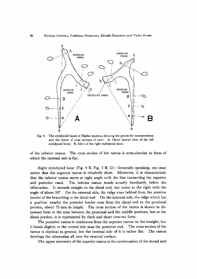
aaFig. 4. The stylohyoid bones of ElePhas maximus, showing the points for measurements
and the forms of cross sections of rami. A, Outer lateral view of the left stylohyoid bone; B, Idem of the right stylohyoid bone.
of the inferior ramus. The cross section of the ramus is semi-circular in form of
which the internal side is flat.
Right stylohyoid bone (Fig. 4 B, Fig. 5 B, D): Generally speaking, one maynotice that the superior ramus is relatively short. Moreover, it is characteristic
that the inferior ramus meets at right angle with the line connecting the superior
and posterior rami. The inferior ramus bends acutely imediately below thebifurcation. It extends straight to the distal end, but twists to the right with the
angle of about IOO. On the external side, the ridge runs behind from the anterior
border ofthe branching to the distal end. On the internal side, the ridge which has
a position nearby the posterior border runs from the distal end to the proximalportion, about 75 mm in length. The cross section of the ramus is shown in de-pressed form in the area between the proximal and the middle portions, but at the
distal portion, it is represented by thick and short crescent form.
The posterior ramus is continuous from the superior ramus on the straight, but
it bends slightly to the ventral side near the posterior end. The cross section of the
ramus is eliptical in general, but the internal side of it is rather flat. The ramus
develops the tuberositas all over the external surface.
The upper extremity ofthe superior ramus is the continuation of the dorsal and

On the Stylohyoid Bone ofNaumann's Elephant (ElePhas naumanni: MA-yAMA) from Lake Nojiri 57
A B
c D
Fig. 5. Diagrarrrs to show the distribution of tuberosity in the stylohyoid
bones of ElePhas maximtts. A, Outer lateral of the left side bone;
B, idem of the right side bone; C, Inner lateral of the left side
bone; D, idem of the right slde bone.
posterior surface. As the internal side of the ramus is flat, the cross section of
ramus seems to be semi-circular of which the external side is convex. The ramusdevelops the tuberositas especially on the anterior border which is continuous from
the inferior ramus.
d. StylohyoidBone ofAfrican Elephant, Loxodontaafricana(BLuMENBAcH) The materials observed are those kept in the National Science Museum, Tokyo.
They belong to a part of the immature individual skeleton. From the view pointof the eruptive stage of teeth by LAws (1966), this elephant is estimated as three
years old. It is characteristic that the inferior ramus of this stylohyoid is very long.
The inferior ramus is also very thin and the length ofit is nearly as twice as the total
length of the posterior and superior rami. The posterior and superior ramimakejointlyastraight line, and from the middle of which, the inferior ramusis branched off.

58 Norihisa INuzuKA, Yoshikazu HAsEGAwA. Hiroshi NoGARiyA and Tadao KAMEi
Left stylohyoid bone (Fig. 6 A, Fig. 7 A, C): In the proximal portion of theinferior ramus, two ridges can be observed at the anterior and posterior borders
respectively. Among them, the ridge ofthe anterior border tends to run externally.
0n the other hand, in the apical portion, another ridge can be seen on the internal
side, which runs obliquely from the anterior border to the back side and joins to meet
with those two ridges stated above. When it reaches to the posterior-internalborder, the cross section of the ramus becomes to be similar to a right-angled triangle
ofwhich the external side meets in right angle with the posterior side.
Succeedingly, the posterior border of the inferior ramus tends to run nearbythe external side and also the anterior border of the ramus goes in the reach of the
external side. And yet the internal border runs obliquely to the anterior side.Therefore, the cross section of the ramus looks like a regular triangle of which the
base corresponds to the anterior side. Near to the apex, the height of the ridge on
the anterior external border becomes low relatively. At the same time, as thewidth of other two ridges increases more and more, the cross section of the ramus
forms consequently bow shape ofwhich the internal side is flat.
As described above, the inferior ramus has ridges in complicated fashion hard
to be explained by simple twisting. But, as a whole, it seems that it twists with the
c
INFERIOR RAMUS
c
D----
Q----•
0------ e
ht /
-
SUPERIOR RAMUS
-<)
l - !p'i
1
/b
Xb>Å~
aPOSTERIOR
g
f
/ /
A
RAMUSINFER1OR RAMUS
----- e
a
9 Ba
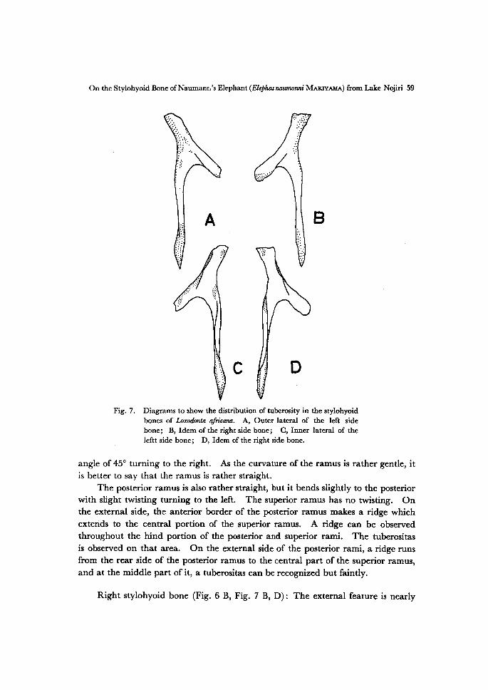
Fig. 6. The stylohyoid bones of Loxodonta africana, showing the points for measurements
and the forms of cross sections of rami. A, Outer lateral of the left side bone; B, Idem of the right side bone.
On the Stylohyoid Bone ofNaumann's Elephant (Elophasnautnanni MAKiyAMA) from Lake Nojiri 59
:i---
1,
A:iE
B
't:'
:' i1.:
c;l --
::
t'
D
Fig. 7. Diagrams to show the distribution of tuberosity in the stylohyoid
bones of Loxodonta afii'cana. A, Outer lateral of the left side
bone; B, Idem ofthe right side bone; C, Inner lateral of the]eftt side bone; D, Idem of the right side bone.
angle of 450 turning to the right. As the curvature of the ramus is rather gentle, it
is better to say that the ramus is rather straight.
The posterior ramus is also rather straight, but it bends slightly to the posterior
with slight twisting turning to the left. The superior ramus has no twisting. Onthe external side, the anterior border of the posterior ramus makes a ridge which
extends to the central portion of the superior ramus. A ridge can be observedthroughout the hind portion of the posterior and superior rami. The tuberositas
is observed on that area. On the external side of the posterior rami, a ridge runsfrom the rear side of the posterior ramus to the central part of the superior ramus,
and at the middle part of it, a tuberositas can be recognized but faintly.
Right stylohyoid bone (Fig. 6 B, Fig. 7 B, D): The external feature is nearly
oo Norihisa INuzuKA, Yoshikazu HAsEGAwA, Hiroshi NoGARryA and Tadao KAMEi
symmetric to the left stylohyoid bone with respect to the median plane. But thepresent material is different in having a sulcus which runs along the hind border of
the posterior and superior rami. In a pit ofsulcus, there can be seen some tuberos-
itas. The angle between the apical plane of articulation and the shaft of thesuperior ramus is less than that ofthe counterpart.
Measurements
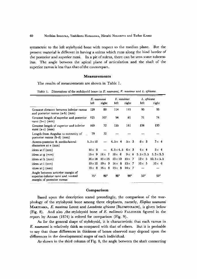
The results of measurements are shown in Table 1.
Table 1. Dimensions ofthe stylohyoid bones in E. naumanni, E. maximus and L. africana.
E. naumanni
left rightE. maximusleft right
L. qfricana
left right
Greatest distance between inferior rarnus 129and posterior ramus (a-b) (mm)
Greatest length ofsuperior and posterior 125rami (b-c) (mm)Greatest length of superior and inferior
rami (a-c) (mm)
Length from Angulus to extremity ofposterior ramus (b-d) (mm)
Antero-posterior & medio-lateraldiameters at e (mm)
idem at f (mm)
idem at g (mm)
idem at h (mrn)
idem ati(mm)idem atj (mm)Angle between anterior margin ofsuperior-inferior rami and ventra1margin of posterior ramus
169
79
5.5Å~ 10
80
107
72
72
114
on
155
115
81
141
- 4.5Å~ 4 5Å~ 3
10Å~ 9 - 6. 5Å~4.5 6Å~ 315Å~ 9 14Å~ 7
16xl4 15Å~1519Å~10 19Å~ 918Å~ 8 16Å~ 8
7so 80e
10Å~ 6 9Å~ 413Å~10 15Å~ 7
14Å~ 8 13Å~ 713Å~ 8 14Å~ 7
9oe 9oo
95
71
134
8Å~ 3
93
74
130
7Å~ 4
4Å~4 5x45.5Å~3.5 5.5Å~3.5
12Å~ 5 10.5Å~5.5
10Å~ 5 10Å~ 6
5oo 5oe
Comparison
Based upon the description stated precedingly, the comparison of the mor-phology of the stylohyoid bone among three elephants, namely, Etephas naumanniMAKiyAMA, E. maximus LiNNE and Loxodonta afn'cana (BLuMENBAcH), is given below
(Fig. 8). And also ,the stylohyoid bone of E. melitensis FALaoNER figured in the
report by ADAMs (1874) is refered for comparison (Fig. 9).
As for the general shape of stylohyoid, it is characteristic that each ramus in
E. naumanni is relatively thick as compared with that of others. But it is probable
to say that those differences in thickness of bones observed may depend upon the
differences in the developmental stages of each individual.
As shown in the third column of Fig. 8, the angle between the shaft connecting

On the Stylohyoid Bone of Naumann's Elephant (ElePhas naumanni MAKiyAMA) from Lake Nojiri 61
E.maximus -L.afrlcana
GENERAL,'SHAPE:GeneralizedEorm
ThicknessofRamus Thick Thin Thin
'
ortuarginofsuperi-er-inferiorrami& 7oo--Boe 9oe Soeventralmarginof
.posterxorramus
Ratiogfsuperior-posteriorramilength'
3:4 2:3 3:Sto.inferiorramuslength
INFERIORRJUeUSt'
ShapeofExtremity gentlyflatantero- withafurrowrunn- expandedantero-posteriorly ingalongtheaxis posteriorly
,
Curvature simple.andslightly stroriglycuryednear straighteurvedposteriorly andtothebranching
AngleofTwisting 9oo loo 4so
TransitionofFormofcrosBsectionfrombranchingto
'exttemxty
ts-- o-R-o K7-R-Y7-x
>:7-fL
POSTERIORRAMUS:'
PositionofRidge in,mediqlsidealong inlater41sideanteriorandposteri- indistinct alonganterior
,ormargm 'margln
Curvature straight bendpostbriorlyin slight}ycurvedtothemiddle posteriorside
GeneralÅ}zedCrosssection
(=>SUPERIORRAbdUS:)SodeofTransition bendatbranching curvedposteriorly straight•
.toPosterlorRamus
AngulusofPosterior present absent absentRarnus
GeneralizedCrosssection
Fig. 8. Comparative morphology of the stylohyoid bones of Elophas nattmanni,ElePhas maximtts and Loxodenta africana.

62 Norihisa INuzuKA, Yoshikazu HAsEGAwA, Hiroshi NoGARiyA and Tadao KAMEi
+t -t:tt t
:t-
",
'
i:.
"
ii
:
:'t
: ,:.:,::,
" ;"• :1
i .:ilt,l}
- --- -- -• :--- --- t--. - t -''-'-'
:;
`
-
Å}-'
-
-Y..li s'-'- "'. t:. •rl"lkl':s
N
Fig. 9. The stylohyoid bone (left side) of ElePhas metitensts
(redrawn from ADAMs, 1874)comparison among those elephants.
It is generally known that the styloglossus muscle arises from the distal extremity
of the inferior ramus and extends to both sides of the tongue. The function of that
muscle is to move the tongue side by side or withdraw it. Only in L. africana, the
distal extremity of the inferior ramus is expanded fore-and-aft with well-marked
tuberositas, In E. mczximzts, the inferior ramus is curved very strongly, especially
near the branching, and bends to ventral side. The twisting angle of the inferior
ramus is 900 in E. naumanni, but only 100 in E. maximtts. AIthough in the case ofL. africana it is about 450, it is curious that the twisting movement is in the opposite
direction to that of others.
The cross section of the inferior ramus varies in shape successively from the part
near the branching to the distal extremity as figured in Fig. 8. The tuberositas of
the inferior ramus is hardly seen in E. naumanni, but can be observed at the extremity
in L. afrieana. In E. maximus, the tuberositas is seen in the sulcus near the extremity
of the external side.
The ridge of the posterior ramus is present at the anterior-posterior border of
internal side in E. naumanni. It is distinct at the anterior border of the external side
in L. africana, but is obscure in E. mcurvimtts. The posterior ramus is straight in
E. naumanni, but in L. afre'cana it bends slightly backward. It turns behind at the
middle of the ramus in E. maximtts. The torsion is commonly observed but is veryweak in those three. The cross section of the posterior ramus is different from each
the inferior and superior rami and that of the posterior
ramus is measured. The angle is larger in E. naumanni
than in L. afn'cana and smaller than in E. maximzas.
Apart from this, the angle of the divergence of theinferior ramus from the posterior ramus is large inE. naumanni, but it is much smaller in both E. maximtts
and L. afn'cana. But the latter may also depend upon
the differences of the developmental stages of those
elephants.
Although the apical portion of the inferior ramus
of the present material is slightly damaged, the ratio
of the total length of the superior and posterior rami
to that of the superior and inferior rami, shown in
Fig. 8, is largest in E. naumanni. It is3:4 in E.naumanni, but is 2:3 in E. maximtts and 3:5 inL. afn'cana. Accordingly, the posterior ramus in E.
naumanni is well developed as compared with those of
E.maximus and L. africana. It is also the same tosay about the relation of relative ramus thickness in
On the Stylohyoid Bone ofNaumann's Elephant (ElePhas naumanni MAKryAMA) from Lake Nojiri 63
other, viz. bow form in E. naumanni, semi-circular in L. africana and eliptical in E.
maximus. The tuberositas is seen on the surface ofboth the internal and the external
sides and near the angulus of the posterior ramus in E. naumanni, but it covers all of
the external side and the posterior half of the internal side in E. maximus. In L.
africana, it can be observed in the distal part of the external side.
As to the relation of the superior ramus to the posterior ramus, a conspicuous
break is observed in E. naumanni near the point ofbifurcation. The superior-poster-
ior rami bend slightly backward in E. maximus and is straight in L. africana. The
cross section of the superior ramus is represented by depressive shape in L. africana,
internally convex shape in E. naumanni and externally convex shape in E. maximus.The tuberositas of the superior ramus is seen in the upper half of the external side of
the ramus in E. naumanni. It is observed in the anterior halfof the external side and
in the posterior to the middle of the internal side in E. maximus. In L. afn'cana,it is distributed in the anterior half of the external side and the posterior border.
A distinct feature of the angulus of the posterior ramus seen only in E. naumanni
is worth to discuss. From this place, a strip of the digastric muscle (Muscalus
digastrctts VenterPosterior) arises. The presence of a strong process, development
of tuberositas around it and a stout posterior ramus, all of them suggest that the
digastric muscle was much developed in E. naumanni. Accordingly, it seems that
Naumann's elephant was vigorous in the motion ofmouth-opening and swallowing.In addition to this, it must be mentioned that only in E. naumanni the tuberositas
can be seen on the internal and ventral sides at the junction of the superior and
posterior rami. The stylopharyngeus muscle which participates in the motion ofthe larynx attaches to this place. Moreover, the tuberositas in the transitional area
of the anterior border from the inferior ramus to the superior ramus is commonlyobserved in all of three elephants. But the tuberositas is developed especially in
E. maximtts and is weakly observed in L. africana. It may be true that stylohyoideus
muscle attaches to this place in E. naumanni, while in L. africana it is uncertain what
kind of muscle, viz., stylopharyngeus muscle or stylohyoideus muscle, attaches to
here.
The stylohyoid bone of E. melitensds (ADAMs, 1873) is similar to that of E.naumanni in having the same curvature in the inferior ramus and the same value in
the ratio of the thickness to the length. On the other hand, the former has also
some characteristics common to L. africana, namely, the same value ofthe bifurcation
angle of the ramus, the absence of the angle of posterior ramus and the straightfeature of the posterior and superior rami. According to the origipal description,
"When we know that fig. 10 could not have belonged to a foetal individual, it wi11
be conceded that its owner must have been a diminutive form of Elephant."(ADAMs, 1874, p.45)
64 Norihisa INuzuKA, Yoshikazu HAsEGAwA, Hiroshi NoGARiyA and Tadao KAMEi
Summary
The results of the observation given to fossil hyoid bone from Lake Nojiri
are summarized as follows.1) Both of two specimens (5NC9-l3, 14) from Lake Nojiti are the stylohyoid bone of Proboscidea. Moreover, it is highly probable that they belong to a part of the hyoid bone of E. naumanni MAKiyAMA,2) As the results of comparison among E. naumanni, E. maximus, L. africana, it becomes clear that there are some morphogical differences among them. i) It is characteristic that the stylohyoid bone of E. naumanni is distinct in
having a distinct process (the angulus of the posterior ramus).
ii) The distal extremity of the inferor ramus is depressed fore-and-aft in L. africna.
iii) The degree of twisting in the inferior ramus is highest in E. naumanni.
As the materials used for comparison were limited in this case, it is untenable
to discuss in this article about the problems of functional and phylogenetic me-anings of them. Therefore, it is necessary to make re-examination on those ma-
terials when more numerous and more useful materials for comparison areobtained in future.
References
ADAMs, A. L. (1874): On theDentition and Osteology ofthe Maltese fossil Elephanbs. Trans. Zool. Soc. Londbn, 9, pt. I, pp. 45, pl. XV, fig. 10.
EALEs, N. B. (1926): The Anatomy of the Head of a Foetal Africal Elephant, Elephas africanus (Loxodonta africana). Trans. Ray. Soc. Edinb. 54, pt. 3, no. 11, pp. 503-505, pl. VI.
FLowER,W. H. (1966): An Introduction to the Osteology of the Mammmalia. A. Asher, Arnsterdam,
pp. 208.GAsc, J. P. (1967): Squelette Hyobranchial, in GRAssE, P. P. ed. "Trait} de Zoologie", tom. 16, fasc.
1, Masson Paris, pp. 551-582.HAsEGAwA, Y. (1972): The Naumann's Elephant, Palaeoloxodon naumanni (MAKiyAMA) from the Late Pleistocene off Shakagahana, Shodoshima Is. in Seto Inland Sea, Japan. Bull. Nat. Sci.
Musettm., 15, 3, pp. 513-591.KAMEi. T. & H. TARuNo (1973) : Note on the Occurrence of the Latest Pleistocene Mammals from Lake Nojiri (Part 1). Mem. Fac, Sci. 1<yoto Univ., ser. geol. min. 39, 2, pp. 99-122, pl. 4-11.
KATo, Y. (1971): The ComParative AnatomJ of the Domestic Animals, an atlas. I. Yokendo, Tokyo.
pp. 52-53. (in Japanese)KAwADA, S. & M. DAiGo (1970) : Altas ef the ComParative Anatom2 of the Domestic Animals. I. Bun-eido,
it Tokyo. pp. 88-89, 160-161. (inJapanese)LAws, R. M. (1966): Age Criteria for the African Elephant, Loxodonta africana. East African Wild Ltfe Joum. 4, pp. 1-37.
MiLLER, M. C., CHRisT[ENsEN & EvANs (1965): Anataay of the Dog. Saunders, Philadelphia & London. pp. 37-38, 148-150,MoRr, O. etal. (1969): Kaib6gtrku (Anatomy) I. (10th rev. ed.). Kanehara Shuppan, Tokyo.pp. 282-286. (in Japanese)

On•the Stylohyoid BoneofNaumann's Elephant(ElePhas naumanni MAKiyAMA) from Lake Nojiri 65
SABAN, R. (1968): Musculature de la Tet6, in GRAssE, P. P. ed."Traitg de Zeologie" tom.I6, fasc.
2. Masson, Paris. pp. 306-322.SicHER, H• & E. L. DuBRuL (1970): Orat Anatoml, (5th ed.), Mosby, Saint-Louis, pp. 49-50.Sisson, S. (1970): TThe AnatomJ of the Demestic Anitnats. (4th rev. ed.), Saunders, Philadelphia &
Tuttle, Tokyo, pp. 68-69 & 140.
Explanation of plate 5
The Stylohyoid bones of ElePhas naumanni MAKiyAMA (all natural size)Fig. 1. 0uter lateral view of the left side bone.
Fig. 2. 0uter lateral view of the right side bone (partly broken).
Explanation of plate 6
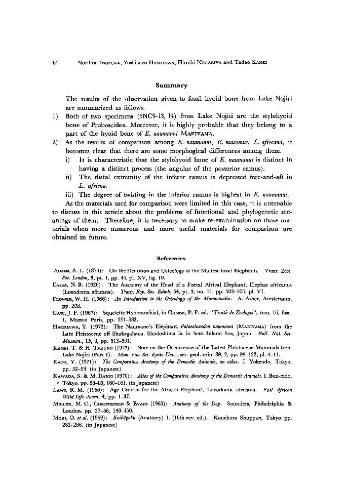
The Stylohyoid bones of ElePhas naumamii MAKiyAMA (all naturalsize)Fig. 1. Inner lateral view of the left side bone.
Fig. 2. Inner lateral view of the right side bone (partly broken).
Explanation of plate 7
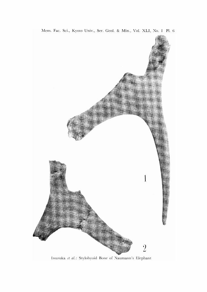
The Stylohyoid bones of Elephas naumanni MAKiyAMA (all natural size)Fig. 1. Anterior view of the left side bone.
Fig. 2. Anterior view of the right side bone.
Fig. 3. Posterior view of the left side bone.
Fig. 4. Posterior view of the right side bone.
(Photo by T. FuJiTA)

Mem.Fac. Sci•o Kyoto Univ., Ser. Geol. & Min., Vol. XLI, No. 1 Pl. 5
'suJ 't't"-' 'w.
.s'xti, 'g' 'Z,lj.
'
ge,lf.
i ge'".,
, ,l•\.l/lis•
I'
it
"la'.' : . m,}ny. s"' . ---'
m' ..'i'fi
sls.
si
'i
IL
es- #-
.tin. ks tc" .
thdiff,I
'en gt ttl/ in
get:t #Vttdi/ .t rel tm.
V{t.'.m
1
ptt
.t m. ttartt
' lvwh aft
-.
w
't
'
'
'i
"L'
ee
get.
`)
-
Inuzuka et al. : Stvlohvoid ii
Bone of Naumann's Elephant
Mem.Fac. Sci., Kyoto Univ., Ser.
•il'•
• •/. /•
tt. t.tt /ttt
tY•/iil,lilii••i••
di..
Geol. & Min., Vol. XLI, No. 1 Pl. 6
.t:ttt tt ttt t tt tt/1 •l-l!i, tt .sttt ,l' 1. ., •/ Me il ''//11
/// . fi.t •lj- •, •,//1, ., 'l, tt.lt . /1 .'yt, ... 1,, "'• 'F' ttt .-t. .. .. ., •va•:.,•.ut'fi•'•.,.''"ii..'ill,•
./.I '. 1/'' t/../ tt/t t tttt .t / t tt ttt tt t .l /. /. /t .. uet /t
'/m. I' ''tt'
, ,x
g 'i 'k
t/t
Inuzuka et al.: Stylohyoid Bone of Naumann's
1
2
Elephant
t/.
•,•//
''i'/t
•i l:•1,
'' i
l,
Mem.Fac. Sci., Kyoto Univ., Ser. Geol. & Min -)Vol. XLI ,
No. 1 Pl. 7
1
'"'iee
di' s'•
.se
4
Inuzuka et al.: Stylohyoid Bone of Naumann's Elephant