three dimensional quantitative coronary angiography … · quately define lesions that are...
TRANSCRIPT
© 2015 The Korean Academy of Medical Sciences.This is an Open Access article distributed under the terms of the Creative Commons Attribution Non-Commercial License (http://creativecommons.org/licenses/by-nc/4.0) which permits unrestricted non-commercial use, distribution, and reproduction in any medium, provided the original work is properly cited.
pISSN 1011-8934eISSN 1598-6357
Three Dimensional Quantitative Coronary Angiography Can Detect Reliably Ischemic Coronary Lesions Based on Fractional Flow Reserve
Conventional coronary angiography (CAG) has limitations in evaluating lesions producing ischemia. Three dimensional quantitative coronary angiography (3D-QCA) shows reconstructed images of CAG using computer based algorithm, the Cardio-op B system (Paieon Medical, Rosh Ha’ayin, Israel). The aim of this study was to evaluate whether 3D-QCA can reliably predict ischemia assessed by myocardial fractional flow reserve (FFR) < 0.80. 3D-QCA images were reconstructed from CAG which also were evaluated with FFR to assess ischemia. Minimal luminal diameter (MLD), percent diameter stenosis (%DS), minimal luminal area (MLA), and percent area stenosis (%AS) were obtained. The results of 3D-QCA and FFR were compared. A total of 266 patients was enrolled for the present study. FFR for all lesions ranged from 0.57 to 1.00 (0.85 ± 0.09). Measurement of MLD, %DS, MLA, and %AS all were significantly correlated with FFR (r = 0.569, 0609, 0.569, 0.670, respectively, all P < 0.001). In lesions with MLA < 4.0 mm2, %AS of more than 65.5% had a 80% sensitivity and a 83% specificity to predict FFR < 0.80 (area under curve, AUC was 0.878). 3D-QCA can reliably predict coronary lesions producing ischemia and may be used to guide therapeutic approach for coronary artery disease.
Keywords: Coronary Angiography; Myocardial Ischemia; Coronary Artery Disease
Woo-Young Chung,1 Byoung-Joo Choi,2 Seong-Hoon Lim,3 Yoshiki Matsuo,4 Ryan J Lennon,5 Rajiv Gulati,5 Gurpreet S. Sandhu,5 David R Holmes Jr,5 Charanjit S Rihal,5 and Amir Lerman5
1Devision of Cardiology, Department of Internal Medicine, Boramae Medical Center, Seoul National University, College of Medicine, Seoul; 2Department of Cardiology, Ajou University School of Medicine, Suwon; 3Division of Cardiology, Department of Internal Medicine, School of Medicine, Dankook University, Cheonan, Korea; 4Department of Cardiovascular Medicine, Wakayama Medical University, Wakayama, Japan; 5Division of Cardiovascular Disease, Mayo Clinic, Rochester, MN, USA
Received: 3 August 2014Accepted: 28 January 2015
Address for Correspondence:Amir Lerman, MD200 First Street S.W., Rochester, MN 55905, Mayo Clinic, Rochester, MN, USATel: +1.507-255-4152, Fax: +1.507-255-2550E-mail: [email protected]
http://dx.doi.org/10.3346/jkms.2015.30.6.716 • J Korean Med Sci 2015; 30: 716-724
INTRODUCTION
Conventional coronary angiography has some limitations in the evaluation of lesions because it provides two dimensional images for a three dimensional structure. And it does not ade-quately define lesions that are angulated, eccentric, or obscured by overlapped vessels (1, 2). In addition, conventional angiog-raphy cannot measure ischemia. Myocardial fractional flow reserve (FFR) is currently a stan-dard method to assess whether lesions are associated with myo-cardial ischemia. Earlier studies showed that FFR < 0.75 had diagnostic value for ischemia as well as prognostic value regard-ing cardiovascular outcomes (3, 4). More recently, FFR < 0.80 has become used as a cutoff value based on several studies (5, 6). Besides two dimensional conventional angiography often does not show stenosis clearly, two-dimensional severity which is considered as significant with more than 50% diameter ste-nosis, is poorly correlated with findings measured with FFR (7). Although FFR is accurate for evaluating stenosis, it is more invasive than coronary angiography and require additional cost, time and efforts. Three-dimensional quantitative coronary
angiography (3D-QCA) uses multiple images acquired from conventional coronary angiography to reconstruct three-di-mensional images by its own algorithm. We and others have demonstrated the accuracy to display coronary anatomy has been documented compared with conventional angiography and intravascular ultrasound (IVUS) (8-12). In contrast to two-dimensional angiography, 3D-QCA could potentially be able to evaluate stenoses and predict lesions producing ischemia more accurately because it analyzes and measures lesions from 3 di-mensional views. The current study was designed to test the hy-pothesis that 3D-QCA can predict lesions producing ischemia as compared to FFR.
MATERIALS AND METHODS
Study populationPatients who underwent both coronary angiography and FFR measurement between February, 2008 and January, 2011 for lesions with intermediate functional significance were screened. Exclusion criteria were lesions 1) in left main stem, 2) bypass graft, 3) instent restenosis, 4) a vessel protected by bypass graft,
ORIGINAL ARTICLECardiovascular Disorders
Chung W-Y, et al. • A Correlation Analysis with Invasive Physiologic Study
http://jkms.org 717http://dx.doi.org/10.3346/jkms.2015.30.6.716
5) poorly visualized, 6) related with myocardial infarction, 7) in patients presented in shock, and 8) located in tandem.
FFR measurement Diagnostic coronary angiography was performed with 5 Fr cath-eter prior to coronary pressure measurement through femoral or radial percutaneous approach. Heparin (5,000-7,000 U intra-venous bolus), nitroglycerin (200 μg intracoronary bolus) were administered and then FFR measurement was done as previ-ously described (13, 14). Catheter changed to 6 Fr guiding cath-eter, a 0.014-in. pressure guidewire (St. Jude Medical, Inc., St. Paul, MN, USA or Volcano, Rancho Cordova, CA, USA) was cal-ibrated, advanced through the guiding catheter into the coro-nary artery, and positioned distal to the stenosis. The pressure proximal to the stenosis was recorded through the guiding catheter using a pressure transducer (Edwards Life Sciences, Irvine, CA, USA). Maximal coronary blood flow was achieved by incremental doses of intracoronary adenosine (18-72 μg for the right coronary artery and 24-72 μg for the left coro-nary artery) or intravenous adenosine (140 μg/kg/min continu-ous infusion via antecubital vein) until a plateau response in
FFR was achieved. FFR was automatically calculated by the ra-tio of distal mean coronary pressure to mean aortic pressure during maximal hyperemia. The minimum FFR value achieved during the administration of the highest dose of adenosine was used for data analysis.
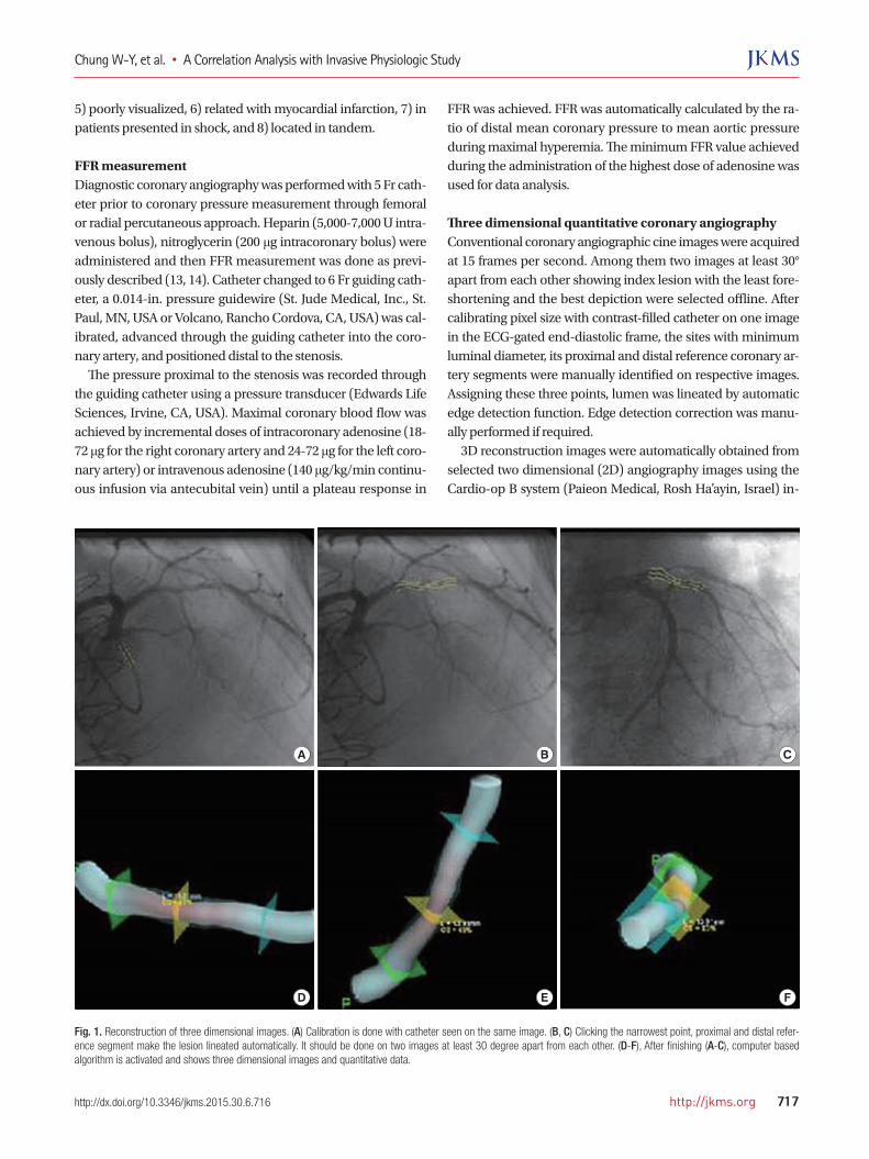
Three dimensional quantitative coronary angiographyConventional coronary angiographic cine images were acquired at 15 frames per second. Among them two images at least 30° apart from each other showing index lesion with the least fore-shortening and the best depiction were selected offline. After calibrating pixel size with contrast-filled catheter on one image in the ECG-gated end-diastolic frame, the sites with minimum luminal diameter, its proximal and distal reference coronary ar-tery segments were manually identified on respective images. Assigning these three points, lumen was lineated by automatic edge detection function. Edge detection correction was manu-ally performed if required. 3D reconstruction images were automatically obtained from selected two dimensional (2D) angiography images using the Cardio-op B system (Paieon Medical, Rosh Ha’ayin, Israel) in-
A
D
B
E
C
F
Fig. 1. Reconstruction of three dimensional images. (A) Calibration is done with catheter seen on the same image. (B, C) Clicking the narrowest point, proximal and distal refer-ence segment make the lesion lineated automatically. It should be done on two images at least 30 degree apart from each other. (D-F), After finishing (A-C), computer based algorithm is activated and shows three dimensional images and quantitative data.
Chung W-Y, et al. • A Correlation Analysis with Invasive Physiologic Study
718 http://jkms.org http://dx.doi.org/10.3346/jkms.2015.30.6.716
stalled in workstation (Fig. 1). This software outlined minimal luminal diameter (MLD), proximal and distal reference diame-ter (PRD, DRD respectively), minimal luminal area (MLA), prox-imal and distal reference luminal area, percent area stenosis (%AS), percent diameter stenosis (%DS), lesion length as well as 3D representation of the arterial lumen. This measurement was undertaken by a experienced cardiologist blinded to FFR values. Interobserver and intraobserver error was reported to be acceptable in previous study (15, 16). To assess the superiority of 3D-QCA to preexisting 2D-QCA in predicting FFR < 0.80, we measured 2D-QCA variables for lesions in randomly selected 39 patients and analysed correla-tion with their corresponding FFR, diagnostic value predicting FFR < 0.80.
Intra- and Inter-observer variation analysisFor intraobserver variation analysis, a single observer recon-structed 3D-QCA image and measured one index lesion twice in 10 patients. For interobserver variation analysis, two inde-pendent observer did the same procedure for 3D-QCA mea-surement separately in 20 patients. The variations were assessed by Tukey method and the difference between two measurements was regarded as not different if P value is more then 0.05, mean-ing the difference is located within 95% confidence interval.
Statistical analysisAn independent statistician performed the statistical analysis. Continuous variables were expressed as mean ± standard devi-ation and categorical variables as percentage and counts. Each 3D-QCA variable was examined by bivariate correlation analy-sis and Pearson’s correlation coefficients were obtained to de-termine if they have linear relation with FFR. Receiver operat-ing characteristic (ROC) curve was drawn and area under curve (AUC) calculated to analyse and quantify the discriminatory ability of individual morphologic variable in predicting FFR < 0.80. For a variable with acceptable AUC, the value with the best sensitivity and specificity was suggested as a diagnostic cutoff for significant ischemia. A two-sided P value of < 0.05 is considered significant. All of statistical analyses were performed using JMP 9.0. (SAS Institute, Cary, NC, USA).
Ethics statementEach patient gave written informed consent for all procedures and the present study was approved by institutional review board (Mayo Clinic, Rochester Minnesota, IRB approval number 11-005843).
RESULTS
Patients and angiographic characteristicsOnly one lesion per patient was screened. Thirteen lesions (4.9%)
of screened 279 lesions were excluded. They were two lesions protected by bypass graft, one a instent restenotic lesion, five poorly visualized, and another five in tandem. Two hundred sixty-six lesions of 266 patients were enrolled for the present study finally. Patients’ mean age was sixty seven and 67% of all were males. 73.7% of all patients had hypertension, 23.4% dia-betes mellitus, 88.2% dyslipidemia, and 15.7% were current smo-kers. Target lesions for comparing FFR and 3D-QCA parameter were located mainly in left anterior descending artery (59.4%), 19.2% in left circumflex artery and 21.4% in right coronary artery. FFR for all target lesions ranged from 0.57 to 1.00. The mean was 0.85 and standard deviation was 0.09. Two dimensional angiographic characteristics of lesions are summarized in Table 1. Reference diameter was 2.66 ± 0.54 mm proximally, 2.45 ± 0.52 mm distally. Lesions were moderately narrowed as percent diameter stenosis (40.9% ± 10.7%), percent area stenosis (60.6% ± 11.0%) has indicated.
Intra- and Inter-observer variationWe assessed intraobserver variation of MLD, %DS, MLA, %AS from 3D-QCA measurement. The difference between two mea-surement by a single observer existed within 95% confidence interval so that P value had more than 0.05. Similar statistical results were obtained in interobserver variation analysis (data not shown).
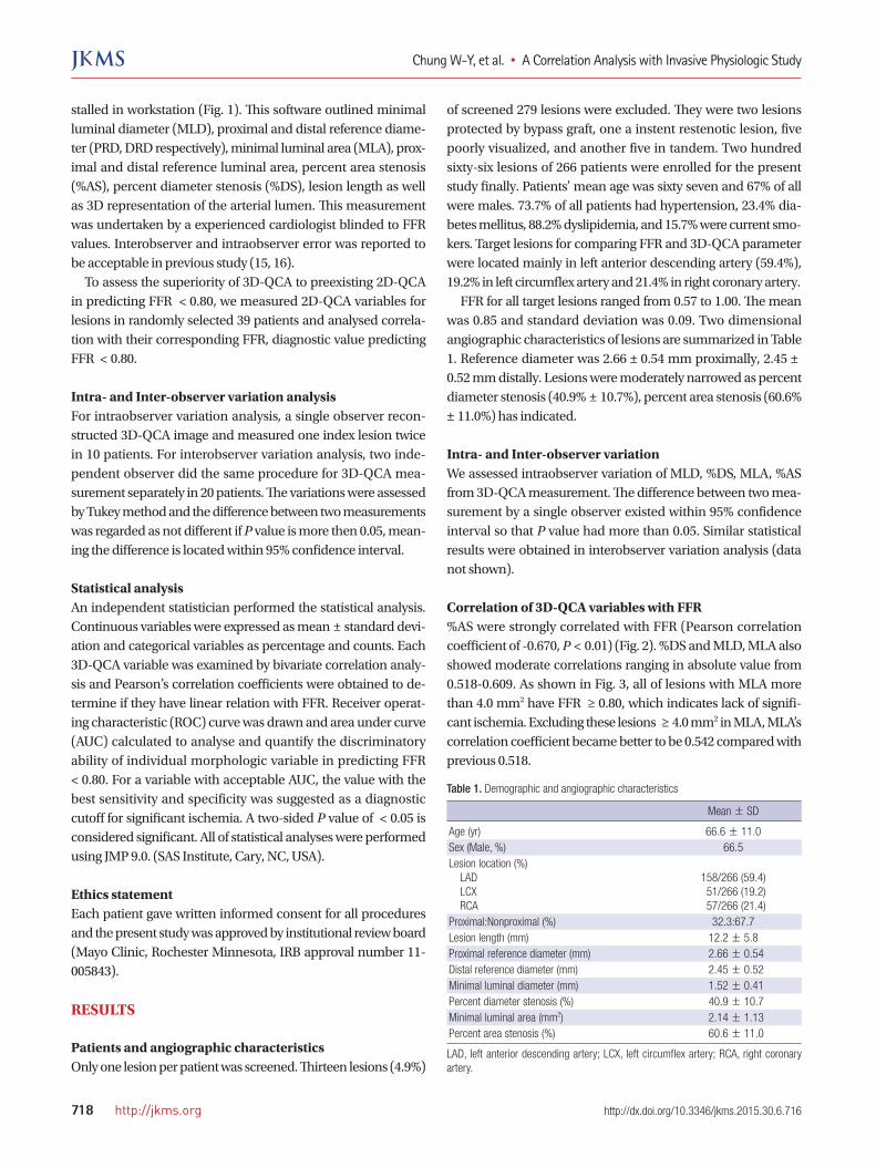
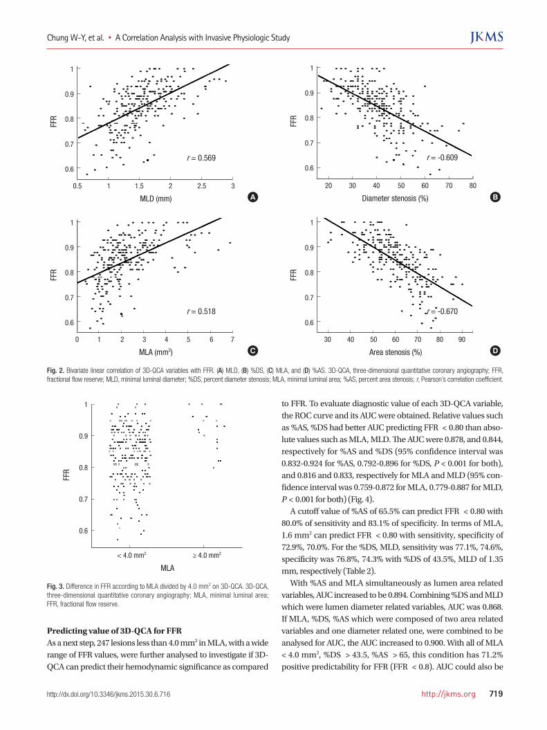
Correlation of 3D-QCA variables with FFR%AS were strongly correlated with FFR (Pearson correlation coefficient of -0.670, P < 0.01) (Fig. 2). %DS and MLD, MLA also showed moderate correlations ranging in absolute value from 0.518-0.609. As shown in Fig. 3, all of lesions with MLA more than 4.0 mm2 have FFR ≥ 0.80, which indicates lack of signifi-cant ischemia. Excluding these lesions ≥ 4.0 mm2 in MLA, MLA’s correlation coefficient became better to be 0.542 compared with previous 0.518.
Table 1. Demographic and angiographic characteristics
Mean ± SD
Age (yr) 66.6 ± 11.0Sex (Male, %) 66.5Lesion location (%) LAD LCX RCA
158/266 (59.4) 51/266 (19.2) 57/266 (21.4)
Proximal:Nonproximal (%) 32.3:67.7 Lesion length (mm) 12.2 ± 5.8Proximal reference diameter (mm) 2.66 ± 0.54Distal reference diameter (mm) 2.45 ± 0.52Minimal luminal diameter (mm) 1.52 ± 0.41 Percent diameter stenosis (%) 40.9 ± 10.7Minimal luminal area (mm2) 2.14 ± 1.13Percent area stenosis (%) 60.6 ± 11.0
LAD, left anterior descending artery; LCX, left circumflex artery; RCA, right coronary artery.
Chung W-Y, et al. • A Correlation Analysis with Invasive Physiologic Study
http://jkms.org 719http://dx.doi.org/10.3346/jkms.2015.30.6.716
Predicting value of 3D-QCA for FFRAs a next step, 247 lesions less than 4.0 mm2 in MLA, with a wide range of FFR values, were further analysed to investigate if 3D-QCA can predict their hemodynamic significance as compared
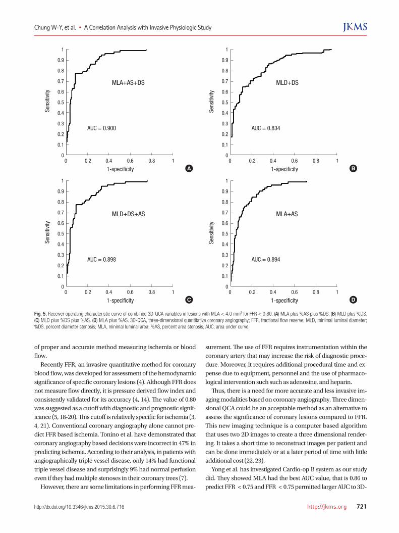
to FFR. To evaluate diagnostic value of each 3D-QCA variable, the ROC curve and its AUC were obtained. Relative values such as %AS, %DS had better AUC predicting FFR < 0.80 than abso-lute values such as MLA, MLD. The AUC were 0.878, and 0.844, respectively for %AS and %DS (95% confidence interval was 0.832-0.924 for %AS, 0.792-0.896 for %DS, P < 0.001 for both), and 0.816 and 0.833, respectively for MLA and MLD (95% con-fidence interval was 0.759-0.872 for MLA, 0.779-0.887 for MLD, P < 0.001 for both) (Fig. 4). A cutoff value of %AS of 65.5% can predict FFR < 0.80 with 80.0% of sensitivity and 83.1% of specificity. In terms of MLA, 1.6 mm2 can predict FFR < 0.80 with sensitivity, specificity of 72.9%, 70.0%. For the %DS, MLD, sensitivity was 77.1%, 74.6%, specificity was 76.8%, 74.3% with %DS of 43.5%, MLD of 1.35 mm, respectively (Table 2). With %AS and MLA simultaneously as lumen area related variables, AUC increased to be 0.894. Combining %DS and MLD which were lumen diameter related variables, AUC was 0.868. If MLA, %DS, %AS which were composed of two area related variables and one diameter related one, were combined to be analysed for AUC, the AUC increased to 0.900. With all of MLA < 4.0 mm2, %DS > 43.5, %AS > 65, this condition has 71.2% positive predictability for FFR (FFR < 0.8). AUC could also be
FFR
MLD (mm)
0.5 1 1.5 2 2.5 3
1
0.9
0.8
0.7
0.6
r = 0.569
FFR
MLA (mm2)
0 1 2 3 4 5 6 7
1
0.9
0.8
0.7
0.6
r = 0.518
FFR
Area stenosis (%)
30 40 50 60 70 80 90
1
0.9
0.8
0.7
0.6
r = -0.670
FFR
Diameter stenosis (%)
20 30 40 50 60 70 80
1
0.9
0.8
0.7
0.6r = -0.609
A
C
B
D
Fig. 2. Bivariate linear correlation of 3D-QCA variables with FFR. (A) MLD, (B) %DS, (C) MLA, and (D) %AS. 3D-QCA, three-dimensional quantitative coronary angiography; FFR, fractional flow reserve; MLD, minimal luminal diameter; %DS, percent diameter stenosis; MLA, minimal luminal area; %AS, percent area stenosis; r, Pearson’s correlation coefficient.
Fig. 3. Difference in FFR according to MLA divided by 4.0 mm2 on 3D-QCA. 3D-QCA, three-dimensional quantitative coronary angiography; MLA, minimal luminal area; FFR, fractional flow reserve.
FFR
MLA
< 4.0 mm2 ≥ 4.0 mm2
1
0.9
0.8
0.7
0.6

Chung W-Y, et al. • A Correlation Analysis with Invasive Physiologic Study
720 http://jkms.org http://dx.doi.org/10.3346/jkms.2015.30.6.716
elevated to be 0.898 with %DS, MLD, %AS, if two diameter re-lated variables were combined with %AS (Fig. 5). However, MLD and %DS obtained by 2D-QCA showed poor correlation with FFR (r = 0.11 for both). Their diagnostic values to detect FFR < 0.80 were also low so that AUC was 0.550, 0.526 for MLD, %DS, respectively.
DISCUSSION
Current study demonstrates that combining several parameters derived from 3D-QCA has high sensitivity and specificity to de-tect hemodynamically significant lesions as assessed by FFR. The current study suggests the use of 3D-QCA may help to guide coronary intervention. Since introduction by Dr. Sones in the 1960s, contrast coro-nary angiography has been a mainstay in diagnosis of coronary artery disease. It visualizes the lumen of the coronary arteries, quantifies diameter stenosis and can evaluate its flow semiquan-titatively by frame count (17). Traditionally more than 50% di-ameter stenosis has been regarded as significant because it com-promises arterial lumen by more than 75%. This angiographical criteria was a long standing cutoff value of functional diagnosis, i.e., ischemia as well as anatomical diagnosis because of the lack
Table 2. Diagnostic value of 3D-QCA variables predicting fractional flow reserve < 0.80 as gold standard of myocardial ischemia
Cutoff Sens (%) Spec (%) PPV (%) NPV (%) Acc (%)
MLD 1.35 mm 74.6 74.3 88.0 53.6 74.5% DS 43.5% 77.1 76.8 56.8 89.4 76.9MLA 1.6 mm2 72.9 70.0 86.0 50.5 72.1% AS 65.5% 80.0 83.1 65.1 91.3 82.8
MLD, minimal luminal diameter; %DS, percent diameter stenosis; MLA, minimal lu-minal area; %AS, percent area stenosis; Sens, sensitivity; Spec, specificity; PPV, posi-tive predictive value; NPV, negative predictive value; Acc, accuracy.
Sens
itivi
ty
1-specificity0 0.2 0.4 0.6 0.8 1
1
0.9
0.8
0.7
0.6
0.5
0.4
0.3
0.2
0.1
0
AUC = 0.833
MLD
A
Sens
itivi
ty
1-specificity0 0.2 0.4 0.6 0.8 1
1
0.9
0.8
0.7
0.6
0.5
0.4
0.3
0.2
0.1
0
AUC = 0.816
MLA
C
Sens
itivi
ty
1-specificity0 0.2 0.4 0.6 0.8 1
1
0.9
0.8
0.7
0.6
0.5
0.4
0.3
0.2
0.1
0
AUC = 0.844
%DS
B
Sens
itivi
ty
1-specificity0 0.2 0.4 0.6 0.8 1
1
0.9
0.8
0.7
0.6
0.5
0.4
0.3
0.2
0.1
0
AUC = 0.878
%AS
D
Fig. 4. Receiver operating characteristic curve of 3D-QCA variables in lesions with MLA < 4.0 mm2 for FFR < 0.80. (A) Minimal luminal diameter. (B) percent area stenosis. (C) minimal luminal area. (D) percent area stenosis. 3D-QCA, three-dimensional quantitative coronary angiography; FFR, fractional flow reserve; MLD, minimal luminal diameter; %DS, percent diameter stenosis; MLA, minimal luminal area; %AS, percent area stenosis; AUC, area under curve.
Chung W-Y, et al. • A Correlation Analysis with Invasive Physiologic Study
http://jkms.org 721http://dx.doi.org/10.3346/jkms.2015.30.6.716
of proper and accurate method measuring ischemia or blood flow. Recently FFR, an invasive quantitative method for coronary blood flow, was developed for assessment of the hemodynamic significance of specific coronary lesions (4). Although FFR does not measure flow directly, it is pressure derived flow index and consistently validated for its accuracy (4, 14). The value of 0.80 was suggested as a cutoff with diagnostic and prognostic signif-icance (5, 18-20). This cutoff is relatively specific for ischemia (3, 4, 21). Conventional coronary angiography alone cannot pre-dict FFR based ischemia. Tonino et al. have demonstrated that coronary angiography based decisions were incorrect in 47% in predicting ischemia. According to their analysis, in patients with angiographically triple vessel disease, only 14% had functional triple vessel disease and surprisingly 9% had normal perfusion even if they had multiple stenoses in their coronary trees (7). However, there are some limitations in performing FFR mea-
surement. The use of FFR requires instrumentation within the coronary artery that may increase the risk of diagnostic proce-dure. Moreover, it requires additional procedural time and ex-pense due to equipment, personnel and the use of pharmaco-logical intervention such such as adenosine, and heparin. Thus, there is a need for more accurate and less invasive im-aging modalities based on coronary angiography. Three dimen-sional QCA could be an acceptable method as an alternative to assess the significance of coronary lesions compared to FFR. This new imaging technique is a computer based algorithm that uses two 2D images to create a three dimensional render-ing. It takes a short time to reconstruct images per patient and can be done immediately or at a later period of time with little additional cost (22, 23). Yong et al. has investigated Cardio-op B system as our study did. They showed MLA had the best AUC value, that is 0.86 to predict FFR < 0.75 and FFR < 0.75 permitted larger AUC to 3D-
Sens
itivi
ty
1-specificity0 0.2 0.4 0.6 0.8 1
1
0.9
0.8
0.7
0.6
0.5
0.4
0.3
0.2
0.1
0
AUC = 0.900
MLA+AS+DS
A
Sens
itivi
ty
1-specificity0 0.2 0.4 0.6 0.8 1
1
0.9
0.8
0.7
0.6
0.5
0.4
0.3
0.2
0.1
0
AUC = 0.898
MLD+DS+AS
C
Sens
itivi
ty
1-specificity0 0.2 0.4 0.6 0.8 1
1
0.9
0.8
0.7
0.6
0.5
0.4
0.3
0.2
0.1
0
AUC = 0.834
MLD+DS
B
Sens
itivi
ty
1-specificity0 0.2 0.4 0.6 0.8 1
1
0.9
0.8
0.7
0.6
0.5
0.4
0.3
0.2
0.1
0
AUC = 0.894
MLA+AS
D
Fig. 5. Receiver operating characteristic curve of combined 3D-QCA variables in lesions with MLA < 4.0 mm2 for FFR < 0.80. (A) MLA plus %AS plus %DS. (B) MLD plus %DS. (C) MLD plus %DS plus %AS. (D) MLA plus %AS. 3D-QCA, three-dimensional quantitative coronary angiography; FFR, fractional flow reserve; MLD, minimal luminal diameter; %DS, percent diameter stenosis; MLA, minimal luminal area; %AS, percent area stenosis; AUC, area under curve.
Chung W-Y, et al. • A Correlation Analysis with Invasive Physiologic Study
722 http://jkms.org http://dx.doi.org/10.3346/jkms.2015.30.6.716
QCA than FFR < 0.80. They also have revealed excellent repro-ducibility. However, the study included relatively small number of lesions and FFR < 0.80 is currently more widely accepted as an ischemic cut-off value. The present study serves as an exten-sion of previous observations and demonstrated that the com-bination of several 3D parameters improves the accuracy and predictive values of 3D-QCA. IVUS is another invasive imaging modality that is well vali-dated. 3D-QCA is similar to IVUS as both can measure luminal area and % area stenosis. Although IVUS does not yield func-tional data, MLA < 4.0 mm2 measured by IVUS was known to have a diagnostic and prognostic value (24, 25). Interestingly, in the present study, all of lesions with MLA more than 4.0 mm2 by 3D-QCA actually have FFR ≥ 0.80. This finding is therefore consistent with IVUS studies, indicating that lesions with more than 4.0 mm2 of MLA may not need further evaluation for isch-emia. For lesions with MLD < 4.0 mm2, the current study demon-strated that %AS revealed the best AUC among variables, and had 65.5% as a cutoff value with acceptable sensitivity and spec-ificity. Similarly Briguori et al. have shown previously that 70% %AS by IVUS is appropriate for predicting ischemia (24). Our cutoff value of MLA, 1.6 mm2 is quite smaller than IVUS’s. How-ever, 4.0 mm2 of MLA on IVUS study had poor specificity 56% although sensitivity was 92%. Recently, Kang et al. suggested that 2.4 mm2 as a new IVUS cutoff value (26). However, it should be notable their AUC was 0.80, specificity 60% which were both inferior to our combined parameters. Previously, there was a debate about which parameter might predict better FFR based ischemia between MLA and %AS (15, 23). Both studies had a small number of patients. In our current study which included larger number of lesions, we have demonstrated that %AS is the best predictor as a single variable. It is reasonable to consider that MLA cannot be uniformly applied to different sized coro-nary artery because it is an absolute value. It becomes evident that FFR values are diffusely distributed for lesions with MLA < 4.0 mm2 (Fig. 3). Because MLA > 4.0 mm2 is sufficient to ex-clude a lesion mediating myocardial ischemia (100% negative predictability), we focused more on lesions with MLA < 4.0 mm2. Finally we evaluated additional variables for a cut-off other than MLA < 4.0 mm2 to enhance the sensitivity and specificity. The ability to predict FFR associated with myocardial isch-emia with 3D-QCA was enhanced if variables were considered in combination. Among many possible combinations, area re-lated values such as %AS and MLA produced the best AUC. If %DS were considered along with %AS and MLA, AUC reached 0.900. In real practice, we can experience the situation in which 3D-QCA derived %AS or MLA alone is debatable. At that time, integrative decision of %AS, MLA, %DS would increase diag-nostic yield. As the first step applying using 3D-QCA for intermediate le-
sions in real world practice, we can exclude the lesions if MLA > 4.0 mm2. Next, we can exclude the lesions again if %AS < 65.5 having the highest AUC with high negative predictability and specificity. For the remained lesion, we can conclude based on MLA < 1.6 mm2 and %DS of 43.5% comprehensively if they would have FFR < 0.80 although it is uncertain to decide when MLA and %DS values are contradictory. There are some limitations in our study. Left main lesions were excluded, and thus the results can not be extended to left main lesions. Second, as 3D-QCA depends on the quality of conventional angiography, the appropriate 3D-QCA images and variables may not be obtained, thereby limit its clinical ap-plication. Third, this study was done in a single center and has small study population. It prevents from modeling well struc-tured diagnostic algorithm suggesting 3D-QCA variables step by step to be more useful in clinical practice. Larger sized study would solve this limitation. In conclusion, parameters derived from 3D-QCA may reli-ably predict the functional significance of lesions based on FFR and may become a useful less invasive method to guide deci-sion making in cardiac catheterization laboratory. It is reason-able that in future practice, the combination of lesion specific parameter such as MLA, %AS and %DS will be calculated and presented simultaneously during angiogram and may create 3D-QCA derived specific parameter of the target lesion to as-sess its functional significance.
ACKNOWLEDGEMENTS
We, all authors, thank Robert Cole and Rebecca E. Nelson for their executive works and efforts to proceed with this study and maintain 3D-QCA software.
DISCLOSURE
The authors have no conflicts of interest to disclose.
AUTHOR CONTRIBUTION
Study concepts and design: Chung WY, Lerman A, Lim SH. Study coordination: Chung WY, Lerman A, Lim SH. Acquisition of data: Chung WY, Lim SH, Choi BJ, Matsuo Y. Data review: Chung WY, Lim SH, Choi BJ, Matsuo Y. Statistical analysis: Chung WY, Lim SH, Lennon RJ. Manuscript preparation: Chung WY, Lerman A, Gulati R, Sandhu GS, Holmes DR Jr, Rihal CS. Manuscript ap-proval: all authors.
ORCID
Woo-Young Chung http://orcid.org/0000-0002-9106-9331Byoung-Joo Choi http://orcid.org/0000-0002-3045-5343

Chung W-Y, et al. • A Correlation Analysis with Invasive Physiologic Study
http://jkms.org 723http://dx.doi.org/10.3346/jkms.2015.30.6.716
Seong-Hoon Lim http://orcid.org/0000-0002-2941-2784Ryan J Lennon http://orcid.org/0000-0002-8366-8422Rajiv Gulati http://orcid.org/0000-0002-2713-0433Gurpreet S. Sandhu http://orcid.org/0000-0002-4456-6183Amir Lerman http://orcid.org/0000-0002-9446-5313
REFERENCES
1. Gottsauner-Wolf M, Sochor H, Moertl D, Gwechenberger M, Stocken-
huber F, Probst P. Assessing coronary stenosis. Quantitative coronary
angiography versus visual estimation from cine-film or pharmacologi-
cal stress perfusion images. Eur Heart J 1996; 17: 1167-74.
2. Topol EJ, Nissen SE. Our preoccupation with coronary luminology. The
dissociation between clinical and angiographic findings in ischemic heart
disease. Circulation 1995; 92: 2333-42.
3. Pijls NH, van Schaardenburgh P, Manoharan G, Boersma E, Bech JW,
van’t Veer M, Bär F, Hoorntje J, Koolen J, Wijns W, et al. Percutaneous
coronary intervention of functionally nonsignificant stenosis: 5-year fol-
low-up of the DEFER Study. J Am Coll Cardiol 2007; 49: 2105-11.
4. Pijls NH, De Bruyne B, Peels K, Van Der Voort PH, Bonnier HJ, Bartunek
JKJJ, Koolen JJ. Measurement of fractional flow reserve to assess the func-
tional severity of coronary-artery stenoses. N Engl J Med 1996; 334: 1703-8.
5. Tonino PA, De Bruyne B, Pijls NH, Siebert U, Ikeno F, van’ t Veer M,
Klauss V, Manoharan G, Engstrøm T, Oldroyd KG, et al.; FAME Study
Investigators. Fractional flow reserve versus angiography for guiding
percutaneous coronary intervention. N Engl J Med 2009; 360: 213-24.
6. Hamilos M, Muller O, Cuisset T, Ntalianis A, Chlouverakis G, Sarno G,
Nelis O, Bartunek J, Vanderheyden M, Wyffels E, et al. Long-term clini-
cal outcome after fractional flow reserve-guided treatment in patients
with angiographically equivocal left main coronary artery stenosis. Cir-
culation 2009; 120: 1505-12.
7. Tonino PA, Fearon WF, De Bruyne B, Oldroyd KG, Leesar MA, Ver Lee
PN, Maccarthy PA, Van’t Veer M, Pijls NH. Angiographic versus func-
tional severity of coronary artery stenoses in the FAME study fractional
flow reserve versus angiography in multivessel evaluation. J Am Coll
Cardiol 2010; 55: 2816-21.
8. Agostoni P, Biondi-Zoccai G, Van Langenhove G, Cornelis K, Vermeersch
P, Convens C, Vassanelli C, Van Den Heuvel P, Van Den Branden F, Ver-
heye S. Comparison of assessment of native coronary arteries by stan-
dard versus three-dimensional coronary angiography. Am J Cardiol
2008; 102: 272-9.
9. Dvir D, Marom H, Guetta V, Kornowski R. Three-dimensional coronary
reconstruction from routine single-plane coronary angiograms: in vivo
quantitative validation. Int J Cardiovasc Intervent 2005; 7: 141-5.
10. Gollapudi RR, Valencia R, Lee SS, Wong GB, Teirstein PS, Price MJ. Util-
ity of three-dimensional reconstruction of coronary angiography to guide
percutaneous coronary intervention. Catheter Cardiovasc Interv 2007;
69: 479-82.
11. Rubinshtein R, Lerman A, Spoon DB, Rihal CS. Anatomic features of the
left main coronary artery and factors associated with its bifurcation an-
gle: a 3-dimensional quantitative coronary angiographic study. Catheter
Cardiovasc Interv 2012; 80: 304-9.
12. Tu S, Huang Z, Koning G, Cui K, Reiber JH. A novel three-dimensional
quantitative coronary angiography system: in-vivo comparison with in-
travascular ultrasound for assessing arterial segment length. Catheter
Cardiovasc Interv 2010; 76: 291-8.
13. Pijls NH, van Son JA, Kirkeeide RL, De Bruyne B, Gould KL. Experi-
mental basis of determining maximum coronary, myocardial, and col-
lateral blood flow by pressure measurements for assessing functional ste-
nosis severity before and after percutaneous transluminal coronary an-
gioplasty. Circulation 1993; 87: 1354-67.
14. Pijls NH, Van Gelder B, Van der Voort P, Peels K, Bracke FA, Bonnier HJ,
el Gamal MI. Fractional flow reserve. A useful index to evaluate the in-
fluence of an epicardial coronary stenosis on myocardial blood flow.
Circulation 1995; 92: 3183-93.
15. Yong AS, Ng AC, Brieger D, Lowe HC, Ng MK, Kritharides L. Three-di-
mensional and two-dimensional quantitative coronary angiography,
and their prediction of reduced fractional flow reserve. Eur Heart J 2011;
32: 345-53.
16. Schuurbiers JC, Lopez NG, Ligthart J, Gijsen FJ, Dijkstra J, Serruys PW,
Van der Steen AF, Wentzel JJ. In vivo validation of CAAS QCA-3D coro-
nary reconstruction using fusion of angiography and intravascular ul-
trasound (ANGUS). Catheter Cardiovasc Interv 2009; 73: 620-6.
17. Gibson CM, Cannon CP, Daley WL, Dodge JT Jr, Alexander B Jr, Marble
SJ, McCabe CH, Raymond L, Fortin T, Poole WK, et al. TIMI frame count:
a quantitative method of assessing coronary artery flow. Circulation 1996;
93: 879-88.
18. Pijls NH, Fearon WF, Tonino PA, Siebert U, Ikeno F, Bornschein B, van’t
Veer M, Klauss V, Manoharan G, Engstrøm T, et al.; FAME Study Inves-
tigators. Fractional flow reserve versus angiography for guiding percuta-
neous coronary intervention in patients with multivessel coronary artery
disease: 2-year follow-up of the FAME (Fractional Flow Reserve Versus
Angiography for Multivessel Evaluation) study. J Am Coll Cardiol 2010;
56: 177-84.
19. Bech GJ, De Bruyne B, Akasaka T, Liistro F, Bonnier HJ, Koolen JJ, Pijls
NH. Coronary pressure and FFR predict long-term outcome after PTCA.
Int J Cardiovasc Intervent 2001; 4: 67-76.
20. Legalery P, Schiele F, Seronde MF, Meneveau N, Wei H, Didier K, Blonde
MC, Caulfield F, Bassand JP. One-year outcome of patients submitted to
routine fractional flow reserve assessment to determine the need for an-
gioplasty. Eur Heart J 2005; 26: 2623-9.
21. Berger A, Botman KJ, MacCarthy PA, Wijns W, Bartunek J, Heyndrickx
GR, Pijls NH, De Bruyne B. Long-term clinical outcome after fractional
flow reserve-guided percutaneous coronary intervention in patients with
multivessel disease. J Am Coll Cardiol 2005; 46: 438-42.
22. Rittger H, Schertel B, Schmidt M, Justiz J, Brachmann J, Sinha AM. Three-
dimensional reconstruction allows accurate quantification and length
measurements of coronary artery stenoses. EuroIntervention 2009; 5:
127-32.
23. Saad M, Toelg R, Khattab AA, Kassner G, Abdel-Wahab M, Richardt G.
Determination of haemodynamic significance of intermediate coronary
lesions using three-dimensional coronary reconstruction. EuroInterven-
tion 2009; 5: 573-9.
24. Briguori C, Anzuini A, Airoldi F, Gimelli G, Nishida T, Adamian M, Cor-
vaja N, Di Mario C, Colombo A. Intravascular ultrasound criteria for
the assessment of the functional significance of intermediate coronary
artery stenoses and comparison with fractional flow reserve. Am J Car-
diol 2001; 87: 136-41.
25. Abizaid AS, Mintz GS, Mehran R, Abizaid A, Lansky AJ, Pichard AD,

Chung W-Y, et al. • A Correlation Analysis with Invasive Physiologic Study
724 http://jkms.org http://dx.doi.org/10.3346/jkms.2015.30.6.716
Satler LF, Wu H, Pappas C, Kent KM, et al. Long-term follow-up after
percutaneous transluminal coronary angioplasty was not performed
based on intravascular ultrasound findings: importance of lumen di-
mensions. Circulation 1999; 100: 256-61.
26. Kang SJ, Lee JY, Ahn JM, Mintz GS, Kim WJ, Park DW, Yun SC, Lee SW,
Kim YH, Lee CW, et al. Validation of intravascular ultrasound-derived
parameters with fractional flow reserve for assessment of coronary ste-
nosis severity. Circ Cardiovasc Interv 2011; 4: 65-71.