this work was supported by - welcome to sora - soraopenaccess.sgul.ac.uk/108555/1/dobinson et al...
TRANSCRIPT
Evaluation of the clinical and microbiological response to Salmonella
Paratyphi A infection in the first paratyphoid human challenge model
Authors
Hazel C Dobinson1a, Malick M Gibani1a, Claire Jones1a, Helena B Thomaides-
Brears1*, Merryn Voysey1,2, Thomas C Darton1, Claire S. Waddington1, Danielle
Campbell1, Iain Milligan1, Liqing Zhou1, Sonu Shrestha1, Simon A Kerridge1, Anna
Peters1, Zoe Stevens1, Audino Podda3, Laura B. Martin3, Flavia D’Alessio4, Duy
Pham Thanh 5, Buddha Basnyat6, Stephen Baker5,7,8, Brian Angus9, Myron M
Levine10, Christoph J Blohmke1, Andrew J Pollard1
1Oxford Vaccine Group, Department of Paediatrics, University of Oxford and the NIHR Oxford
Biomedical Research Centre, Oxford, United Kingdom; 2Nuffield Department of Primary Care Health
Sciences University of Oxford, UK; 3 GSK Vaccines Institute for Global Health (GVGH), Via
Fiorentina 1, 53100 Siena, Italy; 4 European Vaccine Initiative, Heidelberg, Germany; 5 The Hospital
for Tropical Diseases, Wellcome Trust Major Overseas Programme, Oxford University Clinical
Research Unit, Ho Chi Minh City, Vietnam;6 Oxford University Clinical Research Unit, Patan
Academy of Health Sciences, Kathmandu, Nepal; 7 Centre for Tropical Medicine and Global Health,
University of Oxford, Oxford, United Kingdom;8 The London School of Hygiene and Tropical
Medicine, London, United Kingdom; 9Nuffield Department of Medicine, University of Oxford, UK; 10
Center for Vaccine Development, University of Maryland School of Medicine, Baltimore, MD USA
a H.C.D, M.M.G and C.J. contributed equally to this manuscript. *Corresponding
author.
Keywords: Paratyphoid Infection, Enteric Fever, Salmonella enterica Paratyphi A,
Human challenge study, Immune responses
Running title: Human Challenge Model of S. Paratyphi A
1

Corresponding author contact:
Helena Thomaides-BrearsOxford Vaccine GroupCentre for Clinical Vaccinology and Tropical MedicineChurchill HospitalOld Road, HeadingtonOxford, UKOX3 7LETelephone 0044-1865-611364/Fax: 0044-1865 857420; [email protected]
Alternate corresponding author: Malick Gibani contact details as above;
Summary (40 words): The safe establishment of a protocol for a human
challenge model for S. Paratyphi A can be used to expedite the evaluation of
novel vaccine candidates and provides insight into the clinical and immune
response to paratyphoid infection.
2
ABSTRACT (250 words):
Background: To expedite the evaluation of vaccines against paratyphoid fever,
we aimed to develop the first human challenge model of S. Paratyphi A infection.
Methods: Two groups of 20 participants underwent oral challenge with S.
Paratyphi A following sodium bicarbonate pre-treatment at one of two dose
levels (Group 1: 1-5 x 10³ CFU and Group 2: 0.5-1 x 10³ CFU). Participants were
monitored in an outpatient setting with daily clinical review and collection of
blood and stool cultures. Antibiotic treatment was started when pre-specified
diagnostic criteria were met (temperature ≥38°C for ≥12 hours and/or
bacteraemia) or at day 14 post challenge.
Results: The primary study objective was achieved following challenge with 1-5
x 103 CFU (Group 1), which resulted in an attack rate of 12/20 (60%). Compared
with typhoid challenge, paratyphoid was notable for high rates of subclinical
bacteraemia (at this dose 11/20; 55%). Despite limited symptoms, bacteraemia
persisted for up to 96 hours after antibiotic treatment (median duration of
bacteraemia 53hrs; IQR 24-85hrs). Shedding of S. Paratyphi A in stool typically
preceded onset of bacteraemia.
Conclusions: Challenge with S. Paratyphi A at a dose of 1-5 x 10³ CFU was well
tolerated and associated with an acceptable safety profile. The frequency and
persistence of bacteraemia in the absence of clinical symptoms was notable, and
markedly different from that seen in previous typhoid challenge studies. We
conclude that the paratyphoid challenge model is suitable for the assessment of
vaccine efficacy using endpoints that include bacteraemia and/or
symptomatology.
Clinicaltrials.gov Registration: NCT02100397
3

4

Introduction
Efforts aimed at reducing the global burden of enteric fever are likely to require
a co-ordinated strategy comprising improvements in water quality, sanitation
and hygiene measures along with the development of effective vaccines. Whilst
Salmonella Typhi remains the principal aetiological agent of enteric fever
globally, Salmonella enterica serovar Paratyphi A (S. Paratyphi A) is responsible
for an increasing proportion of enteric fever cases. In South and South-East
Asia, annual incidence rates in some areas are estimated to be as high as 150
cases/100,000 person-years.[1]
Relatively little is known regarding the pathophysiology and host response to S.
Paratyphi A infection. Salmonella enterica serovars Typhi and Paratyphi A
possess similar genomic markers of host-restriction, but differ with regards to
expression of certain virulence factors, such as the lack of Vi polysaccharide
capsule expression by S. Paratyphi A/B. [2,3] Additionally, there are currently no
licensed vaccines available for the prevention of paratyphoid fever. The licensed
S. Typhi vaccine Ty21a can induce cross-reactive humoral immune responses to
S. Paratyphi A/B in vitro and there is some evidence from field trials for cross
protection against S. Paratyphi B.[4,5] Several promising vaccine candidates are
in development, including live-attenuated strains and conjugate vaccines
targeted against the O-specific polysaccharide (O:2) of S. Paratyphi A, although
no candidate vaccines have undergone efficacy trials to date.[6] Development
of vaccines to prevent paratyphoid infection is hampered by limited knowledge
of immunological correlates of protection and the lack of a suitable small-animal
model of infection.
Human challenge studies are increasingly used to identify promising vaccine
candidates suitable for evaluation in large-scale field trials.[7] In this study, we
5

sought to establish a S. Paratyphi A human challenge model in healthy adult
volunteers by oral challenge to enable future assessment of S. Paratyphi A
vaccines and to assess host-pathogen interactions under a strictly controlled
setting.
6

Methods
Study Design
We performed an observational, dose-level modification human challenge study
of S. Paratyphi A infection conducted in healthy community adult volunteers, at
the Centre for Clinical Vaccinology and Tropical Medicine, University of Oxford,
Oxford, United Kingdom.[8] Description of the study protocol and enrolment
criteria are detailed elsewhere.[9][10]
The primary objective of the study was to determine the dose in CFU of S.
Paratyphi A strain NVGH308 required to achieve an attack rate of 60–75%, when
ingested with sodium bicarbonate solution to neutralise gastric acid.
Challenge strain
S. Paratyphi A strain NVGH308, was isolated in 2006 from a patient with
paratyphoid fever in Kathmandu, Nepal, and is susceptible to most commonly
used antibiotics, including ciprofloxacin (Supplementary Table 1).[9] The
NVGH308 strain was selected as it is a contemporary, circulating strain isolated
from a patient in a highly endemic country.
Whole genome sequencing was undertaken to identify the phylogenetic
relationship of NVGH308 to other circulating strains (Supplementary Figure 1;
Supplementary Methods).
The challenge inoculum was freshly prepared on day of challenge
(Supplementary Methods). An initial target dose of 1-5 x 10³ CFU (Group 1)
was selected based upon prior S. Typhi challenge studies in a similar study
7

population.[11] Following fulfilment of the primary objective at the initial target
dose, we amended the original study protocol to include a dose de-escalation
group (0.5-1 x 10³ CFU; Group 2) in order to affirm a relationship between dose
and diagnostic endpoints.
Clinical Evaluation
Regular participant safety monitoring occurred through daily clinical review for a
minimum of 14 days and included solicited symptoms (Supplementary Table 2) and
twice daily temperature measurements.[9]
Diagnostic Criteria
The primary outcome was the rate of paratyphoid infection, defined as a positive
blood culture for S. Paratyphi A (taken ≥72 hours after challenge to avoid
detection of primary bacteraemia) and/or oral temperature ≥38°C persisting for
≥12 hours. [9] Treatment was initiated upon fulfilment of diagnostic criteria (or
at day 14 for those without illness) and comprised oral ciprofloxacin 500mg twice
daily for 14 days.
Serological Response
Blood was collected at baseline and day 10, 28 and 90 post challenge. Specific
Immunoglobulin M (IgM), IgG and IgA to S. Paratyphi A lipopolysaccharide (LPS)
and flagellin (H) were measured by an in-house enzyme-linked immunosorbent
assay (ELISA) using goat anti-human IgM, IgG and IgA conjugated to horseradish
peroxidase (see Supplementary Methods).
Statistical Analysis
8

All participants were included in the primary analysis and a post-hoc analysis
was conducted comparing participants challenged with S. Paratyphi A in this
study with those challenged with S. Typhi in our previous study.[11]
Clinical, laboratory and immunological data were collated using a clinical trials
database (OpenClinica, version 3.1). Data analysis was performed as described
in detail in the Supplementary Methods, using R version 3.2.2.[12]
9

Results
Forty healthy adult participants were enrolled into the study between 20th May
and 20th November 2014. (Table 1; Supplementary Figure 2). Twenty participants were
challenged with S. Paratyphi A at a target dose of 1-5 x 10³ CFU (group 1). A
second group of twenty participants was challenged at a target dose of 0.5-1.0 x
103 CFU (group 2).
Paratyphoid infection was diagnosed in 20/40 (50%) of all participants
challenged. The primary study objective was achieved in challenge group 1,
where 12/20 participants met the pre-specified diagnostic criteria for
paratyphoid fever (Attack rate 60%; 95%CI 36–81%) (Figure 1). Eleven participants
(11/12, 92%) in this group were diagnosed following a positive blood culture for
S. Paratyphi A and one was diagnosed based on temperature criteria with
subsequent blood culture confirmation. Median time from challenge to initiation
of treatment was 6.4 days (range 5.9-8.3 days) (Table 2).
Paratyphoid infection was diagnosed less frequently in challenge group 2. In this
group, 8/20 participants met the pre-specified diagnostic criteria following
challenge (Attack rate 40%; 95%CI 19-64%), all of whom were diagnosed
following a positive blood culture (Table 2). Median time from challenge to
initiation of treatment was 8.3 days (range 7.1-12.0 days) (Figure 1).
Attack rates using alternative diagnostic criteria are outlined in Table 2. The
hypothetical attack rate was lower in both study groups if clinical end-points
alone (e.g. fever) were used to define paratyphoid disease.
All participants were managed with daily clinical review and no participants met
the pre-specified criteria for severe enteric fever.[9] Two participants had
positive stool cultures for S. Paratyphi A after completing a 14-day course of
10

ciprofloxacin, neither of whom had evidence of gall-bladder disease at screening.
Both were successfully re-treated with a 14-day course of oral azithromycin
(500mg daily) and all subsequent clearance stools were negative. There were no
episodes of relapse of clinical disease.[9] One serious adverse event was
recorded (appendicitis six months after S. Paratyphi A challenge) and was
assessed as being unrelated to study procedures. One participant developed
mouth ulcers after treatment with ciprofloxacin, which resolved following a
change to azithromycin. A second participant developed vaginal candidiasis
following antibiotic treatment (ciprofloxacin), which resolved following treatment
with topical clotrimazole.
The most common symptom reported by those with paratyphoid illness was
headache (17/20; 85%), followed by malaise (15/20; 75%), abdominal pain
(14/20; 70%) and myalgia (14/20; 70%). The majority of solicited symptoms were
graded as mild and did not result in limitation of usual activity (Figure 1,
Supplementary Table 2). In comparison with participants challenged with S. Typhi in
earlier studies,[11] paratyphoid resulted in a milder symptom profile,
characterised by fewer severe symptoms and lower cumulative symptom scores
(Figure 2, Supplementary Table 3).
Laboratory parameters were in keeping with those expected for acute enteric
fever (Supplementary Figure 3).[13] Amongst those meeting diagnostic endpoints,
peripheral blood biochemistry demonstrated elevated C-reactive protein and
alanine aminotransferase, whilst haematology revealed a modest fall in
haemoglobin, neutrophil and lymphocyte counts.
We documented at least one positive blood culture in all participants diagnosed
with paratyphoid (20/20; 100%; median number positive blood cultures 5.5,
range 1-8). Median bacteraemia clearance times were 2.2 days (IQR 1-3.5 days)
11

for S. Paratyphi (Figure 2, Figure 3). The median time from challenge to bacteraemia
was 5.2 days (interquartile range [IQR], 5.0-6.4) in group 1 and 7 days (IQR 6.7-
7.4) in group 2. Quantitative blood cultures performed prior to antibiotic
treatment demonstrated median bacterial loads of 1.1 CFU/ml (range 0-4.1), in
group 1 and 0.2 CFU/ml (range 0-6.9) in group 2 participants, which are
comparable to those seen in cases of typhoid fever in earlier challenge studies
and in endemic settings.[11,14] The rate of blood culture positive days was twice
as high in S. Paratyphi A diagnosed participants than in S. Typhi diagnosed
participants from our previous study (IRR 2.1; 95%CI 1.46 to 3.1, p<0.001) (Figure
2; Table 2).
Sub-clinical bacteraemia was a common finding following challenge, with 11/20
(55%; 95%CI 32-77%) participants having demonstrable bacteraemia but
remaining persistently afebrile following challenge, although other symptoms
were observed as discussed above (Supplementary Table 2). Afebrile
bacteraemia was less common than in earlier typhoid challenge studies
performed by our group (4/21; 19%; 95%CI 5-42%; p=0.025 Fisher’s exact test).
[11] Fever was recorded less frequently in group 2 (2/8; 25%) than in group 1
(7/12; 58%) (Table 2).
Shedding of S. Paratyphi A in stool occurred sporadically throughout the
challenge period and at least one stool cultures was positive in 17/40
participants (43%; 95%CI 27-59%) (Figure 3). Amongst participants with
demonstrable bacteraemia 11/20 (55%; 95%CI 32-77%) had a positive stool
culture for at least one time point and stool shedding frequently preceded the
onset of bacteraemia (Figure 3). Asymptomatic shedding was identified in 6/20
participants (30%; 95%CI 12-54%) who did not reach the diagnostic criteria
during the observation period. The rate of stool positivity was similar in S.
12

Paratyphi A diagnosed participants as was observed previously in S. Typhi
diagnosed participants (IRR 0.78; 95%CI 0.40 to 1.52, p=0.457).
Baseline anti-LPS IgG geometric mean concentrations were higher in
undiagnosed participants prior to challenge (p=0.034) but did not relate to the
incidence of bacteraemia (Supplementary Figure 4). Anti-LPS antibody responses
peaked at day 28 post-challenge and were significantly higher in those
diagnosed with paratyphoid compared with those without acute disease
(p<0.001). In contrast, anti-H antibody responses were only marginally increased
above baseline at D28 and only in those meeting diagnostic endpoints
(Supplementary Figure 4).
13

Discussion
In 1909, Proescher reported a case of “two individuals who accidentally
swallowed a small amount of a pure culture” of Salmonella Paratyphi A and who
“became ill…five and seven days after imbibing” the culture.[15] Here, we
describe the first deliberate and controlled human challenge model using a
contemporary circulating strain of S. Paratyphi A. Whilst S. Typhi challenge
studies have become well established over the past half-century, we have
uniquely demonstrated that S. Paratyphi A challenge can also be undertaken
safely in an outpatient setting. Challenge at a target dose of 1-5x10³ CFU was
sufficient to achieve an attack rate of 60% in a cohort of naïve volunteers, whilst
challenge using a lower dose resulted in a reduced attack rate and increased
time to bacteraemia.
Whilst Vi-conjugate typhoid vaccines are currently licenced in both India and
China, the paratyphoid A vaccine pipeline is less well developed.[6] The effect of
future Vi-based typhoid vaccine programmes on the burden of paratyphoid fever
is unknown and there is conflicting evidence of serovar replacement following
historical typhoid vaccination campaigns.[16,17] This presents a pressing need
for accelerated paratyphoid A vaccine development, which could be facilitated by
the establishment of a human paratyphoid A challenge model. In support of this,
the WHO has stated that well-validated challenge studies can provide
“considerable supporting evidence of the efficacy” of typhoid Vi-conjugate
vaccines, as well as offering insights into immunological correlates of protection.
[18] We suggest that the model described herein could fulfil an equivalent role
for paratyphoid by providing a platform to assess the efficacy of paratyphoid
vaccines currently in development.
14

The choice of diagnostic endpoints is central to future applications of this model
in the assessment of candidate paratyphoid vaccines. In this study, we used a
composite diagnostic end-point of clinical and/or microbiologically defined
paratyphoid infection and observed that 95% of individuals who met the
diagnostic criteria did so based upon microbiological criteria (Table 2). The
frequency of blood culture sampling in this study resulted in the identification of
several cases of asymptomatic bacteraemia, which may or may not have
progressed to symptomatic paratyphoid fever if left untreated. Whilst the
consistent finding of bacteraemia represents an unambiguous diagnostic
endpoint, this could set an overly stringent threshold in future vaccine efficacy
studies and clinical endpoints might be preferable as this more accurately
reflects the situation in the field (Table 2). Arguably, vaccines demonstrating at
least moderate efficacy in this challenge model would perform better in field
trials, where the endpoints would include a clinical syndrome of paratyphoid
fever with culture confirmation. Vaccine efficacy studies using this model should
include multiple alternative pre-specified secondary diagnostic endpoints, such
as the proportion of cases diagnosed by clinical criteria alone, microbiological
criteria alone or a composite of clinical symptoms with culture confirmation.
To our knowledge, no previous studies have explored the infectious dose
required to induce paratyphoid disease, although it is frequently assumed that S.
Paratyphi A requires a higher dose of bacteria to cause clinical disease than does
S. Typhi.[19,20] In this study, an attack rate of 60% was achieved using a dose
of 1-5x10³ CFU S. Paratyphi A, which is comparable to the attack rate (55%)
observed at an equivalent dose of S. Typhi in earlier studies.[11] Notably
challenge with as few as 700 CFU S. Paratyphi A was able to induce acute
disease, in the context of sodium bicarbonate pre-treatment. Our findings are
broadly consistent with observations from S. Typhi challenge studies, where
15

reducing the challenge inoculum results in a reduced attack rate and longer
incubation period.[21] As with S. Typhi, it is possible that the infectious dose
required to induce paratyphoid disease in a non-endemic setting may differ from
that required in an endemic setting, where prior exposure could lead to partial
immunity.
We performed a post-hoc comparison of symptom data collected during earlier
typhoid challenge studies [11] and noted that paratyphoid challenge resulted in
a milder disease profile than that previously observed following typhoid
challenge. In our model, paratyphoid was characterised by high rates of afebrile
bacteraemia in groups 1 and 2 (11/20; 55%) with minimal symptoms, which may
otherwise have escaped detection in the absence of intensive sampling. Case
series from the pre-antibiotic era reported that S. Paratyphi A caused a spectrum
of clinical manifestations that “closely resembles that of mild enteric fever” [22]
and asymptomatic cases of paratyphoid infection are well described.[23,24]
Although the largest published case series to date indicates that S. Typhi and
Paratyphi A infections cause an indistinguishable clinical syndrome[19], this
study was limited to patients who attended healthcare facilities and may not
account for ambulatory or mild cases of paratyphoid in the community. In
keeping with this, epidemiological data from endemic settings suggest that sub-
clinical paratyphoid infection frequently occurs.[5] In light of our challenge model
data, we interpret earlier data as being supportive of the hypothesis of an
underappreciated and undiagnosed burden of paratyphoid disease in the
community. In a proportion of individuals, exposure to the bacillus is followed by
asymptomatic bacteraemia and asymptomatic stool shedding, potentially
perpetuating both long- and short-cycle transmission. In keeping with this,
asymptomatic shedding of S. Paratyphi A was also observed in our model,
16

including in individuals who did not meet diagnostic criteria for paratyphoid
disease.
Somewhat counter-intuitively, we observed prolonged bacteraemia and
protracted time to blood culture clearance (≥96hrs) in the paratyphoid challenge
model, despite the low rate of fever. In addition, convalescent stool shedding
was noted in two individuals despite a 14 day course of ciprofloxacin, both of
whom were successfully treated with a further course of azithromycin. The
relatively poor response to ciprofloxacin was unexpected, given that the
NVGH308 strain is sensitive to ciprofloxacin according to current CLSI and
EUCAST guidelines (MIC 0.06 μg/ml). [25,26] Studies in endemic settings have
found fluoroquinolone MICs for S. Paratyphi A were typically higher than those for
S. Typhi.[19] The mechanisms underlying the discrepancy between the clinical
and microbiological profiles of paratyphoid infection seen in this study are
unclear, but may represent a strategy of achieving infection-by-stealth that
facilitates onward transmission and persistence of the bacterium within the
environment.
Ethical and safety considerations in the design of this study, including early
initiation of rescue therapy, limit the extent that our findings can be extrapolated
to endemic settings. For example, a proportion of individuals with sub-clinical
bacteraemia could have progressed to symptomatic disease had antibiotic
treatment been delayed, as was observed in historical typhoid challenge studies.
[27] As only a single strain was used in this study we cannot conclusively rule out
a strain-specific effect for our observations. However, the NVGH308 strain is a
recent clinical isolate from a symptomatic case with bacteraemia, and is closely
related to currently circulating strains in South Asia. Additionally, S. Paratyphi A
is a clonal monomorphic pathogen containing limited genomic variation,
suggesting that the pathogenicity and immune response to the NVGH308 strain
17

will translate to other wild-type strains.[28] Future work could identify whether
these observations apply to other strains of S. Paratyphi A and also investigate B
and C strains in the human challenge model. The evaluation of only two
challenge doses is one potential limitation of this study and challenge at of
higher dose ranges and/or re-challenge studies could be undertaken to better
define clinical outcomes.
In summary, we have established the first S. Paratyphi A human challenge model
and have described the clinical and microbiological response to challenge in
healthy adult volunteers, presenting marked differences from those previously
seen with S. Typhi. Whilst S. Typhi challenge models have existed for several
decades, the need for a S. Paratyphi A challenge model arguably exceeds that
for S. Typhi, as far less is understood regarding the responses to infection and
there are no specific control measures yet available. Development of a
successful paratyphoid challenge model could offer distinctive insights into
paratyphoid disease as well as providing a platform to expedite the development
and implementation of much needed vaccines and diagnostics.
18

Funding
This work was supported by the European Vaccine Initiative (ref: PIM) and The
Bill and Melinda Gates Foundation (ref: OPP1084259). The typhoid challenge
study was supported by a Wellcome Trust Strategic Translational Award (grant
number 092661). The study funders are independent of the study design, study
management, analyses and interpretation of the study results. The Oxford
Vaccine Group, University of Oxford, is supported by the National Institute for
Health Research Clinical Research Network (CRN). LBM reports grants from the
Wellcome Trust outside of the submitted work. AJP, TCD and CJB are supported
by the NIHR Oxford Biomedical Research Centre (Translational Research
Fellowship to TCD). CJB is supported by the European Union (FP7) as a Marie
Curie fellow.
Conflict of Interest
19

LBM and AP are employees of GSK, which has partnered with Biological E
(Hyderabad, India) for development of a bivalent typhoid and paratyphoid A
vaccine. GSK Vaccines for Global Health have received a grant from the
Wellcome Trust for development of a bivalent typhoid/paratyphoid A vaccine.
MML reports a license agreement between the University of Maryland and Bharat
Biotech for a bivalent typhoid/paratyphoid vaccine in early pre-clinical
development, for which he is a co inventor. AJP has previously undertaken clinical
studies on behalf of the University of Oxford, which were funded by vaccine
manufacturers, but no longer does so. His department has received unrestricted
educational grants from vaccine manufacturers to support delivery of a course
on childhood infection. All other authors declare that they have no competing
interests.
Acknowledgements
The study team wish to acknowledge the contribution of study participants and
the assistance of the following: Public Health England, Oxfordshire; Oxford
University Hospital Laboratories; clinical and laboratory staff at Oxford Vaccine
Group; University of Maryland provision of antigens and Emergent BioSolutions
for provision of standard sera to complete the antibody work.
20

References
1. Arndt MB, Mosites EM, Tian M, et al. Estimating the burden of paratyphoid a
in Asia and Africa. PLoS Negl. Trop. Dis. 2014; 8:e2925.
2. Gunn JS, Marshall JM, Baker S, Dongol S, Charles RC, Ryan ET. Salmonella
chronic carriage: epidemiology, diagnosis, and gallbladder persistence.
Trends Microbiol. 2014; 22:648–655.
3. McClelland M, Sanderson KE, Clifton SW, et al. Comparison of genome
degradation in Paratyphi A and Typhi, human-restricted serovars of
Salmonella enterica that cause typhoid. Nat. Genet. 2004; 36:1268–74.
4. Wahid R, Simon R, Zafar SJ, Levine MM, Sztein MB. Live oral typhoid
vaccine Ty21a induces cross-reactive humoral immune responses against
Salmonella enterica serovar Paratyphi A and S. Paratyphi B in humans. Clin.
Vaccine Immunol. 2012; 19:825–34.
5. Levine MM, Ferreccio C, Black RE, Lagos R, San Martin O, Blackwelder WC.
Ty21a live oral typhoid vaccine and prevention of paratyphoid fever caused
by Salmonella enterica serovar paratyphi B. Clin. Infect. Dis. 2007; 45:S24–
S28.
6. Martin LB, Simon R, MacLennan CA, Tennant SM, Sahastrabuddhe S, Khan
MI. Status of paratyphoid fever vaccine research and development. Vaccine
2016;
7. Darton TC, Blohmke CJ, Moorthy VS, et al. Design, recruitment, and
microbiological considerations in human challenge studies. Lancet. Infect.
Dis. 2015;
8. England PH. Enteric fever (typhoid and paratyphoid) England, Wales and
Northern Ireland: 2014. 2015. Available at:
21

https://www.gov.uk/government/uploads/system/uploads/attachment_data/
file/488395/Enteric_fever_annual_report_2014_FINAL_.pdf. Accessed 1
January 2016.
9. McCullagh D, Dobinson HC, Darton T, et al. Understanding paratyphoid
infection: study protocol for the development of a human model of
Salmonella enterica serovar Paratyphi A challenge in healthy adult
volunteers. BMJ Open 2015; 5:e007481–e007481.
10. Balasegaram S, Potter AL, Grynszpan D, et al. Guidelines for the public
health management of typhoid and paratyphoid in England: practice
guidelines from the National Typhoid and Paratyphoid Reference Group. J.
Infect. 2012; 65:197–213.
11. Waddington CS, Darton TC, Jones C, et al. An outpatient, ambulant-design,
controlled human infection model using escalating doses of Salmonella
Typhi challenge delivered in sodium bicarbonate solution. Clin. Infect. Dis.
2014; 58:1230–40.
12. The R Core team. R : A Language and Environment for Statistical
Computing. 2016; 0. Available at: https://www.r-project.org.
13. Parry CM, Hien TT, Dougan G, White NJ, Farrar JJ. Typhoid fever. N. Engl. J.
Med. 2002; 347:1770–1782.
14. Wain J, Diep TS, Ho VA, et al. Quantitation of bacteria in blood of typhoid
fever patients and relationship between counts and clinical features,
transmissibility, and antibiotic resistance. J. Clin. Microbiol. 1998; 36:1683–
7.
15. Proescher F. A Report of Forty-Eight new Cases of Paratyphoid Fever (Type
A). J. Am. Med. Assoc. 1909; LII:470. Available at:
22

http://jama.jamanetwork.com/article.aspx?articleid=428987.
16. Dong BQ, Yang J, Wang XY, et al. Trends and disease burden of enteric
fever in Guangxi province, China, 1994-2004. Bull World Heal. Organ 2010;
88:689–696.
17. Bodhidatta L, Taylor DN, Thisyakorn U, Echeverria P. Control of typhoid
fever in Bangkok, Thailand, by annual immunization of schoolchildren with
parenteral typhoid vaccine. Rev. Infect. Dis. 9:841–5.
18. WHO Expert Committee on Biological Standardization. World Heal. Organ
Tech Rep Ser 2014; :1–266,
19. Maskey AP, Day JN, Phung QT, et al. Salmonella enterica serovar Paratyphi
A and S. enterica serovar Typhi cause indistinguishable clinical syndromes
in Kathmandu, Nepal. Clin. Infect. Dis. 2006; 42:1247–53.
20. Vollaard AM, Ali S, Widjaja S, et al. Identification of typhoid fever and
paratyphoid fever cases at presentation in outpatient clinics in Jakarta,
Indonesia. Trans. R. Soc. Trop. Med. Hyg. 2005; 99:440–50.
21. Hornick RB, Greisman SE, Woodward TE, DuPont HL, Dawkins AT, Snyder
MJ. Typhoid fever: pathogenesis and immunologic control. N. Engl. J. Med.
1970; 283:686–91.
22. Bainbridge FA. Paratyphoid Fever and Meat Poisoning. Lancet 1912; :704–
709.
23. Grattan W, Wood L. Paratyphoid fever in india. J. R. Army Med. Corps 1911;
:143–156.
24. Safford AH. Paratyphoid Fever. An account of two epidemics with remakrs
on some clinical features of the disease. J. R. Army Med. Corps 1912;
23

20:567–578.
25. Clinical Laboratory Standards Institute. Performance Standards for
Antimicrobial Susceptibility Testing; Twenty-Fourth Informational
Supplement. 2014.
26. EUCAST. European Committee on Antimicrobial Susceptibility Testing
Breakpoint tables for interpretation of MICs and zone diameters European
Committee on Antimicrobial Susceptibility Testing Breakpoint tables for
interpretation of MICs and zone diameters. 2016; :0–77.
27. Woodward WE. Induced Typhoid Fever and Experimental Tyhpoid Vaccines
- A Study of 1886 Volunteers.
28. Zhou Z, McCann A, Weill FX, et al. Transient darwinian selection in
salmonella enterica serovar paratyphi a during 450 years of global spread
of enteric fever. Proc. Natl. Acad. Sci. U. S. A. 2014; 111:12199–12204.
24

Table 1
Participant demographic characteristicsCharacteristic Group 1 Group 2 All
No. of participants
challenged
20 20 40
Sex, Male, No (%) 10 (50) 11 (55) 21 (52.5)
Age, y, Median (Range) 27 (19-49) 23 (20-50) 25(19 -50)
Ethnicity, White British, No
(%)
18 (90) 17 (85) 35 (88)
Country of Birth, UK, No (%) 15 (75) 17 (85) 32 (80)
Tobacco Smoking, Yes, No (%) 9 (45) 8 (40) 17 (43)
Alcohol Intake, Units/week,
Median (IQR)
4 (0-10) 7 (4-11) 5 (2-10)
BMI, kg/m2, Mean ± SD 23.5 ± 3.2 22.8 ± 4.2 24.1 ± 3.8
25

Table 2
Attack rates and response to S. Paratyphi A challenge
Results
Group 1
(n=20)
Group 2
(n=20)
All
(n=40)
Target challenge dose (CFU) 1-5x10³ 0.5-1x10³ -
Attack Rate, n (% total) (95%
confidence interval)
12 (60)
(36-81)
8 (40)
(19-64)
20 (50)
(34-66)
Actual challenge dose, CFU
x103, median, range2.4 (2.2-2.8) 0.9(0.7-1.3) -
Paratyphoid A diagnosed 2.4 (2.2-2.7) 1.0 (0.7-1.3) -
Paratyphoid A NOT
diagnosed2.5 (2.3-2.7) 0.8 (0.7-1.3) -
Actual challenge dose/Body
surface areaa, CFU/m2 Mean ±
SD
1350 ± 166 519 ± 163 -
Paratyphoid A diagnosed 1336 ± 196 533 ± 171 -
Paratyphoid A NOT
diagnosed1371 ± 119 509 ± 165 -
Mode of diagnosis, n (%
total)
Temperature ≥38°C for ≥12
hours with blood culture
1 (5) 0 (0) 1 (2.5)
26

confirmation
Microbiological diagnosisb 11 (55) 8 (40) 19 (47.5)
Bacteraemia, n (% total) 12 (60) 8 (40) 20 (50)
Time to bacteraemiac, days,
median (interquartile range)
5.2 (5.0-6.4) 7 (6-11) 6.1 (5.1-7.1)
Time to antibioticsd, days,
median (interquartile range)
6.4 (6.2-7.4) 8.3 (8.2-9.2) 8.0 (6.4-8.3)
Overall duration of
bacteraemiae, days, median
(interquartile range)
4.1 (3.4-5.0) 1.0 (1.0 -3.4) 4.0 (1.1-4.9)
Clearance timef, days, median
(interquartile range)
2.9 (2.1-3.6) 0.9 (0.7-1.9) 2.2 (1.0-3.5)
Quantitative blood culture,
CFU/ml, Median (range)1.1 (0-4.1) 0.2 (0-6.9) 1 (0-6.9)
Temperature ≥38°C any
durationg, n (% total)
7 (35) 2 (10) 9 (45)
in Paratyphoid A diagnosed 7 (35) 2 (10) 9 (45)
in Paratyphoid A NOT
diagnosed
0 (0) 0 (0) 0 (0)
Stool Shedding, n (% total) 12 (60) 4 (20) 16 (40)
in Paratyphoid A diagnosed 8 (40) 2 (10) 10 (25)
in Paratyphoid A NOT
diagnosed
4 (20) 2 (10) 6 (15)
27
Attack rates using alternative
diagnostic criteria
Temperature ≥37.5°C (any
duration), n (% total)
8 (40) 4 (20) 12 (30)
Temperature ≥38°C (any
duration), n (% total)
7 (35) 2 (10) 9 (45)
Temperature ≥37.5°C (any
duration) AND positive blood
culture
7 (35) 4 (20) 11 (27.5)
Temperature ≥38°C (any
duration) AND positive blood
culture
7 (35) 2 (10) 9 (22.5)
a. Body surface area calculated according to Mostellar Method, BSA (m2) = (height cm x weight kg/3600) x 0.5
b. Salmonella Paratyphi A isolated from blood culture taken ≥72hours from challengec. Time from challenge to collection of first positive blood culture. d. Time from ingestion of the challenge agent to the fulfilment of diagnostic criteria. e. Time from collection of first positive blood culture to collection of first persistently negative blood
culture.f. Time from initiation of antibiotics to time the collection of first persistently negative blood culture.g. Day 0 (Day of challenge) to Day 14.
28

Figure Legends
Figure 1
(a)Kaplan-Meier plots indicating time to diagnostic end points (i) Time to
diagnosis (ii) Time to first temperature ≥38°C. (b) Clinical symptom profiles
following S. Paratyphi A challenge. Percentage of participants reporting solicited
systemic symptoms on one or more days following S. Paratyphi A challenge,
recorded using an online diary for 21 days. Stacked columns display percentage
of participants reporting maximum symptom severity as mild, moderate or
severe. Individuals meeting pre-specified criteria for paratyphoid disease in
group 1 (i) and group 2 (ii) Individuals who did not meet pre-specified criteria for
paratyphoid disease in group 1 and group 2. None = No reported symptoms; Mild
= Present but no limitation of usual activity; Moderate =some limitation of daily
activity; Severe = unable to perform normal daily activity.
Figure 2
Comparison of the clinical and microbiological response to S. Typhi and S.
Paratyphi A challenge. (a) Solicited systemic symptoms in individuals with acute
typhoid (T, n=24) or paratyphoid (P, n=20) disease recorded using an online
diary (Day 0 = Day of challenge; Day 14 = Final day of treatment initiation).
Percentage of participants reporting one or more events. Stacked columns
display percentage of participants reporting maximum symptom severity as mild
(present but no limitation of usual activity), moderate (some limitation of daily
29

activity) or severe (unable to perform normal daily activity); (b) Comparison of
cumulative symptom severity scores between participants with acute typhoid (T,
n=24) or paratyphoid (P, n=20) disease. Symptoms scores were calculated by
summing numerical values assigned to the severity of individual symptoms
between Day 0 to Day 14 (0=not present; 1=mild; 2=moderate; 3=severe). Box-
and-whiskers display median, IQR and range; (c) Symptom severity scores for
specific symptoms Day 0 to Day 14; (d) Results of quantitative blood cultures
(CFU/ml) collected immediately prior to initiation of antibiotic treatment for
participants with typhoid or paratyphoid disease (See Materials & Methods).
Figure 3
Clinical and Microbiological dynamics for participants challenged with S. Typhi
and S. Paratyphi A. PD1-20 = Confirmed paratyphoid disease; nPD1-20 =
Paratyphoid disease not diagnosed. TD1-TD25 = Confirmed typhoid disease;
nTD1-nTD16 = Typhoid disease not diagnosed. Day 0 = Day of challenge. (a)
Pattern of blood culture results (red = positive; grey = negative; white = sample
not collected). Numbers in parenthesis refer to quantitative blood culture colony
county (CFU/ml) taken immediately prior to antibiotic treatment and correspond
to day of treatment initiation; (b) Pattern of stool shedding following challenge.
Coloured squares indicate stool culture results for S. Paratyphi A or S. Typhi
(brown = positive; grey = negative; white = sample not collected). Tx = Day of
treatment initiation. (c) Maximum temperature measurements by day. Tx= Day
of treatment initiation
30
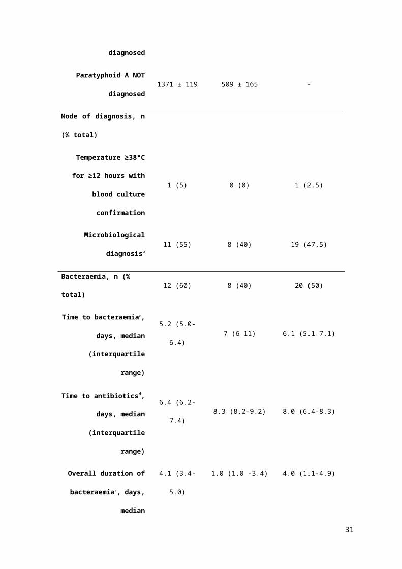
Figure 1

Figure 2
32

33

Figure 3
Salmonella enterica serovar Typhi, Quailes strain
Salmonella enterica serovar Paratyphi A, NVGH308
TemperatureStool cultureBlood culture
