thermophilic fungi: their physiology and enzymes†mmbr.asm.org/content/64/3/461.full.pdf · above...
TRANSCRIPT

MICROBIOLOGY AND MOLECULAR BIOLOGY REVIEWS,1092-2172/00/$04.0010
Sept. 2000, p. 461–488 Vol. 64, No. 3
Copyright © 2000, American Society for Microbiology. All Rights Reserved.
Thermophilic Fungi: Their Physiology and Enzymes†RAMESH MAHESHWARI,1* GIRISH BHARADWAJ,1 AND MAHALINGESHWARA K. BHAT2
Department of Biochemistry, Indian Institute of Science, Bangalore 560 012, India,1 andInstitute of Food Research, Norwich Laboratory, Colney NR4 7UA, United Kingdom2
INTRODUCTION .......................................................................................................................................................461NOMENCLATURE.....................................................................................................................................................462HISTORICAL BACKGROUND................................................................................................................................463PHYSIOLOGY ............................................................................................................................................................464
Growth Medium ......................................................................................................................................................464Minimal Temperature for Growth........................................................................................................................464Homeoviscous Adaptation......................................................................................................................................464Sensitivity to Subminimal Temperatures ............................................................................................................464Oxygen Requirement ..............................................................................................................................................465Economic Coefficient and Growth Rate ...............................................................................................................465Respiration...............................................................................................................................................................465Transport .................................................................................................................................................................466Utilization of Carbon Sources...............................................................................................................................466Mixed-Substrate Utilization ..................................................................................................................................467Protein Breakdown .................................................................................................................................................468Acquired Thermotolerance ....................................................................................................................................468
SECRETORY ENZYMES..........................................................................................................................................469Protease ....................................................................................................................................................................469Lipase .......................................................................................................................................................................470a-Amylase ................................................................................................................................................................471Glucoamylase...........................................................................................................................................................471Cellulase...................................................................................................................................................................472Cellobiose Dehydrogenase .....................................................................................................................................474Xylanase ...................................................................................................................................................................475a-D-Glucuronidase ..................................................................................................................................................477Polygalacturonase ...................................................................................................................................................477Laccase .....................................................................................................................................................................477Phytase .....................................................................................................................................................................478D-Glucosyltransferase .............................................................................................................................................478
CELL-ASSOCIATED ENZYMES.............................................................................................................................478Enzymes of the Pentose Phosphate Pathway and the TCA Cycle....................................................................478Trehalase..................................................................................................................................................................478Invertase...................................................................................................................................................................480b-Glycosidase ..........................................................................................................................................................480Lipoamide Dehydrogenase.....................................................................................................................................481ATP Sulfurylase ......................................................................................................................................................481Protein Disulfide Isomerase ..................................................................................................................................481
MISCELLANEOUS PROTEINS ..............................................................................................................................481CONCLUSIONS AND PROSPECTS.......................................................................................................................482ACKNOWLEDGMENTS ...........................................................................................................................................483REFERENCES ............................................................................................................................................................483
INTRODUCTION
Among the eukaryotic organisms, only a few species of fungihave the ability to thrive at temperatures between 45 and 55°C.Such fungi comprise thermophilic and thermotolerant forms,
which are arbitrarily distinguished on the basis of their mini-mum and maximum temperature of growth (63): the thermo-philic fungi have a growth temperature minimum at or above20°C and a growth temperature maximum at or above 50°C,and the thermotolerant forms have a temperature range ofgrowth from below 20 to ;55°C. Thermophily in fungi is not asextreme as in eubacteria or archaea, some species of which areable to grow near or above 100°C in thermal springs, solfatarafields, or hydrothermal vents (36, 45). Perhaps because of theirmoderate degree of thermophily and because their habitats arenot exotic, thermophilic fungi have not received much publicityand attention. However, considering that the vast majority ofeukaryotes cannot survive prolonged exposure to temperatures
* Corresponding author. Mailing address: Department of Biochem-istry, Indian Institute of Science, Bangalore 560 012, India. Phone:91-80-309-2674. Fax: 91-80-360-0814. E-mail: [email protected].
† This review is dedicated to the late Paul J. Allen of the Universityof Wisconsin, Madison, Wis., and to Alfred S. Sussman of the Univer-sity of Michigan, Ann Arbor, Mich., for their inspiring teaching andoutstanding contributions to fungal biology.
461
on May 17, 2018 by guest
http://mm
br.asm.org/
Dow
nloaded from

above 40 to 45°C (8), the ability of some 30 species, out ofapproximately 50,000 recorded fungal species, to breach theupper temperature limit of eukaryotes is a phenomenon thatdeserves elucidation. Moreover, this group of fungi providesscientists with valuable experimental material for investiga-tions of the mechanisms which, although allowing their growthat moderately high temperatures, limit it beyond 60 to 62°C(243).
Thermophilic fungi are the chief components of the micro-flora that develops in heaped masses of plant material, piles ofagricultural and forestry products, and other accumulations oforganic matter wherein the warm, humid, and aerobic environ-ment provides the basic conditions for their development (10,172). They constitute a heterogeneous physiological group ofvarious genera in the Phycomycetes, Ascomycetes, Fungi Im-perfecti, and Mycelia Sterilia (182).
NOMENCLATUREWhile reviewing the literature, we faced difficulties on ac-
count of the confusing nomenclature of thermophilic fungi.The confusion is due to several reasons. Since the early taxo-
nomic literature is scattered and is often in languages otherthan English, it was difficult to ascertain the priority associatedwith the names of a species. As a result, some species havebeen described repeatedly under different names. As and whenthe earlier names were discovered, the fundamental rule ofpriority was applied and the names of the taxa were changedfrom time to time. For example, the ubiquitous fungus Ther-momyces lanuginosus, which has been frequently used in ex-perimental studies, has several synonyms (Table 1). Even inrecent times, in several papers this fungus has been referred toby its earlier name, Humicola lanuginosa. Another source ofconfusion is the practice of interchangeably using the names ofthe asexual (anamorph) and the sexual (teleomorph) stages ofthe same fungus. For example, Sporotrichum (Chrysosporium)thermophile and Myceliophthora thermophila are, respectively,the anamorph and teleomorph stages of the same fungus. Thisfungus is reportedly a heterothallic ascomycete (182), but theheterothallic nature of an isolate cannot be demonstrated un-less a compatible strain of the opposite mating type is avail-able. There also are instances in the literature of misidentifi-cations of thermophilic fungi. One example is Thermoascus
TABLE 1. Taxonomic status and cardinal temperatures of thermophilic fungia
Fungus (present nomenclature) Other names TOpt (°C) Tmax (°C)
Canariomyces thermophila Guarro & Samson 45Chaetomium mesopotamicum Abdullah & Zora 45 52Chaetomium thermophile La Touche C. thermophilum, C. thermophilium 45–55 58–61Coonemeria aegyptiaca (Ueda & Udagawa) Mouchacca Thermoascus aegyptiacus, Paecilomyces aegyptiaca 40 55Coonemeria crustacea (Apinis & Chesters) Mouchacca Thermoascus crustaceus, Dactylomyces crustaceus,
Paecilomyces crustaceus40 ,60
Coonemeria verrucosa (Yaguchi, Someya et Udagawa)Mouchacca
Thermoascus crustaceus 30–40 55
Corynascus thermophilus (Fergus & Sinden) vanKlopotek
Thielavia thermophila, Myceliophthora fergusii,Chrysosporium fergusii
50 60
Dactylomyces thermophilus Sopp Thermoascus thermophilus, Thermoascusaurantiacus (misapplied name)
40–45
Malbranchea cinnamomea (Libert) van Oorschot & deHoog
Trichothecium cinnamomeum, Thermoidiumsulfureum, Malbranchea pulchella var. sulfurea
45 57
Melanocarpus albomyces (Cooney & Emerson) von Arx Myriococcum albomyces, Thielavia albomyces 45 57Melanocarpus thermophilus (Abdullah & Al-Bader)
Guarro, Abdullah & Al-BaderThielavia minuta var. thermophila 35 50
Myceliophthora hinnulea Awao & Udagawa 40–45 .50Myceliophthora thermophila (Apinis) van Oorschot Sporotrichum thermophilum/thermophile,
Chrysosporium thermophilum, Myceliophthoraindica, Corynascus heterothallicus
45–50 55
Myriococcum thermophilum (Fergus) van der Aa 45 53Paecilomyces varioti Bainierb 50 55Rhizomucor miehei (Cooney & Emerson) Schipper Mucor miehei 35–45 57Rhizomucor pusillus (Lindt) Schipper Mucor pusillus 35–45 55Scytalidium thermophilum (Cooney & Emerson)
AustwickTorula thermophila, Humicola grisea var.
thermoidea, Humicola insolens40 58
Stilbella thermophila Fergus 35–50 55Talaromyces byssochlamydioides Stolk & Samson Paecilomyces byssochlamydioides 40–45 .50Talaromyces emersonii Geosmithia emersonii; Talaromyces duponti and
Penicillium duponti (misapplied names)40–45 55
Talaromyces thermophilus Penicillium duponti 45–50 60Thermoascus aurantiacus Thermoascus aurantiacus sensu Cooney &
Emerson (misapplied name)49–52 61
Thermomyces ibadanensis Apinis & Eggins 42–47 61Thermomyces lanuginosus Tsiklinskaya Humicola lanuginosa 45–50 60Thermomyces stellatus (Bunce) Apinis Humicola stellata 40 50Thielavia australiensis Tansey & Jack 35–40 50Thielavia pingtungia Chen K.-Y. & Chen Z.-C. 40 .50Thielavia terrestris (Apinis) Malloch & Cain Allescheria terrestris, Acremonium alabamensis 40–45 52
a Temperature data are from various sources and should be regarded as approximate. Because of uncertainty about the minimal temperature of growth (see text),this is not given. TOpt, optimal temperature; Tmax, maximum temperature.
b Confusion exists regarding its designation as a thermophilic fungus.
462 MAHESHWARI ET AL. MICROBIOL. MOL. BIOL. REV.
on May 17, 2018 by guest
http://mm
br.asm.org/
Dow
nloaded from

aurantiacus, which has featured in early physiological investi-gations and in several recent reports dealing with enzymologi-cal studies. Some investigators have identified their isolatesbased on a description of T. aurantiacus given by Cooney andEmerson (63), who depicted this taxon as having an asexualstage, although in reality it lacks an asexual stage. Rather, thediagnosis of T. aurantiacus as given by Cooney and Emersonfits that of Dactylomyces crustaceus, which has a Paecilomycesasexual stage (16). Since both T. aurantiacus and D. crustaceusbecame a source of confusion, Mouchacca (182) proposed thatthe name T. aurantiacus be retained whereas D. crustaceus berenamed as Coonemeria crustacea. As is now known, T. auran-tiacus is an ascomycetous fungus with a bright orange color,elliptical ascospores, and no asexual stage. Unfortunately, un-less cultures used by different investigators under the name ofT. aurantiacus are reexamined, it may not be possible to de-termine which fungus was actually used.
To nonmycologists, confusion has also resulted from themerging of what, for many years, had been regarded as differ-ent taxa. For example, several scientific papers deal with po-lysaccharide-degrading enzymes and trehalase of Humicola in-solens, H. grisea var. thermoidea, or Torula thermophila, fungiwhich are commonly found in mushroom composts and in soil.All these are now thought to represent one single variablespecies, Scytalidium thermophilum (236). Supposing a biologistwishes to follow on the report of an interesting enzyme foundin H. grisea var. thermoidea, he or she may be at a loss toreproduce the observations unless the original culture used bythe author is available. Finally, in some cases, the specificepithet thermophilum (or variants thereof) has been used with-out adhering to the proposed definition of a thermophilic fun-gus. To name a few, these cases include Achaetomium ther-mophilum, Sordaria thermophila, or Gilmaniella thermophila,which are thermotolerant rather than thermophilic species(however, the dividing line between the two types of fungi isthin). Mouchacca (182) has attempted to remedy the confusionthat had arisen by performing a critical analysis of the nomen-clature and taxonomic status of thermophilic fungi. The cur-rent names of thermophilic fungi and their synonyms are givenin Table 1. It will be some times before the proposed names ofthermophilic fungi are stabilized. In this paper, when the workof earlier authors is reviewed, the names of the fungi as re-ported in the original publications have been retained, butthese should be cross-checked by reference to Table 1.
HISTORICAL BACKGROUND
The first of the known thermophilic fungi, Mucor pusillus,was isolated from bread and described over a century ago byLindt (148). A little later, Tsiklinskaya discovered anotherthermophilic fungus, Thermomyces lanuginosus, growing on po-tato which had been inoculated with garden soil (252). Boththese molds were essentially discovered as chance contami-nants. The natural habitats of thermophilic fungi and the bioticconditions which favored their growth remained unknown untilHugo Miehe investigated the causes of self-heating and spon-taneous combustion of damp haystacks (172). In solving thepuzzle of thermogenesis of stored agricultural products, Miehewas drawn to study the microflora present therein. He was thefirst person to work extensively on thermophilic microorgan-isms. He isolated four species of thermophilic fungi from self-heating hay: Mucor pusillus, Thermomyces lanuginosus, Ther-moidium sulfureum, and Thermoascus aurantiacus. He comparedthe heating capacities of mesophilic and thermophilic fungi (173,174). He inoculated sterilized hay and other substrates kept insideinsulated flasks with pure cultures of individual fungi and ob-
served that the final temperature of the material depended on themaximum temperature of growth of the fungus used. He demon-strated thereby that heating of packed plant material was causedby the microorganisms present therein. Miehe explained the self-heating of hay and other plant material as follows. Initially, be-cause of the exothermic reactions of the saprophytic, mesophilicmicroflora present therein, the temperature of the material risesto ;40°C. The resulting warmed environment favors the germi-nation of spores of the thermophilic microflora, and eventuallythe latter outgrows the mesophilic microflora; in the process, thetemperature of the mass is raised further to 60°C or even higher.
By the beginning of the 20th century, Miehe’s work had ledto the discovery of a small group of thermophilic fungi and totheir primary habitats. Their unique thermal adaptation at-tracted the attention of Kurt Noack (188), who isolated ther-mophilic fungi from several natural substrates. He was in-trigued by the fact that in addition to self-heating masses of hayand compost heaps of leaves, these fungi were present in placeswhere temperatures conducive to their growth occur only in-frequently, for example in soils of the Temperate Zone. Thispuzzling aspect of the ecology of thermophilic fungi providedthe foundation for Noack’s pioneering investigations of theirphysiology. Using respiration as the probe, Noack sought todetermine if thermophilic fungi had an unusually high rate ofrespiration whereby the released metabolic heat could warmtheir environment, allowing them to complete their life cyclerapidly. He found, however, that the respiration of thermo-philic fungi does not confer any special advantage on them.
During the Second World War, the need for finding alter-nate sources of rubber led to studies of the rubber-producingguayule shrub, Parthenium argentatum. It had been observedthat the extractability and physical properties of rubber fromthe shrub improved when the plant material was chopped andstored in a mass before being milled. Allen and Emerson (10)demonstrated that the observed improvement from the abovetreatment (retting) resulted primarily from utilization andreduction in the amount of resin in crude rubber by a ther-mophilic microflora. From the self-heating mass of choppedguayule, Allen and Emerson isolated several species of ther-mophilic fungi that had temperature limits extending up to60°C. They demonstrated that for optimal development ofthermophilic fungi in the mass of material, its moisture andnutrient content were crucial. In addition, although size ofthe mass was an important factor for reducing the outwarddissipation of heat, the mass of material had to be suffi-ciently porous for air to diffuse inside and allow aerobicrespiration of fungi. Based on the isolates of thermophilicfungi from the retting guayule shrub and on collections ofcultures from other investigators, Cooney and Emerson (63)provided taxonomic descriptions of 13 species known at thattime, an account of their habitats, and the general biology ofthermophilic fungi. This monograph, in English, for the firsttime made mycologists generally aware of the existence ofthermophilic fungi. It stimulated the search for new speciesin order to understand their taxonomic diversity as well as toinvestigate their potential use as sources of commerciallyimportant enzymes. The taxonomy (182) and ecology (153,244) of thermophilic fungi have been reviewed previously.The other important areas that have been studied in thisgroup of fungi include their physiology and the purificationand study of the functional characteristics of their enzymes.This review therefore covers the studies in these two areasfrom the time when experimental work on thermophilicfungi began.
VOL. 64, 2000 PHYSIOLOGY AND ENZYMES OF THERMOPHILIC FUNGI 463
on May 17, 2018 by guest
http://mm
br.asm.org/
Dow
nloaded from

PHYSIOLOGY
Growth Medium
Noack (188) grew Thermoascus aurantiacus, Anixia spadicea(Chaetomium thermophile?), Mucor pusillus, Thermomyces lanugi-nosus, and Thermoidium sulfureum in a glucose-salt liquid me-dium fortified with peptone and, often, with a decoction of hay.Until the 1980s, thermophilic fungi were thought to have complexor unusual nutritional requirements. For example, Miller et al.(176) remarked that “no defined medium could be produced inwhich the thermophilic fungi would grow . . . .” Rosenberg (218)reported that nearly half of the species of thermophilic and ther-motolerant fungi tested required 0.01% yeast extract for growthin a solid medium. Wali et al. (260) reported that for growth in aliquid medium containing glucose and ammonium sulfate, thethermophilic fungi required a supplementation of succinic acid, atricarboxylic acid cycle intermediate. While this observation wasconfirmed in our laboratory (102), we additionally demonstratedthat because of the low phosphate concentration in the culturemedium, the pH of the medium in the absence of the organic aciddropped to ;3.4 after 12 to 24 h and the growth ceased. More-over, any one of the several tricarboxylic acid cycle acids testedstimulated growth, which was due to their buffering action in themedium rather than to their nutritional role: thermophilic fungigrew satisfactorily in a minimal medium if the pH of the mediumwas controlled between 5.5 and 7.0 by increasing the phosphateconcentration in the medium, readjusting the pH by addition ofan alkali, including powdered calcium carbonate as a reservealkali, or replacing the inorganic nitrogen source with an organicnitrogen source (L-asparagine). The low pH reduces the solubilityof CO2 in the growth medium and limits its availability for assim-ilation by the anaplerotic enzyme pyruvate carboxylase (102).Although CO2 is not regarded as a nutritional requirement forfungi, growth of T. lanuginosus was severely affected if the gasphase in the culture flask was devoid of CO2. The concentrationof CO2 inside composts can be as high as 10 to 15% (74); there-fore, it is likely that its assimilation plays nutritional and morpho-genetic roles in the development of thermophilic fungi, which arethe primary components of the microflora of such habitats. It isinteresting that this gas has been identified as essential for theaxenic culture of a rust fungus, which had long been regarded asan obligate parasite on plants (37).
Minimal Temperature for Growth
Thermophilic fungi have a widespread distribution in trop-ical as well as temperate regions (157). Tendler et al. (247)remarked that “the ubiquitous distribution of organisms,whose minimal temperature for growth exceeds the tempera-tures obtainable in the natural environment from whence theywere isolated, still stands as a ‘perfect crime’ story in the libraryof biological systems.” They considered whether eukaryoticthermophily is an artifact of the nutritional environment. Totest this, thermophilic isolates of Humicola, Thermoascus, andAspergillus were incubated in a nutritionally rich liquid mediumthat included glucose, mannitol, starch, Casamino Acids, yeastextract, and peptone. After 10 days at 20°C, these fungi hadgenerated good growth, although they had failed to grow below30°C in a sucrose-salts medium that lacked complex supple-ments. It was suggested that the complex materials contained afactor(s) which the organisms could not synthesize at the lowertemperature. This suggestion was supported by the observationthat the growth of Talaromyces thermophilus at a suboptimaltemperature (33°C) benefited from the supplementation ofculture medium with 5 mg of ergosterol per ml (264).
Furthermore, the choice of inoculum, i.e., spores versus ger-
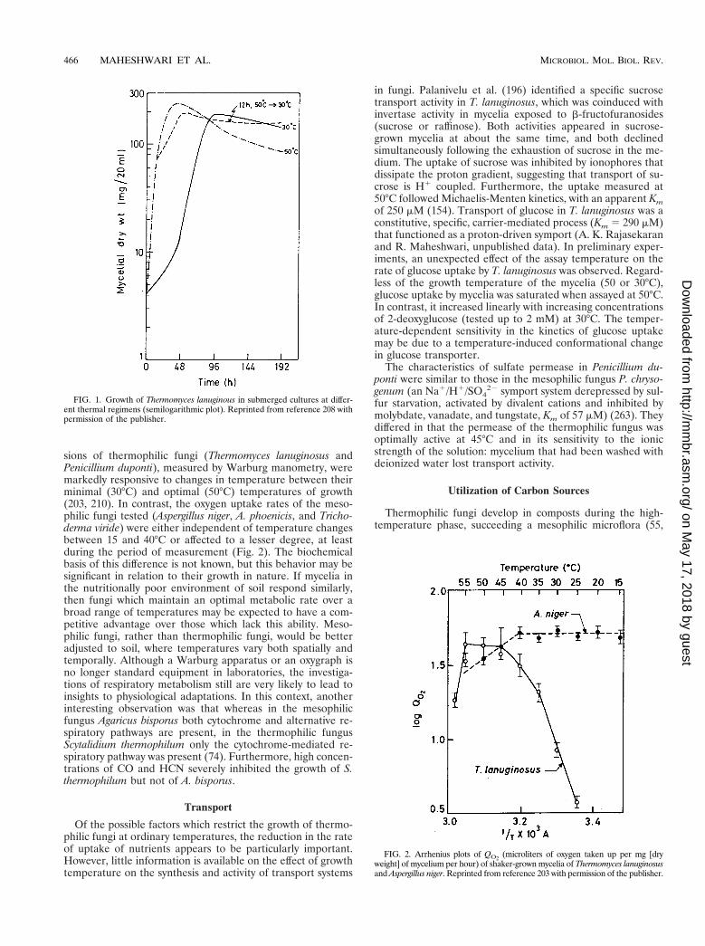
minated spores or mycelium, may also influence the minimaltemperature of growth of thermophilic fungi. Whereas Mucormiehei did not grow at 25°C in submerged cultures when sporeswere used as the inoculum, substantial growth occurred at thistemperature (nearly 64% of that at 48°C in 72 h) in as muchtime when pregerminated spores were used as inocula (237).Similarly, we observed that a mycelial inoculum, but not aspore inoculum, resulted in a near maximal yield of Thermo-myces lanuginosus at 25°C; growth occurred without a percep-tible lag but at a lower rate than at 50°C (Fig. 1). In Thermoas-cus aurantiacus, the lowest temperature at which theascospores germinated was 10 to 12°C higher than that forhyphal growth (71). Presumably this may be a means of min-imizing competition from the mesophilic fungi and ensuringthat warm conditions would be available for yet more timeuntil the mycelium is established for resource capture. In lightof the observations that the conditions for spore germinationcan be more exacting than those for hyphal growth, the re-ported minimal temperature of growth of thermophilic fungishould be redetermined, specifying the method of inoculationand the composition of the medium used. The above observa-tions also suggest that once the spores have been induced togerminate at high temperature, the requirement of high tem-perature for sustaining growth may not be critical.
Homeoviscous AdaptationMany organisms vary the fatty acid composition of their
membrane phospholipids as a function of growth temperatureso that their membrane fluidity is kept constant for the optimalfunctioning of membrane-localized transporters and enzymes.For example, with an increase in temperature, there is anincrease in the proportion of saturated fatty acids incorporatedinto phospholipids, whereas at lower temperature, a higherproportion of unsaturated fatty acids is incorporated. Thisphenomenon is called homeoviscous adaptation (229). Wrightet al. (264) examined whether an inability to regulate mem-brane fluidity may be a reason for the high minimum temper-ature of growth of thermophilic fungi. They reported that whenTalaromyces thermophilus was shifted from a high (50°C) to alow (33°C) growth temperature, the degree of unsaturation offatty acids at the two stated temperatures remained virtuallyunchanged. This was thought to be the result of a metaboliclimitation, presumably due to a nonfunctional fatty acid de-saturase, which restricted the ability of the fungus to convertoleate to linoleate at low temperature. However, results with T.lanuginosus were different. In this fungus, the concentration oflinoleic acid (18:2) was twofold higher at 30 than at 50°C. Thedegree of unsaturation of phospholipid fatty acids was 0.88 inmycelia grown at 50°C but 1.0 in the temperature-shifted cul-tures (from 50 to 30°C) and 1.06 in cultures grown at constant30°C (209). A decrease in the degree of unsaturation was alsoobserved in Chaetomium thermophile when it was subjected toheat shock (190). As mentioned above, some species of ther-mophilic fungi are capable of growth even at mesophilic tem-peratures. Therefore, it seems unlikely that the inability toadjust membrane fluidity is the general reason for their highminimum temperature of growth. As was reasoned for ther-mophilic bacteria (240), the loss of catalytic potential of one ormore vital enzymes, caused by conformational changes and/orribosomal assembly, may be an important determinant of theminimal growth temperature.
Sensitivity to Subminimal TemperaturesThe need to explain the occurrence of thermophilic fungi in
soil, which may warm to favorable temperatures because of
464 MAHESHWARI ET AL. MICROBIOL. MOL. BIOL. REV.
on May 17, 2018 by guest
http://mm
br.asm.org/
Dow
nloaded from

solar radiation, but only for a transitory period, promptedNoack (188) to investigate the effects of subminimal tempera-tures and rewarming. He noticed that when an actively growingculture of Thermoascus aurantiacus was cooled to 31°C (4°Cbelow the lower temperature limit of growth), its respirationafter 2 days declined only minimally but that cultures kept at21°C for 24 h stopped respiring. When the culture medium wasrewarmed to 46°C, practically no respiration was observed.Whether the low resistance to lower temperature displayed byT. aurantiacus is also applicable to other thermophilic fungi isnot known. However, until more information is available, itshould not necessarily be assumed that mycelial cultures ofthermophilic fungi can be stored under refrigeration or atsubminimal temperatures without loss of viability.
Oxygen Requirement
Noack (188) recognized that during the self-heating processthe environment in composts would become oxygen deficient.He therefore studied the behavior of Thermoascus aurantiacussubjected to anaerobiosis and found that the withdrawal ofoxygen severely affected its respiration and growth. Althoughthermophilic fungi do not have the ability to undergo anaero-bic growth (136), Humicola insolens was reported to grow bet-ter under anaerobic or microaerobic conditions than underaerobic conditions at elevated temperatures (116; also see ref-erence 78). Cooney and Emerson (63) reported an interestingmorphogenetic effect of anaerobiosis in Talaromyces (Penicil-lium) duponti. This thermophilic fungus formed only a conidialstage (Penicillium) in aerobic cultures; the sexual stage (Ta-laromyces) was initiated in agar cultures only when they wereflushed with nitrogen. It would be worthwhile to study theeffect of anaerobic conditions in species in which the sexualstage has not been discovered so far or is observed only infre-quently.
Economic Coefficient and Growth Rate
We referred earlier to the work of Kurt Noack (188). Hesought to determine whether thermophilic fungi have an ex-ceptionally high rate of metabolism accompanied by a highrate of substrate conversion. He observed that the economicyield (grams of sugar consumed per gram of mycelial dryweight formed) of Thermoascus aurantiacus grown in a mini-mal medium at 45°C (1.89) was the same as that of the meso-philic fungus Aspergillus niger grown at 25°C. From this, heinferred that the overall metabolisms of the two types of fungimust be quite similar. Moreover, he estimated that on averageboth types of fungi converted 55% of sugar for the synthesis offungal biomass and 45% for metabolism. Since few values ofeconomic coefficients have been reported, we compared themolar growth yield (grams of biomass produced per mole of
glucose utilized) (YG) of fungi. From the data available (Table2), the average YG values of mesophilic and thermophilic fungiat their respective temperature optima are quite comparable(86 to 88 g/mol), suggesting that similar proportions of carbo-hydrate are used by both types of fungi for macromolecularsynthesis. This value is close to that found by Noack for thefungal species studied by him.
Under the culture conditions used by us, some thermophilicfungi (Thermomyces lanuginosus, Penicillium duponti, Sporotri-chum thermophile, and Malbranchea pulchella var. sulfurea)produced exceptionally homogeneous mycelial suspensionswhen grown in a glucose-asparagine medium in shake culturesat 50°C (102, 154, 200). This allowed the sampling of myceliaby using pipettes for quantitative measurements during theirgrowth and facilitated the determination of growth rates,growth yields (see above), the effect of temperature-shift, andother aspects of physiology. The ranges of growth rates ofthermophilic and meophilic fungi were similar (Table 2). In-terestingly, although the growth of T. lanuginosus at a subop-timal temperature was slowed, biomass production was notaffected (Fig. 1). Using CO2 produced as an index of develop-ment of fungal biomass, Wiegant (262) observed that the ex-ponential growth rate of the thermophilic fungus Scytalidiumthermophilum (a common thermophilic fungus in mushroomcompost) at 45°C in a liquid medium supplemented with maltand yeast extracts was 0.41 h21. Thus, contrary to a commonbelief, thermophilic fungi do not, in general, grow faster thanmesophilic fungi. The situation is similar to that in thermo-philic bacteria (44, 240).
Respiration
Using the volume of carbon dioxide evolved over time as ameasure of metabolism, Noack (188) compared a thermophilicfungus (Thermoascus aurantiacus) with a mesophilic fungus(Penicillium glaucum) grown in identical medium. He observedthat the volume of carbon dioxide released by P. glaucum in24 h was equivalent to 67% of its dry weight at 15°C and 133%at 25°C. He argued that if this fungus could grow at 45°C, theextrapolated value of carbon dioxide, according to van’t Hoffrule, would be 532%. However, the actual value for T. auran-tiacus at 45°C was 310%. From this, Noack inferred that at agiven temperature the metabolism of a thermophilic fungus isactually slower than that of a mesophilic fungus. Subsequentstudies have shown that the two types of fungi have nearlycomparable respiratory rates at their respective temperatureoptima (203, 210).
Noack (188) observed that with increases in temperature,the increase in the respiration of Thermoascus aurantiacus waslower than that of mesophilic fungi. However, we found thatthe rates of oxygen uptake of homogeneous mycelial suspen-
TABLE 2. Comparison of the growth parameters of some mesophilic and thermophilic fungi
Fungus Type of fungus Growth temp(°C)
Specific growth rate(h21)
Molar growth yield(YG) (g/mol) Reference
Neurospora crassa Mesophile 30 0.37 NRa 7Trichoderma reesei Mesophile 30 0.20 72 160Aspergillus niger Mesophile 30 0.24 128 208Trichoderma viride Mesophile 30 0.16 59 208Sporotrichum thermophile Thermophile 50 0.23 74 208
30 0.11 65Thermomyces lanuginosus Thermophile 50 0.23 101 208
30 0.06 87
a NR, not reported.
VOL. 64, 2000 PHYSIOLOGY AND ENZYMES OF THERMOPHILIC FUNGI 465
on May 17, 2018 by guest
http://mm
br.asm.org/
Dow
nloaded from
sions of thermophilic fungi (Thermomyces lanuginosus andPenicillium duponti), measured by Warburg manometry, weremarkedly responsive to changes in temperature between theirminimal (30°C) and optimal (50°C) temperatures of growth(203, 210). In contrast, the oxygen uptake rates of the meso-philic fungi tested (Aspergillus niger, A. phoenicis, and Tricho-derma viride) were either independent of temperature changesbetween 15 and 40°C or affected to a lesser degree, at leastduring the period of measurement (Fig. 2). The biochemicalbasis of this difference is not known, but this behavior may besignificant in relation to their growth in nature. If mycelia inthe nutritionally poor environment of soil respond similarly,then fungi which maintain an optimal metabolic rate over abroad range of temperatures may be expected to have a com-petitive advantage over those which lack this ability. Meso-philic fungi, rather than thermophilic fungi, would be betteradjusted to soil, where temperatures vary both spatially andtemporally. Although a Warburg apparatus or an oxygraph isno longer standard equipment in laboratories, the investiga-tions of respiratory metabolism still are very likely to lead toinsights to physiological adaptations. In this context, anotherinteresting observation was that whereas in the mesophilicfungus Agaricus bisporus both cytochrome and alternative re-spiratory pathways are present, in the thermophilic fungusScytalidium thermophilum only the cytochrome-mediated re-spiratory pathway was present (74). Furthermore, high concen-trations of CO and HCN severely inhibited the growth of S.thermophilum but not of A. bisporus.
Transport
Of the possible factors which restrict the growth of thermo-philic fungi at ordinary temperatures, the reduction in the rateof uptake of nutrients appears to be particularly important.However, little information is available on the effect of growthtemperature on the synthesis and activity of transport systems
in fungi. Palanivelu et al. (196) identified a specific sucrosetransport activity in T. lanuginosus, which was coinduced withinvertase activity in mycelia exposed to b-fructofuranosides(sucrose or raffinose). Both activities appeared in sucrose-grown mycelia at about the same time, and both declinedsimultaneously following the exhaustion of sucrose in the me-dium. The uptake of sucrose was inhibited by ionophores thatdissipate the proton gradient, suggesting that transport of su-crose is H1 coupled. Furthermore, the uptake measured at50°C followed Michaelis-Menten kinetics, with an apparent Kmof 250 mM (154). Transport of glucose in T. lanuginosus was aconstitutive, specific, carrier-mediated process (Km 5 290 mM)that functioned as a proton-driven symport (A. K. Rajasekaranand R. Maheshwari, unpublished data). In preliminary exper-iments, an unexpected effect of the assay temperature on therate of glucose uptake by T. lanuginosus was observed. Regard-less of the growth temperature of the mycelia (50 or 30°C),glucose uptake by mycelia was saturated when assayed at 50°C.In contrast, it increased linearly with increasing concentrationsof 2-deoxyglucose (tested up to 2 mM) at 30°C. The temper-ature-dependent sensitivity in the kinetics of glucose uptakemay be due to a temperature-induced conformational changein glucose transporter.
The characteristics of sulfate permease in Penicillium du-ponti were similar to those in the mesophilic fungus P. chryso-genum (an Na1/H1/SO4
22 symport system derepressed by sul-fur starvation, activated by divalent cations and inhibited bymolybdate, vanadate, and tungstate, Km of 57 mM) (263). Theydiffered in that the permease of the thermophilic fungus wasoptimally active at 45°C and in its sensitivity to the ionicstrength of the solution: mycelium that had been washed withdeionized water lost transport activity.
Utilization of Carbon Sources
Thermophilic fungi develop in composts during the high-temperature phase, succeeding a mesophilic microflora (55,
FIG. 1. Growth of Thermomyces lanuginous in submerged cultures at differ-ent thermal regimens (semilogarithmic plot). Reprinted from reference 208 withpermission of the publisher.
FIG. 2. Arrhenius plots of QO2(microliters of oxygen taken up per mg [dry
weight] of mycelium per hour) of shaker-grown mycelia of Thermomyces lanuginosusand Aspergillus niger. Reprinted from reference 203 with permission of the publisher.
466 MAHESHWARI ET AL. MICROBIOL. MOL. BIOL. REV.
on May 17, 2018 by guest
http://mm
br.asm.org/
Dow
nloaded from

115). Since most of the initially available soluble carbonsources (sugars, amino acids, and organic acids) would havebeen depleted, the carbon source available for the growth ofthermophilic fungi would be mainly the polysaccharide constit-uents of the biomass, of which cellulose is the chief constituent.Interestingly, some compost fungi are unable to utilize cellu-lose, for example, Thermomyces lanuginosus (55, 115, 204),Talaromyces duponti, Malbranchea pulchella var. sulfurea, Mu-cor pusillus (55), and Melanocarpus albomyces (156). The non-cellulolytic species in compost can grow commensally by uti-lizing sugars released during the hydrolysis of hemicelluloseand cellulose by the cellulolytic partner. For example, T.lanuginosus showed profuse growth in mixed cultures with acellulolytic fungus, Chaetomium thermophile (115). Moreover,several noncellulolytic species readily utilize xylan, which isexternal to cellulose in the plant cell wall and is apparently amore accessible carbon source (199). Indeed, some fungi (C.thermophile and Humicola insolens) grow even better on xylanthan on simple sugars (55). The secretion of thermostableextracellular polysaccharide-degrading enzymes and the simul-taneous uptake of sugars would be important attributes ofthermophilic fungi in self-heating masses of plant material. InT. lanuginosus, a single transporter was identified for glucose,xylose and mannose, the hydrolytic products of cellulose andhemicellulose (Rajasekaran and Maheshwari, unpublished).
Because the measurement of biomass of fungi growing oninsoluble polysaccharides is indirect, it has rarely been done.We measured the growth of Sporotrichum thermophile on cel-lulose in terms of insoluble nitrogen or as an increase in my-celial dry weight after selectively estimating the amount ofcellulose and subtracting its weight from that of the samples
(32). Interestingly, the exponential growth rate of the funguson cellulose (0.09 to 0.16 h21) was similar to that on glucose(0.1 h21), revealing the remarkable ability of this fungus toutilize cellulose as efficiently as glucose. The visual character-istics of the fungus were strikingly different in submerged cul-tures grown with cellobiose (repeating unit of cellulose) orcellulose (91). The mycelia in cellobiose-grown cultures re-tained a prolonged filamentous and healthy appearance,whereas in cellulose medium they rapidly autolysed and sporu-lated. Perhaps oligosaccharides derived from the hydrolysis ofcellulose regulate the gene expression and metabolic processdifferently from when the fungus is growing on soluble sugars.Another interesting observation was the influence of culturetemperature on fungal morphology when grown with cellulose.At a suboptimal temperature (30°C), the conidia of S. thermo-phile formed a very limited mycelium that precociously devel-oped asexual reproductive structures (microcycle conidiation).Although the mechanism of this cellular response is not un-derstood, microcycle conidiation (Fig. 3) may be a survivalstrategy of producing propagules in the shortest possible timeunder suboptimal conditions.
Mixed-Substrate Utilization
In composting plant material, the hydrolysis of polysaccha-ride constituents by the secreted enzymes would be expected toproduce a mixture of sugars. We determined if thermophilicfungi utilize one sugar at a time or a mixture of sugars simul-taneously (154). In the only study so far with fungi, a combi-nation of glucose and sucrose was chosen, because the concen-trations of these sugars in the medium can easily be determined
FIG. 3. Microcycle conidiation in Sporotrichum thermophile. The fungus was grown in shake cultures with shredded Whatman filter paper as the carbon source. (A)Phase-contrast micrograph of a 24-h-old germling that has produced oval asexual spores. The insoluble particle is a piece of cellulose fiber. (B) Phase-contrastmicrograph showing precocious differentiation of asexual spores in a 72-h-old germling grown at 30°C. The germinated conidium is indicated by an arrow. Bars, 50 mm.
VOL. 64, 2000 PHYSIOLOGY AND ENZYMES OF THERMOPHILIC FUNGI 467
on May 17, 2018 by guest
http://mm
br.asm.org/
Dow
nloaded from

using commercially available enzymes. The fungi studied, Ther-momyces lanuginosus and Penicillium duponti, concurrently uti-lized glucose and sucrose at 50°C, with sucrose being utilizedfaster than glucose. The phenomenon was studied further with T.lanuginosus. Its growth rates on single- or mixed-carbon sourceswere comparable. The rate of utilization of glucose and sucrose inthe mixture was lowered unequally compared to when the sugarswere provided singly, indicating that the two sugars reciprocallyinfluenced their utilization in the mixture. The simultaneous uti-lization of sucrose in the presence of glucose occurred because (i)invertase was insensitive to catabolite repression by glucose and(ii) the activity of the glucose uptake system was repressed byglucose itself as well as by sucrose. Both sugars were also utilizedconcurrently at 30°C but at nearly identical rates. This observationindicates that the activity of nutrient transporters and the sensi-tivity of catabolic enzymes to glucose repression can be influenceddifferently at different temperatures.
Protein Breakdown
One of the early hypotheses put forward to explain ther-mophily in bacteria was that rapid breakdown of proteins atelevated growth temperatures is compensated by their fastresynthesis (9). This rapid-turnover hypothesis came to beknown as the dynamic hypothesis of thermophily. To examineits applicability to thermophilic fungi, Miller et al. (176) com-pared protein breakdown rates in thermophilic (Penicilliumduponti, Malbranchea pulchella, and Mucor miehei) and meso-philic (Penicillium notatum and P. chrysogenum) fungi by mon-itoring the breakdown of pulse-labeled protein. The growingcells in both types of fungi had a negligible rate of breakdownof bulk protein. In the nongrowing cells of both types, thebreakdown rate was similar (5.2 to 6.7% per h). However, thebreakdown rate of the soluble-protein fraction in thermophilicfungi was twice that in the mesophilic fungi. The authors sug-gested that the increased turnover rate of soluble protein isimportant in the survival of thermophilic fungi at the hightemperatures.
The energy expended in increased protein turnover in ther-mophilic fungi would be expected to affect their growth yieldcompared to mesophilic fungi. However, the growth yields ofthermophilic and mesophilic fungi examined were in a similarrange (Table 2). Therefore, we reexamined protein breakdownin fungi (208) by selecting fungi which produced homogeneousmycelia, thereby obviating the sonication treatment used byMiller et al. (176) for rendering mycelia homogeneous forsampling and measuring radioactivity. Moreover, we at-tempted a general labeling of cellular proteins by adding ra-dioactive amino acids at two different times and avoided theuse of toxic concentrations of amino acids during the chase.Furthermore, we used the radioactivity of the whole cells as anindex of the radioactivity of total protein rather than of theprotein that was extractable by ultrasonic disruption of myce-
lia. The results pointed to a definite protein turnover in thegrowing cells of fungi, although the rate of protein turnovervaried among the different species (Table 3). Under differentconditions of incubation, the protein breakdown rate wassomewhat lower in thermophilic fungi than in mesophilic fungi.Although we measured only protein breakdown, it is likely thatthe results would be similar had protein turnover been mea-sured. As in bacteria (240), the rapid-turnover hypothesis inthermophilic fungi is not supported by experimental results.Nonetheless, specific enzymes may well have a high turnoverrate.
Acquired Thermotolerance
Acquired thermotolerance refers to the enhanced survival oforganisms at lethal temperature after a brief exposure to sub-lethal temperatures. A common phenomenon in mesophilicspecies is the synthesis of a set of proteins, called heat shockproteins (HSPs), following a sudden exposure of organisms toelevated temperature. The synthesis of HSPs is thought to bean adaptive response to increased thermotolerance and sur-vival in the face of stressful conditions. Trent et al. (251)demonstrated that in common with thermophilic bacteria andarchaea, thermophilic fungi also synthesize HSPs and acquirethermotolerance. They observed that conidia of T. lanuginosus,germinated at 50°C and heat shocked at 55°C for 60 min priorto exposure to 58°C, showed enhanced survival compared tonon-heat-shocked conidia. Thermotolerance was eliminated ifprotein synthesis during the heat shock period was inhibited bycycloheximide. Pulse-labeling of proteins during the heat shockperiod, followed by their separation by sodium dodecyl sulfate-polyacrylamide gel electrophoresis (SDS-PAGE), showed in-creased synthesis of eight HSPs. Of these, three small HSPsranging from 31 to 33 kDa dominated the heat shock response.Since T. lanuginosus is capable of growth up to 60 to 62°C, therole of HSPs induced at 55°C is not clear. A transient HSPsynthesis was also observed in Chaetomium thermophile var.thermophile (190). In particular, a constitutive, abundantly ex-pressed protein, HSP60, belonging to the intercompartmentaltransport proteins was thought to be important in thermophily.
In closing this section, we should mention that the impres-sion gained is that only cursory explorations of the physiologyof thermophilic fungi have been made, prompted primarily bya desire to explain their widespread distribution in soil—ahabitat characterized by changing temperatures, uncertain sup-plies of nutrients and water, and the coexistence with a numer-ically high mesophilic microflora and fauna consisting of com-petitors and predators. Notwithstanding the reported latentabilities of some species to grow at ordinary temperatures, thecontroversy concerning the growth and reproduction of ther-mophilic fungi in soils is far from settled. Nonetheless, theattempts so far have dispelled certain early notions concerning
TABLE 3. Rate of protein degradation in some mesophilic and thermophilic fungia
Fungus Type of fungusTemp (°C) of growthand assay of protein
degradation
Degradation of protein (% h21) in:
Completemedium
Medium lackingN and S source Buffer
Aspergillus niger Mesophile 30 4.2 3.1 0.8Trichoderma viride Mesophile 30 8.6 8.5 5.7Sporotrichum thermophile Thermophile 50 3.3 4.3 1.3Thermomyces lanuginosus Thermophile 50 3.5 4.2 3.9
a Data from reference 208.
468 MAHESHWARI ET AL. MICROBIOL. MOL. BIOL. REV.
on May 17, 2018 by guest
http://mm
br.asm.org/
Dow
nloaded from

their rates of growth, their nutritional needs, and their overallmetabolism.
SECRETORY ENZYMES
Enzymes of thermophilic fungi have been studied primarilyto explore their suitability in bioprocesses and, to a lesserextent, to probe similarities and differences in physicochemicalproperties between enzymes from mesophilic and thermophilicfungi. Since culture filtrates can be obtained in substantialquantities, the enzymes that are secreted in the growth mediahave been studied more frequently than cell-associated en-zymes, although such investigations have focused mainly on theidentification of suitable thermophilic fungal sources for de-sired enzymes, the development of protocols for the purifica-tion of these enzymes, and the study of their general proper-ties. In a few cases, however, the native or recombinantproteins have been examined to study interspecies differencesin enzyme conformation, amino acid residues involved in sub-strate binding, packing of the hydrophobic core, electrostaticinteractions, and stabilization of helices. In this review of ther-mophilic fungal enzymes, we have attempted to emphasize therealized as well as the potential contributions of such studies tobiology and/or to biotechnology. In this section, the work onsecretory enzymes, also referred to as extracellular enzymes, issummarized.
Protease
Proteases are variously classified on the basis of a criticalamino acid required for the catalytic function (e.g., serineprotease), the pH optimum of their activity (acidic, neutral, oralkaline protease), their site of cleavage (e.g., aminopepti-dases, which act at the free N terminus of the polypeptidechain, or carboxypeptidases, which act at the C terminus of thepolypeptide chain), or their requirement of a free thiol group(e.g., thiol proteinase).
Proteases have long been used in the food, dairy, and deter-gent industries and for leather processing. The need to over-come the limitation of obtaining chymosin, the milk-curdlingenzyme from the stomach contents of milk-feeding calves,which is used in the industrial preparation of cheese, led to asearch for substitutes. Arima et al. (17) screened about 800microorganisms and obtained a soil isolate of Mucor pusillusthat produced an enzyme with a high ratio of milk-clotting toproteolytic activity, enabling the production of high yields ofcurds. Subsequently, a strong milk-clotting activity was alsoobserved in M. miehei (195). The milk-clotting activity of theenzyme was due to its selective attack on the k-casein fraction,which stabilizes the casein micelle in milk. The split k-caseinloses its stabilizing activity, and the micelles of casein coagulatein the presence of calcium (233). The finding of a milk-clottingactivity of utility resulted in serious efforts to isolate otherthermophilic fungi in order to exploit them for industriallyuseful enzymes.
The Mucor rennins were produced by growing the fungus onwheat bran, from which they were extracted with water. Thecrude extract was then purified and crystallized (18, 20, 127,195, 230, 234). Since the rennins hydrolyzed both casein andhemoglobin optimally at pH 3.7, they were classified as acidprotease (EC 3.4.23.6). Both M. pusillus and M. miehei renninshydrolyzed peptide bonds in synthetic peptides with an aro-matic amino acid as the carboxyl donor (20, 195, 233). The M.pusillus enzyme was stable from pH 3.0 to 6.0 and showedmaximum activity at 55°C (273). The enzyme was inhibited bythe aspartic protease inhibitors diazoacetyl-DL-norleucine
methyl ester and pepstatin (241). Sequence comparison withthe other well-characterized aspartic proteases confirmed thepresence of aspartic acid at their active site (25). M. pusillusand M. miehei rennins had similar molecular masses (38.5 and42 kDa) and isoelectric points (3.9 and 4.1), respectively (84).Although the Mucor rennins (aspartic proteases) are structur-ally homologous, they differ from other fungal aspartic pro-teases and from mammalian proteases (25, 187, 249, 269). Thespecificity of Mucor rennins is similar to that of pepsin and calfrennin.
The Mucor rennins are being used as model systems ininvestigations of the heterologous expression of fungal protein,the mechanism of zymogen processing, the refolding of recom-binant protein from inclusion bodies, the effects of glycosyla-tion on secretion, and the activity and stability of mutant en-zymes with amino acid insertions (6, 38, 100, 118). The Mucorrennin genes were cloned in Escherichia coli and sequenced(38, 100, 249). Their deduced amino acid sequence showedthat the enzyme was synthesized as a zymogen that containedan N-terminal leader region constituting a typical signal pep-tide of 22 amino acids and a propeptide of 44 to 47 aminoacids. This leader sequence was not present in the matureprotein (361 amino acids). The expression of the Mucor pro-tease gene in E. coli resulted in the accumulation of unse-creted, inactive polypeptide (38, 100), but when the gene wasexpressed in yeast, a form of the zymogen that was moreglycosylated than the native enzyme was secreted at a concen-tration exceeding 150 mg/liter (267). The prosequence of theheterologous secreted protein was removed by autocatalyticprocessing at acidic pH, yielding an active rennin with proper-ties almost identical to those of the native enzyme (118, 267).A mutation of two of the three glycosylated asparagine sites inthe recombinant M. pusillus rennin significantly reduced theamount of secreted rennin by the yeast cells and decreased itsmilk-clotting activity and thermal stability compared to thoseof nonmutated M. pusillus rennin (6). It has been hypothesizedthat the flexible carbohydrate structures act as “heat reser-voirs” and stabilize the conformation of rennins (269). Therecombinantly produced M. miehei protease in Aspergillusnidulans was similar in specific activity to that produced by M.miehei (100). Subsequently, using an a-amylase promoter ofAspergillus oryzae, a recombinant A. oryzae strain was con-structed that produced heterologous M. miehei protease inexcess of 3 g/liter (61).
The high thermal stability of Mucor rennins turned out to bean undesirable property, since the residual enzyme activity,after cooking, can spoil the flavor of cheese during the longmaturation process (268). Efforts are therefore being made toengineer Mucor rennins with lower thermostability but withthe same milk-clotting potential. Nevertheless, the basic stud-ies on Mucor enzymes have had interesting spinoffs. For ex-ample, the leader peptide of M. pusillus rennin was found to beuseful for the secretion of a heterologous protein by yeast cells:when the human growth hormone gene was fused to the wholepresequence and a part of the prosequence of M. pusillusrennin and expressed in yeast under the control of the yeastGAL7 promoter, the level of the secreted hormone reachedapproximately 10 mg/liter (119).
A source of one of the most thermostable fungal acid pro-teases was a strain of Penicillium duponti isolated from com-post (105, 106). The enzyme was produced in submerged cul-tures, containing rice bran, at 50°C with vigorous aeration andagitation. It was purified by alcohol precipitation, ion-exchangechromatography, and gel filtration (107). Subsequently, it wascrystallized with an yield of 3.3 g from 200 g of crude proteasepreparation (79). The enzyme was most active at pH 2.5 on
VOL. 64, 2000 PHYSIOLOGY AND ENZYMES OF THERMOPHILIC FUNGI 469
on May 17, 2018 by guest
http://mm
br.asm.org/
Dow
nloaded from

casein and at pH 3.0 to 3.5 on hemoglobin. It had a molecularmass of 41 kDa and contained 4.3% carbohydrate. P. dupontiprotease retained its full activity after 1 h at 60°C and pH 4.5.By comparison, the acid protease of M. pusillus was irreversiblydestroyed in 15 min at 65°C (230).
Two fungal sources of thermostable alkaline proteases wereidentified based on the zone of clearing of casein agar byculture filtrates as a semiquantitative assay of proteolytic ac-tivity: Malbranchea pulchella var. sulfurea and Humicola lanugi-nosa (193, 194). Both produced proteases during active growthin the presence of 2 and 8% (wt/vol) casein, respectively, sug-gesting that the enzyme was induced by external protein sub-strate (235). The production of protease by M. pulchella var.sulfurea was repressed by glucose, peptides, amino acids, oryeast extract (194). The M. pulchella protease, named thermo-mycolase, could be concentrated from the culture mediumsimply by vacuum evaporation at 45°C without a loss of activity.After dialysis and removal of pigments by adsorption on acation-exchange column, a homogeneous preparation of theenzyme was obtained by affinity chromatography on a hydro-phobic adsorbent with 78% yield. Its substrate specificity wasinvestigated by analyzing the peptides formed in the reactiondigests of glucagon and the A and B chains of oxidized insulin.At 45°C and pH 7.0, thermomycolase exhibited a general pro-teolytic activity rather than a well-defined specificity for anyparticular amino acid residue (235). The enzyme was classifiedas a serine protease based on inhibition of its activity by diiso-propylfluorophosphate (DFP), which covalently attaches to areactive serine residue. Thermomycolase was optimally activeat pH 8.5 and was stable over a broad pH range (6.0 to 9.5 for20 h at 30°C). As purified thermomycolase autolyzed, it re-sulted in low-molecular-mass peptides. The physicochemicalcharacterization of the protein was therefore carried out usinga DFP-inhibited enzyme. The molecular mass was 11 to 17 kDawhen determined by gel filtration and 32 to 33 kDa whenestimated from sedimentation equilibrium of DFP-thermomy-colase and by SDS-PAGE (256). The thermostability of theenzyme depended on the concentration of calcium ions, aproperty shared by other thermostable enzymes, e.g., thermo-lysin, the neutral protease of Bacillus thermoproteolyticus (257).The t1/2 (time required for the activity of the enzyme to fall to50% of its original value at a given temperature) was 110 minat 73°C in the presence of 10 mM calcium. Calcium (1024 M)was necessary to stabilize the enzyme against autolytic degra-dation at low temperatures: one calcium ion bound to the enzymemolecule with high affinity (258), markedly lowering the rate ofautolytic degradation and of thermal or urea denaturation butcausing no detectable change in the conformation of the en-zyme. It was postulated that the bound calcium stabilized theenzyme by raising the local activation energy for unfolding.
An alkaline protease of Humicola lanuginosa (Thermomyceslanuginosus) was studied by two different groups, and theirenzyme preparations differed in important properties. Sheno-likar and Stevenson (226) purified the enzyme in one stepbased on its specific binding to an organomercury-Sepharosecolumn, from which the enzyme was selectively eluted with abuffer containing mercuric chloride. The mercury-enzymecomplex, reactivated by cysteine in the presence of EDTA,autolyzed rapidly. Based on the inhibition of the enzyme bysome reagents, such as Hg21 or p-chloromercuribenzoate,which react with free thiols, and its reactivation by cysteine inthe presence of EDTA, the enzyme was identified as a uniquethiol proteinase produced by a fungus (226). Both gel filtrationand sedimentation analyses showed that the enzyme had amolecular mass of 237 kDa. It preferentially cleaved its sub-strate at the C-terminal end of the hydrophobic amino acid
residues. However, Hasnain et al. (108) failed to find a pro-tease that specifically bound to the affinity matrix used byShenolikar and Stevenson (226). Rather, the protease activitywas purified by hydrophobic affinity chromatography andshown to have a pH optimum of 8.0 and a molecular mass of38 kDa and was inhibited by phenylmethylsulfonyl fluoride, aninhibitor that is specific to serine protease. Although the en-zyme preparation obtained by Hasnain et al. was also inhibitedby Hg21 and p-chloromercuribenzoate, it was not inhibited byalkylating agents [iodoacetic acid, iodoacetamide or 5,59-di-thiobis(2-nitrobenzoic acid)], suggesting that it was a serineprotease containing a partially buried cysteine. The authors(226) apparently favored the view that the fungus secreted onlya single protease. Such differences in properties are generallyexplained on the basis of strain differences or culture-condi-tion-induced modifications in enzyme structure. We shall en-counter other examples of differences in enzyme propertiesfrom the same (?) fungus.
Lipase
Lipases (EC 3.1.1.3) catalyze the hydrolysis of triacylglycer-ols and the synthesis of esters from glycerol and long-chainfatty acids. These enzymes exhibit the phenomenon of inter-facial activation, i.e., the enhancement of catalytic activity onlipid aggregates (micelles) rather than on lipid monomers inaqueous solution. When used as ingredients of laundry deter-gents, lipases which are stable at pH 10 to 11, at temperaturesfrom 30 to 60°C, and in the presence of surfactants are pre-ferred.
Arima et al. (19) purified an extracellular lipase from Hu-micola lanuginosa strain Y-38, isolated from compost in Japan.The enzyme was produced in a medium containing soybean oil,starch, corn steep liquor, and antifoaming agent. It was puri-fied to homogeneity from 80-h-old culture medium by succes-sive steps of ammonium sulfate precipitation, dialysis, ion-exchange chromatography, and gel filtration chromatography,with 30% recovery. The protein, a single polypeptide (molec-ular weight, 27,500), was optimally active at pH 8.0 and wasstable in the pH range of 4 to 11. Its temperature optimum foractivity was at 60°C. It showed appreciable activity at up to65°C but was inactivated on heating at 80°C for 20 min (149).The enzyme could be stored frozen for more than 6 months.The enzyme molecule contained disulfide linkages but no freeOSH group.
Omar et al. (191, 192) reported that the productivity andthermostability of lipase differed with different strains of H.lanuginosa. These workers developed an optimized mediumcontaining sorbitol, corn steep liquor, silicone oil as an anti-foaming agent, and whale or castor oil as enzyme inducer.With the pH maintained between 7 and 8 and the temperatureset at 45°C, maximum enzyme production by their strain oc-curred after 30 h. Following acetone precipitation and succes-sive chromatographic steps, they obtained a more thermo-stable enzyme (stable at 60°C for 20 h) than was obtained byArima et al. (19). Their preparation was optimally active at pH7.0. The enzyme showed increased activity in organic solvent-aqueous reaction systems, but hydrolysis in complete organicphase reactions did not occur.
A lipase gene from H. lanuginosa was cloned and expressedin Aspergillus oryzae (122). The lipase expressed in the heter-ologous host was purified by a two-step procedure involvinghydrophobic interaction chromatography and ion-exchangechromatography (198). The structure of H. lanuginosa lipase(73) was similar to that of R. miehei lipase. Structural studieshave been directed toward an understanding of the phenom-
470 MAHESHWARI ET AL. MICROBIOL. MOL. BIOL. REV.
on May 17, 2018 by guest
http://mm
br.asm.org/
Dow
nloaded from

enon of interfacial activation. Modification in the lid (de-scribed below) of H. lanuginosa lipase by site-directed mu-tagenesis provided direct evidence of the importance of anumber of residues within the lid in terms of substrate bindingand specificity (120). By spectroscopy and molecular dynamicssimulation, Peters et al. (198) showed that a single mutation ofa serine residue in the active site leads to substantial alter-ations in the motion of the lid and binding affinity of enzyme.
Rhizomucor miehei, formerly called Mucor miehei, also pro-duces active extracellular lipase. The isolation and purificationmethods for M. miehei lipase have been described by Huge-Jensen et al. (123). The lipase produced predominantly in formA (see below) was purified by anion-exchange chromatographyfollowed by affinity chromatography and further purified byhydrophobic interaction chromatography (39). If the purifica-tion steps were carried out at pH 4.5 instead of pH 7.0, form Awas partially deglycosylated and converted to form B. The twoforms showed a high degree of antigenic similarity and wereoptimally active at pH 7.0. However, form A, in contrast toform B, required a prior alkaline (pH 10.5) treatment formaximal activation. The apparent molecular mass of bothlipases was ;32 kDa. The carbohydrate contents of purified R.miehei lipase forms A and B were 11% and 4% (wt/wt), re-spectively. The lipases rapidly hydrolyzed a broad spectrum oflipids found in animal fat and vegetable oil. The enzyme re-mained active even after exhaustive drying (254).
Boel et al. (39) constructed an R. miehei cDNA library in E.coli. From the DNA sequence data and the deduced aminoacid sequence, they inferred that, unlike the characterized bac-terial and mammalian enzymes, the R. miehei lipase was syn-thesized as a zymogen with a signal peptide of 24 amino acidresidues and a further 70-amino-acid propeptide. Maturationinvolved a proteolytic cleavage to remove the propeptide. Infurther work (122), a recombinant plasmid containing thecDNA of R. miehei lipase was expressed in the filamentousfungus Aspergillus oryzae and the enzyme was obtained in largequantities. Heterologous expression did not affect the charac-teristics of the enzyme, showing that the precursor was cor-rectly processed in A. oryzae.
R. miehei lipase has 269 amino acid residues. It was the firstlipase whose three-dimensional structure was deduced by X-ray analysis (42). The lipase is an a/b-type protein, having acore of central, mostly parallel b-sheets connected by a varietyof hairpins, loops, and helical segments. Although the overallprotein structure is quite unrelated to that of the serine pro-teases, the lipase catalytic center has the same three aminoacids (serine, histidine, and aspartic acid) which characterizeserine proteases as well. The finding of this “catalytic triad,”which set up an H1 shuttle or the charge relay system at theactive site of lipase, as in the enzymes of the trypsin family,suggests their convergent evolution. An interesting feature ofthe protein molecule is that the catalytic site is covered by ashort a-helical loop that acts as a “lid.” When the enzyme isadsorbed at the oil-water interface, the lid moves, allowingaccess of the substrate to the active site and at the same timeexposing a large hydrophobic surface which apparently facili-tates the binding of the lipase to the lipid interface (72).
H. lanuginosa lipase (Lipolase; Novo Industri A/S) is beingused in detergent formulations in conjunction with other mi-crobial enzymes (e.g., protease, amylase, and cellulase). Inaddition, lipases have applications in the food industry and areused for the biocatalysis of stereoselective transformations(128). Lipase (Lipozyme; Novo Industri A/S) from R. miehei isused to produce a cocoa butter substitute from a cheaperedible oil, in which oleate exclusively occupies the sn-2 positionbut palmitate rather than stearate predominates at the sn-1
and sn-3 positions (104, 128). Efforts in protein engineering oflipases are being made to obtain improved binding to negativecharges on the lipid surface, to open the lid and activate theenzyme, and to increase stability to high pH and to anionic andnonionic surfactants.
a-Amylase
a-Amylase (EC 3.2.1.1) hydrolyzes a-1,4-glycosidic linkagesin starch to produce maltose and oligosaccharides of variouslengths. All species of thermophilic fungi studied so far secreteamylase (3, 4, 23, 46, 85, 129, 220). However, only T. lanugi-nosus a-amylase has been characterized. The addition ofTween 80 to agitated submerged cultures increased a-amylaseproduction 2.7-fold (22). Although a multiplicity of a-amylasesis common in fungi, only one electrophoretically similar formof the enzyme was detected in culture filtrates of seven strainsof T. lanuginosus (177). a-Amylases from two strains have beenpurified, but the estimations of their molecular masses gaveambiguous results. While the enzyme purified from stationarycultures of strain 1457 had a molecular mass of 54 to 57 kDa bySDS-PAGE (130), the enzyme purified from shaker-grown cul-tures of strain IISc 91 gave different values by different meth-ods: ;24 kDa by SDS-PAGE, ;72 kDa by gel filtration, and;42 kDa by Ferguson analysis on native PAGE (177). Sincethe partial amino acid sequence of IISc 91 a-amylase showeda single N-terminal amino acid, it was suggested that the nativeenzyme is a homodimer with an apparent molecular mass of;42 kDa. This was the first report of a dimeric form of a-amy-lase in fungi. The a-amylases were stabilized by calcium. Forexample, 10 mM calcium increased the thermostability of IISc91 a-amylase by eightfold. After addition of Ca21, its half-lifeat 65°C was 4.5 h. A novel observation was that IISc 91 a-amy-lase underwent structural changes upon heating to 94°C: thenative, dimeric enzyme was progressively and irreversibly in-activated, and the subunits dissociated and reassociated toproduce an inactive, 72-kDa trimeric species which fragmentedon further heating. The enzyme produced exceptionally highlevels of maltose from raw potato starch (177).
Glucoamylase
Glucoamylase (E.C. 3.2.1.3) is an exo-acting enzyme whichhydrolyzes a-1,4-glycosidic linkages and, less frequently, a-1,6-glycosidic linkages from the nonreducing end of starch, pro-ducing b-D-glucose as the sole product. During growth in amedium containing starch, T. lanuginosus also produced glu-coamylase (213, 214, 246), which was separated from a-amy-lase by conventional procedures of protein purification. Al-though maltose had been reported to be a better inducer ofglucoamylase in this fungus (103), growth in a starch mediumallowed both glucoamylase and a-amylase to be produced si-multaneously. Both enzymes were obtained in milligram quan-tities from 2 liters of culture filtrates (177). Although glu-coamylases of T. lanuginosus had similar carbohydrate contents(10 to 12%), different authors have reported different molec-ular masses for the enzyme: ;57 kDa by gel filtration andSDS-PAGE (217), 70 to 77 kDa by SDS-PAGE (131), ;45kDa by SDS-PAGE (178), and 72 kDa by SDS-PAGE (76).Except for this difference in molecular mass, the thermosta-bilities of the glucoamylases were similar. For example, IISc 91glucoamylase was stable for ;7 h at 60°C (177). Unlike a-amy-lase of T. lanuginosus, glucoamylase of the same organism wasless stable in the presence of added calcium.
Although T. lanuginosus produced both a-amylase and glu-coamylase activities simultaneously, the enzymes did not ex-
VOL. 64, 2000 PHYSIOLOGY AND ENZYMES OF THERMOPHILIC FUNGI 471
on May 17, 2018 by guest
http://mm
br.asm.org/
Dow
nloaded from

hibit the synergism observed in the mesophilic fungus Aspergil-lus sp. (1). The thermal resistance of T. lanuginosus glucoamylasewas increased severalfold by its entrapment in polyacrylamidegels (215). Glucoamylase effected up to 76% conversion ofsoluble or raw potato starch to glucose in 24 h, indicating thatit was insensitive to end product inhibition (177). The proper-ties of the enzyme suggested its usefulness in the commercialproduction of glucose syrups.
Another thermophilic fungus with a high potential in starchsaccharification is Humicola grisea var. thermoidea. A strain ofthis fungus, isolated from soil from Brazil, produced 2.5- to3.0-fold-higher glucoamylase activity when grown in a rich me-dium containing maltose as the principal carbon source thanwhen grown on starch (250). The major starch-hydrolyzingenzyme had a molecular mass of 63 kDa, with pH and tem-perature optima of 5.0 and 55°C, respectively. The efficiency(Vmax/Km) of purified protein to hydrolyze starch was twicethat of maltose. Kinetic experiments suggested that both starchand maltose were hydrolyzed at the same catalytic site. An-other strain of H. grisea var. thermoidea produced a glucoamy-lase (74 kDa) that was remarkably insensitive to end productinhibition; it retained 65% activity in the presence of 950 mMglucose (47). Moreover, the enzyme was maximally active atpH 6.0 and 60°C. Increasing the copy number of the encodinggene through transformation (11) increased glucoamylase pro-duction by this fungus nearly threefold.
Cellulase
The cellulase system in fungi is considered to comprise threehydrolytic enzymes: (i) the endo-(1,4)-b-D-glucanase (syn-onyms: endoglucanase, endocellulase, carboxymethyl cellulase[EC 3.2.1.4]), which cleaves b-linkages at random, commonlyin the amorphous parts of cellulose; (ii) the exo-(1,4)-b-D-glucanase (synonyms: cellobiohydrolase, exocellulase, microc-rystalline cellulase, Avicelase [EC 3.2.1.91]), which releasescellobiose from either the nonreducing or the reducing end,generally from the crystalline parts of cellulose; and (iii) theb-glucosidase (synonym: cellobiase [EC 3.2.1.21]), which re-leases glucose from cellobiose and short-chain cellooligosac-charides (33). Although b-glucosidase has no direct action oncellulose, it is regarded as a component of cellulase systembecause it stimulates cellulose hydrolysis (see below).
Based on the view prevalent in the 1970s that the levels ofextracellularly produced cellulase enzymes determine the ex-tent of solubilization of cellulose, several fungi were isolatedand screened for high total cellulase activity in an attempt todevelop a practical process for the enzymatic conversion ofcellulose into glucose (159, 160). Mandels (159) observed thatsome species of thermophilic fungi degraded cellulose rapidlybut that their culture filtrates had low cellulase activity. Thiswas contradicted by reports that the thermophilic fungi Spo-rotrichum thermophile (67) and Talaromyces emersonii (88) pro-duced cellulase activity nearly comparable to that of the me-sophilic fungus Trichoderma reesei, regarded as the best sourceof fungal cellulase. Although cellulase productivity variesamong strains (189), using uniform procedures for the mea-surement of cellulase activity, Bhat and Maheshwari (32) dem-onstrated that the endoglucanase and exoglucanase activitiesin the culture filtrate of their best strain of S. thermophile wereabout 10-fold lower and the b-glucosidase activity was about1.6-fold lower than in T. reesei. Despite these lower activities, S.thermophile degraded cellulose faster and grew at five times therate of T. reesei. That S. thermophile has a powerful cellulolyticsystem was corroborated by the observation that its growthrates on insoluble cellulose and glucose were similar. Of
greater significance, these observations raised strong doubtsabout the notion that secreted levels of cellulase determine therate or extent of cellulolysis.
A question that had remained unresolved was whether cel-lulase formation in fungi is directly correlated with mycelialgrowth; i.e., were the enzymes produced during the tro-phophase or the idiophase? To determine this, the growth of S.thermophile on cellulose was arrested at different times bycycloheximide addition (32). When fungal growth was cur-tailed, some cellulose remained insolubilized in the medium,although cellulase that had already been secreted prior togrowth arrest was present in the culture medium. It was in-ferred that degradation of cellulose is intimately associatedwith fungal growth (32).
In general, crystalline cellulose was found to be a superiorcarbon source for induction of cellulase enzymes in thermo-philic fungi than were its amorphous or “impure” forms (90,93, 166, 167, 211, 217). The exceptions are Thermoascus au-rantiacus (137, 139, 140), Humicola insolens (113), and H. griseavar. thermoidea (270), which produced high cellulase and xyla-nase activities even on hemicellulosic substrates without cellu-lose. In some strains of S. thermophile, the disaccharides cel-lobiose and lactose also induced cellulase, although lessefficiently (50, 189). The time course of the appearance ofvarious cellulase components varied in different species: thus,in H. insolens (271) and T. aurantiacus (138), all three cellulasecomponents appeared simultaneously, while in Chaetomiumthermophile var. coprophile, b-glucosidase activity precededendo- and exoglucanase activities (92) and in S. thermophile itlagged behind endoglucanase and exoglucanase, which weretypically formed during active growth. Rather, the appearanceof b-glucosidase in medium coincided with the time of exten-sive autolysis of mycelium (32, 91). For purification of cellu-lases, aged cultures have been used in which cellulose wasnearly completely solubilized and the b-glucosidase activitywas maximal. The cellulase components have been separatedusing combinations of ion-exchange chromatography and gelfiltration chromatography and/or preparative gel electrophore-sis, but the protein yields have generally been low. Among theexceptions was a soil isolate of T. aurantiacus from India, whichwas grown in shake flasks containing shredded paper and pep-tone. From about 2 liters of culture filtrate, 1,622 mg of de-salted crude protein was obtained, which, on further process-ing, yielded 30 mg of b-glucosidase, 335 mg of exoglucanase,161 mg of endoglucanase, and 258 mg of crystalline xylanase(139). Yoshioka and Hayashida (271) purified cellulases froma strain of H. insolens grown on wheat bran. From 2.5 g ofculture extract protein, 9, 11, and 18 mg, respectively, of pureendoglucanase, exoglucanase, and b-glucosidase were ob-tained (110, 113, 271).
Like the mesophilic fungi, the thermophilic fungi producemultiple forms of the cellulase components. However, twodifferent strains of T. aurantiacus produced one form each ofendoglucanase, exoglucanase, and b-glucosidase, but the formsfrom the two strains had somewhat different properties (139,248). The multiplicity of individual cellulases might be a resultof posttranslational and/or postsecretion modifications of agene product or might be due to multiple genes. For example,T. emersonii produced multiple endoglucanases, exoglucanases,and b-glucosidases (65, 66, 165–168). Its culture filtrate proteinwas resolved by ion-exchange chromatography into four endo-glucanases (Table 4) which, unlike their variable carbohydratecontents (28 to 51%), had similar molecular masses (68 kDa bygel filtration and 35 kDa by SDS-PAGE), isoelectric points, pHand temperature optima, thermal stabilities, and specific activ-ities (178). The different endoglucanases were thought to re-
472 MAHESHWARI ET AL. MICROBIOL. MOL. BIOL. REV.
on May 17, 2018 by guest
http://mm
br.asm.org/
Dow
nloaded from

flect differential posttranslational or postsecretion modifica-tions of a single gene product. However, cloning andexpression of seven endoglucanase genes of H. insolens dem-onstrated a genetic basis of multiple forms. The seven clonedendoglucanases produced in Aspergillus oryzae were purifiedfrom culture medium by affinity chromatography on cellulose(70, 223, 224) and were found to differ in cleavage site speci-ficity and kinetic constants (kcat/Km).
The endoglucanases (30 to 100 kDa) of thermophilic fungiare thermostable, with optimal activity between 55 and 80°C atpH 5.0 to 5.5 and with carbohydrate contents from 2 to 50%(Table 4). The exoglucanases (40 to 70 kDa) are optimallyactive at 50 to 75°C and are thermostable; these enzymes areglycoproteins (Table 5). The partially deglycosylated endo- andexoglucanases of H. insolens showed significantly lower ther-mal and pH stabilities, suggesting a role of carbohydrate instabilization of these proteins (112). The molecular character-istics of b-glucosidases are more variable (Table 6), with mo-lecular masses ranging from 45 to 250 kDa and carbohydratecontents ranging from 9 to 50%. An isolate of H. insolensproduced an unusual b-glucosidase (88 kDa) that was stimu-lated by b-mercaptoethanol and was resistant to SDS even inthe presence of b-mercaptoethanol (212). Although inacti-
vated by 8 M urea, the enzyme regained full activity afterdilution. A b-glucosidase of rather broad specificity was iso-lated from H. grisea var. thermoidea (197). Its affinity (Vmax/Km)for glycosyl residues in aryl glycosides was in the order b-D-fucosyl . b-D-glucosyl . b-D-galactosyl. The rates of hydroly-sis of substrate mixtures were not additive, suggesting that theenzyme had a common catalytic site for the substrates assayed.The enzyme also hydrolyzed cellobiose and lactose, had 23%carbohydrate, and had a molecular mass of 55 kDa. In general,except for the thermostability, the molecular characteristics ofcellulase components of thermophilic and mesophilic fungi aresimilar.
A remarkable feature of the cellulase system is synergism;i.e., the action of a mixture of two or more cellulases is greaterthan the sum of the action of each component. Eriksen andGoksøyr (80, 81) purified one form each of endoglucanase,exoglucanase, and b-glucosidase from Chaetomium thermo-phile var. dissitum. Each of the cellulase components, whenused singly, showed little activity on cotton, but when all thecomponents were combined, extensive degradation of cottonoccurred. Yoshioka et al. (270) purified an endoglucanasefrom H. grisea var. thermoidea which, unlike other fungal en-doglucanases, was completely absorbed onto crystalline cellu-
TABLE 4. Salient properties of b-1,4 endoglucanases of thermophilic fungi
Fungus OptimalpH pI Optimal
temp (°C) t1/2 (min) Mol mass(kDa)
% (wt/wt)Carbohydrate Reference(s)
Chaetomium thermophilevar. dissitum 5.0 ? 4.55 55 NRa 41 NR 81var. coprophile 6.0 NR 60 18 (60°C) 36 NR 94
Humicola grisea var. thermoidea (mutant) 5.0 ? NR NR Stable for 10 min (70°C) 63 NR 109
Humicola insolensYH-8 (Japanese isolate) 5.0 NR 50 5 (95°C) 57 39.0 112, 113Indian isolate 5.6 ? NR 50 NR 45 16 211
Myceliophthora thermophila 4.8 NR 65 Stable for 60 min (70°C) 100 NR 219
Talaromyces emersonii 178EG I 5.5–5.8 3.19 75–80 104 (75°C) 35 27.7EG II 5.5–5.8 3.08 75–80 93 (75°C) 35 29.0EG III 5.5–5.8 2.93 75–80 75 (75°C) 35 44.7EG IV 5.5–5.8 2.86 75–80 66 (75°C) 35 50.8
Thermoascus aurantiacusNew Zealand isolate 4.5 NR 65 Stable for 60 min (65°C) 34 1.8 228, 248Indian isolate 2.9 NR 76 32 (80°C) 32 1.7 139
a NR, not reported.
TABLE 5. Salient properties of b-1,4-exoglucanases from thermophilic cellulolytic fungi
Fungus pHOpt, pH stability, pI TOpt, stable temp (time) Mol mass(kDa)
% Carbohydrate(wt/wt) Reference(s)
Chaetomium thermophileI pHOpt 5 5.8 TOpt 5 75°C, stable at 60°C (1 h) 60 17.0 93II pHOpt 5 6.4 TOpt 5 70°C, stable at 60°C (1 h) 40 23.0
Humicola insolens pHOpt 5 5.0, stable pH 5 3.5–9.5 TOpt 5 50°C, stable at 65°C (5 min) 72 26.0 110
Sporotrichum thermophile pHOpt 5 4.0–5.0, pI 5 4.5 TOpt 5 63°C, stable at 63°C (3 h) 64 8.0 90
Thermoascus aurantiacusNew Zealand isolate pHOpt 5 5.0, stable pH 5 5–12 TOpt 5 60°C, stable at 60°C (1 h) 49 2.6 228, 248Indian isolate pHOpt 5 4.1 TOpt 5 65°C, stable at 50°C (8 h) 58 5.2 139
VOL. 64, 2000 PHYSIOLOGY AND ENZYMES OF THERMOPHILIC FUNGI 473
on May 17, 2018 by guest
http://mm
br.asm.org/
Dow
nloaded from

lose but had no activity on this material. However, when thisendoglucanase was incubated with an exoglucanase, even of adifferent origin, it showed a high synergistic action on crystal-line cellulose. On the other hand, cellulase components of T.aurantiacus showed no synergism (228). Why some cellulaseenzymes show synergism whereas others do not is of greatinterest. In this regard, the observations of Hayashida and Mo(109) on mutants of H. grisea var. thermoidea are relevant. Onemutant produced an endoglucanase (128 kDa) that adsorbedtightly onto crystalline cellulose, disintegrated fibrils (mechan-ical dispersion), and showed strong synergistic activity with theT. reesei cellulase. Another mutant produced an endoglucanase(63 kDa) that did not adsorb onto or disintegrate cellulose andshowed less synergism. The observations suggested that a cru-cial aspect of synergism is adsorption of cellulase componentsonto cellulose. Endoglucanase, exoglucanase, and b-glucosi-dase may form a ternary complex with the substrate (178).
During the 1970s to 1980s, practical considerations of enzy-matic saccharification of cellulose by cellulase preparationsfrom T. reesei generated much interest in b-glucosidase be-cause the addition of this enzyme to reaction mixtures stimu-lated the rate and extent of cellulose hydrolysis (232). Thepotentiating effect of b-glucosidase was explained on the basisthat it cleaved cellobiose and relieved end product inhibition ofcellulase activity. However, in S. thermophile, b-glucosidasestimulated cellulose hydrolysis by some endo- and exoglu-canases even when little or no cellobiose accumulated in thereaction mixture (31). It was hypothesized that b-glucosidasephysically associated with endo- and exoglucanases to form acatalytically more active cellulase complex. The presence ofmultienzyme complexes of cellulase components anchored onthe hyphal wall, similar to the cellulosome in bacteria (34, 145),
may explain the powerful cellulolysis by some thermophilicfungi.
The endoglucanases of H. insolens were cloned and ex-pressed in Aspergillus oryzae. The enzyme had two domains(69): a catalytic domain and a cellulose-binding domain joinedby a 33-amino-acid linker sequence (40). Endoglucanase 1(EG1) was a single-domain enzyme composed of two antipa-rallel b-sheets (152). Although the structures of H. insolens andT. reesei endoglucanases (EG1) were very similar (143), theirpH optima were different, at 7.5 and 4.5, respectively (225).This difference was ascribed to a subtle change, i.e., an active-site alanine of T. reesei enzyme replaced by histidine in H.insolens. Based on analysis of the enzyme structure by X-raydiffraction, it was proposed that this histidine could furnishprotons to a glutamate residue at the active site and shift thepH optimum to the alkaline region. In both endoglucanases,the active site was located in a long open groove. The structureof H. insolens EGV with an oligosaccharide bound to it sug-gested that the enzyme would favor the binding of a transitionstate substrate with an elongated glycosidic bond (69). Theknowledge of the structures of cellulases and the use ofrecombinant DNA technology may allow the design of cel-lulases for use in detergent formulations; these might in-clude cellulases with higher pH optima (143). (In combina-tion with detergent, cellulase removes unwanted “pills” thatare formed in clothes due to repeated washing and wearingand which collect dirt.)
Cellobiose Dehydrogenase
The observation that cellulolysis by culture filtrates of thewhite rot fungus Sporotrichum pulverulentum was enhanced in
TABLE 6. Salient properties of extracellular b-glucosidases from thermophilic fungi
Fungus Specificity pHOpt, pI TOpt, t1/2, stable temp(time)
Mol mass(kDa)
% Carbohydrate(wt/wt) Reference(s)
Humicola grisea var.thermoidea
PNPGa pHOpt 5 4.0–4.5 TOpt 5 60°C,t1/2 5 15 min at 65°C
156 NRb 86
Humicola insolensJapanese isolate Cellobiose, b-glucosides; no
action on cellulosepHOpt 5 5.0,
pI 5 4.2TOpt 5 50°C, stable
at 60°C (5 min)250 2.5 110
Indian isolate Cellobiose, b-glucosides,Cellulose, xylan
pHOpt 5 5.6,pI 5 8.1
TOpt 5 50°C 45 10.0 211
Humicola lanuginosa Cellobiose (Km 5 0.44 mM),PNPG (Km 5 0.5 mM),xylan, b-D-xyloside
pHOpt 5 4.5 TOpt 5 60°C 110 9.0 14
Sporotrichum thermophile Cellobiose (Km 5 0.83 mM),PNPG (Km 5 0.3 mM),cellodextrins; inactive oncellulose
pHOpt 5 5.4 TOpt 5 65°C 240 33
Talaromuces emersonii 65(I) Extracellular Cellobiose pHOpt 5 4.1 TOpt 5 70°C 135 50.0(II) Extracellular 100(III) Extracellular Cellodextrins pHOpt 5 5.1 TOpt 5 70°C 45 26.0(IV) Intracellular Cellobiose pHOpt 5 5.7 TOpt 5 35°C 57 12.0
Thermoascus aurantiacusNew Zealand isolate PNPG (Km 5 0.5 mM),
cellobiose, xylan, lichenanpHOpt 5 5.0 TOpt 5 70°C 87 33.0 228, 248
Indian isolate Cellobiose . PNPG pHOpt 5 4.2 TOpt 5 71°C 98 15.1 139
a PNPG, p-nitrophenyl-b-D-glucopyranoside.b NR, not reported.
474 MAHESHWARI ET AL. MICROBIOL. MOL. BIOL. REV.
on May 17, 2018 by guest
http://mm
br.asm.org/
Dow
nloaded from

the presence of oxygen led to the discovery of an oxidativeenzyme(s) that oxidized cellobiose and cellodextrins using qui-nones as the electron acceptor (cellobiose 1 acceptor 3 cel-lobionolactone 1 reduced acceptor) (82). Subsequently, cello-biose dehydrogenases (CDHs) (EC 1.1.99.18) were isolatedfrom S. thermophile (48, 49, 64, 222) and H. insolens (222) andshown to be hemoflavoproteins of 92 to 95 kDa. The CDHshad temperature optima of 60 to 65°C (222, 238). The pHoptima of H. insolens CDH was 7.5 to 8.0, and that of M. (S.)thermophile was ;4.0 (125, 222). Cloning and sequencing of S.thermophile CDH (238) showed that the enzyme has threedomains: an N-terminal flavin domain, which contains the cat-alytic site (125); a middle heme domain; and a C-terminalcellulose-binding domain with homology to the cellulose-bind-ing domain of cellulase of T. reesei. The role of CDH in cel-lulolysis by fungi is not clear. Although the H. insolens CDHcould use oxygen as an electron acceptor (222), the identity ofthe reduced oxygen species is not known. It is thought thatoxygen is reduced to hydrogen peroxide, which in the presenceof Fe31 can generate hydroxyl radicals (H2O2 1 Fe213 HO·
1 HO2 1 Fe31) that, in cooperation with cellulase, can de-polymerize cellulose (124, 147, 161).
Xylanase
Next to cellulose, xylan is the most abundant structural poly-saccharide in nature. Its complete degradation requires thecooperative action of a variety of hydrolytic enzymes: the en-doxylanases (EC 3.2.1.8), which randomly cleave b-1,4-linkedxylose (the xylan backbone); the b-xylosidases (EC 3.2.1.37),which hydrolyze xylooligomers; and the different side-branch-splitting enzymes, e.g., a-glucuronidase and a-arabinosidase,acetylxylan esterase, and acetyl esterase, which liberate othersugars (glucuronic acid arabinose) that are attached asbranches to the backbone (35). Xylanases of thermophilicfungi are receiving considerable attention because of theirapplication in biobleaching of pulp in the paper industry,wherein the enzymatic removal of xylan from lignin-carbohy-drate complexes facilitates the leaching of lignin from the fibercell wall, obviating the need for chlorine for pulp bleaching inthe brightening process. They also have applications in thepretreatment of animal feed to improve its digestibility.
A variety of materials have been used for induction of xyla-nases: pure xylan (98, 142, 163, 164, 205) and xylan-rich naturalsubstrates, such as sawdust (274), corn cob (26, 99, 205), wheatbran (272), sugar beet pulp (253), and sugarcane bagasse (57,199). Paper of inferior quality was an excellent carbon sourceand inducer for xylanase in Thermoascus aurantiacus (139),Humicola lanuginosa (13, 15), and Paecilomyces varioti (144).In Melanocarpus albomyces (156) and Thermomyces lanugino-sus (205), xylose, the pentosan unit of xylan, could also inducexylanase. Xylanases are often coinduced with cellulases by purecellulose, as in T. aurantiacus (248), Chaetomium thermophilevar. coprophile (95), and H. insolens (183). In M. albomyces(156) and T. lanuginosus (98, 99, 206), xylanase, but little or nocellulase, was produced. Crude culture filtrates of these fungican therefore be used for biobleaching of paper pulp. Themajority of xylan-degrading enzymes from thermophilic fungiare endoxylanases.
Malbranchea pulchella var. sulfurea also produced an extra-cellular xylosidase (162–164), but in H. grisea var. thermoidea(12, 179) and Talaromyces emersonii (253) the xylosidase wasperiplasmic. Interestingly, the best xylanase-producing strainsof T. lanuginosus secreted small amounts of xylan-debranchingenzymes and did not produce b-mannan- and arabinan-de-grading enzymes, whereas the low-xylanase-producing strains
exhibited a higher degree of xylan utilization and also theability to produce a mannan-degrading enzyme system (204).This suggests that utilization of xylan is facilitated by the re-moval of other polysaccharides that are tightly bound or cross-linked to xylan.
Outstanding yields of xylanases, requiring only three- tofourfold purification of culture filtrate protein, have been ob-tained from some wild isolates of thermophilic fungi (139, 144,242). In T. aurantiacus, 258 mg of crystalline xylanase wasobtained from 1,622 mg of desalted crude culture filtrate pro-tein, with 75% yield (139). In P. varioti, the secreted proteinwas mostly xylanase with small amounts of b-glucosidase (144).When the culture filtrate after ammonium sulfate precipitation(50 to 70 mg of protein/ml) was kept at 4°C for 10 to 12 h,xylanase crystallized out of the solution without the need forprior chromatographic purification procedures. Strains of H.lanuginosa, P. varioti, and T. aurantiacus have yielded 2, 52, and136 mg of pure xylanase from 1 liter of culture filtrates (183).All three xylanases were crystallized.
As in the mesophilic fungi, a multiplicity of xylanases hasalso been observed in some thermophilic fungi (Table 7). Mul-tiple forms of xylanases differ in stability (199), catalytic effi-ciency (179, 199), and absorption onto and activity on sub-strates (77, 253). A possible role for the production of xylanaseisozymes of different molecular sizes (199) might be to allowtheir diffusion into the plant cell walls of highly variable struc-tures. The majority of xylanases have pH optima ranging from4.5 to 6.5. T. emersonii xylanases are unusual in having acidicpH optima. The temperature optima of most xylanases rangefrom 55 to 65°C. Xylanases of some strains of T. aurantiacusand T. lanuginosus are optimally active at 70 to 80°C. Themolecular masses of xylanases cover a wide range, from 21 to78 kDa (Table 7). With the exception of the dimeric Xyl I andXyl II of T. emersonii, most xylanases are single polypeptides.Xylanase I of C. thermophile var. coprophile and (95) xylanaseII of H. insolens were remarkable in their low molecular mass(7 kDa) (77). Xylanases are glycoproteins. The carbohydratecontent of the three xylanases of Talaromyces byssochlamy-doides varied from 14 to 37%. Two endoxylanases of T. emer-sonii were unusual in that they had no action on xylan unlessthe arabinose substituents were removed and also in theirability to hydrolyze aryl b-D-xylosides. Xylanases have notshown cooperative interaction in the hydrolysis of xylan (96,97, 199). Polyclonal antibodies showed antigenic cross-reactiv-ity among xylanases of T. aurantiacus, P. varioti, H. insolens,and H. lanuginosa (183).
Generally proteins have a long shelf-life in the dry state.However, the lyophilized xylanase of H. lanuginosa was inac-tivated after 2 months at 220°C, although the purified enzymein solution did not lose activity (13). The lyophilized proteinwas resolved by gel filtration chromatography into a high-molecular-weight inactive protein and minor active proteins.The electrophoretic and ultracentrifugation analyses of theseproteins suggested that even in the dehydrated state at subzerotemperatures, the native xylanase molecules aggregated, lead-ing to a loss of activity. Gomes et al. (99) reported a partial lossof activity in dried culture filtrates of strain DSM 5826 afterseveral months of storage at 220°C but not at 4°C. H. lanugi-nosa xylanase may be useful in testing the stabilization ofprotein by additives, analyzing the mechanistic basis for pres-ervation of the native structure, and developing strategies ofpreservation of proteins during freeze-drying.
Native xylanase of a strain of H. lanuginosa (T. lanuginosus)(245) and recombinant xylanase of strain DSM 5826 (101)were reported to contain a disulfide bond but no free OSHgroup. The native xylanase of DSM 5826 contained only a
VOL. 64, 2000 PHYSIOLOGY AND ENZYMES OF THERMOPHILIC FUNGI 475
on May 17, 2018 by guest
http://mm
br.asm.org/
Dow
nloaded from

single, catalytically important cysteine but no intramoleculardisulfide bond (53). In view of the postulated important role ofdisulfide bonding in the thermostability of xylanase (see be-low), further investigation with fully unfolded xylanase is re-
quired to clarify the situation. As has been commonly ob-served, thermostable proteins exhibit increased stability towarddenaturants as well. Xylanase of H. lanuginosa was remarkablyresistant to denaturation by 8 M urea (245). Heating of xyla-
TABLE 7. Some properties of xylanases of thermophilic fungi
Fungus pI, pHOpt, pH stability TOpt, stable temp(time)
Km(mg/ml)
Mol mass(kDa)
% Carbohydrate(wt/wt)
% Hydrolysisof xylan Reference(s)
Chaetomiumthermophile
95
Xylanase I pHOpt 5 4.8–6.0 TOpt 5 70°C, stableat 50°C (24 h)
0.55 26 NRa NR
Xylanase II pHOpt 5 5.4–6.0 TOpt 5 60°C, stableat 50°C (24 h)
0.1 7 NR NR
Humicola insolensIndian isolate pI 5 9.0, pHOpt 5 5.5–6.5,
stable at 3–10TOpt 5 50–65°C,
stable at 50°C1.33 66 NR NR 183
Novo Nordisk 77Xyl I pI 5 9.0, pHOpt 5 6–6.5 TOpt 5 55–60°C,
stable at 50°CNR 6 NR 12 (LWX)b
Xyl II pI 5 7.7, pHOpt 5 6–6.5 TOpt 5 55–60°C,stable at 50°C
NR 21 NR NR
Humicola lanuginosaJapanese isolate pHOpt 5 6.0, stable at
5.0–8.0TOpt 5 65°C, stable at
45–60°C7.3 (LWX) 21.5 21 142
Indian isolate pHOpt 5 6.0, stable at6.0–9.0
TOpt 5 65°C, stable at60°C (1 h)
0.9 (LWX) 22.5 1.2 NR 13
Bangladesh isolate(DSM 5826)
pI 5 4.1, pHOpt 5 4.5–6.5 TOpt 5 60–70°C See text 25.5 Absent NR 53
ATCC 46882 pI 5 3.7, pHOpt 5 6.0–6.5;stable at 5.0–9.0
TOpt 5 75°C, stable at60°C (5 h)
NR 25.7 NR NR 26
Melanocarpusalbomyces
199
Xyl IA pHOpt 5 6.6, stable at5.0–10.0
TOpt 5 65°C, stable at50°C
0.30 (LWX) 38 7 19–38 (LWX,OSX,b SBXb
Xyl IIIA pHOpt 5 5.6, stable at5.0–10.0
TOpt 5 65°C, lessstable at 50°C
1.69 (LWX) 24 NDa 18–27 (LWX,OSX, SBX)
Paecilomyces varioti pHOpt 5 6.5, stable at3–10
TOpt 5 65°C 2.5 (LWX) 25 4.5 NR 144
Talaromycesbyssochlamydoides
111, 112
Xa pHOpt 5 5.5, stable at3.0–9.0
TOpt 5 75°C, t1/2 ' 5min (95°C)
76 36.6 38 (5 days)
XbI pHOpt 5 4.5, stable at3.5–8.5
TOpt 5 70°C 54 31.5 70 (5 days)
XbII pHOpt 5 5.0, stable at3.0–9.0
TOpt 5 70°C 45 14.2 75 (5 days)
Talaromycesemersonii
Xyl I pI 5 8.9, pHOpt 5 2.5 TOpt 5 60°C NR 181 (dimer) NR 41 253Xyl II pI 5 5.3, pHOpt 5 4.2 TOpt 5 78°C NR 131 (dimer) NR 27Xyl III pI 5 4.2, pHOpt 5 3.5 TOpt 5 67°C 54.2 NR NR
Thermoascusaurantiacus
New Zealandisolate
pHOpt 5 4.5, stable at4.0–8.5
TOpt 5 65°C, stable at70°C (1 h)
78 5.5 228, 248
C436 (Canadianisolate)
pI 5 7.1, pHOpt 5 5.1 TOpt 5 80°C, stable at70°C (24 h) and60°C (97 h)
1.7 (OSX)1.34 (LWX)
32 NR .53 (crude) 242
Indian isolate pHOpt 5 5.2 TOpt 5 63°C, stable at70°C (8 h)
31.8 1.0 50 (24 h) 139, 140
a NR, not reported; ND, not detectable.b LWX, larch wood xylan; OSX, oat spelt xylan; SBX, sugarcane bagasse xylan.
476 MAHESHWARI ET AL. MICROBIOL. MOL. BIOL. REV.
on May 17, 2018 by guest
http://mm
br.asm.org/
Dow
nloaded from

nase to 60°C for 30 min in the presence of b-mercaptoethanolwas necessary to facilitate the reduction of the disulfide bond.The resulting reduced, unfolded protein was functionally inac-tive and structurally distinct in the presence of urea. The pro-tein had the remarkable ability to refold to its native form uponremoval of b-mercaptoethanol, even in the presence of 8 Murea.
Some thermophilic xylanases have been purified, and theirprimary structures have been determined and crystallized forstructural analysis by X-ray crystallography. T. aurantiacus xy-lanase (TAX) (139) contains 301 amino acid residues (150,184, 231, 255). Its primary structure was highly homologous tothat of mesophilic fungal xylanases (150). The substrate-bind-ing site contained glutamate and histidine residues and waslined with negative charges as in other xylanases. Natesh et al.(184) postulated that a disulfide bridge and a preponderance ofsalt bridges stabilize the protein by holding secondary struc-tures together, thereby contributing to thermostability of thebarrel fold. However, when TAX was compared with xylanasesfrom mesophilic sources, there was no correlation between thenumber of potential salt bridges and increased thermostability(150). Instead, the thermostability of TAX appeared to be duemainly to its well-packed hydrophobic core, favorable interac-tions of charged side chains with helix dipoles, and the pres-ence of prolines at the N termini of helices that decreased theconformational freedom of the protein molecule. The struc-ture of the xylanase of T. lanuginosus, a polypeptide of 225amino acids with high homology to other xylanases (221), hasalso been determined (101). The compact, globular proteinshowed a long cleft that contained the active site. Modelingshowed that in the active-site groove, a heptaxylan could beaccommodated with the central three sugar rings tightly boundinside the active site. The peripheral sugar rings could assumedifferent orientations and conformations, indicating that theenzyme could also accept xylan chains which are branched atthese positions. The thermostability of T. lanuginosus xylanasewas ascribed to the presence of an extra disulfide bridge, whichwas absent in the majority of mesophilic xylanases, and to anincreased density of charged residues throughout the protein.Studies are being carried out to determine the structure of axylanase from a thermophilic fungus that was identified as P.varioti (83, 144).
a-D-Glucuronidase
Crude culture filtrate protein of T. aurantiacus grown oninferior-quality filter paper completely degraded larch woodxylan to produce xylose and 4-O-methyl-a-D-glucuronic acid(139). The identification of the enzyme components of theculture filtrate protein led to the purification of an a-D-glucu-ronidase that released an acidic sugar, glucuronic acid, which islinked by an a(132) linkage to xylan (141). The enzyme (EC3.2.1.139), a single polypeptide of 118 kDa, had optimal activ-ity at pH 4.5 and at 65°C. a-D-Glucuronidase was not as ther-mostable as xylanase and cellulase purified from the sameculture filtrates: at 60°C, more than 50% of the original activitywas lost after incubation for 6 h. The three extracellular en-zymes of this fungus, xylanase, a-D-glucuronidase, and b-glu-cosidase, acted together to bring about complete degradationof larch wood xylan.
Polygalacturonase
Since pectin is an important constituent of the plant cellwall, the major pectin-hydrolyzing enzyme, polygalacturonase(E.C. 3.2.1.15), which cleaves the a-1,4 linkages of pectin or
polygalacturonic acid, is expected to be commonly produced bythermophilic fungi involved in the decomposition of vegetablematter. However, very few pectin-degrading thermophilic fun-gal species have been isolated and identified (5). Inamdar(126) screened 40 thermophilic fungal cultures belonging toseven genera by growing them in pectin-containing liquid me-dium at pH 4.5 (to reduce the degradation of pectin duringautoclaving) and assaying the culture filtrates for the presenceof polygalacturonase activity. Most of them showed no detect-able activity, suggesting that polygalacturonase productionmay not be species specific but, rather, may depend on theparticular isolate. One isolate of T. aurantiacus produced morepectinase in medium containing 5% pectin, but the high vis-cosity of the culture filtrate due to undegraded pectin madepurification of the enzyme difficult. A marked enhancement ofenzyme production occurred when citrus peel was used com-pared to purified pectin. Polygalacturonase appeared in theculture fluid before other wall polysaccharide-degrading en-zymes (cellulase and xylanase), but its activity fell rapidlywhereas that of the other enzymes continued to rise. Polyga-lacturonase was purified by precipitation of culture filtrateprotein by ammonium sulfate and acetone, followed by puri-fication of the protein using ion-exchange chromatography atpH 7.5 and gel filtration. The purified enzyme sedimented as asingle homogeneous peak on ultracentrifugation. It had a mo-lecular mass of 37 kDa, a pH optimum of 4.0, and a Km of 0.4 gof polygalacturonic acid/liter. The polygalacturonase of T. au-rantiacus appears to be one of the most stable among thepolygalacturonases reported. It had a temperature optimum of65°C and a half-life of 20 min at 55°C. The enzyme mainlyproduced digalacturonic acid as the end product from hydro-lysis of polygalacturonic acid.
Laccase
Laccases (EC 1.10.3.2) are copper-containing enzymes thatcatalyze the oxidation of phenolic compounds that is accom-panied by reduction of oxygen to water. The range of sub-strates oxidized varies from one laccase to another. The phe-nolic nucleus is oxidized by a one-electron removal, generatinga phenoxy free radical product, which undergoes polymeriza-tion. Laccase-like activity has been demonstrated in compost(60). Laccase activity of thermophilic fungi could therefore beimportant in the polymerization of phenolic substances intohumic substances. The gene encoding laccase of Mycelioph-thora thermophila was cloned and expressed in A. oryzae, andthe recombinant enzyme (r-MtL) was purified from culturebroth with a two- to fourfold-higher yield (11 to 19 mg perliter) than that of native (MtL) laccase (28). r-MtL differedfrom native MtL in three respects: multiplicity of isoforms,higher molecular mass (85 versus 80 kDa), and threefold-higher specific activity. The multiple isoforms contained 40 to60% carbohydrate and were thought to be due to differentialglycosylation. The optimal activity of the enzyme occurred atpH 6.5, and it retained full activity when incubated at 60°C for20 min.
Laccase from culture filtrates of a thermophilic fungus re-ported as Chaetomium thermophilium was purified by ultrafil-tration, anion-exchange chromatography, and affinity chroma-tography (60). The enzyme was a glycoprotein of 77 kDa. Itwas stable at a broad pH range from 5 to 10 and at 50°C (t1/2 512 h). The N-terminal sequence of C. thermophilium laccasedid not show homology to other fungal laccases. The enzymepolymerized low-molecular-weight water-soluble organic mat-ter fraction isolated from compost into a high-molecular-weight product. These properties suggested that the enzyme
VOL. 64, 2000 PHYSIOLOGY AND ENZYMES OF THERMOPHILIC FUNGI 477
on May 17, 2018 by guest
http://mm
br.asm.org/
Dow
nloaded from

might be involved in the humification process during compost-ing.
Phytase
Phytases (myo-inositol hexakisphosphate phosphohydro-lases, EC.3.1.3.8) catalyze the hydrolysis of phytic acid (myo-inositol hexakisphosphate) to the mono-, di-, tri-, tetra-, andpentaphosphates of myo-inositol and inorganic phosphate.Phytic acid is the primary storage form of phosphorus in plantseeds. Thermostable phytases of high catalytic efficiency havecommercial prospects because the supplementation of seed-based poultry and pig feed with phytase increases the avail-ability of phosphorus in the feed. Using a phytase-specificprobe from Aspergillus niger, the phyA gene encoding the ex-tracellular phytase from Thermomyces lanuginosus was clonedand the recombinant gene product was expressed in Fusariumvenenatum (27). The heterologous host produced 2 orders ofmagnitude more enzyme. In contrast to A. niger (mesophile)enzyme, the T. lanuginosus phytase was active at neutral pHand at 65°C. Differential scanning calorimetry showed that ahigher temperature was required to unfold the T. lanuginosusphytase (69°C) than to unfold the A. niger phytase (60°C).
Phytase genes from several fungi, including the thermophilicspecies Myceliophthora thermophila and Talaromyces ther-mophilus, have been cloned and overexpressed in heterologoushosts (265, 266). The phytases of the aforementioned thermo-philic fungi were monomeric glycoproteins with molecularmasses of ;63 and 128 kDa, respectively. The phytases re-leased all five equatorial phosphate groups, and the end prod-uct was myo-inositol 2-monophosphate.
D-Glucosyltransferase
An extracellular D-glucosyltransferase (transglucosidase; EC2.4.1.24) from Talaromyces duponti grown on maltodextrins at37°C was purified by ammonium sulfate precipitation, hydro-phobic interaction chromatography, ion-exchange chromatog-raphy, and chromatofocusing (41). The pH and temperatureoptima of enzyme were 4.5 and 70°C. The enzyme had a mo-lecular mass of 170 kDa and was remarkably thermostable(t1/2 5 73 h at 60°C). It may therefore be suitable for thesynthesis of a-alkylglucoside (used as a nonionic surfactant) bythe transfer of glucosyl moieties from maltooligosaccharidedonors to butanol in biphasic medium, since the high temper-ature can enhance the solubility of the alcohol in the aqueousphase.
In concluding this section, we note that several proteinsproduced in the growth medium by fungi for assimilative rolesare receiving attention from the standpoint of their use inbiotechnology. In general, the proteins secreted by thermo-philic fungi are intrinsically thermostable. Some wild strainssecrete high levels of thermostable proteins which can be usedfor the determination of the general principles that govern theevolution of thermostable protein.
CELL-ASSOCIATED ENZYMES
In this section we summarize information on enzymes thatoccur in the cytosol, in the periplasmic space, within the mul-tilayered structure of the cell wall, or on the surface of cells.Only in rare cases is the precise location of the enzyme known.To stimulate interest in thermophilic fungi, the focus will be onany unusual properties and implications thereof, even if thedata are rather preliminary.
Enzymes of the Pentose Phosphate Pathwayand the TCA Cycle
The first intracellular enzyme studied from a thermophilicfungus was glucose-6-phosphate dehydrogenase (EC 1.1.1.49)of Penicillium duponti (43), which is the first enzyme in thepentose phosphate pathway and is involved in the generationof NADPH for biosynthetic reactions. The P. duponti enzymeexhibited a sigmoidal saturation curve, suggesting that it maybe an allosteric enzyme. The enzyme (Table 8) was very un-stable when purified (t1/2 5 24 h at 4°C); it was more stable inan impure state (227). Another enzyme of this pathway,6-phosphogluconate dehydrogenase, was purified from P. du-ponti and P. notatum (mesophile) by similar methods (175).The two enzymes had identical molecular masses, but the en-zyme from the thermophilic fungus was more resistant to tem-perature and to urea and acetamide, compounds that breakhydrogen bonds. The thermostability of P. duponti enzyme wasindependent of the cultivation temperature. However, aldolasefrom a thermophilic Talaromyces sp. grown at 50 and 30°C haddifferent Km values, indicating temperature-induced synthesisof isozymes (75).
Eight species of thermophilic fungi were studied to examine thevariability in the degree of thermostability in malate dehydroge-nase (261). The enzyme (Table 8) from Humicola lanuginosa andMucor pusillus were similar in physicochemical properties (259).Although H. lanuginosa is the stronger thermophile of the two, itsenzyme was less stable than that of M. pusillus. Monovalent cat-ions (Na1, K1, and NH4
1) protected both enzymes against heatinactivation without influencing the reaction velocity, indicatingthat the effect of the cations was possibly due to induced confor-mational change of the enzyme to a more stable form. The en-zymes were also stabilized by citrate.
Trehalase
The substrate of trehalase (EC 3.2.1.28) is the nonreducingdisaccharide trehalose, which has the unique property of sta-bilizing membranes and enzymes against drying and thermaldenaturation (62, 68). Therefore, it was thought that this sugarmay be present in large amounts in thermophilic fungi. How-ever, trehalose was found in small amounts (less than 5% ofthe dry weight) in conidia and mycelium of Thermomyceslanuginosus (208; G. Bharadwaj and R. Maheshwari, unpub-lished data), presumably because this fungus has a very activetrehalase. In T. lanuginosus, trehalase was produced constitu-tively but was strongly bound to the hyphal wall (200), fromwhich it was solubilized, but only partially, by acetone-butanoltreatment of mycelia (201). A partially purified preparation ofthe enzyme was afforded protection against heat inactivationby unidentified proteins in the mycelial extracts and by anextraneous protein, casein. Subsequently, T. lanuginosus tre-halase was purified to homogeneity and its properties werecompared with those of trehalase from Neurospora crassa (30,202). Trehalases from both sources had acidic pH optima,between 5.0 and 5.5. T. lanuginosus trehalase was a monomericprotein of 145 kDa, whereas the N. crassa enzyme was a ho-motetramer with a subunit molecular mass 92 kDa. Both tre-halases were glycoproteins, with carbohydrate contents of 20%(T. lanuginosus) and 43% (N. crassa), were optimally active at50°C, and exhibited similar thermostability at this temperature(t1/2 . 6 h). The catalytic efficiency, kcat/Km, of N. crassa tre-halase was 1 order of magnitude higher than that of T. lanugi-nosus trehalase. The activation energy for thermal inactivationwas twofold higher for the N. crassa trehalase (92 kcal/mol)than for the T. lanuginosus enzyme (46 kcal/mol). These results
478 MAHESHWARI ET AL. MICROBIOL. MOL. BIOL. REV.
on May 17, 2018 by guest
http://mm
br.asm.org/
Dow
nloaded from

showed that N. crassa trehalase is not only more stable but alsoa better catalyst than T. lanuginosus trehalase.
It is often assumed that enzymes in spores and mycelium aresimilar if not identical, as for example N. crassa trehalase (114).In Humicola grisea var. thermoidea, trehalases in conidia (275)and mycelium (52) were quite different (Table 8). Whereasconidial trehalase was localized on the surface and was a hex-amer composed of three different polypeptides, the mycelialtrehalase was cytosolic and a homotrimer. The conidial andmycelial trehalases also differed in their carbohydrate contents(56 and 12%, respectively) and Km (2.5 and 0.86 mM, respec-tively) but had similar pH optima (5.5) and temperature op-tima (60°C). Moreover, both enzymes were stimulated byCa21, Co21, and Mn21 but inhibited by inorganic phosphate,AMP, ADP, and ATP. The addition of calcium disaggregated
the mycelial trehalase into three catalytically active but lessthermostable forms than the native enzyme, implying a role forself-aggregation of the enzyme molecules in increasing theirthermal stability. From the point of view of development, itwould be interesting to determine how differences in mycelialand spore trehalases originate. A practical-grade preparationof the conidial trehalase of H. grisea var. thermoidea, which wasfree of other glycosidase activities and was stable under storageconditions, may be useful for the quantification of trehalose inbiological samples (186).
As noted above, trehalases are bound to the mycelial wall orare present in the cytosol. A thermophilic fungus, identified asScytalidium thermophilum (133), was unusual in that it secretedlarge amounts of trehalase when grown on starch. Both theintracellular and the extracellular (secreted) forms (Table 8)
TABLE 8. Properties of some cell-associated enzymes from thermophilic fungi
Enzyme Source Mol mass(kDa) Km (mM)d Stability
(t1/2)Ea
(kcal/mol)b Reference(s)
Aminopeptidase Talaromyces duponti 400 5.0 (65°C, pH 7.2)(LPNA)
8 h at 55°C NRc 56
ATP sulfurylasea Penicillium duponti 440 0.19 (30°C) (ATP) 5.5 min at 70°C 9.87 216
a-Galactosidasea Penicillium duponti 500 NR Stabilized by intra-cellular environ-ment
NR 21
b-D-Galactosidase Thermomyces lanuginosus 75 18.2 (lactose) ,1 h at 56°C NR 87
Glucose-6-phosphatedehydrogenasea
Penicillium duponti 120 0.16 (25°C)(G-6-P)
;5 min at 55°C 5.5 43, 158
b-D-Glucosidase Chaetomium thermophileSporotrichum thermophile
NR 0.075 (PNPG) NR 151
440 0.5 (ONPG) 15 min at 48°C NR 17040 0.18 (ONPG) 15 min at 47°C NR
Humicola grisea var. thermoidea 55 0.08 (PNPF) 12 min at 55°C NR 197
Invertase Thermomyces lanuginosus 38 12.5 Unstable 8.2 58
Laccase Chaetomium thermophilium 77 0.10 (L-DOPA) 12 h at 50°C NR 60
Lipoamide dehydrogenase Malbranchea pulchella var.sulfurea
102 NR 50 min at 70°C NR 169
Malate dehydrogenase Humicola lanuginosa 68 4.4 (malate) 14 min at 50°C 4.9 259Mucor pusillus 68 5.9 (malate) 60 min at 50°C 5.6 259
Phosphodiesterase Talaromyces duponti 62 NR 100 min at 60°C NR 89
6-Phosphogluconatedehydrogenasea
Penicillium duponti 89 NR 6 min at 45°C 10.6 175
Trehalase Humicola lanuginosa 170/145 0.4 ;120 min at 60°C 11.4 30, 199, 202Humicola grisea var. thermoidea
Cell surface 580 2.3 NR NR 275Cytosol 360 0.86 NR NR 52
Scytalidium thermophilum 133Secreted 370 3.58 3 min at 60°C NRIntracellular 398 2.24 4.5 min at 60°C NR
b-D-Xylosidase Humicola grisea var. thermoidea 43 0.49 (PNBX) 26 min at 60°C NR 12
a Not purified to homogeneity.b Ea, activation energy.c NR, not reported.d Substrates are given in parentheses. L-DOPA, L-3,4-dihydroxyphenylalanine; G-6-P, glucose-6-phosphate; LPNA, leucine-p-nitroanilide; PNPF, p-nitrophenyl-b-
D-fucoside; PNPG, p-nitrophenyl-b-D-glucopyranoside; PNBX, p-nitrophenyl-b-D-xylopyranoside.
VOL. 64, 2000 PHYSIOLOGY AND ENZYMES OF THERMOPHILIC FUNGI 479
on May 17, 2018 by guest
http://mm
br.asm.org/
Dow
nloaded from
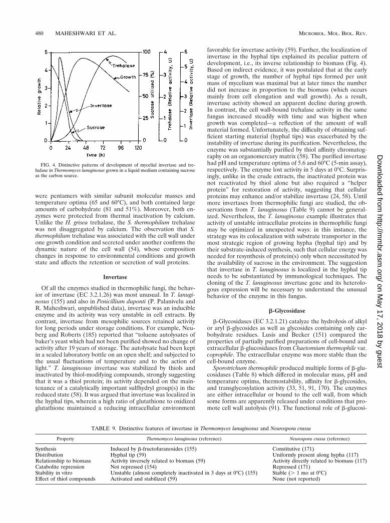
were pentamers with similar subunit molecular masses andtemperature optima (65 and 60°C), and both contained largeamounts of carbohydrate (81 and 51%). Moreover, both en-zymes were protected from thermal inactivation by calcium.Unlike the H. grisea trehalase, the S. thermophilum trehalasewas not disaggregated by calcium. The observation that S.thermophilum trehalase was associated with the cell wall underone growth condition and secreted under another confirms thedynamic nature of the cell wall (54), whose compositionchanges in response to environmental conditions and growthstate and affects the retention or secretion of wall proteins.
Invertase
Of all the enzymes studied in thermophilic fungi, the behav-ior of invertase (EC 3.2.1.26) was most unusual. In T. lanugi-nosus (155) and also in Penicillium duponti (P. Palanivelu andR. Maheshwari, unpublished data), invertase was an inducibleenzyme and its activity was very unstable in cell extracts. Bycontrast, invertase from mesophilic sources retained activityfor long periods under storage conditions. For example, Neu-berg and Roberts (185) reported that “toluene autolysates ofbaker’s yeast which had not been purified showed no change ofactivity after 19 years of storage. The autolysate had been keptin a sealed laboratory bottle on an open shelf; and subjected tothe usual fluctuations of temperature and to the action oflight.” T. lanuginosus invertase was stabilized by thiols andinactivated by thiol-modifying compounds, strongly suggestingthat it was a thiol protein; its activity depended on the main-tenance of a catalytically important sulfhydryl group(s) in thereduced state (58). It was argued that invertase was localized inthe hyphal tips, wherein a high ratio of glutathione to oxidizedglutathione maintained a reducing intracellular environment
favorable for invertase activity (59). Further, the localization ofinvertase in the hyphal tips explained its peculiar pattern ofdevelopment, i.e., its inverse relationship to biomass (Fig. 4).Based on indirect evidence, it was postulated that at the earlystage of growth, the number of hyphal tips formed per unitmass of mycelium was maximal but at later times the numberdid not increase in proportion to the biomass (which occursmainly from cell elongation and wall growth). As a result,invertase activity showed an apparent decline during growth.In contrast, the cell wall-bound trehalase activity in the samefungus increased steadily with time and was highest whengrowth was completed—a reflection of the amount of wallmaterial formed. Unfortunately, the difficulty of obtaining suf-ficient starting material (hyphal tips) was exacerbated by theinstability of invertase during its purification. Nevertheless, theenzyme was substantially purified by thiol affinity chromatog-raphy on an organomercury matrix (58). The purified invertasehad pH and temperature optima of 5.6 and 60°C (5-min assay),respectively. The enzyme lost activity in 5 days at 0°C. Surpris-ingly, unlike in the crude extracts, the inactivated protein wasnot reactivated by thiol alone but also required a “helperprotein” for restoration of activity, suggesting that cellularproteins may enhance and/or stabilize invertase (24, 58). Untilmore invertases from thermophilic fungi are studied, the ob-servations from T. lanuginosus (Table 9) cannot be general-ized. Nevertheless, the T. lanuginosus example illustrates thatactivity of unstable intracellular proteins in thermophilic fungimay be optimized in unexpected ways: in this instance, thestrategy was its colocalization with substrate transporter in themost strategic region of growing hypha (hyphal tip) and bytheir substrate-induced synthesis, such that cellular energy wasneeded for resynthesis of protein(s) only when necessitated bythe availability of sucrose in the environment. The suggestionthat invertase in T. lanuginosus is localized in the hyphal tipneeds to be substantiated by immunological techniques. Thecloning of the T. lanuginosus invertase gene and its heterolo-gous expression will be necessary to understand the unusualbehavior of the enzyme in this fungus.
b-Glycosidase
b-Glycosidases (EC 3.2.1.21) catalyze the hydrolysis of alkylor aryl b-glycosides as well as glycosides containing only car-bohydrate residues. Lusis and Becker (151) compared theproperties of partially purified preparations of cell-bound andextracellular b-glucosidases from Chaetomium thermophile var.coprophile. The extracellular enzyme was more stable than thecell-bound enzyme.
Sporotrichum thermophile produced multiple forms of b-glu-cosidases (Table 8) which differed in molecular mass, pH andtemperature optima, thermostability, affinity for b-glycosides,and transglycosylation activity (33, 51, 91, 170). The enzymesare either intracellular or bound to the cell wall, from whichsome forms are apparently released under conditions that pro-mote cell wall autolysis (91). The functional role of b-glucosi-
FIG. 4. Distinctive patterns of development of mycelial invertase and tre-halase in Thermomyces lanuginosus grown in a liquid medium containing sucroseas the carbon source.
TABLE 9. Distinctive features of invertase in Thermomyces lanuginosus and Neurospora crassa
Property Thermomyces lanuginosus (reference) Neurospora crassa (reference)
Synthesis Induced by b-fructofuranosides (155) Constitutive (171)Distribution Hyphal tip (59) Uniformly present along hypha (117)Relationship to biomass Activity inversely related to biomass (59) Activity directly related to biomass (117)Catabolite repression Not repressed (154) Repressed (171)Stability in vitro Unstable (almost completely inactivated in 3 days at 0°C) (155) Stable (. 1 mo at 0°C)Effect of thiol compounds Activated and stabilized (59) None (not reported)
480 MAHESHWARI ET AL. MICROBIOL. MOL. BIOL. REV.
on May 17, 2018 by guest
http://mm
br.asm.org/
Dow
nloaded from

dases is not known. They may be involved in cell wall biosyn-thesis or remodeling during hyphal elongation. Anotherpossibility is that the cell wall b-glucosidases may combine withcellulase enzymes to form a protein complex that is highlyefficient in solubilizing cellulose (91).
An inducible, glycoprotein b-galactosidase from T. lanugi-nosus was purified by fractional salt precipitation, hydrophobicinteraction chromatography, and anion-exchange chromatog-raphy (87). The enzyme displayed maximum activity at pH 6.7to 7.2 and was only moderately stable at 56°C, losing all activityin 1 h. At 50°C the enzyme activity in ammonium sulfateprecipitates was nearly four times as stable as in the crudeextract. The enzyme was dimeric, with a subunit molecularmass of 75 to 80 kDa. The most favorable substrates were p-and o-nitrophenyl-b-D-galactopyranosides.
Lipoamide Dehydrogenase
Lipoamide dehydrogenase (LD) (EC 1.6.4.3), also calleddihydrolipoyl dehydrogenase, is a component of the multien-zyme pyruvate dehydrogenase complex. It catalyzes the reduc-tion of NAD1 with dihydrolipoamide. LD from the thermo-philic fungus Malbranchea pulchella (Table 8) was purified tohomogeneity by a simple three-step procedure consisting ofsalt precipitation, affinity chromatography, and ion-exchangechromatography (169). M. pulchella var. sulfurea LD was verysimilar to the enzyme purified from several other sources.Although the enzyme showed appreciable resistance to ther-mal denaturation (t1/2, 170 min at 65°C), the activity was com-pletely lost in 1 month at 4°C even in concentrated solutions(greater than 2 mg/ml).
ATP Sulfurylase
Renosto et al. (216) compared the properties of the sulfate-activating enzymes ATP sulfurylase (ATP:sulfate adenylyltransferase [EC 2.7.7.4]) and APS kinase (ATP:adenylylsul-fate-39-phosphotransferase [EC 2.7.1.25]) from Penicillium du-ponti with those of the enzyme from the mesophilic fungus P.chrysogenum. The enzymes catalyze the first and second reac-tions in the assimilation of inorganic sulfate: ATP 1 SO4
223PPi 1 APS (adenosine-59-phosphosulfate) and ATP 1 APS3ADP 1 PAPS (39-phosphoadenosine-59-phosphosulfate). ATPsulfurylase of P. duponti (Table 8) had a temperature optimumat 70°C, and that of P. chrysogenum was close to 55°C. Thethermophile ATP sulfurylase was about 90 times more heatstable (first-order rate constant for denaturation, k 5 0.02min21) than the mesophile enzyme (k 5 1.78 min21) and wasalso more stable in response to urea and to low pH. Bothenzymes were hexamers, composed of nearly equal-size sub-units (69 kDa), and they had nearly identical amino acid com-positions. Antiserum to each enzyme showed cross-reactivity,indicating similar structural determinants. The activation en-ergies of P. duponti and P. chrysogenum enzymes were 127 and106 kcal, respectively, both of which are within the usual range.
The APS kinase of P. duponti was purified by the sameprocedure as that used for the P. chrysogenum enzyme. How-ever, the P. duponti enzyme preparation was not homogeneousand was also not as stable when stored frozen as concentratedsolution. At 42°C, the P. chryosgenum APS kinase dissociatedinto inactive subunits but the P. duponti enzyme remainedintact and active. At 50°C, the specific activity of P. dupontiATP sulfurylase was about twofold higher than that of the P.chrysogenum enzyme, indicating that the thermophile enzymewas a better catalyst than the mesophile enzyme.
Protein Disulfide Isomerase
Some proteins are active only when their sulfhydryl groupsare in the reduced state, and several secretory proteins areactive only when folded into three-dimensional structures bycorrect disulfide bridges. In the cell, protein disulfide isomer-ase (PDI) (EC 5.3.4.1) in the endoplasmic reticulum facilitatesdisulfide interchange by shuffling the disulfide bonds to rapidlyestablish the most thermodynamically stable pairing. The en-zyme is useful for refolding and renaturation of scrambledprotein in vitro, for basic studies, and for industrial uses. Aheat-stable PDI from mycelial extract of Humicola insolens waspurified in a rapid three-step procedure involving anion-ex-change chromatography, concanavalin A affinity chromatogra-phy, and reverse-phase high-pressure liquid chromatography(239). Like the yeast and bovine PDI, the H. insolens enzymewas a homodimer with a subunit molecular mass of 60 kDa.The cDNA of H. insolens PDI was expressed in Bacillus brevis,and the recombinant protein secreted into the culture mediumwas purified to homogeneity in two steps by anion-exchangechromatography and hydrophobic interaction chromatographywith a high yield (equivalent to 2.1 g of bovine liver enzyme perliter) (134, 135). The recombinant PDI was 1 kDa smallerbecause the recombinant protein produced by the bacterialhost is not glycosylated. The similar properties of native andrecombinant PDIs suggested that glycosylation of the enzymeis not essential for enzymatic activity and stability.
MISCELLANEOUS PROTEINS
The mitochondrial protein cytochrome c from severalsources has been studied in detail to determine the evolution-ary relationships among organisms. The primary structure ofcytochrome c from Humicola lanuginosa was quite similar tothat of other organisms, and its sequence in the heme regionwas quite homologous to that of Neurospora crassa (146, 180,181).
A phosphodiesterase (62 kDa) from Talaromyces dupontiwas purified (89). It displayed optimum activity at pH 6.6 andwas activated by cobalt ions. The enzyme retained full activityfor 1 h at 50°C. Its t1/2 was 100 min at 60°C and 10 min at 70°C.
It is of great interest to know whether the protein-synthe-sizing machinery of thermophilic fungi has intrinsic thermosta-bility. A study of the effect of temperature on the aminoacy-lation reaction showed that the maximum activity of the totaltRNA synthetase preparation from H. lanuginosa was observedat 40°C but that at the temperature for optimum growth(50°C), the aminoacylation activity was only one-half of that at40°C (132). One species of valine-tRNA synthetase was puri-fied to homogeneity. The purified enzyme was stabilizedagainst inactivation to various degrees by interaction with mag-nesium ions and the substrates valine, tRNA, and ATP, as wellas spermine.
The problem of growing large amounts of mycelia requiredas the starting material, the difficulty of extracting protein fromfungi which possess a multilayered cell wall, and the low spe-cific activities of their enzymes in the crude extracts have, ingeneral, discouraged studies of intracellular enzymes. Fromthe results of a few studies, mostly carried out with partiallypurified enzyme preparations, the picture that emerges is thatintracellular enzymes of thermophilic fungi are less stable thanthe extracellularly secreted enzymes. This leads to a furtherconsideration that rather than depending on intrinsic thermo-stability as the primary mechanism in thermal adaptation, eu-karyotic thermophily has exploited subtle variations in enzymestructure, catalysis, or mode of regulation.
VOL. 64, 2000 PHYSIOLOGY AND ENZYMES OF THERMOPHILIC FUNGI 481
on May 17, 2018 by guest
http://mm
br.asm.org/
Dow
nloaded from

CONCLUSIONS AND PROSPECTS
Modern studies on thermophilic fungi were stimulated bythe prospect of finding fungi capable of secreting high levels ofenzymes and of finding novel enzyme variants with high tem-perature optima and a long “shelf-life,” which are desirablecharacteristics for commercial applications of enzymes. Unfor-tunately, even before we had a chance to understand or ap-preciate thermophilic fungi, the questions of their adaptationto high temperatures were brushed aside in favor of practicalends. Consequently, our knowledge of their physiology is veryfragmentary. A long-held belief that thermophilic fungi areunable to grow, or grow poorly, at ambient temperatures needsto be reexamined, since some thermophilic fungi exhibit nearoptimal growth even at room temperature (20 to 30°C). Thishas led to uncertainty about their minimal temperature ofgrowth. Because of this, thermophilic fungi may be redefinedas fungi whose optimum growth temperature is 45°C or above.Although it is unlikely that the temperature range of growth ofthermophilic fungi will exceed 32 to 36°C, their latent meso-philic capability may be an advantage, since this would obviatethe need for a strict temperature regimen for their industrialcultivation, resulting in savings in operational costs. From thebasic point of view, the potential of the same fungus for growthunder thermophilic as well as mesophilic conditions posesquestions about the biochemical composition of the funguswhen grown over a wide temperature range, the rapidity withwhich metabolism is reorganized, and the mechanisms in-volved in metabolic adjustments. The question why thermo-philic fungi have not been able to breach the upper tempera-ture limit of growth of 60 to 62°C is still not resolved.
Because thermophilic fungi occur in terrestrial habitatswhich are heterogeneous in terms of temperature and thetypes and concentrations of nutrients, chemicals, gases, wateractivity, competing species, and other variables, they may beable to adapt to several factors besides just high temperature.From this perspective, research should extend to their nutrientuptake systems, their ability to utilize mixed substrates, thenature and concentrations of their intracellular ions and os-molytes, and their effects on enzyme function. In addition, thecomposition of membrane lipids and of membrane-bound en-zymes, the composition of their respiratory chain, and theirenergy production when grown under thermophilic and meso-philic conditions are other aspects that should be investigated.Only when several species are studied and the results arecompared will we be able to determine which of the observedresponses is an adaptive strategy for thermophilic growth. Ingeneral, our knowledge of the above areas of fungal physiologyis very limited. Perhaps as thermophilic fungi become betterknown, a fascination for these organisms will lead to basicstudies and their physiology and biochemistry will be betterunderstood. The ease of their isolation and maintenance ofcultures, the reduced risk of contamination of thermophiliccultures, their simple nutrition and rapid growth, and the for-mation of homogeneous mycelia in suspension cultures aredesirable features for many types of experimental studies.
Although thermophilic fungi have long been known to beinvolved in composting and humification, the mechanisms in-volved in the accelerated decomposition of biomass are notwell understood. However, their role in decomposition of veg-etable matter suggests that thermophilic fungi may be goodsources of a battery of purified enzymes that are capable ofdisassembling plant cell walls, which are required as tools byplant biologists for elucidation of cell wall architecture.
The extracellular enzymes of thermophilic fungi are appre-ciably thermostable, but less so than those of hyperthermo-
philic archaea (2). Currently, enzymes from hyperthermophilesare being favored, with prospects in biotechnology. A realisticassessment of the degree of thermostability needed for indus-trial applications of enzymes is required. Since flexibility ofproteins is essential for catalysis, the enzymes from hyperther-mophiles will have optimal conformational flexibility at thetemperatures at which they are adapted to grow (80°C orabove) but may become too rigid and have low catalytic ratesat the operational temperatures which in most situations rangefrom 50 to 65°C. Therefore, in most situations, enzymes havingmoderately high temperature optima and thermostability, ashas been observed for extracellular proteins of thermophilicfungi, may be better suited than enzymes from hyperthermo-philes. In basic studies, enzymes of thermophilic fungi (eu-karyotes) will provide additional material for comparativeanalysis of the extent of modification in kinetic properties ofenzymes vis-a-vis their thermal stability. Some thermophilicfungi have yielded crystalline preparations of thermostableproteins that are contributing to structural information on therules that determine thermostability of proteins and the criticalgroups involved in catalysis. This information will be essentialfor designing alteration in the genes encoding enzymes forspecific applications.
The available information suggests that the thermostabilityof intracellular enzymes of thermophilic fungi is not apprecia-bly different from that of mesophilic fungi. In fact, instances ofunstable enzymes in thermophilic fungi have come to light.Although the studies so far have been preliminary, there areindications that eukaryotic thermophily involves a repertoire ofmechanisms of stabilization of enzymes, with different mecha-nisms operating for different proteins: intrinsic thermostabilityand stabilization by ions, other cellular proteins includingchaperonin molecules, self-aggregation, and possibly covalentor noncovalent associations with the polymeric constituents ofthe cell wall. The activity of certain sensitive enzymes may beoptimized by their induced synthesis only when needed, bytheir placement in the most strategic location of the hypha, andby their rapid resynthesis. In this regard, the finding in a ther-mophilic fungus of a novel invertase—an enzyme that hasfigured prominently in the development of biochemistry—sug-gests that investigations may reveal other examples of enzymesthat are regulated differently in thermophilic fungi. Such ex-amples pose challenging questions about the significance of theobserved difference, including whether the observed differ-ences are adaptive responses to thermophily or to conditionsthat normally prevail in the habitat. Cloning of some genes ofthermophilic fungi and their heterologous expression have pro-vided answers to questions concerning their biosynthesis, struc-ture, and role of carbohydrate moieties in proteins. Heterolo-gous expression will undoubtedly be extended to otherenzymes, which are difficult to obtain in sufficient amountsfrom the native strain. Some thermophilic fungi produce highlevels of extracellular enzymes that are of interest in biotech-nology. Knowledge of the structure and function of transcrip-tion control regions of the genes encoding these proteins maybe used to construct recombinant genes for their overexpres-sion.
Because thermophilic fungi are found in a variety of habitatsfrom which they are easily isolated, many investigators havebeen prompted to use their own strains in enzyme studies. Thisis of course welcome, since studies with different isolates arerequired to obtain information on intraspecies differences inthe properties of the same enzyme. However, in some cases themarked differences observed in properties of enzymes in“strains” from different geographical backgrounds have raiseddoubts about whether the taxon was, in fact, correctly identi-
482 MAHESHWARI ET AL. MICROBIOL. MOL. BIOL. REV.
on May 17, 2018 by guest
http://mm
br.asm.org/
Dow
nloaded from

fied. A comparison of the properties of an enzyme from two ormore strains by the same method will be necessary to deter-mine if high variability is a characteristic feature of thermo-philic fungi. If so, investigations of the mechanisms generatingvariation may provide opportunities for fundamental discover-ies. Finally, as has been said repeatedly, progress in sciencedepends on prior investigations. It is therefore important thatscientists share cultures of thermophilic fungi and clonedgenes.
ACKNOWLEDGMENTS
We acknowledge discussions with Paul J. Vithayathil, S. Mahadevan,and S. K. Podder. Manjuli Maheshwari and S. Mahadevan suggestedseveral improvements in the manuscript. S. Anuradha, Foreign Lan-guages Department, translated Noack’s article into English.
Research by R.M. on thermophilic fungi at Bangalore was sup-ported by grants from the Department of Science and Technology, theCouncil of Scientific and Industrial Research (CSIR), and the Depart-ment of Biotechnology, Government of India. G.B. thanks CSIR forthe award of a Research Associateship. The research of M.K.B. wassupported by the United Kingdom BBSRC.
REFERENCES
1. Abe, J.-I., K. Nakajima, H. Nagano, S. Hizukuri, and K. Obata. 1988.Properties of the raw-starch digesting amylase of Aspergillus sp. K-27: asynergistic action of glucoamylase and alpha-amylase. Carbohydr. Res. 175:85–92.
2. Adams, M. W. W. 1993. Enzymes and proteins from organisms that grownear and above 100°C. Annu. Rev. Microbiol. 47:627–658.
3. Adams, P. R. 1994. Extracellular amylase activities of Rhizomucor pusillusand Humicola lanuginosa at initial stages of growth. Mycopathologia 128:139–141.
4. Adams, P. R., and J. J. Deploey. 1976. Amylase production by Mucor mieheiand M. pusillus. Mycologia 68:934–938.
5. Adams, P. R., and J. J. Deploey. 1978. Enzymes produced by thermophilicfungi. Mycologia 70:906–910.
6. Aikawa, J., T. Yamashita, M. Nishiyama, S. Horinouchi, and T. Beppu.1990. Effects of glycosylation on the secretion and enzyme activity of Mucorrennin, an aspartic proteinase of Mucor pusillus, produced by recombinantyeast. J. Biol. Chem. 265:13955–13959.
7. Alberghina, F. A. M. 1973. Growth regulation in Neurospora crassa. Effectsof nutrients and of temperature. Arch. Microbiol. 89:83–94.
8. Alexandrov, V. Y. 1977. Cells, molecules and temperature. Translated fromthe Russian by V. A. Bernstam. Springer-Verlag KG, Berlin, Germany.
9. Allen, M. B. 1953. The thermophilic aerobic spore forming bacteria. Bac-teriol. Rev. 17:125–173.
10. Allen, P. J., and R. Emerson. 1949. Guayule rubber, microbiological im-provement by shrub retting. Ind. Eng. Chem. 41:346–365.
11. Allison, D. S., M. W. Rey, R. M. Berka, G. Armstrong, and N. S. Dunn-Coleman. 1992. Transformation of the thermophilic fungus Humicola griseavar. thermoidea and overproduction of Humicola glucoamylase. Curr.Genet. 21:225–229.
12. Almeida, E. M. de, M. de L. T. M. Polizeli, H. F. Terenzi, and J. A. Jorge.1995. Purification and biochemical characterization of b-xylosidase fromHumicola grisea var. thermoidea. FEMS Microbiol. Lett. 130:171–176.
13. Anand, L., S. Krishnamurthy, and P. J. Vithayathil. 1990. Purification andproperties of xylanase from the thermophilic fungus, Humicola lanuginosa(Griffon and Maublanc) Bunce. Arch. Biochem. Biophys. 276:546–553.
14. Anand, L., and P. J. Vithayathil. 1989. Purification and properties of b-glu-cosidase from a thermophilic fungus Humicola lanuginosa (Griffon andMaublanc) Bunce. J. Ferment. Bioeng. 67:380–386.
15. Anand, L., and P. J. Vithayathil. 1990. Production of extracellular xylanaseby thermophilic fungus, Humicola lanuginosa (Griffon and Maublanc)Bunce. Indian J. Exp. Biol. 28:434–437.
16. Apinis, A. E. 1967. Dactylomyces and Thermoascus. Trans. Br. Mycol. Soc.50:573–582.
17. Arima, K., S. Iwasaki, and G. Tamura. 1967. Milk clotting enzyme frommicroorganisms. I. Screening test and the identification of the potent fun-gus. Agric. Biol. Chem. 31:540–545.
18. Arima, K., S. Iwasaki, and G. Tamura. 1968. Milk clotting enzymes frommicroorganisms. V. Purification and crystallization of Mucor rennin fromMucor pusillus Lindt. Appl. Microbiol. 16:1727–1733.
19. Arima, K., W.-H. Liu, and T. Beppu. 1972. Studies on the lipase of ther-mophilic fungus Humicola lanuginosa. Agric. Biol. Chem. 36:893–895.
20. Arima, K., J. Yu, and S. Iwasaki. 1970. Milk-clotting enzyme from Mucorpusillus var. Lindt. Methods Enzymol. 19:446–459.
21. Arnaud, N., D. A. Bush, and M. Horisberger. 1976. Study of an intracellulara-galactosidase from the thermophilic fungus Penicillium duponti. Biotech-nol. Bioeng. 18:581–585.
22. Arnesen, S., S. H. Eriksen, J. Olsen, and B. Jensen. 1998. Increased pro-duction of a-amylase from Thermomyces lanuginosus by the addition ofTween 80. Enzyme Microb. Technol. 23:249–252.
23. Barnett, E. A., and C. L. Fergus. 1971. The relation of extracellular amylase,mycelium, and time, in some thermophilic and mesophilic Humicola spe-cies. Mycopathol. Mycol. Appl. 44:131–141.
24. Basha, S. Y., and P. Palanivelu. 1998. Enhancement in activity of an inver-tase from the thermophilic fungus Thermomyces lanuginosus by exogenousproteins. World J. Microbiol. Biotechnol. 14:603–605.
25. Baudys, M., S. Foundling, M. Pavlik, T. Blundell, and V. Kostka. 1988.Protein chemical characterization of Mucor pusillus aspartic proteinase.Amino acid sequence homology with the other aspartic proteinases, disul-fide bond arrangement and site of carbohydrate attachment. FEBS Lett.235:271–274.
26. Bennett, N. A., J. Ryan, P. Biely, M. Vrsanska, L. Kremnicky, B. J. Macris,D. Kekos, P. Christakopoulos, P. Katapodis, M. Claeyssens, W. Nerinckx,P. Ntauma, and M. K. Bhat. 1998. Biochemical and catalytic properties ofan endoxylanase purified from the culture filtrate of Thermomyces lanugi-nosus ATCC 46882. Carbohydr. Res. 306:445–455.
27. Berka, R. M., M. W. Rey, K. M. Brown, T. Byun, and A. V. Klotz. 1998.Molecular characterization and expression of a phytase gene from thethermophilic fungus Thermomyces lanuginosus. Appl. Environ. Microbiol.64:4423–4427.
28. Berka, R. M., P. Schneider, E. J. Golightly, S. H. Brown, M. Madden, K. M.Brown, T. Halkier, K. Mondorf, and F. Xu. 1997. Characterization of thegene encoding an extracellular laccase of Myceliophthora thermophila andanalysis of the recombinant enzyme expressed in Aspergillus oryzae. Appl.Environ. Microbiol. 63:3151–3157.
29. Bernstein, F. C., T. F. Koetzle, G. J. B. Williams, E. F. Meyer, Jr., M. D.Brice, J. R. Rodgers, O. Kennard, T. Shimanouchi, and M. Tasumi. 1977.The protein data bank: a computer-based archival file for macromolecularstructures. J. Mol. Biol. 112:535–542.
30. Bharadwaj, G., and R. Maheshwari. 1999. A comparison of thermal andkinetic parameters of trehalases from a thermophilic and mesophilic fun-gus. FEMS Microbiol. Lett. 181:187–193.
31. Bhat, K. M., J. S. Gaikwad, and R. Maheshwari. 1993. Purification andcharacterization of an extracellular b-glucosidase from the thermophilicfungus Sporotrichum thermophile and its influence on cellulase activity.J. Gen. Microbiol. 139:2825–2832.
32. Bhat, K. M., and R. Maheshwari. 1987. Sporotrichum thermophile: growth,cellulose degradation, and cellulase activity. Appl. Environ. Microbiol. 53:2175–2182.
33. Bhat, M. K., and S. Bhat. 1997. Cellulose degrading enzymes and theirpotential industrial applications. Biotechnol. Adv. 15:583–620.
34. Bhat, M. K., and S. Bhat. 1998. Clostridium thermocellum cellulosome:dissociation, isolation and characterisation of subunits and the potentialbiotechnological implications. Recent Res. Dev. Biotechnol. Bioeng. 1:59–84.
35. Biely, P. 1985. Microbial xylanolytic systems. Trends Biotechnol. 3:286–290.36. Blochl, E., R. Rachel, S. Burggraf, D. Hafenbradl, H. W. Jannasch, and
K. O. Stetter. 1997. Pyrolobus fumarii, gen. and sp. nov., represents a novelgroup of archaea, extending the upper temperature limit for life to 113°C.Extremophiles 1:14–21.
37. Boasson, R., and M. Shaw. 1984. Further observations on the growth of flaxrust fungus in axenic culture. Can. J. Bot. 62:2175–2180.
38. Boel, E., A.-M. Bech, K. Randrup, B. Draeger, N. P. Fiil, and B. Foltmann.1986. Primary structure of a precursor to the aspartic proteinase fromRhizomucor miehei shows that the enzyme is synthesized as a zymogen.Proteins Struct. Funct. Genet. 1:363–369.
39. Boel, E., B. Huge-Jensen, M. Christensen, L. Thim, and N. P. Fiil. 1988.Rhizomucor miehei triglyceride lipase is synthesized as a precursor. Lipids23:701–706.
40. Boisset, C., R. Borsali, M. Schulein, and B. Henrissat. 1995. Dynamic lightscattering study of the two-domain structure of Humicola insolens endoglu-canase V. FEBS Lett. 376:49–52.
41. Bousquet, M.-P., R.-M. Willemot, P. Monsan, and E. Boures. 1998. Pro-duction, purification, and characterization of thermostable a-transglucosi-dase from Talaromyces duponti—application to a-alkylglucoside synthesis.Enzyme Microbiol. Technol. 23:83–90.
42. Brady, L., A. M. Brzozowski, Z. S. Derewenda, E. Dodson, G. Dodson, S.Tolley, J. P. Turkenburg, L. Christiansen, B. Huge-Jensen, L. Norskov, L.Thim, and U. Menge. 1990. A serine protease triad forms the catalyticcentre of a triacylglycerol lipase. Nature 343:767–770.
43. Broad, T. E., and M. G. Shepherd. 1970. Purification and properties ofglucose-6-phosphate dehydrogenase from the thermophilic fungus Penicil-lium duponti. Biochim. Biophys. Acta 198:407–414.
44. Brock, T. D. 1967. Life at high temperatures. Science 158:1012–1019.45. Brock, T. D. 1995. The road to Yellowstone—and beyond. Annu. Rev.
Microbiol. 49:1–28.
VOL. 64, 2000 PHYSIOLOGY AND ENZYMES OF THERMOPHILIC FUNGI 483
on May 17, 2018 by guest
http://mm
br.asm.org/
Dow
nloaded from

46. Bunni, L., L. McHale, and A. P. McHale. 1989. Production, isolation andpartial characterization of an amylase system produced by Talaromycesemersonii CBS 814.70. Enzyme Microb. Technol. 11:370–375.
47. Campos, L., and C. R. Felix. 1995. Purification and characterization of aglucoamylase from Humicola grisea. Appl. Environ. Microbiol. 61:2436–2438.
48. Canevascini, G. 1988. Cellobiose dehydrogenase from Sporotrichum ther-mophile. Methods Enzymol. 160:443–448.
49. Canevascini, G., P. Borer, and J.-L. Dreyer. 1991. Cellobiose dehydroge-nases of Sporotrichum (Chrysosporium) thermophile. Eur. J. Biochem. 198:43–52.
50. Canevascini, G., M.-R. Coudray, J.-P. Rey, R. J. G. Southgate, and H.Meier. 1979. Induction and catabolite repression of cellulase synthesis inthe thermophilic fungus Sporotrichum thermophile. J. Gen. Microbiol. 110:291–303.
51. Canevascini, G., and H. P. Meyer. 1979. b-Glucosidase in the cellulolyticfungus Sporotrichum thermophile Apinis. Exp. Mycol. 3:203–214.
52. Cardello, L., H. F. Terenzi, and J. A. Jorge. 1994. A cytosolic trehalase fromthe thermophilic fungus Humicola grisea var. thermoidea. Microbiology 140:1671–1677.
53. Cesar, T., and V. Mrsa. 1996. Purification and properties of the xylanaseproduced by Thermomyces lanuginosus. Enzyme Microb. Technol. 19:289–296.
54. Chaffin, W. L., J. L. Lopez-Ribot, M. Casanova, D. Gozalbo, and J. P.Martınez. 1998. Cell wall and secreted proeins of Candida albicans. Iden-tification, function and expression. Microbiol. Mol. Biol. Rev. 62:130–180.
55. Chang, Y. 1967. The fungi of wheat straw compost. II. Biochemical andphysiological studies. Trans. Br. Mycol. Soc. 50:667–677.
56. Chapuis, R., and H. Zuber. 1970. Thermophilic aminopeptidases: AP Ifrom Talaromyces duponti. Methods Enzymol. 19:552–555.
57. Chaudhri, S., I. S. Thakur, R. Goel, and B. N. Johri. 1988. Purification andcharacterization of two thermostable xylanases from Melanocarpus albomy-ces. Biochem. Int. 17:563–575.
58. Chaudhuri, A., and R. Maheshwari. 1996. A novel invertase from a ther-mophilic fungus Thermomyces lanuginosus: its requirement of thiol andprotein for activation. Arch. Biochem. Biophys. 327:98–106.
59. Chaudhuri, A., G. Bharadwaj, and R. Maheshwari. 1999. An unusual pat-tern of invertase activity development in the thermophilic fungus Thermo-myces lanuginosus. FEMS Microbiol. Lett. 177:39–45.
60. Chefetz, B., Y. Chen, and Y. Hadar. 1998. Purification and characterizationof laccase from Chaetomium thermophilium and its role in humification.Appl. Environ. Microbiol. 64:3175–3179.
61. Christensen, T., H. Woeldike, E. Boel, S. B. Mortensen, K. Hjortshoej, L.Thim, and M. T. Hansen. 1988. High level expression of recombinant genesin Aspergillus oryzae. Bio/Technology 6:1419–1422.
62. Colaco, C., S. Sen, M. Thangavelu, S. Pinder, and B. Roser. 1992. Extraor-dinary stability of enzymes dried in trehalose: simplified molecular biology.Bio/Technology 10:1007–1011.
63. Cooney, D. G., and R. Emerson. 1964. Thermophilic fungi. An account oftheir biology, activities and classification. W. H. Freeman & Co., San Fran-cisco, Calif.
64. Coudray, M.-R., G. Canevascini, and H. Meier. 1982. Characterization of acellobiose dehydrogenase in the cellulolytic fungus Sporotrichum (Chryso-sporium) thermophile. Biochem. J. 203:277–284.
65. Coughlan, M. P., and A. McHale. 1988. Purification of b-D-glucoside glu-cohydrolases of Talaromyces emersonii. Methods Enzymol. 160:437–443.
66. Coughlan, M. P., and A. P. Moloney. 1988. Isolation of 1,4-b-D-glucan4-glucanohydrolases of Talaromyces emersonii. Methods Enzymol. 160:363–368.
67. Coutts, A. D., and R. E. Smith. 1976. Factors influencing the production ofcellulases by Sporotrichum thermophile. Appl. Environ. Microbiol. 31:819–825.
68. Crowe, J. H., L. M. Crowe, and D. Chapman. 1984. Preservation of mem-branes in anhydrobiotic organisms: the role of trehalose. Science 223:701–703.
69. Davies, G. J., S. P. Tolley, B. Henrissat, C. Hjort, and M. Schulein. 1995.Structure of oligosaccharide-bound forms of the endoglucanase V fromHumicola insolens at 1.9 Å resolution. Biochemistry 34:16210–16220.
70. Davies, G., S. Tolley, K. Wilson, M. Schulein, H. F. Woldike, and G.Dodson. 1992. Crystallization and preliminary X-ray analysis of a fungalendoglucanase I. J. Mol. Biol. 228:970–972.
71. Deploey, J. J. 1995. Some factors affecting the germination of Thermoascusaurantiacus. Mycologia 87:362–365.
72. Derewenda, Z. S., U. Derewenda, and G. G. Dodson. 1992. The crystal andmolecular structure of the Rhizomucor miehei triacylglyceride lipase at 1.9Å resolution. J. Mol. Biol. 227:818–839.
73. Derewenda, U., L. Swenson, R. Green, Y. Wei, G. G. Dodson, S. Yamaguchi,M. J. Haas, and Z. S. Derewenda. 1994. An unusual buried polar cluster ina family of fungal lipases. Nat. Struct. Biol. 1:36–47.
74. Derikx, P. J. L., H. J. M. Op den Camp, A. M. Wagner, G. Straatsma,L. J. L. D. van Griensven, and G. D. Vogels. 1990. Respiratory pathways in
Agaricus bisporus and Scytalidium thermophilum. FEMS Microbiol. Lett.66:307–312.
75. Desai, S. B., A. K. Mattoo, V. V. Modi, and G. S. Patel. 1973. Growth andfructose-1,6-diphosphate aldolase of a thermophilic Talaromyces sp. IndianJ. Exp. Biol. 11:352–354.
76. Duo-Chuan, L., Y. Yi-Jung, P. You-Liang, and S. Chong-Yao. 1998. Puri-fication and characterization of extracellulae glucoamylase from the ther-mophilic Thermomyces lanuginosus. Mycol. Res. 102:568–572.
77. Dusterhoft, E.-M., V. A. J. M. Linssen, A. G. J. Voragen, and G. Beldman.1997. Purification, characterization, and properties of two xylanases fromHumicola insolens. Enzyme Microb. Technol. 20:437–445.
78. Emerson, R. 1968. Thermophiles, p. 105–128. In G. C. Ainsworth and A. S.Sussman (ed.), The fungi, an advanced treatise, vol. 3. Academic Press, Inc.,New York, N.Y.
79. Emi, S., D. V. Myers, and G. A. Iacobucci. 1976. Purification and propertiesof the thermostable acid protease of Penicillium duponti. Biochemistry15:842–848.
80. Eriksen, J., and J. Goksøyr. 1976. The effect of temperature on growth andcellulase (beta-1,4-endoglucanase) production in the compost fungusChaetomium thermophile var. dissitum. Arch. Microbiol. 110:233–238.
81. Eriksen, J., and J. Goksøyr. 1977. Cellulases from Chaetomium thermophilevar. dissitum. Eur. J. Biochem. 77:445–450.
82. Eriksson, K.-E., B. Pettersson, and U. Westermark. 1974. Oxidation: animportant enzyme reaction in fungal degradation of cellulose. FEBS Lett.49:282–285.
83. Eswaramoorthy, S., P. J. Vithayathil, and M. A. Viswamitra. 1994. Crys-tallization and preliminary X-ray crystallographic studies of thermostablexylanase crystals isolated from Paecilomyces varioti. J. Mol. Biol. 243:806–808.
84. Etoh, Y., H. Shoun, T. Beppu, and K. Arima. 1979. Physicochemical andimmunochemical studies on similarities of acid proteases Mucor pusillusrennin and Mucor miehei rennin. Agric. Biol. Chem. 43:209–215.
85. Fergus, C. L. 1969. The production of amylase by some thermophilic fungi.Mycologia 61:1171–1175.
86. Filho, E. X. F. 1996. Purification and characterization of a b-glucosidasefrom solid-state cultures of Humicola grisea var. thermoidea. Can. J. Micro-biol. 42:1–5.
87. Fischer, L., C. Scheckermann, and F. Wagner. 1995. Purification and char-acterization of a thermotolerant b-galactosidase from Thermomyces lanugi-nosus. Appl. Environ. Microbiol. 61:1497–1501.
88. Folan, M. A., and M. P. Coughlan. 1978. The cellulase complex in theculture filtrate of the thermophyllic fungus, Talaromyces emersonii. Int.J. Biochem. 9:717–722.
89. Fontana, A., E. Boccu’, and F. M. Veronese. 1974. Purification of a phos-phodiesterase from the thermophilic fungus Talaromyces duponti. Hoppe-Seyler’s Z. Physiol. Chem. 355:1436–1442.
90. Fracheboud, D., and G. Canevascini. 1989. Isolation, purification, andproperties of the exocellulase from Sporotrichum (Chrysosporium) thermo-phile. Enzyme Microb. Technol. 11:220–229.
91. Gaikwad, J. S., and R. Maheshwari. 1994. Localization and release ofb-glucosidase in the thermophilic and cellulolytic fungus, Sporotrichumthermophile. Exp. Mycol. 18:300–310.
92. Ganju, R. K. 1986. Isolation and characterization of polysaccharide degrad-ing enzymes of Chaetomium thermophile var. coprophile. Ph.D. thesis. In-dian Institute of Science, Bangalore.
93. Ganju, R. K., S. K. Murthy, and P. J. Vithayathil. 1989. Purification andcharacterization of two cellobiohydrolases from Chaetomium thermophilevar. coprophile. Biochim. Biophys. Acta 993:266–274.
94. Ganju, R. K., S. K. Murthy, and P. J. Vithayathil. 1990. Purification andfunctional characteristics of an endocellulase from Chaetomium thermo-phile var. coprophile. Carbohydr. Res. 197:245–255.
95. Ganju, R. K., P. J. Vithayathil, and S. K. Murthy. 1989. Purification andcharacterization of two xylanases from Chaetomium thermophile var. cop-rophile. Can. J. Microbiol. 35:836–842.
96. Gilbert, M., C. Breuil, M. Yaguchi, and J. N. Saddler. 1992. Purificationand characterization of a xylanase from the thermophilic ascomyceteThielavia terrestris 255B. Appl. Biochem. Biotechnol. 34–35:247–259.
97. Gilbert, M., M. Yaguchi, D. C. Watson, K. K. Y. Wong, C. Breuil, and J. N.Saddler. 1993. A comparison of two xylanases from the thermophilic fungiThielavia terrestris and Thermoascus crustaceus. Appl. Microbiol. Biotech-nol. 40:508–514.
98. Gomes, J., I. Gomes, W. Kreiner, H. Esterbauer, M. Sinner, and W. Steiner.1993. Production of high level of cellulase-free and thermostable xylanaseby a wild strain of Thermomyces lanuginosus using beechwood xylan. J. Bio-technol. 30:283–297.
99. Gomes, J., H. Purkarthofer, M. Hayn, J. Kapplmuller, M. Sinner, and W.Steiner. 1993. Production of a high level of cellulase-free xylanase by thethermophilic fungus Thermomyces lanuginosus in laboratory and pilot scalesusing lignocellulosic materials. Appl. Microbiol. Biotechnol. 39:700–707.
100. Gray, G. L., K. Hayenga, D. Cullen, L. J. Wilson, and S. Norton. 1986.Primary structure of Mucor miehei aspartyl protease: evidence for a zymo-gen intermediate. Gene 48:41–53.
484 MAHESHWARI ET AL. MICROBIOL. MOL. BIOL. REV.
on May 17, 2018 by guest
http://mm
br.asm.org/
Dow
nloaded from

101. Gruber, K., G. Klintschar, M. Hayn, A. Schlacher, W. Steiner, and C.Kratky. 1998. Thermophilic xylanase from Thermomyces lanuginosus: highresolution X-ray structure and modeling studies. Biochemistry 37:13475–13485.
102. Gupta, S. D., and R. Maheshwari. 1985. Is organic acid required for nutri-tion of thermophilic fungi? Arch. Microbiol. 141:164–169.
103. Haasum, I., S. H. Eriksen, B. Jensen, and J. Olsen. 1991. Growth andglucoamylase production by the thermophilic fungus Thermomyces lanugi-nosus in a sysnthetic medium. Appl. Microbiol. Biotechnol. 34:656–660.
104. Harwood, J. 1989. The versatility of lipases for industrial uses. TrendsBiochem. Sci. 14:125–126.
105. Hashimoto, H., T. Iwaasa, and T. Yokotsuka. 1972. Thermostable acidprotease produced by Penicillium duponti K1014, a true thermophilic fun-gus newly isolated from compost. Appl. Microbiol. 24:986–992.
106. Hashimoto, H., T. Iwaasa, and T. Yokotsuka. 1973. Some properties of acidprotease from the thermophilic fungus, Penicillium duponti K1014. Appl.Microbiol. 25:578–583.
107. Hashimoto, H., Y. Kaneko, T. Iwaasa, and T. Yokotsuka. 1973. Productionand purification of acid protease from the thermophilic fungus, Penicilliumduponti K1014. Appl. Microbiol. 25:584–588.
108. Hasnain, S., K. Adeli, and A. C. Storer. 1992. Purification and character-ization of an extracellular thiol-containing serine proteinase from Thermo-myces lanuginosus. Biochem. Cell Biol. 70:117–122.
109. Hayashida, S., and K. Mo. 1986. Production and characteristics of avicel-disintegrating endoglucanase from a protease-negative Humicola grisea var.thermoidea mutant. Appl. Environ. Microbiol. 51:1041–1046.
110. Hayashida, S., K. Ohta, and K. Mo. 1988. Cellulases of Humicola insolensand Humicola grisea. Methods Enzymol. 160:323–332.
111. Hayashida, S., K. Ohta, and K. Mo. 1988. Xylanase of Talaromyces byss-ochlamydoides. Methods Enzymol. 160:675–678.
112. Hayashida, S., and H. Yoshioka. 1980. The role of carbohydrate moiety onthermostability of cellulases from Humicola insolens YH-8. Agric. Biol.Chem. 44:481–487.
113. Hayashida, S., and H. Yoshioka. 1980. Production and purification of ther-mostable cellulases from Humicola insolens YH-8. Agric. Biol. Chem. 44:1721–1728.
114. Hecker, L. I., and A. S. Sussman. 1973. Activity and heat stability oftrehalase from the mycelium and ascospores of Neurospora. J. Bacteriol.115:582–591.
115. Hedger, J. N., and H. J. Hudson. 1974. Nutritional studies of Thermomyceslanuginosus from wheat straw compost. Trans. Br. Mycol. Soc. 62:129–143.
116. Henssen, A. 1957. Uber die Bedeutung der thermophilen Mikroorganismenfur die Zersetzung des Stallmistes. Arch. Mikrobiol. 27:63–81.
117. Hill, E. P., and A. S. Sussman. 1964. Development of trehalase and inver-tase activity in Neurospora. J. Bacteriol. 88:1556–1566.
118. Hiramatsu, R., J. Aikawa, S. Horinouchi, and T. Beppu. 1989. Secretion byyeast of the zymogen form of Mucor rennin, an aspartic proteinase of Mucorpusillus, and its conversion to the mature form. J. Biol. Chem. 264:16862–16866.
119. Hiramatsu, R., T. Yamashita, J.-I. Aikawa, S. Horinouchi, and T. Beppu.1990. The prepro-peptide of Mucor rennin directs the secretion of humangrowth hormone by Saccharomyces cerevisiae. Appl. Environ. Microbiol.56:2125–2132.
120. Holmquist, M., I. G. Clausen, S. Patkar, A. Svendsen, and K. Hult. 1995.Probing a functional role of Glu87 and Trp89 in the lid of Humicolalanuginosa lipase through transesterification reactions in organic solvent. J.Protein Chem. 14:217–224.
121. Holmquist, M., M. Martinelle, I. G. Clausen, S. Patkar, A. Svendsen, andK. Hult. 1994. Trp 89 in the lid of Humicola lanuginosa lipase is importantfor efficient hydrolysis of tributyrin. Lipids 29:599–603.
122. Huge-Jensen, B., F. Andreasen, T. Christensen, M. Christensen, L. Thim,and E. Boel. 1989. Rhizomucor miehei triglyceride lipase is processed andsecreted from transformed Aspergillus oryzae. Lipids 24:781–785.
123. Huge-Jensen, B., D. R. Gaialuzzo, and R. G. Jensen. 1987. Partial purifi-cation and characterization of free and immobilized lipases from Mucormiehei. Lipids 22:559–565.
124. Igarashi, K., M. Samejima, Y. Saburi, N. Habu, and K.-E. L. Eriksson.1997. Localization of cellobiose dehydrogenase in cellulose-grown culturesof Phanerochaete chrysosporium. Fungal Genet. Biol. 21:214–222.
125. Igarashi, K., M. F. J. M. Verhagen, M. Samejima, M. Schulein, K.-E. L.Eriksson, and T. Nishino. 1999. Cellobiose dehydrogenase from the fungiPhanerochaete chrysosporium and Humicola insolens. J. Biol. Chem. 274:3338–3344.
126. Inamdar, A. 1987. Polygalacturonase from Thermoascus aurantiacus: isola-tion and functional characteristics. Ph.D. thesis. Indian Institute of Science,Bangalore.
127. Iwasaki, S., G. Tamura, and K. Arima. 1967. Milk clotting enzyme frommicroorganisms. II. The enzyme production and the properties of crudeenzyme. Agric. Biol. Chem. 31:546–551.
128. Jaeger, K.-E., and M. T. Reetz. 1998. Microbial lipases form versatile toolsfor biotechnology. Trends Biotechnol. 16:396–403.
129. Jaychandran, S., and R. Ramabadran. 1970. Production of amylase by
Thermoascus aurantiacus. Indian J. Exp. Biol. 8:344.130. Jensen, B., and J. Olsen. 1992. Physicochemical properties of a purified
alpha-amylase from the thermophilic fungus Thermomyces lanuginosus. En-zyme Microb. Technol. 14:112–116.
131. Jensen, B., J. Olsen, and K. Allermann. 1988. Purification of extracellularamylolytic enzymes from the thermophilic fungus Thermomyces lanugino-sus. Can. J. Microbiol. 34:218–223.
132. Joshi, A. K., and J. D. Cherayil. 1987. Stabilisation of some of the proteinsynthesis components in the thermophilic fungus, Humicola lanuginosa.J. Biosci. 11:193–202.
133. Kadowaki, M. K., M. L. T. M. Polizeli, H. F. Terenzi, and J. A. Jorge. 1996.Characterization of trehalase activities from the thermophilic fungusScytalidium thermophilum. Biochim. Biophys. Acta 1291:199–205.
134. Kajino, T., C. Miyazaki, O. Asami, M. Hirai, Y. Yamada, and S. Udaka.1998. Thermophilic fungal protein disulfide isomerase. Methods Enzymol.290:50–59.
135. Kajino, T., K. Sarai, T. Imaeda, C. Idekoba, O. Asami, Y. Yamada, M.Hirai, and S. Udaka. 1994. Molecular cloning of a fungal cDNA encodingprotein disulfide isomerase. Biosci. Biotechnol. Biochem. 58:1424–1429.
136. Kane, B. E., and J. T. Mullins. 1973. Thermophilic fungi in a municipalwaste compost system. Mycologia 65:1087–1100.
137. Kawamori, M., K.-I. Takayama, and S. Takasawa. 1987. Production ofcellulases by a thermophilic fungus Thermoascus aurantiacus A-131. Agric.Biol. Chem. 51:647–654.
138. Khandke, K. M. 1986. Isolation and functional characteristics of xylanasesand cellulases of Thermoascus aurantiacus. Ph.D. thesis. Indian Institute ofScience, Bangalore.
139. Khandke, K. M., P. J. Vithayathil, and S. K. Murthy. 1989. Purification ofxylanase, b-glucosidase, endocellulase, and exocellulase from a thermo-philic fungus, Thermoascus aurantiacus. Arch. Biochem. Biophys. 274:491–500.
140. Khandke, K. M., P. J. Vithayathil, and S. K. Murthy. 1989. Degradation oflarchwood xylan by enzymes of a thermophilic fungus, Thermoascus auran-tiacus. Arch. Biochem. Biophys. 274:501–510.
141. Khandke, K. M., P. J. Vithayathil, and S. K. Murthy. 1989. Purification andcharacterization of an a-D-glucuronidase from a thermophilic fungus, Ther-moascus aurantiacus. Arch. Biochem. Biophys. 274:511–517.
142. Kitpreechavanich, V., M. Hayashi, and S. Nagai. 1984. Purification andproperties of endo-1,4-b-xylanase from Humicola lanuginosa. J. Ferment.Technol. 5:415–420.
143. Kleywegt, G. J., J.-Y. Zou, C. Divne, G. J. Davies, I. Sinning, J. Ståhlberg,T. Reinikainen, M. Srisodsuk, T. T. Teeri, and T. A. Jones. 1997. The crystalstructure of the catalytic core domain of endoglucanase I from Trichodermareesei at 3.6 Å resolution, and a comparison with related enzymes. J. Mol.Biol. 272:383–397.
144. Krishnamurthy, S. 1989. Purification and properties of xylanases and b-glu-cosidases elaborated by the thermophilic fungus Paecilomyces variotiBainier. Ph.D. thesis. Indian Institute of Science, Bangalore.
145. Lamed, R., E. Stetter, R. Kenig, and E. A. Beyer. 1983. The cellulosome—adiscrete cell surface organelle of Clostridium thermocellum which exhibitsseparate antigenic, cellulose binding and various cellulolytic activities. Bio-technol. Bioeng. Symp. 13:163–181.
146. Lederer, F., and A. M. Simon. 1974. Neurospora crassa and Humicolalanuginosa cytochromes c: more homology in the heme region. Biochem.Biophys. Res. Commun. 56:317–323.
147. Li, B., S. R. Nagalla, and V. Renganathan. 1997. Cellobiose dehydrogenasefrom Phanerochaete chrysosporium is encoded by two allelic variants. Appl.Environ. Microbiol. 63:796–799.
148. Lindt, W. 1886. Mitteilungen uber einige neue pathogene Shimmelpilze.Arch. Exp. Pathol. Pharmakol. 21:269–298.
149. Liu, W.-H., T. Beppu, and K. Arima. 1973. Physical and chemical propertiesof the lipase of thermophilic fungus Humicola lanuginosa S-38. Agric. Biol.Chem. 37:2493–2499.
150. Lo Leggio, L., S. Kalogiannis, M. K. Bhat, and R. W. Pickersgill. 1999. Highresolution structure and sequence of T. aurantiacus xylanase I: implicationsfor the evolution of thermostability in family 10 xylanases and enzymes withba-barrel architecture. Proteins Struct. Funct. Genet. 36:295–306.
151. Lusis, A. J., and R. R. Becker. 1973. The b-glucosidase system of thethermophilic fungus Chaetomium thermophile var. coprophile n. var. Bio-chim. Biophys. Acta 329:5–16.
152. Mackenzie, L. F., G. Sulzenbacher, C. Divne, T. A. Jones, H. F. Woldike, M.Schulein, S. G. Withers, and G. J. Davies. 1998. Crystal structure of thefamily 7 endoglucanase I (Cel7B) from Humicola insolens at 2.2 Å resolu-tion and identification of the catalytic nucleophile by trapping of the cova-lent glycosyl-enzyme intermediate. Biochem. J. 335:409–416.
153. Maheshwari, R. 1997. The ecology of thermophilic fungi, p. 277–289. InK. K. Janardhan, C. Rajendran, K. Natarajan and D. L. Hawksworth (ed.),Tropical mycology. Oxford & IBH Publishing Co. Pvt. Ltd., New Delhi,India.
154. Maheshwari, R., and P. V. Balasubramanyam. 1988. Simultaneous utiliza-tion of glucose and sucrose by thermophilic fungi. J. Bacteriol. 170:3274–3280.
VOL. 64, 2000 PHYSIOLOGY AND ENZYMES OF THERMOPHILIC FUNGI 485
on May 17, 2018 by guest
http://mm
br.asm.org/
Dow
nloaded from

155. Maheshwari, R., P. V. Balasubramanyam, and P. Palanivelu. 1983. Dis-tinctive behaviour of invertase in a thermophilic fungus, Thermomyceslanuginosus. Arch. Microbiol. 134:255–260.
156. Maheshwari, R., and P. T. Kamalam. 1985. Isolation and culture of athermophilic fungus, Melanocarpus albomyces, and factors influencing theproduction and activity of xylanase. J. Gen. Microbiol. 131:3017–3027.
157. Maheshwari, R., P. T. Kamalam, and P. V. Balasubramanyam. 1987. Thebiogeography of thermophilic fungi. Curr. Sci. 56:151–155.
158. Malcolm, A. A., and M. G. Shepherd. 1972. Purification and properties ofPenicillium glucose-6-phosphate dehydrogenase. Biochem. J. 128:817–831.
159. Mandels, M. 1975. Microbial sources of cellulase. Biotechnol. Bioeng.Symp. 5:81–105.
160. Mandels, M., and D. Sternberg. 1976. Recent advances in cellulase tech-nology. J. Ferment. Technol. 54:267–286.
161. Mansfield, S. D., E. de Jong, and J. N. Saddler. 1997. Cellobiose dehydro-genase, an active agent in cellulose depolymerization. Appl. Environ. Mi-crobiol. 63:3804–3809.
162. Matsuo, M., and T. Yasui. 1985. Properties of xylanase of Malbrancheapulchella var. sulfurea no. 48. Agric. Biol. Chem. 49:839–841.
163. Matsuo, M., T. Yasui, and T. Kobayashi. 1977. Purification and someproperties of b-xylosidase from Malbranchea pulchella var. sulfurea no. 48.Agric. Biol. Chem. 41:1593–1599.
164. Matsuo, M., T. Yasui, and T. Kobayashi. 1977. Enzymatic properties ofb-xylosidase from Malbranchea pulchella var. sulfurea no. 48. Agric. Biol.Chem. 41:1601–1606.
165. McHale, A., and M. P. Coughlan. 1980. Synergistic hydrolysis of cellulose bycomponents of the extracellular cellulase system of Talaromyces emersonii.FEBS Lett. 117:319–322.
166. McHale, A., and M. P. Coughlan. 1981. The cellulolytic system of Talaro-myces emersonii. Identification of the various components produced duringgrowth on cellulosic media. Biochim. Biophys. Acta 662:145–151.
167. McHale, A., and M. P. Coughlan. 1981. The cellulolytic system of Talaro-myces emersonii. Purification and characterization of the extracellular andintracellular b-glucosidases. Biochim. Biophys. Acta 662:152–159.
168. McHale, A., and M. P. Coughlan. 1982. Properties of the b-glucosidases ofTalaromyces emersonii. J. Gen. Microbiol. 128:2327–2331.
169. McKay, D. J., and K. J. Stevenson. 1979. Lipoamide dehydrogenase fromMalbranchea pulchella: isolation and characterization. Biochemistry 18:4702–4707.
170. Meyer, H.-P., and G. Canevascini. 1981. Separation and some properties oftwo intracellular b-glucosidases of Sporotrichum (Chrysosporium) thermo-phile. Appl. Environ. Microbiol. 41:924–931.
171. Metzenberg, R. L. 1962. A gene affecting the repression of invertase andtrehalase in Neurospora. Arch. Biochem. Biophys. 96:468–474.
172. Miehe, H. 1907. Die Selbsterhitzung des Heus. Eine biologische Studie.Gustav Fischer Verlag, Jena, Germany.
173. Miehe, H. 1930. Die Warmebildung von Reinkulturen im Hinblick auf dieAtiologie der Selbsterhitzung pflanzlicher Stoffe. Arch. Mikrobiol. 1:78–118.
174. Miehe, H. 1930. Uber die Selbsterhitzung des Heues. Arb. Dtsch. Land-wirtsch.-Gesellsch. Berlin 111:76–91.
175. Miller, H. M., and M. G. Shepherd. 1972. Purification and properties of6-phosphogluconate dehydrogenase from Penicillium duponti and Penicil-lium notatum. Can. J. Microbiol. 18:1289–1298.
176. Miller, H. M., P. A. Sullivan, and M. G. Shepherd. 1974. Intracellularprotein breakdown in thermophilic and mesophilic fungi. Biochem. J. 144:209–214.
177. Mishra, R. S., and R. Maheshwari. 1996. Amylases of the thermophilicfungus Thermomyces lanuginosus: their purification, properties, action onstarch and response to heat. J. Biosci. 21:653–672.
178. Moloney, A. P., S. I. McCrae, T. M. Wood, and M. P. Coughlan. 1985.Isolation and characterization of 1,4-b-D-glucan glucanohydrolases of Ta-laromyces emersonii. Biochem. J. 225:365–374.
179. Monti, R., H. F. Terenzi, and J. A. Jorge. 1991. Purification and propertiesof an extracellular xylanase from the thermophilic fungus Humicola griseavar. thermoidea. Can. J. Microbiol. 37:675–681.
180. Morgan, W. T., C. P. Hensley, Jr., and J. P. Riehm. 1972. Proteins of thethermophilic fungus Humicola lanuginosa. I. Isolation and amino acid se-quence of a cytochrome c. J. Biol. Chem. 247:6555–6565.
181. Morgan, W. T., and J. P. Riehm. 1973. Proteins of the thermophilic fungusHumicola lanuginosa. II. Some physicochemical properties of a cytochromec. Arch. Biochem. Biophys. 154:415–421.
182. Mouchacca, J. 1997. Thermophilic fungi: biodiversity and taxonomic status.Cryptogamie Mycol. 18:19–69.
183. Naren, A. P. 1992. Structure and function of xylanases from four thermo-philic fungi. Ph.D. thesis. Indian Institute of Science, Bangalore.
184. Natesh, R., P. Bhanumoorthy, P. J. Vithayathil, K. Sekar, S. Ramakumar,and M. A. Viswamitra. 1999. Crystal structure at 1.8 Å resolution andproposed amino acid sequence of thermostable xylanase from Thermoascusaurantiacus. J. Mol. Biol. 288:999–1012.
185. Neuberg, C., and I. S. Roberts. 1946. Invertase. Scientific Reports Series,no. 4. Sugar Research Foundation, Inc., New York, N.Y.
186. Neves, M. J., H. F. Terenzi, F. A. Leone, and J. A. Jorge. 1994. Quantifi-cation of trehalose in biological samples with a conidial trehalase from thethermophilic fungus Humicola grisea var. thermoidea. World J. Microbiol.Biotechnol. 10:17–19.
187. Newman, M., F. Watson, P. Roychowdhury, H. Jones, M. Badasso, A.Cleasby, S. P. Wood, I. J. Tickle, and T. L. Blundell. 1993. X-ray analysesof aspartic proteinases. V. Structure and refinement at 2.0 Å resolution ofthe aspartic proteinase from Mucor pusillus. J. Mol. Biol. 230:260–283.
188. Noack, K. 1920. Der Betriebstoffwechsel der thermophilen Pilze. Jahrb.Wiss. Bot. 59:593–648.
189. Oberson, J., T. Binz, D. Fracheboud, and G. Canevascini. 1992. Compar-ative investigation of cellulose-degrading enzyme systems produced by dif-ferent strains of Myceliophthora thermophila (Apinis) v. Oorschot. EnzymeMicrob. Technol. 14:303–312.
190. Oberson, J., A. Rawyler, R. Brandle, and G. Canevascini. 1999. Analysis ofthe heat-shock response displayed by two Chaetomium species originatingfrom different thermal environments. Fungal Genet. Biol. 26:178–189.
191. Omar, I. C., M. Hayashi, and S. Nagai. 1987. Purification and some prop-erties of a thermostable lipase from Humicola lanuginosa no. 3. Agric. Biol.Chem. 51:37–45.
192. Omar, I. C., N. Nishio, and S. Nagai. 1987. Production of a thermostablelipase by Humicola lanuginosa grown on sorbitol-corn steep liquor medium.Agric. Biol. Chem. 51:2145–2151.
193. Ong, P. S., and G. M. Gaucher. 1973. Protease production by thermophilicfungi. Can. J. Microbiol. 19:129–133.
194. Ong, P. S., and G. M. Gaucher. 1976. Production, purification and charac-terization of thermomycolase, the extracellular serine protease of the ther-mophilic fungus Malbranchea pulchella var. sulfurea. Can. J. Microbiol.22:165–176.
195. Ottesen, M., and W. Rickert. 1970. The isolation and partial characteriza-tion of an acid protease produced by Mucor miehei. C. R. Trav. Lab.Carlsberg 37:301–325.
196. Palanivelu, P., P. V. Balasubramanyam, and R. Maheshwari. 1984. Co-induction of sucrose transport and invertase activities in a thermophilicfungus, Thermomyces lanuginosus. Arch. Microbiol. 139:44–47.
197. Peralta, R. M., H. F. Terenzi, and J. A. Jorge. 1990. b-D-Glycosidaseactivities of Humicola grisea: biochemical and kinetic characterization of amultifunctional enzyme. Biochim. Biophys. Acta 1033:243–249.
198. Peters, G. H., A. Svendsen, H. Langberg, J. Vind, S. A. Patkar, S. Toxvaerd,and P. K. J. Kinnunen. 1998. Active serine involved in the stabilization ofthe active site loop in the Humicola lanuginosa lipase. Biochemistry 37:12375–12383.
199. Prabhu, K. A., and R. Maheshwari. 1999. Biochemical properties of xyla-nases from a thermophilic fungus, Melanocarpus albomyces, and their actionon plant cell walls. J. Biosci. 24:461–470.
200. Prasad, A. R. S., and R. Maheshwari. 1978. Growth of and trehalase activityin the thermophilic fungus Thermomyces lanuginosus. Proc. Indian Acad.Sci. 87B:231–241.
201. Prasad, A. R. S., and R. Maheshwari. 1978. Purification and properties oftrehalase from the thermophilic fungus Humicola lanuginosa. Biochim.Biophys. Acta 525:162–170.
202. Prasad, A. R. S., and R. Maheshwari. 1979. Temperature-response oftrehalase from mesophilic (Neurospora crassa) and a thermophilic fungus(Thermomyces lanuginosus). Arch. Microbiol. 122:275–280.
203. Prasad, A. R. S., C. K. R. Kurup, and R. Maheshwari. 1979. Effect oftemperature on respiration of a mesophilic and thermophilic fungus. PlantPhysiol. 64:347–348.
204. Puchart, V., P. Katapodis, P. Biely, Kremnicky, P. Christakopoulos, M.Vrsanska, D. Kekos, B. J. Macris, and M. K. Bhat. 1999. Production ofxylanases, mannanases, and pectinases by the thermophilic fungus Thermo-myces lanuginosus. Enzyme Microb. Technol. 24:355–361.
205. Purkarthofer, H., M. Sinner, and W. Steiner. 1993. Effect of shear rate andculture pH on the production of xylanase by Thermomyces lanuginosus.Biotechnol. Lett. 15:405–410.
206. Purkarthofer, H., M. Sinner, and W. Steiner. 1993. Cellulase-free xylanasefrom Thermomyces lanuginosus: optimization of production in submergedand solid-state culture. Enzyme Microb. Technol. 15:677–682.
207. Rajasekaran, A. K., and S. Divakar. 1988. Carbon-13 NMR spectroscopicstudy of a thermophilic fungus. Curr. Sci. 57:235–237.
208. Rajasekaran, A. K., and R. Maheshwari. 1990. Growth kinetics and intra-cellular protein breakdown in mesophilic and thermophilic fungi. Indian J.Exp. Biol. 28:46–51.
209. Rajasekaran, A. K., and R. Maheshwari. 1990. Effect of growth tempera-ture on lipid composition of a thermophilic fungus Thermomyces lanugino-sus. Indian J. Exp. Biol. 28:134–137.
210. Rajasekaran, A. K., and R. Maheshwari. 1993. Thermophilic fungi: anassessment of their potential for growth in soil. J. Biosci. 18:345–354.
211. Rao, U. S., and S. K. Murthy. 1988. Purification and characterization of ab-glucosidase and endocellulase from Humicola insolens. Indian J. Bio-chem. Biophys. 25:687–694.
212. Rao, U. S., and S. K. Murthy. 1991. The effects of b-mercaptoethanol and
486 MAHESHWARI ET AL. MICROBIOL. MOL. BIOL. REV.
on May 17, 2018 by guest
http://mm
br.asm.org/
Dow
nloaded from
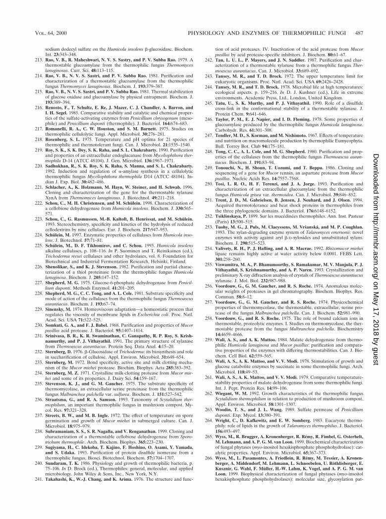
sodium dodecyl sulfate on the Humicola insolens b-glucosidase. Biochem.Int. 23:343–348.
213. Rao, V. B., R. Maheshwari, N. V. S. Sastry, and P. V. Subba Rao. 1979. Athermostable glucoamylase from the thermophilic fungus Thermomyceslanuginosus. Curr. Sci. 48:113–115.
214. Rao, V. B., N. V. S. Sastri, and P. V. Subba Rao. 1981. Purification andcharacterization of a thermostable glucoamylase from the thermophilicfungus Thermomyces lanuginosus. Biochem. J. 193:379–387.
215. Rao, V. B., N. V. S. Sastri, and P. V. Subba Rao. 1981. Thermal stabilizationof glucose oxidase and glucoamylase by physical entrapment. Biochem. J.193:389–394.
216. Renosto, F., T. Schultz, E. Re, J. Mazer, C. J. Chandler, A. Barron, andI. H. Segel. 1985. Comparative stability and catalytic and chemical proper-ties of the sulfate-activating enzymes from Penicillium chrysogenum (meso-phile) and Penicillium duponti (thermophile). J. Bacteriol. 164:674–683.
217. Romanelli, R. A., C. W. Houston, and S. M. Barnett. 1975. Studies onthermophilic cellulolytic fungi. Appl. Microbiol. 30:276–281.
218. Rosenberg, S. L. 1975. Temperature and pH optima for 21 species ofthermophilic and thermotolerant fungi. Can. J. Microbiol. 21:1535–1540.
219. Roy, S. K., S. K. Dey, S. K. Raha, and S. L. Chakrabarty. 1990. Purificationand properties of an extracellular endoglucanase from Myceliophthora ther-mophila D-14 (ATCC 48104). J. Gen. Microbiol. 136:1967–1971.
220. Sadhukhan, R., S. K. Roy, S. K. Raha, S. Manna, and S. L. Chakrabarty.1992. Induction and regulation of a-amylase synthesis in a cellulolyticthermophilic fungus Myceliophthora thermophila D14 (ATCC 48104). In-dian J. Exp. Biol. 30:482–486.
221. Schlacher, A., K. Holzmann, M. Hayn, W. Steiner, and H. Schwab. 1996.Cloning and characterization of the gene for the thermostable xylanaseXynA from Thermomyces lanuginosus. J. Biotechnol. 49:211–218.
222. Schou, C., M. H. Christensen, and M. Schulein. 1998. Characterization ofa cellobiose dehydrogenase from Humicola insolens. Biochem. J. 330:565–571.
223. Schou, C., G. Rasmussen, M.-B. Kaltoft, B. Henrissat, and M. Schulein.1993. Stereochemistry, specificity and kinetics of the hydrolysis of reducedcellodextrins by nine cellulaes. Eur. J. Biochem. 217:947–953.
224. Schulein, M. 1997. Enzymatic properties of cellulases from Humicola inso-lens. J. Biotechnol. 57:71–81.
225. Schulein, M., D. F. Tikhomirov, and C. Schou. 1993. Humicola insolensalkaline cellulases, p. 108–116. In P. Suominen and T. Reinikainen (ed.),Trichoderma reesei cellulases and other hydrolases, vol. 8. Foundation forBiotechnical and Industrial Fermentation Research, Helsinki, Finland.
226. Shenolikar, S., and K. J. Stevenson. 1982. Purification and partial charac-terization of a thiol proteinase from the thermophilic fungus Humicolalanuginosa. Biochem. J. 205:147–152.
227. Shepherd, M. G. 1975. Glucose-6-phosphate dehydrogenase from Penicil-lium duponti. Methods Enzymol. 41:201–205.
228. Shepherd, M. G., C. C. Tong, and A. L. Cole. 1981. Substrate specificity andmode of action of the cellulases from the thermophilic fungus Thermoascusaurantiacus. Biochem. J. 193:67–74.
229. Sinensky, M. 1974. Homeoviscous adaptation—a homeostatic process thatregulates the viscosity of membrane lipids in Escherichia coli. Proc. Natl.Acad. Sci. USA 71:522–525.
230. Somkuti, G. A., and F. J. Babel. 1968. Purification and properties of Mucorpusillus acid protease. J. Bacteriol. 95:1407–1414.
231. Srinivasa, B. R., K. R. Swaminathan, C. Ganapathy, R. P. Roy, S. Krish-namurthy, and P. J. Vithayathil. 1991. The primary structure of xylanasefrom Thermoascus aurantiacus. Protein Seq. Data Anal. 4:15–20.
232. Sternberg, D. 1976. b-Glucosidase of Trichoderma: its biosynthesis and rolein saccharification of cellulose. Appl. Environ. Microbiol. 31:648–654.
233. Sternberg, M. 1972. Bond specificity, active site and milk clotting mecha-nism of the Mucor miehei protease. Biochim. Biophys. Acta 285:383–392.
234. Sternberg, M. Z. 1971. Crystalline milk-clotting protease from Mucor mie-hei and some of its properties. J. Dairy Sci. 54:159–167.
235. Stevenson, K. J., and G. M. Gaucher. 1975. The substrate specificity ofthermomycolase, an extracellular serine proteinase from the thermophilicfungus Malbranchea pulchella var. sulfurea. Biochem. J. 151:527–542.
236. Straatsma, G., and R. A. Samson. 1993. Taxonomy of Scytalidium ther-mophilum, an important thermophilic fungus in mushroom compost. My-col. Res. 97:321–328.
237. Streets, B. W., and M. B. Ingle. 1972. The effect of temperature on sporegermination and growth of Mucor miehei in submerged culture. Can. J.Microbiol. 18:975–979.
238. Subramaniam, S. S., S. R. Nagalla, and V. Renganathan. 1999. Cloning andcharacterization of a thermostable cellobiose dehydrogenase from Sporo-trichum thermophile. Arch. Biochem. Biophys. 365:223–230.
239. Sugiyama, H., C. Idekoba, T. Kajino, F. Hoshino, O. Asami, Y. Yamada,and S. Udaka. 1993. Purification of protein disulfide isomerase from athermophilic fungus. Biosci. Biotechnol. Biochem. 57:1704–1707.
240. Sundaram, T. K. 1986. Physiology and growth of thermophilic bacteria, p.75–106. In D. Brock (ed.), Thermophiles: general, molecular, and appliedmicrobiology. John Wiley & Sons, Inc., New York, N.Y.
241. Takahashi, K., W.-J. Chang, and K. Arima. 1976. The structure and func-
tion of acid proteases. IV. Inactivation of the acid protease from Mucorpusillus by acid protease-specific inhibitors. J. Biochem. 80:61–67.
242. Tan, L. U. L., P. Mayers, and J. N. Saddler. 1987. Purification and char-acterization of a thermostable xylanase from a thermophilic fungus Ther-moascus aurantiacus. Can. J. Microbiol. 33:689–692.
243. Tansey, M. R., and T. D. Brock. 1972. The upper temperature limit foreukaryotic organisms. Proc. Natl. Acad. Sci. USA 69:2426–2428.
244. Tansey, M. R., and T. D. Brock. 1978. Microbial life at high temperatures:ecological aspects. p. 159–216. In D. J. Kushner (ed.), Life in extremeenvironments. Academic Press, Ltd., London, United Kingdom.
245. Tatu, U., S. K. Murthy, and P. J. Vithayathil. 1990. Role of a disulfidecross-link in the conformational stability of a thermostable xylanase. J.Protein Chem. 9:641–646.
246. Taylor, P. M., E. J. Napier, and I. D. Fleming. 1978. Some properties ofglucoamylase produced by the thermophilic fungus Humicola lanuginosa.Carbohydr. Res. 61:301–308.
247. Tendler, M. D., S. Korman, and M. Nishimoto. 1967. Effects of temperatureand nutrition on macromolecule production by thermophilic Eumycophyta.Bull. Torrey Bot. Club 94:175–181.
248. Tong, C. C., A. L. Cole, and M. G. Shepherd. 1980. Purification and prop-erties of the cellulases from the thermophilic fungus Thermoascus auran-tiacus. Biochem. J. 191:83–94.
249. Tonouchi, N., H. Shoun, T. Uozumi, and T. Beppu. 1986. Cloning andsequencing of a gene for Mucor rennin, an aspartate protease from Mucorpusillus. Nucleic Acids Res. 14:7557–7568.
250. Tosi, L. R. O., H. F. Terenzi, and J. A. Jorge. 1993. Purification andcharacterization of an extracellular glucoamylase from the thermophilicfungus Humicola grisea var. thermoidea. Can. J. Microbiol. 39:846–852.
251. Trent, J. D., M. Gabrielsen, B. Jensen, J. Neuhard, and J. Olsen. 1994.Acquired thermotolerance and heat shock proteins in thermophiles fromthe three phylogenetic domains. J. Bacteriol. 176:6148–6152.
252. Tsiklinskaya, P. 1899. Sur les mucedinees thermophiles. Ann. Inst. Pasteur(Paris) 13:500–515.
253. Tuohy, M. G., J. Puls, M. Claeyssens, M. Vrsanska, and M. P. Coughlan.1993. The xylan-degrading enzyme system of Talaromyces emersonii: novelenzymes with activity against aryl b-D-xylosides and unsubstituted xylans.Biochem. J. 290:515–523.
254. Valivety, R. H., P. J. Halling, and A. R. Macrae. 1992. Rhizomucor mieheilipase remains highly active at water activity below 0.0001. FEBS Lett.301:258–260.
255. Viswamitra, M. A., P. Bhanumoorthy, S. Ramakumar, M. V. Manjula, P. J.Vithayathil, S. Krishnamurthy, and A. P. Naren. 1993. Crystallization andpreliminary X-ray diffraction analysis of crystals of Thermoascus aurantiacusxylanase. J. Mol. Biol. 232:987–988.
256. Voordouw, G., G. M. Gaucher, and R. S. Roche. 1974. Anomalous molec-ular weights of proteases in gel chromatography. Biochem. Biophys. Res.Commun. 58:8–12.
257. Voordouw, G., G. M. Gaucher, and R. S. Roche. 1974. Physicochemicalproperties of thermomycolase, the thermostable, extracellular, serine pro-tease of the fungus Malbranchea pulchella. Can. J. Biochem. 52:981–990.
258. Voordouw, G., and R. S. Roche. 1975. The role of bound calcium ions inthermostable, proteolytic enzymes. I. Studies on thermomycolase, the ther-mostable protease from the fungus Malbranchea pulchella. Biochemistry14:4659–4666.
259. Wali, A. S., and A. K. Mattoo. 1984. Malate dehydrogenase from thermo-philic Humicola lanuginosa and Mucor pusillus: purification and compara-tive properties of the enzymes with differing thermostabilities. Can. J. Bio-chem. Cell Biol. 62:559–565.
260. Wali, A. S., A. K. Mattoo, and V. V. Modi. 1978. Stimulation of growth andglucose catabolite enzymes by succinate in some thermophilic fungi. Arch.Microbiol. 118:49–53.
261. Wali, A. S., A. K. Mattoo, and V. V. Modi. 1979. Comparative temperature-stability properties of malate dehydrogenase from some thermophilic fungi.Int. J. Pept. Protein Res. 14:99–106.
262. Wiegant, W. M. 1992. Growth characteristics of the thermophilic fungusScytalidium thermophilum in relation to production of mushroom compost.Appl. Environ. Microbiol. 58:1301–1307.
263. Woodin, T. S., and J. L. Wang. 1989. Sulfate permease of Penicilliumduponti. Exp. Mycol. 13:380–391.
264. Wright, C., D. Kafkewitz, and E. W. Somberg. 1983. Eucaryote thermo-phily: role of lipids in the growth of Talaromyces thermophilus. J. Bacteriol.156:493–497.
265. Wyss, M., R. Brugger, A. Kronenberger, R. Remy, R. Fimbel, G. Osterhelt,M. Lehmann, and A. P. G. M. van Loon. 1999. Biochemical characterizationof fungal phytases (myo-inositol hexakisphosphate phosphohydrolase): cat-alytic properties. Appl. Environ. Microbiol. 65:367–373.
266. Wyss, M., L. Pasamontes, A. Friedlein, R. Remy, M. Tessier, A. Kronen-berger, A. Middendorf, M. Lehmann, L. Schnoebelen, U. Rothlisberger, E.Kusznir, G. Wahl, F. Muller, H.-W. Lahm, K. Vogel, and A. P. G. M. vanLoon. 1999. Biophysical characterization of fungal phytases (myo-insoitolhexakisphosphate phosphohydrolases): molecular size, glycosylation pat-
VOL. 64, 2000 PHYSIOLOGY AND ENZYMES OF THERMOPHILIC FUNGI 487
on May 17, 2018 by guest
http://mm
br.asm.org/
Dow
nloaded from

tern, and engineering of proteolytic resistance. Appl. Environ. Microbiol.65:359–366.
267. Yamashita, T., N. Tonouchi, T. Uozumi, and T. Beppu. 1987. Secretion ofMucor rennin, a fungal aspartic protease of Mucor pusillus, by recombinantyeast cells. Mol. Gen. Genet. 210:462–467.
268. Yamashita, T., S. Higashi, T. Higashi, H. Machida, S. Iwasaki, M. Nish-iyama, and T. Beppu. 1994. Mutation of a fungal aspartic proteinase, Mucorpusillus rennin, to decrease thermostability for use as a milk coagulant.J. Biotechnol. 32:17–28.
269. Yang, J., A. Teplyakov, and J. W. Quail. 1997. Crystal structure of theaspartic proteinase from Rhizomucor miehei at 2.15 Å resolution. J. Mol.Biol. 268:449–459.
270. Yoshioka, H., S.-I. Anraku, and S. Hayashida. 1982. Production and puri-fication of a novel type of CMCase from Humicola grisea var. thermoideaYH-78. Agric. Biol. Chem. 46:75–82.
271. Yoshioka, H., and S. Hayashida. 1980. Purification and properties of b-glu-cosidase from Humicola insolens YH-8. Agric. Biol. Chem. 44:1729–1735.
272. Yoshioka, H., N. Nagato, S. Chavanich, N. Nilubol, and S. Hayashida. 1981.Purification and properties of thermostable xylanase from Talaromyces bys-sochlamydoides YH-50. Agric. Biol. Chem. 45:2425–2432.
273. Yu, J., S. Iwasaki, G. Tamura, and K. Arima. 1968. Physical properties andamino acid composition of Mucor rennin crystal isolated from Mucor pu-sillus var. Lindt. Agric. Biol. Chem. 32:1051–1053.
274. Yu, E. K. C., L. U. L. Tan, M. K.-H. Chan, L. Deschatelets, and J. N.Saddler. 1987. Production of thermostable xylanase by a thermophilic fun-gus, Thermoascus aurantiacus. Enzyme Microb. Technol. 9:16–24.
275. Zimmermann, A. L. S., H. F. Terenzi, and J. A. Jorge. 1990. Purification andproperties of an extracellular conidial trehalase from Humicola grisea var.thermoidea. Biochim. Biophys. Acta 1036:41–46.
488 MAHESHWARI ET AL. MICROBIOL. MOL. BIOL. REV.
on May 17, 2018 by guest
http://mm
br.asm.org/
Dow
nloaded from