therapeutic aphaeresis, technical overview - arpn journal of
TRANSCRIPT

VOL. 2, NO. 5, June 2012 ISSN 2225-7217
ARPN Journal of Science and Technology ©2011-2012. All rights reserved.
http://www.ejournalofscience.org
399
Therapeutic Aphaeresis, Technical Overview 1 Rolf Bambauer, 2 Ralf Schiel, 3 Boris Lehmann, 4 Carolin Bambauer
1 Formerly: Institute for Bloodpurification, 66424 Homburg, Germany, 2 Inselklinik Heringsdorf GmbH, 17424 Seeheilbad Heringsdorf, Germany,
3 Laboratorium of Medicine, 66386 St. Ingbert, Germany 4 Main Hospital Darmstadt, 64283 Darmstadt, Germany
ABSTRACT
Since the mid-1970s, a new process for plasma phrases became available which used membrane modules instead of centrifuge. Many patients suffering from severe diseases with a poor prognosis could be cured. The advantages of therapeutic aphaeresis with membrane modules are a complete separation of the corpuscular components from the plasma, an effective elimination of macromolecular substances and a less cell damage. The development of new membranes has allowed new techniques, and the adsorption technologies allow the most selective separation of plasma components. The technical aspects of the different methods, the interaction between extracorporeal surfaces and blood, and path physiological points are shown. Up to now the cascade filtration, lipid filtration, imunoadsorption, LDL-aphaeresis and the liver support system such as the Bio Logic-DTPP system, Molecular Adsorbent Recycling System and the Prometheus System have been of clinical relevance. The technical aspects and their clinical indication are discussed. In experienced hands, therapeutic has minimal side effects. New approaches are made in the last years, and some companies have developed machines which can be used as well as for intermitted dialysis and for therapeutic aphaeresis methods. Keywords: Therapeutic aphaeresis, LDL-aphaeresis, cascade filtration, lipid filtration, immuno-adsorption, heparin-induced LDL-precipitation (HELP), LDL adsorption with dextran sulphate (Liposorber), Lipoprotein (a) aphaeresis, LDL-hemoperfusion, liver support systems
1. INTRODUCTION
The ancient medical belief that removal of a patient´s blood can also lead to the removal of his disease was widely accepted in the pre-scientific era as bloodletting, could not, of course, be upheld in the light of modern medicine (1). Still, it appears to have returned to our clinical practice within the framework of new theories. While the practice of plasma phrases may also call to mind the ancient practice of bloodletting (2), since blood content is removed from the patients’ body for a period of time, the two therapy methods are, of course, based on entirely different theoretical bases and are used to treat conditions understood within the framework of modern medicine.
More than six thousand years ago, bloodletting or phlebotomy methods for different diseases are mentioned in the Chinese medicine and three thousand years ago, priests in ancient Egypt began the use of bloodletting to treat many diseases (3). The priests of ancient Egypt, the healers of classical Greece and imperial Rome, the learned doctors of feudal Europe, all took for granted the necessity of cleansing the bodies of sick patients to get rid of foul and noxious matter. Thus for 3,000 years of Western civilization, physicians and healers required the sick to sweat; to drink copious amounts of potions to rinse their bodies; and to take emetics, purgatives, and enemas to cleanse their bowels. Finally, they were bled. The practice of bloodletting was based on a simple logical analogy: "You must first draw out the old and stagnant water from a drinking well to allow the fresh spring water to flow in" (1). The practice of bloodletting spread to Greece, where the art of
medicine in the ancient world reached its prime between 500 B.C. and 500 A.D. Well into the late 18th century, the "bloodletting-man" was widely used as an aide-memoire to recall when and for what illness to go to the barber for bloodletting. Around the figure one can see the signs of the Zodiak. They determine the optimal time for the cure of the various ailments in different parts of the body. Behind this image stands the ancient belief in a connection between different parts of the body and the stars and planets in the sky (4). In the early 18th century, bloodletting and related procedures reached their height of popularity. Hedon seems to have been the first to discuss the subject of plasma phrases in 1902. He auto transfused body-own erythrocytes into rabbits (5). The first therapeutic approaches in humans were taken by Fleig in 1909. He reported investigations of auto transfusions with washed blood cells in toxaemia and hetero transfusions in anaemia (6). Five years later in 1914, Abel, Row tree, and Turner studied the effect of plasma removal in dogs (7). They created the term Plasma phrases. In 1926 Gilbert, Tzanck, and Negroni described plasma phrases applied in humans (8). In 1944 Tui, Bartter, Wright, and Holt carried out plasma separation using centrifuges for the first time. Since that time, it has become possible to produce fractions of plasma for transfusions (9). In 1950 Grifols-Lucas established plasma collection on a routine basis in Lisbon (10). In 1952 Adams and his colleagues for the first time successfully treated a patient with multiple myeloma by plasma phrases (11)

VOL. 2, NO. 5, June 2012 ISSN 2225-7217
ARPN Journal of Science and Technology ©2011-2012. All rights reserved.
http://www.ejournalofscience.org
400
During the mid-1970s, an additional process for plasma phrases became available which used membrane modules instead of centrifuges. The first successful application of the so-called membrane plasma separation in humans was reported by Yamazaki et al., Castino et al., and Nose´ et al. in 1976 (12,13). The advantages of this method are a complete separation of the corpuscular components from the plasma and due to increased blood flow rate higher efficacy. Furthermore, cell damage - especially to thrombocytes - occurs less using membranes than centrifuges for cell separation (14). The plasma phrases equipment currently available are, however, not yet perfect, because the filtered plasma fractions have to be discarded. Substitution solutions, electrolyte solutions supplemented with human albumin, human serum protein solutions, or fresh frozen plasma are used to replace the discarded fractions. The development of new, more sophisticated membranes has allowed to new techniques, such as double and triple filtration. These procedures allow for a more selective plasma component removal. The adsorption technologies allow the most selective separation of plasma components. Such techniques hold much future promise. 2. MEMBRANE PLASMPHERESIS Until the development of hollow fibber membranes, therapeutic plasma exchange (TPE) was almost exclusively carried out by the centrifugal technique. These devices are mainly used today - aside from the conventional TPE and selective separation process - for the production of plasma fractions and blood cell concentrates. While in North America TPE is mostly carried out with centrifuges, the membrane plasmapheresis is used mainelyin Europe and Japan. The devices for membrane plasmapheresis have to be operated with the control of the transmembrane pressure. The hemo filtration systems can be used. As early as 1980, physicians adapted the single needle technique to plasmapheresis and simplified the system in the process. Investigators used a double pump in combination with a hollow fibber module, a pressure balancing system, and a heater pump (15). In addition, two level-detectors were added to the system; therefore, the system could work on a semi-automatic principle. The main advantages of the double head pump unit are that:
1. A relatively large flow rate of plasma through the plasma separator is achieved without the maximum pressure exceeding the safety limit.
2. The average pressure in the membrane separator can be adjusted to individual requirements.
3. Haemolysis can then occur at various pressures in different membrane separators.
4. The discontinuous mode of operation of both pumps causes a permanent change in flow and pressure and
delays the membrane fouling by blood cells. In systems for single-needle plasmapheresis, the balancing pump is connected to the blood return air trap of the double head pump through a tubing system. It can pump exact volumes of the substitution solution into this chamber. The filtrate is also pumped off with the substitution fluid through its own tubing. The amount of substitution fluid is equal to the amount of filtrate pumped off with the same balancing pump and over the same pump segments. A second air trap is included in the filtrate line to prevent misbalance. Filtrate flow rates can be changed by adjusting the speed of the roller pumps and by modifying the transmembrane pressure. On commencing TPE, the filtrate flow rate depends on the blood flow rate and the transmembrane pressure (16). Further development and improvements of pump systems are necessary. Recently, new pump systems (pulsatile and non pulsatile) for extracorporeal circulation have found use in clinical practice (17, 18). In 1984, a small double head pump was developed which works according to this principle. It is especially useful in newborns and premature infants where blood volume is limited. Hemo filtration, dialysis, and plasmapheresis treatments may be carried out with such a small double head pump (220 x 255 x 95 mm). Plasmapheresis treatment requires a supplementary heater and balancing pump. With a dialysis system, single-needle hemo dialysis treatments can be carried out also. The double-needle technique may also be used with this system (19). The blood flow must be at least 5–15 ml/min. The arterial and venous tubing system and the hollow fibber module may be filled with donor blood or fresh frozen plasma before the treatment is started. The total volume of the extracorporeal circulation was 30–37 ml (20). In 1981, Cobe/USA independently developed a compact flat plate membrane device which works exclusively by using the double-needle method. It is especially suitable for peripheral vascular connection with a blood flow of at least 40 ml/min. The plate membrane module has a surface area of 0.13 m². Thus, depending to the blood flow by which the pressure on the membrane causes stretching, the wall shear-rate is controlled within narrow limits. The filtration performance can thus be kept constant (21). The filtrate flow increases with blood flow. Other companies have offered devices for membrane plasmapheresis. These devices also work with the double-needle access technique (22). An essential limitation of all these devices is the fact that – aside from their technical complexity - they are only for use in TPE and not in combination with other extracorporeal detoxification methods. The exchange of substances through semi permimeable membranes takes place through diffusion and/or convection. For permeable substances, the
VOL. 2, NO. 5, June 2012 ISSN 2225-7217
ARPN Journal of Science and Technology ©2011-2012. All rights reserved.
http://www.ejournalofscience.org
401
transport is determined by the concentration gradient or hydrostatic gradient. During the diffusion process, the migratory velocity of the substances depends on concentration difference, temperature, as well as on the flow in the system. This is called a passive transport of substances (23). During this process, the high-molecular weight components are separated from low-molecular compounds in the aqueous phase (24). The principle of plasma filtration is based on a convective transport of substances, which is qualitatively pre-determined through the membrane structure and quantitatively by the transmembrane pressure. The ability to support a membrane is additionally determined by the properties of the substances to be filtered. In general, it is higher in blood than in aqueous solutions free particles. This difference arises because in blood a secondary membrane is formed because of the concentration polarization. Through the deposit of proteins and the sedimentation of blood cells on the membrane surface, there is a reduction in the filtrate stream and a decrease in the sieving coefficient, with a simultaneous increase in the capacity to support the membrane (25). According to this approach, the filtration properties of a plasmapheresis membrane are determined less through the pores of the membrane than due to the different composition of the patient blood, the different flow conditions, through a dynamically developing secondary membrane. This secondary membrane has “gel-like“- characteristics and is formed by the macromolecules kept back by plasmapheresis. Its thickness and porosity depend on the flow velocity of the blood (26). Mean pore size determination was based upon analysis of particle rejection. Pore size results of correlations of rejection coefficients with particle size are based on the log-normal probability analysis (27). The different membranes are noted to differ considerably in mean pore size. Therefore, it seems to be important to determine the mean pore size in all membranes used for plasma separation. The primary considerations, however, include blood and plasma viscosities, cell and macromolecule concentrations, cell types and sizes, and the individual properties of the cells. With plasma separation, the whole blood cell concentration increases substantially from the inlet to the outlet (28). The maximum plasma separation rate is related to the inlet cell concentration. The filtration rate of cell-free plasma is associated with the viscosity of the solution and to the interactions, as by adsorption, of plasma proteins with the membrane materials, which lead to pore narrowing (27, 29). Increasing the applied TMP increases the filtration rate of cell-free plasma. Membranes have an asymmetrical structure. Inside on the blood-leading side, they appear at first to have a specific thickness of 1 μm. They also have pores with a diameter of 0.2 to 0.5 μm. A thicker layer of 140 to 160 μm with a tube system dilating to the outside is
connected to this structure. The membrane structure is macro-reticular anisotropic and asymmetric. A certain quantity of these hollow fibbers - according to the request surface between five hundred and several thousand - is put together in one tube. The inside diameters of the hollow fibber modules range from 330 to 370 μm. Aside from these hollow fibber modules, there are also membrane separators in the form of flat membranes. The permeability of all these membranes for macromolecular substances reaches a molecular mass of 0.2 x 10 high 6 to 0.5 x 10 high 6 Dalton. The limit of permeability can be reduced by means of changes in the spin process. Surface modification is carried during the spinning process with modifying solutions which contain different substances with reactive expose groups (16). Available membranes are either hydrophilic and consist of cellulose acetate, poly , poly vinyl alcohol, and polycarbonate, or else hydrophobic and consist of polypropylene, polyvinylchloride, and other synthetic polymers. The term back filtration describes the movement of plasma from the plasma compartment into the blood compartment of the hollow fibre separator in that section of the hollow fibre separator distant from the blood inlet port. The driving force is a transmembrane pressure gradient, which is such that the plasma pressure is higher than the blood pressure. This is in the reverse direction of the TMP set for the removal of plasma from the patient´s blood. At the blood inlet port the blood pressure will always exceed the plasma pressure side of the hollow fibre, so filtration from blood to plasma fluid occurs. However, pressure drops along the length of the hollow fibre separator (from inlet to outlet) and the blood pressure may become lower than the plasma pressure at some point of the hollow fibre separation. The membranes used in TPE are micro porous membranes, through which all plasma solutes can pass. Apart from hydrophilic membranes consisting of cellulose diacetate or polycarbonate, even hydrophobic membranes consisting of materials such as polypropylene have been applied. Hydrophilic (affinity for water) and hydrophobic (lacking affinity for water) properties are primarily based on the properties of the lateral chains - directed at the outside - of the synthetic polymer. Polar lateral chains are hydrophilic, the apolar ones are hydrophobic. Aside from polar or apolar the properties, the surface charge of the filter membrane also plays a crucial role in biocompatibility. Thus, negative surface charges are linked to an inhibited activation of the intrinsic coagulation system (30). Unlike hydrophilic membranes, hydrophobic ones increasingly adsorb proteins with negative effects on the filtrate flow and membrane permeability (31). For this specific reason, membranes consisting of hydrophobic synthetic polymers contain substances having affinity for water. Authors express the effectiveness of the elimination of different substances as a percentage of its
VOL. 2, NO. 5, June 2012 ISSN 2225-7217
ARPN Journal of Science and Technology ©2011-2012. All rights reserved.
http://www.ejournalofscience.org
402
starting concentration (32). This concentration usually ranges from 40 - 60 percent. A qualitative comparison of different membranes in terms of their sieving properties can be made by deter-mining the sieving coefficients (S1, S2). The sieving coefficient is generally defined to be the ratio of filtrate concentration of the measured substance to that in incoming blood. In 1958 Kaplov et al. first reported anaphylactic reactions in chronic hemo dialysis. They observed by chance an acute neutronpenia at the beginning of an hemo dialysis treatment session (33). Based on their tests, Craddock et al. conjectured that various symptoms which can manifest themselves during hemo dialysis treatment, such as back pain, angina pectoris complaints, difficulty in breathing, and even anaphylactic shock, are caused by the interaction of blood with foreign materials. They attributed the increase in leucocyte aggregation with respect to adhesiveness and the many clinical symptoms which might result to the documented complement activation (34). The frequency of acute or chronic symptoms increased with longer treatment time. The basic substances of synthetically manufactured membranes are polymers. Most of these chemical combinations are water-repellent and hydrophobic. To make it possible for them to be moistened or for them to be hydrophilic with blood and water, so-called surfactant substances such as polyethylene glycolglycerine or polyvinyl-pyrolidone have to be added. These additives can themselves be toxic or cause a whole series of side-effects as soon as they come into contact with blood (35). Furthermore, the incorporation of hydrophilic groups enables the moistening of these synthetic membranes. The hydro philization of a membrane is a means of assessing its performance, while with hydrophobic membranes there is an increase in the adsorption of blood cells (36). The synthetic membranes are particularly biocompatible, however, with very different properties. Through a modification of the cellulose, it was possible to significantly improve the biocompatibility of this membrane. a. Interaction between Extracorporeal
Surfaces and Blood: Contact between blood and membranes usually results in the formation of clots.. Adsorption takes place accompanied by a reduction of the free surface energy. With a constant surface area, this means a reduction of the surface tension, which is thus of basic importance for the adsorption of proteins. A surface with high surface tension will be capable of a greater reduction in free surface energy and, in turn, a higher protein adsorption. For this reason, low surface tension should be the objective in the development of biocompatible surfaces (37).
The blood activation process on synthetic surfaces begins with the adsorption of various proteins such as albumin, fibrinogen, gamma globulins, fibro nectin, factor XII, high molecular weight kinogen and others. It is very likely that so-called contact proteins are also adsorbed. These proteins are responsible for activating the intrinsic blood coagulation system such as the factors XII and XI, or for the formation of vasoactive substances such as bradykinin. Of additional importance is the adsorption of coagulation system inhibitors such as anti thrombin III and complement system proteins. The adsorption of the fibrinolysis system factors, such as plasminogen - proactivators, is also important. During the contact between the blood and the foreign matter, a protein layer is formed which depends on the mechanical consistency of the surface tension, the surface’s hydrophilic/hydrophobic state, its electrical energy, and its binding character, as well as the flow conditions to underlying membrane properties (38, 39). Contact between the thrombocytes and extracorporeal surfaces begin with thrombocyte adhesion. Three factors increase this adhesion: the carbohydrate content of the adsorbed proteins, the presence of fatty acids, and surface-charged groups (40). After adhesion of the thrombocytes, adenosine di phosphate (ADP) is released, which trigger thrombocyte aggregation. Various preliminary stages are passed before the eventual formation of thromboxan, which has an opposite effect to prostacyclin and is responsible for thrombocyte aggregation. The release of ß-thromboglobulin also contributes to thrombocyte aggregation. Thrombocyte aggregation is the beginning of clot formation. A slow blood flow is critical, leading to low shear rates and subsequent thrombocyte aggregation and clot formation (41, 42). In cases of activation and adhesion, platelets change their morphology; they form pseudopodia to which other platelets attach via fibrinogen bridges. Often this reversible aggregation of platelets is mediated by adenosine diphosphate and enhanced by collagen, adrenalin and platelet-activating factor, the last being secreted by activated leukocytes. At the same time, thromboxane synthesis and irreversible aggregation of platelets is initiated by thrombin. This leads to degranulation and liberation of a number of biological active substances that accelerate thrombin formation The coagulation system can also be activated by release of platelet factor III from thrombocytes. The thrombin which is then formed also causes an increase in thrombocyte aggregation (43). Inactive precursor proteins (proenzymes, namely the clotting factors XII, XI, X, IX, VII and prothrombin) are transformed into active proteases (so-called enzyme factors) by limited proteolysis. All factors that activate platelets have an import on coagulation activation. Erythrocytes and leucocytes can also adhere to extracorporeal surfaces. As with the thrombocytes, this
VOL. 2, NO. 5, June 2012 ISSN 2225-7217
ARPN Journal of Science and Technology ©2011-2012. All rights reserved.
http://www.ejournalofscience.org
403
adhesion depends on the proteins initially adsorbed on the foreign surface (44). Monocytes, neutrophils and eosinophils show the greatest tendency toward adhesion. These are followed by B and T lymphocytes. The leucocytes are probably deformed and activated through adhesion as they release biologically active substances. It can be demonstrated that with the degranulation of leucocytes triggered by the contact with foreign membranes, not only does a thrombocyte-activating factor enter into the blood, but also numerous other substances from the leucocyte granules. (45). Complement develops its biological effect via ligand-receptor mediated cellular activation with the splitting products C5a and C3a. These complement peptides are also called anaphylatoxins because of their ability to stimulate mast cells to release histamine and other vasoactive substances, cause smooth muscle contraction, increase vascular permeability and lead to anaphylactic shock. A number of oxidative killing processes are activated, too. The oxidative stress refers to an increased production of reactive oxygen species and other prooxidants in combination with a decreased antioxidant defence (46). A consequence of oxidative stress is the oxidation of polyunsatured lipids, proteins and sugars, resulting in advanced lipooxidation end-products, advanced oxidation protein products and advanced glycation end-products, respectively. Oxidative stress is clinically assessed using these parameters rather than measurements of free radicals as free radicals have a very short half-live (47). Cytokines are polypeptides with molecular weights ranging from 10,000 to 45,000 that generated by immunocompetent cells in response to infection, inflammation or trauma. They induce cellular responses, even in femetoto picomolar concentrations, distal of their site of secretion by reaction with the appropriate surface receptor of the target cell. The cytokines like Il-2, Interferon α, γ and tumor necrosis factor can released by stimulated mono cytes or macrophages and they initiate, together with other cytokines, the activation of granulocytes, endothel cells, B and T lymphoblasts, fibroblasts, bone morrow cells and cells in the brain (48). From the basophils and the mast cells, mediators such as histamin and kallikrein can be released, as well as prostaglandin and leucotriens, to which the “slow acting substances” also belong. They can trigger acute allergic reactions, such as bronchial constriction and increased vascular permeability (49). The neutrophil granules release, among other substances, collagen and elastin which can decompose vascular basal membranes. Additionally, lysosome enzymes such as plasminogen and plasminogen activator can enter the blood. Upon contact between monocytes and extracorporeal surfaces, interleukin-1 can be released through the stimulation of lipopolysaccharides, polysaccharides, C3a, C5a, and so forth (50).
Another point to note is that the anticoagulant choise influences the biological system. Differences in complement activation, white blood cell and platelet changes, and coagulation properties of whole blood depend on the anticoagulant choice, in addition to the choice of materials. However, no anticoagulant can completely avoid activation of the hemostatic system. The un fractionated heparin (MW: 2,000 – 40,000) has an antithrombotic III activity and fibrin formation is successfully avoided. The anticoagulant activity of heparin is related to its molecular weights. The un fractionated heparin, together with AT III, inhibits the clotting factors XIIa, XIa, IXa and thrombin, but not factor VIIa. Decreasing of the molecular weight results in an increase in factor Xa inhibition and a reduction in thrombin inhibition. Low molecular weight heparins are fragments of heparin with mean molecular weight ranging from 4,000 to 6,500 which are produced by either chemical or enzymatic depolymerisation (51). 3. SELECTIVE PLASMA SEPARATION
METHODS Plasma separation can be carried out with centrifuges or with a hollow fibber membrane as the primary separation system. A disadvantage of centrifuging blood is contaminating plasma with thrombocytes; the decrease in platelet count after a plasma exchange can be up to 25 percent (52). With the development of new semi-selective or selective plasma separation devices, it has become possible to develop new treatment systems, which have been put on the market. Common to all these systems is primary and a secondary separation, whereby existing primary separation systems are combined with newly developed secondary separation units. The so-called spinner (Autopheresis-C TPS TPACA system, Baxter, USA) combines the advantages of centrifugation and filtration. The Plasma cell C unit has been used millions of times in plasma donation. The rotating membrane consists of nylon with a pore size of 0.7 μm and has a surface of 60 cm² and a filling volume of 0.7 ml. With this small extracorporeal blood volume, it is possible to produce thrombocyte-poor plasma. The blood flow can be reduced up to 30 ml/min. This system can be used for plasma exchange and immunoadsorption (53). A disk plasma separator about the size of a CD-ROM is available from Miltenyi Biotect, Germany. The centre of its rotor is provided with a contact-free and highly effective drive using magnetic induction. The system produces a high plasma yield both at extremely low and high plasma flows. The blood volume is very small at 10 ml, and the system can also be used for the treatment of infants in combination with the Life 18® system and special tube systems (54). Plasma separation takes place at the filter membrane through control of
VOL. 2, NO. 5, June 2012 ISSN 2225-7217
ARPN Journal of Science and Technology ©2011-2012. All rights reserved.
http://www.ejournalofscience.org
404
transmembrane pressure. The fluid flows in the space between the membrane and rotor is controlled by two forces: the sheer force at the driving rotor surface, which draws the blood tangentially in a circle, and centrifugal force, which attempts to drive the blood in a radial direction out of the intermediate space. Both forces are controlled directly by the angular speed and are thus at their highest at the surface of the rotor and zero at the surface of the stationary membrane. An outwardly directed spiral flow from the rotor fulcrum is created at the rotor surface in the direction of its rotation. A corresponding spiral flow in the opposite direction on the blood side of the membrane is directed back towards the fulcrum, taking the place of the outward flowing fluid (54). a. Cascade Filtration: In unselective plasma exchange up to 1.5 plasma volumes are discarded in order to eliminate toxins or pathogenic substances from the blood. Even in the case of highly immunized patients, approximately 70 g/l of proteins (40 grams albumin, 27 grams globulins, 3 grams fibrinogen) have to be removed in order to remove 10 - 30 mg/l of the circulating immunocomplexes (1). Although unselective elimination of so many substances allows an influence over the pathogenic reactions of many diseases, selective elimination is required. Cascade filtration is a semi selective separation technique by which, after the primary separator, a second or even a third separator is installed in the filtrate line, each with a different molecular size cut-off. Its advantage, over conventional TPE, is that with the various membranes of varying pore size, semi selective elimination of certain plasma fractions is possible. Clinically satisfactory separation is currently possible for albumin (molecular weight approximately 65,000 daltons) and macromolecular substances of 500,000 daltons. Separation of albumin and IgG (molecular weight 160,000 daltons) cannot be exactly performed with the membranes currently available, and due to the molecular structure of immunoglobulins will probably not be entirely possible with secondary membranes, either. If the critical value is exceeded, the outlet of the secondary filter must be kept open until the transmembrane pressure has dropped, complicating the technique since the system has to be pressure-controlled. A new term rheopheresis was created for special method of cascade filtration designed to reduce blood viscosity in the management of diseases with impaired microcirculation, like age-related macular degeneration, diabetes melltius, coronary artery disease, peripheral arterial occlusive disease, cerebro- vascular stroke, and sudden deafness (56). The system of lipid filtration consists of a novel lipid filter with enhanced sieving characteristics and capacity, and is completed by an enhanced therapy
machine with an optimized heating unit (56). They found that lipid filtration were as safe and effective as other LDL-apheresis systems with respect to the extracorporeal removal of LDL cholesterol, Lp(a), fibrinogen, by treating identical plasma volumes. A 70 percent reduction can be achieved with lipid filtration. b. Cryo filtration: Malchesky et al. presented cryo filtration as early as 1980 as a semi selective separation technique (57). This method is used to eliminate cryoglobulins in various immune diseases. A drop in temperature to below + 4° C causes cryoglobulins to precipitate and to form a gel, the so-called cryogel. Due to the increase in size of molecular aggregation, the cryoglobulins can no longer pass through the second membrane (57). Cryo filtration is carried out using two membrane separators of the same size and cut-off. After primary separation, the filtrate is cooled in a second membrane separator and the cryogen is removed. The cooled filtrate is then re-warmed again to body temperature and returned to the patient. The formation of cryogen on-line at temperature of 4° C and the removal of cryogen by the membrane filter have been shown to bring positive clinical results in the treatment of autoimmune disease (58). Cryogel as generated in cryo filtration is distinct from cryoprecipitate (cryo protein, cryo globulin) as isolated by different laboratory tests or those substances removed by off-line techniques as cryopheresis and cryo globulin pheresis. Cryogel is formed under conditions of relatively fast cooling (10 – 20 minutes) and is removed during membrane filtration of the plasma. Cryogel concentrates the pathological molecules, and therefore, is selective for their removal. Clinical and in vitro studies of cryo filtration indicate the primary importance that temperature has on the bulk properties of the solution and the filtration process. In general it can be remove more cryo gel from pathological plasma than from normal plasma. Mechanisms involved in the formation of cryo gel are complex and involve multiple plasma solutes. Cold-induced precipitation of macromolecules from plasma is most likely due to the interaction of heparin, fibro nectin (cold insoluble globulin), and fibrin/fibrinogen under cooling conditions. Plasma subjected to the cooling procedures contains sufficient amounts of heparin to produce heparin-fibrinogen complex. This complex acts as a nucleus for cryo gel formation and the binding of macromolecular weight plasma solutes which are then trapped on the cryo filter. For the most part, investigators treated patients with cryo globulinemia and with rheumatic arthritis were treated (59,60). c. Thermo filtration:
VOL. 2, NO. 5, June 2012 ISSN 2225-7217
ARPN Journal of Science and Technology ©2011-2012. All rights reserved.
http://www.ejournalofscience.org
405
Another variation of cascade filtration is the thermo filtration, such as was first described by Nosé et al. (61). As in cryo filtration, two membrane separators with the same cut-off are also used here. After primary separation, the plasma is warmed up to 40° C and fed through the second membrane separator, where separation of very low density lipoprotein (VLDL) and low density lipoproteins (LDL) from high density lipoproteins (HDL) take place. According to Malchesky, this method enables elimination of over more than 60 percent of the cholesterin (62). The separation of VLDL, LDL, and HDL from aggregates at temperatures of over 38° C is possible. Thermo filtration is one of the on-line plasma fractionation techniques used when warming plasma from 37 to 42° C prevent cryo gel formation. Thermo filtration enhanced the performance of a lipo filter (e.g., Kuraray, Japan) and demonstrated better molecular cut off between low density lipoprotein (LDL) cholesterol and high density lipoprotein (HDL) cholesterol than cascade filtration. The indication for thermo filtration is being hyper lipo proteinemia and, particularly, its familial form (62). One advantage is doubtless the low technical expenditure involved compared to the adsorption techniques currently available. A disadvantage is the fact that relatively little is known about the behaviour of other macromolecules when exposed to warming. The development of new membranes with various cut-offs and improved technical apparatus will probably allow for safe and effective double or triple filtration in the near future such as the lipid filter (Asahi, Japan). The implementation of specially constructed hollow fibres made of glass will also facilitate a more effective cascade filtration. These hollow glass fibres will be manufactured with pores of exactly defined measurements, which will enable exact filtration. Thus, it will be possible to manufacture various reusable hollow fibre modules with varying pore sizes, and which are re-usable. Selective plasma separation will, drastically reduce the danger of hepatitis and senibilization by foreign proteins. The biggest advantage clinically is that physiological proteins such as clotting factors, hormones, enzymes, etc. and other such elements will to a lesser extent. 4. ADSORPTION SYSTEMS In recent years various adsorption systems have been established for treatment of various diseases. The parameters which might influence the biocompatibility of the material must be clarified in new materials (63). The different adsorbents can be divided into three groups. The first group includes the activated charcoal and amberlite XAD-4 resin. For direct hemo perfusion, columns are available with Polymxin B bound polystyrene fiber, PMX fiber cartridge for endotoxin adsorption, and polyacrylate coated polyacrylamide gel
(DALI-column). These columns are used for different severe diseases like drug intoxication, endotoxemia, amyloidosis and hypercholesterolemia. The second group includes various adsorbents like anion exchange resins. Examples include styrene-divinyl benzene copolymer adsorb, negatively charged bilirubin, and bile acid for the treatment of icterus, pruritus, and acute hepatic failure. Tryptophan and phenylalanine-fixed polyvinyl alcohol gels are immunoadsorbents for adsorption of immunoactive substances in the treatment of myastenia gravis, Guillain-Barré syndrome, rheumatoid arthritis, and systemic lupus erythematosus. Polyvinyl-alcohol resin has been immobilized for removal of antiacetylcholine receptor antibodies in MG. The protein A column adsorbs IgG and IgG immune complexes and is used in treating immunological diseases and idiopathic thrombocytopenia, and hemo lytic uremic syndrome. Crystallized silica on which synthesized antigens of A, B, or O blood typing have been fixed are available for removal of A and B blood group antibodies for ABO mismatched organ transplantation. Dextran sulfate fixed cellulose fiber, which adsorbs all apolipoprotein B containing substances, is used for LDL-apheresis and in the treatment of nephrotic syndrome resulting from focal glomerular sclerosis and on arteriosclerosis obliterans. Polyclonal sheep antibodies immobilized on sepaharose Cl-4B are used in the therapy of severe hypercholesterolemia. Immunoadsobant columns using immobilized antibodies to proinflammatory cytokine receptors to increase effective anti-tumor immunity are under development. The third group contains various adsorbents for direct blood cell adsorption, like polystyrene unwoven fibber for leukocytapheresis, or unwoven fibber immobilized with anti-CD4+ monoclonal antibody for autoimmune diseases. In some of the absorbers, however, there is concern for the risk of washing out toxic substances or of release of micro particles which can be dangerous for the patients under this treatment. a. Unselective Adsorption: Various adsorbents have been developed for non-selective as well as selective elimination of toxins. As early as 1964 Yatzidis drew attention to the side-effects such as thrombotic-, leucocytic, and charcoal micro- embolism in a first description of hemo perfusion over activated charcoal (63). At the time, these disadvantages prevented further use of granulated active charcoal for hemo perfusion. Active charcoal, a highly porous material, is obtained through the carbonization and activation of organic substances such as wood, coal, turf, and coconut shells, etc. Activation serves to increase the inner surface of the carbonised substrata (64, 65). The inner surface measures approximately 800 – 1200 m²/g and can be
VOL. 2, NO. 5, June 2012 ISSN 2225-7217
ARPN Journal of Science and Technology ©2011-2012. All rights reserved.
http://www.ejournalofscience.org
406
increased up to 2000 m²/g. The adsorption characteristics depend on the size of the inner surface, the number of active centres and on the pore distribution of active charcoal (65). By coating the active carbon with various materials such as cellulose nitrate, cellulose acetate, acrylic-hydrocel, cellulose, methyl methaacrylate, silicone, and albumin, it was possible to reduce the above mentioned side effects to an acceptable level or even to eliminate them completely. The indication for hemo perfusion with active charcoal is mostly intoxication for the most part (66, 67, 68). Hemo perfusion has also been applied with considerable success in the therapy of for liver failure presumably by adsorbing aromatic amino acids and partially relieving the live of amine groups for detoxification (69, 70). Ion exchangers are composed of an insoluble matrix, carrying functional groups with positive or negative charges. Bound to these are complementarily charged ions, which are exchanged against those from the liquid phase. Essential for this process are the electrostatic forces, which are bound to the polar groups of the ion exchanger through the ions. Thus, generally, ions of higher value replace lower-value ions. The principle of chemical balance also plays a role in that large concentrations of loosely bound ions replace more tightly bound ones. For hemoperfusion, however, usually ion exchangers which do not carry any ion- bound groups on the matrix are used. The adsorptive characteristics of the absorber resins are purely surface effects and dependent on the size of the inner surface and the average pore size of the matrix. This matrix can be composed of organic materials, such as condensed polymers or additional polymers, or of organic material (71). Endotoxins, which consist of lipo-polysaccharides are known to be a pathogenic substance in patients with septic schocks. Endo toxin is an important pathogenic for sepsis. Therefore the poly mycin-B immobilized endo toxin removal hemo perfusion cartrige was introduced as an additional treatment of sepsis. b. Plasma perfusion: Plasma perfusion is the term used for the combination of plasma pheresis and the perfusion of active charcoal as an ion exchange with plasma. After primary separation, the plasma is perfused with plasma through using absorbents (72, 73). Because of its effectiveness, plasma perfusion is increasingly replacing hemo perfusion in the therapy of for severe exogenous intoxication. Among other factors, the capacity for elimination depends on plasma protein binding, tissue affinity, distribution volume, and the concentration in the blood of the substance to be eliminated, as well as on the the adsorption capacity of the adsorbing material. In view of the low rate of complications as compared to hemo perfusion, plasma perfusion is a particularly suitable technique for the perfusion of larger volumes of plamsa (6 - 15 L/ treatment session). The perfussion of such volumes leads to a large concentration gradient
between blood and plasma, in particular in the case of intoxication with substances with a large distribution volume and high tissue affinity c. Specific Adsorption: In affinity chromatography antibodies are bound on an agar or cellulose matrix, thus creating selective adsorbents. Based on this principle, immunoadsorbents can also be created, which are able to remove certain antibodies from the blood. Staphylococcal protein-A bound to a sepharose matrix can remove of IgG (74). In addition to immunoadsorbers, specific adsorbents are now available, which are directed against factor VIII, myasthenia gravis, and DNA antibodies. d. Protein-A Immunoadsorption (IA): Protein-A was found to bind more than immunoglobulin or Fc-fragment, demonstrating that protein-A contains two or more identical or similar structures (75). The properties of protein-A vary depending on the source and method of preparation, as well as on the methods used for characterization. The intact protein-A is a single polypeptide chain with five immunoglobulin bindings sides (C, B, A, D, E). Each isolated binding domain has a molecular weight of approximately 7,000 Dalton. Protein A has also various structural elements for linking to the cell wall (76). Insoluble protein-A in the form of formaldehyde fixated S. aureus of the strain Cowan 1 was found to be useful as an anti-IgG reagent and has been used in a number of analytical techniques. Unspecific binding of the plasma proteins in such systems could be lowered by chemical immobilization of purified protein-A to beaded agarose (Sepharose) (Immunosorba®, Fresenius, Germany) or silica matrix (Prosorba®, Cypress Bioscience, USA). The production and purification of protein-A was facilitated as a methicillin resistant strain of staphylococcus aureus. The strain was highly productive of protein-A, secreting it into the medium during fermentation (77). Protein-A binds all subclasses of human IgG, but its reactivity with subclass 3 is not total. Human IgG of the subclasses 1, 2, and 4 has a structure on the Fc-part in the interface region between the C2 and C3 domains that interacts with protein-A. All human antibodies produced using genes within the third variable region on the heavy chain (VH III-region) also present structures interacting with Protein A. The binding capacity of IgG 3, IgA, and IgM varies, therefore, in different individuals. Normally between 30 and 80 percent of the antibodies within these immunoglobulin classes react with protein-A (78). Protein-A was used only in cancer patients in the following years. Given the limited capacity of the columns, two columns are necessary, each containing 200 mg protein-A, which are

VOL. 2, NO. 5, June 2012 ISSN 2225-7217
ARPN Journal of Science and Technology ©2011-2012. All rights reserved.
http://www.ejournalofscience.org
407
used in a parallel line and are perfused one after the other (79).Besides the adsorption of circulating immuno complexes and IgG, the protein-A could be seen as an interferon inducer and possibly of others lymphokines (80, 81). Messerschmitt et al. could show, that protein-A etc. has an anti- tumur tumor effect, and therefore that it has use in therapy (80). Korec et al. observed in 1984 even in the thrombotic-thrombocytopenic purpura (TTP) that there was a long period of an increase of thrombocytes during protein-A immunoadsorption treatment. (82). another disease was the Good pasture syndrome. During the treatment a drastic decrease of the anti basal-membrane antibodies was observed In many publications it could be showed, it has been shown that the protein-A immunoadsorption is a very effective treatment for the elimination of antibodies. With one treatment 30 to 50 grams IgG can be removed and the serum concentration of IgG can be reduced by 70 percent (83). In immunoadsorption, the pathogens are bound specifically, generally no substitution fluids are needed and plasma can be conducted over the immunoadsorption columns as often as needed to achieve any reduction that one aims at, in some instances below the detection limit (e.g., HLA-antibodies in transplantation). Generally, immunoadsorption is used in patients, where less expensive and demanding treatment options have failed, like autoimmune disorders or others (84). e. Anti- IgG Immuno adsorption: Ig – Therasorb® immunoadsorption (Miltenyi Biotect, Germany) has been performed in a variety of clinical indications, especially in the treatment of autoimmune diseases. The system works on an antigen-antibody binding utility. The columns are reusable for the same patient for up to one year. After a primary separation of the blood into cells and plasma, the plasma of the patient is directed to one column where the selected components bind to the immobilized antibodies on the Sepharose. A pre-determined amount of plasma is processed, and then the flow is pumped to the other column. When loaded, this column is regenerated using a buffer with a low pH. The procedure can be repeated until the desired reduction of the substances to be eliminated is achieved. The mode of action of Ig-Therasorb is the specific binding of immunoglobulins and immune complexes by polyclonal anti bodies. The matrix is Sepharose CL 4B coupled with specific antihuman-Ig sheep antibodies housed in a glass column. Ig-Therasorb® IA can be used in all clinical situations in which the elimination of human immunoglobulins of type G of the subclasses 1 to 4, type M, A, E, circulating immune complexes, and fragments of the immunoglobulins seems beneficial for the patient (85).
The indications of IA are all alloantibodies mediated diseases. f. Fibrinogen Adsorption: Rheology, or how material flows and deforms under stress, is a problem, especially in the areas of macro- and microcirculation. Arteriosclerosis is a primary disease of the intima of arteries, comprising the epicardial, coronary, carotid, kidney, and leg arteries. The arteriosclerotic stenosis of the intima by plaques is important in myocardial infarction, stroke, and other circulatory diseases. A decreased macro-circulation is a bad condition for all small vessels with a diameter below 1/100 mm diameter, especially when combined with an increase of viscosity, specifically by high fibrinogen concentration. Ulcerations of the feet is a major complication of diabetes mellitus that results in long-term morbidity (86). The pathogenesis of these ulcerations is usually multi factorial, but fibrinogen elimination in these cases can improve the rheology. The selective fibrinogen absorber is able to remove fibrinogen and fibrin from the plasma of a patient (Rheo-Sorb®, Miltenyi, Biotect, Germany). The absorber consists of a Sepharose CL-4B matrix with covalently coupled pentapeptide (glycine-proline-argenine-proline-lysine), which has a good tolerability. One column has a volume of 168 ml adsorbent; special equipment for the primary plasma separation and the adsorption procedure is necessary (LIFE 18®, Miltenyi Bio tect, Germany). Patients have been treated with this equipment in cycles of 24 to 48 times, or more. The repooling of fibrinogen from tissue is finished with a treatment duration of 3 to 5 weeks. Two columns are necessary for one treatment procedure. By using the cubital veins, one can load each column with 1500 ml plasma for a plasma flow of between 20 and 90 ml/min. Usually heparin is used for anticoagulation. g. Peptid-Goat-Antimouse (GAM®)
Adsorber: Peptides as ligands for immonadsorption exhibit several potential advantages over native proteins. Both newly developed adsorbers Coraffin® and Globaffin® are based on peptides covalently coupled to Sepharose CL-4B. The peptid-GAM® (Globaffin®, Affina, Germany) is the first synthetic adsorber. The adsorber contains synthetic peptid-goat-antimouse, which works like a minireceptor together with an epitop and binds all immunoglobulins independent from their antigen specify and thus, applicable in transplant recipients and several anti body mediated autoimmune diseases (87). Among others the most important disorders suitable for the treatment with Globaffin® are rheumatoid arthritis, SLE, and acute renal transplant rejection. Globaffin®, the first synthetic adsorber, has a high binding capacity for antibodies. Synthetic adsorbers
VOL. 2, NO. 5, June 2012 ISSN 2225-7217
ARPN Journal of Science and Technology ©2011-2012. All rights reserved.
http://www.ejournalofscience.org
408
without any biological materials, such as the synthetic dialyzers, are needed for treatments without allergic reactions. h. Immunoadsorption in Idiopathic Dilated
Cardiomyopathy(IDCM): Idiopathic dilated cardiomyopathy is a severe, relatively common heart disease characterized by decreased myocardial contractility, dilation of the heart and progressive heart failure. The reported incidence is between 60 – 100 patients per million with a prevalence of 300 – 400 patients per million. To date, the etiology of IDCM is unknown and diagnosis is currently made by exclusion of known causes for secondary cardiomyopathy (88). Dilated cardiomyopathy (DCM) is the number one indication for heart transplantation. Therapy has been mainly symptomatically with ACE inhibitors, digitalis, diuretics, and more recently, beta blockers. Mechanical support or transplantation are treatment options at the end of the line and only suitable for selected patients with IDCM. Despite optimized drug therapy the prognosis for patients with IDCM remains poor. Fifty percent of the patients with IDCM die of sudden cardiac death due to arrythmias or thromboembolic events within two to three years of myocardial failure (88). A variety of autoantibodies against different myocardial autoantigens have been identified in patients with IDCM. These include autoantibodies directed against the β1-adrenergic receptor, myosin, muscarinergic M2 receptor, Ca2+-ATPase, laminin. Cardiac autoantibodies have been shown to predispose patients to a higher risk of arrythmias and sudden cardiac death (88, 89). Therefore a special column, Corafin® (Affina, Germany) was developed for the treatment of the IDCM. The special column contains a combination of synthetic peptides. These are working like a minitop and binding auto-antibodies specifically, which inhibit the β1-adrenergic receptor of the heart muscle. The elimination of these auto-antibodies is the sole causal therapy, which is known in DCM. A number of case studies have shown that the removal of cardiac auto antibodies by IA results in a significant improvement of hemodynamic parameters in these patients. Cardiac diameters decreased and clinical signs of heart failure improve. i. Different Adsorbents: There are available immunoadsorption columns as adsorbents for the semi selective adsorption of anti-acetylcholine-receptor antibodies from blood or plasma. The adsorption material consists of a polyvinyl-alcohol gel, which contains covalently-bound tryptophan. Sato et al. discovered that the anti-acetylcholine-receptor antibodies in the plasma of patients with myasthenia gravis could be reduced by 64 – 84 percent (90).
There are also immune absorbers available for the semi selective treatment of systemic lupus erythematosis and multiple sclerosis. The adsorption material is polyvinyl-alcohol gel, which contains covalently bound phenylalanine (91). Hashimoto et al. reported in 1991 of the selective removal of anti-DNA and cardiolipin antibodies using dextran sulfate columns in patients with SLE (92). These adsorbers need a primary separation and are perfused only with plasma. The adsorber is chemically identical to the Liposorber LA-15 system (Selesorb, Kaneka, Japan), which is optimzed by a modification of the porous structure of the absorber material. Further possible indications are reported the anti-phospholipid antibody syndrom, rheumatoid arthritis, and bollous pemphigus. 5. LDL-APHERESIS In 1985 Goldstein and Brown received the Noble Prize for Medicine for their outstanding work on the regulation of cholesterol metabolism. Based on numerous experiments, they were able to show that circulating LDL is absorbed into the cell through receptor-bound endocytosis (93). The adsorption of LDL into the cell is specific and is mediated by an LDL receptor. In patients with familial hypercholesterolemia, this receptor is changed such that the LDL particles can no longer be recognized. As a result, their adsorption can no longer be mediated, leading to an accumulation of LDL in the blood. Furthermore, a surplus of cholesterol also blocks the enzyme HMG-CoA-reductase, which normally inhibits the cholesterol synthesis rate. Goldstein and Brown also discovered the structure of the LDL receptor (94). They found that the structure of this receptor was defective in many patients with familial hypercholesterolemia. The LDL receptors do not exist at all in patients with homozygotic familial hypercholesterolemia. Thus, familial hypercholesterolemia was the first metabolic disease that could be related to the mutation of a receptor gene (94). Through numerous epidemiological examinations, the importance of cholesterol - and of LDL in particular -in the development of coronary sclerosis has not only been qualitatively substantiated, but also a continuing relationship between cholesterol levels and coronary morbidity has been established. The LDL concentration in the blood is particularly significant for in the development of arteriosclerosis and especially of coronary heart disease. The insight into of these processes spurred an innovative impetus throughout both the pharmaceutics and medical industries. This innovation was aimed on the one hand at metabolizing LDL intravascular through medication or at inhibiting cholesterol synthesis, and, on the other hand at eliminating cholesterol from the intravascular spaces. There are various methods for the extracorporeal elimination of cholesterol, which are listed in Table 1. Up to now only cascade filtration, lipid filtration,
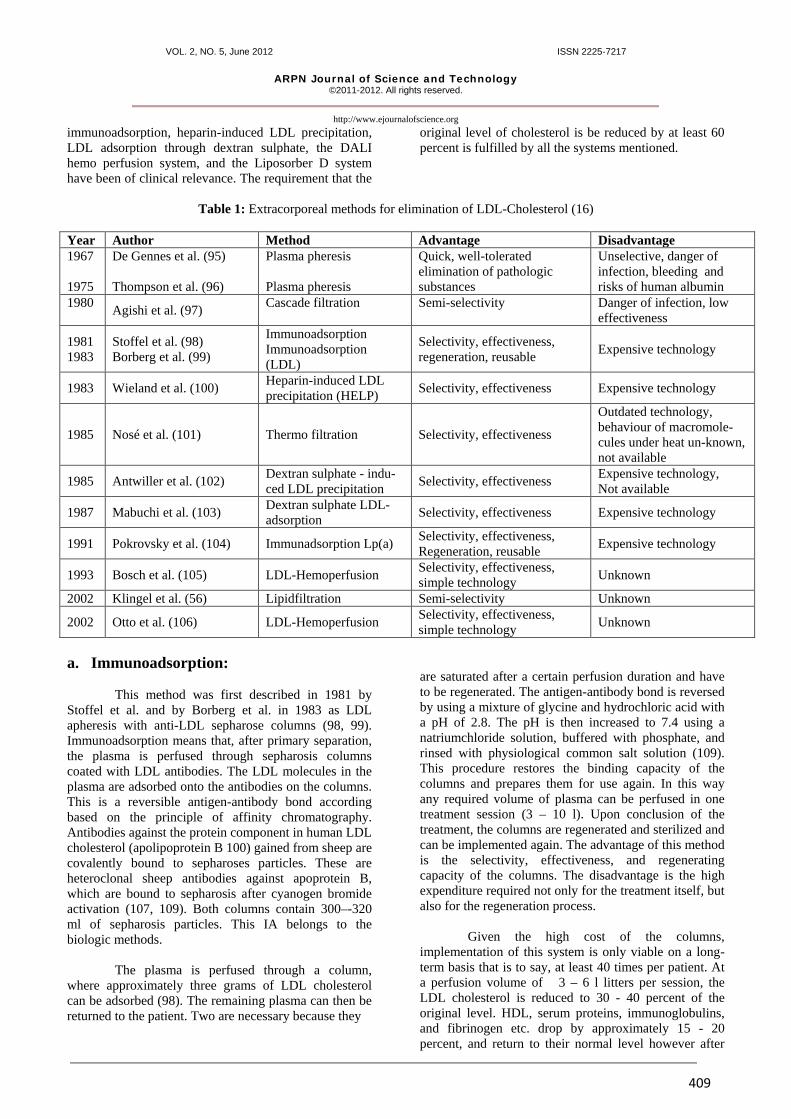
VOL. 2, NO. 5, June 2012 ISSN 2225-7217
ARPN Journal of Science and Technology ©2011-2012. All rights reserved.
http://www.ejournalofscience.org
409
immunoadsorption, heparin-induced LDL precipitation, LDL adsorption through dextran sulphate, the DALI hemo perfusion system, and the Liposorber D system have been of clinical relevance. The requirement that the
original level of cholesterol is be reduced by at least 60 percent is fulfilled by all the systems mentioned.
Table 1: Extracorporeal methods for elimination of LDL-Cholesterol (16)
Year Author Method Advantage Disadvantage 1967 1975
De Gennes et al. (95) Thompson et al. (96)
Plasma pheresis Plasma pheresis
Quick, well-tolerated elimination of pathologic substances
Unselective, danger of infection, bleeding and risks of human albumin
1980 Agishi et al. (97) Cascade filtration
Semi-selectivity
Danger of infection, low effectiveness
1981 1983
Stoffel et al. (98) Borberg et al. (99)
Immunoadsorption Immunoadsorption (LDL)
Selectivity, effectiveness, regeneration, reusable Expensive technology
1983 Wieland et al. (100) Heparin-induced LDL precipitation (HELP) Selectivity, effectiveness Expensive technology
1985 Nosé et al. (101) Thermo filtration Selectivity, effectiveness
Outdated technology, behaviour of macromole-cules under heat un-known, not available
1985 Antwiller et al. (102) Dextran sulphate - indu-ced LDL precipitation Selectivity, effectiveness Expensive technology,
Not available
1987 Mabuchi et al. (103) Dextran sulphate LDL- adsorption Selectivity, effectiveness Expensive technology
1991 Pokrovsky et al. (104) Immunadsorption Lp(a) Selectivity, effectiveness, Regeneration, reusable Expensive technology
1993 Bosch et al. (105) LDL-Hemoperfusion Selectivity, effectiveness, simple technology Unknown
2002 Klingel et al. (56) Lipidfiltration Semi-selectivity Unknown
2002 Otto et al. (106) LDL-Hemoperfusion Selectivity, effectiveness, simple technology Unknown
a. Immunoadsorption: This method was first described in 1981 by Stoffel et al. and by Borberg et al. in 1983 as LDL apheresis with anti-LDL sepharose columns (98, 99). Immunoadsorption means that, after primary separation, the plasma is perfused through sepharosis columns coated with LDL antibodies. The LDL molecules in the plasma are adsorbed onto the antibodies on the columns. This is a reversible antigen-antibody bond according based on the principle of affinity chromatography. Antibodies against the protein component in human LDL cholesterol (apolipoprotein B 100) gained from sheep are covalently bound to sepharoses particles. These are heteroclonal sheep antibodies against apoprotein B, which are bound to sepharosis after cyanogen bromide activation (107, 109). Both columns contain 300–-320 ml of sepharosis particles. This IA belongs to the biologic methods.
The plasma is perfused through a column, where approximately three grams of LDL cholesterol can be adsorbed (98). The remaining plasma can then be returned to the patient. Two are necessary because they
are saturated after a certain perfusion duration and have to be regenerated. The antigen-antibody bond is reversed by using a mixture of glycine and hydrochloric acid with a pH of 2.8. The pH is then increased to 7.4 using a natriumchloride solution, buffered with phosphate, and rinsed with physiological common salt solution (109). This procedure restores the binding capacity of the columns and prepares them for use again. In this way any required volume of plasma can be perfused in one treatment session (3 – 10 l). Upon conclusion of the treatment, the columns are regenerated and sterilized and can be implemented again. The advantage of this method is the selectivity, effectiveness, and regenerating capacity of the columns. The disadvantage is the high expenditure required not only for the treatment itself, but also for the regeneration process. Given the high cost of the columns, implementation of this system is only viable on a long-term basis that is to say, at least 40 times per patient. At a perfusion volume of 3 – 6 l litters per session, the LDL cholesterol is reduced to 30 - 40 percent of the original level. HDL, serum proteins, immunoglobulins, and fibrinogen etc. drop by approximately 15 - 20 percent, and return to their normal level however after

VOL. 2, NO. 5, June 2012 ISSN 2225-7217
ARPN Journal of Science and Technology ©2011-2012. All rights reserved.
http://www.ejournalofscience.org
410
approximately 24 hours (99, 109). Resulting side effects or other complications have, only been seldom reported, however (100). Two different systems are are currently available, the LDL-Therasorb ® system (Miltenyi Biotec, Germany) and the LDL- and Lp (a)-Excorim system (Fresenius, Germany). The matrix sepharose is coupled with specific antihuman apolipoprotein B-100 or antihuman Lp(a) sheep antibodies (107, 110). Both systems are safe and effective in clinical use, even in long-term treatment. Indications for the extracorporeal elimination of LDL cholesterol are primary and secondary dyslipoproteinemia, and for the Lp(a) IA the solitary familial Lp(a) elevation. b. Heparin-induced LDL-precipitation
(HELP system): In 1983 Wieland et al. described the possibility of carrying out LDL preparation using heparin (100). A heparin buffer with a pH of 5.12 is added to the separated plasma, whereby a heparin-LDL complex is formed, which precipitates in the acid environment and which can be retained by means of a filter. The plasma is mixed at a ratio of 1:1 with acetic acid buffer (pH 4.85), resulting in a pH of 5.12. This mixture contains 100,000 E of heparin per liter, and precipitation follows. LDL, fibrinogen, and heparin form an insoluble complex. The remaining heparin is almost fully removed by using a heparin adsorber (DEAE cellulose) (111). More recently, a compact unit has been designed that somewhat reduces the cost of the equipment. The Plasmat® Futura (B. Braun, Germany) is easy to use and safe in handling. The priming rinsing and reinfusion are fully automated. The entire treatment uses only disposable materials; the machine does not need descaling or desinfection and no piped water supply or reverse osmosis required. The user is safely guides through all treatment steps and support with message prompts and warnings. Because C3, C4, fibrinogen, and plasminogen are also reduced to approximately 50 percent of their original concentration, as well as cholesterol, LDL, and triglycerides, the technique is not highly specific (110). Due to the large drop in fibrinogen in the HELP process, the amount of plasma per session is limited to 3 three liters. A treatment using larger amounts of plasma could lead to bleeding complications. With this amount of plasma, cholesterol is reduced by about 50 percent, LDL by about 60 percent, oxidized LDL by about 45 percent, Lp(a) about 50 percent, and triglycerides by about 50 percent of the original levels. The other factors, like CRP, are also reduced by approximately 50 percent. In contrast, HDL is only reduced by 10 - 20 percent. Thus far no severe side effects have been described; however, occasional shivering and drops in blood pressure have been observed (111, 112, 113).
c. LDL Adsorption with Dextransulfate: Mabuchi et al. published their first results on the adsorption of LDL with dextransulphate in 1987 (103). Dextran sulphate, a low-molecular substance with a molecular weight of 4,500, is covalently bound to cellulose micropellets and can predominantly adsorb cholesterol containing apolipoprotein-B, such as LDL, VLDL, and triglycerides from plasma gained through plasma separation. Both columns contain 150 ml of a cellulose-dextran sulphate mixture, and each column can bind approximately 2.5 grams of LDL cholesterol. Almost fully cleansed of cholesterol, LDL, and VLDL, the plasma is returned to the patient. This technique also requires two perfusion columns, which, once saturated, are perfused and regenerated. The adsorption capacity of one column amounts to about 500 ml, then it is regenerated. Using a 4.1 percent solution of common salt and rinsing with Ringer’s solution, this technique cleanses it of adsorbed cholesterol. Given the constant adsorption and regeneration of the two columns, capacity is practically unlimited, which means that plasma perfusion volumes of between six and nine litters are quite common. The levels of cholesterol, LDL, and triglycerides drop by more than 60 - 80 percent of the original levels. Conversely, HDL and immunoglobulins are hardly affected (114). However, there is no 100 percent selectivity here either, as fibrinogen is also reduced by approximately 20 – 30 percent. It is likely that other clotting factors, such as Factor VIII are also adsorbed, since one effect is that there is a drop in the Quick time. No further side effects have been reported, with the exception of occasional slight drops in blood pressure in a pattern common to all extracorporeal treatments. Side effects, in particular allergic reactions to dextransulfate, such as can arise with dextran with a much higher relative molecular density of 40,000 – 80,000, have not been observed. Nor has any evidence been found of antibodies against dextran. No severe side effects are reported until today. The adverse reactions during or after the treatment can be a drop in blood pressure (hyoptension), nausea, vomiting, flushing, and blotching. d. Lipoprotein (a) Apheresis: Lipoprotein (a) or Lp(a) represents a class of lipoprotein particles which have lipid composition similar to LDL and a protein moiety, apoB 100, covalently linked to apo(a), a glycoprotein with striking structural similarities to plasminogen (114). High plasma levels of Lp(a) are associated with an increased risk for atherosclerotic coronary heart disease (CHD) by a mechanism yet to be determined. Because of its structural properties, Lp(a) can have both atherogenic and thrombogenic potentials (115). Uttermann et al. found six different Lp(a) phenotypes (S4, S3, S2, S1, B, F) and their influences on the Lp(a) levels. Phenotypes

VOL. 2, NO. 5, June 2012 ISSN 2225-7217
ARPN Journal of Science and Technology ©2011-2012. All rights reserved.
http://www.ejournalofscience.org
411
S2, S1, and B were associated with high, and phenotypes S4 and S3 with low Lp(a) concentrations (117). It is not yet clear whether the manifestation of the CHD is mainly associated with Lp(a) levels or with the phenotypes. It can, however, be concluded that high levels of Lp(a) are associated with CHD; the iso forms S2, S1, B, F are linked with CHD; and patients with premature CHD showed the highest Lp(a) levels and the iso forms S2, S1, B, and F more frequently than others (118). The means for correcting the high plasma levels of Lp(a) are still limited in effectiveness. All drug therapies tried thus far have failed. The most effective therapeutic methods in lowering Lp(a) are the LDL-apheresis methods. Since 1991, special immunoadsorption polyclonal antibody columns (Pocard, Moscow, Russia) containing sepharose bound anti-Lp(a) have been available for the treatment of patients with elevated Lp(a) serum concentrations (118). Monospecific polyclonal antibody to human Lp(a) was obtained from immune sheep serum (105). For the treatment two columns are necessary. Each, column is filled with 400 ml of sorbent tested for sterility and pyrogenicity. Anti-Lp(a) immunoadsorption columns are reusable. Between the treatments, the columns are stored at 4° C in storage solution, which is rinsed prior to each Lp(a) apheresis procedure. Two personal columns are assigned to each patient (118). The columns can be used in systems after primary plasma separation. The numerous studies provide the epidemiological evidence that Lp(a) is an independent risk factor in the pathogenesis of coronary heart disease and arteriosclerosis. The individual plasma level is genetically determined. Diet or available drugs do not influence the plasma level of Lp(a). All the techniques described here are effective and well-tolerated. Based on an average drop in cholesterol of 50 – 60 percent per session, a treatment intervall of 7 – 14 days is advisable. The constant reduction of cholesterol is meant above all, to prevent the progression or the development of atherosclerosis. By lowering the cholesterol from 400 mg/dl to 200 mg/dl, treatment can almost double a patient’s life expectancy, according to at least one study (119). e. LDL-Hemo Perfusion System: Bosch et al. first described LDL–hemoperfusion (direct adsorption of lipoproteins, DALI, Fresenius, Germany) in 1993. The new absorber, which is compatible with human whole blood, contains polyacrylate in the column (105). In the DALI system, blood is perfused through the adsorber, which contains 480 ml polyacrylate-coated polyacrylamide beads, without need of regeneration. The column has a capacity of more than 1.5 – 2.0 blood volumes for effective adsorption of cholesterol, LDL, Lp (a), and triglyceride, etc. Regeneration is not necessary because the column is used only for one treatment. (120). The elimination of
LDL cholesterol and Lp(a) from the whole blood is achieved by adsorption onto polyacrylate-coated polyacrylamide beads. Polyacrylate, like the LDL receptor, consists of polyanions with negatively charged carboxylate groups. These polyanions interact selectively with the cationic groups in the apos moiety of LDL and Lp(a). This electrochemical interaction immobilizes lipoproteins on the absorber beads (105, 120). Besides lipoproteins, the LDL-hemoperfusion system also adsorbs the positively charged ions calcium and magnesium. Therefore, the columns have to be pre-rinsed with six litters of a priming solution containing these electrolytes. The absorber is thereby saturated with these cations, thus preventing hypocalcemia and hypomagnesemia (121). After passing the absorber, the blood depleted by all apos-containing lipoproteins is given back to the patient. In each patient, 1.0 – 1.5 patient blood volumes were treated per session. The advantages are good selectivity, high effectiveness, and a simple technology. The blood is perfused at a flow rate of 30 – 60 ml/min through the absorber containing 500, 750 750, or 1,000 ml polyacrylate/polyacrylamide gel. The anticoagulatltion consists of an infusion of acide citrate dextrose (ratio of citrate flow: blood flow = 1: 20 – 1: 40. P1 – 3 are pressure monitors. Filled circles correspond to ACD-A- and blood pumps (120). The machine continously monitors the blood from outside the body by pumping the blood, controlling its velocity and preventing air from entering the blood flow. After about one or two hours of treatment, the LDL cholesterol level of the blood can decrease more than 60 percent. In 2003 Otto et al. reported of a new whole-blood absorber is the Liposorber D for DHP (Direct Hemoperfusion, Kaneka, Japan) (106). The adsorption column contains approximately 700 ml of adsorbent of dextran sulfate-cellulose beads, with a particle size of approximately 250 micrometers. Liposorber D is a new whole blood perfusion LDL apheresis system developed on the basis of the technology of the Liposorber LA-15 system. Liposorber D and DALI both adsorb positively charged LDL, VLDL, and Lp(a) particles from whole blood using negatively charged polyanions. In contrast to DALI which uses polyacrylate bound to polyacrylamide, Liposorber D contains negatively charged dextran sulphate covalently bound to cellulose. Three columns with different volumes (DL 50, DL 75, DL 100) are available. Prior to the treatment the adsorption column must be rinsed with 750 ml physiological saline alone or physiological saline mixed with 25 ml of acid dextrose (ACD-A) solution and 25 ml of 8.4 percent sodium bicarbonate solution per litter. Heparin (500 – 1,500 IU) is given as a bolus. ACD-A is used as the main anticoagulant at a flow rate of 1/20 (vol/vol) of blood flow rate at the initial stage. During the treatment, the flow rate is kept in a range of 1/20 to 1/40. The Liposorber D system removes selectively all apolipoprotein B-containing lipoproteins such as
VOL. 2, NO. 5, June 2012 ISSN 2225-7217
ARPN Journal of Science and Technology ©2011-2012. All rights reserved.
http://www.ejournalofscience.org
412
cholesterol, LDL, VLDL, and Lp(a). The negatively charged surface activates the instrinsic coagulation pathway and prolongation of a PTT and shortening of PT have been observed in the LDL apheresis with the dextran sulphate column. Coagulation factors such as Factor XI and XII were reduced by dextran sulphate adsorption, but those coagulation factors returned to normal range within one or two days after the treatment. The newly developed direct hemoperfusion LDL apheresis system Liposorber D is a safe and simple apheresis system. It thus appears and thus to be a useful modality for removing apolipoprotein B-containing lipoproteins from the whole blood, as shown in the initial clinical results (106). Adverse events which were observed were hypocalcemia during treatment caused by ACD-A solution; the symptoms disappeared by administration of calcium, and slight hypotension. Since some years a new machine DX-21for the direct hemoperfusion is available. The system is comparable with the DALI system. The handling is easy and safely, the user is guided through all treatment steps. 6. EZYME ADSORBENTS
(BIOREACTOR) Another method is the immobilization of certain enzymes on adsorption material, such as agarose, cellulose particles, or membranes. These immobilized enzymes activate certain reactions in blood or plasma, serving to inactivate and eliminate toxic substances. Thus, enzymes such as tyrosine, cytochrom P 450, glucoronyltransferase, and sulphotransferase, are immobilized and implemented in the treatment of liver comas, to at least partially relieve the stress on liver (122). 7. IMMUNOPHERESIS OF SOLUBLE
TUMOR NECROSIS FAKTOR FACTOR INHIBITORS IN CANCER
It is well recognized that Tumor Necrosis Factor (TNF, TNF-α), a cytokine bound to macrophages, and Lymphotoxin (LT, TNF-β), a cytokine released by killer lymphocytes, will kill cancer in vitro if the lymphocytes and macrophages are unblocked and make cell-to-cell contact. They must be free of inhibitory factors found in the serum of the cancer patient. The primary, if not the sole blocker/inhibitors of TNF/LT, are the soluble receptors to TNF/LT. TNF inhibitor production is stimulated by IL-2 and other pro-inflammatory cytokines, thus explaining at least in part why the administration of supraphysiologic doses of these cytokines has produced such disappointing clinical results. Previous studies have demonstrated that melanoma and breast cancer does sometimes respond to immunologic stimulation caused by the removal of the soluble inhibitors to TNF (sTNF-Rs) (123, 124).There are two distinct cell surface TNF receptors (TNF-Rs),
designated sTNF-R1 and sTNF-R2 with molecular weights of 55 and 75 kd respectively. These receptors have been sequenced. When shed, they are soluble, truncated versions of membrane TNF-Rs, and consist of only the extracellular binding domain. All the evidence points to this form of apheresis produce a greater clinical effect than previous methods. The optimal length and frequency of repeat courses needs to be investigated. The first clinical trials were begun to observe and document the clinical response of immunoadsorption of sTNFR-1 and sTNFR-2 in patients with metatstatic cancer involving the extracorporeal filtration removal of sTNF-R1 and R2 by plasma fractionation have correlated with the removal of sTNF-Rs with significant clinical response (123, 124). 8. LIVER SUPPORT SYSTEMS In the last few years, different various liver support systems have been developed. These systems try to combine all advantages of the different extracorporeal detoxification methods which have been implemented in recent years in the treatment of liver diseases. a. Bio Logic – DTPP System: The Bio Logic – DTPF system which was first described by Ash et al. in 1992 combines hemodiasorption with push-pull sorbent-based pheresis. The BioLogic-DTPF system is a simple, sorbent-based extracorporeal hemodiasorption system indicated for treatment of acute hepatic failure with encephalopathy or the treatment of serious drug overdose (125). The Bio Logic – DTPF system uses a 2 litter sorbent suspension that surrounds the membranes of a cellulosic plate dialyzer. This system provides a much higher sorbent surface area and greater biocompatibility than conventional sorbent hemo perfusion columns. Changes in pressure of sorbent suspension actuacte the membranes to pull blood from a single-lumen catheter, pass it through the dialyzer, and return it through the same catheter. The sorbent suspension contains 140 grams of powdered charcoal and 80 grams of cation exchangers. The system can selectively remove hepatic toxins of less than 5,000 Daltons with moderate protein binding, such as aromatic amino acids, glutamine, mercaptane, spermidine, putrescine, phenols, bile acids, lactate, false neural transmitters such as glutamate and octopamine, neural inhibitors such as GABA, benzodiacepine-like substances, short fatty acids, ammonium, potassium, and manganesium. Urea is removed only in moderate amounts. The sorbent suspension is preloaded with glucose to return glucose to the patient, while removing the various organic and anorganic toxins from the blood (126). For the toxins of hepatic failure which are strongly protein-bound or lipid-bound including, endotoxins, as well as soluble cytokines such as tumor necrosis factor-α, interleukin-2, and interleukin-6, a
VOL. 2, NO. 5, June 2012 ISSN 2225-7217
ARPN Journal of Science and Technology ©2011-2012. All rights reserved.
http://www.ejournalofscience.org
413
module with the capability to remove the protein-bound toxins and large molecules is added to the system (127). The BioLogic DTPF system includes a plasma-permeable hollow-fiber plasma membrane placed downstream from the plate dialyzer in which alternating positive and negative transmembrane pressure causes plasma to transiently pass into a powdered charcoal sorbent suspension around the hollow fibber membrane. Here, direct interaction between plasma and charcoal can remove protein-bound and large molecular weight toxins, then return plasma without these molecules to the blood. b. Molecular Adsorbent Recycling System
(MARS-System): The molecular adsorbent recycling system (MARS, Gambro, Sweden) system is an extracorporeal hemodia filtration (HDF) method using albumin-enriched dialysate to facilitate the removal of albumin-bound toxins (128). It requires a standard dialysis machine or a continuous venous-venous hemo filtration monitor, and an extra device to run and monitor a cloosed-loop albumin circuit. As vascular access, a double-lumen catheter in the cava superior or inferior must be used. For anticoagulation, heparin is necessary both as initial dosage and in continuously administration. The system is driven by a blood flow rate of 150 - 250 ml/min, depending on the hemodynamic situation of the patient. Blood is passed through a non-albumin-permeable, high-flux dialysis membrane. A closed-loop dialysate circuit, containing 10 percent human albumin is driven by a roller pump of the MARS monitor at 150 ml/min. The dialysate human albumin is passed through the dialysate compartment of the blood dialyzer and subsequently regenerated online by dialysis against a bicarbonate-buffered dialysate, followed by passage through a column with uncoated charcoal and a second column with an anion-exchanger resin. The treatment is performed intermittently for six to eight hours and longer a day. The biochemical efficacy of MARS has been well proven. Toxins as removable by MARS include bilirubin, ammonia, aromatic amino acids, tryptophans, short-chain fatty acids, bile acids, GABA-like substances, nitric oxidase and cytokines (129). Most of the toxins removable by MARS are implicated in the development of hepatic encephalopathy, portal hypertension or other inflammatory changes in liver failure. The physiological benefits demonstrated by the MARS system include lowering of intracranial pressure, and improvement of cerebral edema, reduction of portal pressure, and improvement of blood pressure, and systemic vascular resistance. Reduction of these symptoms may be able to gain more time to bridge to transplantation for a patient. The MARS system is a treatment with high safety standards includes simultaneous selective removal
of albumin-bound and water-soluble substances, high effectiveness and selectivity, mangement of fluid, electrolyte, and acid/base balance, control of glucose and lactate level, and cell free operation. Side effects were reported as platelet reduction, hypoglycemia during treatment, electrolyte disturbances and non cardiogenic pulmonary edema. As MARS is one form of extracorporeal circuit, it shares the potential complications that extracorporeal circuits usually have, including catheter-related infection, hematoma and circuit thrombosis, etc. (129). Today, the MARS system is the most frequently used type of albumin dialysis and most studied liver-support technique. Albumin dialysis enables the selective regeneration of albumin. The resulting increase of albumin binding capacity is paralleled by improvement of central and local hemodynamic and liver, brain, and kidney functions. The treatment can contribute to liver regeneration and prolongation of patient survival in the context of acute liver failure, decompensate chronic liver disease, and bridging of patients to liver transplantation (130). c. Prometheus-System: The Prometheus-system (Fresenius, Germany) is a new device based on the combination of albumin adsorption with high-flux hemo dialysis after selective filtration of albumin fraction through a specific poly sulfon filter (Albu flow®, Fresenius, Germany). The clearance of toxins is achieved in different steps. Albumin is separated from blood by the novel hollow fiber albumin dialyser Albu Flow®. The Albu Flow® is made of polysulfone hollow fibres and is permeable to albumin, and hence to albumin-bound substances (131). The blood flow rate is 150 – 200 ml/min. The albumin filtrate in the fractionated plasma separation, adsorption and dialysis (FPSA) circuit is subsequently perfused through a column with neutral resin (Prometh 01) and through a second column with an anion exchanger resin adsorber (Prometh 02, both Fresenius, Germany), whereby the bound toxins are adsorbed by direct contact with the high-affinity adsorbing material. The “native” albumin is then returned to the patient. The FPSA recirculation circuit is driven by a roller pump at 300 ml/min. After passage through the Albu Flow®, the patient´s blood is dialysed through a high-flux dialyser, whereby water-soluble toxins are eliminated (132). A modified hemodialysis unit (4008 H, Fresenius, Germany) is used to integrate the two parts of the Prometheus® system: a “standard” extracorporeal blood circuit connected to the double-lumen dialysis catheter and a secondary circuit containing the separated plasma. Both circuits are separately monitored by different hardware and software allowing the user to perform hemo dialysis with simultaneous albumin detoxification or conventional hemo dialysis alone. With the Prometheus® system both protein-bound and water-soluble substances that accumulate in liver failure are removed in a single step or for example during a single
VOL. 2, NO. 5, June 2012 ISSN 2225-7217
ARPN Journal of Science and Technology ©2011-2012. All rights reserved.
http://www.ejournalofscience.org
414
passage of the blood through the extracorporeal circuit. The system is based upon the FPSA method, which allows direct purification of the albumin fraction in a secondary circuit (131). Furthermore, a high flux dialyzer is incorporated in the Prometheus® system as well. Both systems (MARS-, Prometheus-system) are cell free extracorporeal non biological liver support systems providing elimination of albumin-bound as well as water soluble toxins that accumulate in liver failure. Both systems were able to clear cytokines from plasma but they did not change serum cytokine level significantly (133). This apparent discrepancy is probably due to a high rate of ongoing cytokine production in acute liver failure counterbalancing elimination within the extracorporeal circuits. The other liver support methods like hemo dialysis, hemo perfusion, plasmapheresis, cascade filtration, plasma perfusion and single pass albumin dialysis are not as selective as the MARS- or Prometheus-system. The bio artificial liver support devices with human or porcine heaptocytes or the extracorporeal liver perfusion have an expensive technology and they are introduced only in special departments and are reported elsewhere. 9. DISCUSSION The mentioned apheresi techniques are a selection of techniques which are used in the clinical routine or are on experimetal phase. A disadvantage is that the most methods have an expensive technology and various hardware types. Simple and user-friendly systems are necessary for routine clinical use. It is therefore desirable that only one machine (hardware) is available for all different extracorporeal methods. This hardware should be equipped with various software programs for running different apheresis methods. The membrane, adsorber and tube sets should be different for different methods. These sets should be only used with special software programs. Only one hardware system for all different apheresis methods could be a great advantage because the machine park in nephrological and intensive medical care units could be reduced. For the physicians and the staff this will be a simplification of their daily work, because they can work with different sets and only one hardware system (16). New approaches are made in the last few years. Some companies have developed machines which can be
used as well as for intermittend and continuous dialysis methods, as hemo perfusion and plasmapheresis but not for immunoadsorption, a successful way. In addition, there are the so-called “artificial ells” now being developed. These, contain individual enzymes or whole enzyme systems in a small capsule, which is then used to trigger various metabolic reactions in the organism (16). This development, in particular, opens up new perspectives, which will facilitate a more effective and gentle form of treatment when used in combination with selective adsorbents. More controlled multi-centre studies are necessary. The controlled clinical studies with TPE which many authors demand are only feasible for a few diseases. Three reasons can be given for the failure of such studies up to now: too few cases (as, for example, in HUS), not acceptable for ethical reasons (e.g., Good pasture syndrome), too costly. Controlled clinical studies require the application of the strictest criteria, such as exact diagnosis, duration of disease, age of patient, previous treatment, TPE standardization, and a control arm. The latter is particularly problematic from an ethical consideration because so often these diseases being treated are fatal and TPE is typically used when other treatments have already failed. Despite all the cases of successful therapy, many of the pathophysiological processes underlying this therapy are still not completely understood; this knowledge is necessary to be able to register and understand reactions that occur and their mutual influences. Further basic research is necessary, especially in the field of immunology, to enable us to understand the pathophysiology of many diseases and then to learn how to control them through specific elimination of immune complexes, antibodies, antigens, cytokines, chemokines and other mediators of pathophysiology. The aim of all apheresis methods is the elimination of pathogen substances or cells to interrupt pathological metabolism pathways. The plasma separation is carried out with centrifuge or hollow fibbers as well for the conventionel plasmapheresis as for the selective separation with secondary membrane or adsorption columns which are loaded with different antibodies or chemical substances. Different artificial methods are available for the temporary detoxification of the liver (16). In Table 2 all mentioned apheresis and other methods are summarized.
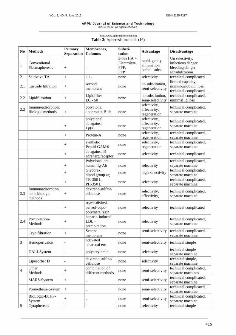
VOL. 2, NO. 5, June 2012 ISSN 2225-7217
ARPN Journal of Science and Technology ©2011-2012. All rights reserved.
http://www.ejournalofscience.org
415
Table 2: Apheresis-methods (16)
No Methods Primary Separation
Membranes, Columns
Substi- tution Advantage Disadvantage
1 Conventional Plasmapheresis
+
-
3-5% HA + Electrolyte, PPF, FFP
rapid, gently elimination pathol. subst.
Un selectivity, infectious danger, bleeding danger, sensibilization
2 Selektive TA + + / - none selectivity technical complicated
2.1 Cascade filtration + second membrane none no substitution,
semi-selectivity
limited capacity, immunoglobulin loss, technical complicated
2.2 Lipidfiltration + Lipidfilter EC - 50 none no substitution,
semi-selectivity technical complicated, minimal Ig loss
2.2 Immunoadsorption, Biologic methods
+
polyclonal apoprotein B-ab
none
selectivity, effectivity, regeneration
technical complicated, separate machine
+
polyclonal ab against Lp(a)
none
selectivity, effectivity, regeneration
technical complicated, separate machine
+ Protein-A none selectivity, regeneration
technical complicated, separate machine
+ synthetic Peptid-GAM® none selectivity,
regeneration technical complicated, separate machine
+ ab against β1 adrenerg receptor none selectivity technical complicated
+
Polyclonal anti- human Ig-Ab
none
selectivity
technical complicated, separate machine
+ Glycorex, blood group ag none high-selectivity technical complicated,
separate machine
+ TR-350 L, PH-350 L none selectivity technical complicated,
separate machine
2.3 Immunoadsorption, none biologic methods
+ dextrane-sulfate/ cellulose
none selectivity, effevtivity,
technical complicated, separate machine
+ styrol-divinyl-benzol-copo-polymere resin
none selectivity technical complicated
2.4 Precipitation- Methods +
heparin-induced LDL - precipitation
none selectivity technical complicated, separate machine
Cryo filtration + Second membrane none semi-selectivity
technical complicated, separate machine
3 Hemoperfusion - activated charcoal etc. none semi-selectivity technical simple
DALI-System - polyacrylamid none selectivity technical simple separate machine
Liposorber D - dextrane-sulfate/ cellulose none selectivity technical simple,
separate machine
4 Other Methods + combination of
different methods none semi-selectivity technical complicated, separate machines
MARS-System + „ none semi-selectivity technical complicated, separate machine
Prometheus-System + „ none semi-selectivity technical complicated, separate machine
BioLogic-DTPP-System + „ none semi-selectivity technical complicated,
separate machine 5 Cytapheresis - - none selectivity technical simple
VOL. 2, NO. 5, June 2012 ISSN 2225-7217
ARPN Journal of Science and Technology ©2011-2012. All rights reserved.
http://www.ejournalofscience.org
416
In experienced hands, TA has minimal side effects, reported in relevant literature as being between 4 and 22 percent. These figures include both severe and clinically insignificant side effects. Through constant technical improvement of the equipment, membrane separators and reduction of extracorporeal volume, severe side effects have become more rare in so far as they are apparatus induced. The fact that TA treatment still incurs high costs is a safeguard against indiscriminate implementation, on the one hand, but its omission can, on the other, prevent causal therapy. Yet again, judicious implementation of TA saves high subsequent costs. Nevertheless, therapeutic plasma exchange is now a part of the standard of care for many life threatening diseases and still at an experimental stage in many diseases, and great efforts must still be made to be able to allocate a secure place in modern therapy to this form of treatment, alongside the few absolute indications. Future studies must show whether chronic TA treatment, already extensively practiced, is superior to other medicational therapy schemes for some chronic diseases. New techniques which already promise interesting developments are the following: immunoadsorption by columns with immobilized monoclonal or polyclonal antibodies, bioreactors, cell filtration and flow cytometry, artificial blood cells, such as erythrocytes, lymphocytes, magnetic separation. A further technical refinement opens up possibilities, in which T lymphocytes can be separated from B lymphocytes and eliminated, for example. Flow cytometry and centrifugal elutriation are modern techniques that enable sorting and separation of leucocytes. Therapeutic application is also feasible here. Different companies are trying to unite various extracorporeal processes in one system, called futuristically the artificial lymph node, with the aim of restoring deficient or disturbed immune function to the patient. Up to now, conventional TPE has proved itself in a series of immunological diseases, metabolic diseases, and intoxication. When selective plasma separation and immunoadsorption with immobilized monoclonal or polyclonal antibodies and the artificial lymph node have secured their place in clinical routine, we will have taken a great step nearer to the wish of our medical ancestors of removing “the disease factors from the blood” and will be able to provide improved treatment for many diseases (1). Due to the high costs of the different apheresis methods it should be necessary to select only a few patients to who insert the apheresis treatment. This problem raises large ethic questions which must be answered. The commissions which should decide on the reimbursement of the apheresis treatment must be
intouch with physicians who are involved in the apheresis treatments. REFERENCES [1] Rother K, K Thies. Plasmapherese-Therapie.
Immunpathologische Konzeption. Internist. 24: 1 (1983).
[2] Kambic HE, Y Nosé. Plasmapheresis, historical
perspective, therapeutic applications, and new frontiers. Artif Org 17: 850 (1993).
[3] Kambic HE, Y Nosé (Eds.). Plasmapheresis,
historical perspective. Therapeutic applications and new frontiers. ISAO Press, Cleveland, USA, 1983.
[4] Brockhaus Enzyklopädie In 20 Bänden.
Brockhaus, Wiesbaden, 1966. [5] Hedon E. Sur l´hemolyse par les glycosides et les
conditions de milieu qui la favorisent ou l´empechent. Arch Intern Pharmacol Gand VII: 381 (1901).
[6] Fleig C. Auto transfusion of washed corpuscles as
a blood washing procedure in toxemia: Hetero transfusion of washed corpuscles in anemia. Bull Mem Acad Sci Let 1: 4 (1909).
[7] Abel JJ, LG Rowntree, BB Turner. Plasma
removal with return of corpuscles (Plasmapheresis). J Pharmacol Exp Ther 5: 625 (1914).
[8] Gilbert A, A Tzanck, T negroni. Emission
sanguine avec restitution globulaire. Paris Méd 21 : 217 (1926).
[9] Tui C, FC Bartter, AM Wright et al. Red cell
reinfusion and the frequency of plasma donations. Preliminary report of multiple donations in eight weeks by each of six donors. JAMA 124: (1944).
[10] Grifols-Lucas JA. Primeras Applicaciones de la
Plasmapheresis en el hombre. Med Clin 18: 301 (1952).
[11] Adams WS, WH Blahid, SH Bassett. A method of
human plasmapheresis. Proc Soc Exp Biol Med 80: 377 (1952).
[12] Castino F, K Scheucher, PS Malchesky et al.
Continuous plasma filtration through micro porous membranes. Proc Ann Conf Eng Med Bio 18: 292 (1976).
VOL. 2, NO. 5, June 2012 ISSN 2225-7217
ARPN Journal of Science and Technology ©2011-2012. All rights reserved.
http://www.ejournalofscience.org
417
[13] Nosé Y, PS Malckesky, F Castino et al. Improved hemoperfusion system for renal hepatic support. Kid Int 10: 244 (1976).
[14] Gurland HJ, MJ Lysaght, W Samtleben et al. A
comparison of centrifugal and membrane-bases apheresis formats. Int J Artif Org 7 : 35 (1984).
[15] Bambauer R, GA Jutzler, H Mauch et al. Ein
vereinfachtes Plasmapherese-Be handlungsverfahren. Technik and erste klinische Ergebnisse. Nieren- Hochdruckkrankh 10: 126 (1981).
[16] Bambauer R, R Latza, MR Lentz. Therapeutic
Plasma Exchange and Selective Plasma Separation methods. Fundamental Technologies, Pathology and Clinical Results. 3rd Edition. Pabst Science Publishers, Lengerich, Berlin, Bremen, Miami, Riga, Viernheim, Zagreb, 2009.
[17] Brant AM, JF Chielewsky, TK Hung et al.
Stimulation in vitro of pulsatile vascular hemo dynamics using a CAD/CAM-designed cam disk and roller follower. Artif Org 10: 419 (1986).
[18] Qian KX, Q Fei, KD Lin et al. The realization of a
pulsatile impeller pump with low hemolysis. Artif Org 13: 162 (1989).
[19] Bambauer R, HJ Jesberger, HG Limbach et al.
Single-needle dia filtration (SN-DF) in acute renal failure in premature infants. Blood Purific 4: 199 (1985).
[20] Bambauer R, GA Jutzler, H Philippi et al. Hemo
filtration and plasmapheresis in premature infants and newborns. Artif Org 12: 20 (1988).
[21] Cobe Laboratories, Inc. Cobe entry TPE system.
Technical Bulletins for TPE operator´s Handbook. Lakewood, Colorado, USA, 1981.
[22] Gurland HJ, W Samtleben, M Blumenstein et al.
Clinical application of macromolecular separations. Trans Am Soc Artif Intern Org 27: 356 (1981).
[23] Cooper AR (Ed.). Ultra filtration membranes and
applications. Plenum, New York, 1979. [24] Rainer H, H Borberg, JM Mishler et al. (eds.).
Cell separation and cryobiology. Schattauer Verlag, Stuttgart, New York, 1978.
[25] Holtz M, R Rautenbach. Technische Aspekte der
klinischen Diafiltration. Biomed Tech 21: 302 (1976).
[26] Porter MC. Concentration polarization with membrane ultra filtration. Ind Eng Chem Prod Res Dev 11: 234 (1972).
[27] Michaels AS. Analysis and prediction of sieving
curves for ultra filtration membranes: A universal correlation separation. Sci Technol 15: 1305 (1980).
[28] Kozinska AA, EN Light food. Protein ultra
filtration: A general example of boundary layer filtration. AICHE 18: 1030 (1986).
[29] Malchesky PS, T Horiuchi, JJ Lewandowsky et al.
Membrane plasma separation and the on-line treatment of plasma by membranes. J Membr Sci 44: 55 (1989).
[30] Mandolfi S, C Tetta, S David et al. In vitro and in
vivo biocompatibility of substituted cellulose and synthetic membranes. Int J Artif Org 20: 603 (1997).
[31] Roberts CG, K Schindhelm, PC Farrell Protein-
membrane interactions in membrane plasma separation. In: Nosé Y, PS Malchesky, JW Smith et al. (eds.). Plasmapheresis: Therapeutic apllications and new technique. Raven Press, New York, pp 81 (1983).
[32] Sieberth HG, WM Klöckner, HH Hirsch et al.
Plasma separation mit Hilfe von Membranen – Untersuchungen am Menschen. Klin Wschr 58: 551 (1980).
[33] Kaplov LS, JA Goffinet. Profound neutropenia
during the early phase of hemo dialysis. JAMA 203: 1135 (1968).
[34] Craddock PR, J Fehr, AP Dalmasso et al. Hemo
dialysis leukopenia. Pulmonary vascular leukostasis resulting from complement activation by dialyzer cellophane membranes. J Clin Invest 59: 879 (1977).
[35] Lysaght M. Hemo dialysis membranes in
transition. Contr Nephrol 61: 1 (1988). [36] Yamamoto T, D Tsikas, G Brunner. Enzymatic
detoxification using lipophilic hollow-fibber membranes: III Oxidation reactions of sulphides. Aritf Org 13: 103 (1989).
[37] Wetzels E, A Colombi, P Dittrich et al.
Hämodialyse, Peitonealdialyse, Membran plasmapherese. Springer Verlag, Berlin, Heidelberg, New York, Tokyo, 1986.
[38] Mailet F, D Laborre, MD Kazatchinke. The role
of naturally occurring antibodies against
VOL. 2, NO. 5, June 2012 ISSN 2225-7217
ARPN Journal of Science and Technology ©2011-2012. All rights reserved.
http://www.ejournalofscience.org
418
manmade materials in biocompatibility. Transfus Sci 11: 33 (1990).
[39] Omokawa S, PS Malchesky, M Jamaskeita et al.
Effect of anticoagulants on biocompatibility in membrane plasmapheresis. Int J Artif Org 13: 766 (1990).
[40] Lee RG, SW Kimm. Adsorbent glycol proteins in
platelet adhesion onto polymer surfaces: Significant of terminal galctose units. Trans Am Soc Artif Intern Org 25: 124 (1979).
[41] Adams GA, IA Feuerstein. Kinetics of platelet
adhesion and aggregation on protein-coated surfaces. ASAIO J 4: 90 (1991).
[42] Leonard EF. The shear rate filtration rate
relationship of membranes for plasmapheresis. In: (2224) pp 135 (1983).
[43] Jaffrin MY, G Vantard, A Granger. A
concentration polarization model of hemo filtration with highly permeable membranes. ASAIO J 2: 84 1984).
[44] Chang HG, D Absolom, A Strong et al. Physical
and hydrodynamic factors effecting erythrocyte adhesion to polymer surfaces. J Biomed Mat Res 22: 13 (1988).
[45] Brasch JL, DJ Lyman. Dependence of albumin-
fibrinogen simple and competitive adsorption on surface properties of biomaterials. J Polym Sci 66 : 377 (1979).
[46] Nguyen-Khoa T, ZA Massy, JP De Brandt et al.
Oxidative stress and haemodialysis: role inflammation and duration of dialysis treatment. Nephrol Dial Transplant 16: 335 (2001).
[47] Locatelli F, B Canaud, KW Eckhardt et al.
Oxidative stress in end-stage renal disease: an emerging threat to patient outcome. Nephrol Dial Transplant 18: 1272 (2003).
[48] Uhlenbusch-Körwer E, E Bonnie-Schorn, J
Vienken et al. (Eds.). Understanding Membranes and Dialysers. Pabst Science Publishers, Lengerich, Berlin, Bremen, Miami, Riga, Viernheim, Wien Zagreb, 2004.
[49] Aljama P, PAF Bird, MK Ward et al. Hemo
dialysis induced leukpenia and activation of complement: effects of different membranes. Proc. EDTA, Pitman Med. 15: 144 (1978).
[50] Bacle F, N Haeffner-Cavaillon, JM Cavaillon et
al. Bio incompatibility of artificial surfaces in extracorporeal circulation: Induction of
interleukin-1 production during hemodialysis. Plasma Ther Transfus Technol 8: 318 (1987).
[51] Hirsh J, NM Levine. Low molecular weight
heparin. Blood 79: 1 (1992). [52] Kalinin NN, MM Petrov, VJ Petrova.
Therapeutische Plasmapherese mittels Zellseparator. Fol Haematol 116: 117 (1989).
[53] Malchesky PS, J Starre, SJ Subichin et al.
Modeling of membrane plasma separation. In: (2229) pp 883 (1986).
[54] Achten B. Rheo Line® 18 – das Schlauchsystem.
PlasmaSelect, Teterow, Germany, 2000. [55] Klingel R, B Erdtracht, V Gauss et al.
Rheopheresis in patients with critical limb ischemia – Results of an open label prospective pilot trial. Ther Apher Dial 9: 473 (2005).
[56] Klingel R, B Göhlen, C Fassbender et al.
Fortschritte der Lipidapherese am Beispiel der Membran-Differential-Filtration. Lipidreport 11: 12 (2002).
[57] Malchesky PS, Y Asanuma, I Zawicki et al. On-
line separation of macromolecules by membrane filtration with cryogelation. In: (2798) pp 133 (1980). Sieberth HG (Ed.). Plasma exchange. Schattauer Verlag, Stuttgart, New York, 1980).
[58] Nosé Y, T Horiuchi, PS Malchesky et al.
Therapeutic cryogel removal in autoimmune disease: What is cryogel? In: (2241) pp 15 (1982). Oda T (Ed.). Therapeutic plasmapheresis (II). Schattauer, Verlag, Stuttgart, New York, 1982.
[59] Malchesky PS, JW Smith, A Koo et al.
Uncontrolled trial of cryofiltration in rheumatoid arthritis. J Clin Apher 4: 156 (1988).
[60] Smith JW, K Kayashima, Y Asanuma. Membrane
plasma separation with on-line plasma cryo filtration for treatment of autoimmune disease. Artif Org (Suppl.) 5: 129 (1981).
[61] Nosé Y, J Usami, PS Malchesky et al. Clinical
thermo filtration: initial application. Artif Org 9: 425 (1985).
[62] Malchesky PS, A Werynski, H Nomura et al.
Kinetic anlysis of cholesterol removal by thermofiltration. ASAIO Trans 34: 965 (1988).
[63] Yatidis H. A convenient hemo perfusion micro-
apparatus over charcoal for the treatment of endogenous and exogenous intoxications. Its use
VOL. 2, NO. 5, June 2012 ISSN 2225-7217
ARPN Journal of Science and Technology ©2011-2012. All rights reserved.
http://www.ejournalofscience.org
419
as an effective artificial kidney. Proc Eur Dial Transpl Ass 1: 83 (1964).
[64] Gupta BB, LH Ding, MY Jaffrin. High shear rate
hemo filtration: influence of fibber dimensions and shear rate. Artif Org 13: 97 (1989).
[65] Walker JM, E Denti, R van Wagenen et al.
Evaluation and selection of activated charbon for hemo perfusion. Kid Int 10: 320 (1976).
[66] Low CL, KR Phelps, GR Bailie. Relative efficacy
of hemo perfusion, hemo dialysis and CAPD in removal of procainamide and NAPA in a patient with severes procainamide toxicity. Nephrol Dial Transplant 11: 881 (1996).
[67] O´Reagan O, PO Rbitailee, JG Mongeau et al.
Charcoal hemoperfusion for drug and poison intoxicition in pediatric patients. Dial Transplant 14: 605 (1985).
[68] Sangster B, A van Heust, J Sischma. The
influence of hemoperfusion on hemo stasis and cellular constituents of the blood in the treatment of intoxications. Arch Toxicol 47: 269 (1981).
[69] Dixit V, TMS Chang. In vitro and clinical studies
of the removal of cortisol, thyroxine, insulin and thyroid-stimulating hormone by coated charcoal hemo perfusion. Life Supp Syst 2: 238 (1984).
[70] Kennedy HJ, M Greaves, DR Triger. Clinical
experience with the use of charcoal haemoperfusion. Is prostacyclin required? Life Supp Syst 3: 115 (1985).
[71] Maini R. Hämoperfusion over ion exchange resins
and polymeric adsorbents. Artif Org 1: 196 (1978).
[72] Lauterberg BH, AA Pineda, EA Burgstaler et al.
Treatment of pruritus of cholestasis by plasma perfusion through USP-charcoal-coated glass beads. Lancet I: 53 (1980).
[73] Pineda AA. Plasmaperfusion and
immunoadsorption. Plasma Ther Transfus Technol 7: 43 (1986).
[74] Pineda AA, HF Taswell. Selective plasma
component removal. Alternation to plasma exchange. Artif Org 5: 234 (1981).
[75] Sjöquist J, B Meloun, H Hjelm. Protein A isolated
from staphylococcus aureus after disgestion with lyostaphin. Eur J Biochem 29: 572 (1972).
[76] Moks T, L Abrahamsen, B Nilson. Staphylococcal protein A consists of five IgG-binding domains. Eur J Biochem 156: 637 (1986)
[77] Lindmark R, K Throen-Tolling, J Sjöquist.
Binding of immunoglobulins to protein A and immunoglobulin levels in mammalian sera. Immunol Meth 62: 1 (1998).
[78] Sasso EH, HJ Silvermann, M Mannik. Human
molecules that bind staphylococcal protein A belong to the VHIII subgroup. Immunology 124: 2778 (1989).
[79] Messerschmidt GL, DH Henry, HW Snyder et al.
Protein A immunotherapy in the treatment of cancer: an update. Sem Hematol 26: 19 (1989).
[80] Messerschmidt GL, DH Henry, HW Snyder et al.
Protein A immunoadsorption in the treatment of malignant disease. Clin Oncol 6: 203 (1988).
[81] Parkinson DR. The status of plasma therapy as
cancer treatment. Clin Oncol 6: 189 (1988). [82] Korec S, FP Smith, PS Schein. Clinical
experiences with plasma perfusion over staph. Protein A filters in cancer patients. Biol Resp Mod 3: 365 (1984).
[83] Gjörstrup P, RM Watt. Therapeutic protein-A
immunoadsorption. A review. Transfus Sci 11: 281 (1990).
[84] Ullrich H, P Kuehnl. New trends in specific
immunoadsorption. Transfus Apher Sci 30: 223 (2004).
[85] Knönle P, K Derfler, L Korninger et al.
Elimination of acquired factor VIII antibodies by extracoproeal antibody-based immunoadsorption (Ig-Therasorb). Thromb Hemost 74: 1025 (1995).
[86] Berrouschot J, H Barthel, C Scheel et al.
Extracorporeal membrane differential filtration: a new and safe method to optimize hemorheology in acute ischemic stroke. Acta Neurol Scan 97: 126 (1998).
[87] Rönspeck W, R Brinckmann, R Egner et al.
Peptide based adsorbers for therapeutic immunoadsorption. Ther Apher Dial 7: 91 (2003).
[88] Wallukat G, P Reinke, WV Dörfler et al. Removal
of autoantibodies in dilated cardiomyopathy by immunoadsorption. Int J Cardiol 54: 191 (1996).
[89] Wallukat G, A Wollenberger. Effect of serum
gamma globulin fraction of patients with allergic asthma and dilated cardiomyopathy on chronic β-
VOL. 2, NO. 5, June 2012 ISSN 2225-7217
ARPN Journal of Science and Technology ©2011-2012. All rights reserved.
http://www.ejournalofscience.org
420
adrenoceptor function in culture neonatal rat heart myocytes. Biomed Biochem Acta 46: 634 (1997).
[90] Sato T, M Arno, K Arai et al. In vitro removal of
anti-acetylcholine receptor antibodies with a new immunoadsorbent in sera from myasthenia gravis patients. Artif Org 7 (A): p 71 (1983).
[91] Niwa T, T Nomura, Y Ozawa et al. Therapeutic
application of phenylalanine immunoadsorbent with on-line regeneration. Artif Org 11: 486 (1987).
[92] Hashimoto H. Selective removal of anti-DNA and
cardiolipin antibodies plasmapheresis using dextran sulphate columns in patients with systemic lupus erythematosus. Rheumatology 18: 545 (1991).
[93] Brown MS, JL Goldstein. Liporotein receptors in
the liver. Control signals for plasma cholesterol traffic. J Clin Invest 72: 743 (1983).
[94] Brown MS, JL Goldstein. A receptor-mediated
pathway for cholesterol homeostasis. Science 232: 34 (1986).
[95] Gennes JL. Homozygous cutaneo-tendinous
forms of hypercholesterolemic xanthomatosis in an exemplary familial case. Trial of plasmapheresis and heroic treatment. Bull Soc Med Hop 118: 1377 (1967).
[96] Thompson GR. Plasma exhange for
hypercholesterolemia. Lancet I: 1248 (1981). [97] Agishi T, J Kaneko, Y Hasuo et al. Double
filtration plasmapheresis. Trans Am Soc Artif Org 26: 406 (1980).
[98] Stoffel W, T Demant. Selective removal of
apolipoprotein B-containing serum lipoproteins from blood plasma. Proc Nat Acad Sci 78: 611 (1981).
[99] Borberg H, W Stoffel, K Oette. The development
of specific plasmaimmunoadsorption. Plasma Ther ransfus Technol 4: 459 (1983).
[100] Wieland H, D Seidel A simple specific method
for precipitation of low-density lipoproteins. J Lip Res 24: 904 (1983).
[101] Nosé Y, J Usami, PS Malchesky et al. Clinical
thermofiltration: initial application. Artif Org 9: 425 (1985).
[102] Antwiller GD, PC Dau, DD Lobdell. Treatment of
familial hypercholesterolemia by precipitation of
low density lipoproteins with dextran sulfate. 1st Intern Congr World Apher Assoc, p 68, 1985.
[103] Mabuchi H, J Michishita, T Mitsuaki et al. A new
low density lipoprotein apheresis system using two dextran sulfate cellulose columns in an automated column regenrating unit (LDL continous apheresis). Atherosclerosis 68: 19 (1987).
[104] Pokrovsky SN, IY Adamova, OI Afanasieva et al.
Immunosorbent for selective removal lipoprotein (a) from human plasma: in Vitro study. Artif Org 15: 136 (1991).
[105] Bosch T, B Schmidt. M Blumenstein et al. Lipid
apheresis by hemo perfusion: in vitro efficacy and ex vivo biocompatibility of a new low-density lipoprotein absorber compatible with human whole blood. Artif Org 17: 640 (1993).
[106] Otto C, P Kern, R Bambauer et al. Efficacy and
safety of a new whole-blood low-density lipoprotein apheresis (Liposorber D) in severe hypercholesterolemia. Artif Org 27 : 116 (2003).
[107] Borberg H. Der derzeitige Stand der LDL-
Apherese. Mitt Klin Nephrol 29: 54 (1990). [108] Renault V, C Rivat, P Marcillier et al. Study of
parameters involved in specific immunoadsorption of apolipoprotein B. Artif Org 13: 760 (1988).
[109] Olbricht CJ. Extracorporeale Elimination von
LDL-Cholesterin durch Apherese. Dtsch Med Wschr 116: 625 (1991).
[110] Eisenhauer T, VW Armstrong, P Schuff-Werner
et al. Die Heparin-induzierte extrakorporale LDL-Präzipitation (HELP). Mitt Klin Nephrol 29: 45 (1990).
[111] Mellwig KP, D Baller, HK Schmidt et al.
Myocardiale Perfusion unter HELP-Apherese. Z Kardiol 92 (Suppl. 3): III/30 (2003).
[112] Schuff-Werner P,.Langzeitergebniss mit der
HELP-Apherese. Z Kardiol 92 (Suppl. 3): III/28 (2003).
[113] Yamamoto A, S Kojima, M Shiba-Harada et al.
Assessment of the biocompatibility and long term effect of LDL-apheresis by dextran sulfate-cellulose column. Artif Org 16: 177 (1992).
[114] Steinmetz A, G Uttermann. Lipoprotein (a) als
Risikofaktor für Arteriosklerose. Internist 33: 24 (1992).
VOL. 2, NO. 5, June 2012 ISSN 2225-7217
ARPN Journal of Science and Technology ©2011-2012. All rights reserved.
http://www.ejournalofscience.org
421
[115] Scanu AM, Lp(a) as a marker for coronary heart disease. Risk Clin 14: 35 (1991).
[116] Utermann G. The mysteries of lipoprotein (a).
Science 246: 904 (1993). [117] Jauhiainen M, P Koskinen, P Ehnholm et al.
Lipoprotein (a) and coronary heart disease risk: A nested case-control study of the Helsinki Heart Study participants. Atherosclerosis 89: 59 (1991).
[118] Assmann G. Indikationen zur LDL-Apherese.
Dtsch Ärztebl 86: 2250 (1989). [119] Bosch T, B Schmidt, and W Kleophas et al. LDL-
hemoperfusion – a new procedure for LDL apheresis: Biocompatibility results from a first pilot study in hyper cholesterol lemic atherosclerosis patirents. Artif Org 21: 1060 (1997).
[120] Bosch T, B Schmidt, and W Kleophas et al. LDL-
hemoperfusion – a new procedure for LDL apheresis: Biocompatibility results from a first pilot study in hyper cholesterol emic atherosclerosis patirents. Artif Org 21: 1060 (1997).
[121] Gerlach J, JK Klöpel, P Stoll et al. Gas supply
across membranes in bioreactors for hepatocyte culture. Artif Org 14: 328 (1990).
[122] Lentz MR. Continous whole blood ultra pheresis
procedures in patients with metastatic cancer. J Biol Resp Mod 8: 511 (1989).
[123] Lentz MR. Phylogeny of oncology. Mol Biother
2: 137 (1990). [124] Lentz MR. The role of therapeutic apheresis in the
treatment of cancer. A review. Ther Apher 3: 40 (1999).
[125] Ash SR, DE Blake, DJ Carr et al. Clinical effects
of a sorbent suspension dialysis system in
treatment of hepatic coma (the Bio Logic – LOGICDT). Artif Org 15: 151 (1992).
[126] Ash SR, A Wilkinson, R Hughes et al. Bio Logic-
DT system in treatment of acute hepatic coma: Results of randomized, prospectively controlled trials. J Hepatol 25: 101 (1996).
[127] Hughes RD, M Nagaki, H Keane et al. Artificial
liver support in acute liver failure: A review of studies at King´s. Artif Org 16: 167 (1992).
[128]Stange J, SR Mitzner, T Risler et al. Molecular
adsorbent recycling system (MARS): Clinical results of a new membrane-based blood purification system for bioartificial liver support. Artif Org 23: 319 (1999).
[129] Chiu A, ST Fan. MARS in the treatment of liver
failure: controversies and evidence. Int J Artif Org 29: 660 (2006).
[130] Mitzner SR, J Klammt, S Klammt et al. Albumin
dialysis MARS: Knowledge from 10 years of clinical investigation. ASAIO J 55: 498 (2009).
[131] Falkenhagen D, W Strobel, G Vogt et al.
Fractionated plasma separation and adsorption system: a novel system for blood purification to remove albumin bound substances. Artif Org 23: 81 (1999).
[132] Laleman W; A Wilmer, P Evenepol et al. Effect
of the molecular adsorbent recirculating system and Prometheus devices on systemic haemodynamics and vasoactive agents in patients with acute-on-chronic alcoholic liver failure. Crit Care 10: R108 (2006).
[133] Stadlbauer V, P Krisper, R Aigner et al. Effect of
extracorporeal liver support by MARS and Prometheus on serum cytokines in acute-on-chronic liver failure. Crit Care 10: R169 (2006).