thepositivelychargedregionofthemyosiniicnon-helical...
TRANSCRIPT

The Positively Charged Region of the Myosin IIC Non-helicalTailpiece Promotes Filament Assembly*□S
Received for publication, July 28, 2009, and in revised form, December 1, 2009 Published, JBC Papers in Press, December 3, 2009, DOI 10.1074/jbc.M109.049221
Daniel Ronen‡1, Masha M. Rosenberg§1, Deborah E. Shalev¶, Michael Rosenberg‡, Shahar Rotem§, Assaf Friedler§2,and Shoshana Ravid‡3
From the ‡Department of Biochemistry and Molecular Biology, The Institute for Medical Research Israel-Canada, The HebrewUniversity-Hadassah Medical School, Jerusalem 91120, Israel and the §Institute of Chemistry and ¶Wolfson Centre for AppliedStructural Biology, The Hebrew University of Jerusalem, Safra Campus, Givat Ram, Jerusalem 91904, Israel
The motor protein, non-muscle myosin II (NMII), mustundergo dynamic oligomerization into filaments to participatein cellular processes such as cell migration and cytokinesis. Asmall non-helical region at the tail of the long coiled-coil region(tailpiece) is a common feature of all dynamically assemblingmyosin II proteins. In this study, we investigated the role of thetailpiece in NMII-C self-assembly.We show that the tailpieceis natively unfolded, as seen by circular dichroism and NMRexperiments, and is divided into two regions of oppositecharge. The positively charged region (Tailpiece1946–1967)starts at residue 1946 and is extended by seven amino acids atits N terminus from the traditional coiled-coil ending proline(Tailpiece1953–1967). Pull-down and sedimentation assaysshowed that the positive Tailpiece1946–1967 binds to assemblyincompetent NMII-C fragments inducing filament assembly.The negative region, residues 1968–2000, is responsible forNMII paracrystal morphology as determined by chimeras inwhich the negative region was swapped between the NMIIisoforms. Mixing the positive and negative peptides had noeffect on the ability of the positive peptide to bind and inducefilament assembly. This study providesmolecular insight intothe role of the structurally disordered tailpiece of NMII-C inshifting the oligomeric equilibrium of NMII-C toward fila-ment assembly and determining its morphology.
Myosin II is a hexameric protein (Fig. 1A) of a family of actin-based molecular motors that are involved in cellular activitiessuch as muscle contraction, cell migration, and cytokinesis(1–4). Each hexamer comprises two identical heavy chains andtwo sets of light chains. The N-terminal region of myosin IIheavy chain is a globular head containing the actin binding andATPase domains, followed by a large coiled-coil rod, whichenables independentmyosin II hexamers to assemble into large
filaments (1). Non-muscle myosin II (NMII)4 is a ubiquitousprotein expressed by all cell types that mediates cellular pro-cesses that require contractility (2–4). In contrast to skeletalmyosin II, NMII undergoes dynamic filament assembly-disas-sembly cycles to facilitate its diverse range of activities (5).NMII can only function when in filament form. Therefore, theprocess of filament assembly is an important regulatory stepcontrolling NMII action (1). Several crucial factors for NMIIfilament assembly have been identified, including charge peri-odicity in the amino acid sequence along the coiled-coil rod andthe presence of two small assembly competence domainslocated at theC terminus of the coiled-coil (6–11). Additionallymyosin II light chain phosphorylation activates the motordomain and promotes filament assembly (12–14).Three isoforms of NMII (NMII-A, NMII-B, and NMII-C)
have been identified in mammals (15–17), each with specifictissue distributions and functions (18–23). The C terminus ofNMII contains a non-helical tailpiece (tailpiece) of 33–47amino acids depending on the isoform (Fig. 1A). All myosin IImolecules that undergo dynamic assembly and disassemblyhave non-helical tailpieces. This tailpiece has been shown to bean important regulatory domain, affecting NMII filamentassembly and cellular functions (24–28). Removing the tail-piece from chicken smooth muscle myosin II and NMIIchanged their solubility and impaired the ability of NMII toform large filamentous structures (24, 28). Furthermore,smooth muscle myosin II has a splice variant lacking the tail-piece, which exhibited significantly different properties of fila-ment assembly that were reflected by different molecular pack-ing inside the filament (26). Swapping the tailpieces among thedifferent NMII isoforms showed that the tailpiece affects NMIIfunction in vivo and determines the specific morphology of NMIIfilaments (28). Additionally, phosphorylating the tailpiece by pro-tein kinase C and casein kinase II resulted in decreased NMIIassembly as determined by sedimentation and critical concentra-tion experiments (29–34). Mutations creating a stop codon thatprevents the NMII tailpiece from being expressed, resulted inplatelet disorders and hearing problems (35). Despite its appar-ent importance no structural data explaining the tailpiecefunction in filament assembly are available.The distribution of hydrophobic, basic, and acidic amino
acids among the different NMII isoform tailpieces is preserved
* This work was supported in part by Israel Ministry of Health Grant 3114 (toS. R.) and a starting grant from the European Research Council (to A. F.).
□S The on-line version of this article (available at http://www.jbc.org) containssupplemental Figs. S1–S5.
1 Both authors contributed equally to this work.2 To whom correspondence may be addressed: Institute of Chemistry, The
Hebrew University of Jerusalem, Safra Campus, Givat Ram, Jerusalem91904, Israel. E-mail: [email protected].
3 To whom correspondence may be addressed: Dept. of Biochemistry andMolecular Biology, The Institute for Medical Research Israel-Canada, TheHebrew University-Hadassah Medical School, Jerusalem 91120, Israel.E-mail: [email protected].
4 The abbreviations used are: NMII, non-muscle myosin II; tailpiece, non-heli-cal tailpiece.
THE JOURNAL OF BIOLOGICAL CHEMISTRY VOL. 285, NO. 10, pp. 7079 –7086, March 5, 2010© 2010 by The American Society for Biochemistry and Molecular Biology, Inc. Printed in the U.S.A.
MARCH 5, 2010 • VOLUME 285 • NUMBER 10 JOURNAL OF BIOLOGICAL CHEMISTRY 7079
at FA
CU
LTY
OF
AG
RIC
ULT
UR
E, on M
arch 8, 2010w
ww
.jbc.orgD
ownloaded from
http://www.jbc.org/content/suppl/2009/12/03/M109.049221.DC1.htmlSupplemental Material can be found at:
although the lengths and sequences vary (Fig. 1C). This distri-bution divides the tailpiece into a positively charged region attheN terminus followed by a negatively charged region at the Cterminus (Fig. 1C). These two conserved charged regions maybe an important feature for the function of the tailpiece in fila-ment assembly.In this study NMII-C served as a model to explore the struc-
ture and mechanism of action of the NMII tailpiece. Using acombination of structural, biophysical, and biochemical meth-ods we show that the NMII tailpiece is unstructured and thatthe newly defined, positively charged region of the tailpiecepromotes filament assembly.
EXPERIMENTAL PROCEDURES
Peptide Synthesis, Labeling, and Purification—Tailpiece pep-tides and a control peptide KKLANAPRRLKKNSS with a pos-itive charge of �6 were synthesized using an Applied Biosys-tems (ABI) 433A peptide synthesizer. Tailpiece1955–2000 andTailpiece1946–2000were synthesized using a Libertymicrowave-assisted peptide synthesizer (CEM). Labeled Tailpiece1946–2000was ordered from GL Biochem (Shanghai). The peptides werelabeled with Trp at their N terminus for UV spectroscopy. Forpull-down experiments, the N termini of the peptides werelabeled with fluorescein (50). Peptide purification was per-formed with a Gilson HPLC using a reverse-phase C8 semi-preparative column (ACE, Advanced Chromatography Tech-nologies) with a gradient of 5–60% acetonitrile in water (bothcontaining 0.1% (v/v) trifluoroacetic acid). Peptide purity wasconfirmed by MALDI-TOF mass spectrometry and analyticalHPLC (supplemental Fig. S1). The peptide concentration wasdetermined using a UV spectrophotometer (Shimadzu Kyoto,Japan) as described previously (51).Proteins Used in This Study—NMII-A, GenBankTM acces-
sion number NP_002464, NMII-B, GenBankTM accessionnumber A59252, and NMII-C, GenBankTM accession numberAY363100.Construction of NMII Mutants—The IIC-Rod1296–1854 con-
struct was created by introducing a stop codon at position 1855on the NMII rods in pET21 as previously described (28) usingQuikChangeTM site-directed mutagenesis kit (Stratagene, LaJolla, CA) with the following primers 5�-GCC CAGGCAGAGGAG CAG TAG TAA GCA GGA GAG CAGGGAGCG CATC-3�. Negatively charged tail swappingmutantswere created byadding a KpnI site after the positively charged region of eachtailpiece isoform by site-directed mutagenesis with the follow-ing primers: NMII-A KpnI 5�-CGG AAA GGC GCC GGGGGTACC TCCGACGACGAAGAGGTAGAT-3�. NMII-BKpnI 5�-GCG CCA GCT GCA CCT TGG TAC CGC TTCCCT GGA GCT CTC-3�. NMII-C KpnI 5�-CGT CAG AAGCCACGCCGGTACCCAGCCGGAACACCTGG-3�. Frag-ments from the tailpieces containing the negatively chargedregion were removed by digestion with KpnI and DraIII fol-lowed by ligation using Mighty-Mix ligation kit (Takara Bio,Shiga, Japan) into the different NMII isoforms in pET21,digested with the same enzymes. Finally, the KpnI site wasrestored to the original sequence using site-directed mutagen-esis with the following primers: NMII-A negative B KpnI fix:5�-GGA AAG GCG CCG GGG AAG GAG CTT CCC TGG
AGCTCTCCG-3�. NMII-A negative CKpnI fix: 5�-GGAAAGGCG CCG GGG AAG AGG GCG TGG CTT CTG ACGAG-3�. NMII-B negative A KpnI fix: 5�-GCG CCA GCT GCACCT TGA TGG CTC CGA CGA AGA GGT AGA TGG-3�.NMII-B negative C KpnI fix: 5�-GCG CCA GCT GCA CCTTGS SGS GGG CGT GGC TTC TGS CGS G-3�. NMII-C neg-ative A KpnI fix: 5�-CAG GTG TTC CGG CTG GAT GGCTCC GAC GAA GAGGTA GAT GGC-3�. NMII-C negative BKpnI fix: 5�-GGT GTT CCG GCT GGA AGG AGC TTC CCTGGA GCT CTC CG-3�. A 6� His tag IIC-Rod1296–1854 con-struct was created by annealing a 6� His tag primer with NdeIsites and ligating with NMII in pET21 constructs describedabove. FWD 6� HIS primer: 5�-TAT GCA TCA CCA TCACCA TCA CCA-3�. REV 6� HIS primer: 5�-TAT GGT GATGGT GAT GCA-3�. The sequences of all constructs were con-firmed by DNA sequencing (Center for Genomic Analysis, TheHebrew University of Jerusalem).Purification of NMII Fragments from E. coli, Sedimentation
Assays, and Negative Staining for Electron Microscopy—NMIIfragment purification was performed as described (36). Assayswere performed as described previously (10, 36) with the fol-lowing modification: Filament formation for electron micros-copy was performed in a buffer containing 25 mM phosphatebuffer, pH 7.5, 12 mM CaCl2, and 18 mM MgCl2. An additionalwash stepwas added after protein deposition on the grid for theelectron microscopy experiments. Filaments were measuredusing Image-Pro plus software (Media Cybernetics) on cali-brated electron micrographs at �88000 magnification. Mea-surements were consequently averaged and subjected to a two-tailed, two-sampled unequal variance Student’s t test using atleast 15 measurements.Pull-down Assay—His6-tagged IIC-Rod1296–1854, was ex-
pressed and purified as described above. The purified proteinwas incubated with Ni-nitrilotriacetic acid beads (QiagenGmbh, Hilden, Germany) for 2 h in buffer A (25 mM phosphatebuffer, pH 7.5, 800 mM NaCl and 10 mM imidazole) at 4 °Cfollowed by three washes in buffer A. The Ni-bound His6-tagged IIC-Rod1296–1854 was quantified by SDS-PAGE andCoomassie Blue staining, and the amounts of protein were nor-malized. His6-tagged IIC-Rod1296–1854-bound Ni-beads wereincubated for 2 h at 4 °C in 300�l of binding bufferwith an ionicstrength of 100 mM (25 mM phosphate buffer, pH 7.5, 42 mM
NaCl) with 0.5 �M fluorescein-labeled peptides. After binding,the beads were washed three times followed by eluting thebound peptides with 200 �l of elution buffer (25mM phosphatebuffer, pH 7.5, and 800 mM NaCl). The amount of peptidebound to the Ni-bead-IIC-Rod1296–1854 was measured with aGalaxy fluorescent spectrophotometer using 485/520 nmfilters. The amount of bound peptide was calculated by sub-tracting the background values obtained for peptide bound toNi-beads only.NMR Measurements—Samples of each peptide (Table 1)
were prepared by dissolving the lyophilized peptide in 25 mM
sodium phosphate buffer, pH 7.2, 100 mM NaCl, and D2O wasadded to give a final 10% solution (v/v). In assays in which thepeptides were mixed with IIC-Rod1296–1854, the peptides weredissolved in IIC-Rod1296–1854 solution (25 mM sodium phos-phate buffer, pH 7.2, 100 mM NaCl). pH values were adjusted
Myosin IIC Tailpiece Promotes Filament Assembly
7080 JOURNAL OF BIOLOGICAL CHEMISTRY VOLUME 285 • NUMBER 10 • MARCH 5, 2010
at FA
CU
LTY
OF
AG
RIC
ULT
UR
E, on M
arch 8, 2010w
ww
.jbc.orgD
ownloaded from
http://www.jbc.org/content/suppl/2009/12/03/M109.049221.DC1.htmlSupplemental Material can be found at:
with 0.1 M NaOH to give pH 6.80 and a final concentration of0.5mM. The final IIC-Rod1296–1854 concentrationwas 0.25mM.NMR measurements were performed on a Bruker Avance 600MHzDMXspectrometer (Bruker,Germany)operatingat thepro-ton frequency of 600.13 MHz. TOSCY (37, 38), and NOESY (39)experimentswere acquired under identical conditions. All spectrawere acquired at 10 °C. Spectra were processed and analyzedwiththeXWINNMRsoftware package (BrukerAnalytischeMesstech-nikGmbH) and SPARKY (provided byGoddardT. D. andKnellerD. G., SPARKY 3, University of California, San Francisco).CD Measurements—Samples of each peptide (Table 1) were
prepared by dissolving lyophilized peptide in 25 mM sodiumphosphate buffer, pH 7.2, 100 mM NaCl. Solutions of IIC-Rod1296–1854 in the presence and absence of each peptide wereprepared by mixing stock solutions of each peptide and protein.The final concentration of IIC-Rod1296–1854 was 0.01 mM. CDspectrawere recordedusinga J-810spectropolarimeter (Jasco) ina0.1-cm quartz cuvette for far-UV CD spectroscopy. Far-UV CDspectra were collected over 190–260 nm at room temperature.
RESULTS
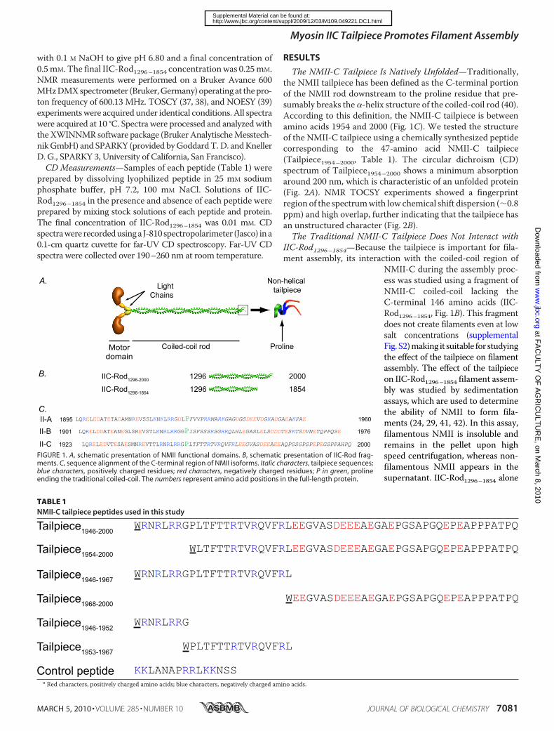
The NMII-C Tailpiece Is Natively Unfolded—Traditionally,the NMII tailpiece has been defined as the C-terminal portionof the NMII rod downstream to the proline residue that pre-sumably breaks the �-helix structure of the coiled-coil rod (40).According to this definition, the NMII-C tailpiece is betweenamino acids 1954 and 2000 (Fig. 1C). We tested the structureof the NMII-C tailpiece using a chemically synthesized peptidecorresponding to the 47-amino acid NMII-C tailpiece(Tailpiece1954–2000, Table 1). The circular dichroism (CD)spectrum of Tailpiece1954–2000 shows a minimum absorptionaround 200 nm, which is characteristic of an unfolded protein(Fig. 2A). NMR TOCSY experiments showed a fingerprintregion of the spectrumwith low chemical shift dispersion (�0.8ppm) and high overlap, further indicating that the tailpiece hasan unstructured character (Fig. 2B).The Traditional NMII-C Tailpiece Does Not Interact with
IIC-Rod1296–1854—Because the tailpiece is important for fila-ment assembly, its interaction with the coiled-coil region of
NMII-C during the assembly proc-ess was studied using a fragment ofNMII-C coiled-coil lacking theC-terminal 146 amino acids (IIC-Rod1296–1854, Fig. 1B). This fragmentdoes not create filaments even at lowsalt concentrations (supplementalFig. S2)making it suitable for studyingthe effect of the tailpiece on filamentassembly. The effect of the tailpieceon IIC-Rod1296–1854 filament assem-bly was studied by sedimentationassays, which are used to determinethe ability of NMII to form fila-ments (24, 29, 41, 42). In this assay,filamentous NMII is insoluble andremains in the pellet upon highspeed centrifugation, whereas non-filamentous NMII appears in thesupernatant. IIC-Rod1296–1854 alone
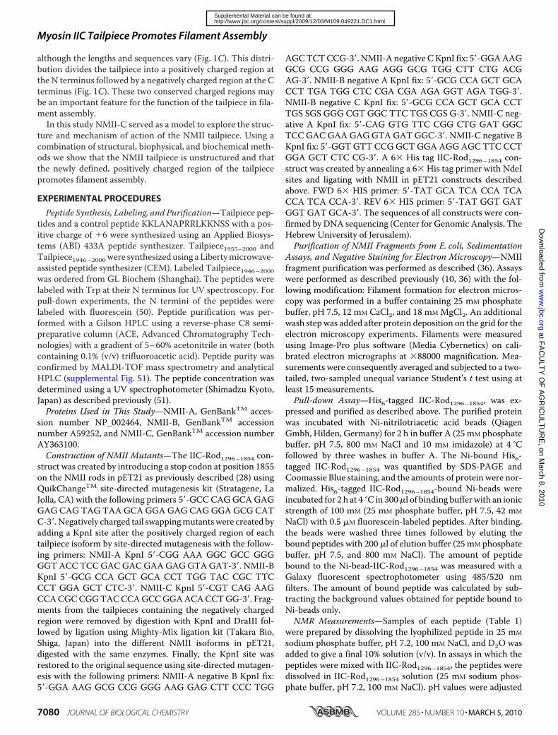
FIGURE 1. A, schematic presentation of NMII functional domains. B, schematic presentation of IIC-Rod frag-ments. C, sequence alignment of the C-terminal region of NMII isoforms. Italic characters, tailpiece sequences;blue characters, positively charged residues; red characters, negatively charged residues; P in green, prolineending the traditional coiled-coil. The numbers represent amino acid positions in the full-length protein.
TABLE 1NMII-C tailpiece peptides used in this study
a Red characters, positively charged amino acids; blue characters, negatively charged amino acids.
Myosin IIC Tailpiece Promotes Filament Assembly
MARCH 5, 2010 • VOLUME 285 • NUMBER 10 JOURNAL OF BIOLOGICAL CHEMISTRY 7081
at FA
CU
LTY
OF
AG
RIC
ULT
UR
E, on M
arch 8, 2010w
ww
.jbc.orgD
ownloaded from
http://www.jbc.org/content/suppl/2009/12/03/M109.049221.DC1.htmlSupplemental Material can be found at:
is completely soluble (Fig. 3), indicating that it is unable toassembleintofilaments. IncubatingIIC-Rod1296–1854withTailpiece1954–2000had no effect on IIC-Rod1296–1854 sedimentation (Fig. 3). Thiswas confirmed byCDanalysis of IIC-Rod1296–1854 in the presenceof increasing concentrations of Tailpiece1954–2000. The resultingCDspectrawere the sumof IIC-Rod1296–1854 andTailpiece1954–2000spectra, indicating that no structural changes occurred(supplemental Fig. S4). NMR TOCSY spectra (Fig. 2B) ofTailpiece1954–2000 alone and in the presence of IIC-Rod1296–1854(at a molar ratio of 2:1 peptide:IIC-Rod1296–1854) showed nodeviations in the chemical shift, further indicating that thetraditional NMII-C tailpiece does not interact withIIC-Rod1296–1854.The Positively Charged Part of Tailpiece1946–1967 Induces
IIC-Rod1296–1854 Filament Assembly—The NMII-C tailpiecehas been traditionally defined as amino acids 1954–2000.How-ever, the margins of the non-helical portion have not beenexactly defined biochemically, and no structural data exist forthe tailpiece. All NMII isoforms have a short positively chargedregion N-terminal to the proline, which is preceded by a nega-tively charged region (Fig. 1C). We propose that the positively
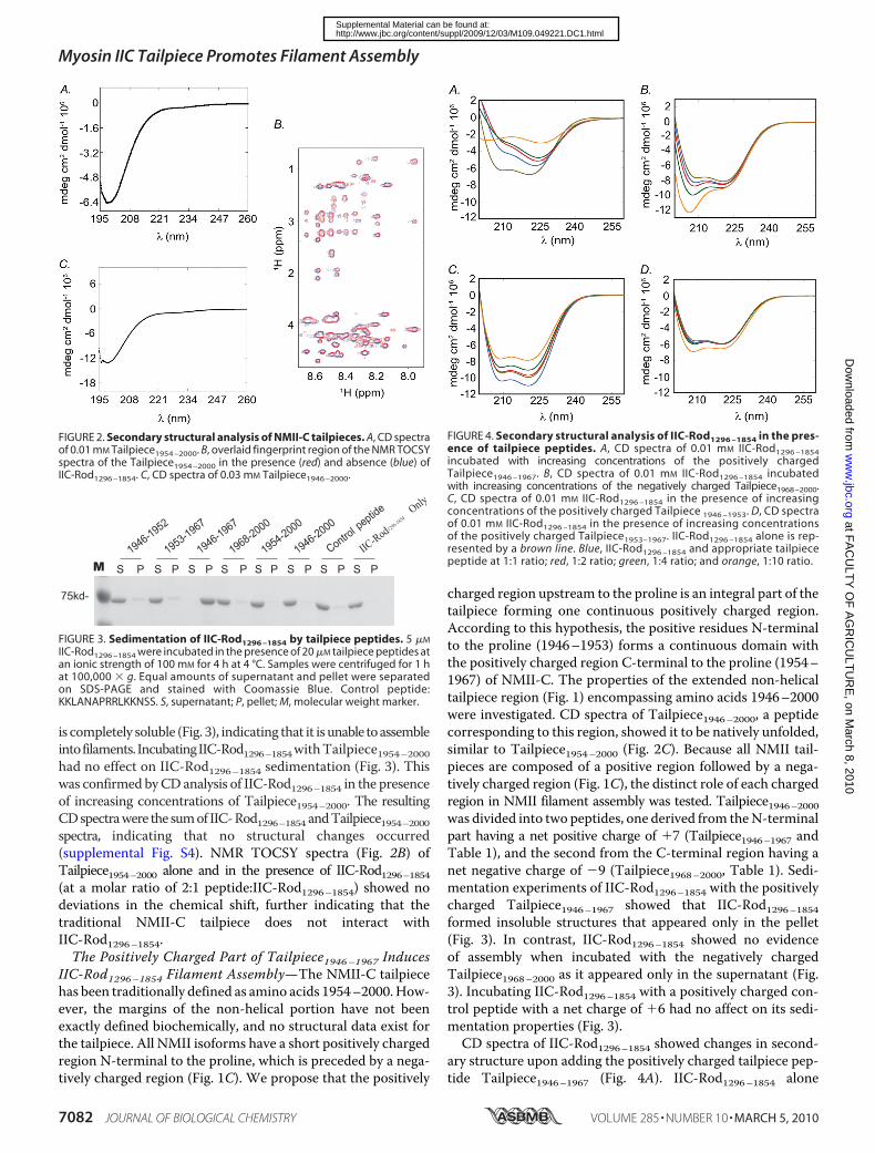
charged region upstream to the proline is an integral part of thetailpiece forming one continuous positively charged region.According to this hypothesis, the positive residues N-terminalto the proline (1946–1953) forms a continuous domain withthe positively charged region C-terminal to the proline (1954–1967) of NMII-C. The properties of the extended non-helicaltailpiece region (Fig. 1) encompassing amino acids 1946–2000were investigated. CD spectra of Tailpiece1946–2000, a peptidecorresponding to this region, showed it to be natively unfolded,similar to Tailpiece1954–2000 (Fig. 2C). Because all NMII tail-pieces are composed of a positive region followed by a nega-tively charged region (Fig. 1C), the distinct role of each chargedregion in NMII filament assembly was tested. Tailpiece1946–2000was divided into two peptides, one derived from theN-terminalpart having a net positive charge of �7 (Tailpiece1946–1967 andTable 1), and the second from the C-terminal region having anet negative charge of �9 (Tailpiece1968–2000, Table 1). Sedi-mentation experiments of IIC-Rod1296–1854 with the positivelycharged Tailpiece1946–1967 showed that IIC-Rod1296–1854formed insoluble structures that appeared only in the pellet(Fig. 3). In contrast, IIC-Rod1296–1854 showed no evidenceof assembly when incubated with the negatively chargedTailpiece1968–2000 as it appeared only in the supernatant (Fig.3). Incubating IIC-Rod1296–1854 with a positively charged con-trol peptide with a net charge of �6 had no affect on its sedi-mentation properties (Fig. 3).CD spectra of IIC-Rod1296–1854 showed changes in second-
ary structure upon adding the positively charged tailpiece pep-tide Tailpiece1946–1967 (Fig. 4A). IIC-Rod1296–1854 alone
FIGURE 2. Secondary structural analysis of NMII-C tailpieces. A, CD spectraof 0.01 mM Tailpiece1954 –2000. B, overlaid fingerprint region of the NMR TOCSYspectra of the Tailpiece1954 –2000 in the presence (red) and absence (blue) ofIIC-Rod1296 –1854. C, CD spectra of 0.03 mM Tailpiece1946 –2000.
FIGURE 3. Sedimentation of IIC-Rod1296 –1854 by tailpiece peptides. 5 �M
IIC-Rod1296 –1854 were incubated in the presence of 20 �M tailpiece peptides atan ionic strength of 100 mM for 4 h at 4 °C. Samples were centrifuged for 1 hat 100,000 � g. Equal amounts of supernatant and pellet were separatedon SDS-PAGE and stained with Coomassie Blue. Control peptide:KKLANAPRRLKKNSS. S, supernatant; P, pellet; M, molecular weight marker.
FIGURE 4. Secondary structural analysis of IIC-Rod1296 –1854 in the pres-ence of tailpiece peptides. A, CD spectra of 0.01 mM IIC-Rod1296 –1854incubated with increasing concentrations of the positively chargedTailpiece1946 –1967. B, CD spectra of 0.01 mM IIC-Rod1296 –1854 incubatedwith increasing concentrations of the negatively charged Tailpiece1968–2000.C, CD spectra of 0.01 mM IIC-Rod1296 –1854 in the presence of increasingconcentrations of the positively charged Tailpiece 1946 –1953. D, CD spectraof 0.01 mM IIC-Rod1296 –1854 in the presence of increasing concentrationsof the positively charged Tailpiece1953–1967. IIC-Rod1296 –1854 alone is rep-resented by a brown line. Blue, IIC-Rod1296 –1854 and appropriate tailpiecepeptide at 1:1 ratio; red, 1:2 ratio; green, 1:4 ratio; and orange, 1:10 ratio.
Myosin IIC Tailpiece Promotes Filament Assembly
7082 JOURNAL OF BIOLOGICAL CHEMISTRY VOLUME 285 • NUMBER 10 • MARCH 5, 2010
at FA
CU
LTY
OF
AG
RIC
ULT
UR
E, on M
arch 8, 2010w
ww
.jbc.orgD
ownloaded from
http://www.jbc.org/content/suppl/2009/12/03/M109.049221.DC1.htmlSupplemental Material can be found at:

adopted an expected �-helical conformation. Adding the posi-tively charged Tailpiece1946–1967 caused a decrease in �-helicalcontent, which was dependent on the concentration of the pos-itively chargedTailpiece1946–1967 (Fig. 4A). This decrease in theabsorption amplitude indicates a decrease in the concentrationof IIC-Rod1296–1854 in solution, possibly because of filamentassembly. Similar experiments using IIC-Rod1296–1854 with thenegatively charged Tailpiece1968–2000 showed a typical CDspectrum representing the sum of IIC-Rod1296–1854 andTailpiece1968–2000 spectra indicating no structural change(Fig. 4B). NMR TOCSY peaks of the positively chargedTailpiece1946–1967 with a 2:1 molar ratio of IIC-Rod1296–1854,disappeared relative to the unreacted peptide, which may becaused by increased correlation times due to binding to thelarge IIC-Rod1296–1854 (supplemental Fig. S3A), whereassimilar NMR experiments using the negatively chargedTailpiece1968–2000 showed no change in the peak intensitiesor chemical shift (supplemental Fig. S3B). This supports thesedimentation and CD results, indicating that the positivelycharged Tailpiece1946–1967 induces IIC-Rod1296–1854 fila-ment assembly into insoluble structures, but the negativelycharged Tailpiece1968–2000 does not.The Entire Positively Charged Region of the Tailpiece Is
Required for Inducing IIC-Rod1296–1855 Filament Assembly—The role of the seven amino acids located N-terminal to theproline (Tailpiece1946–2000, Fig. 1C and Table 1) on IIC-Rod1296–1854 filament assembly was studied using two addi-tional peptides: one corresponding to the seven extended resi-dues (Tailpiece1946–1952, Table 1) and the other correspondingto the positively charged part of the traditional Tailpiece1953–1967(Table 1). Sedimentation experiments of IIC-Rod1296–1854 inthe presence of Tailpiece1946–1952 or Tailpiece1953–1967 showedonly a slight increase in IIC-Rod1296–1854 filament assembly(Fig. 3). CD experiments using the positively chargedTailpiece1946–1952 and Tailpiece1953–1967 showed only a smallconformational change upon incubation with IIC-Rod1296–1854(Fig. 4, C and D). This minimal change in conformation wasstable over time, indicating the absence of subsequently formedhigher order assemblies (supplemental Fig. S5). The positivelycharged region of the tailpiece binds the NMII-C coiled-coilrod and induces NMII-C paracrystal formation. Sedimentationand CD experiments indicated that the positively chargedTailpiece1946–1967 may interact with IIC-Rod1296–1854 and thatthis interaction may induce its assembly. Binding of the differ-ent tailpiece peptides to the coiled-coil rod of NMII-C wasstudied by pull-down assays usingHis-tagged-IIC-Rod1296–1854immobilized on nickel beads and fluorescein-labeled peptides.This assay tests whether the peptides bind to the coiled-coil rodin an assembly-independent manner. Peptides containingamino acids 1953–1967 bound IIC-Rod1296–1854 withTailpiece1946–1967 having the strongest binding (Fig. 5). This isconsistent with Tailpiece1946–1967 results from sedimentationand CD assays. No significant binding of Tailpiece1946–1952to IIC-Rod1296–1854 was detected. The negative region,Tailpiece1968–2000, and control peptide also did not bind IIC-Rod1296–1854 (Fig. 5). These experiments confirm the impor-tance of the positively charged region for filament assembly.
NMII forms large paracrystal filaments at low salt concentra-tions (43). These structures have been used to examine thecapability of NMII to assemble into large filamentous struc-tures (10, 24, 26, 35, 44). The effect of the tailpiece on NMII-Cfilament assembly was studied by inducing IIC-Rod1296–1854 to
FIGURE 5. The interaction between IIC-Rod1296 –1854 and tailpiece pep-tides. Fluorescein-labeled tailpiece peptides were incubated for 2 h at 4 °Cwith Ni-beads-immobilized His-tagged IIC-Rod1296 –1854. Bound peptideswere eluted with 800 mM NaCl, 25 mM phosphate buffer, pH 7.5, and thefluorescence was detected by fluorescence spectrophotometer as describedunder “Experimental Procedures.” The extent of peptide bound to His-tagged-IIC-Rod1296 –1854 was calculated by subtracting the background valueof peptide bound to Ni-beads only from the value of peptide bound to His-tagged-IIC-Rod1296–1854. Values are the average of 3–4 independent experi-ments � S.D. and are normalized to Tailpiece1954–2000 binding. Tailpiece1947–1952and control peptide had binding values below background.
FIGURE 6. Tailpiece peptides effect on IIC-Rod1296 –1854 paracrystal mor-phology. IIC-Rod1296 –1854 was mixed with the different peptides at a 1:4 ratioin filament buffer (25 mM phosphate buffer, pH 7.5, 12 mM CaCl2, and 18 mM
MgCl2). Filaments were negatively stained with uranyl acetate prior to view-ing by electron microscope at �88000 as described under “ExperimentalProcedures.”
Myosin IIC Tailpiece Promotes Filament Assembly
MARCH 5, 2010 • VOLUME 285 • NUMBER 10 JOURNAL OF BIOLOGICAL CHEMISTRY 7083
at FA
CU
LTY
OF
AG
RIC
ULT
UR
E, on M
arch 8, 2010w
ww
.jbc.orgD
ownloaded from
http://www.jbc.org/content/suppl/2009/12/03/M109.049221.DC1.htmlSupplemental Material can be found at:

form paracrystals in the presence of the different tailpiecepeptides. IIC-Rod1296–1854 alone is not capable of creating thedistinctive long paracrystals of wild-typeNMII-C (28), and onlyextremely small, needle-like structures were observed (Fig. 6).Adding Tailpiece1946–1967, corresponding to the full positivelycharged region of the tailpiece, to IIC-Rod1296–1854 resulted inthe formation of filamentous paracrystals similar to thoseformedbywild-typeNMII-C. In accordancewith theCDand sed-imentation experiments, no large filaments were observed uponincubating IIC-Rod1296–1854 with each of the truncated positivelycharged tailpieces (Tailpiece1946–1952 or Tailpiece1953–1967).Furthermore, the traditional Tailpiece1954–2000 or the pep-tide corresponding to the negatively charged Tailpiece1968–2000had no effect on IIC-Rod1296–1854 paracrystal formation (datanot shown). These data are consistent with sedimentationexperiments.The Negatively Charged Region of NMII-C Tailpiece Regu-
lates the Positively Charged Region and Determines FilamentMorphology—The role of the negatively charged region of thetailpiece on IIC-Rod1296–1854 filament assembly was studied bysedimentation and CD experiments in which the full positivelycharged Tailpiece1946–1967 was mixed with the negativelychargedTailpiece1968–2000 and added to IIC-Rod1296–1854. Sed-imentation results showed that IIC-Rod1296–1854 was com-pletely insoluble upon adding themixture of positively and neg-atively charged tailpieces (Fig. 7A). A similar effect on IIC-Rodsolubility was seen using the positively charged Tailpiece1946–1967alone (Fig. 3). The change in CD spectra of IIC-Rod1296–1854after adding an equimolar mixture of positively and negativelycharged peptides was similar to the change seen when the pos-itively chargedTailpiece1946–1967 was added alone (Fig. 7,B andC). As expected similar results were obtained when mixing the
two short positive peptides (Fig. 7A). Thus in contrast to thecomplete tailpiece containing both the positively andnegativelycharged regions, the negatively charged Tailpiece1968–2000 byitself does not alter the effect of the positively chargedTailpiece1946–1967 on IIC-Rod1296–1854 filament assembly.The tailpiece has been shown to be important for determin-
ing the isoform specific morphology of NMII paracrystals (28).As the negatively charged region does not promote IIC-Rod1296–1854 filament assembly it may be important in deter-mining NMII paracrystal morphology. NMII fragment chime-ras were created in which the negatively charged region wasswapped among the NMII-A, NMII-B, and NMII-C isoforms.We have previously shown that rod fragments of the three
FIGURE 7. The effect of mixing the negatively and positively charged tail-piece regions on IIC-Rod1296 –1854 filament assembly. A, sedimentationassay of IIC-Rod1296 –1854 in the presence of either of the positively chargedTailpiece1946 –1952, Tailpiece1953–1967 or Tailpiece1946 –1967 and the negativelycharged Tailpiece1968 –2000 were performed as described in Fig. 3. B, CD spec-tra of 0.01 mM IIC-Rod1296 –1854 incubated with 0.01 mM Tailpiece1946 –1967alone. C, CD spectra of 0.01 mM IIC-Rod1296 –1854 in the presence of both0.01 mM Tailpiece1946 –1967 and 0.01 mM Tailpiece1968 –2000. Brown line, IIC-Rod1296 –1854 alone; Black line, IIC-Rod1296 –1854 and Tailpiece(s) that werecollected after 0 min; Red line, after 1 min; blue line, after 5 min; green line,after 10 min; magenta line after 15 min; light blue line after 30 min; lightgreen line, after 1 h, and orange line, after 2 h.
FIGURE 8. The effect of the tailpiece negatively charged region on NMIIparacrystal morphology. A, amino acids sequence of the negativelycharged tailpiece swapped chimeras. Blue highlight, positively chargedregion, red highlight, negatively charged region. Numbers indicate amino acidposition of the respective region. Only amino acids C-terminal to the prolineare presented. B, NMII isoforms A, B, and C rod fragments with the negativeregion swapped among the isoforms were dialyzed against filament bufferand stained with uranyl acetate prior to viewing by electron microscope at�88000 as described under “Experimental Procedures.” Blue representsNMII-A, red represents NMII-B, and green represents NMII-C originating aminoacid sequence. Scale bars, 50 nm.
Myosin IIC Tailpiece Promotes Filament Assembly
7084 JOURNAL OF BIOLOGICAL CHEMISTRY VOLUME 285 • NUMBER 10 • MARCH 5, 2010
at FA
CU
LTY
OF
AG
RIC
ULT
UR
E, on M
arch 8, 2010w
ww
.jbc.orgD
ownloaded from
http://www.jbc.org/content/suppl/2009/12/03/M109.049221.DC1.htmlSupplemental Material can be found at:
NMII isoforms create different paracrystal morphology:NMII-A and NMII-B form large wide filaments (width mea-suring 1.21 � 0.4 nm and 1.22 � 0.38 nm, respectively) whileNMII-C forms delicate thin filaments (0.32 � 0.07 nm) (28).Swapping the negatively charged region of either NMII-A orNMII-B with the negatively charged region of NMII-C resultedin thin filaments similar to NMII-C (0.269 nm �0.06 and 0.239nm �0.04, respectively) (Fig. 8 and Table 2). Accordingly,swapping the negatively charged region of NMII-C with thenegatively charged region ofNMII-A resulted in large filaments(0.534 � 0.075 nm). Surprisingly swapping the negative tail-piece of NMII-C with the negative of NMII-B increased thefilament width minimally (0.348 � 0.075 nm) (Fig. 8 and Table2). These results indicate that the negative region of the tail-piece plays a role in organizing NMII in the growing filament.
DISCUSSION
Extending the Tailpiece Boundary—As theC-terminal regionof myosin II has not been extensively analyzed, its exact defini-tion is unknown. Our results show that the actual tailpiecedomain extends further toward the N terminus of NMII fromthe putative coiled-coil breaking proline. This region of sevenamino acids is also positively charged, suggesting that it is anintegral part of the positive region together with the traditionaltailpiece. Data from other NMII isoforms have also shown thatthe small region upstream to the proline is important forassembly (28, 45). Both the traditional Tailpiece1954–2000 andthe newly defined Tailpiece1946–2000 were found to be unfoldedby CD (Fig. 2). The entire positively charged Tailpiece1946–1967binds IIC-Rod1296–1854, and induces it to form structures sim-ilar to wild-type NMII-C as seen by CD and EM experiments.Dividing this region into what was traditionally thought to bethe tailpiece of NMII-C (Tailpiece1953–1967) and a peptide rep-resenting only the seven amino acids upstream of the proline(Tailpiece1946–1952), showed that Tailpiece1953–1967 may beresponsible for initial binding to IIC-Rod1296–1854. Howeverthis binding induced only minimal assembly of IIC-Rod1296–1854as seen byCDand sedimentation experiments. This indicates thatthe entire positively charged region is a single domain responsiblefor promoting filament assembly.
The Negatively Charged C Terminus of the Tailpiece Has aRole Distinct from the Positive Region—Peptides correspondingto the negatively charged region of the Tailpiece1968–2000 werefound to be unfolded and did not bind IIC-Rod1296–1854 as seenin pull-down assays. In addition, this peptide did not affect IIC-Rod1296–1854 assembly as it remained completely soluble in thepresence of the peptide. Furthermore, CD experiments showedno conformational changes of IIC-Rod1296–1854 after incuba-tion with the negatively charged Tailpiece1968–2000 (Fig. 4B).This is in agreement with previous experiments in whichremoving the negatively charged region of the tailpiece fromsmooth muscle myosin II had no effect on filament assemblyproperties (24, 46). Nevertheless the negatively chargedTailpiece1968–2000 was sufficient to determine NMII paracrys-tal morphology as seen in chimeric NMII isoforms in whichonly the negatively charged region was swapped among theisoforms (Fig. 8). Each region of the tailpiece seems to have aunique role in NMII filament assembly. Experiments exploringthe individual roles of each region showed that although eachregion has a distinct role, the negatively charged region mayregulate the positively charged region during filament assemblyprocess. Neither the newly defined tailpiece, Tailpiece1946–2000,nor the traditional tailpiece, Tailpiece1954–2000, had any effecton IIC-Rod1296–1854 filament assembly even though these pep-tides contain the positively charged region (Fig. 3). Further-more, binding experiments showed that extending either posi-tively charged peptide to include the negatively charged regiongreatly reduced binding to IIC-Rod1296–1854 (Fig. 5). However,mixing experiments showed that adding the negatively chargedTailpiece1968–2000 did not affect the ability of the positivelycharged Tailpiece1947–1968 to induce IIC-Rod1296–1854 filamentassembly (Fig. 7). This indicates that when in integral form, thenegatively charged region may act as a regulator of the posi-tively charged region.A Model for the Role of the Tailpiece in Regulating Myosin
Assembly—The proposed model for the role of the tailpiece inthe assembly of NMII coiled-coil rod includes a positivelycharged region spanning both sides of the proline (amino acids1946–1967) that is responsible for binding the coiled-coil rodand inducing it to form high oligomeric structures. Once thepositively charged region has bound to the NMII rod, the neg-atively charged region can exert its effect on determining themorphology of the growing filament. This negatively chargedregion is also capable of masking the positively charged region,thereby hindering its ability to bind and hence modulating theassembly process.The Tailpiece Is a Shiftide: A Peptide That Shifts the Olig-
omerization Equilibrium of Proteins—NMII is in equilibriumamong individual hexamers and high order oligomers in fila-mentous form. In cases where proteins are in equilibriumamong several oligomeric states, peptides or small moleculescan bind specifically to one of these oligomeric species, stabilizethe state, and thus shift the oligomerization equilibrium towardthat specific state, according to the law of mass action (47, 48).We have termed such peptides “shiftides.” The positivelycharged Tailpiece1946–1967 is an example for such a shiftide,because it shows an ability to shift IIC-Rod1296–1854 from anindividual hexameric state to a high order oligomeric structure.
TABLE 2Width measurements of NMII negatively charged tailpiece chimerasParacrystal width was measured using ImagePro software from negatively stainedelectronmicrographs as described in the legend to Fig. 8. p values are Student’s t testcomparing to wild-type measurement population.
Name Morphology Width � S.D.p value
(compared towild type)
nmNMII-A wild typea Wide 1.21 � 0.4 N/Ab
NMII-B wild typea Wide 1.22 � 0.4 N/ANMII-C wild type Thin 0.32 � 0.07 N/ANMII-A Neg. Tail B Wide 0.782 � 0.21 7.34e-5NMII-A Neg. Tail C Thin 0.269 � 0.06 1.64e-11NMII-B Neg. Tail A Wide 1.39 � 0.3 0.1NMII-B Neg. Tail C Thin 0.239 � 0.04 1.98e-17NMII-C Neg. Tail A Wide 0.534 � 0.075 3.6e-6NMII-C Neg. tail B Thin 0.348 � 0.075 0.33
a Data taken from Ref. 28.b N/A, not applicable.
Myosin IIC Tailpiece Promotes Filament Assembly
MARCH 5, 2010 • VOLUME 285 • NUMBER 10 JOURNAL OF BIOLOGICAL CHEMISTRY 7085
at FA
CU
LTY
OF
AG
RIC
ULT
UR
E, on M
arch 8, 2010w
ww
.jbc.orgD
ownloaded from
http://www.jbc.org/content/suppl/2009/12/03/M109.049221.DC1.htmlSupplemental Material can be found at:

The mechanism by which the positively charged tailpiece pep-tide acts still needs to be investigated. Because NMII can onlyperform its functions when in filaments, peptides modulatingfilament assembly may shift oligomerization equilibriumtoward the functional form of NMII. This peptide may havetherapeutic potential by shifting the equilibrium toward fila-ment assembly in diseases caused by defects in NMII assembly(49).
Acknowledgment—We thank Dr. Robert S. Adelstein for the NMII-Cconstruct.
REFERENCES1. Sellers, J. R. (1999)Myosins, 2nd Ed., Oxford University Press, Oxford, UK2. Conti, M. A., and Adelstein, R. S. (2008) J. Cell Sci. 121, 11–183. Matsumura, F. (2005) Trends Cell Biol. 15, 371–3774. Lauffenburger, D. A., and Horwitz, A. F. (1996) Cell 84, 359–3695. Sellers, J. R. (2000) Biochim. Biophys. Acta 1496, 3–226. Sohn, R. L., Vikstrom, K. L., Strauss, M., Cohen, C., Szent-Gyorgyi, A. G.,
and Leinwand, L. A. (1997) J. Mol. Biol. 266, 317–3307. Atkinson, S. J., and Stewart, M. (1992) J. Mol. Biol. 226, 7–138. McLachlan, A. D., and Karn, J. (1982) Nature 299, 226–2319. Nakasawa, T., Takahashi, M., Matsuzawa, F., Aikawa, S., Togashi, Y., Sai-
toh, T., Yamagishi, A., and Yazawa, M. (2005) Biochemistry 44, 174–18310. Rosenberg, M., Straussman, R., Ben-Ya’acov, A., Ronen, D., and Ravid, S.
(2008) PLoS ONE 3, e149611. Straussman, R., Squire, J. M., Ben-Ya’acov, A., and Ravid, S. (2005) J. Mol.
Biol. 353, 613–62812. Bresnick, A. R. (1999) Curr. Opin. Cell Biol. 11, 26–3313. Kolega, J., and Kumar, S. (1999) Cell Motil. Cytoskel. 43, 255–26814. Tan, J. L., Ravid, S., and Spudich, J. A. (1992) Annu. Rev. Biochem. 61,
721–75915. Golomb, E., Ma, X., Jana, S. S., Preston, Y. A., Kawamoto, S., Shoham,
N. G., Goldin, E., Conti, M. A., Sellers, J. R., and Adelstein, R. S. (2004)J. Biol. Chem. 279, 2800–2808
16. Shohet, R. V., Conti, M. A., Kawamoto, S., Preston, Y. A., Brill, D. A., andAdelstein, R. S. (1989) Proc. Natl. Acad. Sci. U.S.A. 86, 7726–7730
17. Simons, M., Wang, M., McBride, O. W., Kawamoto, S., Yamakawa, K.,Gdula, D., Adelstein, R. S., and Weir, L. (1991) Circ. Res. 69, 530–539
18. Bao, J., Jana, S. S., and Adelstein, R. S. (2005) J. Biol. Chem. 280,19594–19599
19. Bao, J., Ma, X., Liu, C., and Adelstein, R. S. (2007) J. Biol. Chem. 282,22102–22111
20. Even-Ram, S., Doyle, A. D., Conti, M. A., Matsumoto, K., Adelstein, R. S.,and Yamada, K. M. (2007) Nat. Cell Biol. 9, 299–309
21. Jana, S. S., Kawamoto, S., and Adelstein, R. S. (2006) J. Biol. Chem. 281,24662–24670
22. Sandquist, J. C., Swenson, K. I., Demali, K. A., Burridge, K., and Means,A. R. (2006) J. Biol. Chem. 281, 35873–35883
23. Wylie, S. R., and Chantler, P. D. (2008)Mol. Biol. Cell 19, 3956–396824. Hodge, T. P., Cross, R., and Kendrick-Jones, J. (1992) J. Cell Biol. 118,
1085–109525. Sato, M. K., Takahashi, M., and Yazawa, M. (2007) Mol. Biol. Cell 18,
1009–101726. Rovner, A. S., Fagnant, P. M., Lowey, S., and Trybus, K. M. (2002) J. Cell
Biol. 156, 113–12327. Sinard, J. H., Rimm, D. L., and Pollard, T. D. (1990) J. Cell Biol. 111,
2417–242628. Ronen, D., and Ravid, S. (2009) J. Biol. Chem. 284, 24948–2495729. Dulyaninova, N. G., Malashkevich, V. N., Almo, S. C., and Bresnick, A. R.
(2005) Biochemistry 44, 6867–687630. Even-Faitelson, L., and Ravid, S. (2006)Mol. Biol. Cell 17, 2869–288131. Kelley, C. A., and Adelstein, R. S. (1990) J. Biol. Chem. 265, 17876–1788232. Murakami, N., Chauhan, V. P., and Elzinga, M. (1998) Biochemistry 37,
1989–200333. Rosenberg, M., and Ravid, S. (2006)Mol. Biol. Cell 17, 1364–137434. Straussman, R., Even, L., and Ravid, S. (2001) J. Cell Sci. 114, 3047–305735. Franke, J. D., Dong, F., Rickoll,W. L., Kelley,M. J., andKiehart, D. P. (2005)
Blood 105, 161–16936. Straussman, R., Ben-Ya’acov, A., Woolfson, D. N., and Ravid, S. (2007) J.
Mol. Biol. 366, 1232–124237. Piotto, M., Saudek, V., and Sklenar, V. (1992) J. Biomol. NMR 2, 661–66538. Sklenar, V., Piotto, M., Leppik, R., and Saudek, V. (1993) J. Magn. Reso-
nance A 102, 241–24539. Jeener, J., Meier, B. H., Bachmann, P., and Ernst, R. R. (1979) J. Chem.
Physics 71, 4546–455340. Chou, P. Y., and Fasman, G. D. (1974) Biochemistry 13, 222–24541. Hostetter, D., Rice, S., Dean, S., Altman, D., McMahon, P. M., Sutton, S.,
Tripathy, A., and Spudich, J. A. (2004) PLoS Biol. 2, e35642. Murakami, N., Kotula, L., and Hwang, Y. W. (2000) Biochemistry 39,
11441–1145143. Kendrick-Jones, J., Szent-Gyorgyi, A. S., and Cohen, C. (1971) J. Mol. Biol.
59, 527–52944. Atkinson, S. J., and Stewart, M. (1991) J. Cell Sci. 99, 823–83645. Ikebe, M., Komatsu, S., Woodhead, J. L., Mabuchi, K., Ikebe, R., Saito, J.,
Craig, R., and Higashihara, M. (2001) J. Biol. Chem. 276, 30293–3030046. Turbedsky, K., and Pollard, T. D. (2005) J. Mol. Biol. 345, 351–36147. Hayouka, Z., Rosenbluh, J., Levin, A., Loya, S., Lebendiker,M., Veprintsev,
D., Kotler,M., Hizi, A., Loyter, A., and Friedler, A. (2007) Proc. Natl. Acad.Sci. U.S.A. 104, 8316–8321
48. Jaffe, E. K. (2005) Trends Biochem. Sci. 30, 490–49749. Vicente-Manzanares, M., Ma, X., Adelstein, R. S., and Horwitz, A. R.
(2009) Nat. Rev. 10, 778–79050. Weber, P. J., Bader, J. E., Folkers, G., and Beck-Sickinger, A. G. (1998)
Bioorg. Med. Chem. Lett. 8, 597–60051. Gill, S. C., and von Hippel, P. H. (1989) Anal. Biochem. 182, 319–326
Myosin IIC Tailpiece Promotes Filament Assembly
7086 JOURNAL OF BIOLOGICAL CHEMISTRY VOLUME 285 • NUMBER 10 • MARCH 5, 2010
at FA
CU
LTY
OF
AG
RIC
ULT
UR
E, on M
arch 8, 2010w
ww
.jbc.orgD
ownloaded from
http://www.jbc.org/content/suppl/2009/12/03/M109.049221.DC1.htmlSupplemental Material can be found at: