the yeast nuclear pore complex and transport through it
TRANSCRIPT
YEASTBOOK
CELL STRUCTURE & TRAFFICKING
The Yeast Nuclear Pore Complex and TransportThrough ItJohn D. Aitchison*,1 and Michael P. Rout†*Institute for Systems Biology and Seattle Biomedical Research Institute, Seattle, Washington 98109, and †The Laboratory of Cellular and StructuralBiology, The Rockefeller University, New York, New York 10021
ABSTRACT Exchange of macromolecules between the nucleus and cytoplasm is a key regulatory event in the expression of a cell’sgenome. This exchange requires a dedicated transport system: (1) nuclear pore complexes (NPCs), embedded in the nuclear envelopeand composed of proteins termed nucleoporins (or “Nups”), and (2) nuclear transport factors that recognize the cargoes to betransported and ferry them across the NPCs. This transport is regulated at multiple levels, and the NPC itself also plays a key regulatoryrole in gene expression by influencing nuclear architecture and acting as a point of control for various nuclear processes. Here wesummarize how the yeast Saccharomyces has been used extensively as a model system to understand the fundamental and highlyconserved features of this transport system, revealing the structure and function of the NPC; the NPC’s role in the regulation of geneexpression; and the interactions of transport factors with their cargoes, regulatory factors, and specific nucleoporins.
TABLE OF CONTENTS
Abstract 855
Introduction 856
Structure and Composition of the NPC 856
Overall Composition 857
Membrane Ring 859
Core Scaffold: Inner and Outer Rings 860
Phenylalanine-Glycine Nups 861
Cytoplasmic Filaments and Nuclear Basket 862
Shuttling Nucleoporins 862
NPC Assembly 863
Turnover of NPCs 865
Soluble Phase of Transport: Transport Signals and Carriers 865
Karyopherins and Their Cargoes 866
Competition as a Major Factor in Nuclear Transport 868Continued
Copyright © 2012 by the Genetics Society of Americadoi: 10.1534/genetics.111.127803Manuscript received February 15, 2011; accepted for publication August 1, 2011Available freely online through the author-supported open access option.1Corresponding author: Institute for Systems Biology and Seattle Biomedical Research Institute, 401 Terry Ave. N, Seattle, WA 98109. E-mail: [email protected]
Genetics, Vol. 190, 855–883 March 2012 855
CONTENTS, continued
Not Just Karyopherins: RNA Export 868
Mechanism of Nuclear Transport 870
Accessory Transport and Processing Factors at the NPC 872
Balancing the Books 873
Regulation of Transport by the NPC 873
Beyond Transport: The NPC as a Platform for Other Nuclear Processes 873
Summary 875
ALTHOUGH considered “simple,” its amenability to mo-lecular and genetic interrogation has established baker’s
yeast as an outstanding model system for cell biologists.Moreover, in the context of the Eukaryota, Saccharomycescerevisiae is closely related to humans (both being membersof the opisthokonts). Thus, interrogation of the fundamentalbiology of yeast has proven to be not only comparativelyfacile, but also highly relevant to human biology, both mor-phologically and mechanistically. Indeed, yeast has remainedat the forefront of studies on the nucleus—the defining char-acteristic of eukaryotes—for several decades.
Eukaryotic chromosomes are housed within the nucleus,which is delimited by the two parallel membranes of thenuclear envelope (NE). The evolution of this physical barrierendowed eukaryotes with a critical control mechanismsegregating the sites of gene transcription and ribosomebiogenesis from the site of protein synthesis. This compart-mentalization allows cells to strictly coordinate numerouskey cellular processes, but it also presents cells with thechallenge of selectively managing the transport of a bewil-dering number of proteins and RNAs between the nucleusand cytoplasm. This is accomplished by the presence of “nu-clear pores,” which arise at points where the inner and outerNE membranes conjoin to form circular channels across thenuclear envelope. Within these pores sit large proteinaceouscomplexes, appropriately named nuclear pore complexes(NPCs), which, in conjunction with soluble transport factors,govern all biomolecular transport into and out of the nu-cleus. Beyond this fundamental control of transport, theNPC has adopted a host of other activities by acting as a spa-tial landmark or anchor site for many of the machineriesthat directly control gene activity and transcriptional pro-cessing (reviewed in Ahmed and Brickner 2007; Hetzerand Wente 2009). As a transporter, it must allow small mol-ecules to pass as freely, prevent most macromolecules fromcrossing, and permit the quickest possible passage of se-lected macromolecules bidirectionally across the NE. As ananchor, it must allow free communication between the at-tached control machineries and the chromatin or transcriptsthat they regulate without hindering nuclear transport. Onecan thus also consider the NPC as a major way station ineukaryotes, interacting with and regulating DNA, RNA, and
membranes and communicating between the cytoplasm, nu-cleoplasm, and ER lumen. Because of this, the subject of thenuclear pore complex and nuclear transport is a huge one,far beyond the scope of any single review. Our aim here istherefore to give an overview, including references to manyexcellent reviews that detail particular areas of study.
Structure and Composition of the NPC
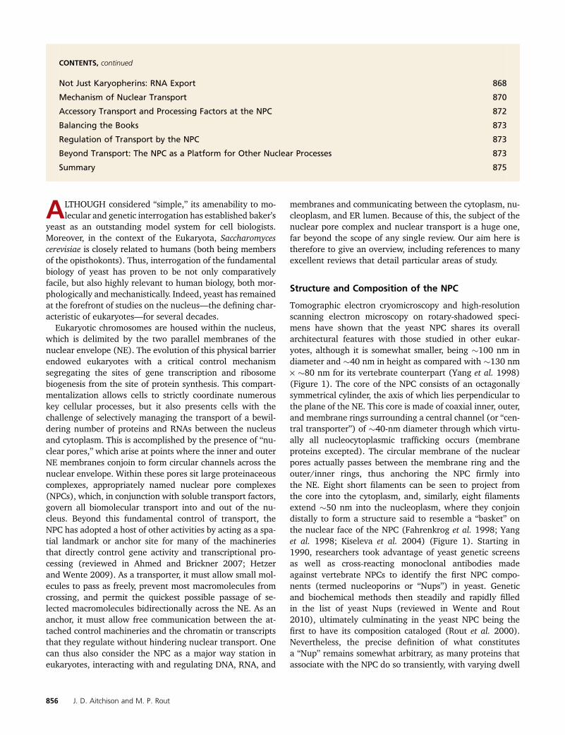
Tomographic electron cryomicroscopy and high-resolutionscanning electron microscopy on rotary-shadowed speci-mens have shown that the yeast NPC shares its overallarchitectural features with those studied in other eukar-yotes, although it is somewhat smaller, being �100 nm indiameter and �40 nm in height as compared with �130 nm· �80 nm for its vertebrate counterpart (Yang et al. 1998)(Figure 1). The core of the NPC consists of an octagonallysymmetrical cylinder, the axis of which lies perpendicular tothe plane of the NE. This core is made of coaxial inner, outer,and membrane rings surrounding a central channel (or “cen-tral transporter”) of �40-nm diameter through which virtu-ally all nucleocytoplasmic trafficking occurs (membraneproteins excepted). The circular membrane of the nuclearpores actually passes between the membrane ring and theouter/inner rings, thus anchoring the NPC firmly intothe NE. Eight short filaments can be seen to project fromthe core into the cytoplasm, and, similarly, eight filamentsextend �50 nm into the nucleoplasm, where they conjoindistally to form a structure said to resemble a “basket” onthe nuclear face of the NPC (Fahrenkrog et al. 1998; Yanget al. 1998; Kiseleva et al. 2004) (Figure 1). Starting in1990, researchers took advantage of yeast genetic screensas well as cross-reacting monoclonal antibodies madeagainst vertebrate NPCs to identify the first NPC compo-nents (termed nucleoporins or “Nups”) in yeast. Geneticand biochemical methods then steadily and rapidly filledin the list of yeast Nups (reviewed in Wente and Rout2010), ultimately culminating in the yeast NPC being thefirst to have its composition cataloged (Rout et al. 2000).Nevertheless, the precise definition of what constitutesa “Nup” remains somewhat arbitrary, as many proteins thatassociate with the NPC do so transiently, with varying dwell
856 J. D. Aitchison and M. P. Rout
times, and some NPC-associated proteins also extend theirfunctions and localizations beyond the NPC (Arib and Akhtar2011).
The sheer size and flexibility of the NPC make it difficult tofully solve its molecular architecture by conventional techni-ques. Therefore, an orthogonal approach has been taken; largeand diverse sets of proteomic data were amassed and acomputational method for using these data was developed todefine the relative positions and proximities of the yeast NPC’sconstituent proteins. A corresponding average protein densitymap represents the position of every Nup with a precision of�5 nm, sufficient to resolve the molecular organization of theentire NPC (Alber et al. 2007a,b) (Figure 2A). The resultingmap agrees with complementary data in both yeast and verte-brates (reviewed in Strambio-de-Castillia et al. 2010).
Overall Composition
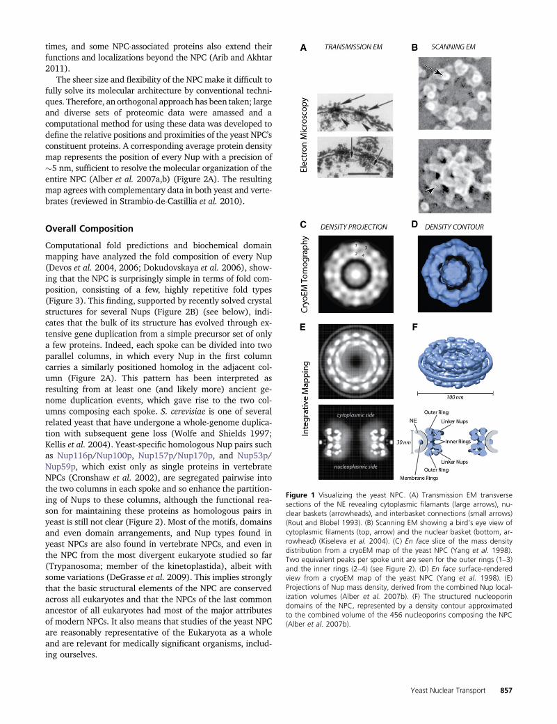
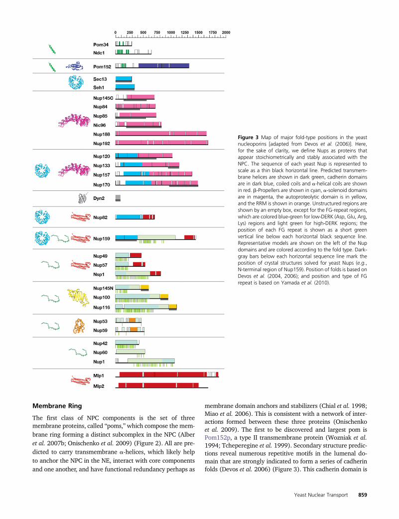
Computational fold predictions and biochemical domainmapping have analyzed the fold composition of every Nup(Devos et al. 2004, 2006; Dokudovskaya et al. 2006), show-ing that the NPC is surprisingly simple in terms of fold com-position, consisting of a few, highly repetitive fold types(Figure 3). This finding, supported by recently solved crystalstructures for several Nups (Figure 2B) (see below), indi-cates that the bulk of its structure has evolved through ex-tensive gene duplication from a simple precursor set of onlya few proteins. Indeed, each spoke can be divided into twoparallel columns, in which every Nup in the first columncarries a similarly positioned homolog in the adjacent col-umn (Figure 2A). This pattern has been interpreted asresulting from at least one (and likely more) ancient ge-nome duplication events, which gave rise to the two col-umns composing each spoke. S. cerevisiae is one of severalrelated yeast that have undergone a whole-genome duplica-tion with subsequent gene loss (Wolfe and Shields 1997;Kellis et al. 2004). Yeast-specific homologous Nup pairs suchas Nup116p/Nup100p, Nup157p/Nup170p, and Nup53p/Nup59p, which exist only as single proteins in vertebrateNPCs (Cronshaw et al. 2002), are segregated pairwise intothe two columns in each spoke and so enhance the partition-ing of Nups to these columns, although the functional rea-son for maintaining these proteins as homologous pairs inyeast is still not clear (Figure 2). Most of the motifs, domainsand even domain arrangements, and Nup types found inyeast NPCs are also found in vertebrate NPCs, and even inthe NPC from the most divergent eukaryote studied so far(Trypanosoma; member of the kinetoplastida), albeit withsome variations (DeGrasse et al. 2009). This implies stronglythat the basic structural elements of the NPC are conservedacross all eukaryotes and that the NPCs of the last commonancestor of all eukaryotes had most of the major attributesof modern NPCs. It also means that studies of the yeast NPCare reasonably representative of the Eukaryota as a wholeand are relevant for medically significant organisms, includ-ing ourselves.
Figure 1 Visualizing the yeast NPC. (A) Transmission EM transversesections of the NE revealing cytoplasmic filamants (large arrows), nu-clear baskets (arrowheads), and interbasket connections (small arrows)(Rout and Blobel 1993). (B) Scanning EM showing a bird’s eye view ofcytoplasmic filaments (top, arrow) and the nuclear basket (bottom, ar-rowhead) (Kiseleva et al. 2004). (C) En face slice of the mass densitydistribution from a cryoEM map of the yeast NPC (Yang et al. 1998).Two equivalent peaks per spoke unit are seen for the outer rings (1–3)and the inner rings (2–4) (see Figure 2). (D) En face surface-renderedview from a cryoEM map of the yeast NPC (Yang et al. 1998). (E)Projections of Nup mass density, derived from the combined Nup local-ization volumes (Alber et al. 2007b). (F) The structured nucleoporindomains of the NPC, represented by a density contour approximatedto the combined volume of the 456 nucleoporins composing the NPC(Alber et al. 2007b).
Yeast Nuclear Transport 857

Figure 2 (A) Major structural features of the yeast NPC (based on the architectural map of Alber et al. 2007a,b); see main text for details. (B) Map ofprotein positions in the yeast NPC (based on the architectural map of Alber et al. 2007a,b), with examples of the atomic structures of pieces of Nupswhere known: Nic96 (2RFO) (Schrader et al. 2008), Nup84/Nup145C/Sec13 (3IKO) (Nagy et al. 2009), Nup85/Seh1 (3EWE) (Brohawn et al. 2008),Nup116 (2AIV) (Robinson et al. 2005), Nup120 (3F7F) (Seo et al. 2009), Nup133 (3KFO), Nup145N (3KEP) (Sampathkumar et al. 2010), Nup159 (1XIP)(Weirich et al. 2004), and Nup170 (3I5P) (Whittle and Schwartz 2009).
858 J. D. Aitchison and M. P. Rout
Membrane Ring
The first class of NPC components is the set of threemembrane proteins, called “poms,”which compose the mem-brane ring forming a distinct subcomplex in the NPC (Alberet al. 2007b; Onischenko et al. 2009) (Figure 2). All are pre-dicted to carry transmembrane a-helices, which likely helpto anchor the NPC in the NE, interact with core componentsand one another, and have functional redundancy perhaps as
membrane domain anchors and stabilizers (Chial et al. 1998;Miao et al. 2006). This is consistent with a network of inter-actions formed between these three proteins (Onischenkoet al. 2009). The first to be discovered and largest pom isPom152p, a type II transmembrane protein (Wozniak et al.1994; Tcheperegine et al. 1999). Secondary structure predic-tions reveal numerous repetitive motifs in the lumenal do-main that are strongly indicated to form a series of cadherinfolds (Devos et al. 2006) (Figure 3). This cadherin domain is
Figure 3 Map of major fold-type positions in the yeastnucleoporins [adapted from Devos et al. (2006)]. Here,for the sake of clarity, we define Nups as proteins thatappear stoichiometrically and stably associated with theNPC. The sequence of each yeast Nup is represented toscale as a thin black horizontal line. Predicted transmem-brane helices are shown in dark green, cadherin domainsare in dark blue, coiled coils and a-helical coils are shownin red. b-Propellers are shown in cyan, a-solenoid domainsare in magenta, the autoproteolytic domain is in yellow,and the RRM is shown in orange. Unstructured regions areshown by an empty box, except for the FG-repeat regions,which are colored blue-green for low-DERK (Asp, Glu, Arg,Lys) regions and light green for high-DERK regions; theposition of each FG repeat is shown as a short greenvertical line below each horizontal black sequence line.Representative models are shown on the left of the Nupdomains and are colored according to the fold type. Dark-gray bars below each horizontal sequence line mark theposition of crystal structures solved for yeast Nups (e.g.,N-terminal region of Nup159). Position of folds is based onDevos et al. (2004, 2006); and position and type of FGrepeat is based on Yamada et al. (2010).
Yeast Nuclear Transport 859

predicted to form homophilic binding interfaces (Bryant andStow 2004) and likely explains the oligomeric lumenal ring.Much less is known about Pom34. It is a small protein con-taining two transmembrane helices and two small domainsboth facing into the core scaffold. As yet, no function has beenassigned to Pom34—even cells lacking both Pom34 andPom152 are viable—and no nonfungal homolog has yet beenfound. However, like Pom152, Pom34 genetically interactswith several core scaffold proteins in ways that suggest thesepoms’ functions partially overlap (see below) (Madrid et al.2006; Miao et al. 2006). Ndc1p (nuclear division cycle 1) isalso a pom but was named differently because it was actuallyfirst characterized due to the effect of one of its mutants onthe assembly of the spindle. Indeed, it turns out that, likeseveral other Nups, Ndc1p plays at least two roles in the cell:in the case of Ndc1p, one in the NPC and one in the spindlepole body (SPB)—themitotic spindle organizer. In yeast, bothmacromolecular assemblies are embedded in the NE, and itseems that Ndc1p helps to insert and attach both into theirrespective nuclear pores. Ndc1p has (at least) six transmem-brane helices and a carboxy-terminal domain that interactswith the core scaffold and other poms (Alber et al. 2007b;Onischenko et al. 2009) and has confirmed homologs in bothSchizosaccharomyces pombe [i.e., cut11, also with known rolesin spindle assembly and an NPC component (West et al.1998)] and metazoa [i.e., NDC1 (Lau et al. 2004; Mansfeldet al. 2006; Stavru et al. 2006)]. Dissecting the NPC functionof Ndc1p from its SPB function has been difficult, but severallines of genetic (Lau et al. 2004) and molecular biological(Onischenko et al. 2009) evidence suggest that Ndc1p playsan important role, with the help of the other poms, in NPCassembly (see also below).
Core Scaffold: Inner and Outer Rings
The second class of NPC components comprises the corescaffold proteins. This scaffold is composed of an inter-locking lattice of roughly a dozen evolutionarily conservedstructural proteins that link together to form a core layergiving the NPC shape and strength (Figure 2). Fold compo-sition analyses revealed that the core scaffold consists ofNups composed of only two fold types in three arrange-ments: consisting almost entirely of a b-propeller fold, oralmost entirely of a-solenoid-like/helix-turn-helix repeatfolds, or a b-propeller followed by an a-solenoid-like domain(Figure 3). These fold types together are characteristic ofcomponents of the clathrin, COPI and COPII membrane ves-icle-coating complexes (reviewed in Field and Dacks 2009),and related complexes such as the intraflagellar transportcomplex (Jekely and Arendt 2006) and the HOPS/CORVETcomplexes (Nickerson et al. 2009). The latter b-propeller/a-solenoid combination is particularly characteristic of thisfamily, as these protein types interlock in a variety of relatedways into a lattice forming their vesicle coats. These simi-larities were recently further underscored by atomic struc-tures solved for several core scaffold Nups (Figure 2B)
(Boehmer et al. 2003; Berke et al. 2004; Hsia et al. 2007;Jeudy and Schwartz 2007; Brohawn et al. 2008; Debler et al.2008; Schrader et al. 2008; Brohawn and Schwartz 2009;Leksa et al. 2009; Nagy et al. 2009; Seo et al. 2009; Whittleand Schwartz 2009) as well as clathrin (reviewed in Owenet al. 2004) and COPII and COPI components (Stagg et al.2006; Fath et al. 2007; Lee and Goldberg 2010; reviewed inStagg et al. 2007, 2008). This fold similarity, analyzed ini-tially in yeast, led to the “protocoatomer hypothesis,” whichproposes that a simple membrane-curving module, madeprimarily from b-propeller and a-solenoid folds, was a com-mon ancestor for NPCs and coated vesicles that originated inthe precursors to the ancient last ancestor common to alleukaryotes (Devos et al. 2004, 2006; Alber et al. 2007a,b).This protcoatomer gave these ancestors the ability to gener-ate internal membrane systems by invagination of theplasma membrane and then to manipulate and elaboratethese systems, eventually leading to the evolution of theER, Golgi, and nucleus that characterize modern eukaryotes(Field and Dacks 2009).
The core scaffold somewhat resembles a vesicle coat, as itforms a discrete layer completely following the curve of thepore membrane, effectively coating it (Figure 2). Thus, itdefines the size of the central tube/transporter of the NPCand the height of the NPC core, and all other Nups and pomsare attached to either the inner or the outer face of this coat(Figure 1). Biochemical studies have shown that the corescaffold Nups compose several subcomplexes that appearto function as “building blocks” during NPC assembly andcan even exchange with a soluble pool in mature NPCs(Lutzmann et al. 2002; D’Angelo et al. 2006; Makio et al.2009). Morphologically, the scaffold is made of two innerrings, sandwiched between and interconnected with twoouter rings, such that the nuclear and cytoplasmic halvesof the NPC have one inner and one outer ring each (Figure2). Although there is still some debate on the matter, a con-sensus remains that in both yeast and vertebrates these in-ner rings are compositionally, as well as morphologically,distinct (Tran and Wente 2006; Alber et al. 2007b).
Four large Nups, each just under 200 kDa in size, composethe inner rings: Nup188p and Nup192p (primarily composedof a-solenoid-like folds) and the homologous Nup170p andNup157p (Figure 2) (made from the clathrin-like pattern ofan amino-terminal b-propeller followed by an a-solenoid-like domain), proteins proposed long ago to be core compo-nents of the NPC (Aitchison et al. 1995b). The inner ringsare adjacent to each other at the equator of the NPC in thesame plane as the three poms of the membrane ring withwhich they interact extensively to anchor the core scaffoldto the NE (Alber et al. 2007b; Onischenko et al. 2009). Nu-merous mutations in all four proteins demonstrate extensivegenetic interaction networks with each other and with themembrane ring components, which likely underscore thefunctional importance of the inner ring in being a keystoneof the core scaffold and in anchoring the NPC to the poremembrane (Aitchison et al. 1995b; Tcheperegine et al. 1999;
860 J. D. Aitchison and M. P. Rout

Miao et al. 2006). Moreover, on both the nuclear and cyto-plasmic sides of each spoke, one copy of the Nup Nic96p isanchored through Nup192p and a second copy throughNup188p, linking the inner ring to the other internal struc-tures of the NPC (Figure 2) (see below).
Structurally, the most extensively studied set of corescaffold proteins are the seven Nups that compose theyeast outer rings, first identified and characterized by theHurt laboratory; Nup133p, Nup120p, Nup145Cp, Nup85p,Nup84p, Seh1p, and Sec13p form a discrete complex thatcan be biochemically isolated and is termed the Nup84 com-plex (Figure 2B) (Siniossoglou et al. 1996, 2000; Lutzmannet al. 2002; Flemming et al. 2009). Importantly, the evolu-tionary link between NPCs and vesicle-coating complexes issupported by the fact that Sec13p is shared with the Sec13/31 COPII vesicle-coating complex (Siniossoglou et al. 1996;Salama et al. 1997; Devos et al. 2004). Moreover, justlike the inner-ring proteins, all Nup84 complex proteinsare formed almost entirely by a b-propeller fold (Seh1p,Sec13p), an a-solenoid-like fold (Nup85p, Nup84p,Nup145Cp), or an N-terminal b-propeller and a carboxy-terminal a-solenoid-like fold (Nup133p, Nup120p), againcommon to vesicle-coating complexes (Devos et al. 2004,2006) (Figure 2B and Figure 3). Excitingly, crystal structuresprimarily from the Blobel and Schwartz laboratories havebegun to piece this complex together at the atomic level(Figure 2B) (Hsia et al. 2007; Brohawn et al. 2008; Debleret al. 2008; Brohawn and Schwartz 2009; Leksa et al. 2009;Nagy et al. 2009; Seo et al. 2009; Whittle and Schwartz2009). Electron microscopy studies have shown that theNup84 complex formed an extended Y structure (Siniossoglouet al. 2000; Lutzmann et al. 2002; Kampmann and Blobel2009), and pioneering work from the Hurt laboratory re-constituted this complex from bacterially expressed pro-teins and showed that the two short arms of this Y arecomposed, respectively, of Nup120p and Nup85p+Seh1p,while Nup133p, Nup84p, and Nup145Cp/Sec13p form themain stalk (Siniossoglou et al. 2000; Lutzmann et al. 2002).There is evidence that this complex is flexible (Kampmannand Blobel 2009), perhaps reflecting the known flexibilityof the NPC in response to changes in NE shape and duringits assembly (see below).
Few interactions have been found between the compo-nents of the Nup84 complex and the rest of the NPC, al-though it connects with the inner rings through, forexample, a Nup157p–Nup120p connection (Lutzmann et al.2005; Alber et al. 2007a,b). Mutations in any of these sevenNups are often characterized by temperature sensitivity,messenger RNA (mRNA) and pre-ribosomal export prob-lems, and aberrant NPC assembly. In particular, a so-called“clustering” phenotype was first described in mutants ofthe Nup84 complex components in which the NPCs can beseen to cluster into one or a few patches in the NE (Doyeet al. 1994; Aitchison et al. 1995a; Heath et al. 1995; Liet al. 1995; Pemberton et al. 1995; Goldstein et al. 1996;Siniossoglou et al. 1996). That outer-ring Nup mutants
cause mislocalization of otherwise reasonably functionalNPCs in the plane of the NE may point to a role for thisstructure in keeping the NPC stably located in the poremembrane. The outer rings are strategically placed at thepoint where the pore membrane joins the coplanar outerand inner NE membranes, and it seems reasonable that amajor role for them is to ensure the smooth transition of thepore membrane into the inner and outer NE membranes(Figure 2) (Alber et al. 2007b).
Phenylalanine-Glycine Nups
It was the field’s catalog of the composition of the yeast NPCthat led to a surprise. No homologs of mechanochemicalproteins or NTPases of any kind that could physically drivea gating process were found as components of the NPC.Instead, strikingly, the cataloging revealed that over one-third of Nups in the NPC shared a highly characteristicrepetitive motif, consisting of multiple repeated phenylala-nine-glycine (FG) pairs spaced by �20 mainly polar aminoacids (Figure 3). Although these proteins are found in alleukaryotes studied, once again they were first sequencedfrom yeast (Hurt 1988; Davis and Fink 1990; Nehrbasset al. 1990), and the yeast “FG Nups” remain the best stud-ied. On the basis initially of work in yeast, two flavors of FGNups were described: FxFG Nups typified by Nsp1p andGLFG Nups typified by Nup100p (on the basis of the typicalsequence of their FG repeat), the former having somecharged amino acids in their spacers and the latter havingrelatively uncharged spacers (reviewed in Rout and Wente1994). These two flavors of FG repeat also appear to beconserved (Figure 3), although by examination of orthologsin syntenic yeasts it was shown that the spacer sequencesbetween each of the FG repeats evolved more rapidly thandid other Nups (Denning and Rexach 2007), a situationcommon to the FG-repeat regions of all eukaryotes(DeGrasse et al. 2009). On the basis of various physicalmeasurements of purified and bacterially expressed pro-teins, a consensus has emerged that the FG-repeat regionsof FG Nups take on a natively unfolded structure both in vitroand in vivo, such that they form long, disordered flexiblefilaments (Denning et al. 2003; Denning and Rexach2007; Lim et al. 2006a,b, 2007a, 2008; Patel et al. 2007;Yamada et al. 2010); the lack of structural constraints there-fore likely explains the lower evolutionary constraints onsequence conservation of these repeat regions (Denningand Rexach 2007).
FG Nups also usually carry small structured domains,which serve to anchor them to the NPC (Alber et al. 2007b).These are generally predicted to be coiled-coil or a-helicalby fold analysis and comparison to vertebrate homologs(Melcak et al. 2007), in addition to b-sandwich and (oddly)RNA recognition motif (RRM) folds (Figure 3) (Devos et al.2006). Nup82p and Nic96p seem to serve as linkers to at-tach many of the FG Nups to the core scaffold (mainlythrough the inner-ring Nups), such that the inner surface
Yeast Nuclear Transport 861
of the scaffold is lined with FG Nups whose filamentous FG-repeat regions fill the central channel and extend into thenucleoplasm and cytoplasm (Figure 2). On both the cyto-plasmic and nucleoplasmic sides of each spoke two copies ofNic96p carry the FG Nups Nsp1p, Nup57p, and Nup49p andanother two copies form interactions to additional copies ofNsp1p, such that these FG Nups face both the nucleus andthe cytoplasm. At the cytoplasmic side, Nup82p associateswith Nsp1p as well as with the cytoplasmically facing FGNups Nup159p, Nup116p, Nup100p, and Nup42p (Grandiet al. 1995; Belgareh et al. 1998; Bailer et al. 2000, 2001;A. K. Ho et al. 2000; Rout et al. 2000; Alber et al. 2007a,b).There are also the FG Nups Nup145Np, Nup1p, and Nup60pfound on the nucleoplasmic side, connecting mainly to theinner-ring Nups. In addition, Nup53p and Nup59p are at-tached to Nup170p and Nic96p, and both face the poremembrane (Figure 2). The latter two Nups may also belongto the class of FG Nups, as they can bind transport factorsand carry degenerate FG-repeat regions that are predicted tobe natively unfolded (Marelli et al. 1998; Fahrenkrog et al.2000b; Lusk et al. 2002; Makhnevych et al. 2003; Alber et al.2007b). One FG Nup, Nup145p, uniquely cleaves itself inhalf at its Phe605-Ser606 peptide bond to produce two sep-arate Nups, Nup145Np (carrying the FG-repeat region andthe autoproteolytic b-sandwich domain at its new carboxy-terminus) and Nup145Cp (a mainly a-solenoid-like proteinthat forms a major component of the Nup84 complex) (Fig-ure 3) (Wente and Blobel 1994; Teixeira et al. 1997, 1999;Rosenblum and Blobel 1999). Although not essential inyeast (Emtage et al. 1997), this cleavage event appears con-served in vertebrates (Fontoura et al. 1999; Hodel et al.2002; Sun and Guo 2008). Nup145Cp and Nup145Np re-main linked as a dynamic complex such that Nup145Np canshuttle between the NPC and the nuclear interior, as does itsvertebrate counterpart (Griffis et al. 2002; Ratner et al.2007). The entirety of Nup145Np is highly conserved withthe homologous yeast nucleoporins Nup100p and Nup116p,neither of which undergoes autoproteolysis as they lack a ho-mologous counterpart for Nup145Cp. It seems that lineage-specific gene duplications of an ancestral Nup145N-likegene gave rise to the truncated versions Nup100p andNup116p, and likely other FG Nups also originated fromsuch, sometimes more ancient, duplication events (Manset al. 2004; Devos et al. 2006).
Collectively, the anchored FG Nups form the business endof the NPC, as the FG-repeat domains form low-affinity, high-specificity interactionswith transport factors involved in activetransport through the NPC and so actually form the selectivebarrier in and around the central tube by providing thebinding sites for transport factors that facilitate their exchangeacross the NE while excluding the passage of macromoleculesnot destined for nucleocytoplasmic transport. In one sense, theNPC can be considered a framework that provides the correctpositioning of the FG repeats, flanking and filling the centraltubewhile defining the upper diameter of the central tube andthe cargoes that transit through it (Figures 1 and 2) (Rout and
Aitchison 2001; Rout et al. 2003; Hetzer and Wente 2009;Walde and Kehlenbach 2010).
Cytoplasmic Filaments and Nuclear Basket
While not as morphologically prominent as their vertebratecounterparts, both cytoplasmic filaments and nuclear bas-kets have been seen to project from the yeast NPC (Figure 1)(Kiseleva et al. 2004). Nup159p, Nup82p, and Nup42p seemto contribute to the cytoplasmic filaments (Kraemer et al.1995; Hurwitz et al. 1998; Strahm et al. 1999; Rout et al.2000; Alber et al. 2007b) and function in the last stages ofexport from the NPC (below). Curiously, the protein Dyn2p,a light chain component of the dynein microtubule motor,binds to Nup159p and helps form a rigid filamentous struc-ture that may stiffen the cytoplasmic filament projecting itout from the core scaffold (Stelter et al. 2007). Yeast lack anobvious homolog of the vertebrate Nup358, which is be-lieved to produce the more prominent cytoplasmic filamentsin the latter (Wu et al. 1995; Matunis et al. 1998). In verte-brates, the bulk of the nuclear basket seems to be made ofTpr (Cordes et al. 1997; Hase et al. 2001; Frosst et al. 2002;Krull et al. 2004; Qi et al. 2004). Tpr is a conserved �200-kDa protein made mainly of extensive coiled-coil domainsthat dimerize into long rods forming the basket struts. TwoTpr homologs, Mlp1p and Mlp2p, exist in yeast and localizeto the region of the nuclear basket (Strambio-de-Castilliaet al. 1999). Unlike metazoa, no lamina lies interwovenbetween NPCs beneath the NE, but both Mlp1p and Mlp2pspread out along the inner face of the NE to form a delicatenetwork interconnecting yeast NPCs, although they are ex-cluded where the dense crescent of the nucleolus pressesagainst the NE (Strambio-de-Castillia et al. 1999). Mlp2pis additionally associated with the SPB (Niepel et al.2005). In yeast, Mlp2p is the result of the specific genomeduplication; however, a spindle organizer-specific copy ofTpr homologs has been independently reinvented severaltimes in evolution for reasons that are still unclear (Jimenezet al. 2000; DeGrasse et al. 2009). Overall, a bewilderingarray of functionalities have been ascribed to the Mlp net-work, including roles in recruitment of transport factors, lateprocessing of transcripts, and epigenetic regulation of geneexpression, as will be discussed below.
Shuttling Nucleoporins
The definition of nucleoporins becomes more difficult whenone considers the dynamics of some of the classically definednucleoporins. Nup2p, for example, was defined as a nucleo-porin as early as 1993 on the basis of its localization to theNPC; however, fluorescence microscopy, subcellular frac-tionation, and experiments monitoring its dynamics in vivo(Dilworth et al. 2001) revealed that Nup2p actually cycles onand off the nuclear basket and in this sense behaves more likea soluble transport factor. Similarly, Yrb2p (yeast ran bindingprotein 2; a.k.a. Nup36p) is primarily nuclear, but contains
862 J. D. Aitchison and M. P. Rout

FG repeats, yet it only transiently associates with the NPC(Floer and Blobel 1996). So far, all such rapidly “shuttling”nucleoporins belong to the FG Nup family. As well as anFG-repeat region, both Nup2p and Yrb2p carry a consensusRan-binding motif and may have a role in promoting thedisassembly of transport cargos; in this way, shuttling nucle-oporins may act as mediators between the stationary andsoluble phases of transport (see below) (Dilworth et al.2001, 2005; Gilchrist et al. 2002). In vertebrates, the dynam-ics of Nups have been comprehensively examined, revealingvarying half-lives of each Nup on the NPC, and it seems likelythat this will be borne out in yeast (Tran and Wente 2006).For example, Nup145Np has a localization that is biased to,but not exclusively on, the nuclear face, while Nup116pand Nup100p are similarly biased to the cytoplasmic face(Suntharalingam and Wente 2003). This variation compli-cates efforts to define a “stoichiometry” for components ofthe NPC, as any number for these more dynamic Nups will bean average of what may be a stochastic variation in Nupnumber and location. We expect that, the closer we look,the more difficult it will be to consider the NPC an autono-mous structure; rather, perhaps it should be considered a dy-namic assembly of proteins which to varying degrees, cyclebetween the stationary and soluble phases during transportand assembly, and functionally link the NPC to numerousother dynamic cellular activities (see below).
NPC Assembly
NPCs are not static structures. They are assembled, and theircomponents appear to be capable of turning over during theNPC’s lifetime. In many organisms, NPCs disassemble uponNE breakdown at the beginning of mitosis or meiosis andreassemble coordinately with the NE around the newly seg-regated chromosomes at its end. However, yeast has a“closed” mitosis in which the NE remains intact, such thatthe NPCs remain assembled throughout the life cycle of thecell and negate the need for NE and NPC disassembly—insharp contrast to the elaborate mitotic nuclear disassemblyand reassembly processes seen in metazoans (Suntharalingamand Wente 2003). Careful analyses of serially sectionedyeast confirmed that NPC assembly occurs continuouslythroughout the entire cell cycle with a typical haploid NEcontaining between �70 NPCs just after mitosis to �140NPCs in late anaphase (Winey et al. 1997). How this assem-bly occurs is still unclear, despite much work in both yeastand vertebrate model systems, with most of that work inmetazoan cells (because researchers generally studied thesynchronized assembly of NPCs in mitosis), and some of thedetails are only just beginning to emerge (as reviewed inFernandez-Martinez and Rout 2009; Hetzer and Wente2009). Nevertheless, the processes of NPC and NE assem-bly—and the reasons why some species opt for a closedmitosis while other related species opt for variants of anopen mitosis [compare the ascomycetes Saccharomyces andAspergillus (De Souza et al. 2004; Osmani et al. 2006; Liu
et al. 2009)], remain somewhat mysterious. Work in verte-brate cell-free systems has established, finally, that newNPCs are indeed inserted de novo into the NE (rather than,e.g., “budding off” from existing NPCs) (D’Angelo et al.2006). In yeast, it is primarily genetic approaches that havegiven some of these insights. As a yeast cell grows, the nu-cleus also grows in volume and the NE enlarges its surfacearea, during which time new NPCs are inserted into the NE(Winey et al. 1997). Although not proven, it seems likelythat this process in yeast is similar to interphase NPC assem-bly in vertebrates, which has been shown to occur throughde novo assembly of precursor building blocks recruited fromboth the nucleoplasm and cytoplasm into the regions of theNE between pre-existing NPCs (D’Angelo et al. 2006). Thecontinued assembly of the NPC and NE throughout the yeastcell cycle has been used as a basis for genetic screens, select-ing for mutants that caused mislocalization of tagged Nups.Initially, mutants in various Nups produced phenotypes that(if not lethal) gave a puzzling collection of different pheno-types that were difficult to interpret in terms of NPC assem-bly. Some made the NPCs cluster (above), whereas othersled to herniations of the NE extending over the cytoplasmicface of NPCs to seal them (Wente and Blobel 1993, 1994).However, more recent approaches have given more inter-pretable phenotypes. By using a photoconvertable Dendratag in cells blocked and then released in NPC assembly, itwas shown that some pre-assembly Nup complexes congre-gate on both the inner and the outer membranes of the NE,including cytoplasmic-facing Nups on the cytoplasmic faceof the NE and nucleoplasmic/basket Nups on the nuclearface, whereas symmetrically disposed Nups were found toaccumulate on both NE faces (Makio et al. 2009; Oni-schenko et al. 2009). These pre-assembly complexes mightcorrespond to the discrete complexes found to compose theNPC, such as the Nup84 complex (see above). Targeting ofthese pre-assembly Nups to the NE seems to require certainsoluble transport factors normally used to chaperone andpower the transport of cargoes through the NPC (see be-low), as genetic screens for conditional mutants in NPCassembly identified Ran, RanGEF, RanGAP, and Ntf2 (seeFigure 4 and below) (Ryan and Wente 2002; Ryan et al.2003, 2007). The karyopherin (Kap) Kap95p was also iden-tified in these screens, and another karyopherin, Kap121p,seems to aid Nup53p in assembling into a complex withNup170p (Lusk et al. 2002).
Interestingly, these mutants correspond to two key com-ponents of the cargo-carrying transport factor pathways,namely the b-karyopherins Kap95p and Kap121p and Rancycle components [Ran, RanGAP, RanGEF, and Ntf2 (re-sponsible for transporting RanGDP into the nucleus)] (Lusket al. 2002; Ryan and Wente 2002; Ryan et al. 2003, 2007).The reasons for the functional associations between NPC as-sembly and transport factors are still being elucidated, butsimilar connections have been seen in vertebrates (D’Angeloet al. 2006). In yeast, Kap121p has been proposed to targetNup53p to the NPC, where it is attached to the core scaffold
Yeast Nuclear Transport 863

component Nup170p. Indeed, recent work has revealed theimportance of the core scaffold to the early stages of NPCassembly. Thus, when the C-terminal domain of Nup170pis overexpressed, what appear to be intermediates of NPCassembly accumulate both in the cytoplasm and at the NE(Flemming et al. 2009). Similarly, in strains lacking bothNup53p and its paralog Nup59p, depletion of Nup170p oreither of two transmembrane nucleoporins that connectwith Nup170p—Pom152p or Pom34p—also caused the ac-cumulation of such intermediates in yeast cells (Onischenkoet al. 2009).
For an NPC to be inserted into the intact NE, both theinner and the outer NE membranes must approach at a siteand fuse to give rise to the pore membrane, upon which thecore scaffold and the rest of the NPC can then assemble. It iscurious, therefore, that two of the three poms (Pom152pand Pom34p) are not essential and so are dispensable forNPC assembly and that all three poms (including Ndc1p) arenot required for NPC assembly in the closely related fungi,Aspergillus (Liu et al. 2009). Taken together, this suggeststhat there must be other transiently or dynamically associ-ating membrane proteins that play key roles in initiating theNPC assembly process and fusion of the inner nuclear mem-brane (INM) and outer nuclear membrane (ONM) to formthe pore membrane.
Indeed, there has been a growing cadre of proteins that,while not strictly Nups, play a key role in yeast NPC assembly.As well as Ran, Ran cofactors, and the Kaps (above; Lusket al. 2002; Ryan et al. 2003, 2007), the two yeast reticulonsRtn1p and Rtn2p and their interacting partner Yop1p havebeen implicated in NPC assembly (Dawson et al. 2009). Rtnsand Yop1/DP1 proteins can deform and mold membranes,having been shown to have roles in both dynamically restruc-turing and maintaining tubular ER (De Craene et al. 2006;Voeltz et al. 2006; Hu et al. 2008) and, in metazoans, even inpostmitotic NE shaping (Anderson and Hetzer, 2008b). Retic-ulons have a segment that can insert into one leaflet of amembrane, which may promote or induce membrane curva-ture (Oertle et al. 2003; De Craene et al. 2006; Voeltz et al.2006; Shibata et al. 2008); indeed, they are depleted inregions of flat membrane, such as the NE between NPCs,and are found to concentrate in curved membrane regionssuch as tubular ER (De Craene et al. 2006; Voeltz et al. 2006;Anderson and Hetzer, 2008a,b). The apparent absence ofthese proteins in the mature NPC suggests that they play onlya transient role at the beginning of the assembly process,perhaps helping the first NE membrane curving and fusionstep to make the pore membrane. Similarly, the NE/ER pro-teins Apq12p and Brr6p are genetically linked to each otherand are necessary for normal NPC assembly and distribution.This work indicates that both proteins are involved in main-taining lipid homoeostasis in the ER, which is necessary forproper NPC insertion and distribution in the NE (Scarcelliet al. 2007; Hodge et al. 2010).
Another candidate NPC assembly factor is Pom33p, iso-lated in a genetic screen for genes that are essential in cells
lacking Nup133p (Chadrin et al. 2010). The transmembraneprotein Pom33p and its paralog Per33p are found in boththe ER and the NE, although Pom33p shows a preferentialdynamic localization at NPCs. Pom33, but not Per33, genet-ically interacts with Nup84 complex components and theinteracting proteins Nup170p and Ndc1p and physicallyforms a direct complex with Rtn1p (Chadrin et al. 2010).These data, plus the fact that depletion of both Nup170 andPom33 significantly impaired assembly of NPCs, point toa role for Pom33p in NPC assembly or maintenance of theNE (Chadrin et al. 2010). Pom33p thus potentially links thereticulon membrane bending and manipulation machinerywith the assembling NPC, which together possibly eitherhelp the transmembrane nucleoporins during the initialmembrane fusion event required for the start of NPC assem-bly or facilitate the stabilization of the nascent nuclear pore.Following this initial pore formation, assembly to form themature NPC must proceed extremely rapidly, as no naturallyoccurring intermediates have been found.
Of course, the NPC core scaffold is composed almostentirely of homologs of vesicle-coating proteins, whosefunction is to mold and fuse membranes into curvedvesicles. On the basis of this similarity it has been suggestedthat the Nup84 complex and the Nup170 inner-ring complex(which interacts directly with poms) could be directly in-volved, after recruitment to the NE, in forming a coat some-what like those in coated vesicles that produces the nascentpore membrane and pinches the inner and outer NE mem-branes together in a manner analogous to pinching offa curved vesicle (reviewed in Fernandez-Martinez and Rout2009; Hetzer and Wente 2009).
In summary, there appear to be several main steps toNPC assembly. Initially, accessory factors collaborate withtransmembrane and inner-ring NPC components to accu-mulate on both the inner nuclear membrane and the outernuclear membrane to warp the latter into a fused pore. The
Figure 4 The nuclear transport cycle for karyopherins and their cargoes.See Fig. 5 legend and main text for details.
864 J. D. Aitchison and M. P. Rout

recruitment of the inner-ring components would recruit theouter-ring components, permitting the assembly of the en-tire membrane-coating core scaffold in the pore (Alber et al.2007b). Rapid association of the remaining FG Nups, otherNPC components, and the nuclear basket would then com-plete the process. However, this sequence of events remainsstrictly speculative, and much remains to be understoodabout the mechanism of NPC assembly in yeast or in anyother eukaryote.
It seems possible that other NE-associated structures shareat least some aspects of the NPC’s assembly process. Curi-ously, Nup60p and Pom152p are also required for the assem-bly and repair of the SPB (Greenland et al. 2010). Recall thatthe pore membrane component, Ndc1p, has been shown tobe a shared component of both the NPC and the SPB and isrequired for the assembly of both (Chial et al. 1998; Lau et al.2004). The functional connection between the SPB and NPCis underscored by the putative nuclear basket componentMlp2p, which may associate with Nup60 (Zhao et al. 2008)and connects to both NPCs and SPBs (Niepel et al. 2005). Itseems that several proteins are found at both locales, raisingthe possibility that, as both NPCs and SPBs are inserted intoa membranous grommet formed from the fusion of the innerand outer nuclear membranes, there are some commonalitiesin their assembly mechanisms.
Turnover of NPCs
No repair mechanism, as such, has been found for the NPC.Rather, it seems that a combination of some pre-emptivereplacement of components by constant turnover and di-lution of “old” NPCs by new ones through cell growth anddivision are the tactics taken to rejuvenate the NPC popula-tion in a growing yeast population. The turnover rates ofyeast Nups are not yet precisely known, although certainlysome FG Nups exchange very quickly (Dilworth et al. 2001;Tran and Wente 2006). Moreover, there is some uncertaintyabout how “old” and “new” NPCs are partitioned betweenthe mother and the daughter cells upon budding. While evi-dence was originally presented that the old NPCs are re-tained preferentially in mother cells, potentially ensuringthat the daughters receive a fresh supply of new NPCs(Shcheprova et al. 2008), more recent work indicates insteadthat new and old NPCs partition equally betweenmother anddaughter at mitosis (Khmelinskii et al. 2010, 2011). As NPCsegregation and turnover have direct implications for agingstudies (Kaeberlein 2010), this and related topics will doubt-less be areas of intensive future investigation.
Soluble Phase of Transport: Transport Signalsand Carriers
While NPC-mediated gating does not require an energyinput, nucleocytoplasmic transport and the accumulationof cargoes in the nucleus and cytoplasm are driven bythe formation and maintenance of concentration gradients
across the NE by GTPases and ATPases in the nucleoplasmand cytoplasm (Figures 4 and 5) (reviewed in Rout et al.2003; Strambio-de-Castillia et al. 2010). Moreover, as is typ-ical for protein sorting throughout eukaryotic cells, proteinssynthesized in the cytoplasm that are destined for the nu-cleus carry targeting signals [generally termed nuclear lo-calization signals (NLSs)] that are recognized by solublereceptors, which mediate their transport. The first transportfactors to be identified and purified to homogeneity werekaryopherin a, karyopherin b, and a small Ras-like GTPasecalled Ran. Through classic biochemical fractionation com-bined with in vitro import assays, these proteins were puri-fied to homogeneity from mammalian systems and shownto mediate transport of reporter proteins carrying an NLSfrom the SV40 large T antigen (reviewed in Pemberton andPaschal 2005; Wente and Rout 2010). Because the yeastgenome had recently been completed, it was then a straight-forward matter to identify orthologs in yeast (reviewed inWozniak et al. 1998), and soon work on the mechanisms ofnucleocytoplasmic transport was progressing in both yeastand mammalian systems. These studies established that kar-yopherin a (Kap60p in yeast) binds to the NLS and thatkaryopherin b (Kap95p in yeast) enhances (or stabilizes)the interaction and in turn binds to FG-repeat-containingnucleoporins. Ran-GTP provides an important source of en-ergy to the reaction by binding to karyopherin b as it entersthe nucleus with karyopherin a and cargo in tow, releasingthe cargo to the nucleoplasm (Figures 4 and 5). Importantly,the versatility of yeast as a model system rapidly led tocomplementary approaches and insights beyond those im-mediately possible in mammalian systems.
It was clear that not all proteins destined for the nucleuscontain an NLS typified by SV40 large T antigen. Thediversity of cargoes and complexes that traverse the NPC ishuge, ranging from proteins to RNAs and ribonucleoproteins(RNPs), including mRNPs and ribosomes, to viruses. Analysisof the yeast genome revealed family transport factorsstructurally related to karyopherin b (and more distantly tokaryopherin a). Other eukaryotes studied, even the mostevolutionarily divergent, seem to retain this same family ofKaps (DeGrasse et al. 2009; Mason et al. 2009). Membersof the b-Kap family are generally large (molecular weightof 100–125 kDa) proteins that share�20% sequence identitywith each other. Each is typified by the presence of up to�20HEAT repeats (amphipathic helix-loop-helix motifs) thatform a large helical solenoid (with a single extended hydro-phobic core) (Figure 5) (Cansizoglu et al. 2007). There areapparently 14 Kaps in S. cerevisiae and at least 19 Kaps inhumans (Stewart 2003), all of which differentially bind dif-ferent classes of nuclear transport signals, FG-repeat nucleo-porins, and Ran; unlike the Kap60p:Kap95p dimer, all otherb-Kaps bind directly to their cargos (Figure 4).
Karyopherins responsible for importing cargoes are oftencalled importins, and exporters are called exportins. Thedirection of transport for each karyopherin is dictated by itsdifferential interaction with cargoes and Ran (Figure 4). In
Yeast Nuclear Transport 865

cells, Ran primarily exists in two forms: in the nucleus, Ran ismaintained in its GTP-bound form by a GTP exchange factor(RanGEF; RCC1, Prp20p, or Srm1p in yeast); this protein ischromatin bound, thus signaling to the nucleocytoplasmictransport system the position of the nucleoplasm by virtueof generating a cloud of RanGTP around it. In contrast, theRan GTPase-activating protein (RanGAP) is localized to thecytoplasm, so that Ran in the cytoplasm predominates inthe GDP form. Karyopherins exploit this property duringtransport. As mentioned above, during an import cycle, Kapsbind to their cargoes in the cytoplasm, and when they reachthe high Ran-GTP in the nucleus, are induced to release theircargoes. In contrast, exportin binding to cargoes is enhancedby the formation of a trimeric complex that includes Ran-GTP. Thus, as this complex meets the RanGAP in the cyto-plasm, the GTP is hydrolyzed and the complex falls apart.Indeed, the direction of karyopherin-mediated transportthrough the NPC can be reversed by inversion of the Rangradient (Nachury and Weis 1999). Most karyopherins arethought to be recycled to their original compartments empty,but in a few instances they are believed to chaperone anothercargo on their return journey (Figure 4).
Studies of prototypical interactions among constituentsof these transport pathways have shed considerable light onthe structural basis of transport (Figure 5). In the classicalpathway, the NLS binds to a long region on the inside ofthe Kap60 superhelix, made of alternating a-helical turns.Kap95p, which is also made of alternating a-helical turns,forms a spiral with two surfaces, and the inner surface wrapsaround an extended N-terminal domain of Kap60p (Figure5) [a.k.a the importin b-binding (IBB) domain] (Cingolaniet al. 1999). The interaction of Kap95p with FG Nups ismediated as the repeated Phe residues on the FG-repeatregions (see below) insert into complementary repeatedpockets formed from the crevices between adjacent a-helicalrepeats all along the outer surface of Kap95p’s spiral.RanGTP binds to Kap95p (Lee et al. 2005) on the inner sur-face of Kap95’s amino-terminal solenoid spiral, which causesconformational changes that lead to release of Kap60p (andcargoes) (Figure 5).
During Kap60p export, Kap60p and RanGTP are bound tothe inner surface of the Cse1p spiral (Matsuura and Stewart2004). In this form, the IBB domain is held tightly againstthe side of Kap60p, inhibiting NLS binding and leaving theouter surface free to interact with FG repeats and therebycarrying the complex through the NPC out of the nucleus(Figure 5). Once in the cytoplasm, the RanGTP hydrolyzesto RanGDP, causing the complex to dissociate. Kap60premains bound to its IBB even when free in the cytoplasm,but binding to an NLS exposes the IBB and allows Kap95p tobind, initializing another round of import.
Karyopherins and Their Cargoes
The apparent presence of a family of karyopherins, and theknowledge that there are numerous classes of cargoes that
must be transported across the NPC, led researchers to beginto identify cargoes for each of the karyopherins. Again, yeasthas been a tremendous model system for investigating thisfundamental question. The mainstay approach for doing sohas been to take advantage of homologous recombinationtechniques to genomically tag karyopherins with an epitopetag (like protein A) and to isolate the Kap and its associatedcargoes (Aitchison et al. 1996). Genetic perturbations of theKap genes have then been used to explore the consequenceswith respect to the potential cargo. This approach was firstapplied to Kap104p to establish that it is responsible forimporting a subclass of RNA-binding proteins (Nab2p andNab4p/Hrp1p) (Aitchison et al. 1996). These proteins aremajor mRNA-binding proteins essential for mRNA process-ing and export (Anderson et al. 1993). They appear to ac-company the mRNA out of the nucleus, and upon reachingthe cytoplasm, they are recycled for another round byKap104p (Lee and Aitchison 1999). Mtr10p/Kap111p alsoappears dedicated to this essential function; it importsNpl3p, another essential mRNA biogenesis factor. Interest-ingly, while both Kaps import essential proteins, neither isessential (under the same conditions) by itself. This suggeststhat Kaps must have the capability to compensate for oneanother and bind to their cargoes with some promiscuity.This was first made obvious upon examination of Kap123p.Deletion of Kap123 is virtually without phenotypic conse-quences in laboratory strains of yeast. Yet, Kap123p is per-haps the most abundant of the Kaps in yeast, conservedthroughout the Eukaryota, and it binds to Lys-rich NLSsshared by a host of ribosomal proteins and ribosome assem-bly factors, which leads to their import into the nucleus priorto their assembly into ribosomes (Rout et al. 1997;Leslieet al. 2002; Timney et al. 2006)—an essential process if everthere was one! Indeed, a host of genetically interactingKaps appear to be involved in the import of proteins criticalto ribosome assembly (e.g., Kap108p/Sxm1p, Kap119p/Nmd5p, Kap121p/Pse1p) (Rosenblum et al. 1997; Routet al. 1997; Sydorskyy et al. 2003; Caesar et al. 2006), andit has been shown explicitly that, in the absence of Kap123p,Kap121p can bind to Kap123p substrates and import theminto the nucleus (Rout et al. 1997).
Perhaps it is not surprising that structurally related Kapscan bind to structurally related NLSs, but it also appears thatKaps can recognize more than one type of NLS. For example,while Kap121p was originally shown to bind to noncanon-ical Lys-rich NLSs (Rout et al. 1997; Kaffman et al. 1998b;Leslie et al. 2002), it, like Kap104p, also imports proteinsthrough rg-NLSs, which are reminiscent of structurally dis-tinct RNA-binding motifs (Dreyfuss et al. 1993; Lee andAitchison 1999; Leslie et al. 2004) characterized by repeatsof Arg and Gly amino acid residues. Moreover, multiplecargo domains exist in Kap114p, and it has been proposedthat this Kap is capable of importing multiple cargoes simul-taneously (Hodges et al. 2005).
Although some of the cargoes for many Kaps have beendefined, there are an estimated 1500–2000 proteins that
866 J. D. Aitchison and M. P. Rout

transit the NPC during their life cycle, and as a field, wehave identified only a handful of the cargoes that they eachrecognize. So, while it has been proposed many times thatevolution has likely exploited their overlapping specificitiesand potential complexity to regulate classes of cargoes byregulating the karyopherins, it remains for the field to morecomprehensively define Kap-cargo complexes to demon-strate how much this is the case and to fully appreciatehow they may have done so.
Protein export from the nucleus is mediated by at leastthree b-karyopherins. The first (“classic”) nuclear exportsignal was defined in vertebrate cells in HIV-Rev protein.Rev binds specifically to unspliced and singly spliced HIVmRNA and ensures that it is exported efficiently. Studiesto define this process identified a short leucine-rich regionwithin Rev that is necessary and sufficient for nuclear ex-port. This sequence is recognized by the karyopherin Xpo1/Crm1 (Stade et al. 1997). As it turns out, there are manyproteins that contain variants of the prototypical sequenceand are exported by Xpo1p. These include the proteins ofthe 40S and 60S preribosomal subunits (J. H. Ho et al. 2000;Stage-Zimmermann et al. 2000; Moy and Silver 2002); nu-merous transcriptional or signaling proteins (Ferrigno et al.
1998; Jensen et al. 2001; Menezes et al. 2004; Chang et al.2006; Martin et al. 2006; Azevedo et al. 2007; Pelaez et al.2009); key regulators of the cell cycle [Cdc14p (Bembeneket al. 2005)], which control exit from mitosis; and certainsmall RNAs (Gallardo et al. 2008; Thomson and Tollervey2010). Xpo1p/Crm1p is also, at least indirectly, required fornormal mRNA production and export (Feng et al. 1999;Strasser et al. 2000; Hammell et al. 2002; Dong et al. 2007).
Msn5p has also been shown to act as a nuclear exportfactor, exporting phosphorylated nuclear transcription fac-tors (Kaffman et al. 1998a; DeVit and Johnston 1999;Gorner et al. 2002; Queralt and Igual 2003; Durchschlaget al. 2004; Ueta et al. 2007), the HO endonuclease (Bakhratet al. 2008), and Whi5p, the yeast ortholog of Rb (Taberneret al. 2009). A consensus nuclear export signal (NES) forMsn5p has been elusive, but its preference for phosphory-lated proteins suggests a role for regulated export. Indeed,regulation of transport provides an exquisite mechanism tocontrol gene expression. Perhaps the best-characterized ex-ample of such regulation in yeast comes from studies ofPho4p. Pho4p is a transcription factor that induces the tran-scription of phosphate-responsive genes. When cells lackphosphate, Pho4p is imported into the nucleus by Kap121p.
Figure 5 The transport cycle ofKap60 and Kap95 is shown dia-grammatically in the center, withrelevant atomic structures shownin the surroundings. (A) The ex-tended NLS attached to a GFP re-porter [green; PDB 1EMA (Ormoet al. 1996)] binds to a long re-gion on the inside of the Kap60superhelix [dark blue; PDB 1EE5(Liker et al. 2000)], made of alter-nating a-helical turns. (B) Thecharacteristic superhelical sole-noid of Kap95 (light blue), madeof alternating a-helical turns ina related fashion to Kap60, formsa spiral with two surfaces. Theinner surface wraps around theextended N-terminal IBB domainof Kap60, which links it tightly toKap95 [PDB 1QGK (Cingolaniet al. 1999)]. (C) As Kap95 passesthrough the NPC, it interactswith FG Nups. The repeated Pheresidues on the FG-repeat region(red) insert into complementaryrepeated pockets formed fromthe crevices between adjacent
a-helical repeats, all along the outer surface of Kap95’s spiral [PDB 2BPT (Liu and Stewart 2005)]. By transferring between the multiple FG repeatsin the NPC, Kap95—together with Kap60 and its NLS-GFP cargo—cross the NPC. (D) In the nucleus, binding of RanGTP (orange) to Kap95 [PDB 2BKU(Lee et al. 2005)] causes a conformational change in the latter, which releases Kap60, and, in doing so, Kap60 is made to release its NLS cargo into thenucleoplasm. In either its Ran bound or free form, Kap95 can bind to FG Nups and thereby cross the NPC to continue the transport cycle. (E) Kap60 isexported from the nucleus by the RanGTP-bound form of the karyopherin Cse1 [magenta; PDB 1WA5 (Matsuura and Stewart 2004)]. In this state, theIBB domain is held tightly against the side of Kap60, inhibiting NLS binding. Both Kap60 and RanGTP are once again held to the inner surface of theCse1 spiral, leaving the outer surface free to interact with FG repeats and carry the complex through the NPC out of the nucleus. Once in the cytoplasm,GTP on Ran is hydrolyzed to form RanGDP, causing the complex to dissociate. Kap60 remains bound to its IBB even when free in the cytoplasm, butbinding to an NLS exposes the IBB and allows Kap95 to bind, initializing another round of import. (F) As a result of the import cycle, NLS-GFPaccumulates in the nucleus over time, shown here by fluorescence microscopy (Timney et al. 2006).
Yeast Nuclear Transport 867

However, in the presence of excess phosphate, Pho4p isphosphorylated adjacent to its NLS, inhibiting Kap121pbinding and consequently its import. In addition, phosphor-ylation at two different sites promotes the factor’s nuclearexport(Kaffman et al. 1998b; Komeili and O’Shea 1999). Asimilar shuttling activity has been described for Tor1p (atarget of the immunosuppressant rapamycin), which is a pro-tein kinase that controls growth in response to nutrientsthrough the regulation of diverse cellular processes in thecytoplasm and nucleus. Interestingly, PolIII transcription isalso regulated by transport; Maf1p, a global inhibitor ofPolIII, is exported by Msn5p when cells are shifted to con-ditions that favor growth and high PolIII activity (Towpiket al. 2008).
Msn5p appears to be the most versatile of the Kaps. Ithas also been shown to import proteins into the nucleus(Yoshida and Blobel 2001), and both Los1p and Msn5phave been shown to be capable of exporting transfer RNAs(tRNAs) from the nucleus. Both appear to bind double-stranded RNA directly (Shibata et al. 2006), and (at least)Los1p appears to play a proofreading role, ensuring that itstRNA substrates are appropriately structured prior to theirexport to the cytoplasm (although splicing per se appears notto be proofread by Los1p binding (Arts et al. 1998; Lipowskyet al. 1999; Cook et al. 2009; Hopper et al. 2010). Thecoupling of transport to function of the cargo is certainlynot limited to the tRNA example; import Kaps such asKap114p, Kap104p, and Mtr10p release their cargoes inthe nucleus in concert with their cargoes binding to DNAand RNA. In effect, nuclear-binding sites compete with Kapsfor their cargoes upon import, suggesting a mechanism forcontrolled release and intranuclear targeting.
Competition as a Major Factor in Nuclear Transport
Similarly, competition effects play a major role in thebehavior of nuclear transport. Using an in vivo assay and bymanipulating the amounts, types, and affinities of Kaps andcargos, it was shown that import rates in vivo are governed ina straightforward manner by the concentrations of Kaps andtheir cargo and the affinity between them and that the mainlimiting factor for import (accounting for the fact that nu-clear accumulation of transported cargo was much slowerthan expected) was the poor ability of Kaps and cargoes tofind each other in the cytoplasm in a background of over-whelming nonspecific competition. In other words, the keyrate-limiting step of the transport cycle is not transitingthrough the NPC itself, but is instead the formation of theKap/cargo complex within the cell’s crowded environment(Timney et al. 2006). The importance of competition seemsto extend to the mechanism of the NPC. A recent computa-tional model indicated how the selectivity of the NPC couldbe enhanced by the exclusion of nonspecific molecules byspecific ones, due to competition for binding sites and limitedspace inside the channel. By using recombinant purified full-length yeast FG Nups and transport factors, it was shown that
FG Nup-functionalized nanopores behave as a nanoselectivefilter, reproducing key features of trafficking through theNPC. It was also confirmed that competition between trans-port factors and nonspecific proteins is a major factor in thetransport mechanism (Jovanovic-Talisman et al. 2009).
Not Just Karyopherins: RNA Export
Many small RNAs, such as small nuclear RNA and tRNAs,are exported using the same RanGTP-powered karyopherin-dependent pathways used by exporting proteins, althoughsome karyopherins seem to specialize in this function, suchas the tRNA exporter Los1 (reviewed in Köhler and Hurt2007). However, the process is considerably more compli-cated for mRNAs and ribosomal RNAs. All such large RNAsare transcribed and assembled into RNP complexes, andeach RNP can be considered an intermediate along an as-sembly line as proteins flit on and off the assembling struc-tures to trim and assemble the RNA. Consider the ribosome.Over 200 proteins are believed to be involved in the assem-bly of ribosomes as they mature during their complex bio-genesis. Similarly, each mRNA is assembled into an RNP(mRNP) particle involving a series of complex assemblyintermediates, which associate with each species of RNA ina dynamic fashion to allow for precise transcript maturation(Fatica and Tollervey 2002; Hopper and Phizicky 2003;Vinciguerra and Stutz 2004).
Surprisingly, mRNA uses a Kap- and Ran-independentmechanism for export (Santos-Rosa et al. 1998; Katahiraet al. 1999). Moreover, mRNP assembly and export involvestrict surveillance mechanisms to ensure that only fully ma-ture and functional RNPs are transported to the cytoplasm(Palancade et al. 2005; Schmid and Jensen 2008; Skruznýet al. 2009). In addition, there are many different species ofmRNA, each potentially with its own particular maturationpathway. This is a topic that has been both extensivelyresearched and comprehensively reviewed, so we willonly summarize these findings briefly and refer the readerto these reviews (Rondon et al. 2010; Stewart 2010;Rodriguez-Navarro and Hurt 2011).
Upon being processed and packaged into mRNP particlesin the nucleus, the non-Kap transport factors Mex67p andMtr2p associate with and chaperone the mRNP through theNPC (Figure 6). These factors are co-transcriptionallyrecruited to the maturing mRNP by a highly coordinatedprocess that couples transcription, post-transcriptional pro-cessing, mRNP assembly, and docking to the nuclear basket,beginning in many cases even before the nascent transcripthas left the gene; and once again, research in yeast haspioneered much of our understanding of these processes.Thus, co-transcriptional recruitment of the THO/TREX com-plex to the nascent mRNA of intron-containing transcriptsultimately leads both to correct 39-end processing and torecruitment of (among other proteins) Yra1p, which in turnrecruits the Mex67p-Mtr2p heterodimer (reviewed in Rodri-guez-Navarro and Hurt 2011) (Figure 6). Another complex,
868 J. D. Aitchison and M. P. Rout

TREX-2, associates with the mRNP at the NPC, apparentlyassuring that it is correctly packaged for its journey acrossthe NPC. The Mex67p-Mtr2p heterodimer, by binding boththe mRNP and the FG Nups, mediates the actual mRNPtransport event, which can be surprisingly rapid; recentwork in vertebrates has suggested that the actual translo-cation event, even for a multi-megadalton mRNP complex,lasts only milliseconds (Grunwald and Singer 2010; Moret al. 2010). After transiting the NPC’s central channel,the mRNP encounters Dbp5p, an ATP-driven RNA helicasetethered to the cytoplasmic filament protein Nup159p. Reg-ulated by Nup42p-tethered Gle1p and the small moleculeIP6 (inositol hexaphosphate), Dbp5p’s action on the exitingmRNP serves to release the transport factors Mex67p andMtr2p as well as mRNP proteins such as Nab2p, both actionspreventing re-import of the mRNP (and thereby helping toconfer directionality to export) and preparing the mRNAfor translation (Figure 6) (reviewed in Carmody and Wente2009; Rodriguez-Navarro and Hurt 2011).
Ribosomal subunit export is still a little less wellcharacterized, and while clearly differing from both proteinand mRNP export, oddly shares elements from both (Hageand Tollervey 2004; Zemp and Kutay 2007; Henras et al.2008; Lo and Johnson 2009). Three transport factors havebeen implicated in yeast in the export of the large ribosomalsubunit: Mex67p-Mtr2p (Yao et al. 2007), Crm1p (whichdocks to the large subunit adaptor protein Nmd3p (J. H.Ho et al. 2000; Gadal et al. 2001), and the noncanonicalreceptor Arx1p (Bradatsch et al. 2007; Hung et al. 2008).Fusions of Mex67p, Los1p, Mtr2p, Cse1p, or Msn5p toNmd3p, lacking its Crm1p-dependent NES, all function inexport, suggesting that there may not be a fundamental re-quirement for any specific export receptor for the large sub-unit, in contrast to the specific export factors required formRNAs (Lo and Johnson 2009). There is some evidence thatup to a dozen a-solenoid proteins, possibly resembling Kaps,such as Rrp12p and Nog1p, may also aid ribosomal subunitexport (Oeffinger et al. 2004; Pertschy et al. 2007). The
Figure 6 Diagrammatic representation of mRNA export, adapted from Strambio-de-Castillia et al. (2010). The SAGA complex is recruited to the promoterof a subset of inducible genes and promotes their transcription. SAGA and the NPC-associated TREX-2 complex may help the genes move to the vicinity ofthe NPC. The nascent transcripts recruit shuttling mRNA-coating factors, THO, TREX, and, subsequently, the mRNA export factors Mex67p and Mtr2p,resulting in the formation of an export-competent mRNP (Rodriguez-Navarro and Hurt 2011); the association of the maturing mRNPs with components ofthe nuclear basket is strengthened in preparation for nuclear translocation, while nuclear basket-associated TRAMP and exosome complex-associatedmRNP surveillance mechanisms ensure that the mRNP is correctly assembled for export (Fasken and Corbett 2009). After translocation through the NPC,the release of mRNA export factors from mRNPs is induced by the combined action of Dbp5p and Gle1p, which are docked to NPC cytoplasmic filamentsvia interaction with Nup42p and Nup159p, respectively, and are thought to act as mRNP-remodelling factors (Carmody and Wente 2009). It is presumedthat this process drives the directionality of mRNP export while at the same time priming mRNAs for translation initiation.
Yeast Nuclear Transport 869

export of 40S subunits is still poorly understood, but may besomewhat simpler than for the 60S subunit (Zemp andKutay 2007; Maggi et al. 2008; Perreault et al. 2008; Carronet al. 2011). Two different yeast nonribosomal proteins,Dim2p and Ltv1p, have been proposed to function as adapt-ers for Crm1p-mediated 40S export in yeast (Seiser et al.2006; Vanrobays et al. 2008). Both are late-acting 40S bio-genesis factors that shuttle between the nucleus and thecytoplasm.
Mechanism of Nuclear Transport
The molecular details are beginning to emerge as to exactlyhow the NPC mediates the active exchange of selectedmacromolecules while excluding all others, although thisremains a subject of vigorous debate. Nevertheless, certainbasic features of the NPC as a transport machine aregenerally accepted.
First, the NPC defines a tube of defined width and heightthat connects between the nucleoplasm and cytoplasm.These dimensions delimit the upper size of the transportcargos, defined in vertebrate NPCs as �35 nm, and, on thebasis of morphological maps, are likely to be similar in yeastand other eukaryotes (reviewed in Strambio-de-Castilliaet al. 2010). Second, the tube is lined with FG-repeat regionscontributed by the �160 copies of the different types of FGNups anchored in and around this tube; work in yeast in-dicated that no ATPases or GTPases are needed as compo-nents of the NPC, such that the NPC does not appear to openand shut as a physical gate, but rather behaves as a “virtual”one (Figures 1 and 2) (Rout et al. 2000, 2003; Peters 2009).Thus, the power for transport is generated in the nucleo-plasm and cytoplasm, and the NPC is chiefly responsiblefor selectivity. On the basis of mapping and deletion muta-genesis experiments, an affinity gradient of FG-binding sitesbetween the nuclear and cytoplasmic faces of the NPC alsodoes not seem to be essential for nuclear transport in yeast(Rout et al. 2000; Strawn et al. 2004). As the general ar-chitecture, distribution, and composition of the FG-repeatregions are similar throughout the eukaryote (see above),the mechanism of gating is likely conserved. The FG-repeatregions do not appear to fold into permanent secondary ortertiary structures, and indeed it is likely that they neverform such structures. Rather, they appear highly flexible,allowing them both to assume many possible conformationsand to dynamically switch between those conformations.Because they are unfolded, the FG-repeat regions fill a vol-ume many times that of a folded protein of the same size.This means that they can extend tens of nanometers fromtheir anchor point, such that the central tube is flanked by,and filled with, filamentous FG repeats, accounting for the“cloud” of filaments seen to surround the yeast NPC by elec-tron microscopy (Fahrenkrog et al. 2000a; Kiseleva et al.2004). Another advantage of disordered filaments as bind-ing sites is that only a little protein is needed to fill a lot ofvolume—a very economical way of having a small amount
of protein generate a huge binding site. As stated above,transport factors bind FG-repeat regions, and it is throughthis binding that they are allowed selective passage throughthe central channel. Regardless of their differing atomicstructures, it seems that all transport factors carry numerouscopies of surface-accessible hydrophobic pockets into whichseveral of the F residues of an FG-repeat region can bind(see above). These appear to have low affinity and rapidexchange rates, although as there are several such interac-tions per transport factor (at least 14 in the case of thekaryopherin transport factor Cse1), the avidity of transportfactors for FG Nups is expected to be high (Isgro and Schul-ten 2005, 2007). In a sense, then, the FG repeats can bethought of as antennae, reaching out in a cloud of bindingsites many tens of nanometers from the nuclear and cyto-plasmic faces of NPCs to efficiently funnel transport factorsand their associated cargoes into the NPC, while generatinga zone of exclusion for nonspecific materials around the NPC(Figure 1) (Rout and Aitchison 2000, 2001; Rout et al. 2000,2003; Macara 2001).
How does it actually work? We still do not know, butattempts have been made to describe the basic physicalprinciples of NPC-mediated gating although this has beendone without a detailed description of FG Nup behavior,considering only the consensus features of the NPC andmaking some basic physical assumptions, thus treating theNPC as a narrow hole lined with binding sites and allowingthat molecules access and transit this hole through normaldiffusion. It has been shown that a narrow channel filledwith FG-repeat regions presents a significant barrier topassage across the NPC, such that the probability of amacromolecule translocating through the channel is low.However, transient trapping by a macromolecule that canbind to the FG repeats (such as a transport factor) increasesthe probability of that molecule remaining in the centralchannel and thus enhances its transport through the channel(Zilman et al. 2007, 2010). Such explanations are similar tothose applied successfully to account for the transport prop-erties of other channels (e.g., Berezhkovskii and Szabo2005; Berezhkovskii and Bezrukov 2005). More elaborateanalyses consider some of the proposed biophysical proper-ties of the FG-repeat regions or invoke others (e.g., Bickeland Bruinsma 2002; Kustanovich and Rabin 2004). Moleculardynamics simulations are also beginning to shed considerablelight on the likely behaviors of FG-repeat regions in the NPC(e.g., Miao and Schulten 2009, 2010), but the sheer complex-ity of computationally simulating this system remains a signif-icant challenge. However, the fact that a narrow hole filledwith a selective polymer is, in principle, all that is needed atthe NPC for gating has been demonstrated by chemical ana-logs (Caspi et al. 2008) and, importantly, by a nanochannelfilled with FG-repeat regions from yeast that exhibited selec-tive passage of transport factors over control proteins andeven transport of a cargo-carrying karyopherin (Jovanovic-Talisman et al. 2009). In this system, gating was exhibitedwithout any other proteins, including an energy-regenerating
870 J. D. Aitchison and M. P. Rout
system; thus gating in principle requires only the FG-repeatregions.
Although perhaps not needed to understand many of thebasic principles of NPC-mediated gating, a full understand-ing of transport will ultimately require an understanding ofhow the FG Nups behave at the molecular level. Indeed, inthe absence of any folded structure, the question of thephysical form and behavior of the FG repeats in and aroundthe NPC’s central channel comes down to what is the precisebalance of the FG-repeat regions’ intramolecular and inter-molecular forces. Cohesive forces have been measured be-tween at least some types of FG-repeat regions and evenwithin individual repeat regions (Krishnan et al. 2008). Con-versely, repulsive forces resulting from entropic exclusion—the tendency of the Brownian motion of a disordered polymerto sweep away other molecules from its vicinity—has alsobeen measured for FG-repeat regions (Lim et al. 2006b). Thedistribution of FG-repeat types has recently been catalogedand characterized extensively, showing that the FxFG (andsimilar) repeat regions are characterized by being highlycharged and in vitro adopt dynamic, extended-coil confor-mations whereas the low-charge-content GLFG regions havebeen reported to form more globular, collapsed coil config-urations in vitro (Yamada et al. 2010). Nsp1p, Nup159p,Nup1p, Nup60p, and Nup2p carry mainly charged (FxFG)regions while Nup100p, Nup116p, Nup145Np, Nup57p,Nup49p, and Nup42p carry mainly uncharged regions;however, many of these FG nucleoporins, although pre-dominantly featuring one type, actually have both types ofregions next to each other, such as Nsp1p, which has an�190-amino-acid GLFG-like amino-terminus followed byan �430-amino-acid region that is canonically FxFG-like(Yamada et al. 2010). The extended regions would tend topush away from each other and are predicted to be moremobile, while the more compact regions would tend to becohesive and less mobile (Ader et al. 2010; Yamada et al.2010). Thus, if intermolecular attraction forces dominate—even rigid, amyloid-like interactions as measured in vitro forthese proteins—then the FG repeats would form a gel; sucha “hydrogel” barrier has been proposed, with the centralchannel filled with a gel of FG Nups cross-linked by inter-molecular cohesion, excluding nonspecific molecules bysieving but through which the transport factors dissolvea tunnel by binding to the FG repeats and thereby unzippingthem (Ribbeck and Gorlich 2002; Frey et al. 2006; Frey andGorlich 2007; Mohr et al. 2009; Ader et al. 2010). If repul-sive forces such as entropic exclusion dominate, then thecentral channel is filled with a polymer brush. The repulsiveforces exclude nonspecific molecules but can be nullified bythe binding forces of transport factors (Lim et al. 2006b,2007a,b, 2008; Rout et al. 2000, 2003). If intra- and inter-molecular cohesions are modulated by entropic exclusioneffects, then various “hybrid” models are possible. One suchmodel is the “reduction in dimensionality” (“oily spaghetti”)model that suggests that the FG-repeat regions are morecompact, such that they form a layer around the inner walls
of the central channel (Macara 2001; Peters 2005). Becausethey bind, transport factors can enter this layer, allowingthem access to the entire central channel, while nonbindingmolecules can access only the narrow middle region of thechannel, which is devoid of FG repeats. In addition, in vitromeasurements led to the suggestion that the reversible bind-ing of transport factors causes the FG repeats to collapse andopen a path (Lim et al. 2006b, 2007b, 2008). Similarly, it hasbeen proposed that the more compact and more elongatedFG-repeat regions are arranged in such a way as to form anorganized but highly dynamic structure, forming a tubulardensity within the central region of the NPC (Yamada et al.2010), and perhaps accounting for the “central transporter”as seen in electron micrographs and tomographic recon-structions of the yeast NPC (Yang et al. 1998; Kiselevaet al. 2004). Different transport factors would have accessto different portions of this transporter, allowing them topass by each other to cross the NPC, while entropic exclu-sion effects prevent nonspecific molecules from crossing(Akey 2010; Yamada et al. 2010). Certainly, there is signif-icant evidence that the different classes of FG repeats arespatially segregated in the yeast NPC (Rout et al. 2000;Alber et al. 2007a,b).
Interestingly, by taking advantage of the molecular ge-netic techniques available to yeast, a set of targeted dele-tions were made of the FG regions of FG Nups, either singlyor in numerous combinations. These experiments showedthat certain of these deletion combinations influenced thetransport of only particular transport factors (Strawn et al.2004; Terry and Wente 2007), and preferences of transportfactors for certain Nups has been indicated in living yeast(Marelli et al. 1998; Makhnevych et al. 2003). This prefer-ence might manifest as spatially distinguished pathways be-cause recent EM studies in yeast have suggested that mRNPstraffic mainly through the center of the NPC’s channelwhereas karyopherins pass through the channel’s periphery(Fiserova et al. 2010). Thus, the different types of FGrepeats might mediate multiple but physically and function-ally segregated transport pathways through the NPC, per-haps so that the passage of one transport factor through thecentral channel does not adversely affect another (Fiserovaet al. 2010; Yamada et al. 2010). Similarly, the observedasymmetric distribution of certain FG Nups along the nucle-ocytoplasmic axis of the NPC (Rout et al. 2000; Alber et al.2007b), although not required for transport (Strawn et al.2004), may aid in biasing the directionality of transport byproviding a high-affinity binding site at the far end of a trans-port factor’s route through the NPC, trapping a transportfactor and its cargo and preventing it from returningthrough the NPC until the transport reaction is terminated(below) (Rout et al. 2000; Rout and Aitchison 2001; Gilchristet al. 2002; Gilchrist and Rexach 2003; Pyhtila and Rexach2003; Strawn et al. 2004; Zilman et al. 2007). Such an ideaagrees with affinity measurements made in vitro, which sug-gested a correlation of the position of asymmetrically lo-cated FG Nups with their affinity for certain transport
Yeast Nuclear Transport 871

factors (Gilchrist et al. 2002; Gilchrist and Rexach 2003;Pyhtila and Rexach 2003).
While, as discussed above, FG Nups theoretically may besufficient for gating the NPC, recent work suggests thatcompetition by transport factors themselves also plays a keyrole in enhancing gating selectivity. Unladen karyopherins,for example, exchange rapidly back and forth across the NPC(Ribbeck et al. 1999), thus occupying space and bindingsites in the channel. Therefore, while nonspecific moleculescould also bind weakly to FG Nups and in principle passthrough the channel, the exchange of transport factors acrossthe NPC, either cargo-laden or free, would sweep these non-specific molecules out of the central channel, the morestrongly binding transport factors effectively out-competingthe weaker binding nonspecific molecules. This competitioneffect has been both predicted theoretically (Zilman et al.2007, 2010) and measured experimentally in an artificialyeast NPC (Jovanovic-Talisman et al. 2009). In effect, thetransport factors may be acting as bouncers at the NPC gate,excluding nonspecific macromolecules and so appearing to beimportant components of the selectivity barrier. Clearly, com-petition effects cannot be ignored and likely play a significantrole in the gating behavior of the NPC.
Accessory Transport and Processing Factorsat the NPC
While the FG-repeat regions serve to concentrate transportfactors in the central channel, thereby aiding their nucleo-cytoplasmic exchange, various more peripherally localizedNups also harbor specific and dynamic binding sites forvarious accessory transport factors—proteins required tomodulate the efficiency, directionality, or energetics of trans-port but which do not themselves carry cargoes across theNPC (Figure 6). At first, some of these factors were identi-fied as potential Nups, for example, both Gle1p and Gle2p inyeast, but all of these factors are also found free in thecytoplasm and nucleoplasm. The function of these bindingsites seems to be to produce a concentrated cloud of thefactors around the entrances of the NPC to make the variousprocesses of cargo loading and release more efficient(reviewed in Strambio-de-Castillia et al. 2010; Wente andRout 2010). Like the shuttling Nups and gatekeeping trans-port factors, then, these clouds blur the distinction betweenNup and transport factor and underscore how the environ-ment in and around the NPC is maintained to be very dif-ferent from the surrounding nucleoplasm and cytoplasm.
A major factor in the efficiency of cellular processes is therate at which macromolecules in a given process caninteract. As the cell size goes up, so the rate of diffusion ofa macromolecule across the cell exponentially decreases(Fick’s law), potentially decreasing the efficiency of cellularprocesses. This effect can be offset by increasing the localconcentration of macromolecules that are part of the sameprocess either by compartmentalizing them (as in eukaryoticinternal membranes) or, as here, by providing anchored
binding sites. The binding sites for accessory transport fac-tors found on the yeast NPC seem less numerous and diversethan those on vertebrate NPCs; the vertebrate Nup358, car-rying up to half a dozen such sites (reviewed in Wente andRout 2010), is absent in yeast. This may be because verte-brate cells are usually significantly larger than yeast cells.
Starting from the nuclear face of the NPC, the nuclearbasket has been implicated by numerous experiments tointeract directly with exporting ribonucleoprotein com-plexes (Figure 6). The best characterized of these interac-tions to date is between the major basket protein Mlp1p andthe essential yeast RNP-binding protein Nab2p, which reg-ulates poly(A) tail length and is important for mRNA nu-clear export. Nab2p’s N-terminal �100 amino acids interactwith the nuclear basket component, Mlp1p, helping to dockmRNPs as they export the nucleus. The atomic structure ofthis N-terminal region has been solved and is typical ofa PWI fold found in numerous RNA-binding proteins (Grantet al. 2008). Another protein on the nuclear face of the NPCand associated with the nuclear basket is Nup1p, which isinvolved in a number of interactions with transport factors.As well as being an FG Nup docking site (above), Nup1pcarries a high-affinity binding site for the Kap95/Kap60complex in its last �40 amino acids; it is believed that thissite increases the translocation efficiency of Kap95p importcomplexes across the NPC by trapping them there prior toRan-mediated dissociation (Gilchrist et al. 2002; Pyhtilaand Rexach 2003). Nup60p may play a similar role, havinghigh-affinity sites for the Kap95/Kap60 complex andKap123p. Nup60p also carries a binding site for Nup2p,and the recruitment of this mobile Nup to a Kap95/Kap60complex binding site may aid in dissociating the cargo fromthis complex and in recycling of Kap60p and Kap95p to thecytoplasm (Denning et al. 2001). In the symmetrical core ofthe NPC, the non-FG Nups of the core scaffold may alsointeract with transport factors to aid their passage acrossthe NPC. The Nup84 complex, and in particular Nup85pwithin it, appears to be able to bind the RNA export factorMex67p and to help it mediate RNA export (Yao et al.2008). Another possible binding site for RNA is in the�100-amino-acid RRM-like domains of Nup53p and its ho-molog Nup59p, although as yet no RNA-binding functionfor these has been assigned in vivo (Devos et al. 2006).Indeed, many RNA-binding and -processing proteins arefound on the cytoplasmic side of the NPC. Thus, a putative�40-amino-acid redundant RNA-binding domain hasbeen assigned to the homologs Nup116p, Nup100p, andNup145Np (Fabre and Hurt 1994), and Nup116p containsa binding site for the conserved poly(A)+ RNA export factorGle2p. The Gle2-binding site (GLEBS) of Nup116p, at resi-dues 110–166, is in the middle of the FG-repeat region,although it is not found in the same region of eitherNup145Np or Nup100p; although removal of the GLEBSfrom Nup116p is normally deleterious, placing it insteadin the same site in Nup100p results in a fully viable strain(Bailer et al. 1998). The precise role of Gle2p in RNA export
872 J. D. Aitchison and M. P. Rout

is still unclear, but seems to be required for mRNP exportunder conditions of heat shock and has been implicated inthe nuclear export of the small ribosomal subunit (Takemuraet al. 2004). Moreover, as mentioned above, the RNA heli-cases Dbp5p and Gle1p are attached to the cytoplasmicfilaments and mediate release mRNP proteins such asMex67p and Nab2p from the mRNPs exiting the NPC anddrive mRNP transport by restructuring them and helping thelong RNP complexes to roll out of the NPC, thereby impart-ing directionality to the process (Figure 6) (Snay-Hodgeet al. 1998; Tseng et al. 1998; Tran et al. 2007; Fan et al.2009).
Balancing the Books
As has been discussed (Rout et al. 2003), the action of theNPC in transport can be likened to that of an enzyme ina biochemical reaction. Enzymes function as catalysts, low-ering the activation energy of a reaction by creating transi-tion states with lowered energy. In this way, they acceleratethe rate of transition between substrate and product. Like anenzyme, the facilitation of nucleocytoplasmic exchange byNPCs also works by lowering the activation barrier, but in-stead of a chemical reaction, they catalyze the movement ofmacromolecules across the NE. In the NPC, the energy bar-rier is overcome by the binding energy of specific transportfactors, but, as in catalysis, this binding should be neithertoo weak nor too strong (Dill and Bromberg 2003). As witha catalyst, this process does not necessarily favor transportin any particular direction. Hence, unladen transport factorscan diffuse back and forth across the NE far faster thansimilarly sized nonbinding macromolecules. However, toprovide unidirectional transport, other cues are needed. Inthe case of nuclear import, NLS cargo accumulates in thenucleus because it becomes “trapped” there; while the cargois allowed to enter the nucleus by virtue of its attachment toa Kap, the high concentration of RanGTP in the nucleuscauses its dissociation from the Kap and so prevents its re-exit from the nucleus. Because Kap-cargo complexes con-stantly form in the cytoplasm and are constantly brokenapart in the nucleus, there is a Kap-cargo complex concen-tration gradient that drives the complexes into the nucleus(Rout et al. 2003). Similarly, export Kaps bind RanGTP andtheir NES cargoes in the nucleus and diffuse down a concen-tration gradient into the cytoplasm where RanGAP dissoci-ates the complexes and releases their cargoes. This buildsa cytoplasmic concentration of free Kaps and RanGDP.These free Kaps can rapidly exchange across the NPC (Rib-beck et al. 1998), allowing them to diffuse between thenucleus and cytoplasm and search for new cargoes (Macara2001). Thus, the hydrolysis of GTP maintains the diffusiongradients that ultimately force cargoes to concentrate onone or the other side of the NE (Figure 4). The accumulationof other kinds of cargos in the cytoplasm or nucleoplasmmust be driven by analogous concentration gradients; forexample, mRNA export is likely driven by the formation of
mRNP complexes in the nucleus and their ATP-poweredhelicase-driven disassembly in the cytoplasm.
Regulation of Transport by the NPC
As an understanding of the principles governing transportacross the NPC has emerged, so has our understanding ofthe regulation of transport. As discussed above, transport isclearly regulated at the level of transport factor recognitionand binding in the soluble phase of transport. However, theNPC itself also regulates transport. Earlier work in verte-brate cells indicated that the functional diameter of thechannel changes as cells progress through the cell cycle(Feldherr et al. 1984). In yeast, the potential for NPC-regu-lated transport has been most extensively studied in thecontext of Nup53p. In contrast to the FG motifs present onnumerous Nups, which bind somewhat indiscriminately tomultiple members of the b-karyopherin family with low af-finity, Nup53p contains a 30-amino-acid residue segmentthat binds specifically and with relatively high affinity toKap121p. Detailing this interaction has led to a modelin which cell-cycle-dependent phosphorylation of Nup53pcauses molecular rearrangements in the NPC during Mphase that expose the high-affinity binding site for Kap121pand that retard Kap121p movement through the NPC (Mar-elli et al. 1998; Makhnevych et al. 2003). While other trans-port pathways function normally throughout the cell cycle,the mitosis-specific inhibition of Kap121p-mediated importand the reinitiation of import in G1 phase are predicted toregulate the subcellular distribution of molecules requiredfor progression through mitosis. Because of the redundancyof the mulitple Kap transport pathways operating in yeast, itis predicted that only a subset of the repertoire of Kap121pcargoes would be specifically affected by this transient trans-port block; however, these cargoes remain to be discovered.Similarly environmental perturbations in yeast lead to dif-ferential phosphorylation of nucleoporins (Saleem et al.2010). Morevover, phosphorylation-triggered changes inthe NPC alter nuclear transport in Aspergillus nidulans (DeSouza et al. 2004; De Souza and Osmani 2007), and Nupphosphorylation is correlated with mitotic NE assembly anddisassembly in mammalian cells (Hetzer and Wente 2009).These results suggest that the NPC may be a suitable targetfor drugs designed to control the cell cycle or specific cell-cycle-regulated transport events.
Beyond Transport: The NPC as a Platform for OtherNuclear Processes
While the NPC is the nexus of communication betweenthe nucleus and cytoplasm, its function is not limited tomacromolecular movement transport. The unique position-ing of NPCs has also provided evolution with the opportu-nity to exploit this structure as a scaffold or positionalbeacon. It is no great surprise, then, that NPCs have beenimplicated in a host of nuclear processes.
Yeast Nuclear Transport 873

The nucleus is a complex, highly organized subcellularcompartment, and the chromatin contained within theorganelle is not uniformly distributed. In yeast and meta-zoans alike, heterochromatin often appears concentrated atthe nuclear periphery under the nuclear envelope. In manyeukaryotes, heterochromatin is readily observed as electron-dense material in electron micrographs. In yeast, regionsof silent chromatin associated with the nuclear peripheryinclude telomeres and the mating-type loci (Cockell andGasser 1999), and silencing of these domains requires periph-eral localization. Indeed, repositioning normally repressedtelomere proximal genes to distal positions along chromo-somes leads to their inappropriate expression (Maillet et al.1996), and physically tethering transcriptionally activegenes to the nuclear periphery induces gene silencing(Andrulis et al. 1998). It is proposed that the peripheraldomain of the nucleus is rich in Sir proteins—silencing fac-tors that promote deacetylation of histones and alter chro-matin structure (Gotta et al. 1996; Maillet et al. 1996). Thus,genes that are positioned near telomeres, which themselvesare generally tethered to the periphery, are exposed to highconcentrations of histone deacetylases and are silenced,while genes distal to telomeres are potentially active. Howchromosomal regions come into association with the periph-ery has been the subject of some debate, but NPC proteinsare certainly central to the picture (Figure 6); the basketproteins such as Nup60p, Mlp1p, and Mlp2p, and the mo-bile nucleoporin, Nup2p, function in the repression of theHMR locus and other subtelomeric genes (Galy et al. 2000;Feuerbach et al. 2002; Dilworth et al. 2005).
In addition to their roles in the formation and mainte-nance of the peripheral silencing apparatus, NPC compo-nents are also linked to the activation of gene expression(Figure 6). For example, while ChIP-chip experiments haverevealed that Nups associate with silent mating-type lociand subtelomeric genes, Nups have also been detected inassociation with active genes (Brickner and Walter 2004;Casolari et al. 2004; Cabal et al. 2006; Taddei et al. 2006;Sarma et al. 2007). NPC-associated genes have been shownto be enriched with the binding site for the transcriptionfactor Rap1p, which, together with its coactivators Gcr1pand Gcr2p, is proposed to link transcriptional machineryto the Nup84 complex (Menon et al. 2005) in a processtermed “reverse recruitment.” Furthermore, highly ex-pressed genes appear to be recruited to the periphery whenactivated (Brickner and Walter 2004; Casolari et al. 2004;Cabal et al. 2006; Taddei et al. 2006; Sarma et al. 2007).Once localized to the periphery, this positioning is main-tained to ensure the potential for rapid reactivation of thegene, providing an important advantage to cells exposed tochanging environmental conditions. This transcriptionalmemory, which can persist through several yeast cell divi-sions, involves the incorporation of the histone variant,Htz1p, into nucleosomes. Htz1p is known to be requiredfor the rapid and robust activation of many environmentallyresponsive genes (Guillemette et al. 2005; Zhang et al.
2005; Wan et al. 2009) and, in the context of the NPC,functions to retain genes at the periphery and to promotetheir reactivation (Brickner et al. 2007; Brickner 2009).
Further obscuring the distinction between the gene-activating and gene-repressing functions of the NPC, someNPC components, for example, Nup2p, have been shown toharbor “boundary activity.” This activity is defined by theability of a protein, when bound to DNA, to prevent thespread of transcriptional repression into adjacent genes bya mechanism that, at least in the case of Nup2p, involveschromosome association with the NPC (Ishii et al. 2002). Inthis case, the NPC appears to both prevent and promotegene expression, and the nuclear periphery appears todefine different territories—both gene activating and repres-sing. Reconciling these apparent contradictions, it is attrac-tive to think that the association of chromatin with NPCsrepresents a transient interaction, perhaps reflecting a rolefor the NPC in transitioning between states, and that thesetranscriptional states are further defined by chromosomemovement between subnuclear territories, a model sup-ported by observations associated with Nup2p (Dilworthet al. 2005). Nup2p has been shown to function in establish-ing boundaries between active and inactive genes, in tran-scriptional memory, and in transitioning genes betweenactive and inactive states (Ishii et al. 2002; Dilworth et al.2005; Brickner et al. 2007) but, unlike typical nucleoporins,Nup2p is not stably associated with NPCs. Rather, this pro-tein transits between the nuclear interior and NPCs, suggest-ing that Nup2p-dependent recruitment and/or maintenanceof genes at the periphery does not dictate stable NPC asso-ciation. This emerging view is exemplified by recent work inhigher eukaryotes, where non-NPC-associated pools of cer-tain nuclear pore complex proteins were shown to associatewith, and aid in, the expression of transcriptionally activegenes deep within the interior of the nucleus (Capelson et al.2010; Kalverda et al. 2010). Thus, a direct association withthe NPC at the nuclear periphery is not a requisite elementin mechanisms of gene regulation by NPC components.
The function of NPCs and nuclear basket-associatedstructures in chromatin organization is not limited totranscriptional regulation. The myriad of physical and ge-netic interactions observed between DNA repair factors,components of the Nup84 complex of the NPC and the Mlpproteins in yeast indicate that this nuclear subdomainalso acts in the maintenance of telomere length and repairof DNA double-stranded breaks (Galy et al. 2000; Zhaoet al. 2004; Zhao and Blobel 2005; Therizols et al. 2006;Palancade et al. 2007; Nagai et al. 2008). Taken together,this convergence of functions at NPCs—in the regulation oftranscription, in the stabilization and repair of DNA ends, andin transcript quality control through mRNA surveillance—suggests that the NPC serves as a nexus to inextricably linkthese processes (Figure 6) (reviewed in Strambio-de-Castilliaet al. 2010).
We now have an impression of the NPC as a largemultimeric structure that not only plays a pivotal role in
874 J. D. Aitchison and M. P. Rout

transport but also operates to facilitate nuclear organization,gene expression, and chromosome maintenance. In manycases, these functions are positionally linked to NPCs and theextended structures emanating from the nuclear basket, butit is becoming increasingly clear that NPC components exertcontrol far beyond this nuclear microenvironment. A well-studied means by which NPCs exert far-reaching control isthrough regulated sequestration. The most well-understoodexamples of this mechanism come from the study of cell cycleregulation in yeast. The overarching theme in these mech-anisms is that NPCs and their associated structures providea scaffold at the nuclear periphery capable of acting as aswitchable molecular reservoir, or sink, for proteins tempo-rally regulated through the cell cycle. One example is the cellcycle regulation of septin sumoylation at the bud neck inyeast. Septins are sumoylated by Siz1p prior to anaphase anddesumoylated by Ulp1p at cytokinesis (Johnson and Blobel1999; Makhnevych et al. 2007). Septin sumoylation by Siz1poccurs earlier in mitosis, preceding desumoylation by Ulp1p,and is also controlled by sequestration; however, in thiscase, Siz1p is sequestered in the nucleus by NES masking.Phosphorylation of Siz1p during mitosis renders the proteincompetent for nuclear export by Msn5p/Kap142p and, as aresult, Siz1p relocalizes to the cytoplasm where it mediatesseptin sumoylation (Makhnevych et al. 2007). Ulp1p is nor-mally sequestered at NPCs, but is specifically released duringmitosis, allowing temporally regulated desumoylation of sep-tins at the bud neck (Takahashi et al. 2000; Makhnevychet al. 2007).
Another cell-cycle-regulated process that employs NPC-mediated sequestration is the spindle assembly checkpoint(SAC), which functions to prevent premature chromosomesegregation during mitosis in cells that lack a properlyformed mitotic spindle. This conserved mechanism involvesthe mitotic-arrest-deficient proteins, Mad1p and Mad2p,and components of the NPC and nuclear basket-associatedstructures (reviewed in Wozniak et al. 2010). In this exam-ple, the Mad proteins are sequestered during interphase atNPCs but transiently associate with kinetochores during mi-tosis until SAC has been passed (Iouk et al. 2002; Scott et al.2005, 2009; Lee et al. 2008; De Souza et al. 2009).
Summary
Starting from the early work to define the both the yeastnuclear pore complex composition and architecture, and alsothe mechanism of transport factor-mediated translocation,studies in yeast have led to fundamental and far-reachingdiscoveries in how the NPC actively participates in transportand many other cellular processes, including chromatinsilencing, gene regulation, and control of the cell cycle. Inhigher eukaryotes, major pathological cellular processes areassociated with altered NPCs, and many viruses targetcomponents of the nucleocytoplasmic transport pathway tousurp it (Faustino et al. 2007; Chahine and Pierce 2009).Hence, nucleoporins and transport factors are key potential
targets for drug therapy. But they have been almost entirelyneglected so far because only recently, through much of thepioneering work in yeast, have we inventoried both the trans-port factors and the molecular constituents of the NPC and sohave begun to gain an understanding of the critical parame-ters of the NPC’s roles in diverse functions. Recent results arerevealing the enormous potential for how we might be ableto specifically control various cellular processes by interven-tions targeting the NPC. Yet, much remains to be discoveredto understand the molecular mechanisms underlying thenumerous roles that the NPC and nuclear transport play insufficient detail to design interventions and to predict theireffects. It is clear that yeast will continue to play a central rolein these discoveries.
Acknowledgments
We wish to acknowledge all members of the Aitchison andRout laboratories for their support, Caterina Strambio deCastilla for helpful comments, and Marlene Oeffinger forcritical reading of the manuscript. We are grateful for thesupport of the National Institutes of Health from grantsU54 RR022220 and U01 GM098256 (M.R. & J.A.), R01GM62427 (M.R.), R01 GM071329 (M.R.), R01 GM075152(J.A.), P50 GM076547 (J.A.).
Literature Cited
Ader, C., S. Frey, W. Maas, H. B. Schmidt, D. Gorlich et al.,2010 Amyloid-like interactions within nucleoporin FG hydro-gels. Proc. Natl. Acad. Sci. USA 107: 6281–6285.
Ahmed, S., and J. H. Brickner, 2007 Regulation and epigeneticcontrol of transcription at the nuclear periphery. Trends Genet.23: 396–402.
Aitchison, J. D., G. Blobel, and M. P. Rout, 1995a Nup120p:a yeast nucleoporin required for NPC distribution and mRNAtransport. J. Cell Biol. 131: 1659–1675.
Aitchison, J. D., M. P. Rout, M. Marelli, G. Blobel, and R. W. Wozniak,1995b Two novel related yeast nucleoporins Nup170p andNup157p: complementation with the vertebrate homologueNup155p and functional interactions with the yeast nuclearpore-membrane protein Pom152p. J. Cell Biol. 131: 1133–1148.
Aitchison, J. D., G. Blobel, and M. P. Rout, 1996 Kap104p: a kar-yopherin involved in the nuclear transport of messenger RNAbinding proteins. Science 274: 624–627.
Akey, C. W., 2010 The NPC-transporter, a ghost in the machine.Structure 18: 1230–1232.
Alber, F., S. Dokudovskaya, L. M. Veenhoff, W. Zhang, J. Kipperet al., 2007a Determining the architectures of macromolecularassemblies. Nature 450: 683–694.
Alber, F., S. Dokudovskaya, L. M. Veenhoff, W. Zhang, J. Kipperet al., 2007b The molecular architecture of the nuclear porecomplex. Nature 450: 695–701.
Anderson, D. J., and M. W. Hetzer, 2008a Reshaping of the en-doplasmic reticulum limits the rate for nuclear envelope forma-tion. J. Cell Biol. 182: 911–924.
Anderson, D. J., and M. W. Hetzer, 2008b Shaping the endoplasmicreticulum into the nuclear envelope. J. Cell Sci. 121: 137–142.
Anderson, J. T., S. M. Wilson, K. V. Datar, and M. S. Swanson,1993 NAB2: a yeast nuclear polyadenylated RNA-binding pro-tein essential for cell viability. Mol. Cell. Biol. 13: 2730–2741.
Yeast Nuclear Transport 875

Andrulis, E. D., A. M. Neiman, D. C. Zappulla, and R. Sternglanz,1998 Perinuclear localization of chromatin facilitates tran-scriptional silencing. Nature 394: 592–595.
Arib, G., and Akhtar, A., 2011 Multiple facets of nuclear peripheryin gene expression control. Curr. Opin. Cell Biol. 23: 346–353.
Arts, G. J., S. Kuersten, P. Romby, B. Ehresmann, and I. W. Mattaj,1998 The role of exportin-t in selective nuclear export of ma-ture tRNAs. EMBO J. 17: 7430–7441.
Azevedo, D., L. Nascimento, J. Labarre, M. B. Toledano, and C.Rodrigues-Pousada, 2007 The S. cerevisiae Yap1 and Yap2transcription factors share a common cadmium-sensing domain.FEBS Lett. 581: 187–195.
Bailer, S. M., S. Siniossoglou, A. Podtelejnikov, A. Hellwig, M. Mannet al., 1998 Nup116p and Nup100p are interchangeablethrough a conserved motif which constitutes a docking site forthe mRNA transport factor Gle2p. EMBO J. 17: 1107–1119.
Bailer, S. M., C. Balduf, J. Katahira, A. Podtelejnikov, C. Rollenhagenet al., 2000 Nup116p associates with the Nup82p-Nsp1p-Nup159p nucleoporin complex. J. Biol. Chem. 275: 23540–23548.
Bailer, S. M., C. Balduf, and E. Hurt, 2001 The Nsp1p carboxy-terminal domain is organized into functionally distinct coiled-coil regions required for assembly of nucleoporin subcomplexesand nucleocytoplasmic transport. Mol. Cell. Biol. 21: 7944–7955.
Bakhrat, A., K. Baranes-Bachar, D. Reshef, O. Voloshin, O. Krichevskyet al., 2008 Nuclear export of Ho endonuclease of yeast viaMsn5. Curr. Genet. 54: 271–281.
Belgareh, N., C. Snay-Hodge, F. Pasteau, S. Dagher, C. N. Cole et al.,1998 Functional characterization of a Nup159p-containingnuclear pore subcomplex. Mol. Biol. Cell 9: 3475–3492.
Bembenek, J., J. Kang, C. Kurischko, B. Li, J. R. Raab et al.,2005 Crm1-mediated nuclear export of Cdc14 is required forthe completion of cytokinesis in budding yeast. Cell Cycle 4:961–971.
Berezhkovskii, A. M., and S. M. Bezrukov, 2005 Optimizing trans-port of metabolites through large channels: molecular sieveswith and without binding. Biophys. J. 88: L17–L19.
Berezhkovskii, A., and A. Szabo, 2005 One-dimensional reactioncoordinates for diffusive activated rate processes in many di-mensions. J. Chem. Phys. 122: 14503.
Berke, I. C., T. Boehmer, G. Blobel, and T. U. Schwartz,2004 Structural and functional analysis of Nup133 domainsreveals modular building blocks of the nuclear pore complex.J. Cell Biol. 167: 591–597.
Bickel, T., and R. Bruinsma, 2002 The nuclear pore complex mys-tery and anomalous diffusion in reversible gels. Biophys. J. 83:3079–3087.
Boehmer, T., J. Enninga, S. Dales, G. Blobel, and H. Zhong,2003 Depletion of a single nucleoporin, Nup107, preventsthe assembly of a subset of nucleoporins into the nuclear porecomplex. Proc. Natl. Acad. Sci. USA 100: 981–985.
Bradatsch, B., J. Katahira, E. Kowalinski, G. Bange, W. Yao et al.,2007 Arx1 functions as an unorthodox nuclear export receptorfor the 60S preribosomal subunit. Mol. Cell 27: 767–779.
Brickner, J. H., 2009 Transcriptional memory at the nuclear pe-riphery. Curr. Opin. Cell Biol. 21: 127–133.
Brickner, J. H., and P. Walter, 2004 Gene recruitment of the acti-vated INO1 locus to the nuclear membrane. PLoS Biol. 2: e342.
Brickner, D. G., I. Cajigas, Y. Fondufe-Mittendorf, S. Ahmed, P. C.Lee et al., 2007 H2A.Z-mediated localization of genes at thenuclear periphery confers epigenetic memory of previous tran-scriptional state. PLoS Biol. 5: e81.
Brohawn, S. G., and T. U. Schwartz, 2009 A lattice model of thenuclear pore complex. Commun. Integr. Biol. 2: 205–207.
Brohawn, S. G., N. C. Leksa, E. D. Spear, K. R. Rajashankar, and T.U. Schwartz, 2008 Structural evidence for common ancestry of
the nuclear pore complex and vesicle coats. Science 322: 1369–1373.
Bryant, D. M., and J. L. Stow, 2004 The ins and outs of E-cadherintrafficking. Trends Cell Biol. 14: 427–434.
Cabal, G. G., A. Genovesio, S. Rodriguez-Navarro, C. Zimmer, O.Gadal et al., 2006 SAGA interacting factors confine sub-diffu-sion of transcribed genes to the nuclear envelope. Nature 441:770–773.
Caesar, S., M. Greiner, and G. Schlenstedt, 2006 Kap120 functionsas a nuclear import receptor for ribosome assembly factor Rpf1in yeast. Mol. Cell. Biol. 26: 3170–3180.
Cansizoglu, A. E., B. J. Lee, Z. C. Zhang, B. M. Fontoura, and Y. M.Chook, 2007 Structure-based design of a pathway-specificnuclear import inhibitor. Nat. Struct. Mol. Biol. 14: 452–454.
Capelson, M., Y. Liang, R. Schulte, W. Mair, U. Wagner et al.,2010 Chromatin-bound nuclear pore components regulategene expression in higher eukaryotes. Cell 140: 372–383.
Carmody, S. R., and S. R. Wente, 2009 mRNA nuclear export ata glance. J. Cell Sci. 122: 1933–1937.
Carron, C., M. F. O’Donohue, V. Choesmel, M. Faubladier, and P. E.Gleizes, 2011 Analysis of two human pre-ribosomal factors,bystin and hTsr1, highlights differences in evolution of ribosomebiogenesis between yeast and mammals. Nucleic Acids Res. 39:280–291.
Casolari, J. M., C. R. Brown, S. Komili, J. West, H. Hieronymuset al., 2004 Genome-wide localization of the nuclear transportmachinery couples transcriptional status and nuclear organiza-tion. Cell 117: 427–439.
Caspi, Y., D. Zbaida, H. Cohen, and M. Elbaum, 2008 Syntheticmimic of selective transport through the nuclear pore complex.Nano Lett. 8: 3728–3734.
Chadrin, A., B. Hess, M. San Roman, X. Gatti, B. Lombard et al.,2010 Pom33, a novel transmembrane nucleoporin requiredfor proper nuclear pore complex distribution. J. Cell Biol. 189:795–811.
Chahine, M. N., and G. N. Pierce, 2009 Therapeutic targeting ofnuclear protein import in pathological cell conditions. Pharma-col. Rev. 61: 358–372.
Chang, J. S., K. Henry, M. I. Geli, and S. K. Lemmon, 2006 Corticalrecruitment and nuclear-cytoplasmic shuttling of Scd5p, a pro-tein phosphatase-1-targeting protein involved in actin organiza-tion and endocytosis. Mol. Biol. Cell 17: 251–262.
Chial, H. J., M. P. Rout, T. H. Giddings, and M. Winey,1998 Saccharomyces cerevisiae Ndc1p is a shared componentof nuclear pore complexes and spindle pole bodies. J. Cell Biol.143: 1789–1800.
Cingolani, G., C. Petosa, K. Weis, and C. W. Muller,1999 Structure of importin-beta bound to the IBB domain ofimportin-alpha. Nature 399: 221–229.
Cockell, M., and S. M. Gasser, 1999 Nuclear compartments andgene regulation. Curr. Opin. Genet. Dev. 9: 199–205.
Cook, A. G., N. Fukuhara, M. Jinek, and E. Conti, 2009 Structuresof the tRNA export factor in the nuclear and cytosolic states.Nature 461: 60–65.
Cordes, V. C., S. Reidenbach, H. R. Rackwitz, and W. W. Franke,1997 Identification of protein p270/Tpr as a constitutive com-ponent of the nuclear pore complex-attached intranuclear fila-ments. J. Cell Biol. 136: 515–529.
Cronshaw, J. M., A. N. Krutchinsky, W. Zhang, B. T. Chait, and M. J.Matunis, 2002 Proteomic analysis of the mammalian nuclearpore complex. J. Cell Biol. 158: 915–927.
D’Angelo, M. A., D. Anderson, E. Richard, and M. Hetzer,2006 Nuclear pores form de novo from both sides of the nu-clear envelope. Science 312: 440–443.
Davis, L. I., and G. R. Fink, 1990 The NUP1 gene encodes anessential component of the yeast nuclear pore complex. Cell61: 965–978.
876 J. D. Aitchison and M. P. Rout
Dawson, T. R., M. D. Lazarus, M. Hetzer, and S. R. Wente,2009 ER membrane-bending proteins are necessary for denovo nuclear pore formation. J. Cell Biol. 184: 659–675.
Debler, E. W., Y. Ma, H. S. Seo, K. C. Hsia, T. R. Noriega et al.,2008 A fence-like coat for the nuclear pore membrane. Mol.Cell 32: 815–826.
De Craene, J. O., J. Coleman, P. Estrada de Martin, M. Pypaert, S.Anderson et al., 2006 Rtn1p is involved in structuring the cor-tical endoplasmic reticulum. Mol. Biol. Cell 17: 3009–3020.
DeGrasse, J. A., K. N. DuBois, D. Devos, T. N. Siegel, A. Sali et al.,2009 Evidence for a shared nuclear pore complex architecturethat is conserved from the last common eukaryotic ancestor.Mol. Cell. Proteomics 8: 2119–2130.
Denning, D. P., and M. F. Rexach, 2007 Rapid evolution exposesthe boundaries of domain structure and function in nativelyunfolded FG nucleoporins. Mol. Cell. Proteomics 6: 272–282.
Denning, D., B. Mykytka, N. P. Allen, L. Huang, B. Al et al.,2001 The nucleoporin Nup60p functions as a Gsp1p-GTP-sensitive tether for Nup2p at the nuclear pore complex. J. CellBiol. 154: 937–950.
Denning, D. P., S. S. Patel, V. Uversky, A. L. Fink, and M. Rexach,2003 Disorder in the nuclear pore complex: the FG repeatregions of nucleoporins are natively unfolded. Proc. Natl. Acad.Sci. USA 100: 2450–2455.
De Souza, C. P., and S. A. Osmani, 2007 Mitosis, not just open orclosed. Eukaryot. Cell 6: 1521–1527.
De Souza, C. P., A. H. Osmani, S. B. Hashmi, and S. A. Osmani,2004 Partial nuclear pore complex disassembly during closedmitosis in Aspergillus nidulans. Curr. Biol. 14: 1973–1984.
De Souza, C. P., Hashmi, S. B., Nayak, T., Oakley, B., and Osmani,S. A., 2009 Mlp1 acts as a mitotic scaffold to spatially regulatespindle assembly checkpoint proteins in Aspergillus nidulans.Mol. Biol. Cell. 20: 2146–2149.
DeVit, M. J., and M. Johnston, 1999 The nuclear exportin Msn5 isrequired for nuclear export of the Mig1 glucose repressor ofSaccharomyces cerevisiae. Curr. Biol. 9: 1231–1241.
Devos, D., S. Dokudovskaya, F. Alber, R. Williams, B. T. Chait et al.,2004 Components of coated vesicles and nuclear pore complexesshare a common molecular architecture. PLoS Biol. 2: e380.
Devos, D., S. Dokudovskaya, R. Williams, F. Alber, N. Eswar et al.,2006 Simple fold composition and modular architecture of thenuclear pore complex. Proc. Natl. Acad. Sci. USA 103: 2172–2177.
Dill, K. A., and S. Bromberg, 2003 Molecular Driving Forces: Sta-tistical Thermodynamics in Chemistry and Biology. Garland Sci-ence, New York/London.
Dilworth, D. J., A. Suprapto, J. C. Padovan, B. T. Chait, R. W.Wozniak et al., 2001 Nup2p dynamically associates with thedistal regions of the yeast nuclear pore complex. J. Cell Biol.153: 1465–1478.
Dilworth, D. J., A. J. Tackett, R. S. Rogers, E. C. Yi, R. H. Christmaset al., 2005 The mobile nucleoporin Nup2p and chromatin-bound Prp20p function in endogenous NPC-mediated transcrip-tional control. J. Cell Biol. 171: 955–965.
Dokudovskaya, S., R. Williams, D. Devos, A. Sali, B. T. Chait et al.,2006 Protease accessibility laddering: a proteomic tool forprobing protein structure. Structure 14: 653–660.
Dong, S., C. Li, D. Zenklusen, R. H. Singer, A. Jacobson et al.,2007 YRA1 autoregulation requires nuclear export and cyto-plasmic Edc3p-mediated degradation of its pre-mRNA. Mol. Cell25: 559–573.
Doye, V., R. Wepf, and E. C. Hurt, 1994 A novel nuclear poreprotein Nup133p with distinct roles in poly(A)+ RNA transportand nuclear pore distribution. EMBO J. 13: 6062–6075.
Dreyfuss, G., M. J. Matunis, S. Pinol-Roma, and C. G. Burd,1993 hnRNP proteins and the biogenesis of mRNA. Annu.Rev. Biochem. 62: 289–321.
Durchschlag, E., W. Reiter, G. Ammerer, and C. Schuller,2004 Nuclear localization destabilizes the stress-regulatedtranscription factor Msn2. J. Biol. Chem. 279: 55425–55432.
Emtage, J. L., M. Bucci, J. L. Watkins, and S. R. Wente,1997 Defining the essential functional regions of the nucleo-porin Nup145p. J. Cell Sci. 110(Pt. 7): 911–925.
Fabre, E., and E. C. Hurt, 1994 Nuclear transport. Curr. Opin. CellBiol. 6: 335–342.
Fahrenkrog, B., E. C. Hurt, U. Aebi, and N. Pante, 1998 Moleculararchitecture of the yeast nuclear pore complex: localization ofNsp1p subcomplexes. J. Cell Biol. 143: 577–588.
Fahrenkrog, B., J. P. Aris, E. C. Hurt, N. Pante, and U. Aebi,2000a Comparative spatial localization of protein-A-taggedand authentic yeast nuclear pore complex proteins by immuno-gold electron microscopy. J. Struct. Biol. 129: 295–305.
Fahrenkrog, B., W. Hubner, A. Mandinova, N. Pante, W. Kelleret al., 2000b The yeast nucleoporin Nup53p specifically inter-acts with Nic96p and is directly involved in nuclear proteinimport. Mol. Biol. Cell 11: 3885–3896.
Fan, J. S., Z. Cheng, J. Zhang, C. Noble, Z. Zhou et al.,2009 Solution and crystal structures of mRNA exporter Dbp5pand its interaction with nucleotides. J. Mol. Biol. 388: 1–10.
Fasken, M. B., and A. H. Corbett, 2009 Mechanisms of nuclearmRNA quality control. RNA Biol. 6: 237–241.
Fath, S., J. D. Mancias, X. Bi, and J. Goldberg, 2007 Structure andorganization of coat proteins in the COPII cage. Cell 129: 1325–1336.
Fatica, A., and D. Tollervey, 2002 Making ribosomes. Curr. Opin.Cell Biol. 14: 313–318.
Faustino, R. S., T. J. Nelson, A. Terzic, and C. Perez-Terzic,2007 Nuclear transport: target for therapy. Clin. Pharmacol.Ther. 81: 880–886.
Feldherr, C. M., E. Kallenbach, and N. Schultz, 1984 Movement ofa karyophilic protein through the nuclear pores of oocytes. J.Cell Biol. 99: 2216–2222.
Feng, W., A. L. Benko, J. H. Lee, D. R. Stanford, and A. K. Hopper,1999 Antagonistic effects of NES and NLS motifs determine S.cerevisiae Rna1p subcellular distribution. J. Cell Sci. 112(Pt. 3):339–347.
Fernandez-Martinez, J., and M. P. Rout, 2009 Nuclear pore com-plex biogenesis. Curr. Opin. Cell Biol. 21: 603–612.
Ferrigno, P., F. Posas, D. Koepp, H. Saito, and P. A. Silver,1998 Regulated nucleo/cytoplasmic exchange of HOG1 MAPKrequires the importin beta homologs NMD5 and XPO1. EMBO J.17: 5606–5614.
Feuerbach, F., V. Galy, E. Trelles-Sticken, M. Fromont-Racine, A.Jacquier et al., 2002 Nuclear architecture and spatial position-ing help establish transcriptional states of telomeres in yeast.Nat. Cell Biol. 4: 214–221.
Field, M. C., and J. B. Dacks, 2009 First and last ancestors: re-constructing evolution of the endomembrane system withESCRTs, vesicle coat proteins, and nuclear pore complexes.Curr. Opin. Cell Biol. 21: 4–13.
Fiserova, J., S. A. Richards, S. R. Wente, and M. W. Goldberg,2010 Facilitated transport and diffusion take distinct spatialroutes through the nuclear pore complex. J. Cell Sci. 123:2773–2780.
Flemming, D., P. Sarges, P. Stelter, A. Hellwig, B. Bottcher et al.,2009 Two structurally distinct domains of the nucleoporinNup170 cooperate to tether a subset of nucleoporins to nuclearpores. J. Cell Biol. 185: 387–395.
Floer, M., and G. Blobel, 1996 The nuclear transport factor kar-yopherin beta binds stoichiometrically to Ran-GTP and inhibitsthe Ran GTPase activating protein. J. Biol. Chem. 271: 5313–5316.
Fontoura, B. M., G. Blobel, and M. J. Matunis, 1999 A conservedbiogenesis pathway for nucleoporins: proteolytic processing of
Yeast Nuclear Transport 877

a 186-kilodalton precursor generates Nup98 and the novel nu-cleoporin, Nup96. J. Cell Biol. 144: 1097–1112.
Frey, S., and D. Gorlich, 2007 A saturated FG-repeat hydrogel canreproduce the permeability properties of nuclear pore com-plexes. Cell 130: 512–523.
Frey, S., R. P. Richter, and D. Gorlich, 2006 FG-rich repeats ofnuclear pore proteins form a three-dimensional meshwork withhydrogel-like properties. Science 314: 815–817.
Frosst, P., T. Guan, C. Subauste, K. Hahn, and L. Gerace, 2002 Tpris localized within the nuclear basket of the pore complex andhas a role in nuclear protein export. J. Cell Biol. 156: 617–630.
Gadal, O., D. Strauss, J. Kessl, B. Trumpower, D. Tollervey et al.,2001 Nuclear export of 60s ribosomal subunits depends onXpo1p and requires a nuclear export sequence-containing factor,Nmd3p, that associates with the large subunit protein Rpl10p.Mol. Cell. Biol. 21: 3405–3415.
Gallardo, F., C. Olivier, A. T. Dandjinou, R. J. Wellinger, and P.Chartrand, 2008 TLC1 RNA nucleo-cytoplasmic traffickinglinks telomerase biogenesis to its recruitment to telomeres.EMBO J. 27: 748–757.
Galy, V., J. C. Olivo-Marin, H. Scherthan, V. Doye, N. Rascalouet al., 2000 Nuclear pore complexes in the organization ofsilent telomeric chromatin. Nature 403: 108–112.
Gilchrist, D., and M. Rexach, 2003 Molecular basis for the rapiddissociation of nuclear localization signals from karyopherin al-pha in the nucleoplasm. J. Biol. Chem. 278: 51937–51949.
Gilchrist, D., B. Mykytka, and M. Rexach, 2002 Accelerating therate of disassembly of karyopherin.cargo complexes. J. Biol.Chem. 277: 18161–18172.
Goldstein, A. L., C. A. Snay, C. V. Heath, and C. N. Cole,1996 Pleiotropic nuclear defects associated with a conditionalallele of the novel nucleoporin Rat9p/Nup85p. Mol. Biol. Cell 7:917–934.
Gorner, W., E. Durchschlag, J. Wolf, E. L. Brown, G. Ammerer et al.,2002 Acute glucose starvation activates the nuclear localiza-tion signal of a stress-specific yeast transcription factor. EMBO J.21: 135–144.
Gotta, M., T. Laroche, A. Formenton, L. Maillet, H. Scherthan et al.,1996 The clustering of telomeres and colocalization withRap1, Sir3, and Sir4 proteins in wild-type Saccharomyces cer-evisiae. J. Cell Biol. 134: 1349–1363.
Grandi, P., S. Emig, C. Weise, F. Hucho, T. Pohl et al., 1995 Anovel nuclear pore protein Nup82p which specifically binds toa fraction of Nsp1p. J. Cell Biol. 130: 1263–1273.
Grant, R. P., N. J. Marshall, J. C. Yang, M. B. Fasken, S. M. Kellyet al., 2008 Structure of the N-terminal Mlp1-binding domainof the Saccharomyces cerevisiae mRNA-binding protein, Nab2.J. Mol. Biol. 376: 1048–1059.
Greenland, K. B., H. Ding, M. Costanzo, C. Boone, and T. N. Davis,2010 Identification of Saccharomyces cerevisiae spindle polebody remodeling factors. PLoS ONE 5: e15426.
Griffis, E. R., N. Altan, J. Lippincott-Schwartz, and M. A. Powers,2002 Nup98 is a mobile nucleoporin with transcription-depen-dent dynamics. Mol. Biol. Cell 13: 1282–1297.
Grunwald, D., and R. Singer, 2010 In vivo imaging of labelledendogenous b-actin mRNA during nucleocytoplasmic transport.Nature 467: 604–607.
Guillemette, B., A. R. Bataille, N. Gevry, M. Adam, M. Blanchetteet al., 2005 Variant histone H2A.Z is globally localized to thepromoters of inactive yeast genes and regulates nucleosomepositioning. PLoS Biol. 3: e384.
Hage, A. E., and D. Tollervey, 2004 A surfeit of factors: Why isribosome assembly so much more complicated in eukaryotesthan bacteria? RNA Biol. 1: 10–15.
Hammell, C. M., S. Gross, D. Zenklusen, C. V. Heath, F. Stutz et al.,2002 Coupling of termination, 39 processing, and mRNA ex-port. Mol. Cell. Biol. 22: 6441–6457.
Hase, M. E., N. V. Kuznetsov, and V. C. Cordes, 2001 Amino acidsubstitutions of coiled-coil protein Tpr abrogate anchorage tothe nuclear pore complex but not parallel, in-register homodi-merization. Mol. Biol. Cell 12: 2433–2452.
Heath, C. V., C. S. Copeland, D. C. Amberg, V. Del Priore, M. Snyderet al., 1995 Nuclear pore complex clustering and nuclear ac-cumulation of poly(A)+ RNA associated with mutation of theSaccharomyces cerevisiae RAT2/NUP120 gene. J. Cell Biol. 131:1677–1697.
Henras, A. K., J. Soudet, M. Gerus, S. Lebaron, M. Caizergues-Ferreret al., 2008 The post-transcriptional steps of eukaryotic ribo-some biogenesis. Cell. Mol. Life Sci. 65: 2334–2359.
Hetzer, M. W., and S. R. Wente, 2009 Border control at the nu-cleus: biogenesis and organization of the nuclear membrane andpore complexes. Dev. Cell 17: 606–616.
Ho, A. K., T. X. Shen, K. J. Ryan, E. Kiseleva, M. A. Levy et al.,2000 Assembly and preferential localization of Nup116p onthe cytoplasmic face of the nuclear pore complex by interactionwith Nup82p. Mol. Cell. Biol. 20: 5736–5748.
Ho, J. H., G. Kallstrom, and A. W. Johnson, 2000 Nmd3p isa Crm1p-dependent adapter protein for nuclear export of thelarge ribosomal subunit. J. Cell Biol. 151: 1057–1066.
Hodel, A. E., M. R. Hodel, E. R. Griffis, K. A. Hennig, G. A. Ratneret al., 2002 The three-dimensional structure of the autopro-teolytic, nuclear pore-targeting domain of the human nucleo-porin Nup98. Mol. Cell 10: 347–358.
Hodge, C. A., V. Choudhary, M. J. Wolyniak, J. J. Scarcelli, R.Schneiter et al., 2010 Integral membrane proteins Brr6 andApq12 link assembly of the nuclear pore complex to lipid homeo-stasis in the endoplasmic reticulum. J. Cell Sci. 123: 141–151.
Hodges, J. L., J. H. Leslie, N. Mosammaparast, Y. Guo, J. Shabano-witz et al., 2005 Nuclear import of TFIIB is mediated byKap114p, a karyopherin with multiple cargo-binding domains.Mol. Biol. Cell 16: 3200–3210.
Hopper, A. K., and E. M. Phizicky, 2003 tRNA transfers to thelimelight. Genes Dev. 17: 162–180.
Hopper, A. K., D. A. Pai, and D. R. Engelke, 2010 Cellular dynam-ics of tRNAs and their genes. FEBS Lett. 584: 310–317.
Hsia, K. C., P. Stavropoulos, G. Blobel, and A. Hoelz,2007 Architecture of a coat for the nuclear pore membrane.Cell 131: 1313–1326.
Hu, J., Y. Shibata, C. Voss, T. Shemesh, Z. Li et al., 2008 Membraneproteins of the endoplasmic reticulum induce high-curvaturetubules. Science 319: 1247–1250.
Hung, N. J., K. Y. Lo, S. S. Patel, K. Helmke, and A. W. Johnson,2008 Arx1 is a nuclear export receptor for the 60S ribosomalsubunit in yeast. Mol. Biol. Cell 19: 735–744.
Hurt, E. C., 1988 A novel nucleoskeletal-like protein located atthe nuclear periphery is required for the life cycle of Saccharo-myces cerevisiae. EMBO J. 7: 4323–4334.
Hurwitz, M. E., C. Strambio-de-Castillia, and G. Blobel, 1998 Twoyeast nuclear pore complex proteins involved in mRNA exportform a cytoplasmically oriented subcomplex. Proc. Natl. Acad.Sci. USA 95: 11241–11245.
Iouk, T., O. Kerscher, R. J. Scott, M. A. Basrai, and R. W. Wozniak,2002 The yeast nuclear pore complex functionally interactswith components of the spindle assembly checkpoint. J. CellBiol. 159: 807–819.
Isgro, T. A., and K. Schulten, 2005 Binding dynamics of isolatednucleoporin repeat regions to importin-beta. Structure 13:1869–1879.
Isgro, T. A., and K. Schulten, 2007 Association of nuclear poreFG-repeat domains to NTF2 import and export complexes. J.Mol. Biol. 366: 330–345.
Ishii, K., G. Arib, C. Lin, G. Van Houwe, and U. K. Laemmli,2002 Chromatin boundaries in budding yeast: the nuclearpore connection. Cell 109: 551–562.
878 J. D. Aitchison and M. P. Rout

Jekely, G., and D. Arendt, 2006 Evolution of intraflagellar trans-port from coated vesicles and autogenous origin of the eukary-otic cilium. Bioessays 28: 191–198.
Jensen, T. H., J. Boulay, M. Rosbash, and D. Libri, 2001 The DECDbox putative ATPase Sub2p is an early mRNA export factor. Curr.Biol. 11: 1711–1715.
Jeudy, S., and T. U. Schwartz, 2007 Crystal structure of nucleo-porin Nic96 reveals a novel, intricate helical domain architec-ture. J. Biol. Chem. 282: 34904–34912.
Jimenez, M., T. Petit, C. Gancedo, and C. Goday, 2000 The alm1+gene from Schizosaccharomyces pombe encodes a coiled-coilprotein that associates with the medial region during mitosis.Mol. Gen. Genet. 262: 921–930.
Johnson, E. S., and G. Blobel, 1999 Cell cycle-regulated attach-ment of the ubiquitin-related protein SUMO to the yeast septins.J. Cell Biol. 147: 981–994.
Jovanovic-Talisman, T., J. Tetenbaum-Novatt, A. S. McKenney, A.Zilman, R. Peters et al., 2009 Artificial nanopores that mimicthe transport selectivity of the nuclear pore complex. Nature457: 1023–1027.
Kaeberlein, M., 2010 Lessons on longevity from budding yeast.Nature 464: 513–519.
Kaffman, A., N. M. Rank, E. M. O’Neill, L. S. Huang, and E. K.O’Shea, 1998a The receptor Msn5 exports the phosphorylatedtranscription factor Pho4 out of the nucleus. Nature 396: 482–486.
Kaffman, A., N. M. Rank, and E. K. O’Shea, 1998b Phosphorylationregulates association of the transcription factor Pho4 with itsimport receptor Pse1/Kap121. Genes Dev. 12: 2673–2683.
Kalverda, B., H. Pickersgill, V. V. Shloma, and M. Fornerod,2010 Nucleoporins directly stimulate expression of develop-mental and cell-cycle genes inside the nucleoplasm. Cell 140:360–371.
Kampmann, M., and G. Blobel, 2009 Three-dimensional structureand flexibility of a membrane-coating module of the nuclearpore complex. Nat. Struct. Mol. Biol. 16: 782–788.
Katahira, J., K. Strasser, A. Podtelejnikov, M. Mann, J. U. Jung et al.,1999 The Mex67p-mediated nuclear mRNA export pathway isconserved from yeast to human. EMBO J. 18: 2593–2609.
Kellis, M., B. W. Birren, and E. S. Lander, 2004 Proof and evolu-tionary analysis of ancient genome duplication in the yeast Sac-charomyces cerevisiae. Nature 428: 617–624.
Khmelinskii, A., P. J. Keller, H. Lorenz, E. Schiebel, and M. Knop,2010 Segregation of yeast nuclear pores. Nature 466: E1.
Khmelinskii, A., M. Meurer, M. Knop, and E. Schiebel,2011 Artificial tethering to nuclear pores promotes partition-ing of extrachromosomal DNA during yeast asymmetric celldivision. Curr. Biol. 21: R17–R18.
Kiseleva, E., T. D. Allen, S. Rutherford, M. Bucci, S. R. Wente et al.,2004 Yeast nuclear pore complexes have a cytoplasmic ringand internal filaments. J. Struct. Biol. 145: 272–288.
Köhler, A., and E. C. Hurt, 2007 Exporting RNA from the nucleusto the cytoplasm. Nat. Rev. Mol. Cell Biol. 8: 761–773.
Komeili, A., and E. K. O’Shea, 1999 Roles of phosphorylation sitesin regulating activity of the transcription factor Pho4. Science284: 977–980.
Kraemer, D. M., C. Strambio-de-Castillia, G. Blobel, and M. P. Rout,1995 The essential yeast nucleoporin NUP159 is located onthe cytoplasmic side of the nuclear pore complex and servesin karyopherin-mediated binding of transport substrate. J. Biol.Chem. 270: 19017–19021.
Krishnan, V. V., E. Y. Lau, J. Yamada, D. P. Denning, S. S. Patelet al., 2008 Intramolecular cohesion of coils mediated by phe-nylalanine–glycine motifs in the natively unfolded domain ofa nucleoporin. PLoS Comput. Biol. 4: e1000145.
Krull, S., J. Thyberg, B. Bjorkroth, H. R. Rackwitz, and V. C. Cordes,2004 Nucleoporins as components of the nuclear pore com-
plex core structure and Tpr as the architectural element of thenuclear basket. Mol. Biol. Cell 15: 4261–4277.
Kustanovich, T., and Y. Rabin, 2004 Metastable network model ofprotein transport through nuclear pores. Biophys. J. 86: 2008–2016.
Lau, C. K., T. H. Giddings Jr. and M. Winey, 2004 A novel allele ofSaccharomyces cerevisiae NDC1 reveals a potential role for thespindle pole body component Ndc1p in nuclear pore assembly.Eukaryot. Cell 3: 447–458.
Lee, C., and J. Goldberg, 2010 Structure of coatomer cage pro-teins and the relationship among COPI, COPII, and clathrinvesicle coats. Cell 142: 123–132.
Lee, D. C., and J. D. Aitchison, 1999 Kap104p-mediated nuclearimport. Nuclear localization signals in mRNA-binding proteinsand the role of Ran and Rna. J. Biol. Chem. 274: 29031–29037.
Lee, S. H., H. Sterling, A. Burlingame, and F. McCormick,2008 Tpr directly binds to Mad1 and Mad2 and is importantfor the Mad1-Mad2-mediated mitotic spindle checkpoint. GenesDev. 22: 2926–2931.
Lee, S. J., Y. Matsuura, S. M. Liu, and M. Stewart, 2005 Structuralbasis for nuclear import complex dissociation by RanGTP.Nature 435: 693–696.
Leksa, N. C., S. G. Brohawn, and T. U. Schwartz, 2009 The struc-ture of the scaffold nucleoporin Nup120 reveals a new andunexpected domain architecture. Structure 17: 1082–1091.
Leslie, D. M., B. Grill, M. P. Rout, R. W. Wozniak, and J. D. Aitch-ison, 2002 Kap121p-mediated nuclear import is required formating and cellular differentiation in yeast. Mol. Cell. Biol. 22:2544–2555.
Leslie, D. M., W. Zhang, B. L. Timney, B. T. Chait, M. P. Rout et al.,2004 Characterization of karyopherin cargoes reveals uniquemechanisms of Kap121p-mediated nuclear import. Mol. Cell.Biol. 24: 8487–8503.
Li, O., C. V. Heath, D. C. Amberg, T. C. Dockendorff, C. S. Copelandet al., 1995 Mutation or deletion of the Saccharomycescerevisiae RAT3/NUP133 gene causes temperature-dependentnuclear accumulation of poly(A)+ RNA and constitutive clus-tering of nuclear pore complexes. Mol. Biol. Cell 6: 401–417.
Liker, E., E. Fernandez, E. Izaurralde, and E. Conti, 2000 Thestructure of the mRNA export factor TAP reveals a cis arrange-ment of a non-canonical RNP domain and an LRR domain.EMBO J. 19: 5587–5598.
Lim, R. Y., U. Aebi, and D. Stoffler, 2006a From the trap to thebasket: getting to the bottom of the nuclear pore complex. Chro-mosoma 115: 15–26.
Lim, R. Y., N. P. Huang, J. Koser, J. Deng, K. H. Lau et al.,2006b Flexible phenylalanine-glycine nucleoporins as entropicbarriers to nucleocytoplasmic transport. Proc. Natl. Acad. Sci.USA 103: 9512–9517.
Lim, R. Y., B. Fahrenkrog, J. Koser, K. Schwarz-Herion, J. Denget al., 2007a Nanomechanical basis of selective gating by thenuclear pore complex. Science 318: 640–643.
Lim, R. Y., J. Koser, N. P. Huang, K. Schwarz-Herion, and U. Aebi,2007b Nanomechanical interactions of phenylalanine-glycinenucleoporins studied by single molecule force-volume spectros-copy. J. Struct. Biol. 159: 277–289.
Lim, R. Y., K. S. Ullman, and B. Fahrenkrog, 2008 Biology andbiophysics of the nuclear pore complex and its components. Int.Rev. Cell Mol. Biol. 267: 299–342.
Lipowsky, G., F. R. Bischoff, E. Izaurralde, U. Kutay, S. Schafer et al.,1999 Coordination of tRNA nuclear export with processing oftRNA. RNA 5: 539–549.
Liu, H. L., C. P. De Souza, A. H. Osmani, and S. A. Osmani,2009 The three fungal transmembrane nuclear pore complexproteins of Aspergillus nidulans are dispensable in the presenceof an intact An-Nup84–120 complex. Mol. Biol. Cell 20: 616–630.
Yeast Nuclear Transport 879

Liu, S. M., and M. Stewart, 2005 Structural basis for the high-affinity binding of nucleoporin Nup1p to the Saccharomycescerevisiae importin-beta homologue, Kap95p. J. Mol. Biol.349: 515–525.
Lo, K. Y., and A. W. Johnson, 2009 Reengineering ribosome ex-port. Mol. Biol. Cell 20: 1545–1554.
Lusk, C. P., T. Makhnevych, M. Marelli, J. D. Aitchison, and R. W.Wozniak, 2002 Karyopherins in nuclear pore biogenesis: a rolefor Kap121p in the assembly of Nup53p into nuclear pore com-plexes. J. Cell Biol. 159: 267–278.
Lutzmann, M., R. Kunze, A. Buerer, U. Aebi, and E. Hurt,2002 Modular self-assembly of a Y-shaped multiprotein com-plex from seven nucleoporins. EMBO J. 21: 387–397.
Lutzmann, M., R. Kunze, K. Stangl, P. Stelter, K. F. Toth et al.,2005 Reconstitution of Nup157 and Nup145N into theNup84 complex. J. Biol. Chem. 280: 18442–18451.
Macara, I. G., 2001 Transport into and out of the nucleus. Micro-biol. Mol. Biol. Rev. 65: 570–594.
Madrid, A. S., J. Mancuso, W. Z. Cande, and K. Weis, 2006 Therole of the integral membrane nucleoporins Ndc1p andPom152p in nuclear pore complex assembly and function. J.Cell Biol. 173: 361–371.
Maggi, L. B. Jr. M. Kuchenruether, D. Y. Dadey, R. M. Schwope, S.Grisendi et al., 2008 Nucleophosmin serves as a rate-limitingnuclear export chaperone for the mammalian ribosome. Mol.Cell. Biol. 28: 7050–7065.
Maillet, L., C. Boscheron, M. Gotta, S. Marcand, E. Gilson et al.,1996 Evidence for silencing compartments within the yeastnucleus: a role for telomere proximity and Sir protein concentra-tion in silencer-mediated repression. Genes Dev. 10: 1796–1811.
Makhnevych, T., C. P. Lusk, A. M. Anderson, J. D. Aitchison, and R.W. Wozniak, 2003 Cell cycle regulated transport controlled byalterations in the nuclear pore complex. Cell 115: 813–823.
Makhnevych, T., C. Ptak, C. P. Lusk, J. D. Aitchison, and R. W.Wozniak, 2007 The role of karyopherins in the regulated su-moylation of septins. J. Cell Biol. 177: 39–49.
Makio, T., L. H. Stanton, C. C. Lin, D. S. Goldfarb, K. Weis et al.,2009 The nucleoporins Nup170p and Nup157p are essentialfor nuclear pore complex assembly. J. Cell Biol. 185: 459–473.
Mans, B. J., V. Anantharaman, L. Aravind, and E. V. Koonin,2004 Comparative genomics, evolution and origins of the nu-clear envelope and nuclear pore complex. Cell Cycle 3: 1612–1637.
Mansfeld, J., S. Güttinger, L. A. Hawryluk-Gara, N. Panté, M. Mallet al., 2006 The conserved transmembrane nucleoporin NDC1is required for nuclear pore complex assembly in vertebratecells. Mol. Cell 22: 93–103.
Marelli, M., J. D. Aitchison, and R. W. Wozniak, 1998 Specificbinding of the karyopherin Kap121p to a subunit of the nuclearpore complex containing Nup53p, Nup59p, and Nup170p. J.Cell Biol. 143: 1813–1830.
Martin, D. E., T. Powers, and M. N. Hall, 2006 Regulation ofribosome biogenesis: Where is TOR? Cell Metab. 4: 259–260.
Mason, D. A., D. E. Stage, and D. S. Goldfarb, 2009 Evolution ofthe metazoan-specific importin alpha gene family. J. Mol. Evol.68: 351–365.
Matsuura, Y., and M. Stewart, 2004 Structural basis for the as-sembly of a nuclear export complex. Nature 432: 872–877.
Matunis, M. J., J. Wu, and G. Blobel, 1998 SUMO-1 modificationand its role in targeting the Ran GTPase-activating protein, Ran-GAP1, to the nuclear pore complex. J. Cell Biol. 140: 499–509.
Melcak, I., A. Hoelz, and G. Blobel, 2007 Structure of Nup58/45suggests flexible nuclear pore diameter by intermolecular slid-ing. Science 315: 1729–1732.
Menezes, R. A., C. Amaral, A. Delaunay, M. Toledano, and C.Rodrigues-Pousada, 2004 Yap8p activation in Saccharomycescerevisiae under arsenic conditions. FEBS Lett. 566: 141–146.
Menon, B. B., N. J. Sarma, S. Pasula, S. J. Deminoff, K. A. Williset al., 2005 Reverse recruitment: the Nup84 nuclear pore sub-complex mediates Rap1/Gcr1/Gcr2 transcriptional activation.Proc. Natl. Acad. Sci. USA 102: 5749–5754.
Miao, L., and Schulten, K., 2009 Transport-related structures andprocesses of the nuclear pore complex studied through molecu-lar dynamics. Structure 17: 449–459.
Miao, L., and K. Schulten, 2010 Probing a structural model of thenuclear pore complex channel through molecular dynamics.Biophys. J. 98: 1658–1667.
Miao, M., K. J. Ryan, and S. R. Wente, 2006 The integral mem-brane protein Pom34p functionally links nucleoporin subcom-plexes. Genetics 172: 1441–1457.
Mohr, D., S. Frey, T. Fischer, T. Guttler, and D. Gorlich,2009 Characterisation of the passive permeability barrier ofnuclear pore complexes. EMBO J. 28: 2541–2553.
Mor, A., S. Suliman, R. Ben-Yishay, S. Yunger, Y. Brody et al.,2010 Dynamics of single mRNP nucleocytoplasmic transportand export through the nuclear pore in living cells. Nat. CellBiol. 12: 543–552.
Moy, T. I., and P. A. Silver, 2002 Requirements for the nuclearexport of the small ribosomal subunit. J. Cell Sci. 115: 2985–2995.
Nachury, M. V., and K. Weis, 1999 The direction of transportthrough the nuclear pore can be inverted. Proc. Natl. Acad.Sci. USA 96: 9622–9627.
Nagai, S., K. Dubrana, M. Tsai-Pflugfelder, M. B. Davidson, T. M.Roberts et al., 2008 Functional targeting of DNA damage toa nuclear pore-associated SUMO-dependent ubiquitin ligase.Science 322: 597–602.
Nagy, V., K. C. Hsia, E. W. Debler, M. Kampmann, A. M. Davenportet al., 2009 Structure of a trimeric nucleoporin complex re-veals alternate oligomerization states. Proc. Natl. Acad. Sci.USA 106: 17693–17698.
Nehrbass, U., H. Kern, A. Mutvei, H. Horstmann, B. Marshallsayet al., 1990 NSP1: a yeast nuclear envelope protein localizedat the nuclear pores exerts its essential function by its carboxy-terminal domain. Cell 61: 979–989.
Nickerson, D. P., C. L. Brett, and A. J. Merz, 2009 Vps-C com-plexes: gatekeepers of endolysosomal traffic. Curr. Opin. CellBiol. 21: 543–551.
Niepel, M., C. Strambio-de-Castillia, J. Fasolo, B. T. Chait, and M. P.Rout, 2005 The nuclear pore complex-associated protein,Mlp2p, binds to the yeast spindle pole body and promotes itsefficient assembly. J. Cell Biol. 170: 225–235.
Oeffinger, M., M. Dlakic, and D. Tollervey, 2004 A pre-ribosome-associated HEAT-repeat protein is required for export of bothribosomal subunits. Genes Dev. 18: 196–209.
Oertle, T., M. Klinger, C. A. Stuermer, and M. E. Schwab, 2003 Areticular rhapsody: phylogenic evolution and nomenclature ofthe RTN/Nogo gene family. FASEB J. 17: 1238–1247.
Onischenko, E., L. H. Stanton, A. S. Madrid, T. Kieselbach, and K.Weis, 2009 Role of the Ndc1 interaction network in yeast nu-clear pore complex assembly and maintenance. J. Cell Biol. 185:475–491.
Ormo, M., A. B. Cubitt, K. Kallio, L. A. Gross, R. Y. Tsien et al.,1996 Crystal structure of the Aequorea victoria green fluores-cent protein. Science 273: 1392–1395.
Osmani, A. H., J. Davies, H. L. Liu, A. Nile, and S. A. Osmani,2006 Systematic deletion and mitotic localization of the nu-clear pore complex proteins of Aspergillus nidulans. Mol. Biol.Cell 17: 4946–4961.
Owen, D. J., B. M. Collins, and P. R. Evans, 2004 Adaptors forclathrin coats: structure and function. Annu. Rev. Cell Dev. Biol.20: 153–191.
Palancade, B., M. Zuccolo, S. Loeillet, A. Nicolas, and V. Doye,2005 Pml39, a novel protein of the nuclear periphery required
880 J. D. Aitchison and M. P. Rout
for nuclear retention of improper messenger ribonucleopar-ticles. Mol. Biol. Cell 16: 5258–5268.
Palancade, B., X. Liu, M. Garcia-Rubio, A. Aguilera, X. Zhao et al.,2007 Nucleoporins prevent DNA damage accumulation bymodulating Ulp1-dependent sumoylation processes. Mol. Biol.Cell 18: 2912–2923.
Patel, S. S., B. J. Belmont, J. M. Sante, and M. F. Rexach,2007 Natively unfolded nucleoporins gate protein diffusionacross the nuclear pore complex. Cell 129: 83–96.
Pelaez, R., P. Herrero, and F. Moreno, 2009 Nuclear export of theyeast hexokinase 2 protein requires the Xpo1 (Crm1)-dependentpathway. J. Biol. Chem. 284: 20548–20555.
Pemberton, L. F., and B. M. Paschal, 2005 Mechanisms of receptor-mediated nuclear import and nuclear export. Traffic 6: 187–198.
Pemberton, L. F., M. P. Rout, and G. Blobel, 1995 Disruption ofthe nucleoporin gene NUP133 results in clustering of nuclearpore complexes. Proc. Natl. Acad. Sci. USA 92: 1187–1191.
Perreault, A., C. Bellemer, and F. Bachand, 2008 Nuclear exportcompetence of pre-40S subunits in fission yeast requires theribosomal protein Rps2. Nucleic Acids Res. 36: 6132–6142.
Pertschy, B., C. Saveanu, G. Zisser, A. Lebreton, M. Tengg et al.,2007 Cytoplasmic recycling of 60S preribosomal factors de-pends on the AAA protein Drg1. Mol. Cell. Biol. 27: 6581–6592.
Peters, R., 2005 Translocation through the nuclear pore complex:selectivity and speed by reduction-of-dimensionality. Traffic 6:421–427.
Peters, R., 2009 Functionalization of a nanopore: the nuclear porecomplex paradigm. Biochim. Biophys. Acta 1793: 1533–1539.
Pyhtila, B., and M. Rexach, 2003 A gradient of affinity for thekaryopherin Kap95p along the yeast nuclear pore complex. J.Biol. Chem. 278: 42699–42709.
Qi, H., U. Rath, D. Wang, Y. Z. Xu, Y. Ding et al., 2004 Megator, anessential coiled-coil protein that localizes to the putative spindlematrix during mitosis in Drosophila. Mol. Biol. Cell 15: 4854–4865.
Queralt, E., and J. C. Igual, 2003 Cell cycle activation of theSwi6p transcription factor is linked to nucleocytoplasmic shut-tling. Mol. Cell. Biol. 23: 3126–3140.
Ratner, G. A., A. E. Hodel, and M. A. Powers, 2007 Moleculardeterminants of binding between Gly-Leu-Phe-Gly nucleoporinsand the nuclear pore complex. J. Biol. Chem. 282: 33968–33976.
Ribbeck, K., and D. Gorlich, 2002 The permeability barrier ofnuclear pore complexes appears to operate via hydrophobic ex-clusion. EMBO J. 21: 2664–2671.
Ribbeck, K., G. Lipowsky, H. M. Kent, M. Stewart, and D. Gorlich,1998 NTF2 mediates nuclear import of Ran. EMBO J. 17:6587–6598.
Ribbeck, K., U. Kutay, E. Paraskeva, and D. Gorlich, 1999 Thetranslocation of transportin-cargo complexes through nuclearpores is independent of both Ran and energy. Curr. Biol. 9:47–50.
Robinson, M. A., S. Park, Z. Y. Sun, P. A. Silver, G. Wagner et al.,2005 Multiple conformations in the ligand-binding site of theyeast nuclear pore-targeting domain of Nup116p. J. Biol. Chem.280: 35723–35732.
Rodriguez-Navarro, S., and Hurt, E., 2011 Linking gene regula-tion to mRNA production and export. Curr. Opin. Cell Biol. 23:302–309.
Rondon, A. G., S. Jimeno, and A. Aguilera, 2010 The interfacebetween transcription and mRNP export: from THO to THSC/TREX-2. Biochim. Biophys. Acta 1799: 533–538.
Rosenblum, J. S., and G. Blobel, 1999 Autoproteolysis in nucleo-porin biogenesis. Proc. Natl. Acad. Sci. USA 96: 11370–11375.
Rosenblum, J. S., L. F. Pemberton, and G. Blobel, 1997 A nuclearimport pathway for a protein involved in tRNA maturation. J.Cell Biol. 139: 1655–1661.
Rout, M. P., and J. D. Aitchison, 2000 Pore relations: nuclear porecomplexes and nucleocytoplasmic exchange. Essays Biochem.36: 75–88.
Rout, M. P., and J. D. Aitchison, 2001 The nuclear pore complexas a transport machine. J. Biol. Chem. 276: 16593–16596.
Rout, M. P., and G. Blobel, 1993 Isolation of the yeast nuclearpore complex. J. Cell Biol. 123: 771–783.
Rout, M. P., and S. R. Wente, 1994 Pores for thought: nuclearpore complex proteins. Trends Cell Biol. 4: 357–365.
Rout, M. P., G. Blobel, and J. D. Aitchison, 1997 A distinct nuclearimport pathway used by ribosomal proteins. Cell 89: 715–725.
Rout, M. P., J. D. Aitchison, A. Suprapto, K. Hjertaas, Y. Zhao et al.,2000 The yeast nuclear pore complex: composition, architec-ture, and transport mechanism. J. Cell Biol. 148: 635–651.
Rout, M. P., J. D. Aitchison, M. O. Magnasco, and B. T. Chait,2003 Virtual gating and nuclear transport: the hole picture.Trends Cell Biol. 13: 622–628.
Ryan, K. J., and S. R. Wente, 2002 Isolation and characterizationof new Saccharomyces cerevisiae mutants perturbed in nuclearpore complex assembly. BMC Genet. 3: 17.
Ryan, K. J., J. M. McCaffery, and S. R. Wente, 2003 The RanGTPase cycle is required for yeast nuclear pore complex assem-bly. J. Cell Biol. 160: 1041–1053.
Ryan, K. J., Y. Zhou, and S. R. Wente, 2007 The karyopherinKap95 regulates nuclear pore complex assembly into intact nu-clear envelopes in vivo. Mol. Biol. Cell 18: 886–898.
Salama, N. R., J. S. Chuang, and R. W. Schekman, 1997 Sec31encodes an essential component of the COPII coat required fortransport vesicle budding from the endoplasmic reticulum. Mol.Biol. Cell 8: 205–217.
Saleem, R. A., R. S. Rogers, A. V. Ratushny, D. J. Dilworth, P. T.Shannon et al., 2010 Integrated phosphoproteomics analysisof a signaling network governing nutrient response and perox-isome induction. Mol. Cell. Proteomics 9: 2076–2088.
Sampathkumar, P., S. A. Ozyurt, J. Do, K. T. Bain, M. Dickey et al.,2010 Structures of the autoproteolytic domain from theSaccharomyces cerevisiae nuclear pore complex component,Nup145. Proteins 78: 1992–1998.
Santos-Rosa, H., H. Moreno, G. Simos, A. Segref, B. Fahrenkroget al., 1998 Nuclear mRNA export requires complex formationbetween Mex67p and Mtr2p at the nuclear pores. Mol. Cell.Biol. 18: 6826–6838.
Sarma, N. J., T. M. Haley, K. E. Barbara, T. D. Buford, K. A. Williset al., 2007 Glucose-responsive regulators of gene expressionin Saccharomyces cerevisiae function at the nuclear periphery viaa reverse recruitment mechanism. Genetics 175: 1127–1135.
Scarcelli, J. J., C. A. Hodge, and C. N. Cole, 2007 The yeast integralmembrane protein Apq12 potentially links membrane dynamicsto assembly of nuclear pore complexes. J. Cell Biol. 178: 799–812.
Schmid, M., and T. H. Jensen, 2008 The exosome: a multipurposeRNA-decay machine. Trends Biochem. Sci. 33: 501–510.
Schrader, N., P. Stelter, D. Flemming, R. Kunze, E. Hurt et al.,2008 Structural basis of the nic96 subcomplex organizationin the nuclear pore channel. Mol. Cell 29: 46–55.
Scott, R. J., C. P. Lusk, D. J. Dilworth, J. D. Aitchison, and R. W.Wozniak, 2005 Interactions between Mad1p and the nucleartransport machinery in the yeast Saccharomyces cerevisiae. Mol.Biol. Cell 16: 4362–4374.
Scott, R. J., L. V. Cairo, D. W. Van de Vosse, and R. W. Wozniak,2009 The nuclear export factor Xpo1p targets Mad1p to kinet-ochores in yeast. J. Cell Biol. 184: 21–29.
Seiser, R. M., A. E. Sundberg, B. J. Wollam, P. Zobel-Thropp, K.Baldwin et al., 2006 Ltv1 is required for efficient nuclear ex-port of the ribosomal small subunit in Saccharomyces cerevisiae.Genetics 174: 679–691.
Seo, H. S., Y. Ma, E. W. Debler, D. Wacker, S. Kutik et al.,2009 Structural and functional analysis of Nup120 suggests
Yeast Nuclear Transport 881

ring formation of the Nup84 complex. Proc. Natl. Acad. Sci. USA106: 14281–14286.
Shcheprova, Z., S. Baldi, S. B. Frei, G. Gonnet, and Y. Barral,2008 A mechanism for asymmetric segregation of age duringyeast budding. Nature 454: 728–734.
Shibata, S., M. Sasaki, T. Miki, A. Shimamoto, Y. Furuichi et al.,2006 Exportin-5 orthologues are functionally divergentamong species. Nucleic Acids Res. 34: 4711–4721.
Shibata, Y., C. Voss, J. M. Rist, J. Hu, T. A. Rapoport et al.,2008 The reticulon and DP1/Yop1p proteins form immobileoligomers in the tubular endoplasmic reticulum. J. Biol. Chem.283: 18892–18904.
Siniossoglou, S., C. Wimmer, M. Rieger, V. Doye, H. Tekotte et al.,1996 A novel complex of nucleoporins, which includes Sec13pand a Sec13p homolog, is essential for normal nuclear pores.Cell 84: 265–275.
Siniossoglou, S., M. Lutzmann, H. Santos-Rosa, K. Leonard, S.Mueller et al., 2000 Structure and assembly of the Nup84pcomplex. J. Cell Biol. 149: 41–54.
Skruzný, M., C. Schneider, A. Rácz, J. Weng, D. Tollervey et al.,2009 An endoribonuclease functionally linked to perinuclearmRNP quality control associates with the nuclear pore com-plexes. PLoS Biol. 7: e8.
Snay-Hodge, C. A., H. V. Colot, A. L. Goldstein, and C. N. Cole,1998 Dbp5p/Rat8p is a yeast nuclear pore-associated DEAD-box protein essential for RNA export. EMBO J. 17: 2663–2676.
Stade, K., C. S. Ford, C. Guthrie, and K. Weis, 1997 Exportin 1(Crm1p) is an essential nuclear export factor. Cell 90: 1041–1050.
Stage-Zimmermann, T., U. Schmidt, and P. A. Silver, 2000 Factorsaffecting nuclear export of the 60S ribosomal subunit in vivo.Mol. Biol. Cell 11: 3777–3789.
Stagg, S. M., C. Gurkan, D. M. Fowler, P. LaPointe, T. R. Foss et al.,2006 Structure of the Sec13/31 COPII coat cage. Nature 439:234–238.
Stagg, S. M., P. LaPointe, and W. E. Balch, 2007 Structural designof cage and coat scaffolds that direct membrane traffic. Curr.Opin. Struct. Biol. 17: 221–228.
Stagg, S. M., P. LaPointe, A. Razvi, C. Gurkan, C. S. Potter et al.,2008 Structural basis for cargo regulation of COPII coat as-sembly. Cell 134: 474–484.
Stavru, F., B. B. Hulsmann, A. Spang, E. Hartmann, V. C. Cordeset al., 2006 NDC1: a crucial membrane-integral nucleoporin ofmetazoan nuclear pore complexes. J. Cell Biol. 173: 509–519.
Stelter, P., R. Kunze, D. Flemming, D. Hopfner, M. Diepholz et al.,2007 Molecular basis for the functional interaction of dyneinlight chain with the nuclear-pore complex. Nat. Cell Biol. 9:788–796.
Stewart, M., 2003 Structural biology. Nuclear trafficking. Science302: 1513–1514.
Stewart, M., 2010 Nuclear export of mRNA. Trends Biochem. Sci.35: 609–617.
Strahm, Y., B. Fahrenkrog, D. Zenklusen, E. Rychner, J. Kantoret al., 1999 The RNA export factor Gle1p is located on thecytoplasmic fibrils of the NPC and physically interacts with theFG-nucleoporin Rip1p, the DEAD-box protein Rat8p/Dbp5p anda new protein Ymr 255p. EMBO J. 18: 5761–5777.
Strambio-de-Castillia, C., G. Blobel, and M. P. Rout, 1999 Proteinsconnecting the nuclear pore complex with the nuclear interior.J. Cell Biol. 144: 839–855.
Strambio-de-Castillia, C., M. Niepel, and M. P. Rout, 2010 Thenuclear pore complex: bridging nuclear transport and gene reg-ulation. Nat. Rev. Mol. Cell Biol. 11: 490–501.
Strasser, K., J. Bassler, and E. Hurt, 2000 Binding of the Mex67p/Mtr2p heterodimer to FXFG, GLFG, and FG repeat nucleoporinsis essential for nuclear mRNA export. J. Cell Biol. 150: 695–706.
Strawn, L. A., T. Shen, N. Shulga, D. S. Goldfarb, and S. R. Wente,2004 Minimal nuclear pore complexes define FG repeat do-mains essential for transport. Nat. Cell Biol. 6: 197–206.
Sun, Y., and H. C. Guo, 2008 Structural constraints on autopro-cessing of the human nucleoporin Nup98. Protein Sci. 17: 494–505.
Suntharalingam, M., and S. R. Wente, 2003 Peering through thepore: nuclear pore complex structure, assembly, and function.Dev. Cell 4: 775–789.
Sydorskyy, Y., D. J. Dilworth, E. C. Yi, D. R. Goodlett, R. W. Woz-niak et al., 2003 Intersection of the Kap123p-mediated nuclearimport and ribosome export pathways. Mol. Cell. Biol. 23:2042–2054.
Taberner, F. J., I. Quilis, and J. C. Igual, 2009 Spatial regulation ofthe start repressor Whi5. Cell Cycle 8: 3010–3018.
Taddei, A., G. Van Houwe, F. Hediger, V. Kalck, F. Cubizolles et al.,2006 Nuclear pore association confers optimal expression lev-els for an inducible yeast gene. Nature 441: 774–778.
Takahashi, Y., J. Mizoi, E. A. Toh, and Y. Kikuchi, 2000 YeastUlp1, an Smt3-specific protease, associates with nucleoporins.J. Biochem. 128: 723–725.
Takemura, R., Y. Inoue, and S. Izawa, 2004 Stress response inyeast mRNA export factor: reversible changes in Rat8p localiza-tion are caused by ethanol stress but not heat shock. J. Cell Sci.117: 4189–4197.
Tcheperegine, S. E., M. Marelli, and R. W. Wozniak, 1999 To-pology and functional domains of the yeast pore membraneprotein Pom152p. J. Biol. Chem. 274: 5252–5258.
Teixeira, M. T., S. Siniossoglou, S. Podtelejnikov, J. C. Benichou, M.Mann et al., 1997 Two functionally distinct domains gener-ated by in vivo cleavage of Nup145p: a novel biogenesis path-way for nucleoporins. EMBO J. 16: 5086–5097.
Teixeira, M. T., E. Fabre, and B. Dujon, 1999 Self-catalyzed cleav-age of the yeast nucleoporin Nup145p precursor. J. Biol. Chem.274: 32439–32444.
Terry, L. J., and S. R. Wente, 2007 Nuclear mRNA export requiresspecific FG nucleoporins for translocation through the nuclearpore complex. J. Cell Biol. 178: 1121–1132.
Therizols, P., C. Fairhead, G. G. Cabal, A. Genovesio, J. C. Olivo-Marin et al., 2006 Telomere tethering at the nuclear peripheryis essential for efficient DNA double strand break repair in sub-telomeric region. J. Cell Biol. 172: 189–199.
Thomson, E., and D. Tollervey, 2010 The final step in 5.8S rRNAprocessing is cytoplasmic in Saccharomyces cerevisiae. Mol.Cell. Biol. 30: 976–984.
Timney, B., J. Tetenbaum-Novatt, D. Agate, R. Williams, W. Zhanget al., 2006 Simple kinetic relationships and nonspecific com-petition govern nuclear import rates in vivo. J. Cell Biol. 175:579–593.
Towpik, J., D. Graczyk, A. Gajda, O. Lefebvre, and M. Boguta,2008 Derepression of RNA polymerase III transcription byphosphorylation and nuclear export of its negative regulator,Maf1. J. Biol. Chem. 283: 17168–17174.
Tran, E. J., and S. R. Wente, 2006 Dynamic nuclear pore com-plexes: life on the edge. Cell 125: 1041–1053.
Tran, E. J., Y. Zhou, A. H. Corbett, and S. R. Wente, 2007 TheDEAD-box protein Dbp5 controls mRNA export by triggeringspecific RNA:protein remodeling events. Mol. Cell 28: 850–859.
Tseng, S. S., P. L. Weaver, Y. Liu, M. Hitomi, A. M. Tartakoff et al.,1998 Dbp5p, a cytosolic RNA helicase, is required for poly(A)+ RNA export. EMBO J. 17: 2651–2662.
Ueta, R., N. Fujiwara, K. Iwai, and Y. Yamaguchi-Iwai,2007 Mechanism underlying the iron-dependent nuclear ex-port of the iron-responsive transcription factor Aft1p in Saccha-romyces cerevisiae. Mol. Biol. Cell 18: 2980–2990.
Vanrobays, E., A. Leplus, Y. N. Osheim, A. L. Beyer, L. Wacheulet al., 2008 TOR regulates the subcellular distribution of
882 J. D. Aitchison and M. P. Rout

DIM2, a KH domain protein required for cotranscriptional ribo-some assembly and pre-40S ribosome export. RNA 14: 2061–2073.
Vinciguerra, P., and F. Stutz, 2004 mRNA export: an assemblyline from genes to nuclear pores. Curr. Opin. Cell Biol. 16:285–292.
Voeltz, G. K., W. A. Prinz, Y. Shibata, J. M. Rist, and T. A. Rapoport,2006 A class of membrane proteins shaping the tubular endo-plasmic reticulum. Cell 124: 573–586.
Walde, S., and R. H. Kehlenbach, 2010 The part and the whole:functions of nucleoporins in nucleocytoplasmic transport.Trends Cell Biol. 20: 461–469.
Wan, Y., R. A. Saleem, A. V. Ratushny, O. Roda, J. J. Smith et al.,2009 Role of the histone variant H2A.Z/Htz1p in TBP recruit-ment, chromatin dynamics, and regulated expression of oleate-responsive genes. Mol. Cell. Biol. 29: 2346–2358.
Weirich, C. S., J. P. Erzberger, J. M. Berger, and K. Weis, 2004 TheN-terminal domain of Nup159 forms a beta-propeller that func-tions in mRNA export by tethering the helicase Dbp5 to thenuclear pore. Mol. Cell 16: 749–760.
Wente, S. R., and G. Blobel, 1993 A temperature-sensitivenup116 null mutant forms a nuclear envelope seal over theyeast nuclear pore complex thereby blocking nucleocytoplasmictraffic. J. Cell Biol. 123: 275–284.
Wente, S. R., and G. Blobel, 1994 NUP145 encodes a novel yeastglycine-leucine-phenylalanine-glycine (GLFG) nucleoporin re-quired for nuclear envelope structure. J. Cell Biol. 125: 955–969.
Wente, S. R., and M. P. Rout, 2010 The nuclear pore complex andnuclear transport. Cold Spring Harb. Perspect. Biol. 2: a000562.
West, R. R., E. V. Vaisberg, R. Ding, P. Nurse, and J. R. McIntosh,1998 cut11(+): a gene required for cell cycle-dependent spin-dle pole body anchoring in the nuclear envelope and bipolarspindle formation in Schizosaccharomyces pombe. Mol. Biol.Cell 9: 2839–2855.
Whittle, J. R., and Schwartz, T. U., 2009 Architectural nucleopor-ins Nup157/170 and Nup133 are structurally related and de-scend from a second ancestral element. J. Biol. Chem. 284:28442–28452.
Winey, M., D. Yarar, T. H. Giddings Jr. and D. N. Mastronarde,1997 Nuclear pore complex number and distribution through-out the Saccharomyces cerevisiae cell cycle by three-dimensionalreconstruction from electron micrographs of nuclear envelopes.Mol. Biol. Cell 8: 2119–2132.
Wolfe, K. H., and D. C. Shields, 1997 Molecular evidence for anancient duplication of the entire yeast genome. Nature 387:708–713.
Wozniak, R. W., G. Blobel, and M. P. Rout, 1994 POM152 is anintegral protein of the pore membrane domain of the yeastnuclear envelope. J. Cell Biol. 125: 31–42.
Wozniak, R. W., M. P. Rout, and J. D. Aitchison, 1998 Karyopherinsand kissing cousins. Trends Cell Biol. 8: 184–188.
Wozniak, R., B. Burke, and V. Doye, 2010 Nuclear transport andthe mitotic apparatus: an evolving relationship. Cellular andmolecular life sciences. Cell. Mol. Life Sci. 67: 2215–2230.
Wu, J., M. J. Matunis, D. Kraemer, G. Blobel, and E. Coutavas,1995 Nup358, a cytoplasmically exposed nucleoporin withpeptide repeats, Ran-GTP binding sites, zinc fingers, a cyclophi-lin A homologous domain, and a leucine-rich region. J. Biol.Chem. 270: 14209–14213.
Yamada, J., J. L. Phillips, S. Patel, G. Goldfien, A. Calestagne-Mor-elli et al., 2010 A bimodal distribution of two distinct catego-ries of intrinsically disordered structures with separate functionsin FG nucleoporins. Mol. Cell. Proteomics 9: 2205–2224.
Yang, Q., M. P. Rout, and C. W. Akey, 1998 Three-dimensionalarchitecture of the isolated yeast nuclear pore complex: func-tional and evolutionary implications. Mol. Cell 1: 223–234.
Yao, W., D. Roser, A. Kohler, B. Bradatsch, J. Bassler et al.,2007 Nuclear export of ribosomal 60S subunits by the generalmRNA export receptor Mex67-Mtr2. Mol. Cell 26: 51–62.
Yao, W., M. Lutzmann, and E. Hurt, 2008 A versatile interactionplatform on the Mex67-Mtr2 receptor creates an overlap be-tween mRNA and ribosome export. EMBO J. 27: 6–16.
Yoshida, K., and G. Blobel, 2001 The karyopherin Kap142p/Msn5p mediates nuclear import and nuclear export of differentcargo proteins. J. Cell Biol. 152: 729–740.
Zemp, I., and U. Kutay, 2007 Nuclear export and cytoplasmicmaturation of ribosomal subunits. FEBS Lett. 581: 2783–2793.
Zhang, H., D. N. Roberts, and B. R. Cairns, 2005 Genome-wide dy-namics of Htz1, a histone H2A variant that poises repressed/basalpromoters for activation through histone loss. Cell 123: 219–231.
Zhao, Q., J. Brkljacic, and I. Meier, 2008 Two distinct interactingclasses of nuclear envelope-associated coiled-coil proteins arerequired for the tissue-specific nuclear envelope targeting ofArabidopsis RanGAP. Plant Cell 20: 1639–1651.
Zhao, X., and G. Blobel, 2005 A SUMO ligase is part of a nuclearmultiprotein complex that affects DNA repair and chromosomalorganization. Proc. Natl. Acad. Sci. USA 102: 4777–4782.
Zhao, X., C. Y. Wu, and G. Blobel, 2004 Mlp-dependent anchor-age and stabilization of a desumoylating enzyme is required toprevent clonal lethality. J. Cell Biol. 167: 605–611.
Zilman, A., S. Di Talia, B. T. Chait, M. P. Rout, and M. O. Magnasco,2007 Efficiency, selectivity, and robustness of nucleocytoplas-mic transport. PLoS Comput. Biol. 3: e125.
Zilman, A., S. Di Talia, T. Jovanovic-Talisman, B. T. Chait, M. P.Rout et al., 2010 Enhancement of transport selectivity throughnano-channels by non-specific competition. PLoS Comput. Biol.6: e1000804.
Communicating editor: T. N. Davis
Yeast Nuclear Transport 883