the three kingdoms of life - welcome to · pdf fileimportant human and animal parasites ......
TRANSCRIPT

1
The Three Kingdoms of Life
Ciliates Dinoflagellates Apicomplexans
Basal Eukaryotes
New Eukaryotic Phylogeny

2
Alveolates - diversity
Ciliates Apicomplexans Dinoflagellates
Phylum Apicomplexa (sporozoa)
Large and diverse group (>5000 species) All members of this phylum are parasitic No cilia or flagella (except for some microgametes) Movement by gliding motility All members possess an apical complex Complex life cycles
Spore-like forms - cysts Sexual and asexual stages Intracellular stages
Class Perkinsasidea Class Conoidasida - Coccidia Class Aconoidasida - Haemosporidia
Perkinsus marinus

3
Apicomplexans
Gregarines parasites of invertebrates, some quite big (used as early
research models) Coccidians
tissue parasites of vertebrates and invertebrates (can have single (e.g. Eimeria) or two host (e.g. Toxoplasma). Many parasites of medical and veterinary importance. Sex produces a sporelike oocyst
Haemosporidians (Plasmodium) and Piroplasms (Babesia & Theileria):
small parasites of blood cells which are transmitted by arthropods
Important human and animal parasites
Plasmodium - Malaria Toxoplasma - Toxoplamosis Cryptosporidium - Cryptosporidiosis Eimeria - Coccidiosis Sarcocystis - Sarcocystosis Cyclospora - Cyclosporosis Isospora – Isosporiasis - rare Babesia – Babesiosis - rare

4
Morphological diversity
Apicomplexans Basic biology and life cycle Host cell invasion Apicomplexan cell division Newly discovered organelles Modification of the host cell Pathogenesis of disease Mechanisms of drug action and resistance Why don’t we have a malaria vaccine?
Will use mainly Plasmodium,Toxoplasma and Eimeria as examples

5
General Apicomplexan Life Cycle
Sporogony - 1 zygote gives rise to many sporozoites Gamogony - gamont gives rise to many gametes Merogony - process that increase the number of
infective cells
Gamogony
Fertilization Sporogony
Merogony
Apicomplexan General Features
Apicomplexans are haplonts and meiosis directly follows fertilization
All replication occurs inside of host cells (with the exception of the conclusion of meiosis in certain species)
There are several invasive zoite stages
6
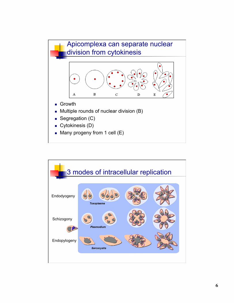
Apicomplexa can separate nuclear division from cytokinesis
Growth Multiple rounds of nuclear division (B) Segregation (C) Cytokinesis (D) Many progeny from 1 cell (E)
3 modes of intracellular replication
Endodyogeny
Schizogony
Endopylogeny

7
Apical complex Ultrastructural complexity
at the anterior end Electron dense structures Concentration of organelles
PR
C
SM
Apicomplexan Ultrastructure
Apical complex plays a role in invasion Rhoptries and Micronemes - modified secretory organelles
Apicoplast

8
Specialized Secretory Organelles
dense granule
rhoptry
microneme
Apicomplexan host cell invasion
9
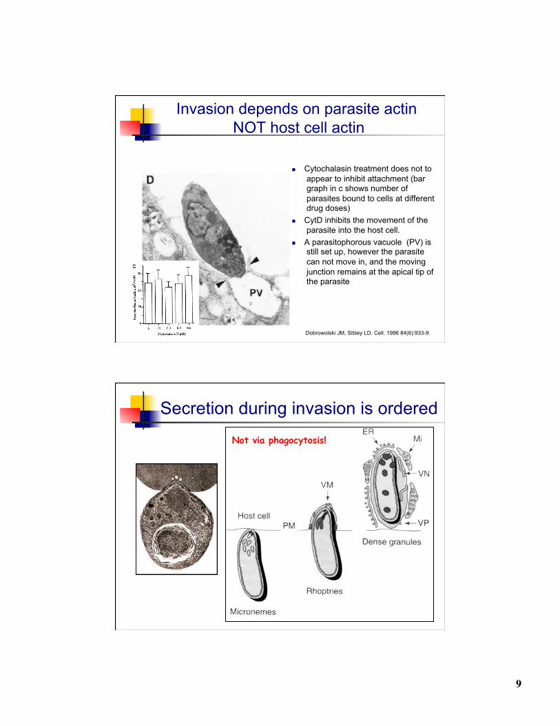
Invasion depends on parasite actin NOT host cell actin
Cytochalasin treatment does not to appear to inhibit attachment (bar graph in c shows number of parasites bound to cells at different drug doses)
CytD inhibits the movement of the parasite into the host cell.
A parasitophorous vacuole (PV) is still set up, however the parasite can not move in, and the moving junction remains at the apical tip of the parasite
Dobrowolski JM, Sibley LD. Cell. 1996 84(6):933-9.
Secretion during invasion is ordered
Not via phagocytosis!

10
Gliding Motility Substrate-dependent motion that
requires an actin-myosin motor Cytochalasins inhibit (actin destabilizer) Gliding is coupled to translocation of cell
surface adhesins (deposit on surface)
Differs from amoeboid movement Also actin based, cell deformation Apicomplexan gliding - no deformation
Assists in 3 vital functions Migration Invasion Egress
Movement includes: Circular, upright twirling, helical
Toxoplasma Motility
Antibody staining of a surface antigen
Circular Twirling
Hakansson et al 1999 Mol Biol Cell 10: 3539
11
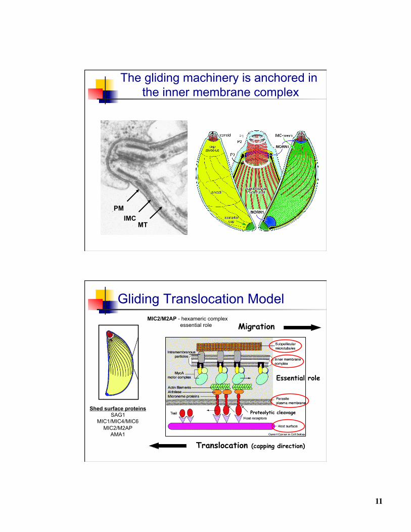
The gliding machinery is anchored in the inner membrane complex
MT IMC
PM
Gliding Translocation Model
Migration
Translocation (capping direction)
Proteolytic cleavage Shed surface proteins
SAG1 MIC1/MIC4/MIC6
MIC2/M2AP AMA1
MIC2/M2AP - hexameric complex essential role
Essential role

12
The Moving Conveyer Belt
The conveyor-belt model Motility depends of parasite actin/myosin (MyoA)
The MyoA is parasite specific - different from host myosin
Myosin is anchored into the outer IMC membrane Complex of proteins (GAP45/50, MyoA, MLC)
Short actin filaments form and are moved towards the posterior end of the parasite by the myosin power stroke
The short actin filaments are linked to microneme proteins by an adaptor Aldolase - moonlighting protein
Movement of actin filaments results in movement of microneme proteins
Microneme proteins are shed at the back end (rhomboid proteases are the best candidates for this activity)
The parasite glides over the substrate
13
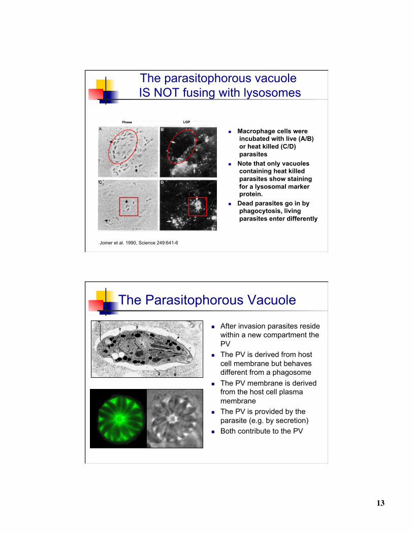
The parasitophorous vacuole IS NOT fusing with lysosomes
Macrophage cells were incubated with live (A/B) or heat killed (C/D) parasites
Note that only vacuoles containing heat killed parasites show staining for a lysosomal marker protein.
Dead parasites go in by phagocytosis, living parasites enter differently
Joiner et al. 1990, Science 249:641-6
The Parasitophorous Vacuole
After invasion parasites reside within a new compartment the PV
The PV is derived from host cell membrane but behaves different from a phagosome
The PV membrane is derived from the host cell plasma membrane
The PV is provided by the parasite (e.g. by secretion)
Both contribute to the PV

14
The PV is highly modified to suite the parasite’s needs
Tubular network increases surface (dense granule)
Sieving pores give access to small nutrient molecules in the host cell cytoplasm (probably dense granule)
Specific host cell organelles are recruited close to the PV membrane (rhoptry)
Dense granules are involved in establishing the intravacuolar network

15
Intestinal Coccidian Species Apicomplexa
Gregarinea Coccidia
Eimeriida Piroplasmida Haemosporida
- Eimeriidae (Eimeria)
- Cryptosporidiidae (Cryptosporidium)
- Sarcocystidae
-Theileriidae - Babesiidae
-Haemosporiidae (Plasmodium)
Similarities
Direct life cycles - no intermediate hosts homoxenous
Oral-fecal transmission Infective stage - oocyst Oocysts in contaminated feces
are not immediately infective Usually contaminated food or
water
Human infections Direct Human-Human
infection is unlikely Oocysts must “mature” Of significance as
opportunistic infections in immunocompromised people

16
Eimeria coccidiosis
Disease of chickens other animals as well! (2500 species)
Can cause high mortality Young birds
Serious disease causing bloody diarrhea, death
Parasite replication causes bleeding, and massive swelling in gut
Once infection is established there is no effective chemotherapy
In US alone, cost of disease is about $80 million/year including coccidiostats (in the feed).
Eimeria Life Cycle
Diagnostic stage
Infectious stage
17
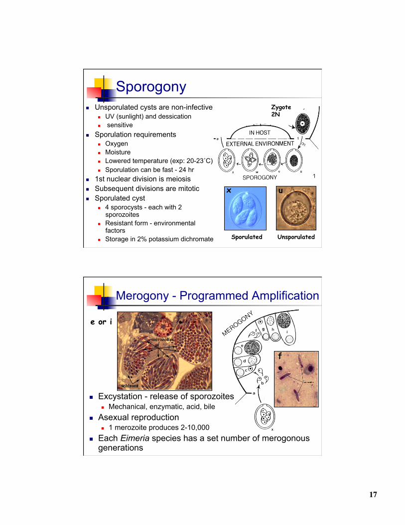
Sporogony
Unsporulated
Zygote 2N
u
Unsporulated cysts are non-infective UV (sunlight) and dessication sensitive
Sporulation requirements Oxygen Moisture Lowered temperature (exp: 20-23˚C) Sporulation can be fast - 24 hr
1st nuclear division is meiosis Subsequent divisions are mitotic Sporulated cyst
4 sporocysts - each with 2 sporozoites
Resistant form - environmental factors
Storage in 2% potassium dichromate Sporulated
x
Merogony - Programmed Amplification
f
e or i
Excystation - release of sporozoites Mechanical, enzymatic, acid, bile
Asexual reproduction 1 merozoite produces 2-10,000
Each Eimeria species has a set number of merogonous generations

18
Gamogony
Sexual reproduction Majority of gamonts produced are macrogamonts
Macrogamont (F) - unicellular Large number of granules - destined to be oocyst cell wall
Microgamont (M)- multiplication, release biflagellated mirogametes
Zygote 2N
Interesting factoids A single oocyst of Eimeria tenella
will produce 1 million more 1 gram of chicken litter (waste)
can contain between 100,000 and 200,000 oocysts
Birds (animals) that are in constant contact with small numbers of oocysts develop immunity to that specific oocyst species
An ounce of prevention….

19
Coccidiosis in Chickens
E. acervulina E. tenella E. brunetti E. necatrix E. mitis E. maxima
Low Low Moderate Moderate High High
10.3 µm 54 µm 30 µm 65.9 µm 11.3 µm 9.4 µm