the subperitoneal space and peritoneal cavity: basic concepts · the subperitoneal space and...
TRANSCRIPT

The subperitoneal space and peritoneal cavity:basic concepts
Harpreet K. Pannu,1 Michael Oliphant2
1Department of Radiology, Memorial Sloan Kettering Cancer Center, 1275 York Avenue, New York, NY 10065, USA2Department of Radiology, Wake Forest University School of Medicine, Winston-Salem, NC, USA
Abstract
The subperitoneal space and peritoneal cavity are twomutually exclusive spaces that are separated by theperitoneum. Each is a single continuous space with in-terconnected regions. Disease can spread either withinthe subperitoneal space or within the peritoneal cavity todistant sites in the abdomen and pelvis via these inter-connecting pathways. Disease can also cross the peri-toneum to spread from the subperitoneal space to theperitoneal cavity or vice versa.
Key words: Subperitoneal space—Peritonealcavity—Anatomy
The article is based on the comprehensive andauthoritative book by Drs Meyers, Charnsangavej, andOliphant titled Meyers’ Dynamic Radiology of theAbdomen [1]. Key concepts from the book are high-lighted in the following text and in Table 1. The text andaccompanying illustrative images are divided into 3 sec-tions on anatomy, an overview of the spread of disease,and disease spread for selected organs.
Anatomy
Lack of organs in the peritoneal cavity
The peritoneum is a serous membrane made up of vis-ceral and parietal layers. The visceral layer of the peri-toneum lines the surface of organs and the parietalperitoneum lines the coelomic cavity. The peritonealcavity is a potential space between the visceral andparietal layers of the peritoneum. There are no organs inthe peritoneal cavity. The potential space of the peri-toneal cavity is normally not visible on imaging as itcontains only a small amount of fluid (about 100 mL).
The peritoneum is analogous to the pleura which has avisceral layer covering lung and a parietal layer lining thethoracic cavity. Similar to the pleural cavity, the peri-toneal cavity is visualized on imaging if it is abnormallydistended by fluid, gas, or masses.
Location of the abdominal and pelvic organs
There are two spaces in the abdomen and pelvis, theperitoneal cavity (a potential space) and the subperi-toneal space, and these are separated by the peritoneum(Fig. 1). Regardless of the complexity of development inthe embryo, the subperitoneal space and the peritonealcavity remain separated from each other, and each re-mains a single continuous space (Figs. 2A, 3A). Distin-guishing the subperitoneal space from the potential spaceof the peritoneal cavity is important for understandingthe distinct patterns of disease spread in each.
The term subperitoneal refers to tissue that is deep tothe peritoneum and includes the extraperitoneal space,the ligaments and the mesenteries and their suspendedorgans (Fig. 2A). Organs whose surfaces are covered byperitoneum are therefore subperitoneal. Subperitonealorgans that are deep to the posterior peritoneum arecalled extraperitoneal. Since there are only 2 spaces in theabdomen and there are no organs in the peritoneal cav-ity, all the abdominal pelvic organs, and their associatedvessels, lymphatics, and nerves are in the subperitonealspace. In other words, all the structures seen in theabdomen and pelvis on cross-sectional imaging are in thesubperitoneal space. The organs lie in the abdominalcavity, not the peritoneal cavity (Figs. 1, 2A). The peri-toneal cavity is a potential space devoid of organs.
Importance of visualizing the subperitoneal spaceas a single space
The subperitoneal space is a large continuous space thatis formed by regions interconnected by ligaments andmesenteries. Ligaments and mesenteries refer to theCorrespondence to: Harpreet K. Pannu; email: [email protected]
ª The Author(s) 2015. This article is published with
open access at Springerlink.com
Published online: 26 May 2015AbdominalImaging
Abdom Imaging (2015) 40:2710–2722
DOI: 10.1007/s00261-015-0429-5
subperitoneal tissue between suspended organs and theextraperitoneal space. Visualizing the subperitonealspace as a single space explains the spread of diseasebetween different regions of the abdomen pelvis andbetween the organs covered by peritoneum and the ex-traperitoneum.
The relationship between the bowel mesenteryand the peritoneum
The mesenteries of the abdomen and pelvis are composedof subperitoneal tissue between 2 layers of visceral peri-toneum. Comparing an axial CT image with a cross-
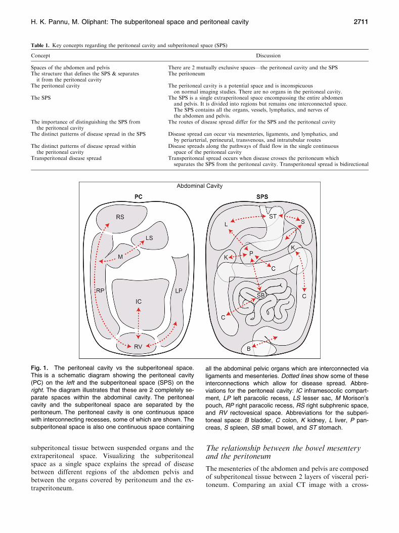
Fig. 1. The peritoneal cavity vs the subperitoneal space.This is a schematic diagram showing the peritoneal cavity(PC) on the left and the subperitoneal space (SPS) on theright. The diagram illustrates that these are 2 completely se-parate spaces within the abdominal cavity. The peritonealcavity and the subperitoneal space are separated by theperitoneum. The peritoneal cavity is one continuous spacewith interconnecting recesses, some of which are shown. Thesubperitoneal space is also one continuous space containing
all the abdominal pelvic organs which are interconnected vialigaments and mesenteries. Dotted lines show some of theseinterconnections which allow for disease spread. Abbre-viations for the peritoneal cavity: IC inframesocolic compart-ment, LP left paracolic recess, LS lesser sac, M Morison’spouch, RP right paracolic recess, RS right subphrenic space,and RV rectovesical space. Abbreviations for the subperi-toneal space: B bladder, C colon, K kidney, L liver, P pan-creas, S spleen, SB small bowel, and ST stomach.
Table 1. Key concepts regarding the peritoneal cavity and subperitoneal space (SPS)
Concept Discussion
Spaces of the abdomen and pelvis There are 2 mutually exclusive spaces—the peritoneal cavity and the SPSThe structure that defines the SPS & separates
it from the peritoneal cavityThe peritoneum
The peritoneal cavity The peritoneal cavity is a potential space and is inconspicuouson normal imaging studies. There are no organs in the peritoneal cavity.
The SPS The SPS is a single extraperitoneal space encompassing the entire abdomenand pelvis. It is divided into regions but remains one interconnected space.The SPS contains all the organs, vessels, lymphatics, and nerves ofthe abdomen and pelvis.
The importance of distinguishing the SPS fromthe peritoneal cavity
The routes of disease spread differ for the SPS and the peritoneal cavity
The distinct patterns of disease spread in the SPS Disease spread can occur via mesenteries, ligaments, and lymphatics, andby periarterial, perineural, transvenous, and intratubular routes
The distinct patterns of disease spread withinthe peritoneal cavity
Disease spreads along the pathways of fluid flow in the single continuousspace of the peritoneal cavity
Transperitoneal disease spread Transperitoneal spread occurs when disease crosses the peritoneum whichseparates the SPS from the peritoneal cavity. Transperitoneal spread is bidirectional
H. K. Pannu, M. Oliphant: The subperitoneal space and peritoneal cavity 2711

sectional diagram of an embryo, the dorsal mesenterycarries vessels from the aorta to the gut (Fig. 2). Thevisceral peritoneum surrounds the mesentery, forms theserosal layer of the gut, and is in continuity with theparietal peritoneum which covers the extraperitonealspace. As the ligaments, mesenteries, and suspended or-gans develop, the peritoneal cavity forms recesses thatremain interconnected as the peritoneal cavity and se-parate from the subperitoneal space (Fig. 3A).
Correlation between embryonic development andthe abdominal ligaments
The spleen, pancreas, liver, and gut form within themesentery that surrounds and suspends the primitive gutin the embryo. The development of these organs resultsin the creation of the abdominal ligaments that can beidentified on CT.
Posteriorly, the spleen and pancreas form in thedorsal mesogastrium, or the part of the dorsal mesenterysuspending the stomach (Fig. 3). The splenic artery runsfrom the aorta through the dorsal mesogastrium to thespleen with branches continuing to the stomach. The partof the dorsal mesogastrium between the stomach and thespleen becomes the gastrosplenic ligament containing theshort gastric vessels. The part of the dorsal mesogastriumcontaining the pancreas fuses with the subperitoneal
tissues anterior to the kidney leaving a connectingsplenorenal ligament that contains the distal splenic ar-tery and vein. The midgut and hindgut form within thedorsal mesentery creating the small intestine mesenteryand the mesocolon.
Anteriorly, the liver forms in the subperitoneal tissuesanterior to the stomach, or the ventral mesogastrium.This divides the subperitoneal tissues into the gastro-hepatic ligament between the stomach and liver, and thefalciform ligament between the liver and abdominal wall.The free edge of the gastrohepatic ligament is thehepatoduodenal ligament. The visceral peritoneum con-tinues over the stomach forming the serosal layer andover the liver and spleen forming their capsules.
Identifying the ligaments and bowel mesenterieson imaging
The ligaments and mesenteries are named according tothe viscera they connect and are identified by the vesselsthat run in them. For example, the hepatoduodenalligament is identified by the portal vein, hepatic artery,and common bile duct. Selected ligaments and their as-sociated vessels are described in Table 2. The ligamentsand mesenteries are a pathway for disease spread be-tween organs. The vasculature within the mesenteriesoften acts as a scaffold for disease spread. The utility of
Fig. 2. Relationship between the mesentery and the peri-toneum. A Diagram of a 4-week-old embryo shows the coe-lomic cavity which will form the peritoneal cavity (PC)surrounding the primitive gut (G). The medial visceral layer(arrows) of the peritoneum is apposed on the gut and the me-sentery, while the parietal layer is lateral. The dorsal mesentery(DM) conveys vessels from the aorta to the gut. Other than theperitoneal cavity, all the tissue in the abdominal cavity portion ofthe diagram is the subperitoneal space (light gray shaded
area). Spine is shown on the diagram only for orientation pur-poses. A aorta,K kidney,S spine, and VM ventral mesentery.BAxial CT image shows the dorsal mesentery of the small bowelas subperitoneal tissue (arrowhead) between 2 layers of vis-ceral peritoneum (black arrows). The parietal peritoneal reflec-tion (white arrows) is also seen anterior to the colon (C), kidney,and in the anterior and lateral abdomen. There is fluid (asterisk)in the peritoneal cavity between the visceral (black arrows) andparietal (white arrows) layers of the peritoneum.
2712 H. K. Pannu, M. Oliphant: The subperitoneal space and peritoneal cavity

identifying the mesenteries and ligaments is to more ac-curately and efficiently recognize sites of disease spread.
Anatomic continuity between the pararenalspaces
The posterior portion of the extraperitoneal space, theretroperitoneum, in the abdomen is divided by the renalfascia into the anterior pararenal, perinephric, and pos-terior pararenal spaces (Fig. 4). The anterior pararenalspace is between the parietal peritoneum and the anteriorrenal fascia; the perinephric space is between the anteriorand posterior renal fascia; the posterior pararenal spaceis between the posterior renal fascia and the transversalisfascia. The posterior renal fascia has 2 layers. An ante-
rior layer that is continuous with the anterior renal fasciaand a posterior layer that forms the lateroconal fasciawhich in turn goes anterolaterally to blend with theperitoneum.
Laterally at the level of the kidney, the lateroconalfascia separates the anterior pararenal space from theposterior pararenal space. Below the level of the kidneyand the iliac crest, the anterior and posterior renal fasciaetend to fuse resulting in anatomic continuity between theanterior and posterior pararenal spaces continuing infe-riorly as the infrarenal space.
The anterior pararenal space contains the pancreas,duodenum, and ascending and descending colon. Theperinephric space contains the kidney and adrenal gland.The posterior pararenal space has no organs and is
Table 2. Selected abdominal and pelvic ligaments and mesenteries
Ligament From To Identified By
Hepatoduodenal Hepatic hilum Duodenum Portal vein, hepatic arteryGastrohepatic
(lesser omentum)Lesser curvature of stomach Fissure of ligamentum venosum Left and right gastric artery, replaced
left hepatic arteryGastrosplenic Greater curvature of proximal
stomachSplenic hilum Left gastroepiploic and short gastric vessels
Gastrocolic Greater curvature of gastric body Transverse colon Right and left gastroepiploic vesselsSplenorenal Left anterior pararenal Splenic hilum Splenic vessels near hilumSmall bowel mesentery root Duodenal-jejunal junction Right iliac fossa Superior mesenteric vesselsGreater omentum Transverse colon Apron anterior to small bowel Epiploic vesselsTransverse mesocolon Transverse colon Pancreas Middle colic vessels, gastrocolic trunkAscending mesocolon Ascending colon Mesenteric root Marginal, ileocolic and right colic vesselsDescending mesocolon Descending colon Left superior duodenal fold Marginal and left colic vessels, inferior
mesenteric veinSigmoid mesocolon Sigmoid colon Root at origin of inferior
mesenteric arteryMarginal and sigmoid vessels
Fig. 3. Abdominal ligaments. A Diagram showing develop-ment of an embryo. The liver develops in the ventral me-sentery anterior to the stomach. The residual part of theventral mesentery between the liver and stomach is called thegastrohepatic ligament in the adult. The spleen and pancreasform in the dorsal mesentery posterior to the stomach. Theresidual part of the dorsal mesentery between the spleen andstomach is called the gastrosplenic ligament in the adult. Thepancreas fuses with the tissues anterior to the kidney to lie inthe anterior pararenal space. The residual part of the dorsal
mesentery between the spleen and pancreas is called thesplenorenal ligament. Dotted lines approximate the paths ofthe ventral and dorsal mesenteries. A aorta, K kidney, L liver,PC peritoneal cavity, S spine, and ST stomach. Subperitonealspace = light gray shaded area in abdominal cavity portion ofdiagram. Spine is shown on the diagram only for orientationpurposes. B Axial CT image of the upper abdomen shows thegastrohepatic (arrow) and gastrosplenic (arrowhead) liga-ments containing the left gastric artery and short gastricvessels, respectively.
H. K. Pannu, M. Oliphant: The subperitoneal space and peritoneal cavity 2713

continuous laterally with the extraperitoneal fat of theproperitoneal flank stripe.
Compartmentalization of the pelvicsubperitoneal tissues
Anteriorly, the umbilicovesical fascia encloses the blad-der and urachus in the perivesical space (Fig. 5). Theumbilicovesical fascia also defines the prevesical spaceanteriorly and laterally. Posteriorly, the rectum is in theperirectal space which is defined laterally by theperirectal fascia, posteriorly by the posterior pelvic fas-cia, and anteriorly by the rectovesical fascia in the maleand the rectovaginal fascia in the female. The aggregateof these fasciae is also referred to as the mesorectal fas-cia. Superiorly, the perirectal space communicates withthe sigmoid mesocolon. The presacral space is posteriorto the perirectal space.
The peritoneum reflects over the dome of the bladder,the uterine body, and the rectouterine recess (Fig. 5).Laterally, in the female, the peritoneum forms the foldsof the broad ligament. In the female, the broad ligamentsuspends the female pelvic organs and is in continuitywith the extraperitoneal space. Regions within the broadligament form the mesometrium, mesosalpinx, and themesovarium. These mesenteries are all in continuitywithin the broad ligament. The serosa of the fallopiantube is continuous as part of the broad ligament and itsfimbriated end is open to the peritoneal cavity. The ovarylies within the broad ligament immediately beneath theperitoneum. The arteries, veins, lymphatics, and nervesto the female pelvic organs all course within the broadligament and connect with the extraperitoneal space.
This forms the pathways for the subperitoneal spread ofdisease.
Overview of disease spread
The pathways of disease spread in the abdominalcavity
Since all organs are subperitoneal, the subperitonealspace is a natural pathway for disease spread (Fig. 6).Potential routes are along the mesenteries and ligaments,via visceral lymphatics to nodes, and by periarterial,perineural, or transvenous routes as well as along ducts.Continuity of the subperitoneal space explains the spreadof disease from one organ to another. Disease can spreadbidirectionally within the subperitoneal space using thepathways created by normal structures. If a tumor ispresent, tracing the organ’s blood supply helps identifynodes that are potential pathways for spread. If an ab-normal node is discovered, knowing its location helps inthe search for the primary site.
For organs which are covered by peritoneum, there isan additional pathway for spread (Fig. 6). Transperi-toneal spread occurs when disease traverses the visceralperitoneum along the surface of the organ. This route ispossible because the peritoneum consists of a single layerof mesothelial cells on a bed of loose connective tissue [2].Neoplastic and inflammatory cells, gas, and hemorrhagein the subperitoneal space can therefore cross the peri-toneum to enter the peritoneal cavity. Subsequently,peritoneal spread occurs via the circulating peritonealfluid to the peritoneal recesses in the abdomen and pelvis.
In summary, the pathways of disease spread aresubperitoneal and peritoneal. In addition, transperi-
Fig. 4. Renal fasciae. A Axial CT image shows the anteriorand posterior renal fascia (black arrows). Arrowheads showthe posterior pararenal space extending laterally as theproperitoneal fat. B Axial CT image shows fluid (arrows) in
between the 2 layers of the posterior renal fascia. Fluid fromthe anterior pararenal space can extend into this potentialspace between the 2 layers. Arrowhead points to the lowerpole of the kidney.
2714 H. K. Pannu, M. Oliphant: The subperitoneal space and peritoneal cavity

toneal spread occurs when subperitoneal disease crossesthe peritoneal lining and spreads within the peritonealcavity.
Omental tumor: subperitoneal disease orperitoneal carcinomatosis
The surface of the greater omentum is lined by visceralperitoneum and the internal composition is of subperi-toneal fat, lymphatics, and vessels. Communicatingchannels allow fluid in the peritoneal cavity to be ab-sorbed by the omental lymphatics. Thus, tumor cells thatare in the peritoneal cavity being disseminated by theperitoneal route can traverse the visceral peritoneum togrow in the richly vascular subperitoneal tissues of theomentum. In addition to the omentum, common sites ofperitoneal tumor implants are the diaphragm and thedependent peritoneal recesses.
Peritoneal fluid flow in the abdomen and pelvis
The transverse mesocolon divides the peritoneal cavityinto the supramesocolic and inframesocolic compart-ments. The latter is divided into right and left infracolicrecesses by the root of the small bowel mesentery. Peri-toneal fluid is drawn into the upper abdomen by lowsubdiaphragmatic pressures and is pulled into the pelvisunder the influence of gravity. Fluid travels from thepelvis to the abdomen via the paracolic gutters andtravels from the abdominal infracolic compartment tothe pelvis (Fig. 7). It pools in dependent recesses, themost prominent of which are the pouch of Douglas in the
Fig. 5. Pelvic spaces. A axial CT image of the pelvis. Theurachus (arrowhead) and obliterated umbilical arteries (ar-rows) form the median umbilical ligament and the medialumbilical folds, respectively, and are encased within the um-bilicovesical fascia forming the perivesical space. The pre-vesical space is anterior and lateral to the bladder. Theperirectal space (asterisk) surrounds the rectum. B SagittalCT image shows the peritoneum (arrows) along the anteriorabdominal wall and reflecting over the bladder and rectum.Peritoneal fluid in the inframesocolic compartment (IM) com-municates with the rectovesical recess (asterisk). The bladderis inferiorly displaced by the peritoneal fluid. B bladder, Rrectum.
Fig. 6. Diagram shows the 2 routes of disease spread forviscera which are covered by peritoneum–transperitoneal andsubperitoneal. Disease can cross the visceral peritoneum(solid arrow) to enter and subsequently spread in the peri-toneal cavity. Alternatively, disease can follow the vesselsand lymphatics (open arrow) of the viscera to spread in thesubperitoneal space (SPS).
H. K. Pannu, M. Oliphant: The subperitoneal space and peritoneal cavity 2715

female and the rectovesical recess in the male, along thesuperior portion of the sigmoid mesocolon, ileocolic re-gion, right paracolic gutter, and Morison’s pouch.
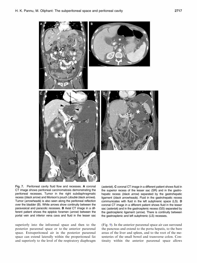
All the peritoneal recesses communicate, however,peritoneal fluid preferentially flows in certain directions andis anatomically limited in some locations. Peritoneal fluidfrom the pelvis primarily goes up the right paracolic gutter(recess) forming continuity of the inframesocolic andsupramesocolic recesses on the right. On the left, thephrenicocolic ligament limits the left paracolic gutter(recess) to the inframesocolic recess. From the right para-colic gutter, fluid enters the right subhepatic space (Mor-ison’s pouch) and may subsequently enter the lesser sac viathe epiploic foramen (ofWinslow) between the main portalvein and the inferior vena cava (Fig. 7). Fluid also goessuperiorly into the right subphrenic space but the falciformligament limits flow from the right to the left subphrenicspace.Abscesses secondary to intraperitoneal infections aretherefore common in the pouch of Douglas, right paracolicgutter, right subhepatic space, and right subphrenic space.Fluid flow patterns are mostly bidirectional.
Although the falciform and phrenicocolic ligamentstypically limit fluid flow across them, large volumes offluid can overflow under the free edge of the falciformligament and over the phrenicocolic ligament. Left sub-phrenic fluid is more commonly seen due to gastric,splenic, or splenic flexure colonic pathology. There iscontinuity between the left subphrenic space, the gas-trohepatic space, and the perisplenic spaces such as thegastrosplenic recess and splenorenal recess. These areseparated from the lesser sac by the gastrohepatic, gas-trosplenic, and splenorenal ligaments, respectively.
Distinguishing intraperitoneal andextraperitoneal fluid in the pelvis
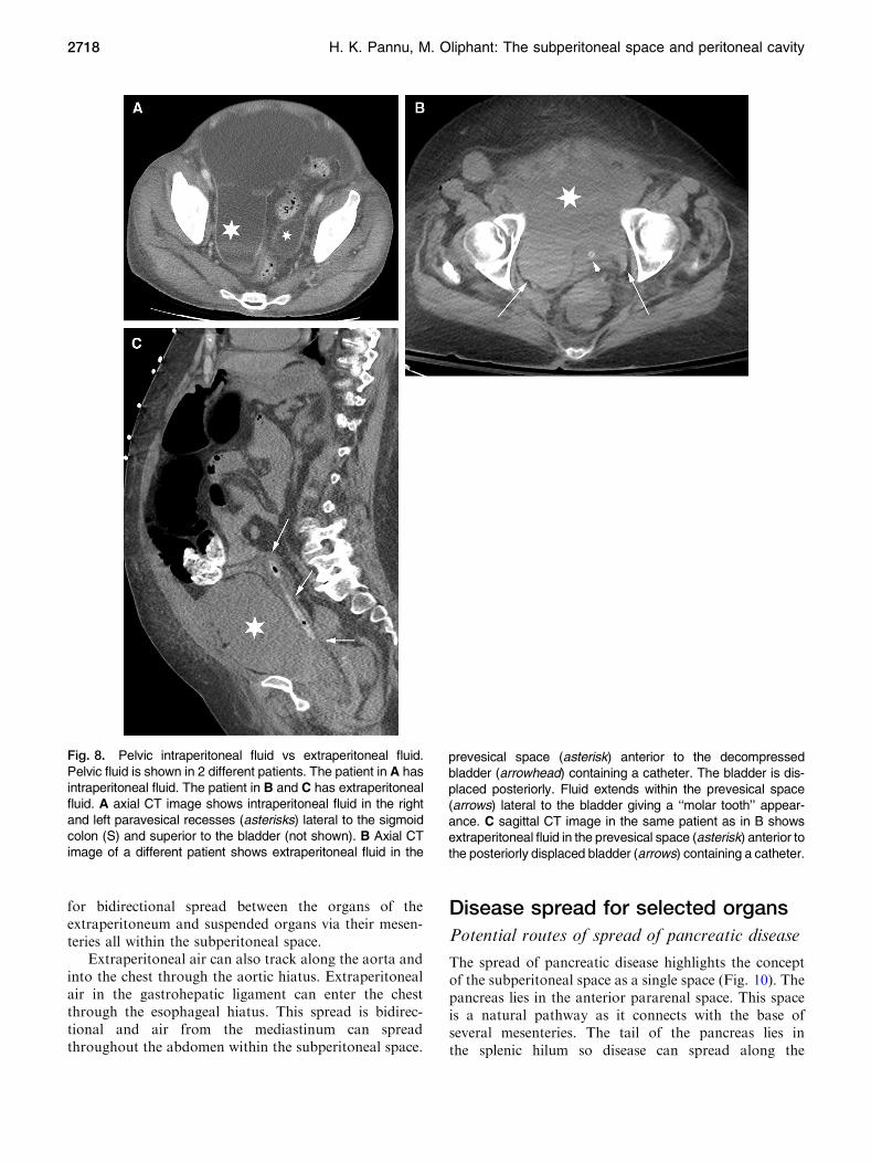
Intraperitoneal pelvic fluid (ascites) occurs in the pouchof Douglas and in the lateral recesses which lie on eitherside of the sigmoid colon and are referred to as par-avesical recesses of the peritoneal cavity (Fig. 8). Ex-traperitoneal pelvic fluid in the subperitoneal spaceoccurs in the prevesical space which is anterior and lat-eral to the bladder. Since the bladder is positioned moreinferiorly in the pelvis than the sigmoid colon, ascites isseen superior to the bladder. Ascites displaces the blad-der inferiorly, while prevesical space fluid displaces thebladder posteromedial. Extraperitoneal fluid in the pre-vesical space extends bilaterally forming a symmetric orasymmetric ‘‘molar tooth’’ appearance of fluid bothanterior and lateral to the bladder (Fig. 8).
Ascites preserves the properitoneal fat which is thelateral extension of the posterior pararenal space andoutlines the medial umbilical folds anteromedial. Ex-traperitoneal fluid in the prevesical space can extend tothe lower abdomen and obliterate the properitoneal fat.The anterior extension spares the midline triangle of the
perivesical fat that encases the urachus (median umbilicalligament) and the obliterated umbilical arteries (medialumbilical ligament). If prevesical fluid penetrates theoverlying transversalis fascia, it can involve the rectusmuscle. Conversely, rectus hematomas can extend intothe prevesical fat.
Ascites travels superiorly from the lateral paravesicalperitoneal recesses to the paracolic gutters. Prevesicalfluid travels superiorly into the infrarenal space andsubsequently into the pararenal spaces. Posteriorly, ex-traperitoneal fluid extends into the presacral space. As-cites can go into the inguinal canal along a hernia, whileprevesical fluid can go to the inguinal ring along the vasdeferens. Unlike ascites, prevesical fluid abuts the lateralpelvic musculature and can extend along the externaliliac vessels and femoral sheath.
Distinguishing intraperitoneal andextraperitoneal free air in the abdomen
The shape of the subdiaphragmatic air and change withrespiration and position help distinguish intraperitonealand extraperitoneal free air. On an upright chest radio-graph, free intraperitoneal air follows the contour of thedome of the hemidiaphragm, while extraperitoneal air isusually medial or lateral to the apex of the hemidi-aphragm. The volume of free intraperitoneal air seenunder the hemidiaphragm increases on inspiration due todecreased subdiaphragmatic pressure. The volume of freeextraperitoneal air seen under the hemidiaphragm in-creases on expiration due to decreased compression by thediaphragm. Free intraperitoneal air fills the potential re-cesses of the peritoneal cavity and can outline ligamentssuch as the falciform ligament. Free extraperitoneal aircan occupy extraperitoneal spaces and can outline thepsoas muscle and follow the flank stripe if within theposterior pararenal space. Peritoneal air shifts readily withposition change, while extraperitoneal air does not.
Potential routes of spread of extraperitoneal freeair
Extraperitoneal free air originating anywhere in the ab-domen can spread throughout the abdomen and pelvisvia the interconnecting subperitoneal space.
Air from a duodenal perforation can go from theanterior pararenal space to the infrarenal space and thento the posterior pararenal space or to the prevesical spaceof the pelvis.
Extraperitoneal pelvic free air from a rectal perfora-tion can go from the mesorectum to the sigmoid meso-colon to the prevesical space. From the prevesical spaceair can go inferiorly along the vas deferens to the scro-tum, laterally outside the pelvic cavity via the sciaticforamen and into the thighs along the scaffold of thefemoral sheath. From the prevesical space air can also go
2716 H. K. Pannu, M. Oliphant: The subperitoneal space and peritoneal cavity
superiorly into the infrarenal space and then to theposterior pararenal space or to the anterior pararenalspace. Extraperitoneal air in the posterior pararenalspace can extend laterally within the properitoneal fatand superiorly to the level of the respiratory diaphragm
(Fig. 9). In the anterior pararenal space air can surroundthe pancreas and extend to the porta hepatis, to the bareareas of the liver and spleen, and to the root of the me-senteries of the small bowel and transverse colon. Con-tinuity within the anterior pararenal space allows
Fig. 7. Peritoneal cavity fluid flow and recesses. A coronalCT image shows peritoneal carcinomatosis demonstrating theperitoneal recesses. Tumor in the right subdiaphragmaticrecess (black arrow) and Morison’s pouch (double black arrows).Tumor (arrowheads) is also seen along the peritoneal reflectionover the bladder (B). White arrows show continuity between theparavesical and paracolic recesses. B Axial CT image in a dif-ferent patient shows the epiploic foramen (arrow) between theportal vein and inferior vena cava and fluid in the lesser sac
(asterisk). C coronal CT image in a different patient shows fluid inthe superior recess of the lesser sac (SR) and in the gastro-hepatic recess (black arrow) separated by the gastrohepaticligament (black arrowheads). Fluid in the gastrohepatic recesscommunicates with fluid in the left subphrenic space (LS). Dcoronal CT image in a different patient shows fluid in the lessersac (asterisk) and in the gastrosplenic recess (GS) separated bythe gastrosplenic ligament (arrow). There is continuity betweenthe gastrosplenic and left subphrenic (LS) recesses.
H. K. Pannu, M. Oliphant: The subperitoneal space and peritoneal cavity 2717
for bidirectional spread between the organs of theextraperitoneum and suspended organs via their mesen-teries all within the subperitoneal space.
Extraperitoneal air can also track along the aorta andinto the chest through the aortic hiatus. Extraperitonealair in the gastrohepatic ligament can enter the chestthrough the esophageal hiatus. This spread is bidirec-tional and air from the mediastinum can spreadthroughout the abdomen within the subperitoneal space.
Disease spread for selected organs
Potential routes of spread of pancreatic disease
The spread of pancreatic disease highlights the conceptof the subperitoneal space as a single space (Fig. 10). Thepancreas lies in the anterior pararenal space. This spaceis a natural pathway as it connects with the base ofseveral mesenteries. The tail of the pancreas lies inthe splenic hilum so disease can spread along the
Fig. 8. Pelvic intraperitoneal fluid vs extraperitoneal fluid.Pelvic fluid is shown in 2 different patients. The patient in A hasintraperitoneal fluid. The patient in B and C has extraperitonealfluid. A axial CT image shows intraperitoneal fluid in the rightand left paravesical recesses (asterisks) lateral to the sigmoidcolon (S) and superior to the bladder (not shown). B Axial CTimage of a different patient shows extraperitoneal fluid in the
prevesical space (asterisk) anterior to the decompressedbladder (arrowhead) containing a catheter. The bladder is dis-placed posteriorly. Fluid extends within the prevesical space(arrows) lateral to the bladder giving a ‘‘molar tooth’’ appear-ance. C sagittal CT image in the same patient as in B showsextraperitoneal fluid in the prevesical space (asterisk) anterior tothe posteriorly displaced bladder (arrows) containing a catheter.
2718 H. K. Pannu, M. Oliphant: The subperitoneal space and peritoneal cavity

gastrosplenic ligament to the gastrocolic ligament alongthe greater curvature of the stomach. The head of thepancreas is connected to the hepatic hilum by thehepatoduodenal ligament so disease can spread along thegastrohepatic ligament to the lesser curvature of thestomach. The root of the transverse mesocolon lies alongthe pancreas so disease can spread to the transverse colon.Similarly, the root of the small bowel mesentery startsadjacent to the anterior inferior aspect of the pancreaticbody so disease can spread to the ileocolic region.
The peritoneum forming the posterior margin of thelesser sac is anterior to the pancreas. Disease can crossthe peritoneum (transperitoneal spread) to enter thelesser sac for subsequent peritoneal spread.
Anatomic basis for a hepatic laceration resultingin a retroperitoneal hematoma
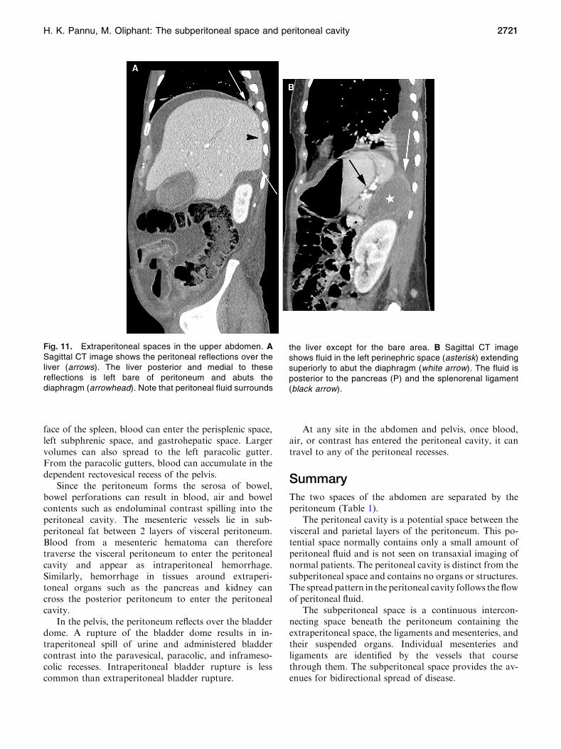
The bare area of the liver is that part of the posterior rightlobe of the liver that is not covered by peritoneum
(Fig. 11). The peritoneum covering the liver reflects onitself to form the right coronary ligament and attach theright lobe to the right hemidiaphragm. The liver posteriorand medial to this reflection is left bare of peritoneum andabuts the diaphragm. In addition, the parietal peritoneumof the inferior right coronary ligament also fuses with theanterior renal fascia. Posteriorly, the diaphragmatic fasciafuses with the right posterior renal fascia. As a result, thespace between the anterior and posterior renal fascia, orthe right perinephric space, communicates with the barearea of the liver. Hemorrhage from the hepatic bare areacan therefore present as a retroperitoneal bleed in the rightperinephric space. Similarly, a right perinephric abscesscan directly spread to the liver and diaphragm.
On the left, the perinephric space communicateswith theleft subphrenic extraperitoneal space (Fig. 11) [2]. This is theresult of the left anterior renal fascia fusing with the gas-trosplenic ligament and the posterior renal fascia fusingwiththe diaphragm [2]. In addition, the peritoneum covers mostof the spleen except for the splenic hilum. The splenorenalligament connects the bare area of the spleen at the hilumwith the left anterior pararenal space. Bleeding from a rup-tured splenic artery aneurysm can therefore result in aretroperitoneal bleed in the anterior pararenal space.
Anatomic basis for a hepatobiliary tumormimicking a pancreatic mass
Tumor from the liver and gallbladder can spread to thenodes within the hepatoduodenal ligament. The anteriorperiportal nodes follow the lymphatics along the hepaticartery to the celiac artery and then to the cisterna chyli. Theposterior periportal nodes follow the lymphatics to theretropancreatic nodes, aortocaval node, and then to thecisterna chyli. Therefore, an enlarged retropancreatic nodefrom a metastatic hepatobiliary tumor can mimic a pan-creatic mass. Other routes of nodal spread are the gastro-hepatic ligament or adjacent to the suprahepatic inferiorvena cava to the juxtaphrenic and paraesophageal nodes.
Diaphragmatic adenopathy secondary toperitoneal carcinomatosis
Similar to the omentum, the diaphragm has abundantlymphatics for the absorption of peritoneal fluid. Thisprocess can result in tumor cells being transported fromthe peritoneal cavity to the diaphragmatic nodes. Me-tastatic diaphragmatic adenopathy can therefore occursecondary to peritoneal carcinomatosis from abdominaland pelvic malignancies.
Potential routes of peritoneal spread of bloodfollowing trauma
Traumatic injuries of viscera can disrupt the capsule ofan organ. The visceral peritoneum forms the capsule of
Fig. 9. Extraperitoneal free air. Sagittal CT image showspneumatosis of the ascending colon and extraluminal air in theadjacent mesentery (asterisk). This extraperitoneal free airtracks in the anterior and posterior pararenal spaces (arrows)to the bare area of the liver and diaphragm (arrowheads).
H. K. Pannu, M. Oliphant: The subperitoneal space and peritoneal cavity 2719

the liver and spleen, except for the bare areas describedearlier. Therefore, a laceration allows for transperitonealspread of blood from these organs into the peritonealcavity. Following a laceration of the peritonealized sur-
face of the liver, blood can spread into the right sub-phrenic space and right subhepatic space and cansubsequently enter the lesser sac and right paracolicgutter. Following a laceration of the peritonealized sur-
Fig. 10. Subperitoneal spread of disease. A Axial CT im-age shows a large pancreatic mass (white arrow) extendingalong the hepatoduodenal ligament to the porta hepatis(arrowheads). Mass also invades the left perinephric spaceand engulfs the adrenal gland (black arrow). Superiorly, themass extended into the gastrohepatic ligament with invasionof the left lobe of the liver (not shown). B Coronal CT imageshows a hematoma in the root of the small bowel mesentery
extending toward the ileocecal junction within the smallintestine mesentery (arrows). C Coronal CT image shows aheterogeneous mass (white arrows) around the inferiorvena cava and aorta. Mass extends superiorly, through theaortic hiatus, to the posterior mediastinum (black arrow-heads). The mass also extended into the root of the smallbowel mesentery (not shown) along the superior mesentericartery.
2720 H. K. Pannu, M. Oliphant: The subperitoneal space and peritoneal cavity
face of the spleen, blood can enter the perisplenic space,left subphrenic space, and gastrohepatic space. Largervolumes can also spread to the left paracolic gutter.From the paracolic gutters, blood can accumulate in thedependent rectovesical recess of the pelvis.
Since the peritoneum forms the serosa of bowel,bowel perforations can result in blood, air and bowelcontents such as endoluminal contrast spilling into theperitoneal cavity. The mesenteric vessels lie in sub-peritoneal fat between 2 layers of visceral peritoneum.Blood from a mesenteric hematoma can thereforetraverse the visceral peritoneum to enter the peritonealcavity and appear as intraperitoneal hemorrhage.Similarly, hemorrhage in tissues around extraperi-toneal organs such as the pancreas and kidney cancross the posterior peritoneum to enter the peritonealcavity.
In the pelvis, the peritoneum reflects over the bladderdome. A rupture of the bladder dome results in in-traperitoneal spill of urine and administered bladdercontrast into the paravesical, paracolic, and inframeso-colic recesses. Intraperitoneal bladder rupture is lesscommon than extraperitoneal bladder rupture.
At any site in the abdomen and pelvis, once blood,air, or contrast has entered the peritoneal cavity, it cantravel to any of the peritoneal recesses.
Summary
The two spaces of the abdomen are separated by theperitoneum (Table 1).
The peritoneal cavity is a potential space between thevisceral and parietal layers of the peritoneum. This po-tential space normally contains only a small amount ofperitoneal fluid and is not seen on transaxial imaging ofnormal patients. The peritoneal cavity is distinct from thesubperitoneal space and contains no organs or structures.The spread pattern in the peritoneal cavity follows the flowof peritoneal fluid.
The subperitoneal space is a continuous intercon-necting space beneath the peritoneum containing theextraperitoneal space, the ligaments and mesenteries, andtheir suspended organs. Individual mesenteries andligaments are identified by the vessels that coursethrough them. The subperitoneal space provides the av-enues for bidirectional spread of disease.
Fig. 11. Extraperitoneal spaces in the upper abdomen. ASagittal CT image shows the peritoneal reflections over theliver (arrows). The liver posterior and medial to thesereflections is left bare of peritoneum and abuts thediaphragm (arrowhead). Note that peritoneal fluid surrounds
the liver except for the bare area. B Sagittal CT imageshows fluid in the left perinephric space (asterisk) extendingsuperiorly to abut the diaphragm (white arrow). The fluid isposterior to the pancreas (P) and the splenorenal ligament(black arrow).
H. K. Pannu, M. Oliphant: The subperitoneal space and peritoneal cavity 2721

Transperitoneal spread occurs when disease spreadsfrom the subperitoneal space to the peritoneal cavity bycrossing the peritoneal lining.
Acknowledgment. The authors would like to thankMs Terry Helms fromthe Medical Graphics department at Memorial Sloan Kettering CancerCenter for her invaluable help and expertise in creating the illustrations forthis paper.
Open Access. This article is distributed under the terms of the CreativeCommons Attribution 4.0 International License (http://creative-commons.org/licenses/by/4.0/), which permits unrestricted use, dis-tribution, and reproduction in any medium, provided you give
appropriate credit to the original author(s) and the source, provide alink to the Creative Commons license, and indicate if changes weremade.
References
1. Meyers MA, Charnsangavej C, Oliphant M (2011) Meyers’ dynamicradiology of the abdomen: normal and pathologic anatomy, 6th edn.Berlin: Springer Science + Business Media, LLC
2. Standring S, Ellis H, Berkovitz BKB (2005) Gray’s anatomy: theanatomical basis of clinical practice, 39th edn. Amsterdam: ElsevierChurchill Livingstone
2722 H. K. Pannu, M. Oliphant: The subperitoneal space and peritoneal cavity