the spectrum of dermatological disorders found in …
TRANSCRIPT

1
THE SPECTRUM OF DERMATOLOGICAL DISORDERS FOUND IN PATIENTS
WITH SARCOIDOSIS PRESENTING TO THE DERMATOLOGY OUTPATIENT
CLINIC AT THE CHRIS HANI BARAGWANATH ACADEMIC HOSPITAL
BABALWA PHINDISWA ZINZISWA MBUQE-LIMBA
Student no. 589938
A research report submitted to the Faculty of Health Sciences, University of the
Witwatersrand, Johannesburg, in partial fulfillment of the requirements for the degree of
Master of Medicine in the branch of Internal Medicine (Dermatology)
Johannesburg 2017

ii
DECLARATION
I, Babalwa Phindiswa Zinziswa Mbuqe-Limba hereby declare that this research report is my
own work. It is being submitted for the degree of Master of Medicine in the branch of Internal
Medicine (Dermatology) in the University of the Witwatersrand, Johannesburg. It has not
been submitted before for any degree or examination at this or any other University.
……………………………….........................................
DR BPZ Mbuqe-Limba, MBChB (WSU), FCDerm (SA).
……. day of……………..........2017

iii
DEDICATION
This research is dedicated to my four children; Xola, Liyema, Alwande and Cwambu, and
their father Lunga, for their encouragement, support and believing in me.

iv
ABSTRACT
Backgrounds
There is a paucity of studies on cutaneous sarcoidosis in the South African setting, with the
last study published 18 years ago. More studies are needed to explore these gaps to inform
referral policy and guidelines for early diagnosis and management of cutaneous sarcoidosis in
the country.
Objectives
The study focuses on the patterns of cutaneous sarcoidosis, demographic features, and
histological associations with the clinical patterns of skin sarcoidosisfound in patients with
sarcoidosis presenting to the dermatology outpatient clinic at the Chris Hani Baragwanath
Academic hospital. In addition, the pattern, chronicity and severity of the cutaneous
manifestations were described. Furthermore, the co-existence of HIV/AIDS and sarcoidosis
was also examined.
Methods
A retrospective descriptive studythat spans from 1991 to 2015, was carried out which
included cases that had a definitive diagnosis of sarcoidosis. One hundred case records of
patients with cutaneous sarcoidosis that attended the Dermatology Outpatient Clinic, from the
study site in Soweto, in the south of Johannesburg, were collected and transferred to a
collection data sheet.
Results
In this predominantly Black African population, women above 45 years old (70%), were most
commonly affected. Papules (68.8%) and plaques (27.1%) were the most frequent skin
findings in Black Africans. The most frequent extra-cutaneous organ affected was the lung
(53%). Subcutaneous lesions were found to be significantly associated (p-value <0. 012) with
Scadding stage 0 and stage 4 lung involvement. On histology 70% of the cases had clean
granulomas, frequently associated with papules clinically. HIV seropositive and sarcoidosis
cases demonstrated an inversely proportional association with the CD4 count, with disease
progression noted with CD4 increments after initiation of therapy.
Conclusion
The variations in the patterns of presentation revealed in this study can improve our knowledge of
cutaneous patterns of sarcoidosis in this study population and assist with the development of
prompt diagnosis and early treatment intervention.

v
ACKNOWLEDGEMENTS
The assistance and guidance of my supervisors Professor Michelle Wong and Professor Colin
Menezes is greatly appreciated. Special thanks to Mr. Ajayi Anthony for assisting with the
statistical analysis and providing intellectual contribution towards the successful completion
of this research report.
The Division of Pulmonology at Chris Hani Baragwanath Academic Hospital and the nursing
staff were generous with their time and support during the period of the data collection. The
National Health Laboratory Services gave access for the collection of data from patients’
histology records.
I am indebted to my colleagues and friends; Dr. Sli Sibisi Shange and Dr. Mpumi Sotobe
Mose for believing in me and with their zealous encouragement. Finally, I am grateful to Dr.
Vincent Adeniyi, for his mentorship and guidance with the research report.

vi
Table of Contents DECLARATION .............................................................................................................................. ii
DEDICATION ................................................................................................................................. iii
ABSTRACT ...................................................................................................................................... iv
ACKNOWLEDGEMENTS ............................................................................................................. v
LIST OF FIGURES ......................................................................................................................... ix
LIST OF TABLES ........................................................................................................................... ix
DEFINITION OF KEY CONCEPTS ............................................................................................ xi
LIST OF ABBREVIATIONS ........................................................................................................ xii
CHAPTER ONE ....................................................................................................................... 1
1.1INTRODUCTION ....................................................................................................................... 1
1.1.1Historical background ............................................................................................................ 1
1.1.2 Sarcoidosis ............................................................................................................................. 2
1.1.3 Cutaneous sarcoidosis ........................................................................................................... 2
1.1.4 Problem Statement ................................................................................................................. 3
1.1.5 Significance of the study ....................................................................................................... 4
1.1.6 Aim of the study .................................................................................................................... 4
1.1.7 Objectives .............................................................................................................................. 5
1.1.8 Research Questions ............................................................................................................... 5
1.1.9. Chapter layout ...................................................................................................................... 5
1.1.10. Conclusion .......................................................................................................................... 6
1.2 LITERATURE REVIEW .................................................................................................. 7
1.2.1. Introduction .......................................................................................................................... 7
1.2.2. Literature Search Strategy .................................................................................................... 7
1.2.3. Clinical manifestations and trends in the epidemiology ....................................................... 7
1.2.4. Aetiopathogenesis ............................................................................................................... 10

vii
1.2.5. Immunology ....................................................................................................................... 11
1.2.6. Diagnosis ............................................................................................................................ 11
1.2.7 The impact of HIV/AIDS pandemic on the clinical manifestation of cutaneous sarcoidosis
...................................................................................................................................................... 13
1.2.8 Management ........................................................................................................................ 13
1.2.9. Conclusion ............................................................................................................................. 15
CHAPTER TWO .................................................................................................................... 16
METHODOLOGY ................................................................................................................. 16
2.1 Introduction ................................................................................................................................. 16
2.2 17 Design .................................................................................................................................... 16
2.3 Study setting ............................................................................................................................... 16
2.4 Study population ......................................................................................................................... 16
2.5 Selection criteriaand sample size ................................................................................................ 17
2.6 Data collection (see Appendix A for the data collection sheet) .................................................. 17
2.6.1 Measurements ...................................................................................................................... 18
2.8 Data entry .................................................................................................................................... 19
2.9 Data analysis ............................................................................................................................... 19
2.10 Ethical approval and other considerations ................................................................................ 20
2.11 Conclusion ................................................................................................................................ 20
CHAPTER THREE ................................................................................................................ 21
RESULTS ................................................................................................................................ 21
3.1Introduction ................................................................................................................................ 21
3.2 Demographic characteristics .................................................................................................... 21
3.3 Distribution of patients with sarcoidosis by occupation ........................................................ 21
3.4 Demographic characteristics of patients and clinical cutaneous manifestations ................ 22
3.5 Extracutaneous organ involvement ......................................................................................... 23
3.5.1 Number of organ involvement ............................................................................................. 24

viii
3.5.2 Patients with four and five organs involved ........................................................................ 25
3.5.3 Association of demographical characteristics and number of organ involvement .............. 26
3.5.6 Extracutaneous organ involvement by specific type lesion ................................................. 27
3.6 Clinical patterns of cutaneous sarcoidosis and the chest X-ray findings ............................. 29
3.7 Chronicity of cutaneous sarcoidosis ........................................................................................ 30
3.8 Treatment .................................................................................................................................. 33
3.9 Histology findings ..................................................................................................................... 34
3.10 Sarcoidosis in people living with HIV ................................................................................... 36
3.11 Summary .................................................................................................................................. 37
CHAPTER FOUR .................................................................................................................. 38
SUMMARY OF FINDINGS, DISCUSSION, CONCLUSION .......................................... 38
4.1 Introduction ............................................................................................................................... 38
4.2 Summary of findings ................................................................................................................ 38
4.3 Discussion of results .................................................................................................................. 39
4.3.1 Where are we today? ........................................................................................................... 39
4.3.2 Correlations between demographic characteristics and patterns of cutaneous sarcoidosis . 40
4.3.3 Patterns, severity and chronicity of cutaneous manifestations of sarcoidosis in patients .... 41
4.3.4 Histopathological findings of biopsy specimens of cutaneous lesions of sarcoidosis ......... 42
4.3.5 Association between HIV and cutaneous or systemic sarcoidosis ...................................... 43
4.4 Limitations ................................................................................................................................. 44
4.5 Conclusion ................................................................................................................................. 44
APPENDIX A (Data collection sheet) ................................................................................... 52
APPENDIX B (Ethics Certificate) ....................................................................................... 58
APPENDIX B (Ethics Certificate continued) ..................................................................... 59
ix
LIST OF FIGURES
Figure 1.1: Step-wise approach to diagnosis of sarcoidosis.................................................12
Figure 1.2: A proposed therapeutic ladder for the treatment of skin sarcoidosis.................15
Figure 2.1: Map showing the study area...............................................................................17
Figure 3.1: Proportion of patients with sarcoidosis by age and sex......................................21
Figure 3.2: Types of cutaneous lesions by age .....................................................................21
Figure 3.3: Extra-cutaneous organ involvement ..................................................................24
Figure 3.4: Number of organ involvement ...........................................................................25
LIST OF TABLES
Table 1.1: Systemic manifestations of sarcoidosis..................................................................1
Table 1.2: Cutaneous manifestations of sarcoidosis................................................................8
Table 1.3: Cutaneous sarcoidosis treatment...........................................................................14
Table 3.1: Types of cutaneous lesions by gender and race................................................22
Table 3.3:Patients with three organ involvement................................................................25
Table3.4: Number of organ involvement by age, gender and race.........................................26
Table 3.5: Organ involvement by specific type lesion...........................................................28
Table 3.6: Severity by pattern of clinical presentation.......................................................29
Table 3.7: Clinical outcomes of sarcoidosis patients according to patterns of presentation…..........31
Table 3.8: Clinical outcomes of sarcoidosis patients based on the number of organs
involved.......................................................................................................................32
Table 3.9: Clinical outcomes of sarcoidosis patients based on number of morphologies on

x
presentation..…………………………………………………...................................33
Table 3.10: Treatment combinations administered to patients..............................................34
Table 3.11: Site of biopsy......................................................................................................35
Table 3.12: Frequency distribution of granulomatous reaction patterns...............................35
Table 3.13: Pattern of presentation versus histological results of patients........................36

xi
DEFINITION OF KEY CONCEPTS
The terms and concepts identified as being central to the research topic are defined below:
• Sarcoidosis: In Greek, sarco means "flesh," eidos means "like," and osis means
"condition"; thus, flesh-like condition (72).
• Cutaneous sarcoidosis lesions: Eruptions caused by sarcoidosis are classified as
“specific” (non-caseating granulomas are present in biopsy specimens) or “non-
specific” (lesions develop as a result of a reactive process without the formation of
granuloma) (32)
• ACCESS: A Case Control Etiological Study Of Sarcoidosis was designed by
Baughman and his colleagues to study the aetiology of sarcoidosis. Judson and
colleagues later updated the instrument used to define the diagnostic criteria for
sarcoidosis, in 2014 (WASOG sarcoidosis organ assessment instrument).
According to this instrument, prerequisites for the acceptance that an organ is involved
by sarcoidosis were (1) histological features of granulomatous inflammation without
a known cause in another organ; (2) exclusion of other causes for the clinical
manifestations. These clinical manifestations were further classified as being due to
sarcoidosis according to the following definitions: (i) highly probable (≥ 90%
likelihood), (ii) probable (50 - 90% likelihood), (iii) possible (< 50% likelihood).
• HIV infection: infection by the Human Immunodeficiency Virus as diagnosed by a
positive HIV antibody test, confirmed by a second HIV antibody test.
• HAART: highly active anti-retroviral therapyis defined as treatment with three or more
antiretroviral agents, including a protease inhibitor.

xii
LIST OF ABBREVIATIONS
ACCESS A Case Control Etiologic Study Of Sarcoidosis
ACE Angiotensin converting enzyme
AIDS Acquired immunodeficiency syndrome
ATS American Thoracic Society
Ca2 Calcium
CHBH Chris Hani Baragwanath Academic Hospital
CXR Chest X-ray
HAART High active anti-retroviral therapy
HIV Human immunodeficiency virus
HRCT High resolution computerized tomography
HXR Hand X-ray
WASOG World Association of Sarcoidosis and Other Granulomatous Disorders

1
CHAPTER ONE
1.1 INTRODUCTION
1.1.1 Historical background
The first dermatological presentation of sarcoidosis was described in the 19th century in 1875
by Hutchinson, a physician of English descent. The clinical pattern he described was that of a
patient who presented with multiple, raised, purplish skin patches which had appeared over a
two year period. In 1899, Caesar Boeck, a nephew to a Norwegian dermatologist, reported the
cutaneous histology of these lesions. The skin lesions resembled a sarcoma but they were
benign clinically and histologically. Boeck then coined the term ‘sarkoid’ and the word
sarcoidosis stemmed from this report (1, 2). It was previously considered as an isolated skin
disease entity, but over the past two centuries, it became clear that sarcoidosis affects multiple
organs (3-5). Table 1.1 shows the systems involved in order of frequency.
Table 1.1 Systemic manifestations of sarcoidosis (adapted from Haimovic et al. 2012 (71))
Extra-cutaneous organ involvement
Frequency (%) Comment
Respiratory system 95% Most commonly involved organ
Cutaneous 15 -25 Erythema nodosum, plaques subcutaneous nodules, lupus pernio
Lymph nodes 15 -30 (hilar 90%) Firm non-tender
Ophthalmic system 12 65% anterior uveitis
Hepatic 12 Granulomas in 40 – 70%
Neurological system 5 -10 Facial nerve palsy most common
Bone marrow 4 – 20 Anaemia and leukopaenia
Metabolic 4 -11 Hypercalciuria 40%, renal stones 10%
Cardiac 5- 10 75% diagnosed at autopsy
Bone/joint 0.5 Joint 25 -40%
Muscle 0.4

2
1.1.2 Sarcoidosis
Sarcoidosis is a multi-systemic granulomatous disorder of unknown aetiology (6). It is
characterised by heterogeneous clinical features of which the respiratory system is commonly
affected with an estimated frequency of 95%, followed by cutaneous involvement in up to
25% of cases (6-8). According to Baughman et al., African American patients have an
increased incidence of cutaneous sarcoidosis compared to the Caucasian population, and a
higher prevalence of skin manifestations of sarcoidosis occurs in females (9-11).
There has been a change in the age of diagnosis demonstrated in recent epidemiologic studies,
with the investigators finding a shift in the peak incidence to 40 – 59 years (13, 14). A study
conducted over three decades at Groote Schuur Hospital in Cape Town demonstrated a
prevalence rate of sarcoidosis of 17/100000 in the Coloured population, 27/100000 in the
Black and 6/100000 in White population (15). Many studies have reported a higher
incidence of sarcoidosis in females (13, 15-17). However, a few reports did not find any
gender preference (13, 18). A South African study of 110 cases conducted in Cape Town
over a 7 year period with 71 Coloured, 25 Black African, and 14 Caucasians described a
discrepancy in gender distribution with 2:1 female to male ratio in the Coloured population
contrary to the other racial groups (1:1 in both Black African and White populations) (19).
The findings were attributed to demographic bias, as the population of the Cape Peninsula at
that time consisted of 610 215 Coloureds, 378 505 Whites and 107 877 Black Africans. A
familial tendency was observed among the African American population with cutaneous
sarcoidosis (9, 20).
1.1.3 Cutaneous sarcoidosis
Overall, skin manifestations occur in 20-35% of the patients diagnosed with sarcoidosis, and
occur mainly at the onset of the disease (12). Isolated cutaneous lesions have been reported
in about a third of the patients with sarcoidosis (3, 12), which are subsequently followed by
extra-cutaneous systemic involvement over a period of four weeks to twelve months (3). Skin
manifestations of sarcoidosis may appear at any age with peaks observed between ages 20 to
40 years (10). Studies on cutaneous sarcoidosis in South Africa are limited.

3
Having worked as a registrar at the CHBH, in Soweto, Johannesburg, the researcher noticed
the lack of guidelines and protocols to assist with the diagnosis of cutaneous sarcoidosis and
treatment options. Because the disease has no specific aetiological source and presents with
variable clinical patterns and an unpredictable course, it is difficult to define a specific
management strategy. It is important to conduct a study to review the clinical spectrum of
cutaneous sarcoidosis in the African context so as to improve on the identification of specific
clinical patterns reported on in Africa and enhance treatment interventions.
1.1.4 Problem Statement
Sarcoidosis is a multi-organ disorder characterized by non-caseating granulomas of unknown
aetiology (6). Cutaneous manifestations occur in 20-35% of cases of sarcoidosis (12, 21-24).
Cutaneous sarcoidosis often develops as an isolated organ involvement (3) or in combination
with multi-organ involvement (12, 25).
Although female predominance of cutaneous sarcoidosis appears to be consistent across
ethnic groups, some variations in the gender ratio of cases across ethnic groups have been
observed. Percentages of female patients with sarcoidosis reported in different continents are
listed as follows: 68% in America; 74% in Northern Europe; 77% in Southern Europe; 81% in
Northern Asia and 83% in sub-Saharan Africa (12, 17, 21, 22, 24). Other authors reported no
gender preference (13, 18). The African American/ Black African female population, is the
racial group mostly affected by cutaneous sarcoidosis (12, 17, 21, 22, 24). an American
author conducted a study on a military veteran population which was racially heterogeneous
and found the highest incidence of cutaneous sarcoidosis in female African Americans (29
versus 12 Caucasian females) (9, 19). Differences in recruitment of study populations may
influence reported gender demographics.
The impact of HIV/AIDS on patterns of presentation of cutaneous sarcoidosis and the
influence of demographics is an additional goal of this study. With a period of 18 years where
no reviews of the condition were done in South Africa, there is justification for a new study to
further understand the pattern of presentation, severity, and histopathology of cutaneous
sarcoidosis. Access to medical care, which was easily open to the White population in the
past and less so to Black South Africans, might have had a significant influence on the
apparent incidence, the clinical spectrum of cutaneous sarcoidosis and the subsequent severity
and chronicity described in the South African literature. This lack of access could have

4
contributed to the reported late presentation and advanced nature of cutaneous sarcoidosis in
the Black Africans, hence giving rise to the notion that the spectrum of cutaneous sarcoidosis
is more florid amongst African Americans and Black Africans (17). Interestingly Rybicki, an
American author, conducted a series of studies to determine the familial and probable genetic
link of cutaneous sarcoidosis in African American and /or Black African race (9, 20).
Together with his colleagues, Rybicki attempted to confirm reports of sarcoidosis in first-
degree relatives of cases and controls by personal interviews, concluding that familial
associations were a risk factor for cutaneous sarcoidosis. He also concluded from his
findings that increased surveillance of relatives for cases of sarcoidosis cases is probably not
warranted, given the small percentage (1%) in whom it will eventually develop (20).
Dermatologists are often the first clinicians to examine and define skin lesions, but thoracic
physicians or rheumatologists publish many reports on cutaneous sarcoidosis, especially in
poorly resourced countries.
Florid and nonspecific atypical cutaneous manifestations of sarcoidosis (e.g. hypo-pigmented
and ichthyosiform lesions, mutilating disfiguring chronic lesions) area ssociated more
commonly with non-Caucasians (11, 26). In South Africa, there is a paucity of literature
examining the spectrum and the management of cutaneous sarcoidosis. This study prompts a
fresh review of this topic amongst Black South Africans.
1.1.5 Significance of the study
Epidemiological data on the incidence, trends, presentations, severity and treatment outcomes
could inform the development of protocols for prompt diagnosis and early management
intervention at the Dermatology Outpatient Clinic at Chris Hani Baragwanath Academic
Hospital. Findings from this study will add to the limited knowledge of cutaneous sarcoidosis
in South Africa.
1.1.6 Aim of the study
The overall aim of the study was to evaluate the spectrum of cutaneous manifestations of
sarcoidosis at Chris Hani Baragwanath Academic Hospital Dermatology outpatient clinic.

5
1.1.7 Objectives
In order to provide direction for the study, the following objectives were formulated:
i. To examine the correlations between demographic characteristics and cutaneous
manifestations of sarcoidosis.
ii. To describe the pattern, severity and chronicity of cutaneous manifestations of
sarcoidosis in patients attending the Dermatology outpatient clinic at the Chris Hani
Baragwanath Academic Hospital.
iii. To describe the histopathological findings of biopsy specimens of cutaneous lesions of
sarcoidosis in the cohort.
iv. To identify any association between HIV and cutaneous or systemic sarcoidosis.
1.1.8 Research Questions
The study will address the following questions:
i. Are there correlations between demographic characteristics and cutaneous
manifestations of sarcoidosis in patients managed at Chris Hani Baragwanath
Academic Hospital?
ii. What are the patterns, severity and chronicity of cutaneous manifestations of
sarcoidosis in patients attending the outpatient clinic at Chris Hani Baragwanath
Academic Hospital?
iii. What are the histopathological findings of biopsy specimens of cutaneous lesions of
sarcoidosis in the cohort?
iv. Is there any association between HIV and cutaneous or systemic sarcoidosis?
1.1.9. Chapter layout
Chapter 1: This chapter covers the protocol for the study and also provides in-depth
literature review on cutaneous sarcoidosis, and an appraisal of studies done globally,
in sub-Saharan Africa and South Africa. The existing gaps in the literature are
identified, thus, providing appropriate context for the new study.
Chapter 2: This chapter highlights the methodology employed in carrying out the
study.
Chapter 3: This chapter provides the results, using tables, flow diagrams and graphs.
Chapter 4: This chapter discusses the findings of the study, limitations of the study
and conclusion.

6
1.1.10. Conclusion
Cutaneous sarcoidosis with its protean clinical manifestations has an unpredictable natural
course (2). Review of the literature has revealed a paucity of studies examining the spectrum
and management of cutaneous sarcoidosis in South Africa. More studies are needed to
explore these gaps to inform referral policy and guidelines for early diagnosis and
management of cutaneous sarcoidosis in the country. The next chapter appraises the existing
literature on cutaneous sarcoidosis globally, regionally and in South Africa.

7
1.2LITERATURE REVIEW
1.2.1. Introduction
This section is designed to provide a review of what is currently known about the research
topic and areas surrounding it. An overview of the current global trends of sarcoidosis and
the cutaneous patterns of presentation with particular emphasis on the South African context
will be presented. The factors that may have an influence on the cutaneous sarcoidosis
spectrum in South Africa are discussed. The last review of this topic was published in 1999,
and prompted the author’s review. A commentary on the co-existence of HIV/AIDS and
cutaneous sarcoidosis is also included.
1.2.2. Literature Search Strategy
A search of published literature was conducted using PubMed, Clinical Key, Scopus, South
African ePublications and Google Scholar. Keywords used in a variety of combinations were:
Cutaneous Sarcoidosis, Clinical patterns of cutaneous sarcoidosis; cutaneous sarcoidosis in
sub-Saharan Africa; co-existence of HIV/AIDS and sarcoidosis. The websites of the National
Health Laboratory Service (NHLS) and Statistics South Africa (Stats SA) were also searched
for relevant information. Electronic journals were accessed using the University of the
Witwatersrand and the Walter Sisulu University online facility. All the articles retrieved were
in English.
1.2.3. Clinical manifestations and Trends in the epidemiology
i. Definition
Sarcoidosis is a multi-organ granulomatous disorder with no known cause (6). This disease
has variable clinical manifestations, with the skin as the second most frequently affected
organ (20 - 35% of the cases on average) (3), preceded only by the respiratory system (3, 14,
27). A table (Table 1.1) highlights a list of other organs affected in order of frequency. The
skin manifestations of sarcoidosis are categorized as “specific” (non-caseating granuloma
present in biopsy specimen of tissue) or “nonspecific” (lesions develop as a result of a
reactive process) (Table1.2) (4). Race, age and gender have an influence on the
manifestations of sarcoidosis, resulting in variations in incidence (28, 29). A number of
studies show that the presence or absence of granulomatous cutaneous involvement has no
prognostic significance (30, 21).

8
Table 1.2 Cutaneous Manifestations Of Sarcoidosis (adapted from Heath et al. 2012 (11))
Specific skin lesions Non-specific skin
lesions
Distinct lesions Non-distinct lesions Erythema nodosum
Papules Subcutaneous nodules Calcification
Plaques Hypopigmented macules Prurigo
Lupus pernio Ulcerative sarcoidosis Dactylitis
Scar sarcoidosis Psoriasiform lesions
Alopecia and nail sarcoidosis
Ichthyosiform presentation
Erythrodemic presentation
The most common specific cutaneous lesions are maculo-papules, plaques, subcutaneous
nodules, lupus pernio and scarsarcoidosis (4). The site of predilection for maculo-papular
eruptions is the face, anterior chest, extremities, and the mucosa. Erythema nodosum (EN) is
the most frequent non-specific cutaneous presentation and is associated with an acute
presentation, good prognosis and spontaneous resolution (31, 22). This presentation consists
of a painful, red and elevated subcutaneous nodule, characteristically on the anterior aspect of
the legs. As the nodules involute, residual post–inflammatory hyperpigmentation is seen.
There is an ethnic difference as these lesions are seen less frequent in Blacks and Asians.EN
and bilateral hilar lymphadenopathy on the chest x-ray are a constellation of features that is
termed Löfgren’s syndrome.
Papular lesions and subcutaneous nodules are more often associated with remission of extra-
cutaneous disease at twenty four months, while plaques and, mainly, lupus pernio are
considered predictive features of chronicity (33, 22). The majority of cutaneous lesions of
sarcoidosis are mild and treatment may not be required. However, lupus pernio (LP) is a
chronic, disfiguring skin lesion and can have a strong psychosocial impact (4, 31, 32). LP is
frequently associated with upper respiratory involvement and pulmonary fibrosis. The
primary pattern arises de novo or from established sarcoid-specific lesions. This characteristic
9
primary pattern of presentation occurs with a raised erythematous or violaceous infiltrate,
with the site of predilection being the head and face (33).
Skin sarcoidosis has an unpredictable natural course (2).
ii. Epidemiology
• Global trends
An increase in reports on case series of sarcoidosis led to better definitions and diagnostic
tools, improving the determination of incidence and outcome of the disease (12,17, 21, 22,
24). Sarcoidosis may appear at any age, with bimodal peaks at age 20 - 40 years and 40 - 59
years (18). African American patients have an increased incidence of 4:1 compared to the
White population, with a female predominance (8, 10, 14). Rybicki and colleagues
conducted a study on a military veteran population which was racially heterogeneous, and
found the highest incidence in female African Americans compared to Caucasian females (29
versus 12 per million) (13). There is an increased familial prevalence, particularly in African
American patients (34) but genetic associations remain unknown (20).
Skin involvement occurs mainly at the onset of disease. A study reviewing specific cutaneous
lesions in southern Europe reported that skin lesions manifest in 30% of patients with
sarcoidosis and are subsequently followed by systemic involvement in a period of one month
to a year (4). The risk of progression from primarily isolated skin lesions to systemic
involvement is not known.
• Sub-saharan African Cutaneous Sarcoidosis trends
Studies on the cutaneous manifestations of sarcoidosis are limited in Africa. The profile of
sarcoidosis has been studied in Nigeria and Ethiopia. The most common clinical picture
reported was that of sarcoidal infiltration of scarification marks. Facial maculo-papular
lesions also were frequently present (23, 35, 36).
• South Africa
The frequency of the skin types described by Jacyk (17) in Black South Africans showed
similarities to those in the general population with sarcoidosis. A comparative study of

10
sarcoidosis (19) conducted among the racial groups in South Africa found an increase in
frequency and much more florid cutaneous manifestations of sarcoidosis amongst the non-
White population. In addition, a more atypical nonspecific spectrum is reported, including
hypopigmented, ichthyosiform, mutilating, ulcerative chronic and severe lesions. In contrast,
EN (nonspecific) was reported to be the most frequent finding amongst the White population
in South Africa, as in the European population. Lupus pernio associated with systemic
involvement, together with subcutaneous lesions, was amongst the least common in the non-
White community (19). Studies done in South Africa on the cutaneous spectrum of sarcoidosis
have been case reports (37, 38) and a small case series (39) describing a subset in the spectrum
of cutaneous sarcoidosis. The last cohort study was concluded almost two decades ago (17). The
pattern of presentation in the Black Africans described as florid and disfiguring is an unusual
manifestation of cutaneous sarcoidosis (15). One may argue that there is no statistical
significance of the findings in the South African literature as these were single case reports
(38) or small case series (37, 39) over a short-term follow-up period. However, these
associations are important to study further and may represent opportunities for further research.
Sarcoidosis was ranked the 50th most common dermatological disease in 2003 in
Johannesburg in a survey of dermatological outpatients in the five academic hospitals serving
the public sector in the Johannesburg area (40). Other studies on cutaneous sarcoidosis done
in South Africa demonstrated differences in the disease pattern, frequency, degree and
laboratory findings (17, 19, 41). However, the paucity of recent epidemiologic studies limits
the true incidence and prevalence of this disease in South Africa.
1.2.4. Aetiopathogenesis
Various trigger factors have been implicated as promoting this granulomatous inflammatory
response. Recently, mycobacteria and proprionibacteria, which demonstrate a similar
histopathological granulomatous process, have been implicated (31). However, as the
microbiology techniques that yield mycobacteria have become more widely available to
diagnose tuberculosis, it is now clear that tuberculosis is not a variant of sarcoidosis (6). The
granulomas formed include oligoclonal CD4 T cells that have been stimulated by an unknown
sarcoidal antigen, giving the characteristic histologic feature of non-caseating granulomas in
multiple organ systems (5). The core, highly organized granuloma, comprises macrophages
and epithelioid cells in 100% of the cases, surrounded by giant cells in 97%, and scanty
lymphocytes, which are predominantly CD4 positive, in 71% of the cases. Other cellular

11
types include neutrophils associated with necrosis. Asteroid bodies and Schaumann bodies
are seen in a minority of cases. These changes are less sensitive and are nonspecific markers
of sarcoidosis (42, 43). Rarely, varying degrees of necrotic changes are found in the
histopathological specimen (44, 45). Of note, there are no significant histopathological
differences found between cutaneous sarcoidosis and extra-cutaneous sarcoidosis (42).
1.2.5. Immunology
The pathogenesis of sarcoidosis involves the contribution of a wide range of cellular
components of the immune system, including lymphocytes, macrophages and antigen-
presenting cells. A cascade of reactions mediated and co-ordinated by various cytokines and
chemokines are implicated in the disease process. Data suggests that a pro-inflammatory
cytokine, tumour necrosis factor-alpha (TNF-α) in particular, is not only required to stimulate
the formation of a granuloma in response to an antigen, but sustains the existence of the
granulomatous process (46, 47). This observation is an important consideration in the light
of the commercially available monoclonal antibodies against TNF-α as a therapeutic option
(46, 48).
1.2.6 Diagnosis
In the early seventies, very few diagnostic indicators were used, i.e., a suspicious lesion, a
suspicious chest X-ray (CXR) or hand X-ray (HXR), or a granulomatous histological specimen
which did not exclude other granulomatous aetiological agents (19). These methods opened gaps
for misdiagnosis of cutaneous sarcoidosis. How the diagnosis is made and how it relates to the
treatment of choice in the 21st century needs further exploration.
The diagnostic process of sarcoidosis is complex; therefore a correct diagnostic workup which
includes clinical, radiologic, and histopathological evaluation needs to be done. Histology of
sarcoidal lesions demonstrates a granulomatous reaction pattern characterized by multiple
discrete, predominantly epithelioid, granulomas without necrosis, the so-called “naked”
granulomas, which are surrounded by sparse lymphocytic infiltrate and mild fibrosis (44, 45).
The definitive diagnosis of cutaneous sarcoidosis has to include: compatible skin and clinical
or radiological features, together with one or more biopsy specimens that demonstrates non-
caseating granulomas , as well as exclusion of other aetiologies of granulomatous diseases

12
such as tuberculosis or deep fungal infections (6, 10, 22, 28,49). America authors Sanchez et
al., developed a simple step ladder approach towards the diagnosis of sarcoidosis (32). This
approach is easy to use, assists towards making a focused diagnosis, and provides guidance
that will eliminate unnecessary delays towards a definitive diagnosis. Figure 1.1 elaborates
on the step-wise approach to diagnosis of sarcoidosis.
Figure 1.1 Step-wise approach to diagnosis of sarcoidosis (adapted from Haimovic A.et al.
2012 (71))
Genetic aspects of sarcoidosis supporting the familial predisposition in the context of case
control study designs address a component of genetic epidemiology of quantifying the
familial risk (20). Harrington et al. described 19% of the African American population and
86% of Caucasians with a positive family history of sarcoidosis (50). Even with the current
advances in the study of genetic components associated with sarcoidosis, the data remains
elusive.
Sarcoidosis is highly likely

13
1.2.7 The impact of HIV/AIDS pandemic on the clinical manifestation of cutaneous
sarcoidosis
Anecdotal reports in recent international literature of the co-existence of sarcoidosis and
HIV/AIDS describe the relationship of the clinical manifestations of sarcoidosis being
inversely proportional to the CD4 count (51-53). However reports fail to demonstrate any
correlation with the pattern, severity or chronicity of this disease in this population.
The granulomatous reaction pattern of sarcoidosis has been implicated to depend on CD4 T–
helper lymphocytes, but infection with HIV depletes these cells and attenuates the expression
of sarcoidosis. Morris et al., American authors, conducted one of the largest studies of HIV
seropositive cases with sarcoidosis in 2003. The findings strongly suggest that a raised level
of cellular immunity is needed for HIV seropositive individuals to manifest the
granulomatous response to the sarcoid agent, unlike mycobacteria or chronic infection (54,
55). The clinical manifestations of sarcoidosis occur either coincidently with, or following, a
diagnosis of HIV seropositivity. The CD4 lymphocyte count was recorded as more than 200
cells/μL at the time of the clinical presentation with granulomatous inflammation.
In about 50% of cases initiated on HAART, there is manifestation of an immune
reconstitution inflammatory syndrome (IRIS) phenomenon (56, 57). Although rare, clinicians
should be aware of the appearance of sarcoidosis lesions in HIV-infected individuals whose
immune systems have improved with the initiation of HAART (51, 53, 58). The clinical,
radiographic, and pathologic features of sarcoidosis in the context of pre-existing HIV
infection are reported to be similar to the well-known features of sarcoidosis in non-HIV-
infected individuals (54).
• South Africa
More studies are necessary to elucidate the relationship of HIV and cutaneous sarcoidosis
especially in the South African setting, where 7.1 million people currently live with HIV (70).
1.2.8 Management
With the available treatment options, patients can go into remission for long periods, but
treatment of sarcoidosis still remains a challenge (39). The decision to initiate therapy in an
individual with cutaneous sarcoidosis is determined by how extensive or disfiguring the

14
presentation is. It can also be limited by other co-existing disease entities such as
hypertension, diabetes and renal insufficiency, that increase the side-effect profile of the
drug used or exacerbate the existing co-morbid condition (e.g. the use of systemic steroids can
cause hypertension and hyperglycaemia; methotrexate can cause renal toxicity (Table 1.3)).
A multidisciplinary approach is crucial for optimal management. There is a paucity of
double-blinded randomized control studies. However, the first-line standard therapy for mild
to severe skin lesions includes topical, intra-lesional and systemic corticosteroids. Topical
steroids or intra-lesional steroids are first-line therapeutics for an isolated or non-threatening
sarcoidal lesion (59, 60). Oral corticosteroids are the therapeutic agents that are preferred for
rapidly progressive cutaneous sarcoidosis eruptions, in combination with topical therapy for
poorly responsive lesions (30, 61). Other anti-inflammatory and immunosuppressive agents
may be prescribed as monotherapy or as adjuvant to slowly reduce the dose of corticosteroids
(34). The table (Table1.3) below highlights the current therapeutic agents available for use.
Table1.3 Cutaneous sarcoidosis treatment (adapted from Heath CR. et al. 2012 (11))
Drug therapy Evidence based category Side effects Topical corticosteroids
2 Hypopigmentation, thinning of the skin
Intra-lesional corticosteroids
2 Hypopigmentation, thinning of the skin
Systemic corticosteroids
2 Short term: Gastric irritation, increased appetite, mood disturbances. Long term: osteoporosis, hypertension, acne, hyperglycemia, Cushing syndrome
Antimalarial 1 chloroquine 2 hydroxychloroquine
Short term: corneal opacity, central retinopathy, visual field disturbances. Long term: bleaching of hair, agranulocytosis.
Methotrexate 2 Liver toxicity, kidney toxicity, gastric disturbances
Level 1 = prospective clinical study with >20 cases, lacking adequate controls
Level 2 = small clinical study with < 20 cases with significant design limitations,
retrospective
Steroid-sparing agents frequently used are anti-malarial agents and methotrexate (11). Co-
administration of corticosteroid-sparing agents and topical steroids are beneficial in
accelerating dose reduction of systemic steroids (32).
A proposed algorithm by an American author, Haimovic, is an easy guide to managing
patients with cutaneous manifestation of sarcoidosis (31). The latest treatment modalities,

15
including tumor necrosis factor alpha inhibitors (TNF-α inhibitors), have been shown to be of
benefit in recalcitrant sarcoidosis subtypes (46).TNF-α inhibitors target the release of the TNF
molecule that plays a role in the formation and sustainability of the non-caseating granuloma
(46, 47). The Figure 1.2 elaborates on the management approach proposed.
Figure 1.2: A proposed therapeutic ladder for the treatment of skin sarcoidosis (adapted
from Haimovic A. et al. 2012 (31))
* Intra-lesional steroids
**Experimental therapy including: laser ablation, surgery; ultraviolet therapy
***Mycophenolate mofetil
CS = corticosteroids
1.2.9. Conclusion
Although recent statistics show a more defined approach in the description of the cutaneous
spectrum of sarcoidosis, there are still gaps in the South African literature, particularly since

16
the last South African study was published 18 years ago. Poor access to healthcare, as well as
migratory patterns, may have had an influence on epidemiological trends of cutaneous
sarcoidosis in this country. The patterns, chronicity and severity associated with systemic
involvement may change as access to healthcare in the post-apartheid era has improved.
CHAPTER TWO
METHODOLOGY
2.1. Introduction
The purpose of this chapter is to provide information on how this investigation was carried
out. The target population, sampling, research design, data collection methods, data analysis
and ethical considerations are discussed in this chapter.
2.2. Study design
This is a descriptive retrospective study.
2.3. Study setting
The Division of Dermatology in the Department of Internal Medicine at the University of the
Witwatersrand in Johannesburg, South Africa, offers dermatological care to patients in a
number of public hospitals, one of which is Chris Hani Baragwanath Academic Hospital
(CHBH). The clinic is staffed by three registrars, one medical officer, a specialist
dermatologist, two staff nurses and two enrolled nurses. The health personnel are assisted by
visiting nursing students from CHBH Nursing School. An average of 400-800 patients a
month attend the Dermatology Clinic, and skin biopsies are performed on all new patients
where a definitive clinical diagnosis cannot be made.
2.4. Study population
The study was conducted at a tertiary academic institution, Chris Hani Baragwanath
Academic Hospital (CHBH), located in Gauteng. CHBH provides healthcare to a large
drainage area in the south of Johannesburg (Figure 3) i.e. Soweto and its neighbouring

17
townships, Lenasia and Eldorado Park. CHBH is a 3000+ bed hospital serving a catchment
population of 1.3million in Soweto, where 40% of the population in Johannesburg resides.
Figure 2.1 Map showing the study area (Source: Google Maps)
2.5. Selection criteria and sample size
This study focused on South African residents who reside in the CHBH drainage area (as
described above) who presented to the Dermatology outpatient department with cutaneous
manifestations of sarcoidosis. The study specifically reviewed the medical records of those
patients that were seen at the Dermatology outpatient clinic at their first visit and those
referred from the Pulmonology outpatient clinic with extra-cutaneous involvement and
suspicious skin lesions suggestive of sarcoidosis. A total of 100 files of patients of all ages,
fulfilling the definitive diagnosis of cutaneous sarcoidosis, with or without systemic
involvement, were included. This study spans from 1991 to 2015. Further information was
obtained from the history and clinical findings that were provided for the histology specimens
submitted to the National Health Laboratory Services (NHLS).
2.6 Data collection (see Appendix A for the data collection sheet)
Patients were included if the records showed features compatible with a diagnosis of
sarcoidosis and the exclusion of other granulomatous diseases. The data extracted also

18
included findings from the history, physical examination and laboratory tests. Some clinical
characteristics of patient information collected demonstrate the chronicity and severity of the
disease progression, an evaluation of the number of areas involved, including the extent of
lung involvement which was assessed by using information from two of the three methods
(10) used to determine the extent of lung involvement (Appendix A, page 58) i.e. these
methods included: chest radiography (CXR) findings including radiographic staging and high
resolution computerized tomography (HRCT) scans (Appendix A, page 58). The grading of
dyspnoea, as a third method to evaluate the extent of lung involvement, was not extracted
from the records. In addition, the histopathology data extracted comprised reports by
designated pathologists who made a clinicopathological conclusion consistent with
sarcoidosis. The site of tissue obtained for histological confirmation was recorded, as well as
the organ systems involved (i.e. skin, lung, eye, lymph node and other organs).
2.6.1 Measurements
Information regarding patients’ demography (gender, age, race), duration of disease, age of
onset, occupations, clinical data on the distribution and the extent of disease, treatment
modality and the type of cutaneous manifestation of sarcoidosis was extracted from the
patient files and transferred into a data collection sheet (attached).In addition, biopsy data,
radiological data (chest X-rays (CXR), hand X-rays (HXR), high resolution computerized
tomography (HRCT)) and pulmonary function tests were recorded. Particular attention to
exclude patients with other granulomatous diseases was ensured.
Methods used in histological specimens to exclude other granulomatous disease aetiologies
included the use of special stains (Periodic acid–Schiff (PAS), Grocott, Ziehl-Neelsen and
FITE stains) to exclude tuberculosis and deep fungal infections, and the GeneXpert
polymerase chain reaction test to exclude tuberculosis. Sputum for acid-fast bacilli (AFB),
where appropriate, was negative. Tissue samples were considered compatible with sarcoidosis
if they demonstrated non-caseating granulomas without evidence of other granulomatous
disease aetiologies. Organ involvement (cutaneous and extra-cutaneous) was determined in
each patient, based on the information obtained from the records such as history, physical
examination, and laboratory testing. The radiological findings were recorded as normal or
abnormal, and the CXR evaluated using the Scadding criteria. The Scadding criteria, is a
chest radiographic staging system used in sarcoidosis and is defined as follows: Stage 0:

19
normal chest X-ray; Stage I: hilar lymphadenopathy alone; Stage II: hilar lymphadenopathy
plus interstitial lung disease; Stage III: interstitial lung disease alone; and Stage IV: lung
fibrosis. The findings of HRCT scans, where performed, were noted. Spirometry was
recorded as obstructive, restrictive or normal. Blood investigations, including complete blood
count, liver functions, serum angiotensin converting enzyme (ACE) and calcium were
extracted and interpreted according to the reference ranges of the local laboratory (NHLS).
Other investigations included electrocardiograms (ECG) and ophthalmological examinations.
CD4 counts and HIV viral loads were recorded for HIV seropositive patients.
Chronicity of cutaneous sarcoidosis was determined by examining the clinical outcomes of
the lesions. The follow-up was determined with a guide from Marcoval et al. (22) of active
lesions over a period of more than 24 months.
2.8 Data entry
Information was collected by the investigator making use of a data collection sheet recording
the following: year of diagnosis, age at diagnosis, gender, race, occupation. It was then
captured into a Microsoft Excel spreadsheet for data cleaning and coding purposes.
2.9 Data analysis
The data were coded and entered into Microsoft Excel (MS Excel) and later transferred into
the Statistical Package for IBM Social Science (SPSS) version 21, 2016; for the purpose of
analysis. Descriptive analyses (means, percentages, frequency, tables and graphs) were
carried out to summarise demographic characteristics of patients and categorical variables
such as race, gender, histology, HIV status, organ involvement and cutaneous lesions.
Bivariate analysis was performed to examine the relationship between independent variables
(age, gender, and race) and dependent variables (clinical presentations and organ
involvement). P-values less than 0.05 were deemed significant. Results are presented with
tables and charts.
20
2.10 Ethical approval and other considerations
Approval was obtained from the Human Research Ethics Committee of the University of the
Witwatersrand. Protocol number: M 150237 (Appendix B). Data for this research will only
be for the purpose of this study. To ensure confidentiality and anonymity of the participants,
code numbers were used during data processing and reporting. Consent was obtained from the
Chief Executive Officer and Heads of the Departments of all clinics at Chris Hani
Baragwanath Academic Hospital.
2.11 Conclusion
This chapter provides information on how this study was carried out, whereby the methods
include the target population, sampling, research design, data collection methods, and data
analysis. The following chapter reports the results that were obtained during the conduction
of the thesis.

21
CHAPTER THREE
RESULTS
3.1 Introduction
This chapter presents the findings of this study. The findings are presented according to the
study objectives with the use of tables and charts.
3.2 Demographic characteristics
Data were available for 100 patients with cutaneous sarcoidosis during the study period 1991-
2015. Eighty-three participants were women and 17 were men. As shown in Figure 3.1, the
age range of patients was 20 - 79 years. In males, the age group 40-49 years was the most
affected, whilst female patients in the age group 50-69 years was most affected.
Figure 3.1: Percentage of patients with cutaneous sarcoidosis by age and sex
3.3 Distribution of patients with sarcoidosis by occupation
Unfortunately, the majority of patients (n=75) did not have any occupational status recorded,
making any statistical conclusions regarding occupational associations impossible.
20-29 yrs 30-39 yrs 40-49 yrs 50-59 yrs 60-69 yrs 70-79 yrsMale 1 4 7 2 2 1Female 0 8 13 29 29 4
0
5
10
15
20
25
30
35
Perc
enta
ge o
f pat
ient
s

22
Most of the female patients with sarcoidosis were above the age of 45 years. Ninety-six
patients were Black Africans and four were Asians, in keeping with the demographics of the
geographic region served by the hospital. Therefore this study reflects data of Black patients
mainly.
3.4 Demographic characteristics of patients and clinical cutaneous manifestations
The most common specific skin lesions were papules and plaques (see Table 3.1). Of the
enrolled patients, 68 had papules, 28 plaques, 17 subcutaneous nodules, and 11 lupus pernio.
Only one patient presented with psoriasiform cutaneous lesions. Although there are marked
differences in the proportion of male to female patients, there appears to be no significant
differences in the manifestations by gender. Papules (68.8%) and plaques (27.1%) were the
most common specific cutaneous lesions in Black African patients. Of the four Asians, only
one presented with papules, two with plaques and the other presented with scar sarcoidosis.
Table 3.1: Types of cutaneous lesions by gender and race
Types of cutaneous lesions Total n=100 (%)
Gender Race Male n=17 (%)
Female n=83 (%)
Black African n=96 (%)
Asian n=4 (%)
Papules 68 (68.0) 8 (47.1) 60 (72.3) 67 (69.8) 1 (25.0) Plaques 28 (28.0) 5 (29.4) 23 (27.7) 26 (27.1) 2 (50.0) Scar 4 (4.0) 1 (5.9) 3 (3.6) 3 (3.1) 1 (25.0) Lupus Pernio 11 (11.0) 1 (5.9) 10 (12.0) 11 (11.5) --- Psoriasiform 1 (1.0) --- 1 (5.9) 1 (1.0) --- Subcutaneous Nodules 17 (17.0) 6 (35.3) 11 (13.3) 17 (17.7) --- Hypopigmented Macules 3 (3.0) --- 3 (3.6) 3 (3.1) --- Ulcerative 4 (4.0) --- 4 (4.8) 3 (3.1) 1 (25.0) Erythrodermic 1 (1.0) --- 1 (1.2) 1 (1.0) --- Ichthyosiform --- --- --- --- --- Sarcoid (Alopecia) and Nails 3 (3.0) -- 3 (3.6) 3 (3.1) --- Prurigo 5 (5.0) 1 (5.9) 4 (4.8) 4 (4.2) 1 (25.0) Dactylitis 12 (12.0) 1 (5.9) 11 (13.3) 12 (12.5) ---
The study also examined the association between cutaneous manifestation and age. As shown
in Figure 3.2, about 70% of the age group 45 years and above presented with papules,
compared to 59.1% of the age group less than 45 years. There was no statistically significant
difference between the cutaneous lesions and the age of patients, except for subcutaneous
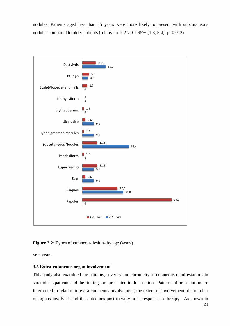
23
nodules. Patients aged less than 45 years were more likely to present with subcutaneous
nodules compared to older patients (relative risk 2.7; CI 95% [1.3, 5.4]; p=0.012).
Figure 3.2: Types of cutaneous lesions by age (years)
yr = years
3.5 Extra-cutaneous organ involvement
This study also examined the patterns, severity and chronicity of cutaneous manifestations in
sarcoidosis patients and the findings are presented in this section. Patterns of presentation are
interpreted in relation to extra-cutaneous involvement, the extent of involvement, the number
of organs involved, and the outcomes post therapy or in response to therapy. As shown in
0
31,8
9,1
9,1
0
36,4
9,1
9,1
0
0
0
4,5
18,2
69,7
27,6
2,6
11,8
1,3
11,8
1,3
2,6
1,3
0
3,9
5,3
10,5
Papules
Plaques
Scar
Lupus Pernio
Psoriasiform
Subcutaneous Nodules
Hypopigmented Macules
Ulcerative
Erytheodermic
Ichthyosiform
Scalp(Alopecia) and nails
Prurigo
Dactylytis
≥ 45 yrs < 45 yrs

24
Figure 3.3, the most frequent extra-cutaneous organ involvement is lung involvement in53
cases, eyes 14 cases, bone/joint 10 cases, lymph nodes and the liver 8 cases each, spleen 6
cases, nervous system and the heart 3 cases each, mucosa 2 cases. Kidney and bone marrow
were observed in one case each in each organ.
Figure 3.3: Extra-cutaneous organ involvement
3.5.1 Number of organs involved
As indicated in Figure 3.4, 36% of the cutaneous sarcoidosis patients had only skin
involvement. Similarly, 36% of the cutaneous sarcoidosis patients had two organs involved.
53
14
108 8
6
3 32
1 10 0
0
10
20
30
40
50
60
Lungs
Eyes
Bone/Joint
Lymph nodes
Liver
Spleen
Heart
Nervous system
Mucosa
Kidney
Blood
Muscle
Bone marrow
Percentage

25
Figure 3.4: Number of organs involved
Of the patients with two organs involved, the skin and lungs were the most common organs
involved (29 of 36). Table 3.3 below elaborates on the number of organs involved.
Table 3.3: Patients with three organs involved
Organs involved Number of patients
Skin, lymph nodes, and bone and joints 2
Skin, lungs and eyes 7
Skin, lungs and lymph nodes 1
Skin, lungs, and heart 1
Skin, lungs, bone and joints 3
Skin, lungs, and spleen 1
3.5.2 Patients with four and five organs involved
Only nine patients had four organs involved. Four percent of the cutaneous sarcoidosis
patients had five organs involved.
36%
36%
15%
9% 4%
Number of organs involved
Only one organ
Two organs
3 organs
4 organs
5 organs

26
3.5.3Association of demographical characteristics and number of organ involvement
The relationship between demographic characteristics and the number of organs involved was
examined and the results are presented in Table 3.4. There was no significance difference in
number of organs involvement and patients’ age or sex.
Table 3.4: Number of organs involved by age, gender and race
Sex Race Age Organ involvement
Male n=17 (%)
Female n=83 (%)
Black African n=96 (%)
Asian n=4 (%)
Age <45
≥45
Only one organ 2 (11.8) 34 (41.0) 36 (37.5) 0 (0.0) 8 (36.4) 26 (34.2)
Two organs 10 (58.8)
26 (31.3) 33 (34.3) 3 (75.0) 7 (31.8) 29 (38.2)
Three Organs 2 (11.8) 13 (15.7) 15 (15.6) 0 (0.0) 4 (18.2) 11 (14.5)
Four organs 3 (17.6) 6 (7.2) 9 (9.4) 0 (0.0) 2 (9.1) 7 (9.2) Five organs 0 (0.0) 4 (4.8) 3 (3.1) 1 (25.0) 1 (4.5) 3 (3.9)

27
3.5.6 Extra-cutaneous organ involvement by specific type lesion
The study also examined the association between organ involvement and patterns of
cutaneous manifestation. The results are presented in Table 3.5. Of the 53 patients with lung
involvement, the most common cutaneous pattern was papules (n=39). The second most
common pattern was plaques (n=20). Regardless of organ involvement, papules remained the
most common pattern of clinical presentation as shown in Table 3.5

28
Table 3.5: Extra-cutaneous organ involvement by specific type lesion
Lung
n=53
LN
n=8
Eyes
n=14
Nervous
n=3
BM
n=0
BV
n=1
Heart
n=3
Bone/joint
n=10
Muscle
n=0
Liver
n=8
Spleen
n=6
Mucosa
n=2
Kidney
N=1
Papules n=68 39 4 10 2 0 1 2 5 0 6 5 2 1
Plaques n=28 20 1 3 1 0 0 0 3 0 2 1 0 0
Lupus n=11 6 0 0 1 0 0 1 0 0 0 0 1 0
Scar n=4 3 0 0 0 0 0 0 0 0 1 1 0 0
Subcutaneous
n=16
7 4 3 0 0 0 1 2 0 2 1 0 0
LN- lymph node; BM- bone marrow; BV- blood vessel

29
3.6 Clinical patterns of cutaneous sarcoidosis and the chest X-ray findings
Pulmonary involvement was assessed using the method of radiographic staging (Scadding criteria) and pulmonary function tests. The majority of cases were classified as Stage 0 or 1 according to the Scadding criteria. Only four patients were in radiographic Stage 4. The patterns of clinical presentation were not associated with the degree of lung involvement. Table 3.6 below elaborates.
Table 3.6: Pattern of clinical presentation by chest X-ray finding
Cutaneous presentation Staging by CXR (Scadding criteria)
STAGE: 0 1 2 3 4
Papules n=58 15 23 9 8 3
Plaques n=26 4 12 4 4 2
Scar n=3 1 2 0 0 0
Lupus Pernio n=8 1 5 1 1 0
Psoriasiform n=1 0 0 1 0 0
Subcutaneous Nodules n=17 8 4 3 2 0
Hypopigmented Macules n=3 0 2 0 1 0
Ulcerative n=4 0 4 0 0 0
Erytheodermic n=1 1 0 0 0 0
Scalp (alopecia) and nails n=2 0 1 1 0 0
Prurigo n=4 3 1 0 0 0
Dactylitis n=10 4 2 2 0 2

30
The pulmonary function test results showed that 45 patients had normal lung function. Of the
41 patients with an abnormal lung function, 19 patients had restrictive lung function, 19
patients had obstructive lung function, and only 3 patients had mixed pattern lung function.
There were no records of pulmonary function tests for 14 patients.
3.7 Chronicity of cutaneous sarcoidosis
Active lesions with progression (worsening) and active lesions with no change (static) while
on therapy that were found on record were classified as not resolved. Lesions that were
recorded as absent and then reappear post-therapy were recorded as waxing and waning. Of
all the patients, there was complete resolution in 28 patients over 24 months, waxing and
waning in 33 patients, static involvement in 12 patients, and worsening lesions in 2 patients.
Clinical outcomes could not be determined in 25 patients due to inadequate records.
The study also examined the clinical outcomes by patterns of presentation (Table 3.7). Of the
patients that presented with papules (n=69), there was complete resolution in only 17. A
significant proportion of patients that presented with papules had either static involvement (10
patients), waxing and waning (24 patients), or worsening (2 patients).
Of the 28 patients presenting with plaques, waxing and waning lesions were the most
common pattern. However, for the four patients that presented with scar sarcoidosis, there
was complete resolution in two. Patients that presented with lupus pernio presented with
waxing and waning or with static lesions. Complete resolution was common only in patients
who presented with subcutaneous nodules.

31
Table 3.7: Clinical outcomes of sarcoidosis patients according to patterns of presentation
The clinical outcomes of cutaneous sarcoidosis patients were examined in relation to the
number of organs involved. There was complete resolution in 7of the 36 patients with only
skin involvement. The rest were either waxing and waning or no change (static) in nature.
Patterns of presentation Clinical Outcomes
Wax &wane Static Worsening Resolved Undetermined
Papules n=69 24 10 2 17 16
Plaques n=28 11 2 0 5 10
Scar n=4 1 0 0 2 1
Lupus pernio n=11 5 2 0 2 2
Psoriasiform n=1 0 1 0 0 0
Subcutaneousnodules
n=17
4 1 0 8 4
Hypopigmented macules
n=3
2 0 0 0 1
Ulcerative n=4 1 0 0 1 2
Erythrodermic n=1 0 0 0 1 0
Scalp (alopecia) and nails
n=2
3 0 0 0 0
Prurigo n=5 3 1 0 1 0
Dactylitis n=12 7 2 0 3 0

32
Of the 36 patients with skin and one more organ involvement, there was complete resolution
in 10 patients. However, there was no record to determine clinical outcomes in 14 patients
with skin and one more organ involvement. Table 3.8 demonstrates the clinical outcomes of
sarcoidosis patients based on the number of organs involved
Table 3.8: Clinical outcomes of sarcoidosis patients based on the number of organs involved
No. of systems
involved
Outcomes
Waxing
& waning
Static Worsening Resolved Undetermined
Skin only n=36 15 7 0 7 7
Skin +1 n=36 9 3 1 10 14
Skin + 2 n=15 4 2 1 5 3
Skin +3 n=9 2 0 0 4 1
Skin +4 n=4 2 0 0 2 0
Some patients presented with multiple morphologies. The clinical outcomes of sarcoidosis
patients based on the number of morphologies were examined and the results are presented in
Table 3.9.

33
Table 3.9: Clinical outcomes of sarcoidosis patients based on number of morphologies on
presentation
No.of morphologies Outcomes
Waxing
&waning
Static Worsening Resolved Undetermined
One lesion (n=57) 15 6 2 18 16
Two lesions (n=31) 11 5 0 8 7
Three lesions (n=10) 5 1 0 2 2
Four lesions (n=1) 1 0 0 0 0
Five lesions (n=1) 1 0 0 0 0
3.8 Treatment
Topical and oral corticosteroids were administered to 35 patients, whilst 24 patients were
given a combination of topical corticosteroids, oral corticosteroids and an anti-malarial drug.
A combination of topical and oral corticosteroids, anti-malarials, and methotrexate was
administered to 10 patients. Ten patients were given topical corticosteroids only. Table 3.10
summarizes the drugs most commonly prescribed.

34
Table 3.10: Treatment combinations administered to patients
Treatment No. of patients Percent
Topical corticosteroids only 10 10.0
Topical and oral corticosteroids 35 35.0
Topical corticosteroids and anti-malarial 2 2.0
Topical and oral corticosteroids and anti-malarial 24 24.0
Topical and oral corticosteroids, anti-malarial and methotrexate 10 10.0
Topical and oral corticosteroids, and methotrexate 6 6.0
Oral corticosteroids only 3 3.0
Topical and oral corticosteroids, anti-malarial and Azathioprine 3 3.0
Topical and oral corticosteroids, methotrexate, and Azathioprine 2 2.0
Topical corticosteroids and methotrexate 1 1.0
Topical corticosteroids, antimalarial, and Azathioprine 1 1.0
Topical and oral corticosteroids and Potassium permanganate 2 2.0
Oral corticosteroids and methotrexate 1 1.0
3.9. Histology findings
Designated pathologists confirmed sarcoidosis in all 100 patients. The summary of the site of
biopsy is summarized in Table 3.11.

35
Table 3.11: Site of biopsy
Site of biopsies taken Frequency Percentage
Skin 79 79.0
Skin &extra-cutaneous site 14 14.0
Extra-cutaneous site only 7 7.0
As shown in Table 3.12, naked granulomas surrounded by a few epithelioid cells were the
most common presentation (70% of cases). The next common presentation was naked
granulomas with epithelioid cells and giant cells (41% of cases). The least common
histological presentations were granulomas extending to the subcutis (4%) and granulomas
with giant cells and asteroid bodies (1%).
Table 3.12: Frequency distribution of granulomatous reaction patterns
Granulomatous reaction pattern Frequency Percent
Naked granulomas surrounded by epithelioid cells and few
lymphocytes
70 70.0
Granulomas with fibrinoid necrosis 14 14.0
Granulomas extending to the subcutis 4 4.0
Naked granulomas comprising epithelioid cells and giant cells 41 41.0
Granulomas with giant cells and asteroid bodies 1 1.0

36
The patterns of presentation versus the histological findings are presented in Table 3.13. The
histological feature of papules showing naked granulomas surrounded by epithelioid cells
with a few lymphocytes was the most common presentation, found in 50 of the cases.
Plaques were the second most common clinical pattern, which was found with naked
granulomas surrounded by epithelioid cells with a few lymphocytes (n=20).Fibrinoid necrosis
was seen in 12% of the cases with papules and the least (4%) in cases with plaque
Table 3.13: Pattern of presentation versus histological results of patients
Pattern of presentation
Histology patterns
Naked granulomas surrounded by epithelioid cells and few lymphocytes
Granulomas with fibrinoid necrosis
Granulomas extending to the subcutis
Naked granulomas comprising epithelioid cells and giant cells
Granulomas with giant cells and asteroid bodies
Papules n=69 50 12 1 28 0
Plaques n=28 20 4 1 15 0
Subcutaneous nodules n=17
10 3 4 6 1
Lupus pernio n=11 7 4 1 5 0
Dactylitis n=12 9 1 0 4 1
Psoriasiform n=1 0 0 0 1 0
Hypopigmented Macules n=3
2 0 0 3 0
Ulcerative n=4 3 0 1 1 0
Erythrodermic n=1 1 1 0 0 0
Scar n=4 2 0 1 1 0
Scalp (alopecia) and nails n=3
3 1 0 0 0
Prurigo n=5 4 0 0 2 0
3.10 Sarcoidosis in people living with HIV
Of the 100 patients included in this study, 12 were living with HIV infection. All were
female and Black African. Their age ranged from 21 to 39 years. Seven were diagnosed
37
between 2010 and 2014, whereas only three of them were diagnosed between 2003 and 2005.
At diagnosis, their CD4 counts were low (mean 248 cells/mm3),but increased as their sarcoid
disease evolved. All on HAART, and those with initial high viral loads responding to
antiretroviral treatment displayed concomitant progression of their sarcoidosis. Four had only
skin involvement, six had both skin and lung involvement, one had skin, lung and eye
involvement and another had disease involving the skin, lungs, bone and joints. Five
presented with papules and plaques; three had papules only. One presented with
erythroderma, and another presented with lupus pernio, papules, and subcutaneous nodules.
Their calcium levels were normal, both before and after therapy. Three had chest X-ray
changes and three had only skin involvement. The remaining patient, who was initially
recorded with skin only involvement, had Stage two chest radiographic changes. The chest x-
ray reports of the six who had skin and lung involvement, indicated that two had Stage three
radiographic changes, and four had Stage one radiographic changes.
3.11 Summary
It is important to describe a sample population in terms of its demographics and clinical profile, in
order to know the population better and to be able to establish areas of concern and areas for
further research.In this study we have managed to identify the demographics of the population
group most affected by cutaneous sarcoidosis (Black females older than 45 years of age). Patterns
of presentation of cutaneous sarcoidosis have been described and the pattern of progress in
patients who have both HIV and cutaneous sarcoidosis has been noted and remarked on. The next
chapter entertains a discussion on the study findings, explores the limitations and makes
recommendations.

38
CHAPTER FOUR
SUMMARY OF FINDINGS, DISCUSSION, CONCLUSION
4.1 Introduction
This chapter presents a discussion of the study findings, conclusions and recommendations.
The discussion is based on the study objectives and attempts are made to explain the key
findings of the study. Where necessary, the findings of this study are compared to previous
studies on cutaneous sarcoidosis.
4.2 Summary of findings
The key findings of this study are summarised as follows:
• Cutaneous sarcoidosis disease was more common in females than males.
• Cutaneous sarcoidosis disease was more common in older females above the age of 45
years.
• The patterns of cutaneous sarcoidosis were predominantly papules and plaques.
• The lung was the most commonly affected extra-cutaneous organ.
• The Scadding staging of the lungs in patients with cutaneous sarcoidosis was mainly
zero and one.
• There was complete resolution of cutaneous sarcoidosis in only 28 cases over 24
months.
• Of the patients that presented with papules (n=69), there was complete resolution only
in 17 of them. The clinical course of a significant proportion of patients that presented
with papules was static (10 patients), waxing and waning (24 patients) or worsening (2
patients).
• Of the 28 patients that presented with plaques, waxing and waning was the most
common outcome. However, of the four patients that presented with scar sarcoidosis,
there was complete resolution in two. Waxing and waning and static were the common
outcomes of patients that presented with lupus pernio. Complete resolution was
common only in patients who presented with subcutaneous nodules.

39
4.3 Discussion of results
This study evaluated the spectrum of cutaneous manifestations of sarcoidosis among patients
attending the Chris Hani Baragwanath Academic Hospital Dermatology outpatient clinic from
1991 – 2015.Studies on cutaneous sarcoidosis in the South African setting are limited.
Besides the Mosam and Morar study in 2004, which focused only on recalcitrant patterns, the
only reported work on the spectrum of cutaneous sarcoidosis in South Africa was conducted
in 1999 by Jacyk et al. (17, 39). The current study provides a detailed description of the
various patterns, severity and chronicity of cutaneous sarcoidosis over two decades. This
study also examines the co-morbidity of HIV/AIDS and cutaneous sarcoidosis.
Results presented below are a summary of the main findings in an attempt to answer the
research questions, with a focus on both the specific and global findings, and what, in
particular, was uncovered that is new to the body of existing knowledge on cutaneous
sarcoidosis in South African patients. Some findings had to be interpreted with caution, since
the sample examined and analysed was a randomly selected cohort of patients seen between
1991 and 2015, and does not reflect the total number of patients who presented with the
disease during this period.
4.3.1 Where are we today?
It appears that cutaneous sarcoidosis was first recognizedin the nineteenth century; due to a
lack of a standardised investigative tools at that time;it is not possible to confirm this, as
investigators relied on the clinical and unique histological appearance of this disease entity
(1). It is only in the mid-twentieth century that radiological findings were used when patients
presented with cutaneous lesions, and lung disease was suspected (6, 62). In the twenty-first
century (ACCESS Research group, 1999) (6), high resolution computerised tomography
became easily available and exposed the X-ray’s limitations (6). With an increased incidence
of tuberculosis in Africa (63),it became apparent that this granulomatous disease had some
histological resemblance to sarcoidosis. This made it difficult to differentiate between the two
pathologies. But with the advance in diagnostic tools, it became possible to exclude other
granulomatous conditions (42, 64, 65).

40
In this study of cutaneous sarcoidosis, all patients were confirmed by biopsy. Some also had
radiological features of intrathoracic sarcoidosis. All had compatible clinical presentations
and exclusion of other granulomatous diseases.
4.3.2 Correlations between demographic characteristics and patterns of cutaneous
sarcoidosis
In answer to the research question of whether there were correlations between demographic
characteristics and cutaneous patterns of sarcoidosis, the researcher was able to confirm the
following:
• Cutaneous sarcoidosis was more common in females than in males.
• Cutaneous sarcoidosis was more common in older women (above the age of 45 years)
than in younger women.
In this study, the peak incidence of cutaneous sarcoidosis was in the age group of 50 to 69
years and is relatively compatible with recent epidemiology studies (14, 21). Although there
is no consensus or evidence for the factors causing the increase in age, which has been shown
in international studies, the hypothesis made here suggests hat changes in the practice and use
of diagnostic imaging technology could account for the higher incidence in older women.
Another proposed reason for the high incidence in older women is that they may seek
professional help for physical disorders more willingly and frequently than younger women
do. This speculation prompts a need for a study.
The predominance of Black females in this study is similar to the findings in literature
globally (8, 10). Of note is the fact that the women were over 45 years of age. Apart from the
role of improved diagnostic tools, the increased tendency for older women to develop skin
lesions maybe related to the lack of exposure to endogenous female hormones in older
women. Hence being pre-menopausal may confer a reduced risk of sarcoidosis (66, 67). The
lack of evidence for this conclusion, however, makes this speculation.
A supportive hypothesis which may contribute to the high female to male ratio includes the
possibility that females have a greater tendency than males to observe their body or skin for
these type of lesions (14, 27).

41
4.3.3 Patterns, severity and chronicity of cutaneous manifestations of sarcoidosis in
patients
In an attempt to answer the research question regarding patterns, severity and chronicity of
cutaneous sarcoidosis, the researcher was able to confirm that
• the patterns of manifestation were predominantly papules and plaques;
• skin and lungs were the most commonly affected organ systems;
• the Scadding staging of lung disease was mainly zero and one.
Recognition of specific lesions is important as they provide clues for diagnosis. Skin lesions
are easily accessible to use as tissue specimens to evaluate for definitive histological
diagnosis; moreover, a punch biopsy is a simple technique to learn and provides a high
diagnostic yield.
Skin lesions often appear at the onset of the disease, but may appear at any stage (31, 32).In
this study, apart from the skin, the most frequent extra-cutaneous organ involvement was the
lungs (53 cases),followed by eyes (14 cases) and bones/joints (10 cases); the least affected
was bone marrow and kidneys. In a recent study by Marcoval et al., it was concluded that
specific cutaneous lesions did not have a predictive factor for the disease severity and
prognosis (22, 27). However, there is an exception with erythema nodosum, a non-specific
pattern of presentation of cutaneous sarcoidosis. It is associated with a good outcome. Lupus
pernio, a more specific pattern of presentation, is associated with severity and chronicity of
cutaneous sarcoidosis (17, 27, 32). In this study, none of our patients presented with
erythema nodosum. However, the subcutaneous pattern of presentation had a p-value =
0.012. It is reported in the literature that due to a limited number of case reports, the
characteristics of subcutaneous sarcoidosis in relation to systemic disease have not been
elucidated (32). The significant finding in this study on the subcutaneous patterns of
presentation is similar to the findings of the current literature. A recent study in America
reported on twenty-one cases and found that this form of cutaneous sarcoidosis has no
correlation with severe systemic involvement and has a relatively good outcome. The strength
of the American study was the significant follow-up period of 32 years (68). Literature from
the east of Africa (Ethiopia) and the west of Africa (Nigeria) reports that the pattern of
presentation mostly demonstrated subcutaneous nodules and scar sarcoidosis respectively (35,
36); these were associated with better prognostic outcomes (32).

42
Regardless of the number of organs involved, papules were the most common pattern of
clinical presentation demonstrated in this study. This concurs with findings globally. Some
authors report that this type of specific cutaneous lesion does not determine the severity and
chronicity of this disease entity (22, 32). This seems to be confirmed in this study, because in
a number of patients the clinical presentation was of one type of lesion co-existing with
another, i.e., poly-morphology (papules with plaques, papules with lupus pernio). It is up to
the individual clinician’s discretion to determine which lesion forms the most predominant
pattern. In a study conducted by Mañá and colleagues, a correlation was found between
specifically papular lesions with an acute form of sarcoidosis, which included involvement of
other organs (hilar lymphadenopathy, peripheral lymphadenopathy, parotid enlargement, EN,
and acute uveitis); this form usually resolved within two years (3, 32).
Regarding chronicity of the presenting patterns, the following is found:
In answer to the research questionregarding chronicity, a significant proportion of patients
with papules (24.6%) and subcutaneous lesions (100%) are frequently associated with
complete resolution of the lesions. Apart from having a patient profile with papules that may
resolve completely, on the contrary papules can be (36 cases) persistent. Similarly patients
who presented with lupus pernio had chronic persistent lesions.
Our population sample is predominantly Black Africans who presented most frequently with
papules and plaques. Whether these features of presentation may be attributed to early
diagnosis or management factors or whether the minimal presence or absence of other
aetiologies causing granulomatous changes may have been misdiagnosed, cutaneous
sarcoidosis remains to be further investigated.
4.3.4 Histopathological findings of biopsy specimens of cutaneous lesions of sarcoidosis
Regarding the histopathological findings of biopsy specimens, and taking into consideration
the frequent clinical presentation of papules, the histological patterns which were seen most
frequently, were classic naked granulomas, in 70% of the cases. Only 12% had findings of
fibrinoid necrosis, similar to the histological findings described in Black South Africans in the

43
literature almost two decades ago by Jacyk, and earlier by Benatar (17, 19). Perhaps
improved access to care and earlier diagnosis and treatment interventions now play a role in
influencing the pattern of presentation, since the natural course of the disease may be changed
or halted with better access and earlier diagnosis. The notion of advanced and mutilating
patterns associated with Black Africans and African Americans race could have been
influenced by late presentations at first visits to outpatient clinics, which in turn could have
been influenced by racially prejudiced health care policies at the time. These factors prevailed
right through the eighteenth, nineteenth and early twentieth century. The current findings
regarding manifestation of the disease entity at first visit may well be strongly influenced by
better access and earlier presentation.
4.3.5 Association between HIV infection and cutaneous or systemic sarcoidosis
The last research question posed at the outset of this study was the question of a possible
correlation between manifestations of cutaneous or systemic sarcoidosis and the presence of
HIV infection. In this study, people living with both HIV and sarcoidosis interestingly
demonstrated an inversely proportional association of the disease entity with the CD4 count.
Disease progression is noted with CD4 increments that occur after initiation of therapy.
Similarly, those with high viral loads experienced progression of sarcoidosis astheir viral
loads declined with HAART. Although half of the patients (six cases) had more than one
organ involvement (skin and lung involvement), there was no correlation between the
Scadding stage and lung function, as 2 patients had normal lung function tests with Stage 3
chest X-rays.
The clinical patterns of sarcoidosis in the HIV-positive patients did not differ from the non-
HIV infected individuals reported globally, including in the histological findings (69). Some
reports in the literature do, however, describe a challenge in the management of cutaneous
and systemic sarcoidosis where an IRIS is present (53, 56).
There is a need for further studies on the coexistence of HIV/AIDS and sarcoidosis. Clarity of
the immunological relationship needs to be investigated further and more patient reports
described. There is opportunity to expand such information into a case series.

44
4.4 Limitations
Limitations are presented here in list form for clarity:
• Many clinical records were incomplete.
• Some patients were excluded because their files were missing. This limited the sample
size. The small sample size hindered the ability to make significant statistical associations.
• The data was dependent on how well clinical and laboratory information was recorded.
• The study was conducted for outpatients only. It would have been interesting to see the
differences in presentation between inpatients and outpatients, to give a broader picture.
• Patient records from this study included patients from Dermatology and Respiratory
outpatient clinics only – this does create a bias for patients with lung involvement as an
extra-cutaneous manifestation.
• This study, which was conducted with records from 1991- 2015 that had not been
collected consecutively, limited the author from obtaining data regarding incidence and
changes in trends.
4.5 Conclusion
The study aimed at describing the cutaneous patterns of sarcoidosis in the population that
presented at the outpatient Dermatology clinic at Chris Hani Baragwanath Academic Hospital.
This was a retrospective review and the first, to my knowledge, to describe this population at
CHBH. Further research and collaboration with other countries in southern Africa is
recommended for a more significant and statistically relevant contribution to the spectrum and
management of this rare disease entity.

45
REFERENCES
1. Boeck C. Multiple benign sarkoid of the skin. Arch Dermatol. 1982;118 (10):710-20.
2. Lazarus A. Sarcoidosis: epidemiology, etiology, pathogenesis, and genetics. Disease-
a-Month. 2009;55 (11):649-60.
3. Mañá J, Marcoval J, Graells J, Salazar A, Peyrí J, Pujol R. Cutaneous involvement in
sarcoidosis: relationship to systemic disease. Arch Dermatol. 1997;133 (7):882-8.
4. Mañá J, Marcoval J. Skin manifestations of sarcoidosis. La Presse Médicale. 2012;41
(6):e355-e74.
5. Allwood B, Ainslie G. Sarcoidosis: CME2013,vol. 31 (9) 326-330.
6. "Statement on sarcoidosis". Joint Statement of the American Thoracic Society (ATS),
the European Respiratory Society (ERS) and the World Association of Sarcoidosis
and Other Granulomatous Disorders (WASOG) adopted by the ATS Board of
Directors and by the ERS Executive Committee, February 1999. Am J Respir Crit
Care Med. 1999;160:736-55.
7. Newman LS, Rose CS, Maier LA. Sarcoidosis. 1997; 336 (17):1224-34.
8. English JC, III, Patel PJ, Greer KE. Sarcoidosis. J Am Acad Dermatol. 2001; 44
(5):725-46.
9. Rybicki BA, Major M, Popovich J, Maliank MJ. Racial differences in sarcoidosis
incidence: a 5-year study in a health maintenance organization. Am J Epidemiol.
1997;145 (3):234-41.
10. Baughman RP, Teirstein AS, Judson MA, Rossman MD, Yeager Jr H, Bresnitz EA, et
al., Clinical characteristics of patients in a case control study of sarcoidosis. Am J
Respir Crit Care Med. 2001; 164 (10):1885-9.
11. Heath CR, David J, Taylor SC. Sarcoidosis: are there differences in your skin of color
patients? J Am Acad Dermatol. 2012; s66 (1):121. e1-. e14.
12. Collin B, Rajaratnam R, Lim R, Lewis H. A retrospective analysis of 34 patients with
cutaneous sarcoidosis assessed in a dermatology department. Clin Exp Dermatol.
2010;35 (2):131-4.
13. Rybicki BA, Iannuzzi MC, editors. Epidemiology of sarcoidosis: recent advances and
future prospects. Seminars in respiratory and critical care medicine; 2007: Copyright©
2007 by Thieme Medical Publishers, Inc., 333 Seventh Avenue, New York, NY
10001, USA.
46
14. Ungprasert P, Carmona EM, Utz JP, Ryu JH, Crowson CS, Matteson EL.
Epidemiology of Sarcoidosis 1946-2013. Mayo Clin Proc. 1991 (2):183-8.
15. Morrison J. Sarcoidosis in the Bantu.Br J Dermatol.1974;90 (6):649-55.
16. Edmondstone W, Wilson A. Sarcoidosis in Causians, Blacks and Asians in London.
Br J Dis Chest. 1985;79:27-36.
17. Jacyk WK. Cutaneous sarcoidosis in black South Africans. Int J Dermatol. 1999;38
(11):841-5.
18. James DG, Neville E, Siltzbach LE, Turiaf J, Battesti JP, Sharma OP, et al. A
Worldwidereview of sarcoidosis. Ann N Y Acad Sci. 1976; 278 (1):321-34.
19. Benatar S. Sarcoidosis in South Africa: a comparative study in Whites, Blacks and
Coloureds. S Afr Med J. 1977; 52 (15):602-6.
20. Rybicki BA, Kirkey KL,Major M, Maliarik MJ, Popovich J, Chase GA, et al. Familial
risk ratio of sarcoidosis in African-American sibs and parents. Am J Epidemiol. 2001;
153 (2):188-93.
21. Yanardag H, Tetikkurt C, Bilir M, Demirci S, Iscimen A. Diagnosis of cutaneous
sarcoidosis; clinical and the prognostic significance of skin lesions.Multidiscip Respir
Med. 2013; 8 (1):26.
22. Marcoval J, Mañá J, Rubio M. Specific cutaneous lesions in patients with systemic
sarcoidosis: relationship to severity and chronicity of disease. Clin Exp Dermatol.
2011; 36 (7):739-44.
23. Jacyk W. Sarcoidosis in the West African. a report of eight Nigerian patients with
cutaneous lesions. Trop Geogr Med. 1984; 36 (3):231-6.
24. Ando M, Miyazaki E, Hatano Y, Nishio S, Torigoe C, Yamasue M, et al.
Subcutaneous sarcoidosis : a clinical analysis of nine patients. Clin Rheumatol.. 2016;
35 (9):2277-81.
25. Olive KE, Kataria YP. Cutaneous manifestations of sarcoidosis relationships to other
organ system involvement, abnormal laboratory measurements, and disease course.
Arch Intern Med.1985; 145 (10):1811-4.
26. Wu M, Lee JY. Cutaneous sarcoidosis in southern Taiwan: clinicopathologic study of
a series with high proportions of lesions confined to the face and angiolupoid variant.
J Eur Acad Dermatol Venereol. 2013; 27 (4):499-505.
27. Yanardağ H, Pamuk ÖN, Karayel T. Cutaneous involvement in sarcoidosis: analysis
of the features in 170 patients. Respiratory medicine. 2003; 97 (8):978-82.
47
28. Hillerdal G, Nöu E, Osterman K, Schmekel B. Sarcoidosis: Epidemiology and
Prognosis: A 15-Year European Study 1. Am Rev Respir Dis. 1984; 130 (1):29-32.
29. Parkes SA, Baker S, Bourdillon R, Murray C, Rakshit M, Sarkies J, et al. Incidence of
sarcoidosis in the Isle of Man. Thorax. 1985; 40 (4):284-7.
30. Veien NK, Stahl D, Brodthagen H. Cutaneous sarcoidosis in Caucasians. J Am Acad
Dermatol. 1987; 16 (3):534-40.
31. Haimovic A, Sanchez M, Judson MA, Prystowsky S. Sarcoidosis: a comprehensive
review and update for the dermatologist: part I. Cutaneous disease. J Am Acad
Dermatol. 2012; 66 (5):699. e1-. e18.
32. Sanchez M, Haimovic A, Prystowsky S. Sarcoidosis. Dermatol Clin. 2015; 33 (3):389-
416
33. Spiteri M, Matthey F, Gordon T, Carstairs L, James D.G. Lupus pernio: a clinico-
radiological study of thirtyfive cases. Br J Dermatol. 1985; 112 (3):315-22.
34. Baughman RP, Lower EE. Evidence-based therapy for cutaneous sarcoidosis. Clin
Dermatol. 2007; 25 (3):334-40.
35. Olumide Y, Bandele E, Elesha S. Cutaneous sarcoidosis in Nigeria. J Am Acad
Dermatol. 1989; 21 (6):1222-4.
36. Tsega E, Getahun B, Teklehaimanot R. Sarcoidosis in Ethiopia. Tubercle. 1978; 59
(4):261-8.
37. Moche M, Glassman S, Modi D, Grayson W. Cutaneous annular sarcoidosis
developing on a background of exogenous ochronosis: A report of two cases and
review of the literature. Clin Exp Dermatol. 2010; 35 (4):399-402.
38. Motswaledi M, Khammissa R, Jadwat Y, Lemmer J, Feller L. Oral sarcoidosis: A
case report and review of the literature. Aust Dent J. 2014; 59 (3):389-94.
39. Mosam A, Morar N. Recalcitrant cutaneous sarcoidosis: an evidence-based sequential
approach. J Dermatolog Treat. 2004; 15 (6):353-9.
40. Hartshorne S. Dermatological disorders in Johannesburg, South Africa. Clin Exp
Dermatol. 2003; 28 (6):661-5.
41. Smith C, Feldman C, Reyneke J, Promnitz D, Kallenbach J, Zwi S. Sarcoidosis in
Johannesburg--a comparative study of black and white patients. S Afr Med J. 1991; 80
(9):423-7.
42. Cardoso J, Cravo M, Reis J, Tellechea O. Cutaneous sarcoidosis: a histopathological
study. J Eur Acad Dermatol Venereol. 2009; 23 (6):678-82.

48
43. Mangas C, Fernández-Figueras MT, Fité E, Fernández-Chico N, Sàbat M, Ferrándiz
C. Clinical spectrum and histological analysis of 32 cases of specific cutaneous
sarcoidosis. J Cutan Pathol. 2006; 33 (12):772-7.
44. Weedon D. - The granulomatous reaction pattern. Weedon's Skin Pathology (Third
edition). Edinburgh: Churchill Livingstone; 2010. p. 169-94.e17.
45. Calonje E, Brenn T, Lazar A, McKee PH. McKee's pathology of the skin:
Elsevier/Saunders; 2012.
46. Wijnen PA, Cremers JP, Nelemans PJ, Erckens RJ, Hoitsma E, Jansen TL, et al.
Association of the TNF-α G-308A polymorphism with TNF-inhibitor response in
sarcoidosis. Eur Respir J. 2014;43 (6):1730-9.
47. Gerke AK, Hunninghake G. The immunology of sarcoidosis. Clinics in chest
medicine. 2008; 29 (3):379-90.
48. Serio RN. Infliximab treatment of sarcoidosis. Ann Pharmacother. 2003; 37 (4):577-
81.
49. Judson MA, Costabel U, Drent M, Wells A, Maier L, Koth L, et al., The WASOG
Sarcoidosis Organ Assessment Instrument: an update of a previous clinical tool.
Sarcoidosis. Vasc Diffuse Lung Dis. 2014; 31 (1):19-27.
50. Harrington D, Major M, Rybicki B, Popovich J, Maliarik M, Iannuzzi M. Familial
sarcoidosis: analysis of 91 families. Sarcoidosis. 1994;11:240-.
51. Blanche P, Passeron A, Gombert B, Ginsburg C, Salmon D, Sicard D. Sarcoidosis and
HIV infection: influence of highly active antiretroviral therapy. Br J Dermatol.
1999;140 (6):1185.
52. Roustan G, Yebra M, Rodriguez-Braojos O, Banos I, Jaurena J, Simon A. Cutaneous
and pulmonary sarcoidosis in a patient with HIV after highly active antiretroviral
therapy. Int J Dermatol. 2007; 46 (1):68-71.
53. Mirmirani P, Maurer TA, Herndier B, McGrath M, Weinsteinb MD, Berger TG.
Sarcoidosis in a patient with AIDS: a manifestation of immune restoration syndrome.
J Am Acad Dermatol.1999;41 (2):285-6.
54. Morris DG, Jasmer RM, Huang L, Gotway MB, Nishimura S, King TE. Sarcoidosis
following HIV infection: evidence for CD4+ lymphocyte dependence. CHEST
Journal. 2003;124 (3):929-35.
55. Gowda KS, Mayers I, Shafran SD. Concomitant sarcoidosis and HIV infection.
CMAJ. 1990;142 (2):136.

49
56. Osei-Sekyere B, Karstaedt A. Immune reconstitution inflammatory syndrome
involving the skin. Clin Exp Dermatol. 2010; 35 (5):477-81.
57. Lehloenya R, Meintjes G. Dermatologic manifestations of the immune reconstitution
inflammatory syndrome. Clin Exp Dermatol. 2006; 24 (4):549-70.
58. Trevenzoli M, Cattelan A, Marino F, Marchioro U, Cadrobbi P. Sarcoidosis and HIV
infection: a case report and a review of the literature. Postgrad Med J. 2003; 79
(935):535-8.
59. Volden G. Successful treatment of chronic skin diseases with clobetasol propionate
and a hydrocolloid occlusive dressing. Acta Derm Venereol. 1991; 72 (1):69-71.
60. Ffrooz A, Tehranchia-Nia Z, Ahmed A.Benefits and risks of intralesional
corticosteroid injection in the treatment of dermatological diseases. Clin Exp
Dermatol. 1995; 20 (5):363-70.
61. Badgwell C, Rosen T. Cutaneous sarcoidosis therapy updated. J Am Acad Dermatol.
2007;56 (1):69-83.
62. Wurm K, Reindell H, Heilmeyer L. Der Lungenboeck im Röntgenbild: Thieme; 1958;
118 (4):229-43
63. Chaisson RE, Martinson NA. Tuberculosis in Africa—combating an HIV-driven
crisis. N Engl J Med. 2008; 358 (11):1089-92.
64. Ball NJ, Kho GT, Martinka M. The histologic spectrum of cutaneous sarcoidosis: a
study of twenty-eight cases. J Cutan Pathol. 2004; 31 (2):160-8.
65. Ishak R, Kurban M, Kibbi AG, Abbas O. Cutaneous sarcoidosis: clinicopathologic
study of 76 patients from Lebanon.Int J Dermatol. 2015; 54 (1):33-41.
66. Cozier YC, Berman JS, Palmer JR, Boggs DA, Wise LA, Rosenberg L. Reproductive
and Hormonal Factors in Relation to Incidence of Sarcoidosis in US Black Women:
The Black Women's Health. StudyAm J Epidemiol. 2012; 176 (7):635-41.
67. Chida K, Shirai M, Sato M, Sato J, Nakamura H. Successful treatment of hepatic
sarcoidosis with hormone replacement in a postmenopausal woman. Respirology.
1999; 4 (3):259-61.
68. Ahmed I, Harshad SR. Subcutaneous sarcoidosis: is it a specific subset of cutaneous
sarcoidosis frequently associated with systemic disease? J Am Acad Dermatol. 2006;
54 (1):55-60.
69. Lowery W.S, Whitlock WL, Dietrich RA, Fine JM. Sarcoidosis complicated by HIV
infection: three case reports and a review of the literature. Am Rev Respir Dis.

50
1990;142 (4):887-9.70. UNAIDS, Jointed United Nations Programme on HIV/AIDS
2016). UNAIDS Report on the global AIDS epidemic 2016.
www.unaids.org/sites/default/files/media_asset/global-AIDS-update-2016_en.pdf
71. Haimovic A, Sanchez M, Judson MA, Prystowsky S. Sarcoidosis: a comprehensive
review and update for the dermatologist: part II. Extracutaneous disease. J Am Acad
Dermatol. 2012; 66, ( 5): 719.e1–719.e10.
72. Chesnutt AN. Enigmas in sarcoidosis.West J Med. 1995;162 (6):519.

51

52
APPENDIX A (Data collection sheet)
DATA COLLLECTION SHEET PATIENT CODE: ------------
Age:………………………………………………………………………………………………….
Sex:
………………………………………………………………………………………………….
Race:………………………………………………………………………………………………
Occupation:……………………………………………………………………………………
Histological confirmation of sarcoidosis……………….Year of diagnosis:……………………
Type of organ specimen: skin non-skin specify type of viscera:………..
For the following, if yes, give details of histological findings:………………………………………………………………………
Granulomatous reaction pattern
Yes No Mixed
Naked granulomas surrounded by epithelioid cells and a few lymphocytes
Granulomas with fibrinoid necrosis
Granulomas extending to the subcutis
Naked granulomas comprising epithelioid cells; giant cells
Granuloma with giant cells and asteroid bodies
Other

53
Co-morbidity:
HIV status Reactive Non-reactive Unknown
HAART therapy yes: no:
CD4: Before HAART Current
Viral load: Before HAART Current:
Organ Involved: Cutaneous involvement: Extra-cutaneous involvement:
Yes
No
Interval between diagnosis of sarcoidosis and eruption of skin lesions:………………………………………………

54
(Body map for back and front)
Documented cutaneous lesion:
Specific skin lesions Non-specific skin lesions
Distinct lesions Non-distinct lesions Erythema nodosum
Papules Subcutaneous nodule Calcification
Plaques Hypopigmented macules Prurigo
Lupus pernio Ulcerative sarcoidosis Dactylitis
Scar sarcoidosis Psoriasiform lesions
Alopecia and nail sarcoidosis
Ichthyosiform presentation
Erythrodemic presentation
Tattoo reactions: other:………………………………………………………………
No. of morphology per lesion:

55
Lesion No.
Associated Pain: Pruritus:
Investigations conducted: Time commenced: before treatment after treatment
Tested normalresultsabnormal result: - raised: low:
Full blood count: …………… …………….
Urea, electrolytes and ………………… …………….
Creatinine .................... ...............
Liver function test: …………… …………….
…………… …………….
Serum calcium: …………… …………….
Serum ACE: ...………… ……………
…………… …………….

56
Chest radiography:…………………..
…………………………grade: zero
.………………………grade: one
.....……………………grade: two
..………..……….……grade: three
…..……….…………….grade: four
Investigations conducted: Tested normal results abnormal result:- raised: low:
Hand radiography: osteolytic changes ……… ………
Pulmonary function tests: …………… …………….
…………… …………….
Electrocardiography: …………… ……………
…………… …………..
Ophthalmology examination: …………… …………..
……………… ………….
Tuberculin Test: …….……… ……………
…………… …………….
Other: specify:…………………… …………… ……………

57
Treatment options:
Name Dose Duration Discontinued/reason
Treatment response: excellent good fair poor
58
APPENDIX B (Ethics Certificate)

59
APPENDIX B (Ethics Certificate continued)