the smile of amphiporus nelsoni sanchez, 1973 (nemertea...
TRANSCRIPT

PROCEEDINGS OF THE BIOLOGICAL SOCIETY OF WASHINGTON118(3):483–498. 2005.
The smile of Amphiporus nelsoni Sanchez, 1973(Nemertea: Hoplonemertea: Monostilifera: Amphiporidae)
leads to a redescription and a change in family
Svetlana A. Maslakova, Martin Thiel, Nelson Vasquez, and Jon L. Norenburg
(SAM) The George Washington University, Washington DC, 20052, U.S.A.,e-mail: [email protected];
(JLN, SAM) Department of Invertebrate Zoology, National Museum of Natural History,Washington D.C. 20560-0163, U.S.A., e-mail: [email protected]; (MT, NV) Facultad Cienciasdel Mar, Universidad Catolica del Norte, Larrondo 1281, Coquimbo, Chile, e-mail: [email protected]
Abstract.—A common intertidal hoplonemertean species, Amphiporus nel-soni Sanchez, 1973, from Chile is re-described based on the investigation ofmaterial from the type locality and one other locality in Chile. The species istransferred to the genus Prosorhochmus Keferstein, 1862 (Prosorhochmidae)based on the presence of a dorsal epidermal fold, i.e., ‘‘prosorhochmid smile’’on the bilobed head, truncated stylet basis, well-developed frontal organ withcharacteristic epithelial specialization, structure of the nephridial system andother characters of internal anatomy. Placement of this species into Proso-rhochmus expands the geographical distribution of the genus, previously knownonly from the North Atlantic and Mediterranean, to the South Pacific. Inter-estingly, Prosorhochmus nelsoni (Sanchez, 1973) is characterized by separatesexes and oviparity, unlike all the other members of the genus, which combinehermaphroditism with ovoviviparity. We compare P. nelsoni to the other spe-cies of Prosorhochmus and reassess some of the morphological characters usedin the systematics of the genus.
The genus Prosorhochmus currentlycomprises four marine intertidal speciescombining two relatively uncommon char-acteristics among nemerteans, hermaphro-ditism and viviparity. A total of nine spe-cies have been described in the genus, buttwo of these have been synonymized withProsorhochmus claparedii Keferstein, 1862and three others are considered species in-quirendae. The four valid species are foundin the North Atlantic or Mediterranean(Gibson & Moore 1985). The original de-scription of Amphiporus nelsoni Sanchez,1973 was mainly based on external appear-ance. In this paper we re-describe A. nelsonion the basis of histological sections of ma-terial from the type locality and recentlycollected material from another locality,and we illustrate the description with a set
of photomicrographs and diagrams. Wedemonstrate that morphological character-istics clearly place A. nelsoni in Proso-rhochmus, and we compare it to the othervalid species of the genus Prosorhochmus.
Materials and Methods
Three attempts over a 15-year periodfailed to locate the type material, consistingof a series of histological sections of a ho-lotype and three unsectioned paratypes in9% sea water formalin originally depositedwith the Museo de Historia Natural, Santi-ago, Chile. The current redescription isbased on investigation of the series of his-tological sections of three specimens(USNM 173162–173164) collected andidentified by Malva Sanchez (who original-

484 PROCEEDINGS OF THE BIOLOGICAL SOCIETY OF WASHINGTON
Fig. 1. Prosorhochmus nelsoni (Sanchez, 1973).External appearance of relaxed individual. Scale bar �3 mm.
Fig. 2. Schematic of anterior end showing proso-rhochmid ‘‘smile’’ and cephalic furrows of Proso-rhochmus nelsoni. A. Dorsal view. B. Ventral view.Scale bar � 1 mm. Abbreviations: cg, cerebral ganglia;cof, cerebral organ furrow; pf, posterior cephalic fur-row; rhy, rhynchopore; sm, prosorhochmid ‘‘smile.’’
ly described the species) from the type lo-cality and five specimens (USNM 1019782,1019784–86) collected from Coquimbo,Chile, by MT, of which three specimenswere sectioned. Because the type materialhas been lost and with the purpose of clar-ifying the taxonomic status of P. nelsoni(Sanchez, 1973), a series of histologicalsections of a reproductive female (USNM173164) collected from the type locality bythe author of the species is hereby desig-nated as the neotype and deposited in theNational Museum of Natural History at theSmithsonian Institution, Washington D.C.,U.S.A. along with the other cataloguedspecimens of this species mentioned above.Additional unsectioned specimens collectedby one of us (MT) in La Pampilla Beachnear Coquimbo, Chile, and preserved forhistological studies have been deposited inthe Museo de Historia Natural, Santiago,Chile (MNHNCL Nemer-No. 1009). Be-cause nemertean species are generally dif-ficult to differentiate and morphologicalcharacters are not interpreted consistently inthe literature, we investigated the type ma-terial of three of the four valid Prosorhoch-mus species: P. americanus Gibson et al.,1986, P. chafarinensis Frutos et al., 1998,and P. adriaticus Senz, 1993. Type materialof P. claparedii Keferstein, 1862 does not
exist; thus we utilized the material on whichthe species redescription was based (Gibson& Moore 1985), as well as additional ma-terial collected by us in reasonable prox-imity of the type locality (Roscoff, France;Bilbao, Spain). A complete list of examinedmaterial is presented in the Appendix.
Abbreviations.—USNM: National Mu-seum of Natural History, Smithsonian In-stitution, Washington, D.C., U.S.A.;VMNH: Natural History Museum, Vienna,Austria; MNHM: Museo Nacional de Cien-cias Naturales, Madrid, Spain; CMZ: Cam-bridge Museum of Zoology, Cambridge,UK.
Results and Discussion
Class Hoplonemertea(sensu Thollesson & Norenburg, 2003)
Order MonostiliferaFamily Prosorhochmidae
Prosorhochmus Keferstein, 1862Prosorhochmus nelsoni (Sanchez, 1973),
new combinationFigs. 1–8, Tables 1, 2
Amphiporus nelsoni Sanchez, 1973:208–213, Figs. 10–13.
External appearance.—Prosorhochmusnelsoni is relatively small, with maximum

VOLUME 118, NUMBER 3 485
Table 1.—Measurements of stylet apparatus of immature specimens of Prosorhochmus nelsoni (Sanchez,1973) from Coquimbo, Chile.
Specimen
Body length(anesthetized,
mm)Stylet length
(S, �m)Basis length
(B, �m)
Basisdiameter
(�m)S/Bratio
12345
159.5
1012
8
88.273.578.49873.5
132.398
107.8122.5112.7
4934.339.253.949
0.670.750.730.80.65
6789
10
1013151512
73.588.2
102.993.178.4
73.5107.8127.4
83.3122.5
24.534.329.458.834.3
10.820.811.10.64
111213
101312
49107.8
88.2
73.5122.5117.6
29.44934.3
0.670.880.75
AverageSDMin
11.882.298
84.0515.4849
107.8020.0073.5
39.9510.7424.5
0.790.140.64
Max 15 107.8 132.3 58.8 1.1
recorded length of reproductive specimens45–50 mm and width 1.2–1.5 mm. The col-or in life is yellowish-orange dorsally, palertoward the ventral side. The body is slenderand compact, dorso-ventrally flattened,wide at the anterior end, gradually taperingtoward the posterior to end in a bluntlyrounded tip (Fig. 1). The head is somewhatspatulate in shape and is wider than the re-maining body, with a characteristic verticalanterior notch giving it a distinct bilobedappearance. An antero-dorsal horizontalepidermal fold anterior to the eyes separatestwo ventral apical lobes from a median dor-sal lobe, creating the appearance of a‘‘smile’’ (Fig. 2A), characteristic of thegenera Prosorhochmus and Pantinonemer-tes. The four reddish-brown eyes are situ-ated in front of the brain; the anterior pairis slightly larger than the posterior. The dis-tance between the eyes of the anterior pairand the posterior pair is larger than betweenthe two pairs. The rudimentary cerebral or-gan furrows, also referred to as the anteriorcephalic grooves, appear as a pair of incon-spicuous latero-ventral, whitish, semi-cir-cular grooves approximately at the level ofthe anterior pair of eyes (Fig. 2B); these are
not visible from the dorsal side. The shal-low posterior cephalic furrow is indistinctand forms a dorsal, posteriorly directed‘‘V’’ immediately behind the brain and aventral, incomplete anteriorly directed ‘‘V’’immediately anterior to the brain (Fig. 2A,B). The rhynchopore is subterminal.
Body wall, musculature and parenchy-ma.—Epidermis is of typical hoplonemer-tean structure (Figs. 3A, 6K). Dermis isrepresented by a thin layer of extracellularmatrix. Body-wall musculature consists ofan outer circular layer and an inner longi-tudinal layer. Diagonal (oblique) muscle fi-bers situated between the circular and lon-gitudinal musculature of the body wall forma thin but distinct layer. This layer is bestvisualized on longitudinal sections (Fig.3A) but also is visible on cross-sections(Fig. 3B). The precerebral septum is of split(Kirsteuer 1974) or mixed type (Chernysh-ev 2002) and corresponds to the descriptiongiven by Gibson & Moore (1985:150, fig.2). It is formed by individual muscle fibersemerging from the body wall longitudinalmusculature at several levels. Behind thebrain, separate bundles of oblique fibers di-verge from the inner margins of the longi-

486 PROCEEDINGS OF THE BIOLOGICAL SOCIETY OF WASHINGTON
Fig. 3. Prosorhochmus nelsoni. A. Transverse section through body wall. B. Longitudinal section throughbody wall. C, D. Longitudinal frontal sections through cerebral region; anterior is to left. E. Transverse sectionshowing well-developed dorso-ventral musculature in foregut region. F. Transverse sections through stomach andcaecum. G. Transverse section through precerebral region showing numerous muscle fibers oriented dorso-ventrally, obliquely and horizontally. H, I. Transverse sections through esophageal region. J. Transverse sectionthrough stomach. K. Transverse section through rhynchodeum. L. Transverse section through rhynchodeum justanterior to proboscis insertion. A, B, H, I, K, L: scale bars � 50 �m; C–G, J: scale bars � 100 �m. Abbreviations:cae, caecum; cm, circular musculature; dm, diagonal musculature; dvm, dorso-ventral musculature; ep, epidermis;int, intestine; lm, longitudinal musculature; oct, oocytes; es, esophagus; pb, proboscis; ps, precerebral septum;rhc, rhynchocoel; rhd, rhynchodeum; spm, foregut or splanchnic musculature; stm, stomach.
tudinal muscle layer and lead forward to-ward the proboscis insertion. Here theoblique fibers are joined by additional (ra-dial) fibers, which turn inward from themain layer (Fig. 3C, D). There is no clear
demarcation between the radial and obliquefibers forming the septum. A few individualfibers from the inner portion of the longi-tudinal musculature continue into the headas cephalic retractors. Dorso-ventral mus-

VOLUME 118, NUMBER 3 487
cles are strongly developed between the go-nads and intestinal diverticula (Figs. 3E,8F). Anteriorly, thick bundles of dorso-ven-tral musculature are found between the lat-eral pouches of the caecum, between dorsallobes of the mucus glands, and between thelateral nerve cords and lateral lobes of themucus glands (Fig. 3F). Muscle fibers ori-ented dorso-ventrally, obliquely, and hori-zontally are abundant in the precerebral re-gion (Fig. 3G). The musculature associatedwith foregut, in the literature often referredto as ‘‘splanchnic musculature,’’ is verywell developed and is continuous with thefibers surrounding the rhynchodeum. A lay-er of longitudinal muscles surrounds theesophagus from the point of its separationfrom the rhynchodeum to the beginning ofthe brain (Fig. 3H). The esophageal mus-culature becomes surrounded at the brainlevel by longitudinal fibers originating atthe proboscis insertion and running parallelto the esophagus (Fig. 3I). These longitu-dinal muscles continue as a thin layer sur-rounding the stomach and are particularlyobvious between the folds of the stomachwall (Fig. 3J). The amorphous extracellularmatrix, the so-called ‘‘parenchyma,’’ is de-veloped somewhat better than in other spe-cies of Prosorhochmus (particularly in theforegut region) but is otherwise unremark-able.
Proboscis apparatus.—The proboscispore opens terminally. It leads into a short,thin-walled rhynchodeum. Rhynchodeal ep-ithelium comprises squamous cells withsmall elongated nuclei (Fig. 3K). Just an-terior to the proboscis insertion, it is com-prised of cells with acidophilic cytoplasmand large nuclei (Fig. 3L). It was not pos-sible with light microscopy to determinewhether rhynchodeal epithelial cells bearcilia or not. The rhynchodeal musculatureis rather well developed and comprises bothlongitudinal and circular muscle fibers (Fig.3K). There is no localized concentration ofcircular muscle fibers representing a distinctrhynchodeal sphincter. The rhynchocoelreaches almost to the posterior end of the
body. Its wall is of typical distromatone-mertean (Thollesson & Norenburg 2003)structure and contains separate outer circu-lar and inner longitudinal muscle layers(Fig. 4A). The thickness of the layerschanges dramatically with the state of con-traction of the animal. The proboscis is thin,longer than the body, somewhat translucent,and whitish or, occasionally, dull cream.The wall of the anterior chamber is com-posed of a tall glandular epithelium, ar-ranged into conical papillae, a thin layer ofextracellular matrix, an outer circular mus-cle layer, a longitudinal muscle layer, thatis divided into concentric layers by the neu-ral sheath, a delicate layer of inner circularmuscles, and a thin endothelium (Fig. 4B).The neural sheath bears 10–12 proboscisnerves (n � 3; Fig. 4C).
The proboscis armature consists of a cen-tral stylet, mounted on a characteristicallytruncated basis, and two pouches each con-taining 1–3 accessory stylets (Fig. 4E).Measurements taken on 13 immature spec-imens (average length about 12 mm), col-lected during November 2003 at La Pam-pilla Beach near Coquimbo (Chile) are pre-sented in Table 1. The average length of thecentral stylet (S) was 84 �m, average basislength was (B) �107.8 �m and average S/B ratio was 0.79. Sanchez (1973) reportedthe central stylet to be 80 �m long, but itis not known whether she measured the sty-let in the mature or immature specimen.The wall of the posterior chamber of theproboscis consists of a glandular epitheliumorganized into papillae, outer longitudinalmuscle layer, thin inner circular muscle lay-er, and a delicate endothelium (Fig. 4D).Similar to other described species of Pro-sorhochmus, but unlike P. claparedii fromAnglesey [Gibson & Moore 1985:151–152,plate II(a)], there is no distinct nerve supplyin the longitudinal muscle layer of posteriorproboscis of P. nelsoni.
Alimentary canal.—The esophagusopens into the rhynchodeum in front of theprecerebral septum. It is enclosed by lon-gitudinal muscle fibers, which are confluent

488 PROCEEDINGS OF THE BIOLOGICAL SOCIETY OF WASHINGTON
Fig. 4. Prosorhochmus nelsoni. A. Transverse section through rhynchocoel wall. B. Transverse (with tan-gential aspect) section through anterior proboscis wall; arrow indicates proboscis nerve. C. Transverse sectionthrough anterior proboscis; proboscis nerves indicated by arrow heads. D. Transverse section through posteriorchamber of proboscis. E. Stylet apparatus. F. Transverse section through densely ciliated posterior esophagus.G. Transverse section through deeply folded glandular stomach. H. Transverse section through posterior regionof stomach and its unpaired lateral diverticulum. I–K. Transverse sections through cephalic blood vessel invarious states of contraction; thickened parts of wall (asterisks) in relaxed region of vessel (I), appear as valvesor pouches in contracted regions (J, K). L. Transverse section through post-cerebral region. M. Transverse sectionthrough vascular plug: note modified rhynchocoelic epithelium (double arrow head). A, B, D, F, I–K, M: scalebars � 50 �m; C, E, G, H, L: scale bars � 100 �m. Abbreviations: ast, accessory stylet; b, basis; cae, caecum;cst, central stylet; icm, inner circular musculature; int, intestine; lm, longitudinal musculature; lum, blood vessellumen; mbv, mid-dorsal blood vessel; ocm, outer circular musculature; os, esophagus; pb, proboscis; pep, pro-boscideal epithelium; rhc, rhynchocoel; spm, foregut or splanchnic musculature; stm, stomach; stp, stomachpouch.
VOLUME 118, NUMBER 3 489
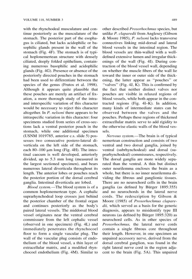
with the rhynchodeal musculature and con-tinue posteriorly as the musculature of thestomach. The posterior part of the esopha-gus is ciliated, but lacks acidophilic or ba-sophilic glands present in the wall of thestomach (Fig. 4F). The stomach is of typi-cal hoplonemertean structure with denselyciliated, deeply folded epithelium, contain-ing numerous basophilic and acidophilicglands (Fig. 4G). Presence or absence of theposteriorly directed pouches in the stomachhad been used to differentiate between thespecies of the genus (Frutos et al. 1998).Although it appears quite plausible thatthese pouches are merely an artifact of fix-ation, a more thorough study of the intra-and interspecific variation of this characterwould be necessary to reject this characteraltogether. In P. nelsoni there appears to beintraspecific variation in this character: fourspecimens studied from series of cross-sec-tions lack a ventral posterior pouch in thestomach, while one additional specimen(USNM 1019785, anterior c.s. slide 5) pos-sesses two consecutive postero-lateral di-verticula on the left side of the stomach,each 80–100 �m long (Fig. 4H). The intes-tinal caecum is well developed, anteriorlydivided, up to 5.3 mm long (measured inthe largest sectioned specimen), and bearsnumerous lateral diverticula throughout itslength. The anterior lobes or pouches reachthe posterior portion of the dorsal cerebralganglia. Intestinal diverticula are lobed.
Blood system.—The blood system is of acommon hoplonemertean type. A cephalicsuprarhynchodeal loop crosses just behindthe posterior chamber of the frontal organand continues posteriorly as the body’spaired lateral vessels. The middorsal bloodvessel originates near the ventral cerebralcommissure from the left cephalic vessel(observed in one specimen) (Fig. 4L) andimmediately penetrates the rhynchocoelfloor to form a single vascular plug. Thewall of the vascular plug consists of endo-thelium of the blood vessel, a thin layer ofextracellular matrix, and a modified rhyn-chocoel endothelium (Fig. 4M). Similar to
other described Prosorhochmus species, butunlike P. claparedii from Anglesey (Gibson& Moore 1985), P. nelsoni lacks transverseconnectives linking mid-dorsal and lateralblood vessels in the intestinal region. Theblood vessels are thin-walled with a well-defined extensive lumen and irregular thick-enings of the wall (Fig. 4I). During con-traction of the blood vessel wall, dependingon whether the muscle fibers of the wall lietoward the inner or outer side of the thick-ening, the latter appear as ‘‘pouches’’ or‘‘valves’’ (Fig. 4J, K). This is confirmed bythe fact that neither distinct valves norpouches are visible in relaxed regions ofblood vessels, while both appear in the con-tracted regions (Fig. 4I–K). In addition,many kinds of intermediate states can beobserved between the valves and thepouches. Perhaps these regions of thickenedextracellular matrix serve to add rigidity tothe otherwise elastic walls of the blood ves-sels.
Nervous system.—The brain is of typicalnemertean construction and consists of twoventral and two dorsal ganglia, joined byventral (subrhynchodeal) and dorsal (su-prarhynchodeal) commissures, respectively.The dorsal ganglia are more widely sepa-rated than the ventral. A thin but distinctouter neurilemma encloses the brain as awhole, but there is no inner neurilemma di-viding the fibrous and ganglionic tissues.There are no neurochord cells in the brainganglia (as defined by Burger 1895:355)and no neurochords in the lateral nervecords. The redescription by Gibson &Moore (1985) of Prosorhochmus clapare-dii, which served as a basis for the genericdiagnosis, appears to misidentify type IIIneurons (as defined by Burger 1895:320) asneurochord cells. As in other species ofProsorhochmus, the lateral nerve cordscontain a single fibrous core throughouttheir length. However, in one specimen anunpaired accessory nerve, derived from thedorsal cerebral ganglion, was found in theright lateral nerve cord in the region adja-cent to the brain (Fig. 5A). This unpaired

490 PROCEEDINGS OF THE BIOLOGICAL SOCIETY OF WASHINGTON
Fig. 5. Prosorhochmus nelsoni. A, B. Consecutive transverse sections through right lateral nerve cord inregion adjacent to brain; note change in position of unpaired accessory nerve (asterisk). C. Transverse sectionthrough left lateral nerve cord; ‘‘upper nerve’’ (asterisk) and muscle fibers (arrows). D. Transverse sectionthrough proboscis insertion, proboscis nerves (arrows). E. Section through eye. F. Longitudinal sagittal sectionthrough both anterior and posterior eyes. G. Longitudinal section through middle part of the frontal organ. H.Transverse section at anterior (slit-like) part of frontal organ. I. Transverse section through frontal organ of sameindividual as (H), acidophilic epithelium (arrows). J. Transverse section through posterior part of frontal organchamber of same individual. K. Transverse section through cerebral organ. L. Transverse section through post-cerebral region. A–C, E–K: scale bars � 50 �m; D, L: scale bars � 100 �m. Abbreviations: bgl, basophilic(mucus) cephalic glands; co, cerebral organ; cof, cerebral organ furrow; foc, frontal organ chamber; lnc, lateralnerve cord; npd, nephridioduct; nph, nephridial tubule; pb, proboscis; pc, eye pigment cup; rhc, rhynchocoel;rhd, rhynchodeum.

VOLUME 118, NUMBER 3 491
Fig. 6. Slightly oblique transverse sections of Prosorhochmus nelsoni from tip of head to brain region:proboscis is absent. A. Opening of frontal organ. B. Frontal organ. C. Section at the level of anterior eye(indicated by asterisk) and anterior portion of ventro-lateral semi-circular cephalic groove, corresponding toanterior cephalic groove. D. Precerebral region. E. Section at level of the right cerebral organ and hind part ofright anterior cephalic groove. F. Ventral cerebral commissure. Scale bar � 200 �m. Abbreviations: agl, acido-philic (proteinaceous) cephalic glands; bgl, basophilic (mucus) cephalic glands; cg, cerebral ganglia; co, cerebralorgan; cof, cerebral organ furrow; cop, cerebral organ opening; fo, frontal organ; es, esophagus; ogl, orange-Gstained (proteinaceous) cephalic glands; rhc, rhynchocoel; rhd, rhynchodeum; vc, ventral brain commissure.
accessory nerve cord later came to lie closeto the dorsal part of the main fibrous core,forming the so-called ‘‘upper nerve,’’ abundle of nerve fibers in the dorsal part of
the fibrous core of the lateral nerve cord,distinguished by their lighter color, whichwe observed in all species of Prosorhoch-mus (Fig. 5B). The difference between this

492 PROCEEDINGS OF THE BIOLOGICAL SOCIETY OF WASHINGTON
Table 2.—Comparison between stylet measurementof Prosorhochmus nelsoni and other described speciesof Prosorhochmus.
Species
Styletlength(�m)
Centralstylet length
to basislength
(S/B) ratio
P. claparedii Keferstein, 1862(after Gibson & Moore 1985) 30–45 0.25–0.33
P. americanus Gibson et al.,1986 90 0.45–0.5
P. adriaticus Senz, 1993 ? 0.25P. chafarinensis Frutos et al.,
1998 99 0.5P. nelsoni (Sanchez, 1973) 84 0.8
upper nerve and a real accessory nerve isthat the upper nerve is never separated fromthe main fibrous core by cell bodies, as isthe accessory nerve, and it is derived fromthe ventral cerebral ganglion. As observedin most monostiliferans studied in the lastthree decades, each lateral nerve cord con-tains a single delicate muscle bundle, con-sisting of 3–7 fibers and running within oradjacent to the fibrous core, near its dorsalborder. In addition, there are several lessconspicuous muscle fibers running alongthe inner lateral side of the fibrous core(Fig. 5C). Muscle fibers associated with thelateral nerve cords can be traced to theirextracerebral origin near the proboscis in-sertion. Numerous cephalic nerves lead an-teriorly from the brain lobes to supply var-ious structures of the head. A stout nervejoins each ventral lobe with the appropriatecerebral sensory organ. A single proboscisnerve trunk originates from the ventral gan-glion on each side near the ventral cerebralcommissure and branches before enteringthe proboscis (Fig. 5D).
Eyes.—The four eyes are of typical pig-ment cup structure and well developed (Fig.5E). The eyes of the anterior pair are slight-ly larger in diameter than the posterior (90�m and 75 �m, respectively). The pigmentcups of the anterior pair are directed an-terolaterally, while those of the posteriorpair are directed posterolaterally (Fig. 5F).
Frontal organ.—The frontal organ,
opening at the tip of the head, is of typicalstructure for the genus Prosorhochmus. Itsstructure, although described slightly dif-ferently for different species of Proso-rhochmus (Gibson & Moore 1985, Gibsonet al. 1986, Frutos et al. 1998), appears tobe remarkably uniform within the genus.The frontal organ of this species consists ofa ciliated canal about 80–100 �m long,lined by a regionally differentiated epithe-lium. Initially narrow and somewhat dorso-ventrally flattened (Figs. 6H, 7A), the canalquickly expands to form a rounded chamber(Figs. 6G, 6I–J, 7B). The ventral wall of thecanal comprises a strongly acidophilic epi-thelium, clad in densely-arranged short cil-ia, whereas the dorsal wall of the canal andventral, dorsal, and posterior walls of thechamber, through which the basophilic mu-cus cephalic glands discharge, has a vacu-olated appearance and bears much longer,sparsely distributed cilia. The acidophilicepithelium divides just in front of the cham-ber to run on its lateral borders. The lateralpart of the lumen of the chamber is dorso-ventrally compressed (Fig. 5I). There are nosubepidermal acidophilic glands associatedwith the acidophilic epidermis of the frontalorgan. It seems that the acidophilic appear-ance comes from the densely arranged elon-gated nuclei of the ciliated cells. The ap-parent higher degree of development of thefrontal organ (comprising the supposed 90�bend) in Prosorhochmus claparedii seemsto be a misinterpretation by Gibson &Moore (1985), carried over to the descrip-tion of Prosorhochmus chafarinensis (Fru-tos et al., 1998). They misinterpreted aslightly oblique cross-section as a verticallongitudinal (i.e., sagittal) section of thefrontal end (Gibson & Moore 1985:149,plate I, fig. e; Frutos et al. 1998:296, fig.3a). As a result, they believed that the canalof the frontal organ first runs horizontallyand then turns ventrally through 90� to endin a flask-shaped chamber. Our re-investi-gation of the original slides of P. claparediiand P. chafarinensis shows that what they

VOLUME 118, NUMBER 3 493
Fig. 7. Slightly oblique transverse sections of Prosorhochmus nelsoni from posterior brain region to intestinalregion. A. Section through posterior region of cerebral ganglia. B. Openings of nephridial ducts. C. Posterior,densely ciliated portion of esophagus and anterior caecal diverticula. D. Stomach. E. End of pylorus. F. Midbodyregion. Scale bar � 200 �m. Abbreviations: agl, acidophilic (proteinaceous) cephalic glands; bgl, basophilic(mucus) cephalic glands; cae, caecum; dvm, dorso-ventral muscles; int, intestine; lnc, lateral nerve cords; mbv,mid-dorsal blood vessel; npd, nephridioduct; nph, nephridial tubule; oct, oocytes; es, esophagus; ogl, orange-G(proteinaceous) cephalic glands; pyl, pylorus; rhc, rhynchocoel; vpl, vascular plug.
believed to be a vertical portion of the canalis actually a ventral ‘‘groove’’ of the canal.
Cephalic glands.—Cephalic glands areextremely well developed. As in othermembers of the genus, they include three
distinct types of cells: basophilic lobuleswith vacuolated appearance (mucusglands), coarsely granular proteinaceousgland cells staining golden-yellow to brownwith Mallory trichrome or orange-G with

494 PROCEEDINGS OF THE BIOLOGICAL SOCIETY OF WASHINGTON
Fig. 8. Diagrammatic cross-section of Prosorhochmus nelsoni in pyloric region. Abbreviations: agl, acido-philic cephalic glands; bgl, basophilic cephalic glands; cm, circular musculature; dvm, dorso-ventral musculature;ep, epidermis; int, intestine; lbv, lateral blood vessel; lm, longitudinal musculature; lnc, lateral nerve cord; mbv,mid-dorsal blood vessel; ncm, nerve cord muscles; nph, nephridial tubule; ogl, orange-G cephalic glands; pyl,pylorus; rhc, rhynchocoel; spm, foregut or splanchnic musculature; upn, upper nerve.
Crandall’s method (orange-G glands), andfinely granular proteinaceous acidophiliccells, staining red with Mallory or Cran-dall’s technique (acidophilic or red glands).However, the degree of development anddistribution of the different types of ce-phalic glands is somewhat different fromthat described for other species of the ge-nus. We refer to these cells as ‘‘glands’’ forthe sake of briefness and also because theyare traditionally referred to as ‘‘glands’’ innemertean literature. These cells are clearlyof secretory nature, but as opposed to realglands they do not form discrete collectives,functioning as units. Moreover, gland cellsof different types may intermix with eachother.
Basophilic (mucus) glands are well de-veloped and, as in other species of the ge-nus, they open through the dorsal and pos-terior epithelium of the frontal organ. Themucus glands are restricted to the dorsalhemisphere precerebrally in some other
prosorhochmids. In this species, the mucusglands are situated centrally, filling most ofthe precerebral region, whereas the acido-philic glands are situated on the periphery,both in dorsal and ventral hemispheres (Fig.6A, B). As acidophilic glands become in-creasingly more abundant in the ventralhemisphere toward the cerebral region, mu-cus glands become interspersed with aci-dophilic glands ventrally and with orange-G glands dorsally and laterally (Fig. 6D–F).Mucus glands in the cerebral region are dis-tributed on the perimeter—dorso-lateral ofthe rhynchocoel, lateral of and under thebrain lobes (Figs. 6F, 7A). Mucus glandsare also well developed postcerebrally andretain essentially the same arrangement asin the cerebral region (Fig. 7B, C). Laterallobes of the mucus glands together with theorange-G glands partially surround excre-tory ducts of the nephridia (Figs. 6L, 7B,C). Dorsal and ventral lobes gradually dis-appear in the stomach-pylorus region (Figs.
VOLUME 118, NUMBER 3 495
7D, E, 8), while lateral lobes gradually di-minish in numbers but reach into the intes-tinal region (Fig. 7D–F).
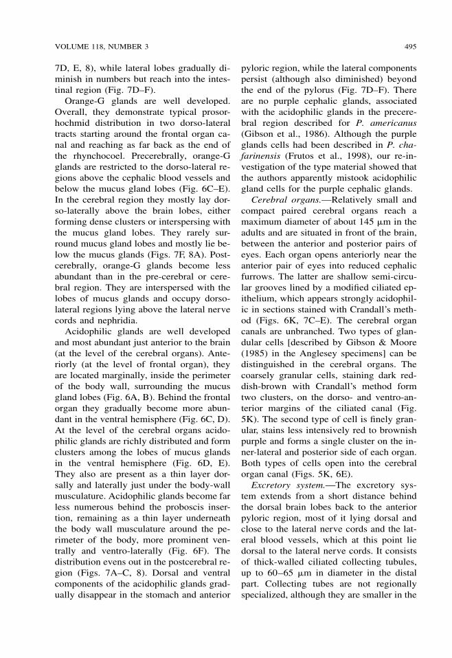
Orange-G glands are well developed.Overall, they demonstrate typical prosor-hochmid distribution in two dorso-lateraltracts starting around the frontal organ ca-nal and reaching as far back as the end ofthe rhynchocoel. Precerebrally, orange-Gglands are restricted to the dorso-lateral re-gions above the cephalic blood vessels andbelow the mucus gland lobes (Fig. 6C–E).In the cerebral region they mostly lay dor-so-laterally above the brain lobes, eitherforming dense clusters or interspersing withthe mucus gland lobes. They rarely sur-round mucus gland lobes and mostly lie be-low the mucus glands (Figs. 7F, 8A). Post-cerebrally, orange-G glands become lessabundant than in the pre-cerebral or cere-bral region. They are interspersed with thelobes of mucus glands and occupy dorso-lateral regions lying above the lateral nervecords and nephridia.
Acidophilic glands are well developedand most abundant just anterior to the brain(at the level of the cerebral organs). Ante-riorly (at the level of frontal organ), theyare located marginally, inside the perimeterof the body wall, surrounding the mucusgland lobes (Fig. 6A, B). Behind the frontalorgan they gradually become more abun-dant in the ventral hemisphere (Fig. 6C, D).At the level of the cerebral organs acido-philic glands are richly distributed and formclusters among the lobes of mucus glandsin the ventral hemisphere (Fig. 6D, E).They also are present as a thin layer dor-sally and laterally just under the body-wallmusculature. Acidophilic glands become farless numerous behind the proboscis inser-tion, remaining as a thin layer underneaththe body wall musculature around the pe-rimeter of the body, more prominent ven-trally and ventro-laterally (Fig. 6F). Thedistribution evens out in the postcerebral re-gion (Figs. 7A–C, 8). Dorsal and ventralcomponents of the acidophilic glands grad-ually disappear in the stomach and anterior
pyloric region, while the lateral componentspersist (although also diminished) beyondthe end of the pylorus (Fig. 7D–F). Thereare no purple cephalic glands, associatedwith the acidophilic glands in the precere-bral region described for P. americanus(Gibson et al., 1986). Although the purpleglands cells had been described in P. cha-farinensis (Frutos et al., 1998), our re-in-vestigation of the type material showed thatthe authors apparently mistook acidophilicgland cells for the purple cephalic glands.
Cerebral organs.—Relatively small andcompact paired cerebral organs reach amaximum diameter of about 145 �m in theadults and are situated in front of the brain,between the anterior and posterior pairs ofeyes. Each organ opens anteriorly near theanterior pair of eyes into reduced cephalicfurrows. The latter are shallow semi-circu-lar grooves lined by a modified ciliated ep-ithelium, which appears strongly acidophil-ic in sections stained with Crandall’s meth-od (Figs. 6K, 7C–E). The cerebral organcanals are unbranched. Two types of glan-dular cells [described by Gibson & Moore(1985) in the Anglesey specimens] can bedistinguished in the cerebral organs. Thecoarsely granular cells, staining dark red-dish-brown with Crandall’s method formtwo clusters, on the dorso- and ventro-an-terior margins of the ciliated canal (Fig.5K). The second type of cell is finely gran-ular, stains less intensively red to brownishpurple and forms a single cluster on the in-ner-lateral and posterior side of each organ.Both types of cells open into the cerebralorgan canal (Figs. 5K, 6E).
Excretory system.—The excretory sys-tem extends from a short distance behindthe dorsal brain lobes back to the anteriorpyloric region, most of it lying dorsal andclose to the lateral nerve cords and the lat-eral blood vessels, which at this point liedorsal to the lateral nerve cords. It consistsof thick-walled ciliated collecting tubules,up to 60–65 �m in diameter in the distalpart. Collecting tubes are not regionallyspecialized, although they are smaller in the

496 PROCEEDINGS OF THE BIOLOGICAL SOCIETY OF WASHINGTON
proximal part (10–15 �m in diameter). Ne-phridial ducts are surrounded by the mucusgland lobes and orange-G glands (Figs. 6L,8A–C, 9). They open dorso-laterally, abouthalfway along the length of the system, viaa single nephridiopore on each side (Figs.5L, 7B). Mononucleate flame cells can beobserved embedded in the extracellular ma-trix in the vicinity of the lateral blood ves-sels. They possess indistinct and irregulartransverse support bars, which can be easilymissed.
Reproductive system and life history.—Unlike all other described species of Pro-sorhochmus, P. nelsoni is gonochoric andoviparous. Reproductive males and femaleswere observed in July–August near Co-quimbo. Gonads are distributed irregularlybetween the lobes of the intestinal divertic-ula. Up to 20–30 mature oocytes can beobserved within the same ovary (Fig. 7F).Females maintained in the laboratory laideggs in cocoons. Eggs are approximately100–150 �m in diameter. Females feedagain following spawning, possibly indicat-ing iteroparity. The embryonic develop-ment from fertilization until the formationof the planuliform swimming larvae takesabout 20 days at room temperature.
Habitat and ecology.—Prosorhochmusnelsoni is fully marine and occurs in themid-intertidal zone on the rocky shore,among the mytilid Perumytilus purpuratusand barnacles Jehlius cirratus, according toSanchez (1973), and on rocks and boulderscovered by algal turf, inhabited by a widevariety of marine invertebrates (polychaets,bivalves, and crustaceans representing themost abundant groups; Thiel et al. 2001).One of us (MT personal observations) con-siders P. nelsoni to be the most commonnemertean species in the rocky intertidal ofthe Chilean coast. It is commonly foundduring morning low tides, particularly un-der overcast conditions.
Geographic distribution.—The Pacificcoast of South America. Only known fromChile. Type locality: Quintero (32�46�S,71�31�W) (Sanchez 1973); other known lo-
calities: La Pampilla Beach near Coquimbo(29�57�S, 71�21�W) (Thiel et al. 2001), Ar-ica (Arica: 18�29�S, 70�20�W) and Concep-cion (36�50�S, 73�03�W) (MT, personal ob-servations).
Conclusions
Characters such as bilobed head with adorsal epidermal fold (the ‘‘smile’’), trun-cated stylet basis, well-developed tubularfrontal organ with laterally differentiatedepithelium, well-developed cephalic glands,combining mucus and proteinaceous com-ponents and nephridial system with thickexcretory tubules, and a single pair of ne-phridioducts place this species into the ge-nus Prosorhochmus. This species differsfrom all other described species of Proso-rhochmus in being gonochoric and ovipa-rous (vs. hermaphroditic and ovovivipa-rous). Additionally, P. nelsoni differs fromother described species of Prosorhochmusin having a greater S/B ratio (Table 2).However, the possibility exists that this isbecause we measured stylets in sexuallyimmature individuals and that the S/B ratiois smaller in mature specimens. Our unpub-lished sequence data from the two mito-chondrial genes 16S rDNA and Cyto-chrome Oxidase Subunit I show that the av-erage sequence divergence between P. nel-soni and other species of Prosorhochmus(P. americanus, P. claparedii, P. cf. adria-ticus and an undescribed species from Be-lize) is comparable to that between the oth-er species: 6.82% for 16S and 8.82% forCOI. Lastly, P. nelsoni is found only on thePacific side of South America (coast ofChile), whereas the other described speciesare known only from the Atlantic and Med-iterranean.
A critical observation for prosorhoch-mids is that presence vs. absence of a 90�bend in the frontal organ is a misinterpre-tation and cannot be used to distinguishmembers of the genus. Reinvestigation ofthe material of all described species of Pro-sorhochmus also revealed that no member

VOLUME 118, NUMBER 3 497
of this genus possesses neurochord cells, incontrast to the generic diagnosis (Gibson &Moore 1985:146). Finally, we suggest thatthe so-called valves and pouches in theblood vessels represent artifacts of fixationand their presence or absence has no diag-nostic significance. Morphological charac-ters previously used in Prosorhochmus sys-tematics, such as presence/absence of dor-so-ventral musculature in the foregut re-gion, esophageal musculature, ciliation inthe posterior esophageal region, neural sup-ply in the posterior proboscis chamber, pos-terior stomach pouch, and number of mus-cle fibers in the lateral nerve cords must beused with caution and need to be thorough-ly re-evaluated, as they might be inconsis-tent with species boundaries. That is outsidethe scope of this article but is work in prog-ress.
Acknowledgments
This work was supported by the NationalScience Foundation PEET grant DEB-9712463 to JLN and Diana Lipscomb. Weare grateful to Malva Sanchez for providingthree specimens of P. nelsoni collected byher in the type locality. Specimens of sev-eral previously described species of Pro-sorhochmus were loaned to us by the Cam-bridge Museum of Zoology, Cambridge,UK; Museo Nacional de Ciencias Natura-les, Madrid, Spain, and Natural HistoryMuseum, Vienna, Austria. Ray Gibsonkindly provided material of P. claparediifrom his personal collection. We also thankBarbara Littman for her help with histolog-ical preparations and the students of Zool-ogia I at the Universidad Catolica del Nor-te, Coquimbo, Chile for taking the mea-surements of the stylet apparatus.
Literature Cited
Burger, O. 1895. Die Nemertinen des Golfes von Nea-pel und der angrenzenden Meeres-Abschnit-te.—Fauna und Flora des Golfes von Neapel 22:1–743.
Chernyshev, A. V. 2002. Description of a new speciesof the genus Poseidonemertes (Nemertea; Mon-
ostilifera) with establishment of the family Po-seidonemertidae. (In Russian).—ZoologicheskiiZhurnal. 81:909–916.
Frutos I., S. Montalvo, & J. Junoy. 1998. A new spe-cies of Prosorhochmus (Hoplonemertea, Mon-ostilifera) from the Chafarinas Islands (westernMediterranean).—Journal of Zoology (London)245:293–298.
Gibson, R., & Moore, J. 1985. The genus Prosorhoch-mus Keferstein, 1862 (Hoplonemertea).—Jour-nal of Zoology (London) 206A:145–162.
Gibson, R., J. Moore, E. E. Ruppert, & J. M. Turbe-ville. 1986. A new species of Prosorhochmus(Hoplonemertea, Monostilifera) from SouthCarolina.—Journal of Zoology (London) 209A:327–335.
Keferstein, W. 1862. Untersuchungen uber niedereSeethiere.—Zeitschrift fur WissenschaftlicheZoologie 12:1–147.
Kirsteuer, E. 1974. Description of Poseidonemertescaribensis sp. n., and discussion of other taxaof Hoplonemertini Monostilifera with dividedlongitudinal musculature in the body wall.—Zoologica Scripta, 3:153–166.
Sanchez, M. 1973. Sobre 4 especies de nemertinos deQuintero (Chile).—Studies on the Neotropicalfauna 8:195–214.
Senz, W. 1993. Nemertinen europaischer Kustenber-eiche (Nebst erganzenden Angaben zur Anato-mie von Apatronemertes albimaculosa Wilfert& Gibson 1974).—Annalen des Naturhisto-rischen Museums in Wien Serie B Botanik undZoologie 94–95:47–145.
Thiel, M., N. Ulrich, & N. Vasquez. 2001. Predationrates of nemertean predators: the case of a rockyshore hoplonemertean feeding on amphipods.—Hydrobiologia 456:45–57.
Thollesson, M., & J. L. Norenburg. 2003. Ribbonworm relationships—a phylogeny of the phy-lum Nemertea.—Proceedings of the Royal So-ciety. London. B Biology. 270:407–415.
Associate Editor: Stephen L. Gardiner
AppendixExamined histological material
P. nelsoni (Sanchez, 1973). USNM (173164): seriesof cross-sections of anterior part and foregut region.Coll. Sanchez, Quintero, Chile. Reproductive female;USNM (173163): series of frontal sections of anteriorpart and cross-sections of posterior part. Coll. Sanchez,Quintero, Chile. Immature individual; USNM(173162): series of sagittal sections of anterior part,cross-sections of posterior part. Coll. Sanchez, Quin-tero, Chile. Reproductive female; USNM (1019782):series of cross-sections of anterior third of body. Coll.MT, Coquimbo, Chile. Reproductive female; USNM

498 PROCEEDINGS OF THE BIOLOGICAL SOCIETY OF WASHINGTON
(1019784): series of cross-sections of anterior fourthof body. Coll. MT, Coquimbo, Chile. Reproductive fe-male; USNM (1019785): series of cross-sections of an-terior part and adjacent midbody region. Coll. MT, Co-quimbo, Chile. Reproductive female; USNM(1019786): two unsectioned specimens in alcohol.Coll. MT, Coquimbo, Chile.
P. claparedii Keferstein, 1862. CMZ (A6): series ofcross-sections of single specimen. Coll. Gibson, An-glesey, UK; series of transverse and longitudinal sec-tions of four specimens from personal collection ofRay Gibson. Coll. Gibson. Anglesey, England; USNM(1020508, 1020509): series of cross-sections of ante-rior parts of two immature individuals. Coll. SAM.
Bilbao, Spain; USNM (1020510–1020513): series ofcross-sections of anterior ends of two specimens, seriesof cross-sections of one whole specimen and series offrontal sections of anterior end of one specimen. Coll.SAM. Roscoff, France.
P. americanus Gibson et al., 1986. Series of cross-sections of holotype (USNM 98550), two paratypes(USNM 98551, 98552) and three additional specimensfrom Florida, USA collected by JLN.
P. chafarinensis Frutos et al., 1998. Series of cross-sections of holotype (MNHM AA1) and two paratypes(MNHM AA2, AA5).
P. adriaticus Senz, 1993. Series of cross-sections ofholotype (VMNH 3254) and two other specimens fromtype locality collected by Senz. (VMNH 4292, 4293).