the signigicance of brittle reaction layers in fusing of
TRANSCRIPT

Department of Stomatognathic Physiology and Prosthetic Dentistry
Institute of Dentistry
University of Helsinki, Helsinki, Finland
Department of Oral and Maxillofacial Diseases
Helsinki University Central Hospital
Helsinki, Finland
Department of Electronics
Faculty of Electronics, Communications and Automation
School of Science and Technology
Aalto University, Espoo, Finland
The significance of brittle reaction
layers in fusing of dental ceramics to
titanium
Mikko Tapani Saloniemi
ACADEMIC DISSERTATION
To be publicly discussed with the permission of the Faculty of Medicine of the
University of Helsinki, in the Institute of Dentistry, Lecture Hall 1,
Mannerheimintie 172, on December 17th
2010, at 12h00.
Helsinki 2010
2
Supervised by
Professor Mauno Könönen
Department of Stomatognathic Physiology and Prosthetic Dentistry
Institute of Dentistry, Faculty of Medicine
University of Helsinki, Helsinki, Finland
and
Professor emeritus Jorma Kivilahti
Department of Electronics
Faculty of Electronics, Communications and Automation
Helsinki University of Technology, Espoo, Finland
Reviewed by
Professor Pekka Vallittu
Department of Biomaterials Science
Institute of Dentistry, Faculty of Medicine
University of Turku, Turku, Finland
and
Docent Jarmo Hautaniemi
Faculty of Technology and Maritime Management Rauma (Technology)
Satakunta University of Applied Sciences, Rauma, Finland
Discussed by
Professor Timo Närhi
Department of Prosthetic Dentistry
Institute of Dentistry, Faculty of Medicine
University of Turku, Turku, Finland
ISBN 978-952-92-8246-3 (paperback) Helsinki University Print
ISBN 978-952-10-6716-7 (PDF) Helsinki 2010
3
ACKNOWLEDGEMENTS
This study was carried out at the Institute of Dentistry in the University of Helsinki and the
Department of Electronics in the School of Science and Technology – former Helsinki
University of Technology. I wish to thank Finnish Dental Society Apollonia and the Helsinki
University Central Hospital for making it financially possible to complete this study.
I would like to express my deep gratitude to my supervisor, Prof. Mauno Könönen. Prof.
Könönen demonstrated open-minded confidence in inviting a young student into an important
and intriguing research project, and in providing constant encouragement and support. I also
wish to thank my other supervisor, Prof. (emer.) Jorma Kivilahti, for his extensive and
profound knowledge on material sciences, for our long discussions during the final stages of
completing this study, and for providing me with broad access to facilities and equipment.
Thanks to the special nature of this joint study, I was able to work as research assistant and
researcher in the Helsinki University of Technology, gaining significant interdisciplinary
experience from a variety of other projects and studies, as well as teaching experience.
I gratefully acknowledge my skilled referees, Prof. Pekka Vallittu and Docent Jarmo
Hautaniemi, for their insightful commentary and constructive criticism. I also wish to thank
my honored opponent, Prof. Timo Närhi, for agreeing to take on this task.
To Mr. Jukka Wichmann I owe my sincere thanks for the use of his dental laboratory facilities
and for his expert guidance in dental techniques. His constant positive attitude and cheerful
disposition made working on this study an enjoyable experience.
My collective thanks go to the personnel of the Helsinki University of Technology, who
readily accepted an outsider in their midst and introduced him to the use of advanced
analytical equipment, also beyond the button-pushing level. Our cooperation will certainly be
of future benefit to dental medicine; the skills I have acquired in the process are also bound to
prove invaluable in my research career.
Finally, I would like to apologize to my wife Tiina, our cat Questor and my family for so
completely depending on their warm and patient support. My brother Timo bore the brunt of
difficult questions and late-night revision sessions, while my father Hannu guided me through
the world of academic writing, and my mother and colleague Elina kept alive my faith in
practical clinical dentistry.
Mikko Saloniemi
Helsinki, October 2010
ACKNOWLEDGEMENTS

4
TABLE OF CONTENTS
TABLE OF CONTENTS ......................................................................................................................... 4
ABBREVIATIONS .................................................................................................................................. 6
ABSTRACT ............................................................................................................................................. 7
1. INTRODUCTION ................................................................................................................................ 9
2. REVIEW OF THE LITERATURE .................................................................................................... 10
2.1. Titanium in comparison to other metals in dental metalloceramics............................................ 10
2.2. Factors influencing joint integrity ............................................................................................... 14
2.2.1. Coefficient of thermal expansion (CTE) ............................................................................. 14
2.2.2. Wetting of titanium surfaces ................................................................................................ 16
2.2.3. α-case layer .......................................................................................................................... 17
2.2.4. Oxygen and titanium oxides ................................................................................................ 19
2.2.5. Titanium silicides ................................................................................................................ 20
2.3. Methods to increase joint strength .............................................................................................. 22
2.3.1. Surface roughening .............................................................................................................. 22
2.3.2. Bonding agents and interlayers ............................................................................................ 23
2.4. Testing joints between dental ceramics and titanium.................................................................. 27
3. AIMS OF THE STUDY ..................................................................................................................... 30
4. SIGNIFICANCE OF TITANIUM OXIDE LAYERS ....................................................................... 31
4.1. Materials and methods ................................................................................................................ 32
4.1.1. Sample fabrication ............................................................................................................... 32
4.1.2. Sample characterization ....................................................................................................... 34
4.2. Results ......................................................................................................................................... 36
4.3. Discussion ................................................................................................................................... 39
4.3.1. On ALD manufactured TiO2 layers ..................................................................................... 39
4.3.2. On scanning acoustic microscopy ....................................................................................... 39
4.3.3. On three-point bending testing ............................................................................................ 40
4.3.4. On titanium and oxygen ....................................................................................................... 40
4.4. Conclusions ................................................................................................................................. 46
5. PHOTOINDUCED SUPERHYDROPHILICITY .............................................................................. 47
5.1. Materials and methods ................................................................................................................ 49
5.1.1. Sample fabrication ............................................................................................................... 49
TABLE OF CONTENTS
5
5.1.2. Photoinduced hydrophilicity characterization ..................................................................... 50
5.2. Results ......................................................................................................................................... 51
5.3. Discussion ................................................................................................................................... 54
5.3.1. On titanium as photoactive material .................................................................................... 54
5.3.2. On light source, UV parameters and achieved hydrophilicity ............................................. 56
5.3.3. On Ti wetting with dental ceramics ..................................................................................... 57
5.4. Conclusions ................................................................................................................................. 58
6. SILVER INTERLAYERS .................................................................................................................. 59
6.1. Materials and methods ................................................................................................................ 60
6.1.1. Sample fabrication ............................................................................................................... 60
6.1.2. Sample characterization ....................................................................................................... 61
6.2. Results ......................................................................................................................................... 62
6.3. Discussion ................................................................................................................................... 68
6.3.1. On the biocompatibility of Ag ............................................................................................. 69
6.3.2. On oxygen dissolution ......................................................................................................... 69
6.3.3. On plastic deformation and bond strength results ............................................................... 69
6.3.4. On applicability and blasting particle contamination .......................................................... 71
6.4. Conclusions ................................................................................................................................. 72
7. PHOTOLITHOGRAPHIC ETCHING ............................................................................................... 73
7.1. Materials and methods ................................................................................................................ 74
7.1.1. Sample fabrication ............................................................................................................... 74
7.1.2. Sample characterization ....................................................................................................... 76
7.2. Results ......................................................................................................................................... 76
7.3. Discussion ................................................................................................................................... 80
7.3.1. On test results and reaction layers ....................................................................................... 80
7.3.2. On photolithographic etching .............................................................................................. 81
7.4. Conclusions ................................................................................................................................. 82
8. GENERAL DISCUSSION ................................................................................................................. 83
8.1. On the need for novel and enhanced titanium metalloceramics techniques................................ 83
8.2. On metalloceramic joints and fractography ................................................................................ 83
8.3. On future aspects ......................................................................................................................... 86
9. CONCLUSIONS ................................................................................................................................ 89
10. TIIVISTELMÄ SUOMEKSI ........................................................................................................... 90
REFERENCES ....................................................................................................................................... 94
TABLE OF CONTENTS
6
ABBREVIATIONS
AFAP………………………………………. area fraction of adherent porcelain
ALD………………………………………... atomic layer deposition
ASM…………………………………...…… American Society for Metals
CAD………………………..………………. computer-aided design
CAM……………………………..………… computer-aided manufacturing
CB……………………………………..…… conductance band
cpTi……………………………………….... commercially pure titanium
CTE……………………………………….... coefficient of thermal expansion
EBG…………………………………………. band gap energy
EDS……………………………………….... energy dispersive X-ray spectroscopy
FPD……………………………………….... fixed partial denture
IMC……………………………………….... intermetallic compound
MPIIID……………………………………... metal plasma immersion ion implantation and
deposition
Nd/YAG……………………………………. neodymium-doped yttrium aluminum garnet
(laser)
SAM………………………………………... scanning acoustic microscope
SEM………………………………………... scanning electron microscope
TI[O]x……………………………………..... titanium oxygen solid solution (oxygen atoms in
octahedral spaces of titanium lattice, not titanium
oxide), see section 4
TPB……………………………………….... three-point bending
VB………………………………………….. valence band
WEDM……………………………………... wire electric discharge machine
WDS………………………………………... wavelength dispersive X-ray spectroscopy
XPS……………………………………….... X-ray photoelectron spectroscopy
XRD………………………………………... X-ray diffraction
ABBREVIATIONS
7
ABSTRACT
This thesis comprises four intercomplementary parts that introduce new approaches to brittle
reaction layers and mechanical compatibility of metalloceramic joints created when fusing
dental ceramics to titanium.
The first part investigates the effects of TiO2 layer structure and thickness on the joint
strength of the titanium-metalloceramic system.
Three groups of standard metalloceramic samples with different TiO2 layer thickness and
crystal structure were tested. The TiO2 layers were produced using atomic layer deposition
(ALD). Scanning acoustic microscopy (SAM), three-point bending (TPB), cross-section
microscopy, scanning electron microscopy (SEM), and energy dispersive X-ray spectroscopy
(EDS) were employed.
Samples with all TiO2 thicknesses displayed good ceramics adhesion to Ti, and uniform
TPB results. The fracture mode was independent of oxide layer thickness and structure.
Cracking occurred deeper inside titanium, in the oxygen-rich Ti[O]x solid solution surface
layer.
During dental ceramics firing TiO2 layers dissociate and joints become brittle with
increased dissolution of oxygen into metallic Ti and consequent reduction in the metal
plasticity. To accomplish an ideal metalloceramic joint this needs to be resolved.
The second part introduces photoinduced superhydrophilicity of TiO2.
Test samples with ALD deposited anatase TiO2 films were produced. Band gap energy
(EBG) for the TiO2 layers was estimated from transmittance measurements. Samples were
irradiated with UV light (> EBG) to induce superhydrophilicity of the surfaces through a
cascade leading to increased amount of surface hydroxyl groups. Samples were divided into
two groups D and E to study the required irradiation time. Hydrophilicity of the TiO2 surfaces
was assessed by sessile drop contact angle measurements.
The reference contact angle prior to UV radiation was ~55˚. Superhydrophilicity (contact
angle ~0˚) was achieved within 2 minutes of UV radiation. After initial partial recovery
during the first 10 minutes, the contact angle remained below 20˚ for 1h. Total recovery was
not observed within 24h storage.
Photoinduced ultrahydrophilicity can be used to enhance wettability of titanium surfaces,
an important factor in dental ceramics veneering processes.
The third part addresses interlayers designed to restrain oxygen dissolution into Ti during
dental ceramics fusing.
ABSTRACT
8
The main requirements for an ideal interlayer material are proposed. Based on these
criteria and systematic exclusion of possible interlayer materials silver (Ag) interlayers were
chosen. Six groups of standard metalloceramic samples were studied. Groups F, J, K and L
were Al2O3-blasted, Groups G and H were left polished. Thin silver interlayers were produced
on Groups G, H, J and K by using the DIARC® plasma coating method and thicker interlayers
on Group J by electrochemical baths. Analysis methods were as in the first part.
Good ceramics adhesion to titanium was observed in all test groups save for G and H,
which both exhibited several areas of poor contact. SEM/EDS analyses revealed attachment
of alumina particles on the Al2O3-blasted titanium. Ag covered this contamination in Group
L. TPB results were significantly better in Group L samples compared to Group F. Generally,
cracking occurred inside titanium in oxygen-rich Ti[O]x solid solution (F, G, H, J, K), locally
also between Ti and dental ceramics (K, L). In Group L multiple cracks occurred inside dental
ceramics, none inside Ti structure.
Ag interlayers of 5 μm on Al2O3-blasted samples can be efficiently used to retard
formation of the brittle oxygen-rich Ti[O]x layer, thus enhancing metalloceramic joint
integrity. Based on the literature, isolation of alumina blasting particle contamination was also
considered beneficial. The most brittle component in metalloceramic joints with 5 μm Ag
interlayers was bulk dental ceramics instead of Ti[O]x.
The fourth part investigates the importance of mechanical interlocking and presents a new
approach to overcome mechanical problems of brittle reaction layers.
Mechanically polished, Al2O3-blasted, and photolithographically etched standard
metalloceramic samples, Groups M, N and P, showed no significant TPB test differences.
Cracking occurred through Ti[O]x, but in photolithographically etched samples also locally
through dental ceramics.
Hence, the significance of mechanical interlocking achieved by conventional surface
treatments can be questioned as long as the formation of the brittle layers (mainly oxygen-rich
Ti[O]x) cannot be sufficiently controlled. Photolithographically etched pits can be used to
cause cracking of dental ceramics instead of the more brittle reaction layers. The current depth
and steepness of the pits, however, were insufficient for extensive stress redistribution.
In summary – in contrast to former impressions of thick titanium oxide layers – this thesis
clearly demonstrates diffusion of oxygen from sintering atmosphere and SiO2 to Ti structures
during dental ceramics firing and the following formation of brittle Ti[O]x solid solution as
the most important factors predisposing joints between Ti and SiO2-based dental ceramics to
low strength. This among other predisposing factors such as residual stresses created by the
coefficient of thermal expansion mismatch between dental ceramics and Ti frameworks can
be avoided with Ag interlayers.
ABSTRACT
9
1. INTRODUCTION
Titanium is a widely used material in prosthetic dentistry. It first became renowned after the
pioneering research and good clinical results on titanium dental implant fixtures by P-I
Brånemark et al., and has been used in metalloceramic crowns since the 1970s. Recent
progress in CAD/CAM processing methods has made wrought titanium solutions possible in
addition to ordinary cast titanium applications, increasing titanium’s applicability and use in
dental metalloceramics. Current developments in framework production techniques, excellent
corrosion resistance and documented biocompatibility have made titanium a reasonable
choice compared to considerably more expensive gold and palladium alloys in crown and
bridge designing as well as overlay dentures. This brings esthetic and durable fixed
prosthetics into the reach of a vast number of new people offering a chance to enhance
patients’ quality of life.
The high reactivity of titanium presents a problem in fusing of dental ceramics to
titanium structures, however: joints are relatively brittle. There are many factors behind this,
including differences in the coefficient of thermal expansion (CTE) between metallic
frameworks and ceramics materials; use of too high fusing temperatures or framework casting
and subsequent α-case layer formation; and brittle reaction layers formed through diffusion of
oxygen and ceramics material into the titanium lattice. While different approaches including
low-fusing dental ceramics, surface roughening and various bonding agents have been
adapted to enhance mechanical integrity of joints, dissolution of oxygen into metallic titanium
during dental ceramics firing, related solid solution strengthening and significantly reduced
plasticity of surface layers of metallic titanium remain a practical problem.
Recent publications have stated a concern about the insufficient amount of research of
titanium-based metalloceramics [Garbelini et al. 2003, Vásquez et al. 2008]. While adherence
problems between titanium and dental ceramics have been considered the main limiting factor
in manufacturing titanium-ceramic restorations for decades [Wang et al. 1999], understanding
the effects of different surface treatments on the formation of critical reaction layers in
titanium-ceramics joints is still defective. Thorough fractographic analyses, i.e. identification
of crack initiation, pattern of crack propagation, energetics of the fracture and classification of
the phases along the fracture plane have been infrequent [Marshall et al. 2010]. Use of
titanium-based metalloceramics, however, is already established in routine dental technology,
healthcare practice and patients’ knowledge. Thus it is of the foremost clinical importance and
duty to study and solve problems associated with fusing dental ceramics to titanium.
1. INTRODUCTION

10
2. REVIEW OF THE LITERATURE
The focus of the study is on brittle reaction layers in joints between dental ceramics and
titanium. The first part of the literature overview gives a general introduction to titanium in
comparison to other metals in dental metalloceramics – such as gold, palladium, their alloys
and cobalt chromium – and to clinical performance of titanium-based metalloceramics. The
second part discusses the most important factors influencing integrity of joints between Ti and
dental ceramics. The third part of the review addresses methods designed to increase joint
strength are discussed. The last section introduces available methods to study metalloceramic
joints.
2.1. Titanium in comparison to other metals in dental metalloceramics
Expensive conservative noble alloys in fixed prosthodontics have been gradually replaced by
more affordable base metal alloys [Yilmaz & Dinçer 1999, Roach 2007, Roberts et al. 2009].
Noble alloys are usually based on gold or palladium and comprise alloys such as Au-Pt-Pd,
Au-Pd, Au-Pd-Ag, Pd-Ag, Pd-Cu and Pd-Ga. Base metal alloys consist of nickel- (Ni) and
cobalt- (Co) based solutions, both containing chromium (Cr) as their second largest
constituent. In addition to high cost, low sag resistance of noble alloys inflicts limitation of
applications to crowns and fixed partial dentures (FPDs) with restricted amount of units, and
some alloys also display discoloration of dental ceramics. [Roberts et al. 2009] Casting
techniques and handling of noble alloys is, however, well known and has been in clinical use
and the focus of follow-up for decades. Investigation on fusing of dental ceramics to gold
alloys was roughly at the same stage in Finland in the 1970s [Yli-Urpo 1975] as the
corresponding study of titanium alloys presently is. Unfortunately, some of the methods
subsequently developed for improving on this joint are not applicable to titanium [Kimura et
al. 1990].
Also base metal alloys portray several problems. Carpenter and Goodkind [1979]
stated, based on their investigations and evaluation of surface texture for roughness, surface
area, re-entrant angles and stress concentration of Au-Ag-Pd and Ni-Cr alloys, that in dental
ceramics, veneering wetting of metal surfaces of the precious alloy was better than that of the
nonprecious alloy. Further disadvantages of base metal alloys are poor biocompatibility, low
corrosion resistance, and discoloration of porcelain [Yilmaz & Dinçer 1999]. Nickel-
chromium-beryllium alloys can no longer be recommended because of the health concerns
associated with beryllium. For patients allergic to nickel, cobalt-chromium alloys have been
the most common base-metal alternative. [Roberts et al. 2009]
2. REVIEW OF THE LITERATURE
11
Titanium provides a competitive alternative both in affordability and biocompatibility.
It has many advantages and favorable properties over the other dental alloys and Ti
restorations are definitely much cheaper than their gold alloy counterparts [Vásquez et al.
2008]. Titanium is considered to be the most biocompatible metal for dental prostheses
[Roberts et al. 2009]. Even with gold (Au), a positive relationship between contact allergy to
gold and the amount of dental gold surfaces in oral cavity and dose-related release of gold
into blood plasma from dental gold restorations has been suggested [Ahlgren et al. 2002,
Ahlgren et al. 2007]. Thus, Ti has been a welcomed material in prosthodontics.
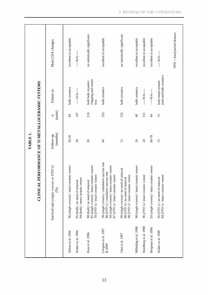
Even though there are not as many or as long follow-up studies with titanium
metalloceramics as with traditional noble alloy metalloceramics, the first results of clinical
performance look promising [Nilson et al. 1994, Walter et al. 1994, Kaus et al. 1996, Lövgren
et al. 1997, Chai et al. 1997, Milleding et al. 1998, Smedberg et al. 1998, Bergman et al. 1999,
Walter et al. 1999, Lövgren et al. 2000]. Survival rate varying from 84% to 99% in follow-ups
of 1 to 6 years, as well as changes mainly from excellent to acceptable in the California
Dental Association (CDA) ratings for surface, color and anatomic form, have been reported
[Table 1]. Good margin integrity, high patient acceptance and low incidence of caries have
also been stated. Modern individual design of outer surfaces of titanium copings not
excecuted in some of the older studies might further improve the success rates and reduce
ceramic material failures by optimizing stress distribution [Nilson et al. 1994, Bergman et al.
1999].
Nilson et al. [1994] compared results from their clinical studies with titanium
metalloceramics to previous follow-up studies where survival rates concerning ceramics
material defects were 90% with cast palladium (Pd) base alloy FPDs after 3 years and 97-98
% in conventional metalloceramic restorations after 10 years. In a 5-year study comparing
veneers on titanium and a gold alloy [Walter et al. 1999], the respective intact dental ceramics
survival rates of 84 % and 98 % were reported. No significant difference, however, was found
in defects requiring removal of the studied FPDs. A single such case, from metalloceramic
joint fracture, was observed with titanium framework. The rarity of such defects, however,
suggested clinical suitability of titanium veneers.
In addition to clinical studies, suitability of titanium for long term use has been studied
by evaluating the effects of thermal and mechanical cycling on flexural strength of
metalloceramic joints [Vásquez et al. 2007, Oyafuso et al. 2008, Vásquez et al. 2009]. Bond
strength results are influenced by numerous factors difficult to equalize, yet they can be used
for rough comparison between different systems. Oyafuso et al. [2008] compared gold alloy
and titanium-based metalloceramic systems, showing on average higher three-point bending
test results for the gold alloy systems (55 MPa vs. 32 MPa), regardless of the fatigue
conditions imposed. Mechanical and thermomechanical fatigue conditions decreased the test
results significantly for both systems. Vásquez et al. [2009] tested three dental ceramics
materials designed for titanium and one control group designed for Au-Pd alloy. Shear bond
strength results of the titanium-based systems ranged between 42 and 64 MPa, one system
2. REVIEW OF THE LITERATURE
12
even exceeding results achieved with Au-Pd alloy (61 MPa). The study also included
mechanical and thermal cycling, which had a significant impact on the dental ceramics
material systems designed for titanium but not on the reference system designed for Au-Pd. In
an earlier study Vásquez et al. [2007] studied the effects of mechanical and thermal cycling
on three-point bending test results of three different dental ceramics materials fused to
titanium. Control values between 33 MPa and 39 MPa were decreased to 27-28 MPa,
significantly for all the tested metalloceramic combinations.
Pang et al. [1995] reported bond strength of dental ceramics fired on cast titanium or
fired on machine-milled titanium to be statistically comparable yet also statistically inferior to
results achieved with palladium-copper alloy dental ceramics system. The respective mean
loads at bond failure in three-point bending test were 8 N, 7 N and 13.5 N, which is still
equaivalent to bond strength close to 40 MPa for the dental ceramics system used with
titanium. The elastic modulus of the cast titanium is not stated in the paper, however, and so
the bond strength results are not easily comparable with other studies. Dérand and Herø
[1992] reported high bond strength values of 40-50 MPa in a four-point bending setup with
titanium-based metalloceramics close to values recorded with palladium alloys veneered with
dental ceramics in a previous study by Syverud, Kvam and Herø [1987]. Homann et al. [2006]
studied strain energy release rate in four-point bending for Ti metalloceramic test samples and
a gold-palladium reference group. One low fusing dental ceramics bonder and two gold
materials were also tested with the titanium system. Higher interfacial toughness values were
achieved with the two gold bonder materials than with the gold-palladium reference samples.
Normal paste bonder performed worst in these tests. Yilmaz and Dinçer [1999] suggested
bond compatibility cast titanium-based metalloceramics to be comparable with the Ni-Cr
dental ceramics system – even without reporting of special removal of the α-case layer [see
section 2.2.3.]. Atsü and Berksun [2000] reported three-point bending test values of three
different dental ceramics systems with titanium to range from 33% to 60% of the results
achieved with a Ni-Cr control group. All bond strength results were considerably poor. Özcan
and Uysal [2005] reported three-point bending test values of SiO2-coated titanium samples
with applied dental ceramic systems and titanium to be 47% to 64% of the values achieved in
the control group with dental ceramics applied on Ni-Cr.
Even though the most typical failures in clinical tests occur inside dental ceramics, and
only rarely in joints between titanium and ceramic material, mechanical tests generally imply
inferior bond strength in titanium-based systems compared to noble alloys [Pang et al. 1995,
Oyafuso et al. 2008, Vásquez et al. 2009]. According to recent reviews titanium-based
metalloceramics would appear to have a promising future in fixed prosthodontics as a
biocompatible and less expensive solution, though much work remains in optimizing the
dental ceramics fusing methods [Haag & Nilner 2007, Roberts et al. 2009].
2. REVIEW OF THE LITERATURE
13
2. REVIEW OF THE LITERATURE
TA
BL
E 1
.
CL
INIC
AL
PE
RF
OR
MA
NC
E O
F T
i M
ET
AL
LO
CE
RA
MIC
SY
ST
EM
S
Su
rviv
al r
ate
(sin
gle
cro
wn
s o
r F
PD
’s)
F
oll
ow
-up
n
Fai
lure
in
M
ain C
DA
ch
ang
es
(
%)
(
mo
nth
s)
(
un
its)
N
ilso
n e
t al
. 19
94
9
6 (
sin
gle
cro
wn
s) /
in
tact
cer
amic
ven
eer
2
6-3
0
4
4
bu
lk c
eram
ics
ex
cell
ent
to a
ccep
tab
le
Wal
ter
et a
l. 1
994
9
5 (
bo
th)
/ no
nee
d o
f re
mov
al
36
1
47
---
--N
/A--
---
--
---N
/A--
---
8
4 (
bo
th)
/ in
tact
cer
amic
ven
eer
Kau
s et
al.
19
96
.
98
(b
oth
) /
no
nee
d o
f re
mov
al
30
1
16
bo
th b
ulk
cer
amic
s
no
sta
tist
ical
ly s
ign
ific
ant
8
5 (
sin
gle
cro
wn
s) /
in
tact
cer
amic
ven
eer
chip
pin
g a
nd v
enee
r
5
9 (
FP
D’s
) /
inta
ct c
eram
ic v
enee
r
loss
Lö
vg
ren
et
al. 19
97
9
9 (
sin
gle
cro
wn
s) /
cu
mula
tiv
e su
cces
s ra
te
6
0
3
33
bu
lk c
eram
ics
ex
cell
ent
to a
ccep
tab
le
& 2
000
9
8 (
FP
D’s
) /
cum
ula
tiv
e su
cces
s ra
te
9
4 (
sin
gle
cro
wn
s) /
in
tact
cer
amic
ven
eer
8
7 (
FP
D’s
) /
inta
ct c
eram
ic v
enee
r
Ch
ai e
t al
. 1
99
7
9
9 (
sin
gle
cro
wn
s) /
no n
eed
of
rem
ov
al
1
2
2
32
bu
lk c
eram
ics
n
o s
tati
stic
ally
sig
nif
ican
t
9
8 (
FP
D’s
) /
no
nee
d o
f re
mo
val
9
8 (
FP
D’s
) /
inta
ct c
eram
ic v
enee
r
Mil
led
ing
et
al.
19
98
9
8 (
sin
gle
cro
wn
s) /
in
tact
cer
amic
ven
eer
2
4
4
0
bu
lk c
eram
ics
ex
cell
ent
to a
ccep
tab
le
Sm
edb
erg
et
al. 19
98
96
(F
PD
’s)
/ in
tact
cer
amic
ven
eer
24
1
51
----
-N/A
----
-
exce
llen
t to
acc
epta
ble
Ber
gm
an e
t al
. 19
99
. 9
3 (
sin
gle
cro
wn
s) /
in
tact
cer
amic
ven
eer
6
0-7
8
4
4
----
-N/A
----
-
exce
llen
t to
acc
epta
ble
Wal
ter
et a
l. 1
999
9
5 (
FP
D’s
) /
no
nee
d o
f re
mo
val
7
2
7
5
bo
th m
etal
-cer
amic
----
-N/A
----
-
8
4 (
FP
D’s
) /
inta
ct c
eram
ic v
enee
r
join
t an
d b
ulk
cer
amic
s
FP
D =
fix
ed p
art
ial
den
ture
.

14
2.2. Factors influencing joint integrity
Success of porcelain-fused-to-alloy restorations has traditionally been considered to depend
acutely on success of strong bonding between dental ceramics and metal frameworks [Wang
et al. 1999]. There are several different factors that influence joints between titanium and
dental ceramics. Most of the major complications of joint integrity arise from the dental
ceramics fusing processes. Different furnaces, dental ceramics materials and firing programs
have been developed in attempt to find the most suitable way to produce an esthetical and
functional tooth like ceramic veneer on titanium supporting structure.
Dental ceramics fusing processes consist of consecutive material application and firing
steps. Ceramics material is stored as powder to which a lubricant agent is added prior to use.
The material is applied onto titanium surfaces with fine instruments such as various sized
paintbrushes. Sintering of the applied ceramics is performed in a dental ceramics firing
furnace, including first an optional bonder material firing step, then opaque powder firing
steps, bulk/dentin material firing steps and a final glaze firing. Recommended parameters of
the steps vary between dental ceramics manufacturers. The material and equipment choices
affect parameters such as coefficient of thermal expansion (CTE) compatibility between
dental ceramics and Ti, and firing temperature, atmosphere and vacuum influencing oxygen
dissolution during the firing process, both critical factors to joint integrity between titanium
and dental ceramics.
Some of the quintessential problems are solved but some only depressed. The most
important factors influencing joint integrity are reviewed in the following section.
2.2.1. Coefficient of thermal expansion (CTE)
Strengthening of ceramics material occurs through one or two mechanisms: by the
development of residual compressive stresses within the surface of the ceramics material, and
by interruption of crack propagation through the material [Reyes et al. 2001]. The most
common way of strengthening ceramics is by introducing residual compressive stresses
within the ceramics surface so that tensile stresses must first exceed the applied residual
compresion before any net tension will develop and lead to crack initiation and propagation.
A common way to achieve the wanted compressive stress on the ceramics surface is through
thermal tempering; a rapid cooling of the material surface is performed while the material
remains hot and in its molten state. The pull of the slower solidifying and shrinking ceramics
core develops net residual compressive stresses within the outer surface.
When objects consisting of titanium and dental ceramics mass go through a glass
sintering firing step, a joint forms between the ceramics and titanium. During the cooling
process, the component with the greater CTE value will shrink more, causing a residual stress
state to the joint even if the metalloceramic restoration is not subjected to any external stress.
2. REVIEW OF THE LITERATURE

15
If the CTE of the metal coping is greater than that of dental ceramics, the porcelain will be
placed under compression along the interface on cooling. Some authors suggest the CTE of
dental ceramics and titanium should be equal in behavior, while others suggest a slight
mismatch leads to creation of the required residual compressive stresses of dental ceramics to
strengthen the complete restoration [Yilmaz & Dinçer 1999, Bieniaś et al. 2009]. Even higher
CTE values for dental ceramics than for Ti structure have been encouraged [Dérand and Herø
1992]. This would lead to an unfavorable stress state in joints, however, causing residual
tensile stresses. For example, the CTE of a common commercial dental ceramics material
designed for titanium copings, DuceratinPlus®
(DeguDent, Dentsply International), is
advertised to be 9.5 ·10-6
K-1
for the first Opaque layer and 8.7 ·10-6
K-1
for the bulk Dentine
ceramics. The mean CTE of linear expansion for commercially pure titanium (cpTi, ASTM
grades 1-4) is 10 ·10-6
K-1
between 0-815 ºC and 9.7 ·10-6
K-1
between 0-540 ºC [Boyer et al.
1994] (Tmax of DuceratinPlus firing protocol is 780 ºC). This will produce a two-stage
compression effect; one on the ceramics layer closest to titanium compared to the Dentine
ceramics of the bulk ceramics restoration and another between the titanium and the Opaque
ceramics perhaps strengthening the ceramics and the joint.
Kimura et al. [19902] showed how metalloceramic joint of low fusing dental ceramics
with CTE greater than that of titanium failed during dental ceramics firing. Kimura et al. also
suggested that thermal compatibility of porcelain-metal systems appears more dependent on
the opaque ceramics than the body ceramics. Yilmaz and Dinçer [1999] showed a CTE value
lower than that for titanium for the tested dental ceramics material at all temperatures. The
bond compatibility between this dental ceramics material and titanium was found to be
comparable to the conventional NiCr alloy system used in the study. They also suggested that
the expansion coefficient mismatch between titanium and the used dental ceramics was above
the proposed thermal compatibility and thus thermal compatibility was not observed. This
CTE mismatch was later questioned in another study since the values disclosed by
manufacturers for the same dental ceramics material were higher and thus the mismatch more
appropriate [Garbelini et al. 2003].
Nielsen and Tuccillo [1972] studied a gold-porcelain system. They considered shear
strength of the joint to be about 90 MPa and if thermal shear stresses that arise from a CTE
mismatch approach or even exceed 90 MPa the joint would be destroyed. They also stated
that it is difficult to select the upper temperature of the cooling range, at which the dental
ceramics begin to behave as a shear-supporting solid. This temperature can be defined as the
lowest temperature of a glass with an internal equilibrium in the distribution of clusters and
domains that comprise the intermolecular structure, or when the glass begins to behave as a
true shear-stress-supporting solid, instead of stress relaxing viscous glass. Both rate of cooling
and degree of stress, however, changed this transition temperature. Nielsen and Tuccillo
experimentally determined the temperature below that the used dental ceramics behaved as a
rigid solid to be about 590 ºC. Thus a CTE mismatch of 0.23 ·10-6
mm/mm·K-1
would be safe,
whereas one as high as 1.08 ·10-6
mm/mm·K-1
could be very harmful. The calculations were
2. REVIEW OF THE LITERATURE
16
based on stresses generated during cooling after one firing of opaque dental ceramics. Nielsen
and Tuccillo also stated that evidence indicates that upon refiring the interfacial shear stress
builds up to higher values due to transition temperature increase.
Besides individual material CTE values, the three dimensional design of the final
restorations where metal copings are surrounded from multiple sides by dental ceramics must
be accounted for; the net forces should never pull joints open and ceramics portions apart
from more shrinking metal cores. The three dimensional shape of a metal ceramic restoration
has a significant impact on the stresses that arise from a mismatch of expansion coefficients
between metal and dental ceramics as represented already in 1972 [Nielsen & Tuccillo 1972].
Thus the effect of CTE mismatch differs greatly between simple veneers and full coverage
crowns. Attention should also be paid in the design of supporting titanium structures and
layering technique of ceramics material by dental technicians.
2.2.2. Wetting of titanium surfaces
Proper wetting of metal surfaces during dental ceramics veneering is essential [Carpenter&
Goodkind 1979, Reyes et al. 2001]. The key principles for good interface formation can be
considered to be creation of a clean surface; generation of a rough surface for interfacial
interlocking; good wetting of frameworks by applied materials; adequate flow and adaptation
for intimate interaction; and acceptable curing [Marshall et al. 2010] – i.e. sufficient sintering
time and temperature, in the case for metalloceramics. Poor wetting prior to dental ceramics
sintering may cause development of voids or air pouches in joints, which disrupt the ceramics
structures during iterative firing cycles, or render metalloceramic constructions intact when
observed visually, but at the same time joints incomplete and weak. Wetting is categorized
from liquid contact angle [Figure 1] as non-wetting (>90˚), wetting (<90˚), and complete
spreading (~0˚). Although all liquids do wet all solids to some extent, degree of wetting can
be considered comparable to degree of adhesion, so that the goal should always be to select
conditions that promote spreading [Marshall et al. 2010], as long as no other property crucial
to good joint formation is compromised by doing so.
2. REVIEW OF THE LITERATURE

17
Figure 1. Schematic representation of contact angle. γxy is the tension between liquid, solid
and gas phases, the phases being identified by x & y.
2.2.3. α-case layer
When dental ceramics for titanium were first developed, a problem related to titanium’s phase
transformation emerged in the titanium casting processes and dental ceramics sintering
processes using high fusing temperatures occupied from gold framework based
metalloceramics. Pure titanium has two crystal forms: a close-packed hexagonal α-structure
below 882 ºC and a body-centered cubic β-structure above it. In the hexagonal structure, there
is one octahedral site for each Ti atom. Dissolved oxygen atoms can occupy these octahedral
sites, and as oxygen dissolves in titanium, a large amount of energy is released, making even
dilute solutions of oxygen thermodynamically very stable. [Könönen & Kivilahti 2001]
The difference in oxygen contents between α- and β-structures at elevated
temperatures can be substantial, owing to the hexagonal crystal structure of α-titanium.
Increasing the amount of oxygen stabilizes the α-structure, making a larger percentage of the
α/β-solution hexagonal. During cooling (e.g. after casting a fixed partial denture framework),
the β-titanium will transform back to α-titanium. The α-titanium with high oxygen content
formed during elevated temperatures does not convert into low-oxygen content α-titanium,
because of the low kinetics, but forms instead the notorious α-case layer on top of underlying
α-titanium with low oxygen content, transformed from the β-titanium [Figure 2]. Oxygen in
the octahedral sites of the α-case layer expands the crystal lattice and reduces mobility of
dislocations in the metal, making the layer harder, less plastic and more brittle than the
underlying α-titanium. Consequential detachment of the α-case-layer from the underlying
titanium even in low stress is a problem – specifically in current conventional titanium
casting, as high dental ceramics fusing temperatures exceeding the phase transformation
temperature are no longer used. Instead, low-fusing dental ceramics materials with sintering
2. REVIEW OF THE LITERATURE
Saturated vapor
Liquid

18
temperatures below 882 ˚C are presently used, and the firing times are short. Thus, during
normal fusing of modern dental ceramics designed for titanium, temperature of the titanium
structure surfaces can never exceed the phase transformation temperature. After titanium
casting, it is essential to remove the α-case-layer of the titanium frameworks prior to dental
ceramics firing. [Könönen & Kivilahti 2001]
Figure 2. Section of the binary phase diagram of the Ti-O system illustrating formation of the
α / β phase boundary and α-case layer [adapted from Massalski 1996]
There are still many misinterpretations of the composition of the α-case layer in
current papers, as it is commonly misreferred to as a thick layer of titanium oxide [Atsü &
Berksun 2000, Garbelini et al. 2003, Vásquez et al. 2008]. Some studies even refer to using a
dental ceramics material based bonding agent in a bid to circumvent the incompatibility
problem between the α-case layer and dental ceramics [Vásquez et al. 2008].
Misunderstandings of this sort in the development, nature and handling of the α-case layer
possess a clear threat to the clinical success of titanium metalloceramics. Large cast titanium
0x
0x
0x
2. REVIEW OF THE LITERATURE
19
structures are progressively being supplanted by CAD/CAM processed frameworks, which
fortunately diminishes the amount of α-case layer inflicted problems.
2.2.4. Oxygen and titanium oxides
Oxygen has several important roles regarding the integrity of joints between dental ceramics
and titanium. As portrayed by the α-case layer, abrupt differences of oxygen content below
the titanium surface can be detrimental to metalloceramic restorations. Even without a distinct
layer caused ultimately by the phase transformation during cooling from casting temperatures
or the high sintering temperatures of early dental ceramics, the dissolution of oxygen into the
titanium lattice, subsequent occupation of vacant octahedral sites and solid solution
strengthening can make oxygen exposed surfaces of metallic frameworks brittle. The focus in
most of the earlier and current publications, however, has been on titanium oxide layers rather
than on the effects of dissoluted oxygen in metallic titanium.
Kimura et al. [19901] used X-ray diffraction analyses to study oxide formation during
fusing of dental ceramics to titanium. A heat treatment resembling a single firing cycle of
dental ceramics was conducted in an ordinary dental ceramics fusing furnace on titanium
samples without any dental ceramics application. A minor vacuum was used with half of the
samples during ramping of temperature, and no vacuum was used with the other half. The
maximum temperature (Tmax ranging between 600 to 1000˚C) was held for 60 s prior to a
cooling period of several minutes. Vickers hardness measurements and X-ray diffraction
analyses were performed. A small peak of TiO2 was observed on titanium samples heat
treated at 800 ˚C and yet larger ones on samples heat treated at 900 ˚C and 1000 ˚C. After
these measurements, dental ceramics were fired on the heat treated titanium samples.
Tension-shear bond measurements were carried out. Titanium oxide detected in the first tests
without dental ceramics application was blamed for adhesive failures observed in the tension-
shear bond tests at what was stated as the metal-metal oxide interface of the second group of
samples. Vickers hardness rose significantly when using a temperature of 1000 ˚C instead of
900 ˚C. In a similar study, Adachi et al. [1990] used 750 ˚C and 1000 ˚C heat treatments in an
attempt to create oxide films and to measure oxide adherence strength. Heating started at 650
˚C and the temperature was increased 55 ˚C/min to 750 ˚C or to 1000 ˚C. X-ray diffraction
analyses revealed peaks of rutile TiO2 and hexagonal close-packed Ti (α-titanium) on samples
heated in 1000 ˚C. No new peaks were detected in samples heated in 750 ˚C (close to the
maximum firing temperature of modern low fusing dental ceramics designed for titanium)
compared to unheated samples. The 1000 ˚C heat treatment reportedly produced oxide films
with low oxide adherence strength, approximately 1 μm thick by visual estimate from SEM
images.
These two papers published in the early 1990s designating titanium oxide to be the
primary reason for poor strength of titanium-ceramics joints have subsequently been widely
2. REVIEW OF THE LITERATURE
20
referenced to [Atsü & Berksun 2000, Suansuwan & Swain 2003, Garbelini et al. 2003,
Yamada et al. 2005, Özcan & Uysal 2005, Vásquez et al. 2008]. The two studies, however,
regarded neither exceeding the phase transformation temperature of titanium, nor the
temperature accelerated dissolution of oxygen into the metallic titanium and the related solid
solution strengthening. No conclusive measurement of oxide layer thickness was done
[Kimura et al. 19901], or oxide layer thickness evalution was done based on visual estimates
[Adachi et al. 1990]. Thus, no clear evidence was portrayed as to whether cracking truly
occurred through the metal-metal oxide interface as claimed. In addition, oxide formation was
only studied on titanium samples exposed to dental ceramics sintering temperatures, but
without actual application of the SiO2-based dental ceramics, most propably influencing
oxygen diffusion from atmosphere inside dental ceramics furnaces [Özcan & Uysal 2005].
Blaming titanium oxide for poor bonding without confirming the existence of a true oxide
layer or performing a deeper study of joint interfaces is also a feature of more recent papers
[Atsü & Berksun 2000, Garbelini et al. 2003, Özcan & Uysal 2005, Vásquez et al. 2008]. The
problems associated with microanalyses of the oxygen-titanium system, solid solutions of Ti
and oxygen and effects of actual TiO2 layers and their significance in fusing of dental
ceramics to titanium are discussed in later sections of this monograph.
2.2.5. Titanium silicides
Könönen and Kivilahti [1994 & 2001] have presented another brittle reaction layer
responsible for the low strength of the titanium-dental ceramics system, the titanium silicide
layers. The Ti-Si system comprises one non-stoichiometric (Ti5Si3) and four stoichiometric
(TiSi2, TiSi, Ti5Si4, Ti3Si) compounds. As dental ceramics fused to titanium are usually based
on SiO2 the most important elements reacting in metalloceramic joints are Ti, O and Si. While
oxygen atoms can occupy vacant octahedral sites in the solid solution of titanium and oxygen,
Si atoms reside in the same substitutional lattice as Ti atoms. The most prominent compound
in titanium silicide layer is Ti5Si3(O) which contains 37.0-40.0 at% silicon. Könönen and
Kivilahti [1994] have previously presented the Ti-O-Si phase diagram at 750 ˚C. The
isothermal section of the system at 750 ˚C is depicted in Figure 3 with a predicted diffusion
path of descending activity. As atoms cannot intrinsically diffuse in a direction in which their
activities will increase, the phase diagram data, together with the related activity diagram, can
be used to predict the layer sequence of the joint of titanium and dental ceramics:
SiO2 → Ti5Si3(O) → Ti[O]x → Ti
2. REVIEW OF THE LITERATURE

21
Silicate glasses formed during the diffusion of Si from dental ceramics to titanium are
extremely brittle. Despite the relatively low mobilities of the elements in the titanium
silicides, extensively long firing times or iterative firing that might be needed in large
multiple unit metalloceramic restorations could create a detectable thick brittle silicide layer,
compromising the joint integrity. Using extended firing times at 800 ˚C, Könönen and
Kivilahti demonstrated dual crack propagation through a silicide layer and titanium oxygen
solid solution. As reactivity is necessary for chemical bonding, whereas in titanium-ceramic
systems, brittle reaction products may impair mechanical compatibility of joints, complexity
in fusing of dental ceramics to titanium is obvious. [Könönen & Kivilahti 1994]
Figure 3. Isothermal section of the Ti-O-Si system at 750 ˚C, near the dental ceramics fusing
temperature, with predicted reaction path (dotted line) [adapted from Könönen & Kivilahti
1994 and 2001]
2. REVIEW OF THE LITERATURE
22
2.3. Methods to increase joint strength
Several different approaches have been tried in order to enhance joint strength between
titanium frameworks and dental ceramics. Out of these, predominately Al2O3-blasting and
dental ceramics paste bonders are in routine use. Even these methods need to be controlled
and properly executed to attain beneficial results.
2.3.1. Surface roughening
To improve dental ceramics’ adhesion to titanium, the metal framework surfaces are
commonly roughened. Aluminum oxide blasting is one method recommended by researchers
and ceramics’ manufacturers [Mo et al. 2003, Al Hussaini & Al Wazzan 2005]. Al2O3-
blasting has been reported to substantially improve the ceramics bonding, compared to
polishing, Si-coating or pre-oxidation, by enabling microscopic interlocking, increasing the
joint area and reducing the maximal tensile stresses in the interfacial regions [Carpenter &
Goodkind 1979, Könönen & Kivilahti 1994, Mo et al. 2003]. In addition to enhancing
mechanical retention, surface irregularities increase wetting of the metal surfaces by dental
ceramics [Reyes et al. 2001]. Excessive surface roughening can result in stress concentration
at ceramics-alloy interfaces, however, and steep re-entrant angles may not allow complete
wetting and therefore result in air entrapment and voids at metalloceramic joints [Carpenter&
Goodkind 1979]. Thus, it must be kept in mind that increasing surface roughness does not
always lead to increased bond strength [Reyes et al. 2001].
Al2O3-blasting of metal surfaces prior to dental ceramics fusing has also been
proposed for use in removal of debris that might cause surface contamination [Carpenter&
Goodkind 1979]. Impurities in metalloceramic joints lead to lowering the external stresses
that the titanium dental ceramics interfaces can sustain without failure [Dérand & Herø 1992].
Blasting particles themselves have been reported to cause abrasive contamination, however,
which can potentially modify the physicochemical interaction capacity of blasted surfaces
[Dérand and Herø 1992, Kern & Thompson 1994, Darwell et al. 1995, Papadopoulos et al.
1999, Cai et al. 2001, Bagno & Di Bello 2004]. Excess of Al in joints of titanium alloy and
dental ceramics have also been suggested as the reason for reduced bond strength compared to
the use of pure titanium [Yamada et. al 2005]. Ultrasonic cleaning of Al2O3-blasted titanium
frameworks to remove loose surface particles prior to dental ceramics fusing has been
recommended [Kern & Thompson 1994].
Blasting particle sizes between 50-250 μm, pressures of 200-400 kPa and times up to
10 seconds have been studied in surface roughening of cast and wrought Ti surfaces [Dérand
& Herø 1992, Papadopoulos et al. 1999, Garbelini et al. 2003, Özcan & Uysal 2004, Vásquez
et al. 2008]. No consensus of the best parameters exists, though particle size of 150 μm and
blasting pressure of 200 kPa is a frequent combination [Garbelini et al. 2003, Vásquez et al.
2. REVIEW OF THE LITERATURE

23
2008]. Higher surface microhardness, small porosities, lower bond strength and increased
particle number and area of contamination were all associated with blasting by the smallest
Al2O3 particles [Papadopoulos et al. 1999, Dérand & Herø 1992]. Nevertheless, surfaces not
receiving airborne-particle abrasion are considered to be prone to an unsatisfactory titanium-
ceramic bond [Al Hussaini & Al Wassan 2005].
Titanium surface roughness can also be increased by chemical etching without the
adverse effects of surface Al2O3 contamination [Reyes et al. 2001, Al Hussaini & Al Wassan
2005, Troia et al. 2008]. Multiple combinations of different acidic or caustic treatments are
available, including 10-35 % HCl, 5-10 % HF, 50 % NaOH, 35 % HNO3, H2SO4, H2O2 and
HF/HNO3/H2O (1:1:2) preceded or followed by Al2O3-blasting, but not all of them are useful
in enhancing bond strength, even if they are successful in increasing surface roughness [Troia
et al. 2008, Reyes et al. 2001]. Application of caustic bath combined or non-combined with
acidic baths was shown to be beneficial when used with following alumina blasting compared
to solely Al2O3-blasted samples. Changes in elemental composition of the titanium surfaces
were proposed to cause the improvement in test results, even though no compositional
analysis of the titanium surfaces was reported [Troia et al. 2008]. The most promising of the
acid treatments was HCl, yet some controversy exists [Reyes et al. 2001, Al Hussaini & Al
Wassan 2005, Troia et al. 2008]. Since benefits of one and the same treatment cannot be
considered reproducible, acid treatments will need much further study before they can be
valuable for clinical use.
Some novel surface roughening techniques have also been proposed and tested to
investigate their effectiveness [Inan et al. 2006, Kim & Cho 2009]. Laser etching with
neodymium-doped yttrium aluminum garnet laser (Nd/YAG) resulted in markedly lower Ra
values than Al2O3-blasting, but no significant difference emerged in shear bond strength test
values. Since there was no alumina contamination of the Ti surfaces when using Nd/YAG, the
question of possible disadvantages of Al2O3 residues to metalloceramic joint integrity is again
evident [Kim & Cho 2009]. Surface roughening with spark erosion also produced improved
Ra-values, yet Al2O3-blasted samples showed a tendency of slightly better TPB results. Spark
erosion deforms the surface layers of titanium and causes microcracking, residual tensile
stresses and other defects possibly detrimental to integrity of the titanium layers in contact
with dental ceramics, thus complicating assessment of the effects of surface roughness or use
of alumina on joint strength [Inan et al. 2006].
2.3.2. Bonding agents and interlayers
In addition to surface roughness modification, several different interlayer systems have been
developed to enhance bond strength of dental ceramics fused to titanium. The most traditional
interlayer between bulk dental ceramics material and titanium framework is dental ceramics
based bonding agents also known as bonders. Bonding agents can be subdivided into low
2. REVIEW OF THE LITERATURE

24
viscosity ceramic bonders known as paste bonders, and special bonders containing additional
elements like gold, not observed in bulk ceramics portions [Dérand & Herø 1992, Suansuwan
& Swain 2003, Homann et al. 2006]. Even though bonding agents are considered an essential
factor, playing a major role in metalloceramic bonding, and most of the modern commercial
dental ceramics systems are provided with a characteristic bonder, the results achieved with
bonding agents remain inconsistent [Suansuwan & Swain 2003].
Paste bonders of common dental ceramics materials are low fusing dental ceramics
with low viscosity at the firing temperature, containing no fillers so that they could readily
wet titanium surfaces and prevent diffusion of oxygen from the firing atmosphere to titanium
and/or oxide layer formation during the next iterative firing cycles [Gilbert et al. 1994,
Suansuwan & Swain 2003]. Generally, they are considered beneficial [Atsü & Berksun 2000,
Al Hussaini & Al Wassan 2005]. Yet it has been debated whether paste bonders are
sufficiently tough to resist crack propagation, or in fact render metalloceramic joints to failure
[Suansuwan & Swain 2003]. Overly thick bonding agent layers should in any case be
avoided. Atsü and Berksun [2000] suggested that bonder thickness of 30-40 μm could be
considered an excessive width for an optimal bond.
Out of special bonders, bonding agents containing gold have been of special interest
[Dérand & Herø 1992, Suansuwan & Swain 2003, Homann et al. 2006]. Suansuwan and
Swain [2003] found that when using a gold-containing bonder, they could achieve greater
critical strain energy release rate values (Gc) at crack tips for a specific load or stress than
with conventional systems. They suggested that gold particles found in the bonding agent
contributed to resisting crack extension through plastic deformation and thus in inflicting a
more tortuous path for crack propagation. Also Dérand and Herø [1992] studied effects of a
similar bonding agent, yet with opposite results. They observed significantly lower bond
strengths in four-point bending tests. Flakes of gold were detected near the Ti surfaces. No
trace of bond formation in the vicinity of these particles was observed. Homann et al. [2006]
studied strain energy release rates in four-point bending test configuration for titanium
metalloceramic test samples and a gold-palladium reference group. Two gold and one dental
ceramics bonder material were tested in water and in kerosene that was supposed to serve as
an inert environment. Higher interfacial toughness values were achieved with the two gold
bonder materials than with the dental ceramics bonder. Reduced interfacial toughness values
in water in the dental ceramics bonder group and the reference group were suggested to be
related to stress corrosion and thus weakening of the bond strength.
As a noble metal, gold has been considered a good candidate for restraining oxygen
diffusion from dental ceramics and firing atmosphere to titanium frameworks in many
research hypotheses, and thus has also been researched as an interlayer material [Sadeq et al.
2003, Lee et al. 2004]. Sadeq et al. [2003] used approximately 1 μm of gold sputter coating on
Al2O3-blasted titanium surfaces. Results acquired after artificial crack initiation using area
fraction of adherent porcelain (AFAP) to evaluate bonding were inconsistent. Lee et al.
[2004] studied 300 μm gold paste layers and 1-1.5 μm gold sputtering, alone and in
2. REVIEW OF THE LITERATURE

25
combination. Significant improvement in AFAP was found in samples with only sputter
coating. X-ray diffraction (XRD) analyses revealed formation of rutile TiO2 on uncoated
control samples subjected to dental ceramics firing protocol without dental ceramics
materials. No protection against fusing atmosphere originated oxygen was provided by a
dental ceramics layer. Rutile was also detected on sputter coated samples without dental
ceramics application. The authors suggested that given the limitations of XRD it was not
possible to determine whether this layer was created through diffusion of oxygen through thin
sputtered gold layer or through diffusion of titanium on the surface of gold. The authors did
point out that the sputter coated gold layer contained many structural defects that could have
provided diffusion paths for elements during dental ceramics firing. Conforming to this, a
change in color of sputter coated samples was observed gradually along dental ceramics firing
protocol. No change in color was observed on the group with thick gold paste during iterative
firing sequences. Intermetallic compounds (IMCs) between gold and titanium were observed.
Increased adhesion results were suggested to result mainly from the interfacial chemical
bonding. It was stated that gold coatings used in this study were not effective barriers to
completely protect titanium from oxygen during the dental ceramics firing but might alter the
oxygen dissolution and formation of Ti[O]x solid solution. Gold intermediate layers have also
been studied in metalloceramic systems using palladium as framework material. Okazaki et
al. [1998] proposed good chemical bonding between gold and palladium and better bending
test results with gold intermediate layers. Problems associated with IMCs and gold as an
oxygen diffusion barrier for titanium during elevated temperatures are further discussed in
section 6.
Wang and Fung [1997] investigated sputter coated and electroplated chromium as an
oxygen diffusion barrier on titanium during dental ceramics firing. Oxidation at 750 ˚C, 850
˚C and 950 ˚C was analyzed using thermogravimetry. Five groups with varying combination
and application time of electroplating and/or sputter coating and one control group were
tested. The best effect was achieved with combining introductory sputter coating with
electroplating and using the longest application times. No attention was paid to the phase
transformation of titanium during the heat treatment, nor was any discussion of the ratio of
titanium oxide vs. the amount of oxygen diffused in metallic titanium stated. No tests were
done to demonstrate applicability of chromium coatings with common dental ceramics
materials. Wang et al. [1998] also studied palladium as an interlayer material using a scanning
acoustic microcope (SAM) and four-point bending. Palladium coated samples had fewer
interfacial defects and a higher load to failure than other groups. Here the use of chromium
produced no differences from the control group.
The SiO2-based interlayers with special application methods other than the normal
dental ceramics layering technique used with SiO2 paste bonders have been proposed in
addition to different metallic interlayers [Wang et al. 1999, Özcan & Uysal 2005, Bieniaś et
al. 2009]. Wang et al. [1999] studied 1 μm thick silicon nitride coatings, implemented through
metal plasma immersion ion implantation and deposition (MPIIID), as an oxygen diffusion
2. REVIEW OF THE LITERATURE

26
barrier. Si3N4 was chosen because it was believed to be corrosion resistant at dental ceramics
sintering temperatures and to bond well with Ti as well as dental ceramics. Ti samples coated
with Si3Ni4 coating had mean four-point bending load to failure values six times higher than
samples without the protective coatings. Cracking in samples with protective coatings
occurred inside the dental ceramics and not at the Ti/Si3Ni4 coating or at the Si3Ni4-
coating/dental ceramics interface. Coated samples thermocycled without dental ceramics, in
simulation of dental ceramics firing process, demonstrated localized oxide formation on top
of the Ti in areas of Si3Ni4 coating cracks. Here, nothing protected the framework from the
firing atmosphere. Thus, one could expect accelerated oxygen dissolution into titanium lattice
in the areas of protective coating cracks and subsequent oxide formation in the surface parts
of Ti with oxygen amount exceeding the saturation solubility needed for formation of the first
oxide. Özcan and Uysal [2005] used magnetron sputtering to create 500 nm thick SiO2
coatings on titanium samples before applying dental ceramics, a method resembling the use of
common low viscosity bonder ceramics. A well-attached and intact layer of SiO2 on a
titanium surface accomplished without excessive heating prior to the first high temperature
cycle of dental ceramics firing can possibly diminish oxygen dissolution from the surrounding
atmosphere even better than low viscosity ceramic bonders, thereby improving bond strength.
Özcan and Uysal showed up to a 30% increase in bond strength when using SiO2 coatings.
Depending on the manufacturer, bonder ceramics include various amounts of Na, Al, K and
Sn as components in contrast to pure SiO2. According to Özcan and Uysal, these differences
of bonder compositions might significantly affect their performance in metalloceramic joints.
In a recent study Bieniaś et al. [2009] used a sol gel dipping technique to create 3 µm SiO2
and SiO2-TiO2 interlayers, achieving significantly improved three-point bending test results.
The SiO2-TiO2 interlayers proved more efficient than SiO2-only interlayers. Cracking of joints
after three-point bending occurred at the interfaces between the interlayers and dental
ceramics material. Good adhesion of the interlayers to titanium remained after three-point
bending. Therefore bond strength between sol gel interlayers and titanium was better than the
bond strength between dental ceramics and titanium, but the bond strength between the
interlayers and dental ceramics was still inferior to the strength of the used dental ceramics.
The sol gel technique required two long heating steps; first 30 min at 500 ˚C and then 30 min
at 750 ˚C [Bieniaś et al. 2009]. It should be noted here that SiO2 is a source for formation of
brittle titanium silicides in long firing cycles that can be detrimental for joints if a thick
continuous layer is formed. SiO2 is in itself one source for oxygen that can diffuse to titanium
frameworks during elevated temperatures; thus it cannot be considered as an entirely ideal
interlayer material [see sections 2.2.5.; 4.3.4.; section 6.].
2. REVIEW OF THE LITERATURE
27
2.4. Testing joints between dental ceramics and titanium
Various tests have been designed and selected by researchers to evaluate and compare bond
strengths of different metalloceramic systems. These tests can be classified according to the
nature of stresses created such as shear, tensile, flexural strength, torsion or a combination of
the previous. Unfortunately there is no consensus as to which method is the best or the most
valid for clinical implications. [Hammad & Talic 1996] Studies concerning dental ceramics
bond strength to framework alloys are very difficult because of the inherent myriad of
variables that evolve during the fabrication of test samples and within the test designs
themselves [Carpenter & Goodkind 1979].
Flexural tests belong to the most common mechanical crack initiating tests used
amongst researchers. The most common bending test is the DIN 13 927 / ISO 9693 standard
three-point bending test [Könönen & Kivilahti 1990, Boening et al. 1992, Gilbert et al. 1994,
Pang et al. 1995, Pröbster et al. 1996, Oshida et al. 1997, Yilmaz & Dinçer 1999, Atsü &
Berksun 2000, Reyes et al. 2001, Al Hussaini & Al Wazzan 2005, Özcan & Oysal 2005, Inan
et al. 2006, Troia et al. 2008, Bieniaś et al. 2009, Papadopoulos & Spyropoulos 2009].
Various unstandardized four-point bending test setups have also been used [i.a. Carpenter and
Goodkind 1979, Wang et al. 1998, Suansuwan and Swain 2003, Yamada et al. 2005, Homann
et al. 2006]. Carpenter and Goodkind [1979] stated that four-point bending and similar three-
point flexural bend tests are affected by inherent design-induced stress concentration
characteristics and that the stress concentration in the tests is nonuniform and varied, causing
the test results to be prone to error. Because test results are dependent on the elasticity of the
tested alloy, an alloy with a greater modulus of elasticity will also resist bending to a greater
extent, producing higher bond strength results. This can complicate comparison of bond
strength results of metalloceramic samples with different framework materials. The evident
advantages of flexural tests are simplicity and ease of fabrication. When the cpTi frameworks
are used, rough comparison of results between different studies can be done.
Various additional crack initiating mechanical tests including pull-through and push-
through tests, planar interface shear tests, oblique shear tests, conical interface shear tests,
tension tests, bending tests and torsion tests have been presented. Experimental variables in
each test set are common and must be recorded well and minimized by means of
standardization within the chosen research frame. No single test method can be universally
nominated to be the most valid for clinical implications [Carpenter and Goodkind 1979,
Hammad & Talic 1996]. Because the stress states generated by different tests are remarkably
complex the term bond strength in separate studies can consist of various different
subcomponents and be in itself misleading. All bond strength results should thus be
considered as results of the specific test used in the given study and best comparable to other
results achieved using the same test method in the very same or possibly another similar
study.
2. REVIEW OF THE LITERATURE

28
Thorough evaluation of crack propagation in metal-ceramic failures is always
important, even though cohesive failures within dental ceramics have been used as an
indication of clinically acceptable bond strength [Hammad & Talic 1996]. Currently this is
considered insufficient, and thorough fractography – identification of fracture origin (crack
initiation), direction and pattern of crack propagation, energetics of the fracture (single event
or fatigue; brittle or ductile), and the phases included along the fracture plane – found rare and
one of the future challenges in dental materials research [Marshall et al. 2010]. Studying
cracks initiated by bending tests can be much more informative than bond strength results
achieved from the actual bending tests. Prevailing means for examining cracked samples is
scanning electron microscopy (SEM) and the related specimen preparation methods including
cross sectioning, epoxy casting, and sputtering techniques. Cracked specimens can be studied
either with or without invasive cross sectioning. When a metalloceramic sample is separated
into two individual samples, the fracture surfaces can be examined without excessive invasive
methods. Many studies publish images and findings from surfaces of framework alloys, such
as evaluations of area fraction of adherent porcelain (AFAP) [Adachi et al. 1990, Sadeq et al.
2003, Lee et al. 2004]. Only the examination of both detached samples will, however, reveal
the level of cracking and the adjacent material zones along the fracture plane. This allows the
determination of whether poor bonding resulted in cracking in an adhesive manner between
the dental ceramics material and alloy frameworks or has the cracking occured inside the
brittle surface layers of titanium in which case traces of Ti can be found on the surfaces of
detached dental ceramics parts. Even though the latter is not truly an adhesive failure type,
typically only cracking of dental ceramics and coexistent intact joints between dental
ceramics and framework alloy are referred to as cohesive cracking. Some studies have shown
no correlation between AFAP and the force needed to joint failure [Papazoglu & Brantley
1998]. The benefits of using AFAP alone in evaluation of joint quality can therefore be
questioned.
SEM studies can be further extended with X-ray microanalyses [Atsü & Berksun
2000, Suansuwan & Swain 2003, Lee et al. 2004, Vásquez et al. 2008, Vásquez et al. 2009].
Many SEM units are equiped with energy dispersive X-ray spectroscopy (EDS) capability.
EDS can be used to evaluate the element compositions of single measurement points, defined
as areas of specimen surface or used in line scan mode to study elemental gradients across
selected interface or joint. It can be efficiently used to examine interface of Si and Ti. After
personal communication with E. Heikinheimo (Chief engineer at Dept. of Materials Science
& Engineering, Helsinki University of Technology), however, it is evident that microanalyses
have some important limitations in studying brittle reaction layers of titanium-based
metalloceramics, especially when using EDS, that are perhaps not emphasized enough in
previous research. Low X-ray yield and high absorption of oxygen combined with low current
used in energy dispersive X-ray spectroscopy all account for low count rates when analyzing
solutions of Ti and O, the most brittle layers of titanium-ceramics joints [Hautaniemi et al.
1992, Könönen & Kivilahti 1994]. Another error source is created by sensitivity to surface
2. REVIEW OF THE LITERATURE

29
contamination of metalloceramic microanalysis samples and low peak to backround ratio
when analyzing crossection samples with native TiO2 scales only 3-8 nm thick [Lausmaa
1996, Poilleau et al. 1997, McCafferty & Wightman 1999, Sittig et al. 1999]. Peak overlaps
can also predispose to inaccurate interpretation of EDS microanalysis results. Peaks for O-Kα
and Ti-Lα are close to each other – 0.523 keV and 0.452 keV respectively – and easily
misread. Therefore, evaluating the ratio of Ti and O using only energy dispersive X-ray
spectroscopy can not be considered exact or reliable. Wavelength dispersive X-ray
spectroscopy (WDS) offers better accuracy due to a higher peak to backround ratio and higher
spectral resolution (typically EDS resolution is ~150 eV, versus WDS resolution which is ~5
eV) constituting fewer peak overlaps (sharper peaks) and less spectral interferences.
Expensive WDS equipment is not as common as EDS equipment and thus is used only in an
extremely limited number of existing literature of titanium-ceramics joints studies [Könönen
& Kivilahti 1994]. EDS is more than adequate, however, for obtaining quantitative data about
elements such as Si, Ti and Al, all important factors at metalloceramic joint interfaces.
Pouilleau et al. [1997] studied structure and composition of passive titanium oxide
films with a wide array of other microanalysis and surface analysis methods including x-ray
photoelectron spectroscopy (XPS), Rutherford back scattering, X-ray diffraction and atomic
force microscopy. Native oxide film of SiC-polished samples, heat treatment generated films
(400 ˚C during 10 min in air), and films generated by anodic oxidation were studied. Even
though the study does not discuss dental metalloceramics, it provides good insight into the
various methods and study of titanium oxide on titanium samples that are similar to those
used in almost all dental titanium metalloceramics studies.
Metalloceramic joints can also be studied before any crack initiation in order to study
inherent weaknesses related to poor wetting or cracking during dental ceramics fusing. Design
of common DIN 13 927/ISO 9693 [1990 and 1999] metalloceramic three-point bending test
samples with dental ceramics of standard dimensions fused on titanium are ideal for non-
invasive analyses using a scanning acoustic microscope (SAM). SAM analyses are based on
differences in the spesific acoustic impedance of studied materials and can efficiently detect
interfaces between dense materials such as titanium and voids possibly created in
metalloceramic joints. The most pronounced benefit of this method is the possibility for non-
destructive evaluation of test samples [Wang et al. 1998]. Resulting data can be used to
concentrate further crossection sample analyses on the most interesting areas. The results of
SAM analyses have been shown to correspond well to bending test results [Könönen &
Kivilahti 1991, Könönen & Kivilahti 1994, Wang et al. 1998].
2. REVIEW OF THE LITERATURE

30
3. AIMS OF THE STUDY
This study of modern dental titanium metalloceramics demonstrates the significance of brittle
reaction layers as the limiting factor for strength of joints between titanium structures and
dental ceramics, and presents new, interdisciplinary solutions to increase bond strength.
The main aims are:
I. To investigate the direct effects of crystallinity and thickness of the titanium
frameworks’ TiO2 layers on bond strength. The hypothesis is that TiO2 layer thickness
prior to dental ceramics fusing is insignificant to the joint brittleness.
II. To introduce special photoinduced wetting properties of TiO2. According to
hypothesis, superhydrofilicity of TiO2 surfaces can be achieved within minutes of
ultraviolet activation.
III. To restrain oxygen dissolution into the titanium lattice during dental ceramics fusing.
According to the hypothesis, Ag interlayers produced on titanium surfaces can
suppress diffusion of oxygen into titanium structures and thus prevent formation of
excessively thick and brittle oxygen-rich Ti[O]x layers.
IV. To study stress redistribution in photolithographically etched samples and the
importance of mechanical interlocking. According to the hypothesis, preventing the
formation of brittle reaction layers is more important than only enhancing the
mechanical interlocking. Photolithographic etching increases the joint area and divides
mechanical stress between brittle reaction layers and dental ceramics, which should
prevent cracking of continuous planar reaction layers.
3. AIMS OF THE STUDY

31
4. SIGNIFICANCE OF TITANIUM OXIDE
LAYERS
A native oxide layer of 3–8 nm [Lausmaa 1996, Poilleau et al. 1997, McCafferty &
Wightman 1999, Sittig et al. 1999, Brunette et al. 2001] almost instantaneously forms on
metallic titanium at ambient atmosphere. This passivating, corrosion resistant oxide surface
can be accredited for much of titanium’s biocompatibility. Titanium oxide has a variety of
different stoichiometries from Ti3O to Ti2O, Ti3O2, TiO, Ti2O3, Ti3O5 and, most importantly,
TiO2 which is the main constituent of natural surface oxide films [Poilleau et al. 1997]. TiO2
exists in three crystallographic forms (rutile, anatase and brookite), but although a certain
degree of short-range order in the nanometer scale is likely to occur, thin surface oxides on
bulk titanium are essentially non-crystalline [Kasemo & Lausmaa 1988, Browne & Gregson
1994, Brunette et al. 2001].
The titanium oxide layer is generally considered pernicious for dental metalloceramic
systems’ mechanical integrity [Adachi et al. 1990, Kimura et al. 19901, Hautaniemi et al.
1992, Atsü & Berksun 2000]. Simultaneously, however, research suggests that the cracking of
titanium metalloceramics occurs deeper within the bulk metal, inside a solid solution of Ti
and oxygen and/or a layer of brittle titanium silicides [Hautaniemi et al. 1992, Könönen &
Kivilahti 1994]. To react with mostly SiO2-based dental ceramics during the relatively high
temperatures in dental ceramics firing, metallic Ti has to at least partly dissociate its own
oxide layer, since the diffusion path of silicon through TiOx would be against silicon’s
increasing activity and thus against the general principles of diffusion [Könönen & Kivilahti
1994, Könönen & Kivilahti 2001]. Thus, in principle the titanium oxide layers also play an
important role in chemical bonding and in preventing the formation of unfavorable reaction
layers such as thick titanium silicide layers.
In prosthodontics, titanium oxide layers are commonly modified either by
electropolishing, anodic oxidation or thermal oxidation [Lausmaa 1996, Poilleau et al. 1997].
These pretreatment methods have not been unproblematic, and the results considering the
interfacial bond strength with dental ceramics and titanium have not always been
advantageous [Mo et al. 2003]. To avoid disadvantages found in conventional titanium
surface treatments affecting the oxide layer composition, the TiO2 layers in this section and
later in the study are produced using atomic layer deposition (ALD). The ALD method
enables production of extremely controlled and considerably thick TiO2 layers without
excessive and long lasting heating of titanium frameworks at high temperatures. This reduces
dissolution of oxygen into titanium lattice during oxide layer manufacturing, which could
otherwise impair the metal-ceramic bonding [Hautaniemi et al. 1992, Könönen & Kivilahti
1994, Könönen & Kivilahti 2001]. Most importantly, it allows reliable study of the specific
influence of titanium oxide layer thickness on dental ceramics bonding.
4. SIGNIFICANCE OF TITANIUM OXIDE LAYERS

32
ALD is a chemical gas phase thin film deposition method based on separate precursor
vapor pulses with self-limiting surface reactions, so that each precursor pulse creates a
monomolecular layer of material onto a given substrate. The method was developed by
Suntola and co-workers in Finland in the late 1970s, and was first introduced as atomic layer
epitaxy [Suntola & Antson 1977, Suntola et al. 19831, Suntola et al. 1983
2]. The self-limiting
nature of film growth enables excellent conformality, uniformity and accurate film thickness
control even onto complex 3D surfaces or trenches [Nalva 2002]. Although there are many
distinguished studies of TiO2 thin layers and atomic layer deposition [Aarik et al. 1995, Aarik
et al. 1996, Aarik et al. 1997, Sammelselg et al. 1998], no previous studies have applied ALD
within dental metalloceramics.
The purpose of this section of the study was to investigate the role of the TiO2 layer
properties in critical reaction layer formation and in the joint strength of the titanium-
metalloceramic system. The hypothesis is that the TiO2 layer thickness prior to dental
ceramics fusing is insignificant to the joint bond strength.
4.1. Materials and methods
4.1.1. Sample fabrication
Titanium test samples, sized 25 mm x (3.0 ± 0.1) mm x (0.50 ± 0.05) mm according to DIN
13 927/ISO 9693 three-point bending test standard [1990 and 1999], were fabricated. A
titanium strip was cut from a 1.4 mm thick, commercially pure, grade 1 titanium plate with an
ordinary metal cutter and cold rolled to 0.7 mm thickness. From this strip, standard samples
were cut with a wire electric discharge machine (WEDM: Charmilles Robofil 400). The
WEDM was programmed to leave an excess of 100 μm in width, in addition to the 200 μm in
thickness. All samples were then mechanically polished with 1200 grit (Federation of the
European Producers of Abrasives, FEPA) wet polishing (Struers LaboPol-21) SiC paper to
the standard dimensions mentioned above. After polishing, all the titanium substrates went
through a uniform cleaning cycle, including a bath in Slotoclean S20 cleansing liquid
containing mild sulfuric acid, an acetone bath and an isopropanol bath, to remove possible
polishing debris and grease. Substrate surfaces were dried with compressed air, and smudging
of the adhesion surfaces was carefully avoided afterwards.
Three test groups (A, B, C) with varying oxide layer thickness and crystallinity were
produced. The TiO2 and TiO2-SiO2 films were deposited by using a flow-type F-120 ALD
reactor by ASM Microchemistry at Laboratory of Inorganic and Analytical Chemistry,
Helsinki University of Technology. TiCl4 and -aminopropyltrimethoxysilane (APTMS) were
used as metal precursors. H2O and O3 were used as oxygen sources for titanium and silicon,
respectively. O3 was used as oxidiser and it was generated from O2 (99.999 %) in an ozone
4. SIGNIFICANCE OF TITANIUM OXIDE LAYERS

33
generator (Fischer model 502). Nitrogen (>99.999 %, Schmidlin UHPN 3000 N2 generator)
was used as carrier and purging gas.
During the first step of the TiO2 deposition cycle, TiCl4 molecules react with the
functional –OH groups of the titanium surface. The following purge removes excess, unbound
precursors from the chamber before dosing of H2O. Next, the H2O molecules react with the
Cl-ligands, producing TiO2 and additional reactive surface –OH sites. This reaction cycle was
repeated until the desired film thickness was obtained [Figure 4].
Figure 4. Schematic representation of the ALD reaction cycle
The oxide layers were deposited both onto the titanium samples and the Si(100) and
Al2O3 reference substrates. The oxide layers of the Group A were 95 nm thick, with a Si/(Si +
Ti) ratio of 16% to preserve an amorphous nature in the deposited oxide layers. The reaction
temperature was 300 ºC. In Groups B (95 nm TiO2) and C (460 nm TiO2), the reaction
temperature was 350 ºC, and no Si was added to the TiCl4 precursor pulses, thus crystalline
films were obtained. Test group specifications are given in Table 2.
Throughout the thesis, the dental ceramics material used in the samples was
DuceratinPlus® (DeguDent, Dentsply International) to keep the fundamental test setting
invariable and results comparable. The fusing of dental ceramics was carefully done
according to manufacturer’s protocol. First, DuceratinPlus® Haftbond
® bonding agent was
fired on the length of 8.0 (±0.1) mm [DIN 13 927/ISO 9693] in the middle of the TiO2-coated
4. SIGNIFICANCE OF TITANIUM OXIDE LAYERS

34
samples as a very thin coating, followed by two layers of opaque ceramics and two layers of
bulk ceramics. Before each firing, excess ceramics was carefully cleaned from the sides of the
titanium samples. The firing was executed in a vacuum of ca 5 kPa (5 ∙10-2
atm) and the
maximum temperature during the firing was 780 ºC. After polishing the ceramics into a
thickness of 1.0 (±0.1) mm [DIN 13 927/ISO 9693] with a dental technician’s grinding stone,
a glazing firing was performed at 730 ºC. The standard dimensions were controlled by optical
microscopy and micrometer.
TABLE 2.
CHARACTERISTICS OF GROUPS A, B AND C FOR TPB
Group N Oxide layer thickness Crystal structure Deposition temperature
A 7 95 nm amorphous 300 ˚C
B 7 95 nm anatase 350 ˚C
C 7 460 nm anatase 350 ˚C
4.1.2. Sample characterization
Oxide film thickness was evaluated by using the optical fitting method [Ylilammi & Ranta-
Aho 1993]. The reflectance and transmittance spectra for thickness analyses were obtained
from Si(100) samples in a Hitachi U-2000 double beam spectrophotometer. Crystallinity of
the deposited films was determined by X-ray diffraction (XRD) using Cu Ka radiation
(Philips MPD 1880). The silicon to titanium ratio was measured by X-ray fluorescence
spectroscopy from samples deposited onto Al2O3 substrates in a Philips PW 1480 WDS
spectrometer using Rh excitation. Data were analyzed with the UniQuant 4.34 program,
which is based on fundamental parameters and experimentally determined instrumental
sensitivity (Omega Data Systems, Netherlands).
After dental ceramics fusing, the metalloceramic interface was studied using a
scanning acoustic microscope (SAM: Hitachi mi-scope hyper). Non-destructive analyses by
SAM were performed in immersion with water as a coupling medium, in 25 MHz mode to
achieve good resolution for detection of small defects in the joint regions. In preparation, the
uncoated sides of the titanium-ceramics samples from each test group were polished with
1200 grit wet SiC paper to remove possible ceramics firing debris. To ensure the correct
probing depth, echoes from the metalloceramic interface were compared to data from areas
without ceramics application. The acquired data are represented as images in which areas
4. SIGNIFICANCE OF TITANIUM OXIDE LAYERS

35
without excessive reflection at the joint depth, i.e. with good ceramics bonding, appear in
shades of blue (dark in grayscale version), and delaminated areas as well as areas without
ceramics application appear in shades of yellow and red (lightly colored in grayscale version).
To verify the ultrasonic examination results, cross-sectional sample microscopy was done.
Details of the SAM methods have been given elsewhere [Könönen & Kivilahti 1991, Wang et
al. 1998].
After the aforementioned examinations, the crack-initiation (bond) strength of the
titanium-porcelain joints was analyzed by the three-point bending test according to DIN 13
927/ISO 9693 standard. Three-point bending tests were performed on seven randomly
selected samples of each three groups with a MTS 858 bending machine (MTS 661.18D-01
with 1kN force sensor, maximal inaccuracy 5 % of the measured force) and a standard
bending jig. The specimens were placed ceramics side down on the bending jig, and loaded
with a bending piston [Figure 5] by increasing applied force to keep the piston speed constant
(1.5 (±0.5) mm/min according to the standards). A disruption in the load deflection curve
indicated cracking of specimen. The maximal forces (Fmax) reached before cracking were
recorded. The crack-initiation strengths (τ) [DIN 13 927 / ISO 9693] were calculated using
the formula τ = kτ · Fmax (given in the DIN/ISO standard) where the coefficient kτ is a function
of the elastic modulus and sample thickness. A curve for the k-values is also given in the
standard.
Figure 5. DIN 13 927/ISO 9693 metalloceramic test sample and the three-point bending test
setup.
8.0 ± 0.1 mm
0.50 ± 0.05 mm
1.0 ± 0.1 mm
3.0 ± 0.1 mm
25 mm
4. SIGNIFICANCE OF TITANIUM OXIDE LAYERS
36
The value for the elastic modulus of commercially pure titanium (grade 1) was
acquired from the literature [Boyer et al. 1994; see 4.3.3.]. In order to check that the high
fusing temperatures used in this study do not alter the value of the modulus – due to increase
in Ti grain size – the following measurements were implemented, however. Four titanium
strips sized 70 mm x 0.65 mm x 6 mm were cut and polished from the cold rolled titanium
plate used in all of the standard specimens. These strips went through the same pretreatments
and ceramics firing sequences as the standard specimens, without any ceramics application.
The strips were stretched with the MTS 858 unit, increasing the tensile force one Newton per
second up to 90 N; the strain was recorded. The elastic modulus (E) was calculated with the
formula E = F/(A·) where equals strain (Δl/l).
Fracture surfaces and cross-sectional samples of the cracks were analyzed with optical
microscopy and scanning electron microscope (SEM: JEOL JSM-6335F) accompanied with
energy dispersive X-ray spectroscopy (EDS: ISIS & INCA, Oxford Microanalyzer). Samples
randomly selected for the cross-section analyses were embedded into EpoFix resin, ground
and mechanically polished perpendicular to the metal/ceramic interface, and sputter coated
with chromium.
4.2. Results
X-ray diffraction (XRD) analyses of the ALD-manufactured TiO2-surfaces confirmed the film
crystal structure. Group A samples had an amorphous oxide layer structure, whereas Groups
B and C had anatase crystal structure. The film thicknesses of Groups A, B and C were 95
nm, 95 nm, and 460 nm, respectively.
After the dental ceramics application, SAM analyses of the metalloceramic interface
showed good adhesion between the ceramics and the underlying titanium in every test group;
no large delamination areas could be found. Minor (d < 0.15 mm) delaminations were
observed somewhat more often, the thinner the created TiO2 layer was. The best results of
interfacial adherence were achieved in samples from test Group C of which an example is
given in Figure 6, yet no significant difference between the test groups was found. SEM
results of test sample cross-sections were congruent with the non-destructive analyses,
revealing minimal delamination only.
Three-point bending test results for Groups A, B and C were 31.5 MPa (SD 7.3 MPa),
30.4 MPa (SD 3.6 MPa) and 32.0 MPa (SD 6.7 MPa) respectively. The elastic modulus for
titanium, determined as the arithmetic average of the measurements from the four test strips
(105.1 GPa, SD 8.8 GPa), yielded a k-value of 4.8 mm-2
. The one-way analysis of variance
showed no significant difference between the groups with a significance level (α) of 0.01.
During the three-point bending, the entire porcelain part detached from 2, 1 and 2 samples
from Groups A, B and C, respectively. On the margins of the titanium side of Group A and B
samples, some areas with remaining dental ceramics were found during optical and scanning
4. SIGNIFICANCE OF TITANIUM OXIDE LAYERS

37
electron microscopy. The diameter of the residual porcelain sites was about 30 μm. No
residual porcelain was observed on Group C samples. The detached ceramics sides of samples
of all the groups were covered with a titanium-colored reaction layer. SEM/EDS analyses
showed that this reaction layer contained mostly titanium.
Figure 6. The SAM test setup using water as coupling medium. Scanning acoustic
microscope image of a sample from Group C. The dark area between the four arrows
indicates good bonding without large delamination. Light gray area indicates area without
dental ceramics.
4. SIGNIFICANCE OF TITANIUM OXIDE LAYERS

38
Cross-section analyses of samples with dental ceramics still partly attached showed
cracks that had propagated relatively far along the reaction zone, beginning from either end of
the ceramics applied on the sample. The cracking mainly occurred 0.8 to 1.2 μm deep in the
titanium substrate in all the sample groups. SEM images of Groups A, B, C and EDS line scan
analysis graph of one representative sample from Group C are given in Figure 7.
Figure 7. Compositional backscattered electron images of cracks in samples from Groups A,
B and C. The 0.8 μm to 1.2 μm exfoliated titanium layer (light gray between arrows 1. and 2.)
is observed above the main crack (dark area between arrows 2. and 3.). The thickness of the
layer is independent of sample group. EDS linescan shows that this layer contains mostly Ti.
Opaque ceramics (above arrow 1.) contain Si and small amounts of Sn and Al.
4. SIGNIFICANCE OF TITANIUM OXIDE LAYERS

39
4.3. Discussion
4.3.1. On ALD manufactured TiO2 layers
Atomic layer deposition offers many benefits in studying thin surface layers on metallic
samples. Thickness and quality control of ALD fabricated thin layers is accurate and simple.
The self-limiting growth process makes deposition rate dependent only on the number of
deposition cycles without need for reactant flux homogeneity, and enables uniform film
thickness deposition even onto various 3-dimensional objects [Nalva 2002]. In addition,
processing temperatures as low as 100 ºC are possible [Aarik et al. 1995], thus avoiding
excessive heating inflicting accelerated oxygen dissolution into the bulk metal during thin
layer production. All this allows the researcher to concentrate on the effects of the thin surface
layer, free of the artifacts of coarser surface processes.
The samples of Groups A and B represent oxide films over eleven times and of Group
C over fifty times as thick as the natively formed oxide layers [Lausmaa 1996, Poilleau et al.
1997, McCafferty & Wightman 1999, Sittig et al. 1999] – prior to the effects of dental
ceramics firing. Oxide films twice as thick have been previously deposited by ALD [Rosental
et al. 1997]. Due to the relative slowness, the most evident limitation of the method, this is
often impractical, however. Even though the TiO2 layers used in this study are not near the
maximal stable thickness processable by ALD they can be considered thick compared to the
natively formed TiO2 layers.
TiCl4 is a very common [Ritala et al. 1993, Kumagai et al. 1995, Aarik et al. 1997,
Rosental et al. 1997, Sammelselg et al. 1998, Rosental et al. 1999] but by no means the only
possible precursor of titanium for ALD-based production of TiO2 layers. A wide array of
combinations including e.g. TiCl4/H2O2; TiI4/H2O; TiI4/H2O2; TiI4/O2; Ti(OMe)4/H2O; are
presented in a recent review [Puurunen 2005].
4.3.2. On scanning acoustic microscopy
Acoustic microscopy has an advantage over other spectroscopy techniques in that it allows
non-destructive evaluation of materials [Wang et al. 1998]. It produces reliable results
comparable with cross-sectional microscopy [Könönen & Kivilahti 1991, Könönen &
Kivilahti 1994], and correlating with results of the four-point bending test [Wang et al. 1998].
The challenge in ultrasonic analyses is to ascertain the acute probing depth. The time
based wave pattern depends directly on the specific acoustic impedance of the sample and can
be affected by heterogeneity in the specimen composition and density. Probing from the
polished titanium side of the metalloceramic DIN/ISO standard specimens is ideal for
acoustic microscopy, as the acoustic impedance and thickness of titanium substrate is constant
4. SIGNIFICANCE OF TITANIUM OXIDE LAYERS
40
in practice and the area without ceramics application gives a good reference of the joint level
of the current specimen. Visible defects have previously been found at the metalloceramic
interface in electrolytically polished titanium samples using congruent SAM methods
[Könönen & Kivilahti 1991, Könönen & Kivilahti 1994]. No defects of this magnitude could
be found from current test groups, indicating good ceramics adhesion.
4.3.3. On three-point bending testing
If results from different studies are to be comparable, the same standard test methods must be
used. The three-point bending test executed according to DIN/ISO standard was chosen for
the thesis because of the explicit test instructions and the wide use of the method [Könönen &
Kivilahti 1990, Boening et al. 1992, Gilbert et al. 1994, Pang et al. 1995, Pröbster et al. 1996,
Oshida et al. 1997, Yilmaz & Dinçer 1999, Atsü & Berksun 2000, Reyes et al. 2001, Al
Hussaini & Al Wazzan 2005, Özcan & Oysal 2005, Inan et al. 2006, Troia et al. 2008, Bieniaś
et al. 2009, Papadopoulos & Spyropoulos 2009]. In TPB, the actual interfacial strength of the
test samples does not consist of plain shear or tensile strength, and the terminology used in
literature has been inconsistent. This monograph uses the inaccurate “bond strength” term to
indicate the results acquired from the three-point bending test. Bond strength results are
commonly reported in literature in either N/mm2 or in MPa (1 MPa = 1 N/mm
2).
The coefficient kτ used in calculating the bond strength according to the DIN/ISO
standard is a function of the titanium substrate thickness and its elastic modulus. The
Young’s modulus (E) for titanium is somewhat dependent of the concentration of the alloying
element and temperature [Boyer et al. 1994, Kikuchi et al. 2005]. Typical values of E for pure
titanium at room temperature lie in the range of 100 to 110 GPa [Boyer et al. 1994], but some
dental material reference books give values as high as 120 GPa [Boyer & Gall 1991]. This
inconsistency between the E-values given in the literature, the importance of the Young’s
modulus for bond strength determination, and the possible effects of ceramics firing
sequences rationalize the elastic modulus measurement. Our test results are consistent with
the ASM International values [Boyer et al. 1994].
4.3.4. On titanium and oxygen
Several difficulties associated with titanium oxide and dental ceramics bonding have been
proposed in the literature. Epitaxial stresses may arise in very thin oxide scales, and the
different characteristics of oxide and metallic titanium can generate residual stresses in the
joints after ceramics firing, resulting in unsatisfactory joint strength [Hautaniemi et al. 1992].
Previous research [Adachi et al. 1990, Kimura et al. 19901, Wang & Fung 1997, Yamada et
al. 2005] has reported surface oxygen layer thickening during heat treatments resembling
4. SIGNIFICANCE OF TITANIUM OXIDE LAYERS
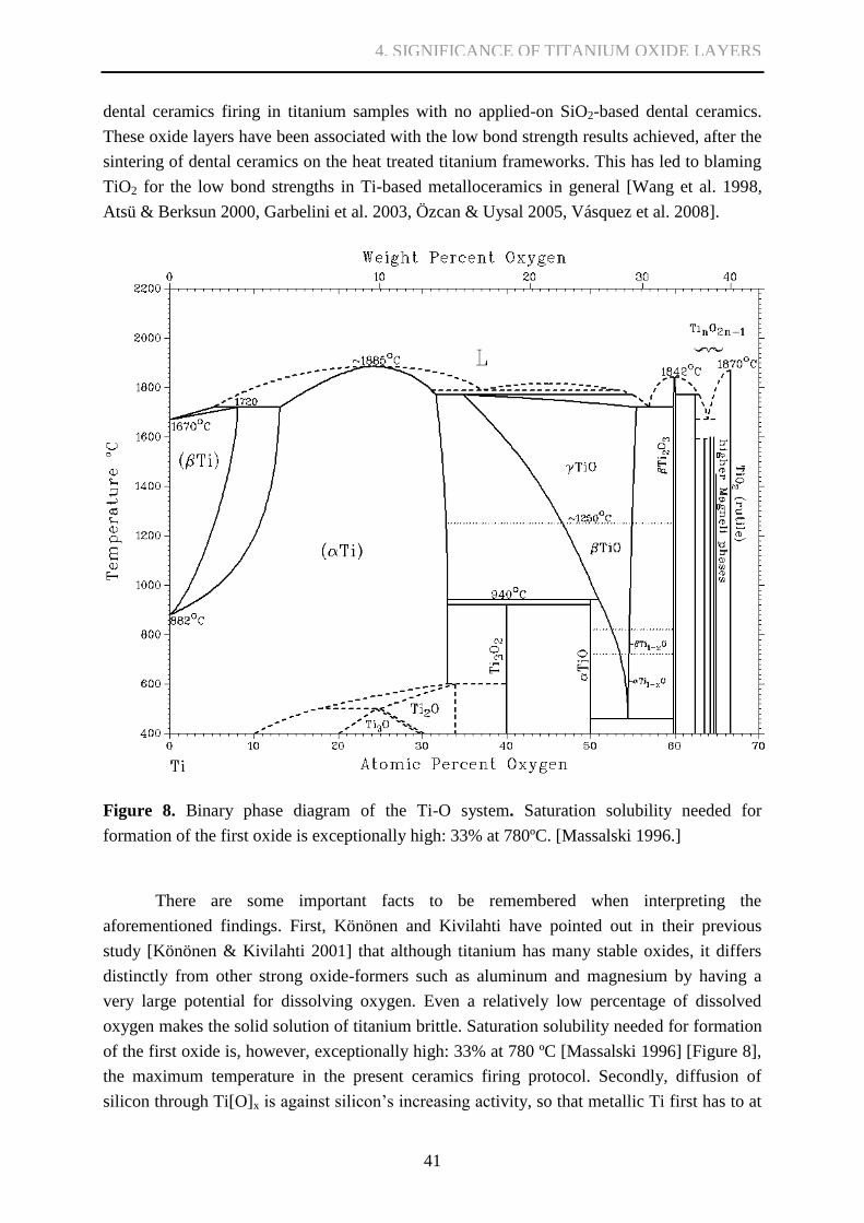
41
dental ceramics firing in titanium samples with no applied-on SiO2-based dental ceramics.
These oxide layers have been associated with the low bond strength results achieved, after the
sintering of dental ceramics on the heat treated titanium frameworks. This has led to blaming
TiO2 for the low bond strengths in Ti-based metalloceramics in general [Wang et al. 1998,
Atsü & Berksun 2000, Garbelini et al. 2003, Özcan & Uysal 2005, Vásquez et al. 2008].
Figure 8. Binary phase diagram of the Ti-O system. Saturation solubility needed for
formation of the first oxide is exceptionally high: 33% at 780ºC. [Massalski 1996.]
There are some important facts to be remembered when interpreting the
aforementioned findings. First, Könönen and Kivilahti have pointed out in their previous
study [Könönen & Kivilahti 2001] that although titanium has many stable oxides, it differs
distinctly from other strong oxide-formers such as aluminum and magnesium by having a
very large potential for dissolving oxygen. Even a relatively low percentage of dissolved
oxygen makes the solid solution of titanium brittle. Saturation solubility needed for formation
of the first oxide is, however, exceptionally high: 33% at 780 ºC [Massalski 1996] [Figure 8],
the maximum temperature in the present ceramics firing protocol. Secondly, diffusion of
silicon through Ti[O]x is against silicon’s increasing activity, so that metallic Ti first has to at
4. SIGNIFICANCE OF TITANIUM OXIDE LAYERS

42
least partly dissociate its own surface oxide layer to enable formation of titanium silicides at
the joint interface. Previously portrayed dual crack propagation through a silicide layer
(mostly Ti5Si3(O)) and relatively thick titanium oxygen solid solution in titanium
metalloceramic samples with dental ceramics already applied and subjected to extended firing
times at 800 ºC [Könönen & Kivilahti 1994] shows that the dental ceramics firing process
inevitably leads to dissociation – not thickening – of the underlying native titanium oxide
layer and to accelerated diffusion of oxygen into titanium lattice. The EDS analyses of the
present study also suggest Si diffusion into Ti in the regions closest to dental ceramics.
Color changes in the sample surface observed during the first oxygen firing step
support the notion that dissociation of the oxide layer may take place even without the
presence of dental ceramics atop it [Figure 9].
Hautaniemi et al. previously investigated titanium ceramics bonding using various
firing times, vacuum levels, four-point bending and X-ray photoelectron spectroscopy (XPS)
[Hautaniemi et al. 1992]. According to their results, the fractures that were created in the four-
point bending tests occurred between metallic titanium and titanium oxide (for the samples
with dental ceramics firing time of only few minutes) or in metallic titanium with high
oxygen solubility (for the samples of longest ceramics firing time). The more complex or
esthetically demanding the clinical metalloceramic restoration, the greater number of firings
and the greater heat load involved. Cumulative firing times equal to the longest firing time
used in the study by Hautaniemi et al. are thus by no means uncommon. As oxygen
dissolution to titanium is highly increased with rising temperature, there exist two main
supplies for the increasing amount of oxygen needed. First, the oxygen can be obtained by the
Figure 9: 95 nm anatase samples
on firing stand: yellow color created
by optical properties of ordered
TiO2 layer is disrupted immediately
during the first sintering cycle in
dental ceramics firing furnace,
indicating disruption of the
homogeneous TiO2 layer.
Group B.
4. SIGNIFICANCE OF TITANIUM OXIDE LAYERS

43
reduction of SiO2-based dental ceramics, since the Gibbs free energy of solution of oxygen in
titanium is more negative than the Gibbs free energy of formation of SiO2 (per mole O2)
[Pajunen & Kivilahti 1992, Könönen & Kivilahti 1994]. This source was also presented by
Hautaniemi et al. [1992], as their group found out during XPS measurements that silicon near
the ceramics-titanium interface appeared only partly in the oxidized state, whereas further
away from the interface it appeared mostly in the oxidized state.
The other source for oxygen is naturally the surrounding atmosphere. Indeed, there are
studies on the influence of different firing atmospheres on ceramics bonding [Atsü & Berksun
2000, Sadeq et al. 2003]. Atsü and Berksun [2000] studied the effect of the firing atmosphere
on bond strength. They studied three different dental ceramics systems on cast and wire
electrical discharge machine worked non-cast titanium. The furnace used in dental ceramics
firing was designed to be flushed with 10 pulses of high purity argon gas to minimize the
amount of residual oxygen present during firing. The three-point bending test results of half
of the tested groups were improved when using protective atmosphere. Unfortunately, no
testing was done to measure the amount of residual oxygen in the firing chamber. Sadeq et al.
[2003] performed biaxial flexure test and reported higher AFAP values of titanium
metalloceramics fired in argon atmosphere compared to samples fired in minor vacuum of 3-4
kPa.
The current commercial firing recommendation is for a vacuum of ca 5 kPa (5 ∙10-2
atm). Rotary pumps of older dental furnaces used by dental technicians create a vacuum of
roughly 1.3 ∙10-3
kPa [Wang et al. 1999]. Preventing any significant oxygen dissolution into
titanium at above 700 ºC, however, requires the vacuum to be of very high level, preferably
10-6
kPa or better. Thermodynamically the equilibrium of titanium/oxygen systems, based on
Ellinghams diagram, requires approximately 10-110
atm to 10-90
atm of oxygen partial pressure
between 400 ˚C to 800 ˚C. It is impossible to achieve this low an oxygen partial pressure by
any means imaginable [Lee et al. 2004].
In addition, oxygen released from dissociation of TiO2 layer in the beginning of dental
ceramics firing somewhat increases the amount of dissolved oxygen in titanium [Figure 10].
4. SIGNIFICANCE OF TITANIUM OXIDE LAYERS
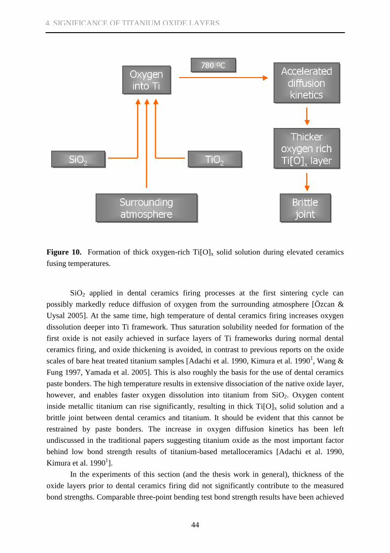
44
Figure 10. Formation of thick oxygen-rich Ti[O]x solid solution during elevated ceramics
fusing temperatures.
SiO2 applied in dental ceramics firing processes at the first sintering cycle can
possibly markedly reduce diffusion of oxygen from the surrounding atmosphere [Özcan &
Uysal 2005]. At the same time, high temperature of dental ceramics firing increases oxygen
dissolution deeper into Ti framework. Thus saturation solubility needed for formation of the
first oxide is not easily achieved in surface layers of Ti frameworks during normal dental
ceramics firing, and oxide thickening is avoided, in contrast to previous reports on the oxide
scales of bare heat treated titanium samples [Adachi et al. 1990, Kimura et al. 19901, Wang &
Fung 1997, Yamada et al. 2005]. This is also roughly the basis for the use of dental ceramics
paste bonders. The high temperature results in extensive dissociation of the native oxide layer,
however, and enables faster oxygen dissolution into titanium from SiO2. Oxygen content
inside metallic titanium can rise significantly, resulting in thick Ti[O]x solid solution and a
brittle joint between dental ceramics and titanium. It should be evident that this cannot be
restrained by paste bonders. The increase in oxygen diffusion kinetics has been left
undiscussed in the traditional papers suggesting titanium oxide as the most important factor
behind low bond strength results of titanium-based metalloceramics [Adachi et al. 1990,
Kimura et al. 19901].
In the experiments of this section (and the thesis work in general), thickness of the
oxide layers prior to dental ceramics firing did not significantly contribute to the measured
bond strengths. Comparable three-point bending test bond strength results have been achieved
4. SIGNIFICANCE OF TITANIUM OXIDE LAYERS

45
in Al2O3 particle blasted samples without excessively thick TiO2 layers [Al Hussaini & Al
Wassan 2005]. The cracking occurred much deeper in Ti substrates than what was the
combined thickness of oxide layers artificially manufactured (95-460 nm) and natively
formed (can be estimated to be less than 10 nm). Based on the above discussion and
thermodynamical considerations, the oxygen-rich Ti[O]x solid solution zone should be
identified as the region of vulnerability, and mechanisms of oxygen dissolution into this zone
should be considered the factor most affecting bond strength.
There exists no sharp boundary between the oxygen-rich titanium and the bulk
titanium. The crack may propagate at any solid solution depth where the quantity of oxygen
diffused into octahedral sites sufficiently affects the solution so that mobility of dislocations
over slip planes, i.e. plasticity, no longer occurs. Therefore even a careful analysis of oxygen
concentration is insufficient in precisely predicting the crack propagation occurrence depth; at
most a zone of probable occurrence could be identified if an oxygen depth profile were
available. As previously indicated [see section 2.4.], such depth profiling is a complex
undertaking, perhaps best sidestepped in favor of direct measures to limit oxygen propagation.
The relatively low mobilities of the elements in the titanium silicides leave the silicide
layer comparatively thinner than the titanium oxygen solid solution [Könönen & Kivilahti
1994]. As no dual crack propagation was observed during the thesis study, the relatively thick
TiO2 layers formed prior to dental ceramics application can be considered to have prevented
excessive formation of continuous thick titanium silicide layers [see section 2.2.5.]. In the
cross-section SEM images of cracked specimens, the two-layered structure of the remaining
Ti containing layer on the dental ceramics side is barely visible. EDS line scan data suggests
Si diffusion to Ti, and the upper 0.5 μm thick, intact segment of the remaining Ti containing
layer closest to dental ceramics can thus be interpreted as a titanium silicide layer [Figure 7;
see also 2.2.5]. The diffusion is observed at the limits of the resolution of the EDS method,
however. It will further be shown that even a native oxide layer is sufficient in preventing
extensive dual crack propagation [see sections 6.2. and 7.2.], albeit again a segment of the
remaining Ti containing layer can be seen as a silicide layer (as a darker band in SEM
images). The cracking in the native oxide samples occurs identically to that of thick oxide
layer samples. Thus the TiO2 layers can be considered benign if not beneficial, and
completely doing away with these layers would in any case be a futile prospect.
The results from TiO2 layer application in this section of the study are in accordance
with those findings and interpretations in the literature concentrating on the effects of
dissolution of oxygen in titanium [Könönen & Kivilahti 1994, Könönen & Kivilahti 2001].
On the other hand, the results contradict the research emphasizing the effects of TiO2
thickness [Wang et al. 1998, Atsü & Berksun 2000, Garbelini et al. 2003, Özcan & Uysal
2005, Vásquez et al. 2008]. Those only appear relevant for certain rather artificial heat
treatment sequences [Adachi et al. 1990, Kimura et al. 19901, Yamada et al. 2005].
4. SIGNIFICANCE OF TITANIUM OXIDE LAYERS

46
The third part of this work [section 6] will tackle the issue of restraining the
aforementioned dissolution of oxygen from surrounding atmosphere and SiO2-based ceramics
to the bulk titanium.
4.4. Conclusions
A thick TiO2 layer on titanium is not as disadvantageous as previously claimed for the
mechanical integrity of titanium-ceramic system. Scanning acoustic microscopy revealed
good ceramics adhesion to titanium in all the test groups, and no significant connection
between bond strength and TiO2 film thickness or crystal structure could be found. The
fracture mode was uniform and thus independent of oxide layer thickness. On the other hand,
no dual crack propagation could be observed in the test specimens; a uniform oxide layer is
expected to sufficiently restrain unwanted thickening of the silicide reaction product layer
between SiO2-based ceramics and titanium. The problem of excessive oxide dissolution
remains to be solved so that an ideal metal-ceramic bond can be accomplished.
4. SIGNIFICANCE OF TITANIUM OXIDE LAYERS

47
5. PHOTOINDUCED SUPERHYDROPHILICITY
Titanium has several interesting aspects not widely discussed in research associated with
dental metalloceramics. Before further discussing restraining oxygen dissolution during dental
ceramics firing, a new aspect to TiO2 is presented. TiO2 possesses two distinct semiconductor
photochemistry-based photoinduced features: a better-known photocatalytic phenomenon,
which leads to breakdown of organics; and the more recently discovered photoinduced
superhydrophilicity [Fujishima et al. 2000, Mills & Lee 2002]. Here, the latter phenomenon is
introduced to dental metalloceramics research.
A semiconductor material such as TiO2 comprises electrons at the valence band (VB)
and, at a higher energy state, largely vacant electron energy levels called the conduction band
(CB). The energy difference between these two bands is called the bandgap, EBG, which is
characteristic to each respective semiconductor material. When a semiconductor material is
subjected to radiation with ultra-bandgap light, i.e. photons with hv > EBG, promotion of a VB
electron to CB may occur. The subsequent formation and fate of an electron-hole pair (e- &
h+) largely determines the photoactivity of the semiconductor material. [Benedix et al. 2000,
Mills & Lee 2002].
Typically in non-photoactive conductors such as metals, electron-hole pairs recombine
immediately [Benedix et al. 2000] and produce heat. Recombination is promoted by defects,
and amorphous materials usually portray poor photoactivity [Mills & Lee 2002]. If some of
the photoexcited electron-hole pairs diffuse to the surface of the semiconductor material, it is
possible for them to interact with surface species [Benedix et al. 2000, Mills & Lee 2002].
This is the basis for photosynthesis, photocatalysis and photoinduced superhydrophilicity. In
photosynthesis, the Gibbs free energy change for reactions of electron-hole pair and their
surrounding environment is positive; in photocatalysis, negative [Mills & Le Hunte 1997].
With TiO2, in the absence of large amounts of adsorbed competing species such as dirt, the
surface species participating in reactions with the electron-hole pair are the oxides’ own
Ti(VI) and bridging O2-
groups. The Ti(III) species are generated via reduction of the Ti(IV)
surface species by photogenerated electrons, and oxygen vacancies are generated via
oxidation of the bridging O2-
groups to oxygen by photogenerated holes [Fujishima et al.
2000, Mills & Lee 2002]. Water molecules can then occupy these oxygen vacancies,
producing adsorbed OH groups, which tend to make the surface hydrophilic [Fujishima et al.
2000] [Figure 11]. In addition to this chemically adsorbed H2O a lesser amount of physically
adsorbed H2O can also be found, at least in porous oxide films [Yu et al. 2000]. With the
increase of chemically adsorbed OH on the surface of TiO2, van der Waals forces and
hydrogen bonds interactions between H2O and the hydroxyl groups will be increased and
water can easily spread across the surface [Yu et al. 2000]. The process is reversible, as Ti(III)
sites are in the dark slowly oxidized back to Ti(IV) by ambient oxygen, hydroxyl groups
5. PHOTOINDUCED SUPERHYDROPHILICITY
48
reactively desorb, and the oxygen vacancies are filled again by O2-
ions, now generated as a
consequence of the oxidation reaction. Unlike other semiconductor materials, dual
photoinduced phenomena are almost exclusively seen with TiO2 [Fujishima et al. 2000, Mills
& Lee 2002].
Photoinduced superhydrophilicity was originally discovered by accident in 1995
[Fujishima et al. 2000]. It has since produced many commercial solutions including non-
fogging glass applications and self-cleaning surfaces.
In dental metalloceramics, good wetting of metal surfaces during ceramics veneering
is of great importance [Carpenter& Goodkind 1979, Reyes et al. 2001]. It is also considered
one of the key principles for good interface formation [Marshall et al. 2010]. Areas that
exhibit poor wetting by the first applied ceramics layer or the paste bonder prior to sintering
may develop into voids or air pouches, disrupting the ceramics structure during iterative firing
cycles; or worse, leaving the finalized metalloceramic product visually intact, but the joint
incomplete and failing under stress generated by the occlusal forces. Good examples of this
are shown in section 6. Improving wettability of titanium (or more exactly its surface TiO2)
might avoid generation of faults endangering integrity of the joints.
There exists a discrepancy in reported time frames needed for achieving good
superhydrophilicity (i.e. contact angle near 0˚); results vary from ~30 min to 5 min [Fujishima
et al. 2000, Miyachi et al. 2000, Miyachi et al. 2002, Miron et al. 2004]. A corresponding
discrepancy exists in assumed duration of superhydrophilicity; it has been proposed to last for
as long as from 2 days up to weeks [Sakai et al. 1998, Benedix ym, 2000], or to gradually
return close to normal during 12 hours [Miron et al. 2004]. Various parameters of light source
and TiO2 are known to have an influence on photoactivity.
The hypothesis is that superhydrophilicity of the TiO2 surface can be achieved within
a reasonable time (minutes) of ultraviolet activation, so that it can be of possible benefit in
dental ceramics veneering process. The purpose of this section of the study is to investigate
the achievable time frame for inducing superhydrophilicity and to present photoactive nature
of TiO2 to dental metalloceramics research.
5. PHOTOINDUCED SUPERHYDROPHILICITY

49
Figure 11. Schematic representation of photoinduced superhydrophilicity.
5.1. Materials and methods
5.1.1. Sample fabrication
TiO2 films 200 nm thick were deposited on 10 (±0.1) mm x 10 (±0.1) mm x 1.0 mm glass
samples by using a Beneq TFS 500 ALD reactor at Micronova laboratory in Helsinki
University of Technology. Glass was chosen as the test specimen framework material for the
actual TiO2 film to facilitate characterization of the deposited oxide, since the framework
material has previously been found to have no evident impact on the photoactivity of the film
[Vilhunen et al. 2009]. Glass plates were fabricated from ordinary microscopy glass slides
using a diamond glass cutter, and mechanical polishing (Struers LaboPol-21) with 1200 grit
5. PHOTOINDUCED SUPERHYDROPHILICITY
50
wet polishing SiC paper on the specimen sides. Samples went through ultrasonic and
chemical cleaning with aceton, isopropanol and water prior to ALD application.
In the ALD process, TiCl4 and H2O were used as metal and oxygen sources,
respectively, and nitrogen as carrier and purging gas. The reaction temperature was 300 ºC.
The TiO2 crystal structure was anatase. The TiO2 process parameters were chosen based on a
recent study on photoinduced systems [Vilhunen et al. 2009].
5.1.2. Photoinduced hydrophilicity characterization
Transmittance of the TiO2 films was measured from 4 randomly chosen samples using the
Perkin-Elmer Lambda 900 spectrophotometer to evaluate the optimal wavelength for the UV
source. From the acquired transmittance data, absorption coefficient kabs was evaluated
according to kabs = d-1
ln (1/T) where d is the TiO2 film thickness and T the transmittance
[Mardare et al. 2000, Wang et al. 2001, Zhang et al. 2009]. Extrapolating the linear parts of
experimental curves to kabs = 0, one can find approximation of the optical band gap for each
curve. The UV source wavelength was chosen to top the approximated EBG and to ensure no
unnecessary transmittance.
The contact angle between the TiO2 surface and water prior and after UV radioation
was measured using sessile drop method. Regulated drops (2.5 μl) of distilled water were
applied onto the sample surface using constant application height and speed with automated
equipment and software (KSV Instruments Ltd. CAM 200). Prior to UV radiation, reference
contact angle values for each sample were first measured after sample storage in dark for
several days. The samples were then irradiated using a 1000 watt Mercury Arc Lamp (Oriel
Instruments Model 6287). Radiation wavelength was controlled with a monochromator
(Bentham Instruments TMc300) to 312 (±2.5) nm, based on transmittance measurement
results. Irradiance on the TiO2 surface was set to 2.5 mW/cm2 and confirmed with
pyroelectric measurements (LaserProbe Inc. Power Probe RkP-575, Power Meter Rk-5710).
UV radiation was applied for 10 min on the first two samples (Group D) and for 2 min
on second two samples (Group E) at normal room temperature and humidity [Table 3]. After
UV exposure, the contact angle measurements were performed with 5 min intervals until 30
min time point, and then at 60 min, 120 min, 180 min and 240 min after the beginning of UV
exposure. Samples were stored in the dark between measurements.
5. PHOTOINDUCED SUPERHYDROPHILICITY

51
TABLE 3.
CHARACTERISTICS OF GROUPS D AND E FOR SESSILE DROP TESTS
Group N Oxide layer thickness Crystal structure UV
D 2 200 nm anatase 10 min
E 2 200 nm anatase 2 min
5.2. Results
Transmittance values and the UV source wavelength are shown in Figure 12. The absorption
coefficient kabs and EBG were approximated from the transmittance results as shown in Figure
13.
The reference contact angle average for all samples was 54.2˚ (±5.2˚). Both Group D
and Group E samples displayed superhydrophilicity (contact angle close to 0˚, unmeasurable)
after UV radiation [Figure 14]. After initial recovery to 10-15˚ during the first two 5 min
intervals, the contact angle remained below 20˚ for one hour [Figure 15]. Total recovery was
slow and not observed during the first 240 min.
Figure 12. Average transmittance, standard deviations and applied UV wavelength.
5. PHOTOINDUCED SUPERHYDROPHILICITY

52
Figure 13. Approximation of EBG from measured transmittance data.
5. PHOTOINDUCED SUPERHYDROPHILICITY

53
Figure 14. Sessile drop measurements before and immediately after UV irradiation.
Figure 15. Average contact angle vs. time after UV irradiation.
5. PHOTOINDUCED SUPERHYDROPHILICITY

54
5.3. Discussion
5.3.1. On titanium as photoactive material
Photoactivated materials are usually called photocatalysts, owing to the earlier discovery of
photocatalysis compared to photoinduced superhydrophilicity. The term photocatalysis is,
however, a subject of some debate [Mills, Le Hunte 1997]. It is argued that the idea of a
photocatalyzed reaction is fundamentally incorrect, since it implies that light is acting as a
catalyst, whereas it rather acts as a reactant, which is consumed in the process. The term is in
such widespread use that it is here to stay; it is not meant to, nor should it ever be used to,
imply catalysis by light, but rather the “acceleration of a photoreaction by the presence of a
catalyst” [Mills, Le Hunte 1997]. The term photoreaction is sometimes elaborated on as a
photoinduced or photoactivated reaction, all to the same effect [Mills, Le Hunte 1997].
Traditionally an ideal semiconductor photocatalyst material should be chemically and
biologically as inert as possible, photocatalytically active, easy to produce and use, cheap as
material and – owing to its common use in self cleaning outdoor surfaces – activated by
sunlight [Mills & Lee 2002]. TiO2 does not entirely meet these terms, yet it is one of the most
ideal photoactivated materials, since it is highly stable chemically, relatively inexpensive, and
the photogenerated holes are highly oxidizing [Fujishima & Zhang 2006]. The two most
common crystal structures of TiO2 are rutile and anatase. The mobility of charge carriers is
easier in anatase, which makes it electrically the more ideal crystal structure [Ilola et al.
2003]. It can be considered the most photoactive crystal structure of TiO2 [Mills & Lee 2002].
The EBG for anatase is slightly higher than that of rutile (3.2 eV and 3.0 eV correspondingly)
[Mogyorósi et al. 2003]. In previous research, a commercial mixture of 75% anatase and 25%
rutile called P25 (Degussa Chemical Company, Germany) has been used in various solutions
[Blake et al. 1999, Mills & Lee 2002, Balasubramanian et al. 2003]. According to Riley et al.
[2005], a TiO2 layer already present on the surface of commercial dental implants may also
act as photocatalyst, even though photoinduced superhydrophilicity was not studied. In the
thesis study, anatase TiO2 was chosen because of the general conception of its higher
photoactivity.
The existence of dual photoinduced phenomena is almost unique for TiO2 [Fujishima
et al. 2000, Mills & Lee 2002]. For example WO3 and V2O5 are only active in photoinduced
superhydrophilicity [Miyauchi et al. 2002] and SrTiO3 does not become hydrophilic under
UV irradiation, even though it has a similar photocatalytic oxidation power to TiO2 [Miyauchi
et al. 2000]. In addition to TiO2, on which most of the study of photoinduced
superhydrophilicity has concentrated, SnO2 and ZnO also portray both photocatalytic
oxidation and photoinduced superhydrophilicity [Miyauchi et al. 2002].
5. PHOTOINDUCED SUPERHYDROPHILICITY
55
Possibly a more straightforward explanation for photoinduced superhydrophilicity
compared to Fujishima et al. [2000] and Mills & Lee [2002] is the one formulated by
Miyauchi et al. [2002]: photogenerated holes are trapped at the surface lattice oxygen atoms,
causing a dissociation of their bonds with the Ti atoms, followed by the adsorption of water
molecules at these sites; or that UV illumination simply causes the dissociation of atomic
bonds between oxygen and metal atoms in the topmost surface layers of TiO2 followed by
adsorption of water molecules at these defect sites. Thus it is easy to understand that bonding
strength between metal ions and oxygen ions is supposedly important for photoinduced
superhydrophilicity. Miyachi et al. argue that photoinduced superhydrophilicity can also be
due to surface structural changes of different origins, and that multiple kinds of surface
oxygen vacancies might be in agreement with increased wettability. They showed that when
bombarding TiO2 and WO3 films known to portray photoinduced superhydrophilicity with
Ar+ ions for 1 min, the films became highly hydrophilic with a contact angle of 0˚.
Noticeably, SrTiO3 films exhibited better crystal stability during Ar+ bombardment, and did
not portray photoinduced superhydrophilicity. Miyachi et al. also elucidate how the strength
of oxidation activities of different metal oxides is roughly explained by the position of
valence and conduction bands, while the surface morphology, crystallinity and optical
absorption properties also play an important role.
Processing the properties of the TiO2 surface can be used to influence the proportion
of photocatalytic character to superhydrophilic character [Fujishima & Zhang 2006].
Breakdown and washing of dirt from the surface increase hydrophilicity, as part of the
contaminant compounds tend to change the surface to hydrophobic [Madaeni & Ghaemi
2007]. Even though hydrophilicity is not induced only by the photocatalytic oxidation of
organic hydrophobic (dirt) compounds on the surface [Miyauchi et al. 2002], hydrophilicity is
considered to increase the photocatalytic activity of TiO2 since most of the OH groups can be
adsorbed on the photocatalyst surface due to hydrophilicity [Madaeni & Ghaemi 2007].
Photoinduced hydrophilic conversion and photocatalytic oxidation process may also
competitively proceed on the TiO2 surface under UV radiation, though even a slight decrease
in the quantum efficiency of photocatalysis can increase quantum efficiency of the
hydrophilic conversion drastically [Sakai et al. 2003]. The photoactivity of TiO2 films may be
affected by influencing on many possible factors such as film composition, porosity,
crystallinity, crystal size, surface area and roughness in attempt to widen the excitation
wavelength (i.e. EBG) and reduce recombination of electron-hole pairs [Balasubramanian et al.
2003, Fu et al. 2005]. Consequently, there are many attempts to customize photoactivity to
wanted levels, such as by use of inorganic and organic doping agents, and these can be found
e.g. in extensive reviews [i.a. Carp et al. 2004].
5. PHOTOINDUCED SUPERHYDROPHILICITY

56
5.3.2. On light source, UV parameters and achieved hydrophilicity
Typical light sources used in previous research and commercial photoactivated solutions are
365 nm blacklight bulbs and low-pressure mercury lamps (λmax = 254 nm), which have
extremely long lifetimes up to >14 000 h, and even gallium nitride based semiconductor
LEDs have been used [Mills & Lee, 2002]. Doping agents such as Au have been used to
widen the absorption spectra so that high pressure sodium bulbs (λ > 400 nm) could also have
been used in addition to UV-B or UV-A wavelengths [Li & Li 2001].
In order for light to induce photoactivity, energy of photons must top the EBG of the
semiconductor surface, 3.2 eV for bulk anatase. Light wavelength should thus be typically
less than 385-388 nm [Maness et al. 1999, Benedix et al. 2000]. The EBG for artificially
created TiO2 surfaces, however, can be considerably higher, e.g. 3.66 eV or 3.75 eV
corresponding to wavelengths of 338 nm and 330 nm, depending on the oxide properties
[Mogyorósi et al. 2003]. Thus the optical properties of the TiO2 surface designated for use
must be properly characterized to select optimal parameters for the light source.
The transmittance measurements in this section proved to be sufficient for estimating
efficient wavelength for photoinduced superhydrophilicity. In the transparency region,
scattering of light influences the optical absorption spectrum calculated from transmission
spectrum. At shorter wavelengths, however, the fundamental absorption evidently dominates
the scattering losses. Therefore, some information about the optical band gap can be obtained
from the transmission spectra [Aarik et al. 1997, Mardare et al. 2000] and the simplified
relation kabs = d-1
ln (1/T) [Zhang et al. 2009, Mardare et al. 2000, Wang et al. 2002] can be
used to evaluate the absoprion coefficient from the measured transmittance. The results
achieved in this thesis are close to the values of pure anatase and rutile. The 3.15 eV values
obtained by Mardare et al. [2000] were considered to represent a mixed structure of rutile and
anatase. It is possible that the TiO2 structure used in the thesis also contained some rutile
components. It must be kept in mind that the obtained band gap value is, however, only an
approximation.
Sakai et al. [2003] stated that the hydrophilic conversion rate strongly depends on the
wavelength of the light source. The onset wavelength for hydrophilic conversion is almost
consistent with that for absorbance spectrum, and the conversion rate increases as absorbance
increases. Previously Miron et al. [2004] achieved superhydrophilicity using a broad spectrum
light source covering transmittance change from maximum to minimum, and Vilhunen et al.
[2009] used TiO2 films – similar to those used in the thesis – with light wavelengths well over
band gap energies (85% of the radiation in the range of 250-260 nm) to successively induce
photocatalysis. In comparison, the narrow width of the spectrum used in this thesis ensured
complete supra band gap energy of the employed radiation as well as minimal transmittance,
and gave more accurate information about the irradiance participating in the photoinduced
phenomena. This information will markedly simplify future selection of radiation sources in
comparable photoinduction experiments.
5. PHOTOINDUCED SUPERHYDROPHILICITY

57
The level of irradiance used here can be considered sufficient for inducing
superhydrophilicity, but a radiation source of optimal parameters could meet the needs at
lower power. Miron et al. [2004] reported that 1 mW/cm2 UV radiation was sufficient to
induce surface superhydrophilicity in 5 min on amorphous TiO2 surface with a short range,
anatase-like atom ordering. The irradiance of 2.5 mW/cm2 used in the thesis systematically
resulted in superhydrophilicity and the spreading of the water droplet on the anatase TiO2 in
only 2 minutes (Group E). Increase in irradiance does not, however, necessary shorten the
time, as Sakai et al. [1998] reported superhydrophilicity only after 10 min when using 40
mW/cm2 Hg-Xe lamp (though the contact angle was already markedly reduced in the first
measurement point at 5 min). Sakai et al. [2003] stated that, based on the comparison of the
hydrophilic conversion rate with the rate for the reverse process or relaxation, the critical
contact angle (the lowest value of the contact angle under UV irradiation) is obtained when
the reconstruction of the surface hydroxyl groups and their relaxation process are equilibrated
under UV irradiation, and that domination of the recombination of photogenerated electron-
hole pairs limits the hydrophilic conversion rate under high UV intensity [Sakai et al. 2003].
Optimation of UV intensity and wavelength of the light needs further study, since they
directly affect the inducing of superhydrophilicity. Even though it has been proposed that it
can take over 20-30 min to achieve superhydrophilicity [Fujishima et al. 2000, Miyachi et al.
2000, Miyachi et al. 2002] it is obvious that if the EBG of the oxide can be evaluated and a
sufficiently optimal wavelength and intensity of the radiation chosen, superhydrophilicity can
be induced in reasonable time (minutes) and lasts long enough to be practical for dental
technicians use during the metalloceramics manufacturing step. Measurements of this sort are
not easily performed alongside dental technicians’ daily work or with equipment commonly
found in dental laboratories. Thus adjustable or wide spectrum light sources are needed, and
general guidelines for the irradiance and wavelength needed for photoinduced
superhydrophilicity on different titanium oxide structures must be provided by the basic
research. The light source used in the current study was ideal for research use owing to its
high controllability, and it proved efficient in promoting photoinduced hydrophilicity.
Relaxation to normal was recorded as featuring a sharp initial rise in contact angle
during the first half an hour, followed by a more gradual rise. The results roughly agree with
Miron et al. [2004] and Sakai et al. [2003], who further discuss the subject and the variables
involved in the exact nature of the relaxation process. If necessary, the relaxation period can
possibly be hastened with the use of ultrasonic treatment, and reinduced again with a new UV
exposure [Sakai et al. 1998].
5.3.3. On Ti wetting with dental ceramics
Though wetting is essential for achieving good bonding [Carpenter& Goodkind 1979, Reyes
et al. 2001, Marshall et al. 2010], restraining excessive oxygen diffusion into the titanium
5. PHOTOINDUCED SUPERHYDROPHILICITY

58
lattice during dental ceramics firing must be considered the first priority for mechanical
integrity of titanium-ceramic systems in modern dental metalloceramics. No pronounced
wetting problems were detected in the SAM investigations of the previous section of the
thesis. In the next section, however, evident drastic consequences of poor contact between
dental ceramics and metal structure are shown. It is thus essential to further study the wetting
properties of titanium-ceramic systems – different dental ceramic materials, Ti frameworks
(CAD/CAM manufactured vs. cast, different alloys) and their native oxide layers – after the
dissolution of oxygen can be controlled or constrained.
This section illustrates how Ti and especially TiO2 possess several notable
characteristics that remain novel and unfamiliar to dental metalloceramics research even
though titanium has already been used for quite some time in clinical practice.
5.4. Conclusions
Photoinduced superhydrophilicity is an easy and effective method to increase wetting of
TiO2-covered titanium surfaces into extreme hydrophilicity. This can possibly be used to
circumvent the emergence of detrimental flaws – voids and air pouches – in joints between
titanium and dental ceramics, thus improving joint integrity and success of metalloceramic
restorations. Photoactivity of surface TiO2 in CAD/CAM manufactured titanium frameworks
after normal processes prior to dental veneering needs further research.
5. PHOTOINDUCED SUPERHYDROPHILICITY

59
6. SILVER INTERLAYERS
The most brittle component in the joints of common dental ceramics and Ti is the oxygen-rich
solid solution Ti[O]x in the surface layer of titanium frameworks, as stated earlier. The
plasticity of Ti[O]x is significantly reduced through the diffusion of oxygen into titanium.
Resultant stresses caused by iterative dental ceramics firing cycles and thermal expansion
mismatch of the two materials burden the metalloceramic structures. Also, the brittle reaction
products such as titanium silicides and possible contamination debris such as aluminum from
Al2O3 blasting particles might further weaken the joints. Even though several different
methods to improve joint strength have been tried out, only a few account for all of the
aforementioned factors.
A vacuum produced by conventional dental furnaces is not even close to sufficient in
order to prevent dissolution of the firing atmosphere originated oxygen into titanium [Wang et
al. 1999, Lee et al. 2004]. Using high vacuum fusing furnaces or a protective gas environment
can perhaps reduce the oxygen dissolution, yet such furnaces are relatively expensive and
rare. More economical ceramics bonding agents have been designed to efficiently wet and
cover the application area of titanium, thus sealing the firing atmosphere after the first firing
sequence. Results of their efficacy in this particular function are not well documented,
however, and the SiO2 will serve as an excellent oxygen source in itself [Pajunen & Kivilahti
1992, Könönen & Kivilahti 1994]. Thus, use of an oxygen diffusion barrier material other
than SiO2 based ceramics bonder would seem reasonable.
The ideal interlayer material must fulfill several requirements. It must be
biocompatible and suitable for oral environment, act as a barrier to diffusion of oxygen, be
strongly adherent to Ti structures, and be capable of establishing a strong bond to dental
ceramics [Wang et al. 1999, Könönen & Kivilahti 2001]. Even though strong bonding
requires chemical bonding, the interlayer must not produce excess reaction products with Ti
or dental ceramics, and/or must sufficiently reduce reactions between Ti and Si to avoid
excess formation of brittle titanium silicides [Könönen & Kivilahti 2001]. The interlayer must
possess plastic deformability, so that it can attenuate residual stresses afflicting the joints
[Könönen & Kivilahti 2001]. Sealing of the blasting particle contamination from dental
ceramics contact can be considered beneficial [Carpenter& Goodkind 1979, Dérand & Herø
1992, Kern & Thompson 1994, Darwell et al. 1995, Papadopoulos et al. 1999, Cai et al. 2001,
Bagno & Di Bello 2004, Yamada et. al 2005]. Finally, the interlayer material needs to be
applicable on titanium with practical methods. After application, it should also maintain its
original properties as much as possible [Könönen & Kivilahti 2001]
Metallic interlayers of a noble metal familiar as dental biomaterial could fulfill many
of these criteria. Metallic materials offer important inherent capability to withstand plastic
deformation and thus residual stresses inflicted during dental ceramics fusing, e.g. due to
6. SILVER INTERLAYERS
60
differences in CTE of dental ceramics and titanium frameworks can be attenuated to maintain
integrity of metal-ceramic joints. The following interlayer metal alternatives were considered
based on criteria for suitability as an oxygen diffusion barrier: Ag, Au, Pd, Pt, Co and Cr.
Cobolt and chromium were discarded owing to their inferior mechanical properties. Out of the
four remaining possibilities, Ag was chosen on the basis of higher reactivity of Au, Pd and Pt
with Ti, so that fast formation of thick brittle reaction layers between the interlayer material
and Ti could be avoided, and the interlayers would remain sufficiently plastic to reduce
residual stresses. Also, Ag has only two intermetallic compounds (IMCs) with Ti, TiAg and
Ti2Ag, less than for example Au. A more thorough comparison on the thermodynamics of
different interlayer metals will be published later by Könönen, Saloniemi, Varpavaara and
Kivilahti.
The purpose of the present investigation is to propose an interlayer material that best
meets the aforementioned criteria, and to compare mechanical compatibility of this new
system with one commercial titanium-ceramic system using the three-point bending test.
According to the hypothesis, Ag interlayers produced on titanium surfaces can suppress
diffusion of oxygen into titanium and thus prevent formation of excessively thick and brittle
oxygen-rich Ti[O]x layers.
6.1. Materials and methods
6.1.1. Sample fabrication
DIN 13 927/ISO 9693 standard titanium test samples were fabricated, mechanically polished
with 1200 grit wet polishing SiC paper, and chemically cleaned in the aforementioned manner
[see section 4.1.1.].
Five test groups with varying Ag interlayer thickness and surface roughness and one
control group were produced. Each group contained eight samples. Samples in Groups F, J, K
and L were blasted with Al2O3 particles (Q-dent High-Grade Corundum) for a few seconds
with 250 kPa pressure from the distance of 3 cm. The particle size used was 110 μm as
recommended by the ceramics manufacturer. The blasting direction was angled to the
titanium surface to avoid excessive alumina attachment. After resting for 10 to 15 minutes,
the samples were cleaned with a steam cleaner (Emmevi VAP 6) before veneering, again
according to recommendation of the ceramics manufacturer. Samples in Group G and H were
left polished. Group F samples served as a control group and had no Ag coating. The Ag
coatings were prepared on Ti substrates of Groups G, H, J, K and L by DIARC-Technology
Inc. using the DIARC® plasma coating method. Coatings were deposited in a vacuum at a
temperature below 100 ˚C and were between 50-200 nm thick. On this Ag layer a thicker Ag
layer up to 5 μm was constructed on Group L samples using electrochemical baths containing
6. SILVER INTERLAYERS

61
80.5% silver cyanide, potassium cyanide, distilled water, sulphuric acid and nitric acid (Silver
Glo 3K, Rohm and Haas Electronic Materials). The current density range was 0-7.0 A/dm3
and the operating temperature was 21-29 ˚C. Test group specifications are given in Table 4.
The fusing of dental ceramics was carefully done according to manufacturer’s protocol
and the DIN/ISO standards as described in section 4.1.1..
TABLE 4.
CHARACTERISTICS OF GROUPS F, G, H, J, K and L
Group N Ti surface Ag layer thickness Deposition method
F 8 Al2O3-blasted ----- -----
G 8 1200 grit SiC-polished 50 nm DIARC®
H 8 1200 grit SiC-polished 200 nm DIARC®
J 8 Al2O3-blasted 50 nm DIARC®
K 8 Al2O3-blasted 200 nm DIARC®
L 8 Al2O3-blasted 5 μm DIARC® + electroplating
6.1.2. Sample characterization
Additional Ti samples with surface treatments of Groups F-L were analyzed with SEM and
EDS prior to dental ceramics fusing. After dental ceramics firing the metalloceramic
interfaces were studied using a scanning acoustic microscope, as detailed in section 4.1.2.. To
verify the ultrasonic examination results, cross-sectional sample microscopy was done.
After the aforementioned examinations, the crack-initiation (bond) strengths of the
metalloceramic samples were analyzed by the DIN 13 927/ISO 9693 standard three-point
bending tests, again as described in section 4.1.2.. Fracture surfaces and cross-sectional
samples of the cracks were analyzed with optical microscopy, SEM and EDS.
6. SILVER INTERLAYERS

62
6.2. Results
Before dental ceramics firing, SEM studies of samples in different groups showed a distinct
difference in surface topography as shown in Figure 17. No significant difference was,
however, detected between 50 nm and 200 nm coated samples. Sharp edges and corners are
smoothened especially in Group L samples. The EDS element maps also show that alumina
residues from Al2O3-blasting are completely insulated from contact to dental ceramics [Figure
18].
One sample in Group G and one in Group H were destroyed during dental ceramics
firing owing to air filled delaminations in the joint. SAM analyses of metalloceramic
interfaces in visually intact samples revealed clear areas of delamination and/or inadequate
bonding in polished samples of Groups G and H. The areas varied from roughly spherical
small areas 0.1 mm in diameter to large delaminated areas of almost 1 mm2 as shown in
Figure 19. The green areas represent areas with poor contact between titanium and dental
ceramics or areas of titanium with no applied dental ceramics. No large delaminations could
be found in samples of Groups F, J, K or L. SEM results confirmed these results.
The three-point bending test results of Groups F, G, H, J, K and L were 37.8 MPa (SD
2.4 MPa), 33.3 MPa (SD 6.1 MPa), 31.0 MPa (SD 6.0 MPa), 37.8 MPa (SD 6.2 MPa), 41.2
MPa (SD 7.2 MPa) and 48.2 MPa (SD 2.7 MPa). The corresponding average displacement in
μm that the samples lasted before cracking was 95.6 μm (SD 10.3 μm), 91.1 μm (SD 17.7
μm), 83.6 μm (SD 11.8 μm), 105.9 μm (SD 11.3 μm), 122.5 μm (SD 35.8 μm) and 150.0 μm
(SD 20.3 μm) in Groups F, G, H, J, K and L. The three-point bending test results are shown in
Table 5 and Table 6. One-way analysis of variance showed a significant difference between
the TPB results of control Group F and Group L (significance level α = 0.01, p = 1.27∙10-6
)
indicating a better joint strength in samples with ca. 5 μm Ag interlayer. No significant
difference emerged between the other groups compared and the control group.
In Groups F, H and J cracking caused by three-point bending occurred 1.4 to 2.5 μm
deep from titanium surface inside Ti[O]x solid solution. In samples of Group G and H
cracking between titanium and dental ceramics could be seen locally. In these areas a brittle
solid solution could be observed on the surface of the titanium structure as shown in cross-
section sample [Figure 20]. No continuous Ag layers were visible between dental ceramics
and titanium in samples of Groups G, H, J and K. Cracking of Group L samples did not occur
inside the brittle surface layers of the titanium structure, but multiple fracture lines can be
observed inside dental ceramics. A clear layer of Ag between titanium and dental ceramics,
and Ti diffusion through this layer – making adequate chemical bonding to dental ceramics
possible – can be observed [Figure 21]. The Ag layers remain unbroken, but the thickness is
uneven, between 4 to 10 μm, and locally only less than 1 μm.
6. SILVER INTERLAYERS
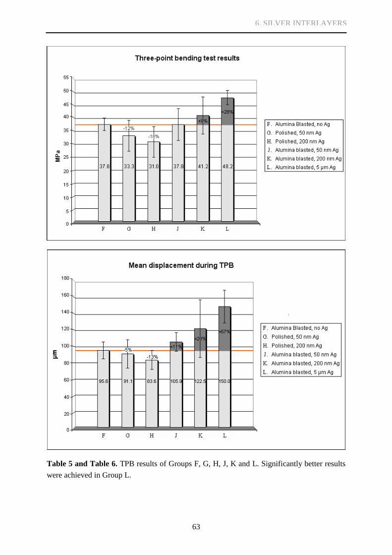
63
Table 5 and Table 6. TPB results of Groups F, G, H, J, K and L. Significantly better results
were achieved in Group L.
6. SILVER INTERLAYERS

64
Polished
5μm Ag
Al2O3-blasted
Figure 17. Different surface
topography is observed in SEM
micrographs prior to dental
ceramics firing. Sharp edges and
corners observed in solely Al2O3-
blasted samples are smoothened
by an applied silver interlayer.
6. SILVER INTERLAYERS

65
Figure 18. Top: compositional backscattered electron image of alumina residues (dark) on
Al2O3-blasted Ti (light grey). Bottom left: EDS element map of Al2O3-blasted sample of
Group F prior to dental ceramics veneering, indicating Al in red and Ti in green. Bottom
right: EDS element map of Al2O3-blasted Ti surface with Ag interlayer in a Group L sample
with the same color scale and no dental ceramics. Alumina contamination is efficiently
insulated from possible interactions with dental ceramics.
6. SILVER INTERLAYERS

66
Group F.
Group J.
Group K.
Group H.
Group L.
Group H.
Group G.
Figure 19. SAM images of samples
from Groups F, G, H, J, K and L after
dental ceramics firing. Blue areas
indicate good bonding between Ti and
dental ceramics. Green areas represent
Ti without applied dental ceramics
(ends of the standard samples) as well
as delamination and/or inadequate
bonding. Samples in Groups F, J, K and
L were found flawless. In Groups G and H (polished samples with 50 nm or 200 nm Ag
interlayers) several samples exhibited delaminated areas.
6. SILVER INTERLAYERS

67
Figure 20. Compositional backscattered electron images of samples from Groups F, H, K and
L after TPB. Cracking inside Ti[O]x in samples of Groups F, H and K and multiple cracks
inside dental ceramics in Group L can be observed. In Group H cracking between dental
ceramics and titanium also occurred locally. A brittle solid solution is observable on the
surface of Ti in these areas. No continuous lightly colored Ag interlayer is observed in Groups
H or K, whereas a clear unbroken layer of Ag between titanium and dental ceramics is found
in Group L samples. No significant differences were found between samples in Group G and
Group H, or between Group J and K.
Group F.
Group H.
Group K.
Group L.
6. SILVER INTERLAYERS
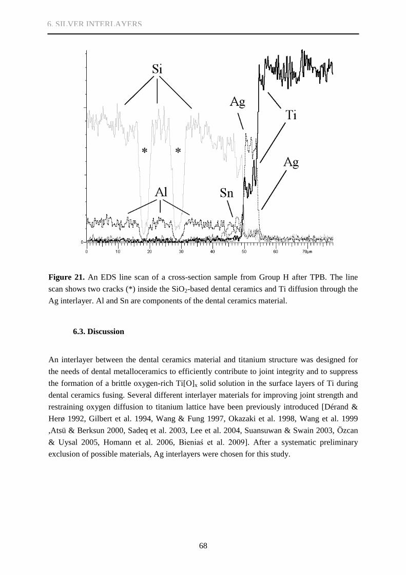
68
Figure 21. An EDS line scan of a cross-section sample from Group H after TPB. The line
scan shows two cracks (*) inside the SiO2-based dental ceramics and Ti diffusion through the
Ag interlayer. Al and Sn are components of the dental ceramics material.
6.3. Discussion
An interlayer between the dental ceramics material and titanium structure was designed for
the needs of dental metalloceramics to efficiently contribute to joint integrity and to suppress
the formation of a brittle oxygen-rich Ti[O]x solid solution in the surface layers of Ti during
dental ceramics fusing. Several different interlayer materials for improving joint strength and
restraining oxygen diffusion to titanium lattice have been previously introduced [Dérand &
Herø 1992, Gilbert et al. 1994, Wang & Fung 1997, Okazaki et al. 1998, Wang et al. 1999
,Atsü & Berksun 2000, Sadeq et al. 2003, Lee et al. 2004, Suansuwan & Swain 2003, Özcan
& Uysal 2005, Homann et al. 2006, Bieniaś et al. 2009]. After a systematic preliminary
exclusion of possible materials, Ag interlayers were chosen for this study.
6. SILVER INTERLAYERS

69
6.3.1. On the biocompatibility of Ag
The biocompatibility of Ag in dental solutions has been discussed recently in various papers.
Despite Ag being a noble metal, it has been proposed to be more cytotoxic to osteoblasts and
gingival fibroblasts than Au or Pd [Cortizo et al. 2004, Locci et al. 2000, Grill et al. 20001].
Experimental titanium-silver casting alloys (Ti 75% wt, Ag 25% wt) are concluded, however,
to impose minimal risks associated with cytotoxicity [Wang & Li 1998], and transient
exposure to an acidic oral environment does not significantly increase elemental release from
Ag containing alloys [Wataha et al. 1998]. Longer exposure can cause some limited release
[Wataha & Lockwood 1998]. The two intermetallic compounds produced in the Ti-Ag joint,
TiAg and Ti2Ag, are highly stable. Generally alloys presenting higher amounts of noble
elements, including Ag, are more biocompatible [Grill et al. 20002]. Ag has been used in
present clinical solutions in alloys with gold and palladium such as Au-Pd-Ag, Pd-Ag, Pd-Ag-
Au [Roberts et al. 2009]
6.3.2. On oxygen dissolution
The Ag interlayers served as excellent oxygen diffusion barriers in Group L samples. No
indications of brittle oxygen-rich Ti[O]x layers were observed and the cracking occurred
inside less brittle dental ceramics material. Joints between the silver interlayers and titanium
structures remained intact showing excellent strength, as did many parts of joints between the
interlayers and dental ceramics.
6.3.3. On plastic deformation and bond strength results
The mobility of dislocations generates the charateristic capacity of plasticity in metals. The
high plasticity of silver is ideal for its use as an interlayer material, e.g. enabling relief of
residual stresses formed due to mismatches in the CTE of dental ceramics and framework
metal. The silver interlayers maintained plasticity even after reacting with Ti that diffused
through the entire thickness of the interlayers, whereas brittle and faster forming IMCs in Au-
Ti systems have been considered disadvantegous to joint strength [Könönen & Kivilahti
2001]. In a yet unpublished study of Könönen, Saloniemi, Varpavaara and Kivilahti compared
Al2O3-blasted cpTi samples applied with only Duceratin® (Ducera, Dental-Gesellschaft,
Germany); Duceratin® with first applied designated Haftbond
® (Ducera, Dental-Gesellschaft,
Germany) bonding material; or with electrochemically composed Ag layers of up to 10 μm
thickness. The 1200 grit SiC polished cpTi samples with the aforementioned Ag layers were
also studied. Congruent to the study of this monograph with thinner Ag layers, the Al2O3-
blasted samples treated with Ag-plating performed best in three-point bending tests, resulting
6. SILVER INTERLAYERS

70
in crack initiation (bond) strength values 51.1% higher than the values of solely Al2O3-blasted
samples without use of any bonding agent. In the samples without Ag-plating, load curves
recorded during the three-point bending tests showed a very sharp dropping at the bond
failure loads compared to the Ag-plated samples where corresponding curves showed more
plasticity at failure load levels [Figure 22]. In the curves of Al2O3-blasted samples with Ag
interlayers, multiple sharp peaks represent multiple cracks inside the dental ceramics material
in contrast to only one sharp drop in samples without Ag-plating, representing cracking of
continuous planar layers of brittle Ti[O]x. These observations strengthened micrograph
findings concerning the mode of joint failure and were congruent to findings of the study in
this monograph. The percentual increase achieved with the 5 μm Ag interlayers was also
larger in mean displacement and better withstood prior cracking than in bond strength results
[Table 5 and Table 6]. This can be explained by the very same plasticity of the Ag interlayers.
Figure 22. Sharp dropping at the joint failure load is observed in samples without Ag
interlayers (groups 1 and 2). TPB results with Ag interlayers are better even without Al2O3-
blasting and the cracking represents more elasticity (group 4). Multiple sharp peaks
representing multiple cracks inside dental ceramics can be observed in samples both Al2O3-
blasted and Ag-plated (group 3) [unpublished data of Könönen, Saloniemi, Varpavaara and
Kivilahti]
6. SILVER INTERLAYERS

71
Özcan and Uysal [2005] stated that it must be remembered that results achieved with
oxygen diffusion barriers can vary substantially with different dental ceramics. There is no
indication that Ag would perform worse as a diffusion barrier with any other dental ceramics
material. Bonding between the interlayers and dental ceramics can, however, be dependant on
the ceramics material composition. Even though bonding between the interlayers and other
SiO2-based ceramics material can be presupposed as similar, it needs further research. Ti
diffusion through the interlayers and IMCs formed between Ti and Ag have a critical role in
chemical bonding to SiO2 based dental ceramics. In this test, the three-point bending test
performance was considered to be limited by the strength of the chosen dental ceramics
material.
Previous results achieved with SiO2-TiO2 coatings [Bieniaś et. all 2009] represented
good bonding to cpTi and the Ti6Al4V alloy and had markedly better success in the three-
point bending tests compared to uncoated cpTi or Ti6Al4V structures. Three-point bending
test results were 25.2% better when the SiO2-TiO2 coatings were used with cpTi and 27.8%
better when used with Ti6Al4V compared to uncoated samples. The Ag interlayers improved
the three-point bending test results 27.5% [Table 5]. Cracks in SiO2-TiO2 coated samples,
however, occured between the coating and dental ceramics, whereas in Ag-coated samples the
cracking predominantly occured inside dental ceramics material, proving markedly better
bonding between the interlayer material and dental ceramics. Thus, the three-point bending
test results were limited by the strength of dental ceramics material and not by the bonding
between the interlayers and dental ceramics.
Ag interlayers do not rule out one of the oxygen sources responsible for Ti[O]x solid
solution formation at elevated temperatures, the native TiO2 formed on surfaces of every Ti
sample in ambient atmosphere. This layer, 3-8 nm thick [Lausmaa 1991, Poilleau et. al 1997,
McCafferty & Wightman 1999, Sittig et al. 1999], dissociates during dental ceramics firing
and is alone too insignificant a source for oxygen to create a brittle solid solution layer thick
or oxygen-rich enough to compromise joint integrity as seen in the three-point bending test
cross-sectional samples.
6.3.4. On applicability and blasting particle contamination
Several different methods have been demonstrated for the application of intermediate layers
between dental ceramics and titanium, including metal pastes [Lee et al. 2004], sputter
coating [Wang & Fung 1997, Sadeq et al. 2003, Lee & al. 2004, Özcan & Uysal 2005],
electroplating [Wang & Fung 1997], sol gel technique [Bieniaś et al. 2009] and even MPIIID
[Wang et al. 1999]. In this study, the DIARC® plasma coating method proved insufficient for
creating Ag layers thick and continuous enough to function as oxygen diffusion barriers. It
demonstrated no harmful effects prior to electrochemical deposition of thicker Ag layers. No
significant benefits of using the method were detected when used together with
6. SILVER INTERLAYERS

72
electrochemical deposition, however, as joint integrity between Ag and Ti remained excellent
even in samples with direct electrochemical silver plating (unpublished data of Könönen,
Saloniemi, Varpavaara and Kivilahti). Electrochemical baths can be easily adopted into dental
technicians’ equipment. Easier chemical silver baths can possibly be produced in the future.
Surface topography of samples with thick Ag interlayer differed from the topography
of solely Al2O3-blasted surfaces. Sharp edges and corners were smoothened, which might
beneficially limit the amount of stress concentration and crack initiation points. Surface
topography and material differences also contributed to surface wetting properties. No
problem of dental ceramics bonder material wetting was detected in SAM examinations or
SEM micrographs of cross-section samples with thick Ag layers, however. Application of
dental ceramics material on surface roughened samples with an electrochemically deposited
silver interlayer felt as easy as applying dental ceramics on solely Al2O3-blasted samples
(author’s first-hand opinion).
Electrochemically produced silver layers also removed two variants from the interface
next to dental ceramics. Aluminum contamination detected after Al2O3-blasting and possibly
unfavourable for joint integrity [Carpenter& Goodkind 1979, Dérand & Herø 1992, Kern &
Thompson 1994, Darwell et al. 1995, Papadopoulos et al. 1999, Cai et al. 2001, Bagno & Di
Bello 2004, Yamada et. al 2005] was effectively insulated from contact to dental ceramics.
Excessive reactions between Si and Ti and following thick titanium silicide layer production
were also forestalled.
6.4. Conclusions
Crack-initiation (bond) strengths of Al2O3-blasted samples with a 5 μm Ag interlayer were
considerably higher than corresponding strengths of samples that were only Al2O3-blasted.
The samples with the 5 μm Ag interlayers withstood larger displacement during bending.
The cracking of samples conventionally Al2O3-blasted or with thin plasma coated Ag
layers occurred inside brittle oxygen-rich metallic titanium (Ti[O]x). Cracking of samples
with the 5 μm Ag interlayers occurred inside dental ceramics material; Ti/Ag joints remained
intact as well as many parts of joints between the silver interlayers and dental ceramics.
Plasma coated Ag layers without consequent electrochemical plating were not
beneficial in increasing bond strength. Voids and their related poor dental ceramics bonding
were observed at joint interfaces between plasma coated polished titanium and dental
ceramics.
The Ag interlayers can be efficiently used to retard dissolution of oxygen into
titanium, preventing the formation of oxygen-rich Ti[O]x solid solution in the surface layers
of supporting titanium structures, thereby enhancing joint integrity between titanium and
dental ceramics.
6. SILVER INTERLAYERS

73
7. PHOTOLITHOGRAPHIC ETCHING
It is generally believed that the surface texture of titanium greatly influences the mechanical
integrity of metal-ceramic systems [Lee 2004] and is therefore of great concern to
prosthodontics in determining the durability of the interface between a metallic framework
and the dental ceramics applied on it. To improve bond strength between dental ceramics and
titanium, roughening of the metal surface by aluminum oxide blasting is commonly
recommended [Mo et al. 2003, Al Hussaini & Al Wazzan 2005]. Roughening increases the
joint area, mechanical retention and wetting, and reduces maximal tensile stresses in the
interfacial regions [Carpenter & Goodkind 1979, Könönen & Kivilahti 1994, Reyes et al.
2001 ,Mo et al. 2003, Al Hussaini & Al Wazzan 2005].
According to findings of the thesis and other research [Hautaniemi et al. 1992,
Könönen & Kivilahti 1994, Könönen & Kivilahti 2001], however, cracking in the Ti/SiO2-
based metalloceramics does not occur inside the dental ceramics or between the ceramics and
titanium, but inside the brittle reaction layers formed during diffusion of oxygen and – in long
firing processes – ceramics material to titanium framework. Excessive roughening should also
be avoided, as too rough a surface texture can result in stress concentration at the joint
interface and re-entrant angles too steep to allow complete wetting [Carpenter& Goodkind
1979]. Thus solely increasing surface roughness does not always lead to an increase in bond
strength [Reyes et al. 2001]. Blasting particles also reportedly cause abrasive contamination,
which can potentially modify the physicochemical interaction capacity of blasted surfaces
[Dérand and Herø 1992, Kern & Thompson 1994, Darwell et al. 1995, Papadopoulos et al.
1999, Cai et al. 2001, Bagno & Di Bello 2004]. Generally, impurities in the joint lead to
lowering the external stresses that the titanium dental ceramics interface can sustain without
failure [Dérand & Herø 1992].
To overcome the mechanical problems of the brittle reaction layers, an approach of
larger scale compared to Al2O3-blasting is introduced in this study. Uniform titanium surface
indentations were formed using photolithographic etching to locally redistribute the
mechanical stress between the brittle layers and dental ceramics, and to prevent crack
propagation in continuous planar reaction layers. Much like conventional surface roughening
methods, photolithographic etching also increases the joint area.
The purpose of this section was to investigate dental metalloceramic fusing, titanium
surface roughening, and to present a new approach to overcome the mechanical problems of
the brittle reaction layers. According to the hypothesis, based on the preceding results of this
study, the degree of mechanical interlocking does not have as great an effect on bond strength
as the degree of brittle reaction layer formation, because cracking occurs below the
interlocking level. Sections 4 and 6 suggested a difference in bond strength between polished
and roughened samples; this is further investigated in this section, eliminating various
7. PHOTOLITHOGRAPHIC ETCHING

74
interfering variables relating e.g. to the firing processes. To further study the hypothesis, the
section also introduces a nonconventional method of improving mechanical interlocking;
increasing the joint area and dividing the mechanical stress between brittle reaction layers and
dental ceramics should prevent cracking of continuous planar reaction layers.
7.1. Materials and methods
7.1.1. Sample fabrication
Metal-ceramic samples were fabricated according to the DIN 13 927/ISO 9693 standard
specifications and went through a chemical cleaning cycle [see section 4.1.1.]. After these
preparations, three distinct surface treatments were applied to the titanium samples. One
group was left polished (Group M); one was blasted with 110 μm Al2O3 particles (Group N);
and one was etched with the modified titanium macroetch acid [Petzow 1999] blended for this
experiment (Group P) [Table 7].
TABLE 7.
CHARACTERISTICS OF GROUPS M, N AND P FOR TPB
Group N Surface
M 7 Polished
N 7 Al2O3 -blasted
P 7 Photolithographically etched
Samples randomly chosen for etching (Group P) were dehumidified at 120 ºC for 20
minutes in a convection furnace (Heraus UT6). A commercial 4500-series AZ positive tone
photopolymer (Clariant GmbH) was used as an etching mask on the polished titanium
samples. After cooling on a ceramic stand, the AZ photoresist was spin-coated on the
samples. The samples rested for 5 minutes to assure planarity and were then baked on a hot
plate to evaporate excess solvent and to densify the photoresist film. The baking consisted of
three phases, starting with five minutes at 70 ºC, followed by a ramping period of five
7. PHOTOLITHOGRAPHIC ETCHING

75
minutes to 110 ºC, and a final phase of three minutes at 110 ºC. The samples rested again for
60 min on plastic surfaces to avoid overly rapid cooling.
The AZ photopolymer protects the coated area during the etching procedure. A regular
pattern of disks (d = 75 μm, 75 μm apart from each other) was exposed through a polymer
film contact mask on the AZ resist on the titanium samples as illustrated in Figure 23A, to
produce an etching mask for manufacturing anchoring pits in the titanium, outlined in Figure
23B. After exposure, the samples were developed to remove the exposed photoresist. The
backsides of the samples were protected with the AZ resist against uncontrolled etching, and
the samples were postbaked on a hotplate. The transfer and developing times were controlled
by optical microscopy. Disturbances could be readily spotted, and all the coated and
developed samples were practically identical.
An HF:HNO3:H2O (3:4:5, HF 10%, HNO3 69%) acid was used as the etchant, mostly
because of its high etching speed and reliability. Photoresist remaining after the etching
procedure was removed with acetone. The etching results were controlled by optical, stereo
and SE microscopy. Prior to ceramics application, the etched samples were cleaned with the
same cleaning procedure as previously outlined [see section 4.1.1.].
The ceramics application procedure was excecuted in the same manner as in previous
parts of the thesis [see section 4.1.1.], and identically for all three types of samples as
instructed by the manufacturer. 14 SiC-polished, 12 Al2O3-blasted, and 12
photolithographically etched samples were chosen for ceramics firing.
Figure 23A. Optical
microscope image of the AZ
etching mask.
Figure 23B. Schematic
representation of the etching
profile.
A.
B.
7. PHOTOLITHOGRAPHIC ETCHING
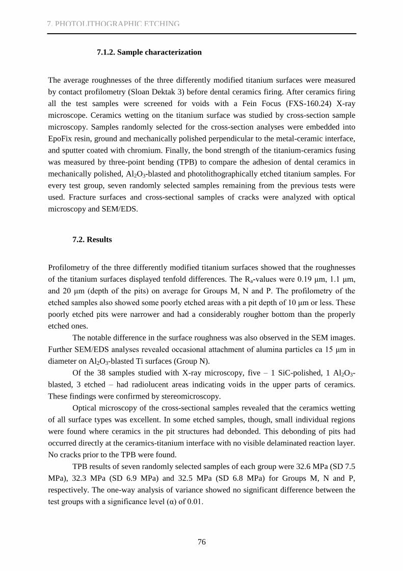
76
7.1.2. Sample characterization
The average roughnesses of the three differently modified titanium surfaces were measured
by contact profilometry (Sloan Dektak 3) before dental ceramics firing. After ceramics firing
all the test samples were screened for voids with a Fein Focus (FXS-160.24) X-ray
microscope. Ceramics wetting on the titanium surface was studied by cross-section sample
microscopy. Samples randomly selected for the cross-section analyses were embedded into
EpoFix resin, ground and mechanically polished perpendicular to the metal-ceramic interface,
and sputter coated with chromium. Finally, the bond strength of the titanium-ceramics fusing
was measured by three-point bending (TPB) to compare the adhesion of dental ceramics in
mechanically polished, Al2O3-blasted and photolithographically etched titanium samples. For
every test group, seven randomly selected samples remaining from the previous tests were
used. Fracture surfaces and cross-sectional samples of cracks were analyzed with optical
microscopy and SEM/EDS.
7.2. Results
Profilometry of the three differently modified titanium surfaces showed that the roughnesses
of the titanium surfaces displayed tenfold differences. The Ra-values were 0.19 μm, 1.1 μm,
and 20 μm (depth of the pits) on average for Groups M, N and P. The profilometry of the
etched samples also showed some poorly etched areas with a pit depth of 10 μm or less. These
poorly etched pits were narrower and had a considerably rougher bottom than the properly
etched ones.
The notable difference in the surface roughness was also observed in the SEM images.
Further SEM/EDS analyses revealed occasional attachment of alumina particles ca 15 μm in
diameter on Al2O3-blasted Ti surfaces (Group N).
Of the 38 samples studied with X-ray microscopy, five – 1 SiC-polished, 1 Al2O3-
blasted, 3 etched – had radiolucent areas indicating voids in the upper parts of ceramics.
These findings were confirmed by stereomicroscopy.
Optical microscopy of the cross-sectional samples revealed that the ceramics wetting
of all surface types was excellent. In some etched samples, though, small individual regions
were found where ceramics in the pit structures had debonded. This debonding of pits had
occurred directly at the ceramics-titanium interface with no visible delaminated reaction layer.
No cracks prior to the TPB were found.
TPB results of seven randomly selected samples of each group were 32.6 MPa (SD 7.5
MPa), 32.3 MPa (SD 6.9 MPa) and 32.5 MPa (SD 6.8 MPa) for Groups M, N and P,
respectively. The one-way analysis of variance showed no significant difference between the
test groups with a significance level (α) of 0.01.
7. PHOTOLITHOGRAPHIC ETCHING
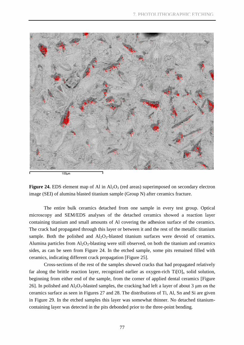
77
Figure 24. EDS element map of Al in Al2O3 (red areas) superimposed on secondary electron
image (SEI) of alumina blasted titanium sample (Group N) after ceramics fracture.
The entire bulk ceramics detached from one sample in every test group. Optical
microscopy and SEM/EDS analyses of the detached ceramics showed a reaction layer
containing titanium and small amounts of Al covering the adhesion surface of the ceramics.
The crack had propagated through this layer or between it and the rest of the metallic titanium
sample. Both the polished and Al2O3-blasted titanium surfaces were devoid of ceramics.
Alumina particles from Al2O3-blasting were still observed, on both the titanium and ceramics
sides, as can be seen from Figure 24. In the etched sample, some pits remained filled with
ceramics, indicating different crack propagation [Figure 25].
Cross-sections of the rest of the samples showed cracks that had propagated relatively
far along the brittle reaction layer, recognized earlier as oxygen-rich Ti[O]x solid solution,
beginning from either end of the sample, from the corner of applied dental ceramics [Figure
26]. In polished and Al2O3-blasted samples, the cracking had left a layer of about 3 μm on the
ceramics surface as seen in Figures 27 and 28. The distributions of Ti, Al, Sn and Si are given
in Figure 29. In the etched samples this layer was somewhat thinner. No detached titanium-
containing layer was detected in the pits debonded prior to the three-point bending.
7. PHOTOLITHOGRAPHIC ETCHING

78
Figure 25. SEI and compositional backscattered electron images of dental ceramics posts
remaining in titanium anchoring pits after the three-point bending test.
Figure 26. Crack initiation in the corner of applied dental ceramics where the applied dental
ceramics starts to spread on the Ti surface. Cracking occurs through oxygen-rich Ti[O]x along
the joint if the dissolution of oxygen from SiO2 and firing atmosphere to Ti is not restrained.
A slightly darker layer closest to dental ceramics in the Ti layer remaining on dental ceramics
can be seen, and interpreted as a titanium silicide layer [see 2.2.5 and 4.3.4].
Group N.
Group P.
7. PHOTOLITHOGRAPHIC ETCHING
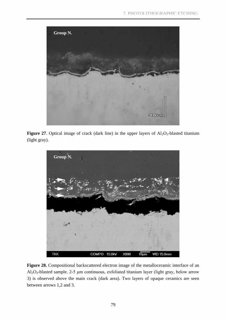
79
Figure 27. Optical image of crack (dark line) in the upper layers of Al2O3-blasted titanium
(light gray).
Figure 28. Compositional backscattered electron image of the metalloceramic interface of an
Al2O3-blasted sample. 2-5 μm continuous, exfoliated titanium layer (light gray, below arrow
3) is observed above the main crack (dark area). Two layers of opaque ceramics are seen
between arrows 1,2 and 3.
Group N.
Group N.
7. PHOTOLITHOGRAPHIC ETCHING
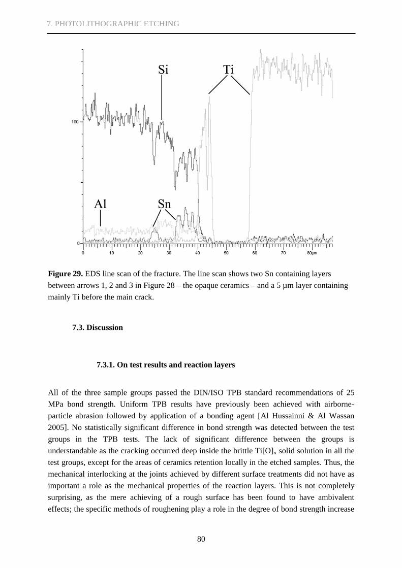
80
Figure 29. EDS line scan of the fracture. The line scan shows two Sn containing layers
between arrows 1, 2 and 3 in Figure 28 – the opaque ceramics – and a 5 µm layer containing
mainly Ti before the main crack.
7.3. Discussion
7.3.1. On test results and reaction layers
All of the three sample groups passed the DIN/ISO TPB standard recommendations of 25
MPa bond strength. Uniform TPB results have previously been achieved with airborne-
particle abrasion followed by application of a bonding agent [Al Hussainni & Al Wassan
2005]. No statistically significant difference in bond strength was detected between the test
groups in the TPB tests. The lack of significant difference between the groups is
understandable as the cracking occurred deep inside the brittle Ti[O]x solid solution in all the
test groups, except for the areas of ceramics retention locally in the etched samples. Thus, the
mechanical interlocking at the joints achieved by different surface treatments did not have as
important a role as the mechanical properties of the reaction layers. This is not completely
surprising, as the mere achieving of a rough surface has been found to have ambivalent
effects; the specific methods of roughening play a role in the degree of bond strength increase
7. PHOTOLITHOGRAPHIC ETCHING

81
achieved [see 2.3.1]. In this case at least, brittle reaction layers consistently demonstrated a
crucial role that was independent of the surface roughness.
Nevertheless, the difference in results between sections 4 and 6 supports the
fundamental importance of surface roughening. Here one has to take into account the effects
of random variation in e.g. handicraft and conditions in manufacturing the samples, as a
considerable time passed between the experiments of these two sections. Since section 7
minimized such variables and failed to demonstrate a difference in bond strength, one again
has to question whether various previous studies on the effect of roughening are fully cross-
comparable.
After the fusing of dental ceramics, some debonded pits without a visible delaminated
reaction layer were found. These pits were originally filled with ceramics, because the
negative of the pit form could be identified in the debonded material. Previous Duceratin
Dentine® has shown good thermal stability after repeated firing [Isgrò et al. 2005], hence
minor thermal variation after the application should also have no adverse effect on bond
strength [Tróia et al. 2003]. Multiple firing cycles, the explicit geometry of the samples,
thermal contraction difference between metal and ceramics [Yilmaz & Dinçer 1999], the glass
transition temperature of ceramics, and the elastic constants of the materials used, however,
could all have effected residual stresses debonding these areas [Könönen & Kivilahti 2001].
According to the findings in the present study, the actual cracking of the Ti[O]x occurs during
bending, not during the high temperature fusing.
7.3.2. On photolithographic etching
Some previous studies have applied photolithographic surface microfabrication in cell
experiments [Brunette 1986, Hallgren et al. 2001, Lu et al. 2005, Zhao et al. 2006, Troia et al.
2008]. Although various etching techniques have been tried to enhance dental ceramics
bonding [Reyes et al. 2001, Gilbert et al. 1994, Al Hussaini & Al Wazzan 2005], to our
knowledge there are no studies published concerning photolithographic methods in
prosthodontics.
The three-point bending produces a complex stress state at the metalloceramic joint
owing to the presence of pit structures as well as to different mechanical properties of
titanium and dental ceramics. Between the pits, the maximum tensile stress component acting
nearly perpendicular to the planar reaction layer surface opens the crack tip and facilitates
cracking. At vertical walls of the pits, the crack opening effect of this stress component is
reduced. Consequently cracking along the direction of the joint surface is impeded and the
crack is more likely to cut ceramic posts inside the pit structures. Compared to Al2O3-blasting
or polishing, the photolithographic etching created more retention sites in which dental
ceramics was found after the cracking.
7. PHOTOLITHOGRAPHIC ETCHING

82
The three-point bending, however, also creates another tensile stress component large
enough for cracks to propagate along the curved reaction layer of the pits in multiple
locations. Therefore, in most cases the cracking of the ceramic posts instead of the more
brittle reaction layers was not achieved. To improve the interfacial stress distribution, steeper
pit structures could be better, further reducing the effects of the stress component normal to
the planar reaction layer and thereby forcing the crack more readily to the ceramics. Using the
current etching protocol, this proved complicated. The openings in the photoresist mask
cannot be made much smaller, and as the etchant propagates equally in all directions, deeper
pits would also get wider. Longer etching time inflicts delamination of the AZ mask, causing
overetching of the titanium surface around the pit structure. This would compromise the crack
directing capability of the surface treatment. The best option could be to use pressurized
etching methods in generating deeper but not much wider anchoring structures. Etching via a
negative mask, i.e. creating controlled titanium rods or spikes instead of indentations, could
also give all the wanted stress directing benefits. Small titanium anchors would also have a
considerably greater shear, tensile and compressive strength than ceramics anchors [White et
al. 1996].
7.4. Conclusions
The results confirm that the strength of modern dental metalloceramic joints is limited by the
strength of mechanically brittle reaction layers formed during the fusing of dental ceramics.
Photolithographically etched pits can be used to redistribute the crack producing stress,
causing cracking of dental ceramics instead of the more brittle reaction layers. The current
depth and steepness of the pits, however, was insufficient for extensive stress redistribution.
Mechanical interlocking achieved by conventional surface treatments was also found to be
less than crucial in light of the prevalence of reaction layer cracking.
7. PHOTOLITHOGRAPHIC ETCHING

83
8. GENERAL DISCUSSION
8.1. On the need for novel and enhanced titanium metalloceramics techniques
Dental prosthetics is expensive, both to patients and health care organizations. From an
epidemiologic viewpoint, however, one can assume an increasing need for prosthetic therapy
[Jokstad 2002]. Treatment need on the population level is to a large extent reflected by the
fact that there is an increasingly larger proportion of elders today who retain their teeth, owing
to a considerable improvement in oral health, in addition to improved general knowledge of
the benefits of adequate oral care [Jokstad 2002]. Increased need leads to increased costs.
Expenses in Department of Oral and Maxillofacial diseases of Helsinki University Central
Hospital used in implants and prostheses increased by 32% from 2008 to 2009 alone [Helsinki
University Central Hospital data]. The significant increase in the price of gold in the 1970s
prompted development of less expensive dental alloys [Al Hussaini & Al Wassan 2005].
Replacing the high cost noble alloys has been one of the reasons behind the development of
titanium-based systems [Yilmaz & Dinçer 1999, Roach 2007, Roberts et al. 2009]. Even
though metalloceramic methods have their own solid place in large multiple-unit restorations
[Vásquez et al. 2008], one always has to consider other treatment options such as all-ceramic
restorations and fiber reinforced composite constructions when establishing the most suitable
care for the patient. These are outside the focus of this study, however.
Novel CAD/CAM solutions suitable for multiple-unit framework design have
experienced a rapid increase in sale rates. For example, the sale of CAD/CAM produced units
by one example dental company increased in Scandinavian countries by over 150% from
2008 to 2009 [Straumann Nordic sales data]. As titanium would appear to have a promising
future in fixed prosthodontics as a biocompatible and less expensive solution [Haag & Nilner
2007, Roberts et al. 2009], the resolving of fundamental problems, such as oxygen dissolution
into titanium during dental ceramics fusing, is essential. Simple additional methods easily
adaptable into dental technicians’ laboratories, such as silver interlayers, are thus more than
welcome.
8.2. On metalloceramic joints and fractography
The thesis work concentrated on four aspects of Ti-metalloceramic joints and the fusing of
dental ceramics: TiO2 layers, wetting, oxygen dissolution into titanium lattice, and mechanical
interlocking. The methodology and results of the four principal research approaches of this
thesis have been discussed in detail in the respective sections.
8. GENERAL DISCUSSION

84
Consistent with the hypothesis, the TiO2 layer thickness prior to dental ceramics
fusing was insignificant to the joint brittleness – contrary to general belief – and no significant
difference was found in TPB results. Titanium oxide layers have been considered detrimental
to joint integrity based on research on Ti samples without applied dental ceramics and in heat
treatments exceeding phase transformation temperature [Adachi et al. 1990, Kimura et al.
19901], and later widely referenced to when blaming titanium oxides for poor bonding
without confirming the existence of a true oxide layer or performing a deeper study of the
joint interface [Atsü & Berksun 2000, Garbelini et al. 2003, Özcan & Uysal 2005, Vásquez et
al. 2008]. TiO2 layers, however, dissociate during long firing protocols with applied SiO2-
based dental ceramics [Könönen & Kivilahti 1994]. Instead, during dental ceramics firing,
oxygen acquired from the firing atmosphere (before insulation by the first dental ceramics
layers), SiO2-based dental ceramics (during the rest of the firing process), and dissociating
TiO2 (an insignificant amount to joint integrity on its own) [Hautaniemi et al. 1992, Pajunen
& Kivilahti 1992, Könönen & Kivilahti 1994, Atsü & Berksun 2000, Könönen & Kivilahti
2000, Özcan & Uysal 2005] diffuses into vacant octahedral sites of the Ti lattice. This reduces
titanium plasticity and makes the surface of the Ti structure brittle. It is this solid solution of
titanium and oxygen that limits mechanical strength of the joint. High firing temperatures
further increase oxygen diffusion kinetics – a subject left undiscussed in the traditional papers
suggesting titanium oxide as the most important factor behind low bond strength [Adachi et
al. 1990, Kimura et al. 19901]. As the vacuum in dental furnaces cannot be sufficient for
inhibiting oxygen from diffusing to Ti [Wang et al. 1999, Lee et al. 2004], and dental
ceramics material remains another excellent oxygen source [Pajunen & Kivilahti 1992,
Könönen & Kivilahti 1994], methods limiting interstitial oxygen diffusion into Ti are needed
in order to achieve an ideal joint.
Good wetting is also needed for good interface formation and is of great importance in
dental metalloceramics during ceramics veneering [Carpenter & Goodkind 1979, Reyes et al.
2001, Marshall et al. 2010]. When good wetting is achieved, low fusing dental ceramics paste
bonders can cover the Ti surface during the first firing cycle, and provide insulation against
the firing atmosphere [Suansuwan & Swain 2003], a more rapid source of oxygen compared
to SiO2. Consistent with the hypothesis, extreme hydrophilicity of TiO2 was achieved with
only a few minutes of suitable UV activation. The timeline presented by Miron et al. [2004]
was supported, and even slightly faster results were achieved. With current dental ceramics,
however, wetting is a lesser problem for joint integrity than the oxygen dissolution during
dental ceramics firing.
Suppression of oxygen dissolution and the consequent formation of excessively thick
and brittle oxygen-rich Ti[O]x was shown possible with Ag interlayers, congruent with the
hypothesis, leaving dental ceramics as the most brittle part of the metalloceramic joint. Even
though several different interlayer materials for improving joint strength by restraining
oxygen diffusion have been tested [Dérand & Herø 1992, Gilbert et al. 1994, Wang & Fung
1997, Okazaki et al. 1998, Wang et al. 1999, Atsü & Berksun 2000, Sadeq et al. 2003, Lee et
8. GENERAL DISCUSSION

85
al. 2004, Suansuwan & Swain 2003, Özcan & Uysal 2005, Homann et al. 2006, Bieniaś et al.
2009], and some guidelines for ideal interlayer are given [Wang et al. 1999], no consistent
plan for ideal interlayer material selection prior to this thesis has been portrayed. In addition
to serving as oxygen diffusion barrier, it is suggested that plasticity of the Ag layers is also
beneficial in reducing stresses from the CTE mismatch of Ti and dental ceramics, well-known
to be detrimental to the joint [Könönen & Kivilahti 1994]. Also, aluminum contamination
detected after Al2O3 blasting and possibly unfavorable for joint integrity [Carpenter &
Goodkind 1979, Dérand & Herø 1992, Kern & Thompson 1994, Darwell et al. 1995,
Papadopoulos et al. 1999, Cai et al. 2001, Bagno & Di Bello 2004, Yamada et al. 2005] was
effectively insulated from contact to dental ceramics.
Increasing surface roughness of the metal framework is generally believed beneficial
in dental metalloceramics, as it increases joint area, mechanical retention and wetting, and
reduces maximal tensile stresses in the interfacial regions [Carpenter & Goodkind 1979,
Könönen & Kivilahti 1994, Reyes et al. 2001, Mo et al. 2003, Al Hussaini & Al Wazzan
2005]. In titanium-based metalloceramics, however, the most brittle part of the joints is not
the very interface between the dental ceramics and titanium, but the brittle oxygen-rich Ti[O]x
solid solution. Photolitographically etched pits succeeded locally in preventing cracking of
continuous planar reaction layers, and cracking occurred through dental ceramics instead of
Ti[O]x, in accordance to the hypothesis. Stress redistribution, however, was achieved in only
some pit structures and no significant difference in TPB results was seen. Perhaps with
steeper pit formations, a more uniform effect could have been achieved, resulting in improved
results in bond strength. Results from samples with Ag interlayers clearly show that cracking
of dental ceramics only occurs when the entire joint withstands a larger stress. Results
achieved in this section, in conjunction with results from earlier sections, clearly state that the
role of mechanical interlocking achieved by conventional surface treatments can be
questioned as long as the formation of the brittle reaction layers, most importantly oxygen-
rich Ti[O]x, cannot be controlled sufficiently well.
The thesis work provided also information on fractography – identification of crack
initiation, pattern of crack propagation, energetics of the fracture and classification of the
phases along the fracture plane – longed for in dental materials research [Marshall et al.
2010]. Cracking occurred in a similar manner in both Al2O3-blasted and polished samples,
and in samples with native or artificially thickened TiO2 layer. Crack initiation could be
identified at the corner of the dental ceramics portion, where the tensile stress in the three-
point bending test has the highest value. Cracking was considered to have proceeded as a
single event brittle crack over the midline of the sample, or in few samples completely
removing the ceramics portion, as can be observed in the sharp single disruption of the load-
deflection curve in TPB test. The optical microscopy, cross-sectional and SEM/EDS studies
after specimen bending clearly authenticate a cohesive nature of cracking through the near-
interface parts of titanium but far deeper than the normal titanium oxide layer thickness. On
the basis of thermodynamics the brittle reaction zone was considered to be the solid solution
8. GENERAL DISCUSSION

86
of titanium and oxygen, where oxygen atoms in the octahedral spacings of Ti restrained the
metallic plasticity. Könönen and Kivilahti have reported similar cracking in a previous study
[Könönen & Kivilahti 1994].
Different cracking was observed in samples with 5μm Ag interlayers. Cracking
occurred inside dental ceramics material, with multiple brittle cracks along the joint, and
significantly better TPB results were achieved. In a yet unpublished study of Könönen,
Saloniemi, Varpavaara and Kivilahti samples without Ag-plating presented load-deflection
curves recorded during TPB similar to the ones in this thesis, representing very sharp
dropping at bond failure load, owing to cracking of the continuous planar layer of brittle
Ti[O]x solid solution. The Ag-plated samples showed more plasticity at the failure load level,
and multiple sharp peaks representing multiple cracks inside the dental ceramics material.
These observations are congruent to findings in this monograph.
In photolithographically etched samples, stress distribution between dental ceramics
and brittle reaction layers was attempted with a different, more mechanical approach. Single
crack propagation through brittle Ti[O]x was forestalled, but only in some of the pit
formations, where cracking through dental ceramics occurred. Therefore no significant
difference in TPB results was achieved.
Restraining the formation of brittle oxygen-rich Ti[O]x can be considered of the
foremost importance in achieving ideal strength of the joints between dental ceramics and
titanium frameworks.
8.3. On future aspects
This thesis portrayed five major factors influencing joint integrity: compatibility of the CTE
values, the wetting of titanium surfaces, the α-case layer, the influence of titanium oxides and
oxygen, and titanium silicides.
Compatibility between the coefficient of thermal expansion of titanium and that of
dental ceramics is extremely important [Nielsen & Tuccillo 1972, Kimura et al. 19902,
Dérand and Herø 1992, Yilmaz & Dinçer 1999, Garbelini et al. 2003, Bieniaś et al. 2009].
Some debate is appropriate as to whether the compressive stress should be on the side of
brittle dental ceramics, or the side of the even more brittle Ti[O]x solid solution. The
temperature at which modern dental ceramics begin to behave as a shear-supporting solid
during cooling should also be estimated [Nielsen & Tuccillo 1972]. With the use of plastically
deforming Ag interlayers, however, the mismatch in the CTE between dental ceramics and Ti
was considered insignificant, so further research in this sector is not so highly topical.
Wetting of titanium surfaces with dental ceramics is also of great importance
[Carpenter & Goodkind 1979, Reyes et al. 2001, Marshall et al. 2010], however, no extreme
problems with the current dental ceramics material used in the thesis were detected.
Comparison between different materials should, however, be carried out in the future. The
8. GENERAL DISCUSSION

87
importance of wetting is even more pronounced, if oxygen diffusion barrier interlayers are not
used. Wetting of the Ag interlayers was found adequate in samples Al2O3-blasted prior to
interlayer deposition. Wetting in polished samples, however, was found inconsistent. To
obtain the best results with Ag interlayers, wetting of the interlayer material with dental
ceramics must be carefully studied further. Perhaps even thin TiO2 films with the capacity of
photoinduced superhydrophilicity can be applied on top of interlayer material in the most
vulnerable areas of large metalloceramics restorations to ensure perfect wetting. The
mechanical strength of nanometer scale TiO2 films compared to dental ceramics needs further
research.
The risks involved in titanium casting and the α-case layer should be well known by
now [Könönen & Kilivalhti 2001]. It has been misreferred to as a thick layer of titanium
oxide [Atsü & Berksun 2000, Garbelini et al. 2003, Vásquez et al. 2008], however, and some
recent studies even refer to using a dental ceramics material based bonding agent in a bid to
circumvent the incompatibility problem between the α-case layer and dental ceramics
[Vásquez et al. 2008]. Misunderstandings of this sort possess a clear threat to the clinical
success of titanium metalloceramics. Substituting the manufacturing of large cast titanium
structures with the more modern CAD/CAM processed frameworks fortunately diminishes
the amount of α-case layer inflicted problems.
The formation of Ti[O]x solid solution needs further research. Even though it can be
sufficiently restrained by the use of Ag interlayers, a deeper knowledge in Ti[O]x formation
kinetics and critical oxygen concentration for mechanical integrity will assist in interlayer
development. Due to the limitations of EDS microanalyses [see section 2.4.] in previous
research [Suansuwan & Swain 2003, Atsü & Berksun 2000, Lee et al. 2004, Vásquez et al.
2008, Vásquez et al. 2009], further WDS studies are required to supplement the previous
studies [Könönen & Kivilahti 1994].
Thick titanium silicide layers [Könönen & Kivilahti 1994, Könönen & Kivilahti 2001]
present a problem mostly after highly extended firing times, and their formation is also
restrained by Ag interlayers.
In addition to these five approaches, future research on effects of surface
microstructure and larger scale shape of underlying titanium framework on stress
concentration to dental ceramics is welcome. If results from different studies are to be
comparable, the same standard test methods must be used. The three-point bending test
executed according to the DIN/ISO standard was chosen for this monograph because of the
explicit test instructions and the wide use of the method [Könönen & Kivilahti 1990, Boening
et al. 1992, Gilbert et al. 1994, Pang et al. 1995, Pröbster et al. 1996, Oshida et al. 1997,
Yilmaz & Dinçer 1999, Atsü & Berksun 2000, Reyes et al. 2001, Al Hussaini & Al Wazzan
2005, Özcan & Oysal 2005, Inan et al. 2006, Troia et al. 2008, Bieniaś et al. 2009,
Papadopoulos & Spyropoulos 2009]. In TPB, the actual interfacial strength of the test samples
does not consist of plain shear or tensile strength, and the terminology used in literature has
been rather inconsistent. This monograph uses the inaccurate “bond strength” term to indicate
8. GENERAL DISCUSSION

88
the results acquired from the three-point bending test. In addition to TPB, mechanical tests
better resembling clinical stress need to be developed.
Although crack initiating mechanical laboratory tests and the following microanalyses
and evaluation of crack propagation give many important insights into the weak and brittle
elements in fusing of dental ceramics to titanium, more clinical follow up studies are needed.
The oral environment is able to induce physico-chemical alterations in the dental materials
[Vásquez et al. 2009], and cyclic longterm mechanical and thermal stress might decrease bond
strength of titanium metalloceramics [Oyafuso et al. 2008, Vásquez et al. 2008, Vásquez et al.
2009]. More research should therefore be done on the effects of clinical wear on the joint
reaction layers, on clinical fractography, and on the expected longevity of the final
restorations.
8. GENERAL DISCUSSION

89
9. CONCLUSIONS
The thesis is composed of four intercomplementary parts introducing new approaches to
brittle reaction layers and mechanical compatibility of metalloceramic joints created by the
fusing of dental ceramics to titanium. The following conclusions are stated:
I. The thickness of the TiO2 layer on titanium prior dental ceramics sintering is
insignificant for mechanical integrity of titanium-ceramic system. Instead, during
dental ceramics firing the TiO2 layer dissociates, interstitial diffusion of oxygen into
metallic titanium is increased, and the joint is made brittle by the resulting reduction in
plasticity of the metal structure surface.
II. Photoinduced superhydrophilicity was achieved within 2 minutes consuming as little
time and effort as Al2O3-blasting. It can be used to enhance wettability of titanium
surfaces, an important factor in dental ceramics veneering processes.
III. Plastically deforming Ag interlayers can be used to restrain oxygen diffusion into the
Ti lattice and prevent formation of excessively brittle oxygen-rich Ti[O]x solid
solution, thus giving significantly better bond strength results compared to only
conventionally blasting titanium frameworks with Al2O3. The most brittle component
of the joints with Ag interlayers was the bulk dental ceramics instead of Ti[O]x.
IV. Photolithographic etching can be used to cause cracking of dental ceramics instead of
the more brittle reaction layers – only locally, however, as it was used in this study.
The significance of mechanical interlocking achieved by conventional surface
treatments can be questioned as long as the formation of the brittle layers (mainly
oxygen-rich Ti[O]x) cannot be sufficiently controlled.
In summary, in contrast to former impressions of thick titanium oxide layers, this thesis study
clearly points out diffusion of oxygen from sintering atmosphere and SiO2 to Ti structures
during dental ceramics firing and the following formation of brittle oxygen-rich Ti[O]x solid
solution as the most important factors predisposing joints between Ti and SiO2-based dental
ceramics to low strength. This among other predisposing factors such as residual stresses
created by the coefficient of thermal expansion mismatch between dental ceramics and Ti
frameworks can be avoided with the use of Ag interlayers.
9. CONCLUSIONS
90
10. TIIVISTELMÄ SUOMEKSI
Titaani on ollut olennainen osa hammaslääketieteen materiaalivalikoimaa jo pitkään ja tullut
tunnetuksi P-I Brånemarkin tutkimuksista implantologiassa. Viimeaikainen tuotantotekninen
kehitys, titaanin erinomainen korroosionkestävyys, biokompatibiliteetti, saatavuus ja
edullisuus ovat tehneet myös titaanimetallokeramiasta varteenotettavan vaihtoehdon
proteettisiin ratkaisuihin. Vaikka titaanisovelluksilla on jo lyhytkestoisissa kliinisissä
seurantatutkimuksissa saatu tyydyttäviä, hyviä ja jopa erinomaisia tuloksia niin kestävyydessä
kuin estetiikassakin, titaanimetallokeramiaan liittyy edelleen ratkaisemattomia ongelmia,
jotka rajoittavat titaanin käyttöä. Merkittävin ongelma aiheutuu hammaskeraamien
polttoprosesseissa muodostuvista liitoksiin muodostuvista hauraista reaktiokerroksista, jotka
tekevät liitoksista titaanin ja keraamimateriaalin välillä verrattain heikkoja. Liitoslujuuden
parantamiseksi on kehitetty lukuisia eri menetelmiä, mutta niiden vaikutus mekaanisten
ominaisuuksien kannalta kriittisten reaktiokerrosten muodostumiseen on edelleen epäselvä.
Koska titaanimetallokeramia on jo vakiintunut kliiniseen käyttöön, on hauraista
reaktiokerroksista aiheutuvien ongelmien ratkaiseminen ja liitoslujuuden maksimoiminen
ensiarvoisen tärkeää.
Väitöskirjan kirjallisuuskatsauksessa käsitellään hammaskeraamien ja titaanin välisten
liitosten menestyksen kannalta olennaisia tekijöitä, kuten lämpölaajenemiskertoimien
yhteensopivuutta, pinnan kostutusta, faasitransitiolämpötilan ylittämisestä aiheutuvaa hauraan
reaktiokerroksen muodostumista (nk. ”α-case layer”), happea, sekä titaanin ja piin välisiä
reaktiotuotteita. Lisäksi esitellään liitosten parantamiseksi kehitettyjä menetelmiä, vertaillaan
titaaniametallokeramiaa ja muita metallokeramiamuotoja sekä liitosten tutkimukseen
soveltuvia menetelmiä.
Väitöskirjatyö lähestyy titaanin ja SiO2-pohjaisten hammaskeraamien liitosongelmia
neljästä lähtökohdasta ja hypoteesista:
I. Titaanipinnan TiO2-kerros. Hypoteesin mukaan polttoprosesseja edeltävällä TiO2-
kerroksen paksuudella ei ole merkitystä liitoksen kestävyyteen.
II. Titaanipinnan kostutus. Hypoteesin mukaan TiO2-pinnasta saadaan voimakkaan
hydrofiilinen vain minuuttien ultraviolettialtistuksella.
10. TIIVISTELMÄ SUOMEKSI

91
III. Hapen diffuusio titaanihilaan keraamipolttoprosessien aikana. Hypoteesin
mukaan titaanirakenteen pinnalle valmistettavan hopeavälikerroksen avulla voidaan
tehokkaasti ehkäistä polttoprosessien aikaista hapen liukenemista titaaniin ja tästä
seuraavaa titaanin pinnan haurastumista.
IV. Mekaanisen rasituksen jakautuminen liitosalueella. Hypoteesin mukaan hauraiden
reaktiokerrosten muodostumisen ehkäisy on tärkeämpää kuin mekaaninen retention
aikaansaaminen pinnan karhennuksella. Fotolitografista etsausmenetelmää käyttämällä
voidaan välttää yhtäjaksoisten tasomaisten reaktiokerrosten haurasmurtuma rasituksen
kohdistuessa paikoitellen Ti päälle sintrattuun hammaskeraamiin.
Titaanin pinnalle muodostuu normaaliympäristössä välittömästi muutamien
nanometrien paksuinen ns. natiivioksidikerros, joka koostuu pääosin amorfisesta TiO2:sta.
Vaikka titaanin erinomainen biokompatibiliteetti ja korroosionkestävyys ovat ensisijaisesti
juuri tämän oksidikerroksen ansiota, titaanioksidien vaikutuksia metallokeraamisten liitosten
muodostumisessa on perinteisesti pidetty erittäin haitallisina perustuen osin käytännön
metallokeramiasta poikkeavissa olosuhteissa saatuihiin testituloksiin. Vallalla on vielä myös
harhaluuloja ja väärinkäsityksiä faasitransitiolämpötilan ylittämisen jälkeen jäähtymisen
aikana titaanin pinnalle muodostuvasta, erittäin runsaasti happea sisältävästä nk. ”α-case
layer”:sta, jota on väärin perustein luultu muun muassa paksuksi titaanioksidikerrokseksi.
Väitöskirjan ensimmäisessä osion kokeellisessa ja kirjallisessa osassa esitetään kuitenkin työn
hypoteesia vastaavasti, kuinka normaaleissa keraamien polttoprosesseissa paksukin TiO2-
kerros dissosioituu, hapen diffuusiokinetiikka nopeutuu kohonneen lämpötilan seurauksena, ja
uunin sisäilmasta, SiO2:sta ja dissosioituneesta oksidikerroksesta lähtöisin oleva happi
diffundoituu syvälle titaanin sisälle, rajoittaen titaanitukirakenteen ylimpien osien
plastisuutta; sitä enemmän mitä korkeampi on titaaniin liuenneen hapen pitoisuus eli Ti[O]x.
3-pistetaivutustesteissä ei keraamipolttoja edeltävien TiO2-kerrosten paksuudella havaittu
merkittävää vaikutusta liitoksen kestoon liitosten murtuessa rasitustesteissä järjestelmällisesti
syvemmältä titaanitukirakenteen sisältä, Ti[O]-kiinteäliuoskerroksesta. Koska
hammaskeraamien polttoprosesseissa käytettävien uunien alipainepumput eivät riitä estämään
hapen diffuusiota ympäristöstä titaaniin, vaaditaan hapen diffuusiota rajoittavia menetelmiä
pinnan haurastumisen ehkäisemiseksi.
Hyvän liitoksen aikaansaamiseksi vaaditaan hyvää pinnan kostutusta, mikä on myös
muun muassa mikromekaanisen retention kannalta ensiarvoisen tärkeää kerrostettaessa
keraamimateriaalia metallipinnoille. Hyvä pinnan kostutus mahdollistaa
keraamiliitosaineiden, bonderien, tehokkaan leviämisen peittämään Ti pintaa heti
ensimmäisessä keraamipolttoprosessissa rajoittaen polttoympäristöstä lähtöisin olevan hapen
pääsyä liitosalueelle. Hypoteesia vastaavasti monografian toisen osion kokeellisessa osiossa
10. TIIVISTELMÄ SUOMEKSI

92
osoitetaan, kuinka TiO2-pinnan superhydrofiilisyys voidaan aikaansaada fotoindusoidusti vain
muutamissa minuuteissa. Nykyisiä keraamimateriaaleja käytettäessä titaanipinnan kostuvuus
on kuitenkin vähäisempi ongelma verrattuna hapen diffuusioon polttoprosessien aikana.
Vaikka polttouunin sisältämä ilma on ensisijainen ja tehokkain hapenlähde, myös SiO2-
pohjaiset keraamit toimivat erinomaisena hapenlähteenä kohonneissa polttolämpötiloissa,
minkä vuoksi bonderiaineiden käyttö ei voi täysin estää hapen pääsyä titaanin.
Tutkimuksen kolmannessa osiossa osoitetaan, kuinka hopeavälikerroksilla voidaan
ehkäistä hapen diffundoituminen polttoprosessien aikana titaanitukirakenteisiin.
Käsittelemällä pinta Ag-välikerroksin Al2O3-puhalluksen jälkeen saavutettiin tilastollisesti
merkittävästi parempi tulos 3-pistetaivutustesteissä (p = 1.27∙10-6
) verrattuna pelkkään pinnan
karhentamiseen Al2O3-puhalluksella. Hopeakerroksia käytettäessä liitoksen haurain osa oli
poikkeavasti itse hammaskeraami, missä havaittiin lukuisia erillisiä haurasmurtumia
titaanitukirangan säilyessä ehjänä. Vaikka lukuisia eri välikerroksia on tutkittu aiemmin
liitoslujuuden parantamiseksi ja hapen diffuusion ehkäisemiseksi ja joitakin vaatimuksia
ideaalisille välikerroksille on esitetty, ei yhtenäistä suunnitelmallista valintaa parhaimman
mahdollisen välikerrosmateriaalin löytämiselle ole ennen tätä väitöskirjaa julkaistu.
Diffuusioeristeenä toimimisen lisäksi Ag-kerrosten plastisuus on suotuisaa keraamin ja
titaanin lämpölaajenemiskertoimien eroista johtuvien jäännösrasitusten laukaisemisessa.
Näiden toistuvissa polttoprosesseissa ja jäähtymisissä syntyvien jännitysten haitallisuus
liitoksen mekaaniselle kestolle on hyvin tunnettu. Myös pinnan karhennuksessa käytettävistä
Al2O3-partikkeleista syntyvien mahdollisesti haitallisten titaanipinnan vierasainejäämien
eristyminen keraamikontaktista hopeakerrosten ansiosta osoitettiin.
Tyypillisesti metallipinnan karhentamisen on uskottu parantavan liitoksen laatua
liitospinta-alaa, mekaanista retentiota ja kostutusta lisäämällä, sekä liitokseen kohdistuvaa
jännitystä vähentämällä. Titaanimetallokeramiassa liitoksen heikoin kohta ei kuitenkaan ole
varsinainen rajapinta keraamimateriaalin ja titaanin välillä vaan runsashappinen Ti[O]x-
kiinteäliuos titaanin pinnassa. Tutkimuksen neljännessä osiossa
mikroelektroniikkateknologian tarpeisiin kehitettyä fotolitografista etsaustekniikka soveltaen
titaanipintaan muodostetaan säännönmukaisia syvennyksiä muokkaamaan mekaanisen
rasituksen jakautumista liitosrajapinnassa hammaskeraamin ja hauraiden reaktiokerrosten
välillä sekä ehkäisemään murtuman etenemistä yhtenäisissä, tasomaisissa reaktiokerroksissa
parantaen täten liitoksen kestävyyttä. Kolmipistetaivutustesti aikaansaa monikomponenttisen,
kompleksin rasitustilan metallokeraamiseen liitokseen litografisesti tuotettujen syvennysten
sekä titaanin ja hammaskeraamin ominaisten materiaaliominaisuuksien johdosta. Syvennysten
välissä maksimaalinen vetojännityskomponentti vaikuttaa lähes kohtisuoraan
reaktiokerrostasoa vasten avaten murtuman kärkeä ja helpottaen murtuman etenemistä.
Pystysuorilla syvennysten seinämillä tämän jännityskomponentin vaikutus on
vähäisimmillään; murtuman eteneminen hauraiden reaktiokerrosten suunnassa vaikeutuu ja
murtuma todennäköisemmin etenee titaanirungon pinnan suuntaisesti leikaten läpi
syvennyksissä olevan vähemmän hauraan hammaskeraamin. Koska hammaskeraamin
10. TIIVISTELMÄ SUOMEKSI

93
murtuminen Ti[O]-kiinteäliuoksen sijasta saavutettiin kokeellisessa osassa vain osassa
syvennyksiä, ei 3-pistetaivutustestissä havaittu merkittäviä eroja kontrolliryhmään verrattuna.
Mahdollisesti säännönmukaisesti syvempiä ja jyrkempiä syvennyksiä tuottamalla voitaisiin
saada merkittävä parannus myös rasitustesteissä; Ag-välikerroksilla tehdyt testi osoittavat
selvästi, että hammaskeraami murtuu vain koko liitoksen kestäessä tavanomaista merkittävästi
suurempia voimia. Neljännen tutkimusosion tulokset osoittavat myös selkeästi, kuinka
perinteisten pinnan karhennusmenetelmien avulla saatavan mekaanisen lukittumisen merkitys
liitoslujuudelle on vähäinen, ellei hauraiden reaktiokerrosten – erityisesti runsaasti happea
sisältävän Ti[O]-kiinteäliuoksen – muodostumista saada kontrolloitua.
Titaanimetallokeramiaa sovelletaan jo laajalti kliinisessä protetiikassa. Titaanin
materiaaliominaisuudet tekevät kuitenkin metallokeraamisen liitoksen suunnittelusta erityisen
haastavaa. Väitöskirjatyö antaa kaivattua informaatiota liitosten heikkouksien syistä ja
mahdollisuuksista vaikuttaa liitosten kestävyyteen. Työssä osoitetaan, että liitosten haurauden
syy on mekaanisesti heikko reaktiokerros ja siinä erityisesti kiinteä happirikas Ti[O]x-kerros,
joka muodostuu polttouunien sisäilmasta ja SiO2:sta/keraamista peräisin olevan hapen ja
titaanin välisessä liukenemisreaktiossa. Vaikka alueella riittää edelleen paljon tutkittavaa,
liitoksen erityisominaisuuksien tunteminen voi mahdollistaa
titaanimetallokeramiamenetelmien entistä luotettavamman ja kestävämmän soveltamisen,
siten että titaanin merkittävimpiä materiaalietuja voidaan täysipainoisesti hyödyntää myös
metallokeraamisissa ratkaisuissa. Tämä lisää entisestään edullisen titaanin
käyttömahdollisuuksia hammaslääketieteellisessä protetiikassa vähentäen proteettisen hoidon
kustannuksia niin potilaiden kuin hoitavan tahon osalta.
10. TIIVISTELMÄ SUOMEKSI

94
REFERENCES
Aarik J, Aidla A, Kiisler A-A, Uustare T, Sammelselg V. Effect of crystal structure on
optical properties of TiO2 films grown by atomic layer deposition. Thin Solid Films,
1997;305:270-3.
Aarik J, Aidla A, Sammelselg V, Siimon H, Uustare T. Control of thin film structure by
reactant pressure in atomic layer deposition of TiO2. J Cryst Growth, 1996;169:496-502.
Aarik J, Aidla A, Uustare T, Sammelselg V. Morphology and structure of TiO2 thin films
grown by atomic layer deposition. J Cryst Growth, 1995;148:268-75.
Adachi M, Mackert J, Parry E, Fairhurst C. Oxide adherence and porcelain bonding to
titanium and Ti-6Al-4V alloy. J Dent Res. 1990;69:1230-5.
Ahlgren C, Ahnlide I, Björkner B, Bruze M, Liedholm R, Möller H, Nilner K. Contact allergy
to gold is correlated to dental gold. Acta Derm Venereol, 2002;82:41-4.
Ahlgren C, Molin M, Lundh T, Nilner K. Levels of gold in plasma after dental gold inlay
insertion. Acta Odontol Scand, 2007;65:331-4.
Al Hussaini I, Al Wazzan K. Effect of surface treatment on bond strength of low-fusing
porcelain to commercially pure titanium. J Prosthet Dent, 2005;94:350-6.
Atsü S, Berksun S. Bond strength of three porcelains to two forms of titanium using two
firing atmospheres. J Prosthet Dent, 2000;84:567-74.
Bagno A, Di Bello C. Surface treatments and roughness properties of Ti-based biomaterials. J
Mater Sci Mater Med, 2004;15:935-49.
Balasubramanian G, Dionysiou D, Suidan M, Subramanian V, Baudin I, Laîné J-M. Titania
powder modified sol-gel process for photocatalytic applications. J Mater Sci, 2003;38:823-31.
Benedix R, Dehn F, Quaas J, Orgass M. Application of titanium dioxide photocatalysis to
create self-cleaning building materials. Lacer, 2000;5:157-68.
Bergman B, Nilson H, Andersson M. A longitudinal clinical study of Procera ceramic-
veneered titanium copings. Int J Prosthodont, 1999;12:135-9.
Bieniaś J, Surowska B, Stoch A, Matraszek H, Walczak M. The influence of SiO2 and SiO2-
TiO2 intermediate coatings on bond strength of titanium and Ti6Al4V alloy to dental
porcelain. Dent Mater, 2009;25:1128-35.
Blake D, Maness P-C, Huang Z, Wolfrum E, Huang J. Application of the photocatalytic
chemistry of titanium dioxide to disinfection and the killing of cancer cells. Separation and
Purification Methods, 1999;28:1-50.
Boening K, Walter M, Reppel P. Non-cast titanium restorations in fixed prosthodontics. J
Oral Rehabil, 1992;19:281-7.
REFERENCES

95
Boyer R, Gall T. Metals Handbook, Desk Edition. 1991. American Society for Metals. USA.
Boyer R, Welsch G, Collings E. Materials Properties Handbook, Titanium Alloys: 131-135.
1994. ASM International. USA.
Browne M, Gregson P. Surface modification of titanium-alloy implants. Biomaterials,
1994;15:894-8.
Brunette D, Tengvall P, Textor M, Thomsen P. Titanium in medicine: 171-230. 2001.
Springer.
Brunette M. Spreading and orientation of epithelial cells on grooved substrata. Exp Cell Res,
1986;167:203-17.
Cai Z, Bunce N, Nunn M, Okabe T. Porcelain adherence to dental cast CP titanium: effects of
surface modifications. Biomaterials, 2001;22:979-86.
Carp O, Huisman C, Reller A. Photoinduced reactivity of titanium dioxide. Progress in Solid
State Chemistry, 2004;32:33-177.
Carpenter M, Goodkind R. Effect of varying surface texture on bond strength of one
semiprecious and one nonprecious ceramo-alloy. J Prosthet Dent, 1979;42:86-95.
Chai J, McGivney G, Munoz C, Rubenstein J. A multicenter longitudinal clinical trial of a
new system for restorations. J Prosthet Dent, 1997;77:1-11.
Cortizo M, De Mele M, Cortizo A. Metallic dental material biocompatibility in osteoblastlike
cells: correlation with metal ion release. Biol Trace Elem Res, 2004;100:151-68.
Darvell B, Samman N, Luk W, Clark RK, Tideman H. Contamination of titanium castings by
aluminium oxide blasting. J Dent, 1995;23:319-22.
Dérand T, Herø H. Bond strength of porcelain on cast vs. wrought titanium. Scand J Dent
Res, 1992;100:184-8.
DIN 13 927. Zahnheilkunde, Metall-Keramik-Systeme, Anforderungen, Prüfung. 1990.
Germany.
Fujishima A, Rao T, Tryk D. Titanium dioxide photocatalysis. J Phochem Photobiol C,
2000;1:1-21.
Fujishima A, Zhang X. Titanium dioxide photocatalysis: present situation and future
approaches. C R Chimie, 2006;9:750-60.
Fy G, Vary P, Lin C-T. Anatase TiO2 nanocomposites for antimicrobial coatings. J Phys
Chem B, 2005;109:8889-98.
Garbelini W, Henriques G, Troia M, Mesquita M, Dezan C. Evaluation of low-fusing ceramic
systems combined with titanium grades II and V by bending test and scanning electron
microscopy. J Appl Oral Sci, 2003;11:354-60.
REFERENCES

96
Gilbert J, Covey D, Lautenschlager E. Bond characteristics of porcelain fused to milled
titanium. Dent Mater, 1994;10:134-40.
Grill V, Sandrucci M, Di Lenarda R, Cadenaro M, Narducci P, Bareggi R, Martelli A.
Biocompatibility evaluation of dental metal alloys in vitro: expression of extracellular matrix
molecules and its relationship to cell proliferation rates. J Biomed Mater Res, 20002;52:479-
87.
Grill V, Sandrucci M, Di Lenarda R, Dorigo E, Narducci P, Martelli A, Bareggi R. Cell
proliferation rates and fibronectin arrangement as parameters for biocompatibility evaluation
of dental metal alloys in vitro. J Oral Sci, 20001;42:1-7.
Petzow G. Metallographic Etching, 2nd edition: 139-144. 1999. ASM International. USA.
Haag P, Nilner K. Questions and answers on titanium-ceramic dental restorative systems: a
literature study. Quintessence Int, 2007;38:5-13.
Hallgren C, Reimers H, Gold J, Wennerberg A. The importance of surface texture for bone
integration of screw shaped implants: an in vivo study of implants patterned by
photolithography. J Biomed Mater Res, 2001;57:485-96.
Hammad I, Talic Y. Designs of bond strength tests for metal-ceramic complexes: review of
the literature. J Prosthet Dent, 1996;75:602-8.
Hautaniemi J, Herø H, Juhanoja J. On the bonding of porcelain on titanium. J Mater Sci Mater
Med, 1992;3:186-191.
Homann F, Waddell J, Swain M. Influence of water, loading rate and bonder on the adhesion
of porcelain to titanium, J Dent, 2006;34:485-90.
Ilola R, Romu J, Papula S, Hänninen H. Katalyyttipinnat ja niiden valmistus. Teknillisen
korkeakoulun materiaalitekniikan laboratorion julkaisuja, 2003;1:1-51.
Inan O, Acar A, Halkaci S. Effects of sandblasting and electrical discharge machining on
porcelain adherence to cast and machined commercially pure titanium. J Biomed Mater Res B
Appl Biomater, 2006;78:393-400.
Isgrò G, Kleverlaan C, Wang H, Feilzer A. The influence of multiple firing on thermal
contraction of ceramic materials used for the fabrication of layered all-ceramic dental
restorations. Dent Mater, 2005;21:557-64.
ISO 9693. Metal-ceramic dental restorative systems. 1999. Switzerland.
Jokstad A. Oral prosthetics from a Nordic perspective. Int J Prosthodont, 2002;15:145-53.
Kasemo B, Lausmaa J. Biomaterial and implant surfaces: a surface science approach. Int J
Oral Maxillofac Implants, 1988;3:247-59.
Kaus T, Pröbster L, Weber H. Clinical follow-up study of ceramic veneered titanium
restorations--three-year results. Int J Prosthodont, 1996;9:9-15.
REFERENCES
97
Kern M, Thompson V. Effects of sandblasting and silica-coating procedures on pure titanium.
J Dent, 1994;22:300-6.
Kim J, Cho S. The effects of laser etching on shear bond strength at the titanium ceramic
interface. J Prosthet Dent, 2009;101:101-6.
Kimura H, Horng C, Okazaki M, Takahashi J. Oxidation effects on porcelain-titanium
interface reactions and bond strength. Dent Mater J, 19901;9:91-9.
Kimura H, Horng C, Okazaki M, Takahashi J. Thermal compatibility of titanium-porcelain
system. J Osaka Univ Dent Sch, 19902;30:43-52.
Könönen M, Kivilahti J. Bonding of low-fusing dental porcelain to commercially pure
titanium. J Biomed Mater Res, 1994;28:1027-35.
Könönen M, Kivilahti J. Fusing of dental ceramics to titanium. J Dent Res, 2001;80:848-54.
Könönen M, Kivilahti J. Joining of dental ceramics to titanium by metallic interlayers, in:
Proceedings of the 7th International Meeting on Modern Ceramic Technologies. P.
Vinzencini, ed., Elsevier, Amsterdam, 1990:41-7.
Könönen M, Kivilahti J. Testing of metal-ceramic joint using scanning acoustic microscopy.
Dent Mater, 1991;7:211-4.
Kumagai H, Matsumoto M, Toyoda K, Obara M, Suzuki M. Fabrication of titanium oxide
thin films by controlled growth with sequential surface chemical reactions. Thin Solid Films,
1995;263:47-53.
Lausmaa J. Surface spectroscopic characterization of titanium implant materials. J Electron
Spectrosc Relat Phenom, 1996;81:343-61.
Lee H-R. Comparative study of bond characteristics between titanium/titanium alloy and
ceramic. 2004. Der Medizinischen Fakultät der Eberhard-Karls-Universität zu Tübingen.
Germany.
Lee K, Cai Z, Griggs J, Guiatas L, Lee D, Okabe T. SEM/EDS evaluation of porcelain
adherence to gold-coated cast titanium. J Biomed Mater Res B Appl Biomater, 2004;68:165-
73.
Li X, Li F. Study of Au/Au3+
-TiO2 photocatalysts toward visible photooxidation for water and
wastewater treatment. Environ Sci Technol, 2001;35:2381-7.
Locci P, Marinucci L, Lilli C, Belcastro S, Staffolani N, Bellocchio S, Damiani F, Becchetti
E. Biocompatibility of alloys used in orthodontics evaluated by cell culture tests. J Biomed
Mater Res, 2000;51:561-8.
Lövgren R, Andersson B, Bergøvist S, Carlsson G, Ekström P, Odman P, Sundqvist B.
Clinical evaluation of ceramic veneered titanium restorations according to the Procera
technique. Swed Dent J, 1997;21:1-10.
Lövgren R, Andersson B, Carlsson G, Odman P. Prospective clinical 5-year study of ceramic-
veneered titanium restorations with the Procera system. J Prosthet Dent, 2000;84:514-21.
REFERENCES

98
Lu X, Leng Y, Zhang X, Xu J, Qin L, Chan W. Comparative study of osteoconduction on
micromachined and alkali-treated titanium alloy surfaces in vitro and in vivo. Biomaterials,
2005;26:1793-8.
Madaeni S, Ghaemi N. Characterization of self-cleaning RO membranes coated with TiO2
particles under UV irradiation. Journal of Membrane Science, 2007;303:221-33.
Maness P-C, Smolinski S, Blake D, Huang Z, Wolfrum E, Jacoby W. Bactericidal activity of
photocatalytic TiO2 reaction: toward an understanding of its killing mechanism. Appl Environ
Microbiol, 1999;65:4094-8.
Mardare D, Tasca M, Delibas M, Rusu G. On the structural properties and optical
transmittance of TiO2 r.f. sputtered thin films. Appl Surf Sci, 2000;156:200-6.
Marshall S, Stephen C, Baier R, Tomsia A, Marshall G. A review of adhesion science. Dent
Mater, 2010;26:11-6.
Massalski T. Binary Alloy Phase Diagrams. 1996. ASM International. USA.
McCafferty E, Wightman JP. An X-ray photoelectron spectroscopy sputter profile study of
the native air-formed oxide film on titanium. Appl Surf Sci, 1999;143:92-100.
Milleding P, Haag P, Neroth B, Renz I. Two years of clinical experience with Procera
titanium crowns. Int J Prosthodont, 1998;11:224-32.
Mills A, Le Hunte S. An overview of semiconductor photocatalysis. J Phochem Photobiol A,
1997;108:1-35.
Mills A, Lee S-K. A web-based overview of semiconductor photochemistry-based current
commercial applications. J Phochem Photobiol A, 2002;152:233-47.
Miron C, Roca A, Hoisie S, Cozorici P, Sirghi L. Photoinduced bactericidal activity of TiO2
thin films obtained by radiofrequency magnetron sputtering deposition. Journal of
Optoelectronics and Advanced Materials, 2004;7:915-9.
Miyachi M, Nakajima A, Fujishima A, Hashimoto K, Watanabe T. Photoinduced surface
reactions on TiO2 and SrTiO3 films: photocatalytic oxidation and photoinduced
hydrophilicity. Chem Mater, 2000;12:3-5
Miyachi M, Nakajima A, Watanabe T, Hashimoto K. Photocatalysis and photoinduced
hydrophilicity of various metal oxide thin films. Chem Mater, 2002;14:2812-6.
Mo A, Cen Y, Liao Y. The influence of surface conditioning on the shear bond strength of
La-Porcelain and titanium. Hua Xi Kou Qiang Yi Xue Za Zhi, 2003;21:104-6.
Mogyorósi K, Dékány I, Fendler J. Preparation and characterization of clay mineral
intercalated titanium dioxide nanoparticles. Langmuir, 2003;19:2938-46.
Nalva H. Handbook of Thin Film Materials, Volume 1. 2002. Academic Press. USA.
Nielsen J, Tuccillo J. Calculation of interfacial stress in dental porcelain bonded to gold alloy
substrate. J Dent Res, 1972;51:1043-7.
REFERENCES
99
Nilson H, Bergman B, Bessing C, Lundqvist P, Andersson M. Titanium copings veneered
with Procera ceramics: a longitudinal clinical study. Int J Prosthodont, 1994;7:115-9.
Okazaki M, Wang X, Toguchi M, Taira M, Takahashi J, Matsuo C, Hino Y. Improvement of
bond strength in metal-ceramic systems using a gold intermediate layer. Dent Mater J,
1998;17:163-73.
Oshida Y, Fung L, Isikbay S. Titanium-porcelain system. Part II: Bond strength of fired
porcelain on nitrided pure titanium. Biomed Mater Eng, 1997;7:13-34.
Oyafuso D, Ozcan M, Bottino M, Itinoche M. Influence of thermal and mechanical cycling on
the flexural strength of ceramics with titanium or gold alloy frameworks. Dent Mater,
2008;24:351-6.
Özcan I, Uysal H. Effects of silicon coating on bond strength of two different titanium
ceramic to titanium. Dent Mater, 2005;21:773-9.
Pajunen M, Kivilahti J. Thermodynamics of the Titanium-Oxygen System. Zeitschrift für
Metallkunde, 1992;83:17-20.
Pang I, Gilbert J, Chai J, Lautenschlager E. Bonding characteristics of low-fusing porcelain
bonded to pure titanium and palladium-copper alloy. J Prosthet Dent, 1995;73:17-25.
Papadopoulos T, Spyropoulos K. The effect of a ceramic coating on the cpTi-porcelain bond
strength. Dent Mater, 2009;25:247-53.
Papadopoulos T, Tsetsekou A, Eliades G. Effect of aluminium oxide sandblasting on cast
commercially pure titanium surfaces. Eur J Prosthodont Restor Dent, 1999;7:15-21.
Papazoglou E, Brantley W. Porcelain adherence vs force to failure for palladium-gallium
alloys: a critique of metal-ceramic bond testing. Dent Mater, 1998;14:112-9.
Poilleau J, Devilliers D, Garrido F, Durand-Vidal S, Mahé E. Structure and composition of
passive titanium oxide films. Mater Sci Eng, 1997;47:235-43.
Pröbster L, Maiwald U, Weber H. Three-point bending strength of ceramics fused to cast
titanium. Eur J Oral Sci, 1996;104:313-9.
Puurunen L. Surface chemistry of atomic layer deposition: A case for the
trimethylaluminum/water process. J Appl Phys, 2005;97:121301.
Reyes M, Oshida Y, Andres C, Barco T, Hovijitra S, Brown D. Titanium-porcelain system.
Part III: effects of surface modification on bond strengths. Biomed Mater Eng, 2001;11:117-
36
Riley D, Bavastrello V, Covani U, Barone A, Nicolini C. An in-vitro study of the sterilization
of titanium dental implants using low intensity UV-radiation. Dent Mater, 2005;21:756-60.
Ritala M, Leskelä M, Nykänen E, Soininen P, Niinistö L. Growth of titanium dioxide thin
films by atomic layer epitaxy. Thin Solid Films, 1993;225:228-95.
REFERENCES
100
Roach M. Base metal alloys used for dental restorations and implants. Dent Clin North Am,
2007;51:603-27.
Roberts H, Berzins D, Moore B, Charlton D. Metal-ceramic alloys in dentistry: a review. J
Prosthodont, 2009;18:188-94.
Rosental A, Adamson P, Gerst A, Koppel H, Tarre A. Atomic layer deposition in traveling-
wave reactor: In situ diagnostics by optical reflection. Appl Surf Sci, 1997;112:82-6.
Rosental A, Tarre A, Adamson P, Gerst A, Kasikov A, Niilisk A. Surface of TiO2 during
atomic layer deposition as determined by incremental dielectric reflection. Appl Surf Sci,
1999;142:204-9.
Sadeq A, Cai Z, Woody R, Miller A. Effects of interfacial variables on ceramic adherence to
cast and machined commercially pure titanium. J Prosthet Dent, 2003;90:10-7.
Sakai N, Fujishima A, Watanabe T, Hashimoto K. Quantitative evaluation of the
photoinduced hydrophilic conversion properties of TiO2 thin film surfaces by the reciprocal
contact angle. J Phys Chem B, 2003;107:1028-35.
Sakai N, Wang R, Fujishima A, Watanabe T, Hashimoto K. Effect of ultrasonic treatment on
highly hydrophilic TiO2 surfaces. Langmuir, 1998:14;5918-20.
Sammelselg V, Rosental A, Tarre A, Niinistö L, Heiskanen K, Ilmonen K, Johansson L-S,
Uustare T. TiO2 thin films by atomic layer deposition: a case of uneven growth at low
temperature. Appl Surf Sci, 1998;134:78-86.
Sittig C, Textor M, Spencer ND, Wieland M, Vallotton PH. Surface characterization of
implant materials c.p. Ti, Ti-6Al-7Nb and Ti-6Al-4V with different pretreatments. J Mater Sci
Mater Med, 1999;10:35-46.
Smedberg J, Ekenbäck J, Lothigius E, Arvidson K. Two-year follow-up study of Procera-
ceramic fixed partial dentures. Int J Prosthodont, 1998;11:145-9.
Suansuwan N, Swain M. Adhesion of porcelain to titanium and a titanium alloy. J Dent,
2003;31:509-18.
Suntola T, Antson J. U.S. Patent 4058430, 1977.
Suntola T, Pakkala A, Lindfors S. U.S. Patent 4389973, 19831.
Suntola T, Pakkala A, Lindfors S. U.S. Patent 4413022, 19832.
Syverud M, Kvam K, Herø H. A new dental Pd-Co alloy for ceramic veneering. Dent Mater,
1987;3:102-6.
Troia M, Henriques G, Mesquita M, Fragoso W. The effect of surface modifications on
titanium to enable titanium-porcelain bonding. Dent Mater, 2008;24:28-33.
Tróia M, Henriques G, Nóbilo M, Mesquita M. The effect of thermal cycling on the bond
strength of low-fusing porcelain to commercially pure titanium and titanium-aluminium-
vanadium alloy. Dent Mater, 2003;19:790-6.
REFERENCES

101
Tu J, Zhao X, Zhao Q, Wang G. Preparation and characterization of super-hydrophilic porous
TiO2 coating films. Mater Chem Phys, 2001:68;253-9.
Vásquez V, Özcan M, Kimpara E. Evaluation of interface characterization and adhesion of
glass ceramics to commercially pure titanium and gold alloy after thermal- and mechanical-
loading. Dent Mater, 2009;25:221-31.
Vásquez V, Özcan M, Nishioka R, Souza R, Mesquita A, Pavanelli C. Mechanical and
thermal cycling effects on the flexural strength of glass ceramics fused to titanium. Dent
Mater J, 2008;27:7-15.
Vilhunen S, Bosund M, Kääriäinen M-L, Cameron D, Sillanpää M. Atomic layer deposited
TiO2 films in photodegradation of aqueous salisylic acid. Separation and Purification
Technology, 2009;66:130-4.
Walter M, Böning K, Reppel P. Clinical performance of machined titanium restorations. J
Dent, 1994;22:346-8.
Walter M, Reppel P, Böning K, Freesmeyer W. Six-year follow-up of titanium and high-gold
porcelain-fused-to-metal fixed partial dentures. J Oral Rehabil, 1999;26:91-6.
Walter M, Reppel P, Böning K, Freesmeyer W. Six-year follow-up of titanium and high-gold
porcelain-fused-to-metal fixed partial dentures. J Oral Rehabil, 1999;26:91-6.
Wang R, Fung K. Oxidation behavior of surface-modified titanium for titanium-ceramic
restorations. J Prosthet Dent, 1997;77:423-34.
Wang R, Li Y. In vitro evaluation of biocompatibility of experimental titanium alloys for
dental restorations. J Prosthet Dent, 1998;80:495-500.
Wang R, Meyers E, Katz J. Scanning acoustic microscopy study of titanium-ceramic interface
of dental restorations. J Biomed Mater Res, 1998;42:508-16.
Wang R, Welsch G, Monteiro O. Silicon nitride coating on titanium to enable titanium–
ceramic bonding. J Biomed Mater Res, 1999;46:262-70.
Wang T, Zheng S, Hao W, Wang C. Studies on photocatalytic activity and transmittance
spectra of TiO2 thin films prepared by r.f. magnetron sputtering method. Surface and Coatings
Technology, 2002;155:141-5.
Wataha J, Lockwood P, Khajotia S, Turner R. Effect of pH on element release from dental
casting alloys. J Prosthet Dent, 1998;80:691-8.
Wataha J, Lockwood P. Release of elements from dental casting alloys into cell-culture
medium over 10 months. Dent Mater, 1998;14:158-63.
White S, Ho L, Caputo A, Goo E. Strength of porcelain fused to titanium beams. J Prosthet
Dent, 1996;75:640-8.
Yamada K, Onizuka T, Endo K, Ohno H, Swain M. The influence of Goldbonder and pre-
heat treatment on the adhesion of titanium alloy and porcelain. J Oral Rehabil, 2005;32:213-
20.
REFERENCES

102
Yilmaz H, Dinçer C. Comparison of the bond compatibility of titanium and an NiCr alloy to
dental porcelain. J Dent, 1999;27:215-22.
Ylilammi M, Ranta-Aho T. Optical determination of the film thicknesses in multilayer thin
film structures. Thin Solid Films, 1993;232:56-62.
Yli-Urpo A. Investigation of a dental gold alloy and its ceramic bonding. Acta Odontol
Scand, 1975;33:1-184.
Zhang Y, Yu J, Tu R, Goto T. Preparation of Ca2Ti2O6 pyrochlore films by MOCVD. Journal
of Physics: Conference Series, 2009;152:1-6.
Zhao G, Zinger O, Schwartz Z, Wieland M, Landolt D, Boyan B. Osteoblast-like cells are
sensitive to submicron-scale surface structure. Clin Oral Implants Res, 2006;17:258-64.
REFERENCES