the role of tumor infiltrating lymphocytes in immunotherapy · federico rojo....
TRANSCRIPT

The role of tumor
infiltratinglymphocytes inimmunotherapy
Federico Rojo
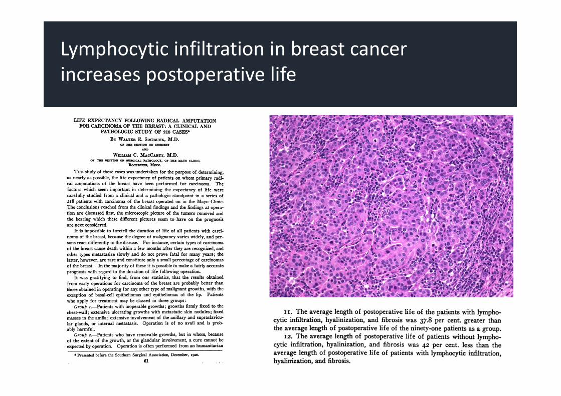
Lymphocytic infiltration in breast cancerincreases postoperative life

Lymphocytic‐predominant phenotype is in 20‐28% of breast cancers and correlates withoutcome
1.Clinical importance of TILs in cancer
2.Immune‐inflammed cancer phenotype and benefit to immunotherapy
3.Assesing TILs in practice: recommendations and reproducibility
Outline

5. ¿Cuál debe de ser el planteamiento futuro en el diagnóstico morfológico?
Denkert, C et al. SABCS 2016
Metaanalysis of 3,771 patients from 6 neoadjuvant trials
TILs as predictive and prognostic biomarker in breast cancer

5. ¿Cuál debe de ser el planteamiento futuro en el diagnóstico morfológico?
Denkert, C et al. SABCS 2016
Metaanalysis of 3,771 patients from 6 neoadjuvant trials
TILs as predictive and prognostic biomarker in breast cancer

Association of immunoscore with prognosis in colorectal cancer
Pages, F et al. NEJM 2005Pages, F et al. J Clin Oncol 2009Mlecnik, B et al. J Clin Oncol 2011

Fridman, WH et al. Nat Rev Oncol 2015
124 studies
Association of immunoscore with prognosis in various types of cancer

1.Clinical importance of TILs in cancer
2.Immune‐inflammed cancer phenotype and benefit to immunotherapy
3.Assesing TILs in practice: recommendations and reproducibility
Outline

Hedge, PS et al. Clin Cancer Res 2015
Tumor immunity continuum

Paucity of T cells in the stroma of the tumor. Although myeloid cells may be present, the general feature of this profile is the presence of a non‐inflamed tumour microenvironment with few or no CD8‐carrying T cells
Rarely respond to anti‐PD‐L1/PD‐1 therapy
This phenotype probably reflects the absence of pre‐existing antitumour immunity, which suggests that the generation of tumour‐specific T cells is the rate‐limiting step
Tumor immunity continuum:1. Immune‐desert phenotype

Herbst, RS et al. Nature 2014
1. Immune‐desert phenotype
Atezolizumab activity is associatedwith presence of TILs in stroma

Abundant immune cells in the stroma that surrounds nests of tumor and not penetrate the parenchyma
After treatment with anti‐PD‐L1/PD‐1 agents, stroma‐associated T cells can show evidence of activation and proliferation but not infiltration, and clinical responses are uncommon
These features suggest that a pre‐existing antitumor response might have been present but was rendered ineffective by a block in tumor penetration through the stroma or by the retention of immune cells in the stroma
Tumor immunity continuum:2. Immune‐excluded phenotype

Herbst, RS et al. Nature 2014
2. Immune‐excluded phenotype
Atezolizumab activity is associatedwith presence of TILs in stroma

Presence of both CD4‐ and CD8 cells, often accompanied by myeloid and monocytic cells, which are positioned in proximity to the tumor cells
Inflamed tumors exhibit staining for PD‐L1 on infiltrating immune cells and, in some cases, tumor cells
Many proinflammatory and effector cytokines can be detected
This profile suggests the presence of a pre‐existing antitumour immune response that was arrested probably by immunosuppression
Clinical responses to anti‐PD‐L1/PD‐1 therapy occur most often in patients with inflamed tumors
However, a response is not assured in these individuals, which indicates that immune‐cell infiltration is necessary but insufficient for inducing a response.
Chen, DS and Mellman, I. Nature 2017
Tumor immunity continuum:3. Immune‐inflamed phenotype

Li, B et al. Genom Biol 2016
3. Immune‐inflamed phenotype
Association between mutational burdenand immune cells in tumor stroma

Taube, JM et al. Clin Cancer Res 2014
3. Immune‐inflamed phenotype
TILs are associated with PD‐L1 expressionin melanoma, NSCLC and RCC

Relationship between CD8 and PD‐L1 expression in advanced melanoma
Tumeh, PC et al Nature 2015
3. Immune‐inflamed phenotype
CD8+ TILs are associated with PD‐L1 expression in advanced melanoma

KEYNOTE016: pembrolizumab and MSI colorectal and non‐colorectal tumors
3. Immune‐inflamed phenotype
Mismatch‐repair deficiency and PD‐1 blockade benefit in CRC and others

Tumeh, PC et al. Nature 2015
Pembrolizumab benefit in advanced melanoma and TILs
3. Immune‐inflamed phenotype
CD8+ TILs are associated with benefit topembrolizumab in advanced melanoma

3. Immune‐inflamed phenotype
Immune repertoire predicts nivolumabresponse in melanoma
Inoue, H et al. Oncoinmunology 2016

Haratani, K et al. Ann Oncol 2017
3. Immune‐inflamed phenotype
Nivolumab efficacy in T790 EGFR mutated NSCLC is associated with presence of CD8+ TILs
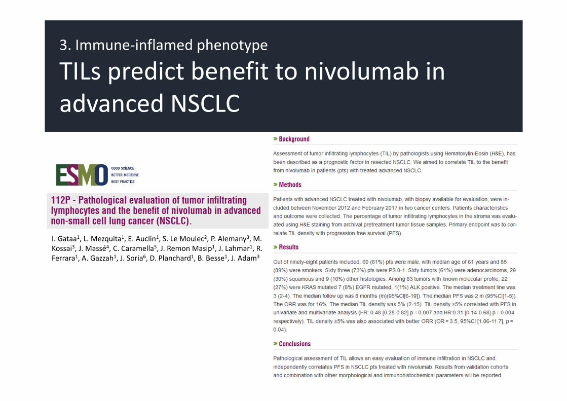
I. Gataa1, L. Mezquita1, E. Auclin1, S. Le Moulec2, P. Alemany3, M. Kossai3, J. Massé4, C. Caramella5, J. Remon Masip1, J. Lahmar1, R. Ferrara1, A. Gazzah1, J. Soria6, D. Planchard1, B. Besse1, J. Adam3
3. Immune‐inflamed phenotype
TILs predict benefit to nivolumab in advanced NSCLC

3. Immune‐inflamed phenotype
TILs predict response to pembrolizimabin mTNBC

1.Clinical importance of TILs in cancer
2.Immune‐inflammed cancer phenotype and benefit to immunotherapy
3.Assesing TILs in practice: recommendations and reproducibility
Outline

Lymphocyte‐predominantcancer (LPC) Stromal TILs Intratumoral TILs
Definitions vary across studies with stromal TILs of 50–60% used as a threshold. LPC can be used for predefined subgroup analyses in tumors with a particularly high immune infiltrate. However, TILs are a continuous parameter and the threshold is arbitrary.
Stromal TILs have been shown to be predictive for increased response to neoadjuvantchemotherapy as well as improved outcome after adjuvantchemotherapy. This parameter is the best one for characterizationof TILs.
Several studies have shown that intratumoral TILs are more difficult to evaluate and do not provide additional predictive/ prognostic informationcompared to stromal TILs.
Standardized methodology for pathological TILs evaluation in cancer

S Hendry, R Salgado, T Gevaert, PA Russell, T John, B Thapa, M Christie, K van de Vijver, MV Estrada, PI Gonzalez‐Ericsson, M Sanders, B Solomon, C Solinas, G Van den Eynden, Y Allory, M Preusser, J Hainfellner, G Pruneri, A Vingiani, S Demaria, F Symmans, P Nuciforo, L Comerma, EA Thompson, S
Lakhani, SR Kim, S Schnitt, C Colpaert, C Sotiriou, SJ Scherer, M Ignatiadis, S Badve, RH Pierce, G Viale, N Sirtaine, F Penault‐Llorca, T Sugie, S Fineberg, S Paik, A Srinivasan, A Richardson, Y Wang, E Chmielik, J Brock, DB Johnson, J Balko, S Wienert, V Bossuyt, S Michiels, N Ternes, N Burchardi, SJ Luen, P
Savas, F Klauschen, PH Watson, B Nelson, C Criscitiello, S O’Toole, D Larsimont, R de Wind, G Curigliano, F Andre, M Lacroix‐Triki, M van de Vijver, F Rojo, G Floris, S Bedri, J Sparano, D Rimm, T Nielsen, Z Kos, S Hewitt, B Singh, G Farshid, S Loibl, K Allison, N Tung, S Adams, K Willard‐Gallo, H Horlings, L
Gandhi, A Moreira, F Hirsch, M Dieci, M Urbanowicz, I Brcic, K Korski, F Gaire, H Koeppen, A Lo, J Giltnane, M Rebelatto, K Steele, J Zha, K Emancipator, J Juco, C Denkert, J Reis‐Filho, S Loi and S Fox

Standardized methodology for pathological TILs evaluation
Recommendations for assessing tumor‐infiltrating lymphocytes (TILs) in breast cancer

Standardized methodology for pathological TILs evaluation
Recommendations for assessing TILs in melanoma, NSCLC, glioma, GU, endometrial, ovarian, GI and HN tumors

Denkert, C et al. Mod Pathol 2016
Standardized methodology for pathological TILs evaluation
Ring studies for standardized evaluation of TILs in breast cancer