the role of smc3 in mouse embryonic and adult hematopoiesis
TRANSCRIPT
Washington University in St. LouisWashington University Open Scholarship
Arts & Sciences Electronic Theses and Dissertations Arts & Sciences
Spring 5-15-2019
The Role of Smc3 in Mouse Embryonic and AdultHematopoiesisTianjiao WangWashington University in St. Louis
Follow this and additional works at: https://openscholarship.wustl.edu/art_sci_etds
Part of the Cell Biology Commons
This Dissertation is brought to you for free and open access by the Arts & Sciences at Washington University Open Scholarship. It has been acceptedfor inclusion in Arts & Sciences Electronic Theses and Dissertations by an authorized administrator of Washington University Open Scholarship. Formore information, please contact [email protected].
Recommended CitationWang, Tianjiao, "The Role of Smc3 in Mouse Embryonic and Adult Hematopoiesis" (2019). Arts & Sciences Electronic Theses andDissertations. 1862.https://openscholarship.wustl.edu/art_sci_etds/1862

WASHINGTON UNIVERSITY IN ST. LOUIS
Division of Biology and Biomedical Sciences
Molecular Cell Biology
Dissertation Examination Committee:
John Welch, Chair
Grant Challen
Timothy Ley
Matthew Walter
Katherine Weilbaecher
The Role of Smc3 in Mouse Embryonic and Adult Hematopoiesis
by
Tianjiao Wang
A dissertation presented to
The Graduate School
of Washington University in
partial fulfillment of the
requirements for the degree
of Doctor of Philosophy
May 2019
St. Louis, Missouri

© 2019, Tianjiao Jephne Wang

ii
Table of Contents
List of Figures ............................................................................................................................... iv
List of Tables ................................................................................................................................ vi
Acknowledgements ..................................................................................................................... vii
Abstract of the Dissertation ........................................................................................................ xi
Chapter 1: Introduction ............................................................................................................... 1
1.1. Acute Myeloid Leukemia ............................................................................................ 2
1.2 Cohesin in Cancer ....................................................................................................... 16
Figure Legends ........................................................................................................................ 25
Figures ...................................................................................................................................... 26
Tables ....................................................................................................................................... 28
References ................................................................................................................................ 30
Chapter 2: Smc3 is required for mouse embryonic and adult hematopoiesis ....................... 42
Abstract .................................................................................................................................... 43
Introduction ............................................................................................................................. 43
Methods .................................................................................................................................... 45
Results ...................................................................................................................................... 50
Discussion................................................................................................................................. 56
Figure Legends ........................................................................................................................ 62
Figures ...................................................................................................................................... 67
References ................................................................................................................................ 79

iii
Chapter 3: Exome analysis of treatment-related AML after APL suggests secondary
evolution ....................................................................................................................................... 84
Figure Legends ........................................................................................................................ 90
Figures ...................................................................................................................................... 91
Tables ....................................................................................................................................... 92
References ................................................................................................................................ 94

iv
List of Figures
Chapter 1
Figure 1.1. Eight functional categories of genes that are frequently mutated in AML ........ 26
Figure 1.2. The cohesin complex ................................................................................................ 27
Chapter 2
Figure 2.1. Generation of Smc3 conditional deficient mice and allele validation ................. 67
Figure 2.2. Embryonic hematopoietic Smc3 deletion .............................................................. 68
Figure 2.3. Homozygous somatic Smc3 deletion ...................................................................... 69
Figure 2.4. Hematopoeitic Smc3 haploinsufficiency ................................................................ 70
Figure 2.5. Competitive transplantation of Smc3 haploinsufficient bone marrow cells ...... 71
Figure 2.6. Effect of Dnmt3a haploinsufficiency on competitive disadvantage in Smc3
haploinsufficient BM cells .................................................................................................. 72
Figure 2.7. Splicing analysis of exon 3 to exon 5 in wild-type and Smc3fl/+/Vav1-Cre+/- KL
cells ....................................................................................................................................... 73
Figure 2.8. Representative plot of intracellular flow cytometry data (Figure 2.1.D-E) ....... 74
Figure 2.9. Analysis of homozygous somatic Smc3 deletion.................................................... 75
Figure 2.10. Analysis of germline heterozygous Smc3 deletion .............................................. 76
Figure 2.11. Immunophenotypic analysis of colonies in serial replating assay ex vivo (Figure
2.4.C-E) ................................................................................................................................ 77
Figure 2.12 Competitive transplantation of ERT2-Cre+/- bone marrow cells ........................ 78

v
Chapter 3
Figure 3.1. Exome analysis of patient 10DD-1029 ................................................................... 91
vi
List of Tables
Chapter 1
Table 1.1. Core subunits and regulatory proteins of the cohesin complex ............................ 28
Table 1.2. Studies of cohesin mutations in hematopoiesis ....................................................... 29
Chapter 2
Chapter 3
Table 3.1. Clinical data of the patient ....................................................................................... 92
Table 3.2. APL patients with cytogenetic abnormalities in chromsomes 5 or 7 and evaluable
subclonal architecture ........................................................................................................ 93

vii
Acknowledgements
First and foremost, I would like to thank my thesis advisor, Dr. John Welch. This research could
not have been completed without his continued guidance, support, and patience. I learned so
much from John’s enthusiasm and dedication to improving treatment options for leukemia
patients. He has been extremely encouraging over the years, especially in the last year of my
graduate studies when I encountered difficulties. Thank you, John for believing in me when I had
doubts and for cheering me all the way to the finish line.
I would like to express my gratitude to my wonderful thesis committee, Dr. Timothy Ley, Dr.
Matthew Walter, Dr. Katherine Weilbeacher, Dr. Grant Challen. Tim is the best chair anyone
could ask for. Thank you, Tim for all the great scientific discussion, kind advice, and for always
keeping us on track. I rotated with Matt and began my journey on the floor in his lab. Thank you,
Matt for introducing me to hematopoietic malignancies research and for providing insights
whenever I have questions. Also, thank you for letting me use your printer all these years and for
inviting me to your lab potlucks, cake sessions, and BBQ. Kathy was a huge support when I got
scooped. Thank you, Kathy for teaching me to treat the incidence as a learning experience and
for bringing your delightful enthusiasm for science to every committee meeting. I often run into
Grant either in the mouse house or on my way to the mouse house. Thank you, Grant for being a
role model of working hard and for giving me helpful suggestions on experiments.
Many thanks to members of the Welch lab. Thank you, Brandi for helping me with the project
and for becoming my close friend. I miss the time we spend in and outside of lab together. Thank
you for letting me be a part of Bradin’s life. It was a surreal experience welcoming him into the

viii
world. Thank you, Orsola, my Italian sister. I wish you had come to the lab earlier and we would
have more time together. It was a true pleasure getting to know you and being your friend. I love
our coffee chats. Thank you for shinning on me when the days were dark and for always caring
about me. Thank you, Gayla for keeping our lab organized and for helping me with cloning
Smc3. I have learned so much from your work ethic and your superb note-keeping skills. Thank
you, Debbie and Conner for taking care of the mice over the years. I would like to also thank my
past students Marti and David for the opportunity to mentor and learn from them.
I have been so lucky to be in a nurturing environment for my thesis research. The 6th and 7th floor
of the Southwest tower is such a collaborative community. I want to thank Nikki for helping me
with the cDNA library prep, Drs. Dave Spencer and Chris Miller for analyzing sequencing data,
Julie for injecting mice for all my transplant experiments. I thank Bill, Dan, Matt at the Siteman
Flow Cytometry Core for their expertise and help with my project. I am grateful for the
instrumental support and guidance provided by Stacy and the MCB directors Drs. Jason Weber
and Heather True. I am also thankful for the Siteman Cancer Center Cancer Biology Pathway
funding that helped to make this work possible.
I would like to thank all the friends I have met during graduate school. Special thanks to Peter
for being my person, whom I can always talk to and count on, and for going through the PhD
years together. I also thank Brian, Ling, Tanzir, Monique, KK, Cara, Neal, Hamza for help with
my experiments, words of wisdom, and great memories. Thank you to my friends from the
entering class of 2012 - Kyle, Linxuan, Bhavna, Melissa, Sarem, Terin for making this journey
so memorable.

ix
I would like to thank my families away from home, the Capozzola’s and the Serlin’s. Thank you,
Kate, Shannon, Mr & Mrs. Capozzola, Grandma and Grandpa Toomey for taking me in as your
sister, daughter, and granddaughter and loving me for the past 10 years. Thank you, Robert for
all the wonderful conversations and career help. Guillemette, Felix, Lukas, Navit, and Marec, I
love all of you guys. Words cannot express my gratitude to my dear friends, Mo, Jeff, Fujun,
Sicong, Huakang, and Qianwen. Thank you for the late nights, early mornings, and the countless
messages. Thank you for all your love and support.
Finally, I would like to thank my family. Heartfelt thanks to my grandparents for being my
biggest cheerleaders and for giving me strength. Thank you, Dad for the witty comfort and the
unconditional support. Thank you, Mom for all the sacrifices you have made to help me achieve
my goals, for taking care of me and keeping me company. Thank you, Dumpling, my dear cat for
finding me on the street 2 years ago and following me home. You have been the source of my
happiness. Thank you for always being with me.
Tianjiao Jephne Wang
Washington University in St. Louis
May 2019

x
Dedicated to my parents and grandparents
This work is impossible without your love and unwavering support
And to my beloved cat, Dumpling
Who is always with me, for better, or for worse

xi
The Role of Smc3 in Mouse Embryonic and Adult Hematopoiesis
by
Tianjiao Wang
Doctor of Philosophy in Biology in Biomedical Sciences
Molecular Cell Biology
Washington University in St. Louis, 2019
Dr. John Welch, Chair
Acute myeloid leukemia (AML) is a heterogeneous disease, characterized by recurrent genetic
mutations. Mutations in the cohesin complex are one of the 8 functional categories of mutations
in AML. SMC3 encodes a subunit of the cohesin complex, which has important roles in
chromosome segregation, genome instability, and gene expression. In the first chapter of the
dissertation, we discuss the genetics of AML, normal functions of the cohesin complex, and the
interplay between cohesin mutations and myeloid malignancies.
SMC3 is recurrently mutated in AML and other myeloid malignancies. In the second chapter of
the dissertation, we compare the consequences of Smc3 deficient and haploinsufficient mouse
models to determine whether the heterozygous missense mutations in SMC3 might have
dominant-negative effects or phenocopy loss-of-function effects. We found that homozygous
deletion of Smc3 during embryogenesis or in adult mice resulted in hematopoietic failure. SMC3
missense mutations are therefore unlikely to be associated with simple dominant negative
phenotypes due to incompatibility with hematopoiesis. Smc3 haploinsufficiency, in contrast, was

xii
tolerated during embryonic and adult hematopoiesis. Under steady-state conditions, Smc3
haploinsufficiency did not alter colony forming capacity ex vivo and led to modest transcriptional
and chromatin accessibility changes in Lin-cKit+ progenitor cells. However, following
tamoxifen-induced deletion in competitive transplantations, we observed a significant
hematopoietic competitive disadvantage in Smc3 haploinsufficient bone marrow cells across
myeloid and lymphoid lineages and within the stem/progenitor compartments. The competitive
disadvantage was not affected by different conditions of hematopoietic stresses, but was partially
abrogated by concurrent Dnmt3a haploinsufficiency, suggesting that antecedent mutations may
be the prerequisites to realize the leukemogenic potential of Smc3 mutations.
In the third chapter of the dissertation, we present a case of an older women that initially
appeared to be treatment-related AML following non-cytotoxic all-trans retinoic acid
(ATRA)/arsenic trioxide (ATO) therapy for acute promyelocytic leukemia (APL), but upon
further analysis found to be more consistent with secondary AML. Exome sequencing revealed a
TET2-mutated dominant clonal process that preceded the APL diagnosis, persisted, and gave rise
to an AML-associated new subclone with a NPM1 mutation. Review of additional cytogenetic
abnormalities observed in APL patients showed that cytogenetic abnormalities commonly occur
as subclones of the APL clone, although one rare case with del(7) independent of the APL clone
was identified. These results demonstrated that APL may emerge within the context of clonal
hematopoiesis and caution must be exercised when interpreting the development of tAML after
ATRA/ATO therapy, especially in older patients.

1
Chapter 1:
Introduction

2
1.1 Acute Myeloid Leukemia (AML)
1.1.1 Disease statistics
Acute myeloid leukemia is an aggressive myeloid neoplasm characterized by accumulation of
myeloblasts in the blood or bone marrow.1 Proliferating immature myeloblasts impair the
development of normal hematopoiesis, leading to severe infections, cytopenias, anemia, immune
compromise, and death.2 AML is the most common acute leukemia in adults, with 19,520
estimated new cases in 2018, accounting for 1.1% of all new cancer cases in the US.3, 4 AML is
slightly more common among men than women, and approximately 0.5% of the population will
be affected at some point during their lifetime based on 2013-2015 data.4, 5 Although AML can
occur in any age group, AML is primarily a disease of the elderly, with a median age at diagnosis
of 68 years.5, 6 Advances in the treatment of AML have significantly improved the outcomes for
younger adult patients, with 5-year survivals of 35 to 40% among those who are 60 years of age
or younger.7 However, prognosis in older patients, who account for the majority of new cases,
remains dismal, with 2-year survivals of only 5 to 15% among patients who are older than 60
years of age, as much as 70% of the elderly will die within 1 year of diagnosis.7, 8 Across all age
groups, the 5-year overall survival of AML is 27.4%, with an estimated 10,670 deaths in 2018,
consisting of 1.8% of all cancer deaths in the US.4
1.1.2 Genetics
1.1.2.1 AML with recurrent genetic abnormalities
AML is a heterogeneous disease. The cytogenetic heterogeneity of AML has been recognized for
more than three decades. Based on karyotype analysis, AML with recurrent genetic

3
abnormalities can be divided into two subtypes: (1) AML with chromosomal aneuploidies; (2)
AML with balanced genomic rearrangements.9
AML with chromosomal aneuploidies
Over 60% of cases in the subgroup of AML with chromosomal aneuploidies have at least 3
chromosomal events, of which the most frequent are -5/5q, -7/7q, -12/12p, -17/17p, and +8/8q.
Approximately 50% of patients with deletions in chromosomes 5, 7, 12, or 17 have TP53
mutations, and these are more commonly observed in older patients; the median age of patients
with chromosomal aneuploidy and TP53 mutations is 58 years vs. 49 years with aneuploidy
alone.9 Patients with both complex karyotype and TP53 mutations have significantly inferior
prognosis to the poor overall outcomes associated with either subset alone, but recent data by
groups at Washington University in St. Louis suggest that this unique subgroup of AML may
respond favorably to hypomethylating agent, decitabine.10
AML with balanced genomic rearrangements
AML with balanced genomic rearrangements tend to present at a younger age and have, on
average, 1 genomic rearrangement and lower overall number of acquired mutations, most
frequently concurrent with activating mutations FLT3- internal tandem duplication (ITD), KIT,
NRAS, tyrosine or serine-threonine kinases, and protein tyrosine phosphatases.11 There are at
least 7 distinct subtypes of recurrent genomic rearrangements in AML, each defining a
clinicopathologic entity.12 These translocations and inversions are considered leukemia-initiating
and are almost uniformly present in patients who subsequently relapse.
The most common translocation fusion gene is PML-RARA, defined by t(15;17)(q22;q21), which
occurs in 5-13% of patients and is characteristic of acute promyelocytic leukemia (APL). FLT3-

4
ITD and WT1 mutations co-occur with PML-RARA in approximately 35% and 15% of APL
cases, respectively. APL patients with PML-RARA that are FLT3 negative are associated with
favorable outcomes when treated with combinational chemotherapy that includes all-trans-
retinoic acid (ATRA).13 Outcomes in APL patients treated with chemotherapy alone were
historically dismal, demonstrating the adaptive relevance of mutation: treatment interactions.
RUNX1-RUNXIT1 AML, defined by t(8;21)(q22;q22.1), occurs in 1-6% of patients, is associated
with good risk following treatment with high dose cytarabine. KIT mutations co-occur with
t(8;21) in approximately 25% of RUNX1-RUNXIT1 AML, and these patients have inferior
outcomes compared to KIT wild type patients. 13, 14
The CBFB-MYH11 fusion results from inv(16)(p13.1q22) and occurs in 1-6% of AML patients.
CBFB-MYH11 AML also has favorable prognosis in the absence of KIT mutations. NRAS
mutations co-occur with CBFB-MYH11 in approximately 40% of AML cases. The less frequent
genomic rearrangements, affecting about 1% or less of AML patients include: MLLT3-KMT2A,
defined by t (9;11)(p21.3;q23.3); DEK-NUP214, defined by t(6;9)(p23;q34.1); GATA2,
MECOM, defined by inv(3)(q21.3q26.2), and RBM15-MKL1 ,defined by (t1;22)(p13.3;q13.3).9,
13
1.1.2.2 AML with gene mutations
Over the past 15 years, advances in next-generation sequencing (NGS) have tremendously
increased our knowledge of the molecular heterogeneity of AML. AML was the first primary
cancer to be studied by massively-parallel sequencing technologies.15 In 2008, the first AML
genome was published in a landmark study done by groups at Washington University in St.
Louis.16 Subsequent studies have identified numerous novel recurrent somatic mutations with

5
biologic, prognostic, and therapeutic relevance and have demonstrated that AML is a complex
and dynamic disease. Emerging data with the use of NGS are revolutionizing our view of the
spectrum and frequency of mutations, their distinct patterns of cooperativity and mutual
exclusivity, their subclonal architecture, the epigenetic landscape of the disease, and the clonal
evolution during AML.17
Clonal Evolution
Studies have shown that most cases of AML are characterized by clonal heterogeneity at the time
of diagnosis, with more than half of the patients exhibiting at least one subclone in addition to a
founding clone.11 Data from clonal evolution studies provide support for a model that mutations
in genes involved in epigenetic regulation (specifically genes involved in the regulation of DNA
methylation and chromatin modifications, most commonly DNMT3A, TET2, IDH1, IDH2, and
ASXL1) are present in preleukemic hematopoietic stem cells (HSCs) and occur early in the
evolution of AML, preceding secondary leukemogenic events such as mutations in
nucelophosmin (NPM1) or signaling genes (FLT3, RAS).13, 17 Furthermore, the epigenetic
modifying genes are frequently found to be mutated in elderly individuals along with clonal
expansion of hematopoiesis that confers and increased risk for the development of hematologic
cancers.13 Such ancestral preleukemic stem cells are capable of multilineage differentiation. For
example, preleukemic DNMT3A-mutant HSCs were shown to have a multilineage repopulation
advantage over wild type HSCs and were detected in samples collected from patients who were
in morphologic complete remission, indicating their potential to be resistant and survive
chemotherapy.18 Thus, preleukemic hematopoietic clones can persist over time, survive
chemotherapy, expand during remission, and eventually leading to relapse and the various
patterns of clonal composition that occur at relapse may contribute to resistance to therapy.19

6
Clonal Hematopoiesis
Recent studies of large population-based cohorts show that clonal hematopoiesis with recurrent
mutations in epigenetic regulators DNMT3A, TET2, and ASXL1 (and less frequently in splicing
factor genes SRSF2, SF3B1 and in the genotoxic sensor TP53) increases as people age and
confers an increased risk of hematologic cancer and death.20, 21, 22, 23, 24 Expanded clones
containing these somatic mutations can be identified in the blood or bone marrow of patients
without evidence of overt hematologic malignancy and decades before the development of AML.
This defines a new entity, termed either “age-related clonal hematopoiesis” (ARCH) or “clonal
hematopoiesis of indeterminate potential (CHIP)”, which has been identified in approximately
10% of patients 70-80 years old.22, 23 A recent study conducted by groups at Washington
University in St. Louis using bar-coded sequencing found a higher incidence of ARCH if the
threshold of detection is lowered to 0.5%.25 The incidence of CHIP increases with age,
predisposes patients to AML and other hematologic malignancies, including myelodysplastic
syndromes (MDS), and the transformation rate of CHIP into a hematologic disease is about 0.5-
1% per year.26
The Cancer Genome Atlas (TCGA) Project
The Cancer Genome Atlas Research Network analyzed the mutational profiling of 200 patients
with de novo AML by either whole-genome (n=50) or whole-exome (n=150) sequencing, along
with RNA and microRNA expression and DNA methylation analysis.11 Significantly mutated
genes in AML were organized into 8 functional categories, summarized in Figure 1.117. (1)
Mutations in NPM1, encoding a multifunctional nucleo-cytoplasmic shuttling protein, resulting
in the aberrant cytoplasmic localization of NPM1 and NPM1-interacting proteins; (2) Mutations
in signaling genes such as kinases FLT3, KIT, or RAS family members KRAS, NRAS that confer

7
a proliferative advantage through the RAS-RAF, JAK-STAT, and PI3K-AKT signaling
pathways; (3) Mutations in myeloid transcription factors such as RUNX1 and CEBPA, leading to
transcriptional deregulation and impaired hematopoietic differentiation; (4) Mutations in tumor-
suppressor genes such as TP53 and WT1 that result in transcriptional deregulation and impaired
degradation through the mouse double minute 2 homologue (MDM2) and the phosphatase and
tensin homologue (PTEN); (5) Mutations in DNA methylation-associated genes DNMT3A and
TET2 that deregulate DNA methylation patterns and lead to transcriptional deregulation of
leukemia-associated gens or in IDH1 and IDH2 that act through the 2-hydroxyglutarate (2-HG)
oncometabolite production and impact DNA methylation via impairment of TET2; (6) Mutations
in chromatin-modifying genes such as AXL1 and PHF6, leading to deregulation of chromatin
modification, for instance methylation of histones H3 and H2A; (7) Mutations in spliceosome-
complex genes such as SRSF2, SF3B1, U2AF1, and ZRSR2 that are involved in impaired
spliceosome function and deregulated RNA processing; (8) Mutations in cohesin-complex genes
such as SMC3, STAG2, and RAD21 that may impair accurate chromosome segregation and
transcriptional regulation.17
NPM1 mutations
NPM1 encodes a phosphoprotein that normally shuttles between the nucleus and the cytoplasm
and plays a role in in epigenetic control, ribosomal protein assembly, and regulation of p53
tumor suppressor pathway.27 NPM1 mutations are the most common genetic mutations in AML,
found in approximately 30% of all AML and 45-60% of AML with normal karyotype.17, 28 They
are mutually exclusive to other genomic rearrangements and frequently co-exist with DNMT3A
(approximately 50%), FLT3-ITD (approximately 40%), NRAS (approximately 20%), cohesin
genes SMC3, SMC1A, RAD21 (approximately 20%), TET2 (approximately 15%), IDH1

8
(approximately 15%), IDH2R140 (approximately 15%) mutations, and PTPN11 (approximately
15%).13 Mutations in DNA hydroxymethylation genes (DNMT3A, TET2, IDH1, and IDH2)
typically represent the first acquired event and are present in the founding clone while NPM1 is
acquired as a secondary event during leukemogenesis, together with mutations in FLT3, NRAS,
and PTPN11. In younger patients (<60 years old), NPM1 mutations in cytogenetically normal
AML without FLT3-ITD mutations portend a favorable prognosis.29 However, patients with
concomitant mutations in NPM1, FLT3-ITD, and DNMT3A, which represent the most frequent
triple genotype in AML, have significantly shorter event-free survival and inferior overall
survival.9
Mutations in signaling genes
FLT3 encodes a receptor tyrosine kinase involved in hematopoiesis. There are two common
mutations that occur in FLT3: ITD in the juxtamembrane domain and a point mutation of the
tyrosine kinase domain (TKD), both mutations lead to constitutive activation. Approximately
20% of all AML cases harbor a FLT3-ITD mutation, which is associated with an unfavorable
prognosis and the mutation is more common in cytogenetically normal AML, accounting for
approximately 30% of these cases.30 The frequency of FLT3-ITD mutations decreases with older
age and FLT3 mutations are associated with NPM1 mutations.13 There is variability in the size of
the FLT3-ITD, ranging from a few base pairs to over 1000 base pairs, the number of FLT3-ITD
mutations, approximately 14-25% of FLT3-ITD positive patients will have more than one FLT3-
ITD mutation.31, 32 Sequencing of FLT3-ITD reveals that the sequence and site of the mutations
are variable: in fact, only about two-thirds of the FLT3 mutations are true tandem duplications
while the remaining are insertions or complex duplications and insertions; approximately 30% of
FLT3-ITD occur outside the juxtamembrane domain and instead occur in the TKD, usually in the

9
β1 sheet.32, 33, 34 The less common FLT3-TKD mutation is found in approximately 10% of
AML.28
KIT encodes a receptor tyrosine kinase that plays important roles in proliferation, differentiation,
and cell survival. The ligand for KIT is stem cell factor (SCF). Binding of SCF to the
extracellular domain of KIT induces receptor dimerization and activation of downstream
signaling pathways that are involved in mediating pro-growth and pro-survival signals within the
cell, including the MAPK signaling pathway (RAS-RAF-MEK-ERK), the PI3K pathway (PI3K-
AKT-mTOR), and the STAT3 pathway.35 KIT mutations are gain-of-function mutations that
occur in less than 5% of all AML cases and are higher, 25-35% of cases in core-binding factor
leukemia.2, 30 KIT mutations occur primarily in exon 17 and affect the activation loop of the
kinase domain, resulting in improved proliferation and survival of leukemic cells.36 KIT
mutations confer unfavorable prognosis in AML with t(8;21), RUNX1-RUNXIT1 AML.13
KRAS and NRAS belong to the RAS GTPase family of genes. KRAS mutations are less common
in adults, found in only 2% of cases vs. 9% of cases in children.37, 38 NRAS mutations occur in
approximately 15% of AML cases in adults and children.13 The concurrent mutations of NRAS
are NPM1 and biallelic CEPBA. RAS mutations do not appear to have a clear impact on outcome
except for NRASG12/G13, which confers superior outcomes in presence of NPM1 and DNMT3A
mutations.39
Mutations in myeloid transcription factors
RUNX1 encodes the alpha subunit of the heterodimer core binding factor, which is involved in
transcription.30 Somatic RUNX1 mutations occur in 5-20% of AML and the incidence increases
with older age.2 They co-segregate with mutations in SRSF2 (approximately 25%), ASXL1

10
(approximately 20%), KMT2A (15-20%), IDH2R140 (approximately 12%).13 They are mutually
exclusive with NPM1, biallelic CEBPA, and AML with recurrent cytogenetic abnormalities.9
RUNX1 mutations are associated with male sex, inferior outcome, and secondary AML evolving
from MDS.40 Germline RUNX1 mutations are found in the autosomal dominant familial platelet
disorder, conferring a predisposition to AML.41
CEBPA encodes a transcription factor involved in granulocytes differentiation. CEBPA
mutations are found in approximately 10% of AML and are more common in cytogenetically
normal AML or with 9q deletions.42 The incidence of CEBPA mutations declines with older age.
Approximately 2/3 of CEBPA mutations may be biallelic, which usually include one N-terminus
and one C-terminus mutation, leading to null expression of CEBPA, and the rest are monoallelic,
which can be truncating N-terminal mutations resulting in a shortened CEBPA with a dominant
negative effect or C-terminal mutations that decrease dimerization or DNA binding.43, 44, 45, 46
Biallelic CEBPA mutations co-occur with NRAS (approximately 30%), GATA2 (approximately
30%), WT1 (approximately 20%), CSF3R (approximately 20%), and 9q- (approximately 15%),
and confer a favorable prognosis.13
Mutations in tumor-suppressor genes
TP53 is a tumor suppressor gene and frequently referred to as the “guardian of the genome” that
regulates the cell cycle in response to cellular stresses. TP53 mutations occur in 5-20% of adult
AML and approximately 1% of pediatric AML.13, 37 The incidence of TP53 mutations
significantly increases with older age. TP53 mutations are predominantly detected in AML with
complex karyotype (56-78% of cases) and are associated with very poor outcome in AML as in
other cancers.17, 47

11
WT1 encodes a transcription factor important for normal cellular development and cell survival
that appears to play a tumor suppressor role in renal tissues, but an oncogenic role in leukemia.48
WT1 mutations can be found in 4-11% of AML cases and are linked with poor outcome in AML
with a normal karyotype.30
Mutations in DNA-methylation-associated genes
DNMT3A encodes a DNA methyltransferase involved in the epigenetic regulation of the genome
through methylation. DNMT3A mutations are quite common in AML, occurring in
approximately 20% of patients and frequently co-occur with NPM1, FLT3-ITD, IDH1, IDH2,
and SMC3 mutations.17, 49 The most common mutation is a substitution of arginine at position
882 (R882).50 DNMT3A with heterozygous R882H mutation forms stable heterodimers with
wild type DNMT3A, disrupting the ability of the wild type DNMT3A protein to form active
tetramers and leading to a hypomorphic effect on the methyltransferase activity of the enzyme
and also a dominant negative effect on the wild type DNMT3A.51, 52, 53 The incidence of
DNMT3A mutations increases with older age. They are associated with CHIP and secondary
AML evolving from MDS and are early events in leukemogenesis. The frequency of DNMT3A
R882 mutations is less than one-third of CHIP DNMT3A mutations, but more than two-thirds of
AML DNMT3A mutations. DNMT3A mutations have moderate adverse effect on outcome, which
can be overcome by high doses anthracycline chemotherapy.47, 38
TET2 encodes an epigenetic modifier that converts methylcytosine to 5-hydroxymethylcytosine
and is also involved in myelopoiesis. TET2 mutations are found in 7-25% of adult AML and 1.5-
4% of pediatric AML and are early events in leukemogenesis. Mutations in TET2 are highly

12
variable, including nonsense mutations, deletions, missense mutations, and splice-site mutations,
which all appear to cause loss-of-function and decrease hydroxymethylation of DNA.54 NPM1
mutations and TET2 mutations statistically co-occur with FLT3-ITD and -TKD aberrations.55 In
contrast, IDH mutations seldom co-exist with TET2 mutations possibly because 2-HG inhibits
the activity of TET2 (see below).55, 56 The incidence of TET2 mutations in AML increases with
older age and TET2 mutations have been found in healthy elderly individuals with CHIP.20
IDH1 and IDH2 are genes involved in metabolism and may also play an epigenetic role in
histone and DNA methylation.57 Mutations in IDH1 and IDH2 occur at the active isocitrate
binding site, which alters the enzymatic activity and leads to the generation of a novel
oncometabolite, 2-HG.58 IDH mutations statistically co-occur with NPM1 mutations (except for
IDH2R172).59 They are associated with CHIP in healthy elderly individuals (although much less
commonly than DNMT3A, TET2, ASXL1, and TP53 mutations) and are early events in
leukemogenesis.13 IDH1 mutations affect the arginine at position 132 or 170 (R132 or R170) and
can be found in 7-14% of adult AML cases, but only 1% of pediatric AML.17, 37 These mutations
are mutually exclusive and exclusive of the IDH2 mutations. IDH1 mutations are associated with
unfavorable outcome.60 IDH2 mutations affect the arginine at position 140 or 172 (R140 or
R172) and occur in 8-19% of adult AML, but only 1-2% of pediatric cases.17, 61 The incidence of
IDH2R140 mutation increases with older age and has been shown to have a favorable prognosis in
intermediate risk AML with NPM1 mutations.38
Mutations in chromatin-modifying genes
ASXL1 encodes a chromatin binding protein, which regulates gene transcription in localized
areas via modifying chromatin structure. ASXL1 mutations are frequently found in MDS and
AML, with a frequency of 5-15%, but appear to be enriched in secondary AML and intermediate
13
risk AML.62 ASXL1 mutations are associated with male sex and CHIP in healthy elderly people
and they also increase with older age, more prevalent in patients over 60 years old and quite rare
(approximately 1%) in children.23, 63 Frequent concomitant mutations are RUNX1 (approximately
20%), IDH2R140 (approximately 13%), and SRSF2.13 ASXL1 mutations are early events in
leukemogenesis, with most studies showing they are predictive of inferior outcome, particularly
genotypes ASXL1mut/RUNX1mut and ASXL1mut/SRSF2mut.17
PHF6 is an X-linked gene that appears to be a highly dynamic chromatin adaptor protein that
interacts with a growing number of partners (nucleosome remodeling and deacetylation complex,
PAF1, UBF) to regulate transcription.64 Germline loss-of-function mutations in PHF6 are the
cause of the Börjeson-Forssman-Lehmann X-linked intellectual disability syndrome.65 Somatic
PHF6 mutations occur in 2-3% of adult AML and are more frequent in males than females.66, 67
They are associated with adverse prognosis in intermediate risk AML patients who are negative
for FLT3-ITD.38
Mutations in spliceosome-complex genes
Mutations in splicing factor genes SRSF2, SF3B1, U2AF1, and ZRSR2 lead to impaired
spliceosome function and deregulated RNA processing resulting in aberrant splicing patterns.
Mutations in spliceosome-complex genes account for 14% of AML patients in the TCGA
cohort.11 They are associated with CHIP in healthy elderly persons and poor outcome, shown by
a few studies on clinical significance.13 Moreover, mutations of splicing factors occur in high
frequencies in MDS. Refractory anemia with ringed sideroblasts (RARS) is a subtype of MDS
characterized by the accumulation of erythroid precursor cells and 15% or more ring sideroblasts
in the bone marrow. SF3B1 is highly mutated in RARS, whereas U2AF1 mutations are not
linked with ringed sideroblasts and RARS.68

14
SRSF2 mutations are also found in chronic myelomonocytic leukemia (CMML) and confer an
increased risk of transformation to acute leukemia. Mutations can occur in multiple domains,
although the most recurrent mutations affect the RNA recognition motif and arginine/serine-rich
protein interaction domain of the protein.69, 70 Functional studies have shown that the P95 SRSF2
mutations have an altered RNA-binding activity resulting in mis-splicing of many important
genes including EZH2.71
SF3B1 is the most commonly mutated spliceosomal gene in hematological cancers, including
MDS, with almost half of SF3B1 mutations in lysine 700. Heterozygous SF3B1 mutations are
mostly missense substitutions in addition to hotspots in the HEAT repeat domains.71
U2AF1 is frequently mutated in codons S34 and Q157 in approximately 11% of MDS patients.
Heterozygous insertions and deletions have also been reported.72, 73 U2AF1 mutations appear to
interfere with 3’ splice site binding function of the protein, leading to aberrant alternative
splicing of numerous U2-dependent introns potentially and constructing an entirely novel
transcriptome specific to MDS.74
ZRSR2 mutations are distributed throughout the gene in MDS patients, interrupting the coding
capacity by creating in-frame stop codons and therefore suggestive of loss-of-function
phenotypes.72 Knockdown of ZRSR2 revealed a distinct splicing defect pattern of the U12-
dependent introns, affecting a large number of U12-type intron-containing genes that play a
significant role in MAPK signaling pathways and E2F transcription activities, and impaired in
vitro erythroid differentiation while promoted myeloid differentiation of cord blood-derived
CD34+ cells, which supports MDS phenotype.75
Mutations in cohesin-complex genes
15
Mutations in cohesin complex genes SMC3, STAG2, RAD21, and SMC1A may cause defects in
chromatid cohesion or impact transcriptional regulation. Cohesin mutations occurred in about
10% of non-M3 AML cases and were identified in 13% of AML patients in the TCGA cohort.11,
49 Cohesin mutations frequently co-occur with NPM1 mutations and RUNX1-RUNXIT1. Other
common mutations concurrent with cohesin mutations in AML include RAS, RUNX1, TET2,
ASXL1, and EZH2.11, 39, 76 Cohesin mutations are not only found in AML, but also in other
myeloid malignancies such as CMML, chronic myeloid leukemia (CML), myeloproliferative
neoplasms (MPNs), and MDS.77, 78 Notably, more than 50% of patients with Down syndrome-
associated acute megakaryocytic leukemia (DS-AMKL) have mutations in STAG2, which can
co-occur with mutations in RAS, ASXL1, EZH2, JAK2, and JAK3.79
SMC3 and RAD21 mutations are nearly universally heterozygous and the majority of SMC3
mutations are missense mutations. Intriguingly, SMC3 mutations frequently co-occur with
DNMT3A mutations, one of the most commonly mutated genes in AML. STAG2 and SMC1A are
encoded on the X chromosome, and therefore mutations would be thought to result in null
alleles.80 Additionally, cohesin mutations tend to be mutually exclusive, implying that either they
may not be tolerated by a cell when co-occurring or alteration in one component may be
sufficient to disrupt the entire complex.49, 76 Although cohesin mutations are often observed as
early subclonal events during leukemia development, conceivably facilitating disease initiation,
they are not observed in CHIP; thus, they are unlikely to be the initiating event.21, 81, 82 In most
AML cases, cohesin mutations are not associated with karyotypic abnormalities, suggesting
cohesin mutations contribute to leukemogenesis through alternative pathways other than
inducing genomic instability.11, 76
16
This thesis focuses on understanding the contribution of cohesin mutations, particularly SMC3
mutations to the pathogenesis of AML.
1.2 Cohesin in Cancer
1.2.1 Roles of cohesin
The cohesin complex consists of four core subunits, structural maintenance of chromosomes
(SMC) proteins SMC1A and SMC3, RAD21, and STAG.83 In mammals, there are two related
STAG proteins, STAG1 or STAG2. Both SMC proteins are rod-shaped proteins containing ATP-
binding cassette (ABC)-like ATPase motifs and are characterized by a globular hinge domain
flanked by two alpha-helical domains, which fold back on themselves at the hinge, forming a
long antiparallel coiled coil arm that brings the N- and C-termini together. SMC1A and SMC3
form a V-shaped heterodimer at the hinge domains. At the distal end of the two coiled coil arms,
the N- and C-termini of each SMC protein form an ATPase head domain.84 The kleisin family
protein RAD21 physically connects the ATPase heads of SMC1A and SMC3, thus forming a
tripartite ring-like structure, with an internal diameter of about 40nm.85 The STAG subunit
interacts with RAD21 and further stabilizes the cohesin ring. In addition to the four core
subunits, cohesin loaders (Scc2/NIPBL, Scc4/MAU2), cohesin regulators (PDS5, SORORIN,
and WAPL), cohesin protector (SGOL), and cohesin modifiers (ESCO and HDAC8) also bind to
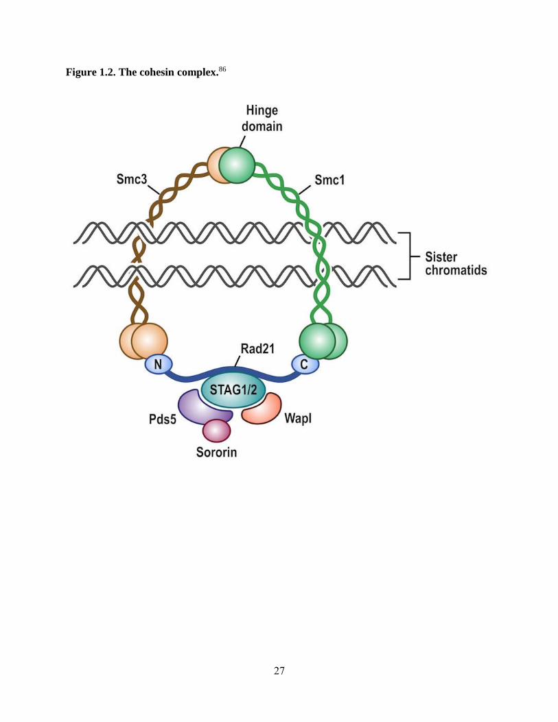
or modify the cohesin complex (Figure 1.2).86
The cohesin complex is highly conserved through evolution with homologs in yeast, fruit flies,
and mammals (Table 1.184).87 Among the several models have been proposed to depict how the
cohesin complex associates with chromatin, the one-ring “embrace” model and the two-ring
“handcuff” model are supported by experimental data.88, 89 The one-ring model suggests that the
17
cohesin ring embraces two chromatins until their segregation.88 The two-ring model describes
each cohesin ring entraps one chromatin and cohesion is mediated by interactions between the
two cohesin rings.89 The canonical role of the cohesin complex is to ensure proper segregation of
chromosomes during mitosis and meiosis. In addition to its essential role in sister chromatids
cohesion, cohesin contributes to genome maintenance and functions by involving in DNA
damage repair and gene expression.90
1.2.1.1 Cohesin functions in chromosome segregation
Cohesins are loaded to the chromatins at the G1/S phase in yeast and at telophase in mammalian
cells by loading complex NIPBL-MAU2.91, 92, 93, 94, 95 During DNA replication at S phase, each
cohesin ring embraces one of the sister chromatids. After DNA replication at the S phase,
acetylation of SMC3 by cohesin acetyltransferases ESCO1 and ESCO2 establishes stable
cohesion between the newly replicated sister chromatids. PDS5 and SORORIN form a complex
to maintain the cohesion throughout the G2 phase until prophase when SORORIN is
phosphorylated and destabilized.96, 97, 98, 99 The removal of cohesins are facilitated by the
formation of PDS5-WAPL complex. At prophase, cohesins on the chromosomal arms are
removed by the phosphorylation of RAD21 and STAG1/2 by PLK1.100 Centromeric cohesion is
protected by SGOL1 until Separase gets activated and cleaves RAD21 at anaphase and therefore
separating the sister chromatids.101, 102 The dissociated cohesins can be recycled after the acetyl
groups are removed from SMC3 proteins by cohesin deacetylases HDAC8.103, 104 In meiosis, a
similar biphasic removal of cohesin occurs, with RAD21 replaced by REC8.105
The main roles of cohesin during cell cycle are to keep sister chromatids together and to provide
resistance when sister chromatids are pulled by microtubules towards the opposing spindle poles,
thus ensuring accurate separation of sister chromatids during the transition from metaphase to

18
anaphase.106 Failure in the formation and maintenance of sister chromatid cohesion results in
premature chromosome segregation, which is thought to be a major pathway to aneuploidy, a
characteristic observed in many human cancers.107
1.2.1.2 Cohesin functions in genome instability
Cellular DNA is exposed to single and double strand breaks (DSBs) through multiple
endogenous and exogenous mechanisms. Cells respond to DNA damage by activation of DNA-
damage checkpoints that halt cell cycle progression until the damaged DNA is repaired. If the
damage cannot be repaired properly, cells may undergo apoptosis. Eukaryote cells have two
distinct mechanisms to repair DSBs, the homologous recombination (HR) between sister
chromatids in the S and G2 phases and the non-homologous end joining (NHEJ), involving re-
ligation of broken DNA, which occurs throughout the cell cycle.84
The function of cohesin in DNA damage repair is evolutionarily conserved from yeast to
humans.108, 109, 110 Rad21 was cloned originally by complementing the γ-radiation sensitivity in
fission yeast with a function in DSB repair, before its role in sister chromatid cohesion was
identified.111 In response to laser-induced DNA damage in human cells, cohesins are recruited to
the DSB site and de novo cohesion, named damage-induced cohesion (DI-cohesion), is
established.112 Besides cohesins, factors that are required to load cohesins to chromatin, establish
cohesion, and maintain cohesion are all needed for DNA damage repair. Defects in the cohesin-
loading complex NIPBL-MAU2, cohesin acetyltransferase ESCO, or maintenance factor
SORORIN block the accumulation of cohesins at DSBs and prevent DNA damage repair,
suggesting the presence of cohesins on chromatin is not sufficient to mediate DNA repair and
instead, additional cohesion is required.113, 114, 115 DI-cohesion may help to structurally stabilize
chromosomes whose DNA backbone has been fragmented by DSBs and to provide the proximity
19
between the damaged sister chromatid and the template, allowing HR to occur. DNA damage-
induced phosphorylation and acetylation on SMC3 were found to be important for genome-wide
DI-cohesion caused by DSB in the G2 phase and DSB repair.112
Moreover, cohesins are required to activate checkpoints when DSBs occur.116 In C. elegans,
when SCC2, a component of the cohesin loading complex, is mutated, cohesins cannot be loaded
onto chromatin in meiosis, resulting in failure of both checkpoint activation and DNA damage
repair.117 This shows the importance of cohesin recruitment to the damaged chromatin. The
checkpoint role of cohesins is independent of its function in sister chromatid cohesion because
cohesins are required for the phosphorylation and activation of Chk2 although no sister
chromatid cohesion occurs in the G1 phase. As evidence, depletion of SORORIN, a protein
essential for the generation and maintenance of sister chromatid cohesion, leads to checkpoint
activation but DSB repair failure.116
1.2.1.3 Cohesin functions in gene expression
The first evidence that cohesin factors regulate gene expression and development came from the
studies of Drosophila cut and Ultrabithorax genes: heterozygous Nipped-B mutants showed
reduced cut expression, whereas loss of Smc1, Rad21, or SA led to increased cut expression.118,
119 Cohesins also facilitate expression of c-myc, a function conserved across Drosophila,
zebrafish, mouse, and humans and cohesin depletion reduces myc transcription.120 Furthermore,
cohesins present in non-cycling and even post-mitotic cell in higher eukaryotes.121 Accumulating
evidence implies an important non-canonical role of cohesin in regulating gene expression,
which is independent of cohesins’ role in cell division.83 In non-dividing mouse thymocytes,
genetic deletion of cohesin resulted in reduced transcription and rearrangements at the T cell
receptor, thereby affecting thymocyte differentiation.122

20
Cohesins have been shown to mediate long-range transcriptional regulation by controlling the
spatial conformation of chromatin at multiple gene loci that are important for normal
development and differentiation.123 Studies revealed two distinct types of cohesin sites: sites that
coincide with the binding of CTCF (CCCTC-binding factor) vs. sites that map to active
enhancers and promoters and are usually cell-type specific. The CTCF-dependent interaction of
cohesins with insulator blocks enhancer activity and disrupts distal enhancer-promoter
interactions required for gene activation.121 Moreover, cohesin has a CTCF-independent role in
tissue-specific transcriptional regulation.124 ChIP-Seq data suggest that cohesins co-localize with
master regulators in several tissues, such as liver-specific transcription factors in HepG2 cells
and estrogen receptor α in MCF-7 cells.124, 125 Cohesin also co-localize with transcriptional
coactivators, such as mediator to facilitate chromatin looping between the enhancer and promoter
of some pluripotency genes (e.g POU5F1) in mouse embryonic stem cells.126 The cohesin
complex lacks a definitive DNA-binding domain. Therefore, DNA localization appears to be
facilitated through binding to CTCF and transcription factors, thus forming a regulatory network
for transcriptional programs of specific cell type.121
Cohesins play an essential role in the maintenance of pluripotency. Depletion of cohesins blocks
self-renewal, induces spontaneous differentiation, and interferes with reprogramming of
fibroblasts to pluripotent cells.127 Mutations in core components of the cohesin complex can
cause developmental defects in a number of species. For instance, heterozygous mutations in
cohesin loader NIPBL or less frequently, in cohesin subunits SMC1A and SMC3 result in
Cornelia de Lange syndrome (CdLS), a neurodevelopmental disorder with upper extremity
malformations.90
1.2.2 Cohesin deregulation in cancer

21
Mutations of cohesins have been found in many cancers including leukemias19, 49, 128, colorectal
carcinomas129, ovarian carcinomas11, 130, glioblastoma, melanomas, and Ewing’s sarcomas131.
The first somatic mutations of cohesin in cancer were reported in 2008 when heterozygous
missense mutations in SMC1A, SMC3, STAG3 (a component of meiotic cohesin) and NIPBL
were identified in aneuploid colorectal cancers.129 In 2010, deletions of RAD21 in a CML and
deletions of STAG2 in an AML were reported.132 In 2011, STAG2 mutations were reported to
result in cohesion defects and aneuploidy in glioblastoma cell lines, melanomas, and Ewing’s
sarcomas.131 STAG2 is the most frequently mutated gene of the cohesin complex. Because
STAG2 is located on the X chromosome, only a single mutational event is required to inactivate
it in both males and females (due to X inactivation). STAG2 mutations are considered loss-of-
function mutations because: 1) the majority of mutations are truncating, 2) truncating mutations
are present in early exons, resulting in a very short protein, 3) in many cases a truncated STAG2
protein is absent, likely due to nonsense-mediated decay of the mutant STAG2 mRNA.133, 134 In
2013, three studies reported frequent somatic mutations of STAG2 in bladder cancer.135, 136, 137 In
addition, SMC1A has been shown to be overexpressed in gliomas and reducing its levels inhibits
glioma cell growth in vitro.138 Upregulation of ESCO2 and WAPL is associated with tumor
progression in melanomas and cervical cancer, respectively.139, 140 Overexpression of Separase is
sufficient to induce tumorigenesis in mammary epithelial cells in a TP53-mutant background.141
Pan-cancer analysis of the TCGA data found that the cohesin complex was recurrently mutated
across 12 cancer types and identified the cohesin complex as one of the 16 significantly mutated
subnetworks.142
1.2.2.1 Cohesin mutations in myeloid malignancies
Cohesin mutations in AML

22
Recurrent mutations in all four members of the cohesin complex, SMC1A, SMC3, RAD21, and
STAG2 were first identified in M1 AML cases. They co-occurred with NPM1, RUNX1,
DNMT3A, or TET2 mutations in 17/19 cases, indicating cooperation with other leukemogenic
pathways.49 Subsequently, the TCGA data confirmed and extended these results, identifying a
cumulative cohesin mutation frequency of 13% (26/200).11 The frequency of cohesin mutations
was assessed in both de novo and secondary AML. One study showed a higher frequency of
cohesin mutations in de novo AML (13%, 16/120 samples studied) than in secondary AML (8%,
3/37 samples studied).77 In contrast, another study showed a higher frequency of cohesin
mutations in secondary AML (20%, 30/149) than in de novo AML (11%, 32/301).78 In both
studies, the most frequently mutated cohesin gene was STAG2, followed by SMC3 and RAD21.77,
78 Cohesin mutations are nearly always mutually exclusive and are mostly found in samples with
normal karyotypes. Based on allelic burden analysis, cohesin mutations are often, but not always,
observed as early event during leukemogenesis.76
Cohesin mutations in MDS
STAG2 mutations were identified in MDS samples from patients whose disease later progressed
to secondary AML.143 Subsequent studies showed that 7% (10/150) of MDS samples harbor
STAG2 mutations and 8% (18/224) of MDS samples harbor cohesin mutations, the majority of
which were STAG2 mutations, with lower mutation frequencies in SMC3 and RAD21.77, 144
Cohesin mutations were also identified in 17% of high-risk MDS samples and 11% of low-risk
samples, respectively.78
Cohesin mutations in DS-AMKL
Down syndrome is associated with trisomy 21, and individuals with trisomy 21 are more
susceptible to hematologic abnormalities. Up to 10% of children with Down syndrome will

23
present with transient abnormal myelopoiesis at birth, a necessary predecessor to DS-AMKL.
Virtually all DS-AMKL patients have an inactivating mutation in GATA1, which results in
exclusive expression of a shorter isoform, named GATA1s.80
Cohesin mutations are prominent in DS-AMKL and are predicted to be heterozygous, loss-of-
function, and early events during leukemia development. Deep sequencing revealed that 53% of
the DS-AMKL samples had acquired cohesin mutations that were not in the self-limiting pre-
leukemic transient abnormal myelopoiesis, suggesting cohesin haploinsufficiency may drive
oncogenic transformation and progression to DS-AMKL.79 In addition to the presence of trisomy
21 and GATA1s, cohesin mutations likely cooperate with chromosome 21 genes such as
RUNX1, ERG, and ETS2 to promote the development of DS-AMKL.80, 145 Furthermore,
mutations in CTCF occur in approximately 20% of DS-AMKL and are not mutually exclusive to
cohesin mutations.80 Cohesins and CTCF interact to regulate chromatin architecture, and thus
mutations in either could have non-overlapping effects on genomic structure and induce global
changes on gene expression.
Phenotypic consequences of cohesin mutations on hematopoiesis
Four recent studies sought to elucidate the phenotypic consequences of loss of cohesin and
cohesin mutations on hematopoiesis in mouse and human models (Table 1.2146).147, 148, 149, 150
Viny et al. showed a dose-dependent role for Smc3 in regulating hematopoietic stem and
progenitor cell (HSPC) function and chromatin structure. Biallelic loss of Smc3 in mice led to
bone marrow aplasia with premature sister chromatid separation, revealing an absolute
requirement for cohesin in HSPC function; whereas, Smc3 haploinsufficiency increased self-
renewal in vitro and in vivo. Furthermore, Smc3 haploinsufficiency reduced expression of
24
transcription factors and lineage commitment-associated genes and cooperated with FLT3-ITD
mutation to induce AML in vivo.147
Mullenders et al. generated a series of inducible shRNA mouse models targeting each of the four
cohesin subunits. Knockdown of cohesin resulted in gain of replating capacity of mouse HSPCs
and altered hematopoiesis with skewing towards myeloid differentiation. Upregulation of genes
involved in myeloid differentiation and increased chromatin accessibility around those genes
were also observed. In addition, aged cohesin knockdown mice developed a clinical picture
closely resembling MPNs, implying that cohesin mutations can occur as an early event in
leukemogenesis and facilitate the potential development of a myeloid malignancy.148
Complementary work in cohesin mutant human HSPCs showed that depletion of cohesin
subunits increased replating capacity in vitro and led to myeloid-skewed differentiation,
consistent with phenotypes seen in mouse models. Mazumdar et al. found that introduction of
cohesin mutants into AML cell lines and primary human cord blood HSPCs resulted in a
differentiation block with an increased frequency of CD34+ cord blood progenitors. Cohesin
mutants augmented the serial replating capability of human HSPCs in vitro and elevated
chromatin accessibility and predicted transcription factor binding for HSPC regulators including
RUNX1, GATA2, and ERG, measured by ATAC-Seq and ChIP-Seq.149
Similarly, Galeev et al. identified several members of the cohesin complex SMC3, RAD21,
STAG1/2 in an RNAi screen as critical modifiers of self-renewal and differentiation in human
HSPCs. They showed that cohesin deficiency induced HSC-specific gene programs and the
reconstitution potential of cohesin-deficient HSPCs was increased in primary and secondary
transplantation studies.150

25
Figure Legends
Figure 1.1. Eight functional categories of genes that are frequently mutated in AML.17
Mutations in signaling genes such as FLT3 (upper left box). Mutations in tumor-suppressor
genes such as TP53 (upper middle box). Mutations in DNA-methylation-associated genes such
as DNMT3A, TET2, IDH1, and IDH2 (upper right box). Mutations in myeloid transcription
factors such as RUNX1 (center left box). Mutations in cohesin-complex genes such as STAG2
and RAD21 (center middle box). Mutations in chromatin-modifying genes such as ASXL1 and
PHF6 (center right box). Mutations in NPM1 (lower left box). Mutations in spliceosome-
complex genes such as SRSF2, SF3B1, U2AF1, and ZRSR2 (lower right box).
Figure 1.2. The cohesin complex.86
Cohesin is a ring-shaped complex, composed of four core subunits SMC1A, SMC3, RAD21, and
STAG1/2. SMC1A and SMC3 form intramolecular antiparallel coiled coils and fold back on
themselves, creating a hinge domain at one end and an ATPase head at the other. SMC1A and
SMC3 dimerize at the hinge domains and their ATPase heads are bound by RAD21. STAG1/2
interacts with the central region of RAD21. PDS5, SORORIN, and WAPL are regulatory
proteins of cohesin.

26
Figure 1.1. Eight functional categories of genes that are frequently mutated in AML.17
27
Figure 1.2. The cohesin complex.86

28
Table 1.1. Core subunits and regulatory proteins of the cohesin complex.84
Mammals D. melanogaster S. cerevisiae S. pombe Function
SMC1A Smc1 Smc1 Psm1
Core subunit (mitosis)
SMC1B Core subunit (meiosis)
SMC3 Smc3 Smc3 Psm3 Core subunit
RAD21 Rad21/Vtd Scc1/Mcd1 Rad21 Core subunit (mitosis)
REC8 C(2)M Rec8 Rec8 Core subunit (meiosis)
STAG1/SA1 SA
Scc3 Psc3
Core subunit (mitosis)
STAG2/SA2 SA2 Core subunit (mitosis)
STAG3/SA3 / Rec11 Core subunit (meiosis)
NIPBL/SCC2 Nipped-B Scc2 Mis4 Cohesin loading
MAU2/SCC4 Scc4 Scc4 Ssl3 Cohesin loading
ESCO1 Eco/Deco Eco1/Ctf7 Eso1 Cohesion establishment
ESCO2 San
PDS5A Pds5 Pds5 Pds5 Cohesion maintenance
PDS5B
WAPL Wapl Wpl1/Rad61 Wpl1 Cohesion maintenance
SORORIN/CDCA5 Dalmatian / / Cohesion maintenance
HDAC8 / Hos1 / Cohesin dacetylase
Shugosin1/SGOL1 Mei-S332 Sgo1 Sgo1 Cohesin protection
Separase Sse1 Esp1 Separase Cohesin removal
Polo like Kinase 1
(PLK1) Polo Cdc5 Plk1 Cohesin removal

29
Table 1.2. Studies of cohesin mutations in hematopoiesis.146
Model system Approach Conclusions
Mouse model
Smc3biallelic and
haploinsufficient
conditional
knockout
Increased replating, enrichment of HSPC
gene signature, chromatiin accessibiligy
changes, dose dependent
Mouse model
shRNA
knockdown of
cohesin subunits
Increased replating, enrichment of HSPC
gene signature, chromatiin accessibiligy
changes, MPN-like phenotype in aged mice
Human cord blood
(HSPCs)
Lentiviral
transduction of
cohesin mutants or
shRNA
knockdown
Increased replating, enrichment of HSPC
gene signature, chromatiin accessibiligy
changes
Human cord blood
(HSPCs) RNAi screen
Increased replating, enrichment of HSPC
gene signature, increased secondary transplant
engraftment

30
References
1. Hasserjian RP. Acute myeloid leukemia: advances in diagnosis and classification. Int J Lab
Hematol. 2013;35(3):358-366. doi:10.1111/ijlh.12081
2. Short NJ, Rytting ME, Cortes JE. Acute myeloid leukaemia. Lancet Lond Engl.
2018;392(10147):593-606. doi:10.1016/S0140-6736(18)31041-9
3. Yamamoto JF, Goodman MT. Patterns of leukemia incidence in the United States by subtype
and demographic characteristics, 1997-2002. Cancer Causes Control CCC. 2008;19(4):379-
390. doi:10.1007/s10552-007-9097-2
4. Acute Myeloid Leukemia - Cancer Stat Facts. SEER.
https://seer.cancer.gov/statfacts/html/amyl.html. Accessed December 10, 2018.
5. Key Statistics for Acute Myeloid Leukemia (AML). https://www.cancer.org/cancer/acute-
myeloid-leukemia/about/key-statistics.html. Accessed December 10, 2018.
6. Patel JP, Levine RL. How do novel molecular genetic markers influence treatment decisions
in acute myeloid leukemia? Hematol Am Soc Hematol Educ Program. 2012;2012:28-34.
doi:10.1182/asheducation-2012.1.28
7. Döhner H, Estey EH, Amadori S, et al. Diagnosis and management of acute myeloid
leukemia in adults: recommendations from an international expert panel, on behalf of the
European LeukemiaNet. Blood. 2010;115(3):453-474. doi:10.1182/blood-2009-07-235358
8. Meyers J, Yu Y, Kaye JA, Davis KL. Medicare fee-for-service enrollees with primary acute
myeloid leukemia: an analysis of treatment patterns, survival, and healthcare resource
utilization and costs. Appl Health Econ Health Policy. 2013;11(3):275-286.
doi:10.1007/s40258-013-0032-2
9. Moarii M, Papaemmanuil E. Classification and risk assessment in AML: integrating
cytogenetics and molecular profiling. Hematol Am Soc Hematol Educ Program.
2017;2017(1):37-44. doi:10.1182/asheducation-2017.1.37
10. Welch JS, Petti AA, Miller CA, et al. TP53 and Decitabine in Acute Myeloid Leukemia and
Myelodysplastic Syndromes. N Engl J Med. 2016;375(21):2023-2036.
doi:10.1056/NEJMoa1605949
11. Cancer Genome Atlas Research Network, Ley TJ, Miller C, et al. Genomic and epigenomic
landscapes of adult de novo acute myeloid leukemia. N Engl J Med. 2013;368(22):2059-
2074. doi:10.1056/NEJMoa1301689
12. Arber DA, Orazi A, Hasserjian R, et al. The 2016 revision to the World Health Organization
classification of myeloid neoplasms and acute leukemia. Blood. 2016;127(20):2391-2405.
doi:10.1182/blood-2016-03-643544
31
13. Bullinger L, Döhner K, Döhner H. Genomics of Acute Myeloid Leukemia Diagnosis and
Pathways. J Clin Oncol Off J Am Soc Clin Oncol. 2017;35(9):934-946.
doi:10.1200/JCO.2016.71.2208
14. Grimwade D, Hills RK, Moorman AV, et al. Refinement of cytogenetic classification in
acute myeloid leukemia: determination of prognostic significance of rare recurring
chromosomal abnormalities among 5876 younger adult patients treated in the United
Kingdom Medical Research Council trials. Blood. 2010;116(3):354-365. doi:10.1182/blood-
2009-11-254441
15. Shivarov V, Bullinger L. Expression profiling of leukemia patients: key lessons and future
directions. Exp Hematol. 2014;42(8):651-660. doi:10.1016/j.exphem.2014.04.006
16. Ley TJ, Mardis ER, Ding L, et al. DNA sequencing of a cytogenetically normal acute
myeloid leukaemia genome. Nature. 2008;456(7218):66-72. doi:10.1038/nature07485
17. Döhner H, Weisdorf DJ, Bloomfield CD. Acute Myeloid Leukemia. N Engl J Med.
2015;373(12):1136-1152. doi:10.1056/NEJMra1406184
18. Shlush LI, Zandi S, Mitchell A, et al. Identification of pre-leukaemic haematopoietic stem
cells in acute leukaemia. Nature. 2014;506(7488):328-333. doi:10.1038/nature13038
19. Ding L, Ley TJ, Larson DE, et al. Clonal evolution in relapsed acute myeloid leukaemia
revealed by whole-genome sequencing. Nature. 2012;481(7382):506-510.
doi:10.1038/nature10738
20. Busque L, Patel JP, Figueroa ME, et al. Recurrent somatic TET2 mutations in normal elderly
individuals with clonal hematopoiesis. Nat Genet. 2012;44(11):1179-1181.
doi:10.1038/ng.2413
21. Xie M, Lu C, Wang J, et al. Age-related mutations associated with clonal hematopoietic
expansion and malignancies. Nat Med. 2014;20(12):1472-1478. doi:10.1038/nm.3733
22. Genovese G, Kähler AK, Handsaker RE, et al. Clonal hematopoiesis and blood-cancer risk
inferred from blood DNA sequence. N Engl J Med. 2014;371(26):2477-2487.
doi:10.1056/NEJMoa1409405
23. Jaiswal S, Fontanillas P, Flannick J, et al. Age-related clonal hematopoiesis associated with
adverse outcomes. N Engl J Med. 2014;371(26):2488-2498. doi:10.1056/NEJMoa1408617
24. McKerrell T, Park N, Moreno T, et al. Leukemia-associated somatic mutations drive distinct
patterns of age-related clonal hemopoiesis. Cell Rep. 2015;10(8):1239-1245.
doi:10.1016/j.celrep.2015.02.005
25. Wong TN, Miller CA, Jotte MRM, et al. Cellular stressors contribute to the expansion of
hematopoietic clones of varying leukemic potential. Nat Commun. 2018;9(1):455.
doi:10.1038/s41467-018-02858-0

32
26. Steensma DP, Bejar R, Jaiswal S, et al. Clonal hematopoiesis of indeterminate potential and
its distinction from myelodysplastic syndromes. Blood. 2015;126(1):9-16.
doi:10.1182/blood-2015-03-631747
27. Federici L, Falini B. Nucleophosmin mutations in acute myeloid leukemia: a tale of protein
unfolding and mislocalization. Protein Sci Publ Protein Soc. 2013;22(5):545-556.
doi:10.1002/pro.2240
28. Ofran Y, Rowe JM. Genetic profiling in acute myeloid leukaemia--where are we and what is
its role in patient management. Br J Haematol. 2013;160(3):303-320. doi:10.1111/bjh.12135
29. Schlenk RF, Döhner K, Krauter J, et al. Mutations and treatment outcome in cytogenetically
normal acute myeloid leukemia. N Engl J Med. 2008;358(18):1909-1918.
doi:10.1056/NEJMoa074306
30. Yohe S. Molecular Genetic Markers in Acute Myeloid Leukemia. J Clin Med.
2015;4(3):460-478. doi:10.3390/jcm4030460
31. Kayser S, Schlenk RF, Londono MC, et al. Insertion of FLT3 internal tandem duplication in
the tyrosine kinase domain-1 is associated with resistance to chemotherapy and inferior
outcome. Blood. 2009;114(12):2386-2392. doi:10.1182/blood-2009-03-209999
32. Schnittger S, Bacher U, Haferlach C, Alpermann T, Kern W, Haferlach T. Diversity of the
juxtamembrane and TKD1 mutations (exons 13-15) in the FLT3 gene with regards to mutant
load, sequence, length, localization, and correlation with biological data. Genes
Chromosomes Cancer. 2012;51(10):910-924. doi:10.1002/gcc.21975
33. Breitenbuecher F, Schnittger S, Grundler R, et al. Identification of a novel type of ITD
mutations located in nonjuxtamembrane domains of the FLT3 tyrosine kinase receptor.
Blood. 2009;113(17):4074-4077. doi:10.1182/blood-2007-11-125476
34. Blau O, Berenstein R, Sindram A, Blau IW. Molecular analysis of different FLT3-ITD
mutations in acute myeloid leukemia. Leuk Lymphoma. 2013;54(1):145-152.
doi:10.3109/10428194.2012.704999
35. KIT Exon 8 Mutation in Acute Myeloid Leukemia - My Cancer Genome.
https://www.mycancergenome.org/content/disease/acute-myeloid-leukemia/kit/273/.
Accessed January 19, 2019.
36. Ashman LK, Griffith R. Therapeutic targeting of c-KIT in cancer. Expert Opin Investig
Drugs. 2013;22(1):103-115. doi:10.1517/13543784.2013.740010
37. Liang D-C, Liu H-C, Yang C-P, et al. Cooperating gene mutations in childhood acute
myeloid leukemia with special reference on mutations of ASXL1, TET2, IDH1, IDH2, and
DNMT3A. Blood. 2013;121(15):2988-2995. doi:10.1182/blood-2012-06-436782

33
38. Patel JP, Gönen M, Figueroa ME, et al. Prognostic relevance of integrated genetic profiling
in acute myeloid leukemia. N Engl J Med. 2012;366(12):1079-1089.
doi:10.1056/NEJMoa1112304
39. Papaemmanuil E, Gerstung M, Bullinger L, et al. Genomic Classification and Prognosis in
Acute Myeloid Leukemia. N Engl J Med. 2016;374(23):2209-2221.
doi:10.1056/NEJMoa1516192
40. Gaidzik VI, Teleanu V, Papaemmanuil E, et al. RUNX1 mutations in acute myeloid
leukemia are associated with distinct clinico-pathologic and genetic features. Leukemia.
2016;30(11):2282. doi:10.1038/leu.2016.207
41. Preudhomme C, Renneville A, Bourdon V, et al. High frequency of RUNX1 biallelic
alteration in acute myeloid leukemia secondary to familial platelet disorder. Blood.
2009;113(22):5583-5587. doi:10.1182/blood-2008-07-168260
42. Martelli MP, Sportoletti P, Tiacci E, Martelli MF, Falini B. Mutational landscape of AML
with normal cytogenetics: biological and clinical implications. Blood Rev. 2013;27(1):13-22.
doi:10.1016/j.blre.2012.11.001
43. Pabst T, Mueller BU, Zhang P, et al. Dominant-negative mutations of CEBPA, encoding
CCAAT/enhancer binding protein-alpha (C/EBPalpha), in acute myeloid leukemia. Nat
Genet. 2001;27(3):263-270. doi:10.1038/85820
44. Nerlov C. C/EBPalpha mutations in acute myeloid leukaemias. Nat Rev Cancer.
2004;4(5):394-400. doi:10.1038/nrc1363
45. Mueller BU, Pabst T. C/EBPalpha and the pathophysiology of acute myeloid leukemia. Curr
Opin Hematol. 2006;13(1):7-14.
46. Wouters BJ, Löwenberg B, Erpelinck-Verschueren CAJ, van Putten WLJ, Valk PJM, Delwel
R. Double CEBPA mutations, but not single CEBPA mutations, define a subgroup of acute
myeloid leukemia with a distinctive gene expression profile that is uniquely associated with
a favorable outcome. Blood. 2009;113(13):3088-3091. doi:10.1182/blood-2008-09-179895
47. Kihara R, Nagata Y, Kiyoi H, et al. Comprehensive analysis of genetic alterations and their
prognostic impacts in adult acute myeloid leukemia patients. Leukemia. 2014;28(8):1586-
1595. doi:10.1038/leu.2014.55
48. Yang L, Han Y, Suarez Saiz F, Saurez Saiz F, Minden MD. A tumor suppressor and
oncogene: the WT1 story. Leukemia. 2007;21(5):868-876. doi:10.1038/sj.leu.2404624
49. Welch JS, Ley TJ, Link DC, et al. The origin and evolution of mutations in acute myeloid
leukemia. Cell. 2012;150(2):264-278. doi:10.1016/j.cell.2012.06.023
50. Ibrahem L, Mahfouz R, Elhelw L, Abdsalam EM, Soliman R. Prognostic significance of
DNMT3A mutations in patients with acute myeloid leukemia. Blood Cells Mol Dis.
2015;54(1):84-89. doi:10.1016/j.bcmd.2014.07.015
34
51. Russler-Germain DA, Spencer DH, Young MA, et al. The R882H DNMT3A mutation
associated with AML dominantly inhibits wild-type DNMT3A by blocking its ability to form
active tetramers. Cancer Cell. 2014;25(4):442-454. doi:10.1016/j.ccr.2014.02.010
52. Yamashita Y, Yuan J, Suetake I, et al. Array-based genomic resequencing of human
leukemia. Oncogene. 2010;29(25):3723-3731. doi:10.1038/onc.2010.117
53. Holz-Schietinger C, Matje DM, Reich NO. Mutations in DNA methyltransferase
(DNMT3A) observed in acute myeloid leukemia patients disrupt processive methylation. J
Biol Chem. 2012;287(37):30941-30951. doi:10.1074/jbc.M112.366625
54. Aslanyan MG, Kroeze LI, Langemeijer SMC, et al. Clinical and biological impact of TET2
mutations and expression in younger adult AML patients treated within the
EORTC/GIMEMA AML-12 clinical trial. Ann Hematol. 2014;93(8):1401-1412.
doi:10.1007/s00277-014-2055-7
55. Gaidzik VI, Paschka P, Späth D, et al. TET2 mutations in acute myeloid leukemia (AML):
results from a comprehensive genetic and clinical analysis of the AML study group. J Clin
Oncol Off J Am Soc Clin Oncol. 2012;30(12):1350-1357. doi:10.1200/JCO.2011.39.2886
56. Tian X, Xu Y, Yin J, et al. TET2 gene mutation is unfavorable prognostic factor in
cytogenetically normal acute myeloid leukemia patients with NPM1+ and FLT3-ITD -
mutations. Int J Hematol. 2014;100(1):96-104. doi:10.1007/s12185-014-1595-x
57. Lu C, Ward PS, Kapoor GS, et al. IDH mutation impairs histone demethylation and results in
a block to cell differentiation. Nature. 2012;483(7390):474-478. doi:10.1038/nature10860
58. Ward PS, Cross JR, Lu C, et al. Identification of additional IDH mutations associated with
oncometabolite R(-)-2-hydroxyglutarate production. Oncogene. 2012;31(19):2491-2498.
doi:10.1038/onc.2011.416
59. Paschka P, Schlenk RF, Gaidzik VI, et al. IDH1 and IDH2 mutations are frequent genetic
alterations in acute myeloid leukemia and confer adverse prognosis in cytogenetically
normal acute myeloid leukemia with NPM1 mutation without FLT3 internal tandem
duplication. J Clin Oncol Off J Am Soc Clin Oncol. 2010;28(22):3636-3643.
doi:10.1200/JCO.2010.28.3762
60. Abbas S, Lugthart S, Kavelaars FG, et al. Acquired mutations in the genes encoding IDH1
and IDH2 both are recurrent aberrations in acute myeloid leukemia: prevalence and
prognostic value. Blood. 2010;116(12):2122-2126. doi:10.1182/blood-2009-11-250878
61. Ho PA, Kutny MA, Alonzo TA, et al. Leukemic mutations in the methylation-associated
genes DNMT3A and IDH2 are rare events in pediatric AML: a report from the Children’s
Oncology Group. Pediatr Blood Cancer. 2011;57(2):204-209. doi:10.1002/pbc.23179
62. Lindsley RC, Mar BG, Mazzola E, et al. Acute myeloid leukemia ontogeny is defined by
distinct somatic mutations. Blood. 2015;125(9):1367-1376. doi:10.1182/blood-2014-11-
610543

35
63. El-Sharkawi D, Ali A, Evans CM, et al. ASXL1 mutations are infrequent in young patients
with primary acute myeloid leukemia and their detection has a limited role in therapeutic risk
stratification. Leuk Lymphoma. 2014;55(6):1326-1331. doi:10.3109/10428194.2013.833332
64. Todd MAM, Ivanochko D, Picketts DJ. PHF6 Degrees of Separation: The Multifaceted
Roles of a Chromatin Adaptor Protein. Genes. 2015;6(2):325-352.
doi:10.3390/genes6020325
65. Lower KM, Turner G, Kerr BA, et al. Mutations in PHF6 are associated with Börjeson-
Forssman-Lehmann syndrome. Nat Genet. 2002;32(4):661-665. doi:10.1038/ng1040
66. Van Vlierberghe P, Patel J, Abdel-Wahab O, et al. PHF6 mutations in adult acute myeloid
leukemia. Leukemia. 2011;25(1):130-134. doi:10.1038/leu.2010.247
67. Yoo NJ, Kim YR, Lee SH. Somatic mutation of PHF6 gene in T-cell acute lymphoblatic
leukemia, acute myelogenous leukemia and hepatocellular carcinoma. Acta Oncol Stockh
Swed. 2012;51(1):107-111. doi:10.3109/0284186X.2011.592148
68. Shukla GC, Singh J. Mutations of RNA splicing factors in hematological malignancies.
Cancer Lett. 2017;409:1-8. doi:10.1016/j.canlet.2017.08.042
69. Papaemmanuil E, Gerstung M, Malcovati L, et al. Clinical and biological implications of
driver mutations in myelodysplastic syndromes. Blood. 2013;122(22):3616-3627; quiz 3699.
doi:10.1182/blood-2013-08-518886
70. Makishima H, Visconte V, Sakaguchi H, et al. Mutations in the spliceosome machinery, a
novel and ubiquitous pathway in leukemogenesis. Blood. 2012;119(14):3203-3210.
doi:10.1182/blood-2011-12-399774
71. Kim E, Ilagan JO, Liang Y, et al. SRSF2 Mutations Contribute to Myelodysplasia by
Mutant-Specific Effects on Exon Recognition. Cancer Cell. 2015;27(5):617-630.
doi:10.1016/j.ccell.2015.04.006
72. Yoshida K, Sanada M, Shiraishi Y, et al. Frequent pathway mutations of splicing machinery
in myelodysplasia. Nature. 2011;478(7367):64-69. doi:10.1038/nature10496
73. Przychodzen B, Jerez A, Guinta K, et al. Patterns of missplicing due to somatic U2AF1
mutations in myeloid neoplasms. Blood. 2013;122(6):999-1006. doi:10.1182/blood-2013-01-
480970
74. Shirai CL, Ley JN, White BS, et al. Mutant U2AF1 Expression Alters Hematopoiesis and
Pre-mRNA Splicing In Vivo. Cancer Cell. 2015;27(5):631-643.
doi:10.1016/j.ccell.2015.04.008
75. Madan V, Kanojia D, Li J, et al. Aberrant splicing of U12-type introns is the hallmark of
ZRSR2 mutant myelodysplastic syndrome. Nat Commun. 2015;6:6042.
doi:10.1038/ncomms7042

36
76. Thol F, Bollin R, Gehlhaar M, et al. Mutations in the cohesin complex in acute myeloid
leukemia: clinical and prognostic implications. Blood. 2014;123(6):914-920.
doi:10.1182/blood-2013-07-518746
77. Kon A, Shih L-Y, Minamino M, et al. Recurrent mutations in multiple components of the
cohesin complex in myeloid neoplasms. Nat Genet. 2013;45(10):1232-1237.
doi:10.1038/ng.2731
78. Thota S, Viny AD, Makishima H, et al. Genetic alterations of the cohesin complex genes in
myeloid malignancies. Blood. 2014;124(11):1790-1798. doi:10.1182/blood-2014-04-567057
79. Yoshida K, Toki T, Okuno Y, et al. The landscape of somatic mutations in Down syndrome-
related myeloid disorders. Nat Genet. 2013;45(11):1293-1299. doi:10.1038/ng.2759
80. Fisher JB, McNulty M, Burke MJ, Crispino JD, Rao S. Cohesin Mutations in Myeloid
Malignancies. Trends Cancer. 2017;3(4):282-293. doi:10.1016/j.trecan.2017.02.006
81. Conese M, Liso A. Cohesin complex is a major player on the stage of leukemogenesis. Stem
Cell Investig. 2016;3:18. doi:10.21037/sci.2016.05.04
82. Jaiswal S, Natarajan P, Silver AJ, et al. Clonal Hematopoiesis and Risk of Atherosclerotic
Cardiovascular Disease. N Engl J Med. 2017;377(2):111-121. doi:10.1056/NEJMoa1701719
83. Faure AJ, Schmidt D, Watt S, et al. Cohesin regulates tissue-specific expression by
stabilizing highly occupied cis-regulatory modules. Genome Res. 2012;22(11):2163-2175.
doi:10.1101/gr.136507.111
84. Bhardwaj S, Gullerova M. Cohesin: It's Functions and Relevance to Cancer. J Genet Syndr
Gene Ther. 2013;04(05). doi:10.4172/2157-7412.1000148
85. Nasmyth K, Haering CH. Cohesin: its roles and mechanisms. Annu Rev Genet. 2009;43:525-
558. doi:10.1146/annurev-genet-102108-134233
86. Research | Solomon Lab. https://solomonlab.ucsf.edu/research. Accessed January 11, 2019.
87. Peters J-M. The many functions of cohesin--different rings to rule them all? EMBO J.
2012;31(9):2061-2063. doi:10.1038/emboj.2012.90
88. Nasmyth K. How might cohesin hold sister chromatids together? Philos Trans R Soc Lond B
Biol Sci. 2005;360(1455):483-496. doi:10.1098/rstb.2004.1604
89. Zhang N, Kuznetsov SG, Sharan SK, Li K, Rao PH, Pati D. A handcuff model for the
cohesin complex. J Cell Biol. 2008;183(6):1019-1031. doi:10.1083/jcb.200801157
90. Remeseiro S, Cuadrado A, Gómez-López G, Pisano DG, Losada A. A unique role of
cohesin-SA1 in gene regulation and development. EMBO J. 2012;31(9):2090-2102.
doi:10.1038/emboj.2012.60

37
91. Ciosk R, Shirayama M, Shevchenko A, et al. Cohesin’s binding to chromosomes depends on
a separate complex consisting of Scc2 and Scc4 proteins. Mol Cell. 2000;5(2):243-254.
92. Rollins RA, Korom M, Aulner N, Martens A, Dorsett D. Drosophila nipped-B protein
supports sister chromatid cohesion and opposes the stromalin/Scc3 cohesion factor to
facilitate long-range activation of the cut gene. Mol Cell Biol. 2004;24(8):3100-3111.
93. Bernard P, Drogat J, Maure J-F, et al. A screen for cohesion mutants uncovers Ssl3, the
fission yeast counterpart of the cohesin loading factor Scc4. Curr Biol CB. 2006;16(9):875-
881. doi:10.1016/j.cub.2006.03.037
94. Watrin E, Schleiffer A, Tanaka K, Eisenhaber F, Nasmyth K, Peters J-M. Human Scc4 is
required for cohesin binding to chromatin, sister-chromatid cohesion, and mitotic
progression. Curr Biol CB. 2006;16(9):863-874. doi:10.1016/j.cub.2006.03.049
95. Seitan VC, Banks P, Laval S, et al. Metazoan Scc4 homologs link sister chromatid cohesion
to cell and axon migration guidance. PLoS Biol. 2006;4(8):e242.
doi:10.1371/journal.pbio.0040242
96. Losada A, Yokochi T, Hirano T. Functional contribution of Pds5 to cohesin-mediated
cohesion in human cells and Xenopus egg extracts. J Cell Sci. 2005;118(Pt 10):2133-2141.
doi:10.1242/jcs.02355
97. Zhang J, Shi X, Li Y, et al. Acetylation of Smc3 by Eco1 is required for S phase sister
chromatid cohesion in both human and yeast. Mol Cell. 2008;31(1):143-151.
doi:10.1016/j.molcel.2008.06.006
98. Rowland BD, Roig MB, Nishino T, et al. Building sister chromatid cohesion: smc3
acetylation counteracts an antiestablishment activity. Mol Cell. 2009;33(6):763-774.
doi:10.1016/j.molcel.2009.02.028
99. Nishiyama T, Ladurner R, Schmitz J, et al. Sororin mediates sister chromatid cohesion by
antagonizing Wapl. Cell. 2010;143(5):737-749. doi:10.1016/j.cell.2010.10.031
100. Sumara I, Vorlaufer E, Gieffers C, Peters BH, Peters JM. Characterization of vertebrate
cohesin complexes and their regulation in prophase. J Cell Biol. 2000;151(4):749-762.
101. Kitajima TS, Sakuno T, Ishiguro K, et al. Shugoshin collaborates with protein phosphatase
2A to protect cohesin. Nature. 2006;441(7089):46-52. doi:10.1038/nature04663
102. Hauf S, Waizenegger IC, Peters JM. Cohesin cleavage by separase required for anaphase
and cytokinesis in human cells. Science. 2001;293(5533):1320-1323.
doi:10.1126/science.1061376
103. Borges V, Lehane C, Lopez-Serra L, et al. Hos1 deacetylates Smc3 to close the cohesin
acetylation cycle. Mol Cell. 2010;39(5):677-688. doi:10.1016/j.molcel.2010.08.009

38
104. Deardorff MA, Bando M, Nakato R, et al. HDAC8 mutations in Cornelia de Lange
syndrome affect the cohesin acetylation cycle. Nature. 2012;489(7415):313-317.
doi:10.1038/nature11316
105. Watanabe Y, Nurse P. Cohesin Rec8 is required for reductional chromosome segregation at
meiosis. Nature. 1999;400(6743):461-464. doi:10.1038/22774
106. Nasmyth K. Disseminating the genome: joining, resolving, and separating sister chromatids
during mitosis and meiosis. Annu Rev Genet. 2001;35:673-745.
doi:10.1146/annurev.genet.35.102401.091334
107. Panigrahi AK, Pati D. Road to the crossroads of life and death: linking sister chromatid
cohesion and separation to aneuploidy, apoptosis and cancer. Crit Rev Oncol Hematol.
2009;72(3):181-193. doi:10.1016/j.critrevonc.2008.12.002
108. Sjögren C, Nasmyth K. Sister chromatid cohesion is required for postreplicative double-
strand break repair in Saccharomyces cerevisiae. Curr Biol CB. 2001;11(12):991-995.
109. Schär P, Fäsi M, Jessberger R. SMC1 coordinates DNA double-strand break repair
pathways. Nucleic Acids Res. 2004;32(13):3921-3929. doi:10.1093/nar/gkh716
110. Atienza JM, Roth RB, Rosette C, et al. Suppression of RAD21 gene expression decreases
cell growth and enhances cytotoxicity of etoposide and bleomycin in human breast cancer
cells. Mol Cancer Ther. 2005;4(3):361-368. doi:10.1158/1535-7163.MCT-04-0241
111. Birkenbihl RP, Subramani S. Cloning and characterization of rad21 an essential gene of
Schizosaccharomyces pombe involved in DNA double-strand-break repair. Nucleic Acids
Res. 1992;20(24):6605-6611.
112. Kim B-J, Li Y, Zhang J, et al. Genome-wide reinforcement of cohesin binding at pre-
existing cohesin sites in response to ionizing radiation in human cells. J Biol Chem.
2010;285(30):22784-22792. doi:10.1074/jbc.M110.134577
113. Schmitz J, Watrin E, Lénárt P, Mechtler K, Peters J-M. Sororin is required for stable
binding of cohesin to chromatin and for sister chromatid cohesion in interphase. Curr Biol
CB. 2007;17(7):630-636. doi:10.1016/j.cub.2007.02.029
114. Ström L, Karlsson C, Lindroos HB, et al. Postreplicative formation of cohesion is required
for repair and induced by a single DNA break. Science. 2007;317(5835):242-245.
doi:10.1126/science.1140649
115. Unal E, Heidinger-Pauli JM, Kim W, et al. A molecular determinant for the establishment
of sister chromatid cohesion. Science. 2008;321(5888):566-569.
doi:10.1126/science.1157880
116. Watrin E, Peters J-M. The cohesin complex is required for the DNA damage-induced G2/M
checkpoint in mammalian cells. EMBO J. 2009;28(17):2625-2635.
doi:10.1038/emboj.2009.202
39
117. Lightfoot J, Testori S, Barroso C, Martinez-Perez E. Loading of meiotic cohesin by SCC-2
is required for early processing of DSBs and for the DNA damage checkpoint. Curr Biol CB.
2011;21(17):1421-1430. doi:10.1016/j.cub.2011.07.007
118. Rollins RA, Morcillo P, Dorsett D. Nipped-B, a Drosophila homologue of chromosomal
adherins, participates in activation by remote enhancers in the cut and Ultrabithorax genes.
Genetics. 1999;152(2):577-593.
119. Dorsett D, Eissenberg JC, Misulovin Z, Martens A, Redding B, McKim K. Effects of sister
chromatid cohesion proteins on cut gene expression during wing development in Drosophila.
Dev Camb Engl. 2005;132(21):4743-4753. doi:10.1242/dev.02064
120. Rhodes JM, Bentley FK, Print CG, et al. Positive regulation of c-Myc by cohesin is direct,
and evolutionarily conserved. Dev Biol. 2010;344(2):637-649.
doi:10.1016/j.ydbio.2010.05.493
121. Merkenschlager M, Odom DT. CTCF and cohesin: linking gene regulatory elements with
their targets. Cell. 2013;152(6):1285-1297. doi:10.1016/j.cell.2013.02.029
122. Seitan VC, Hao B, Tachibana-Konwalski K, et al. A role for cohesin in T-cell-receptor
rearrangement and thymocyte differentiation. Nature. 2011;476(7361):467-471.
doi:10.1038/nature10312
123. Seitan VC, Merkenschlager M. Cohesin and chromatin organisation. Curr Opin Genet Dev.
2012;22(2):93-100. doi:10.1016/j.gde.2011.11.003
124. Schmidt D, Schwalie PC, Ross-Innes CS, et al. A CTCF-independent role for cohesin in
tissue-specific transcription. Genome Res. 2010;20(5):578-588. doi:10.1101/gr.100479.109
125. Fullwood MJ, Liu MH, Pan YF, et al. An oestrogen-receptor-alpha-bound human chromatin
interactome. Nature. 2009;462(7269):58-64. doi:10.1038/nature08497
126. Kagey MH, Newman JJ, Bilodeau S, et al. Mediator and cohesin connect gene expression
and chromatin architecture. Nature. 2010;467(7314):430-435. doi:10.1038/nature09380
127. Zhang H, Jiao W, Sun L, et al. Intrachromosomal looping is required for activation of
endogenous pluripotency genes during reprogramming. Cell Stem Cell. 2013;13(1):30-35.
doi:10.1016/j.stem.2013.05.012
128. Walter MJ, Payton JE, Ries RE, et al. Acquired copy number alterations in adult acute
myeloid leukemia genomes. Proc Natl Acad Sci U S A. 2009;106(31):12950-12955.
doi:10.1073/pnas.0903091106
129. Barber TD, McManus K, Yuen KWY, et al. Chromatid cohesion defects may underlie
chromosome instability in human colorectal cancers. Proc Natl Acad Sci U S A.
2008;105(9):3443-3448. doi:10.1073/pnas.0712384105

40
130. Gorringe KL, Ramakrishna M, Williams LH, et al. Are there any more ovarian tumor
suppressor genes? A new perspective using ultra high-resolution copy number and loss of
heterozygosity analysis. Genes Chromosomes Cancer. 2009;48(10):931-942.
doi:10.1002/gcc.20694
131. Solomon DA, Kim T, Diaz-Martinez LA, et al. Mutational inactivation of STAG2 causes
aneuploidy in human cancer. Science. 2011;333(6045):1039-1043.
doi:10.1126/science.1203619
132. Rocquain J, Gelsi-Boyer V, Adélaïde J, et al. Alteration of cohesin genes in myeloid
diseases. Am J Hematol. 2010;85(9):717-719. doi:10.1002/ajh.21798
133. Kim J-S, He X, Orr B, et al. Intact Cohesion, Anaphase, and Chromosome Segregation in
Human Cells Harboring Tumor-Derived Mutations in STAG2. PLoS Genet.
2016;12(2):e1005865. doi:10.1371/journal.pgen.1005865
134. Hill VK, Kim J-S, Waldman T. Cohesin mutations in human cancer. Biochim Biophys Acta.
2016;1866(1):1-11. doi:10.1016/j.bbcan.2016.05.002
135. Solomon DA, Kim J-S, Bondaruk J, et al. Frequent truncating mutations of STAG2 in
bladder cancer. Nat Genet. 2013;45(12):1428-1430. doi:10.1038/ng.2800
136. Balbás-Martínez C, Sagrera A, Carrillo-de-Santa-Pau E, et al. Recurrent inactivation of
STAG2 in bladder cancer is not associated with aneuploidy. Nat Genet. 2013;45(12):1464-
1469. doi:10.1038/ng.2799
137. Guo G, Sun X, Chen C, et al. Whole-genome and whole-exome sequencing of bladder
cancer identifies frequent alterations in genes involved in sister chromatid cohesion and
segregation. Nat Genet. 2013;45(12):1459-1463. doi:10.1038/ng.2798
138. Ma Z, Lin M, Li K, et al. Knocking down SMC1A inhibits growth and leads to G2/M arrest
in human glioma cells. Int J Clin Exp Pathol. 2013;6(5):862-869.
139. Ryu B, Kim DS, Deluca AM, Alani RM. Comprehensive expression profiling of tumor cell
lines identifies molecular signatures of melanoma progression. PloS One. 2007;2(7):e594.
doi:10.1371/journal.pone.0000594
140. Oikawa K, Ohbayashi T, Kiyono T, et al. Expression of a novel human gene, human wings
apart-like (hWAPL), is associated with cervical carcinogenesis and tumor progression.
Cancer Res. 2004;64(10):3545-3549. doi:10.1158/0008-5472.CAN-03-3822
141. Meyer R, Fofanov V, Panigrahi A, Merchant F, Zhang N, Pati D. Overexpression and
mislocalization of the chromosomal segregation protein separase in multiple human cancers.
Clin Cancer Res Off J Am Assoc Cancer Res. 2009;15(8):2703-2710. doi:10.1158/1078-
0432.CCR-08-2454

41
142. Leiserson MDM, Vandin F, Wu H-T, et al. Pan-cancer network analysis identifies
combinations of rare somatic mutations across pathways and protein complexes. Nat Genet.
2015;47(2):106-114. doi:10.1038/ng.3168
143. Walter MJ, Shen D, Ding L, et al. Clonal architecture of secondary acute myeloid leukemia.
N Engl J Med. 2012;366(12):1090-1098. doi:10.1056/NEJMoa1106968
144. Walter MJ, Shen D, Shao J, et al. Clonal diversity of recurrently mutated genes in
myelodysplastic syndromes. Leukemia. 2013;27(6):1275-1282. doi:10.1038/leu.2013.58
145. Malinge S, Bliss-Moreau M, Kirsammer G, et al. Increased dosage of the chromosome 21
ortholog Dyrk1a promotes megakaryoblastic leukemia in a murine model of Down
syndrome. J Clin Invest. 2012;122(3):948-962. doi:10.1172/JCI60455
146. Mazumdar C, Majeti R. The role of mutations in the cohesin complex in acute myeloid
leukemia. Int J Hematol. 2017;105(1):31-36. doi:10.1007/s12185-016-2119-7
147. Viny AD, Ott CJ, Spitzer B, et al. Dose-dependent role of the cohesin complex in normal
and malignant hematopoiesis. J Exp Med. 2015;212(11):1819-1832.
doi:10.1084/jem.20151317
148. Mullenders J, Aranda-Orgilles B, Lhoumaud P, et al. Cohesin loss alters adult
hematopoietic stem cell homeostasis, leading to myeloproliferative neoplasms. J Exp Med.
2015;212(11):1833-1850. doi:10.1084/jem.20151323
149. Mazumdar C, Shen Y, Xavy S, et al. Leukemia-Associated Cohesin Mutants Dominantly
Enforce Stem Cell Programs and Impair Human Hematopoietic Progenitor Differentiation.
Cell Stem Cell. 2015;17(6):675-688. doi:10.1016/j.stem.2015.09.017
150. Galeev R, Baudet A, Kumar P, et al. Genome-wide RNAi Screen Identifies Cohesin Genes
as Modifiers of Renewal and Differentiation in Human HSCs. Cell Rep. 2016;14(12):2988-
3000. doi:10.1016/j.celrep.2016.02.082
42
Chapter 2:
Smc3 is required for mouse embryonic and adult hematopoiesis

43
Abstract
SMC3 encodes a subunit of the cohesin complex that has canonical roles in regulating sister
chromatids segregation during mitosis and meiosis. Recurrent heterozygous mutations in SMC3
have been reported in acute myeloid leukemia (AML) and other myeloid malignancies. In this
study, we investigated whether the missense mutations in SMC3 might have dominant-negative
effects or phenocopy loss-of-function effects by comparing the consequences of Smc3 deficient
and haploinsufficient mouse models. We found that homozygous deletion of Smc3 during
embryogenesis or in adult mice led to hematopoietic failure, suggesting that SMC3 missense
mutations are unlikely to be associated with simple dominant negative phenotypes. In contrast,
Smc3 haploinsufficiency was tolerated during embryonic and adult hematopoiesis. Under steady-
state conditions, Smc3 haploinsufficiency did not alter colony forming in methylcellulose, only
modestly decreased mature myeloid cell populations, and led to limited expression changes and
chromatin alteration in Lin-cKit+ bone marrow cells. However, following transplantation,
engraftment, and subsequent deletion, we observed a hematopoietic competitive disadvantage
across myeloid and lymphoid lineages and within the stem/progenitor compartments. This
disadvantage was not affected by hematopoietic stresses but was partially abrogated by
concurrent Dnmt3a haploinsufficiency, suggesting that antecedent mutations may be required to
optimize the leukemogenic potential of Smc3 mutations.
Introduction
Acute myeloid leukemia (AML) is an aggressive hematopoietic malignancy, characterized by the
accumulation of myeloblasts in the blood or bone marrow (BM) with maturation arrest and
retained self-renewal.1 Tremendous progress has been made in identifying recurrent gene

44
mutations in AML, yet we are still in the early stages of understanding the mechanisms through
which these genetic alterations contribute to the onset of the disease.2
Recurring mutations in the cohesin complex occur in four core components, SMC3, SMC1A,
RAD21, and STAG2, and have been identified in AML and other myeloid malignancies.3, 4, 5 Over
50% of patients with Down syndrome-associated acute megakaryocytic leukemia (DS-AMKL)
have cohesin mutations, specifically in STAG2.6 Somatic cohesin mutations have also been
observed in a variety of solid cancers, including colorectal carcinoma, ovarian carcinoma,
glioblastoma, bladder carcinoma, and Ewing’s sarcoma.7, 8, 9, 10, 11, 12 Additionally, germline
mutations of the cohesin complex are causally related to developmental disorders, particularly
cohesinopathies such as Cornelia de Lange syndrome (CdLS).13, 14
SMC3 and RAD21 mutations are nearly universally heterozygous, whereas mutations in SMC1A
and STAG2 may be hemizygous because they are X-linked. Cohesin mutations also tend to be
mutually exclusive, implying that alteration in one component may be sufficient to disrupt the
entire complex or alternatively, they may not be tolerated by a cell when co-occurring.15, 16
Cohesin mutations are often observed as early subclonal events in AML, conceivably facilitating
disease initiation, although they are not observed in cases of clonal hematopoiesis of
indeterminate potential (CHIP), suggesting they are unlikely to be the initiating event.15, 17, 18, 19
The majority of SMC3 mutations are missense mutations; only one-third of SMC3 mutations are
nonsense or splice-site variants. The missense mutations are scattered across all domains,
although a few recurrently mutated nucleotides have been observed (R381Q, R661P). This
pattern suggests that many of these mutations may result in simple loss-of-function
consequences, although novel dominant negative activities cannot be dismissed within the hot-
spot variants. Intriguingly, DNMT3A mutations, one of the most commonly mutated genes in
45
AML, frequently coincided with SMC3 mutations, suggesting there may be leukemogenic
interactions between these mutations.5, 15, 16, 20
In yeast and cell line-based studies, cohesin has been shown to play essential roles in sister
chromatid segregation during cell cycle, DNA damage repair, transcriptional regulation via
chromatin looping, and maintenance of chromatin architecture.21, 22, 23, 24 Notably, AML patients
who harbor cohesin mutations typically have normal karyotype, indicating that hematopoietic
cohesin mutations do not lead directly to chromosomal instability.25, 16
To define the hematopoietic consequences of SMC3 mutations and to determine whether these
could reflect dominant negative or loss of function phenotypes, we characterized the in vivo
effects of Smc3 deficiency and Smc3 haploinsufficiency on murine hematopoiesis using
conditionally deleted strategies. In contrast to our expectations that these leukemia-associated
mutations would lead to expansions of hematopoietic stem cell populations or augmented self-
renewal, we observed a competitive disadvantage in Smc3 deficient and haploinsufficient BM
cells in vivo without an associated increase in maturation-arrested stem cells.
Methods
Animal Studies
Smc3trap mice were obtained from the European Conditional Mouse Mutagenesis Program
(EUCOMM) (Smc3<tm1a(EUCOMM)Wtsi>, MGI:4434007). To generate Smc3fl mice, the
gene-trap was removed by crossing Smc3trap mice with Flp deleter mice (B6.129S4-
Gt(ROSA)26Sortm2(FLP*)Sor/J), and subsequently outbreeding the Flp allele with C57BL/6J
intercrosses. We generated Smc3 conditional deficient mice by breeding the Smc3fl/fl mice with
Vav1-Cre (B6.Cg-Commd10Tg(Vav1-icre)A2Kio/J), ERT2-Cre (B6.Cg-Tg(cre/Esr1)5Amc/J),

46
and CMV-Cre (B6.C-Tg(CMV-cre)1Cgn/J), obtained from the Jackson Laboratory. We
characterized Smc3 conditional deficient mice at 6-8 weeks old and both genders were used.
Whenever possible, littermate controls were used for all experiments. CBCs were measured
using Hemavet 950 (Drew Scientific Group).
All mice were on the C57BL/6 background and were cared for in the Experimental Animal
Center of Washington University School of Medicine. The Washington University Animal
Studies Committee approved all animal experiments.
Intracellular Smc3 staining
Intracellular Smc3 was detected with the Pharmingen™ Transcription Factor Buffer Set (562574
BD Biosciences) according to the manufacturer’s instructions. BM cells were isolated from
femurs and tibias and lysed with ACK lysis buffer (150mM NH4Cl, 10mM KHCO3, 0.1mM
Na2EDTA [Na2-ethylenediaminetetraacetic acid], PH7.2-7.4). Cells were stained with cell-
surface markers to identify cell type by flow cytometry and then fixed for 40 minutes at 4°C.
Cells were washed with perm wash buffer and incubated with primary antibody against Smc3
(1:100 dilution, ab9263, Abcam) for 30 minutes at 4°C. Cells were washed in perm wash buffer
and incubated in secondary antibody (1:500 dilution, chicken anti-rabbit Alexa Fluor 647,
Molecular Probes) for 30 minutes at 4°C. Cells were rinsed in perm wash buffer and analyzed by
flow cytometry. The mean fluorescence intensity was calculated for the AF647 signal.
Flow cytometry
After lysis of red blood cells by ACK lysis buffer, peripheral blood, BM, spleen cells, or
thymocytes were treated with anti-mouse CD16/32 (eBioscience; clone 93) and stained with the
indicated combinations of the following antibodies (all antibodies are from eBioscience unless

47
noted otherwise): CD34 FITC (clone RAM34), CD11b PE (clone M1/70), c-Kit PECy7 (clone
2B8) or BV421 (BioLegend, clone 2B8), Sca1 PE-Dazzle™ 594 (BioLegend, clone D7) or APC
(clone D7), Gr-1 FITC, PECy7, APC (clone RB6-8C5), or BV421 (BioLegend, clone RB6-8C5),
B220 PE, PECy7, APC (clone RA3-6B2), or APC-Cy7 (BioLegend, clone RA3-6B2), CD3
PECy7 (clone 145-2C11), CD71 PE(clone R17217), Ter-119 PECy7 or APC (clone TER-119),
CD16/32 BV510 (clone 93), CD150 PE (BioLegend 115903, clone TC15-12F12.2), CD48 APC-
Cy7 (BioLegend, clone HM48-1), Ly5.1 APC (clone A20) or AF700 (BioLegend, clone A20),
Ly 5.2 PE or e450 (clone 104). The following flow phenotypes were used for stem and
progenitor cell flow: Lin- (lineage negative): B220-, CD3e-, Gr-1-, Ter-119-, CD4-, CD8-,
CD19-, CD127-; KL: Lin-, cKit+, Sca1-; KLS: Lin-, cKit+, Sca-1+; KLS-SLAM: Lin-, cKit+,
Sca-1+, CD150+, CD48-; GMP: Lin-, cKit+, Sca-1-, CD34+, CD16/32+; CMP: Lin-, cKit+, Sca-
1-, CD34+, CD16/32-; and MEP: Lin-, cKit+, Sca-1-, CD34-, CD16/32-.
Analysis was performed using a FACScan (Beckman Coulter) or Gallios flow cytometer
(Beckman Coulter). Cell sorting was performed using I-Cyt Synergy II sorter (I-Cyt
Technologies). Flow cytometry data were analyzed with FlowJo Software Version 10 (TreeStar),
Excel (Microsoft), and Prism 7.02 (GraphPad Software).
Competitive transplantation
Competitive transplantation was performed using 0.5 x 10⁶ whole BM cells from indicated donor
mice (CD45.2) mixed with 0.5 x 10⁶ competitor whole BM cells wild-type CD45.1 (Ly5.1) x
CD45.2 mice. Mixture cells were injected intravenously into 6-8 weeks old CD45.1 recipient
mice that received 1,100 cGy total body irradiation (Mark 1 Cesum irradiator, J.L. Shepard) 24
hours prior to transplantation. For Smc3fl/fl/ERT2-Cre+/- or Smc3fl/+/ERT2-Cre+/- transplantation,
recipient mice were treated with tamoxifen (dissolved in sterile corn oil, Sigma-Aldrich) 6 weeks
48
post-transplant via oral gavage for 9 doses (3 mg/day/mouse, 3 days/week). Peripheral blood was
examined for donor cell chimerism at indicated time points after transplantation. Recipient mice
BM were analyzed at the end of experiment.
Colony replating assay
BM cells were harvested and plated in duplicate (10,000 BM cells/plate) in complete mouse
methylcellulose medium with stem cell factor, IL-3, IL-6, and Epo (R&D Systems). Colonies
were counted on day 7, and cells were collected from methylcellulose in warm Dulbecco
modified Eagle medium containing 2% fetal bovine serum, washed, and replated as before. An
aliquot of cells was taken for analysis of myeloid (Gr1, CD11b) and mast cell markers (cKit,
FcER1) by flow cytometry. This process was repeated for 4 weeks or until colony formation
failed.
RNA sequencing of multipotent progenitors and analysis
Multipotent progenitors (KLs; Lin-, cKit+, Sca1-) were sorted from three wild-type or
Smc3fl/+/Vav1-Cre+/- mice into DMEM media. Flow cytometry of samples after sorting
validated >93% sort accuracy. RNA was extracted from cell pellets using a miRNeasy kit
(QIAGEN) and genomic DNA was removed by RNase-Free DNase Set (QIAGEN). RNA was
analyzed for degradation using the RNA Nano Chip (Agilent #5067-1521). An input of 300ng
was taken forward for each sample using the TruSeq Stranded Total RNA with Ribo-Zero
Globin Kit (Ilumina #20020612). Final Libraries were analyzed using the High Sensitivity DNA
Chip (Agilent# 5067-4626). All Libraries were pooled and run across 3 lanes of HiSeq4000.
RNAseq data were aligned to the human reference with Tophat v2.0.8 (denovo mode, params: --
49
library-type fr-firststrand --bowtie-version=2.1.0). Expression levels were calculated with
Cufflinks v2.1.1 (params: --max-bundle-length 10000000 --max-bundle-frags 10000000).52
ATAC-Sequencing of multipotent progenitor and analysis
Chromatin accessibility assays using the bacterial Tn5 transposase were performed using
multipotent progenitors (KLs; Lin-, cKit+, Sca1-) sorted from Smc3fl/+ or Smc3fl/+/Vav1-Cre+/-
mice in triplicate. DNA was prepared from 75,000 sorted cells and >93% sorting accuracy
verified with post-sort analysis. ATAC libraries were generated exactly as described53 and
pooled and sequenced on a HiSeqX instrument (Illumina) to obtain between 133 and 152 million
2x150 bp paired-end reads. Raw sequencing reads were adapter trimmed with trim galore using
cutadapt version 1.8.1 (Martin EMBnet 2011) and then aligned to the mouse reference genome
(mm10) using bwa mem (Li H. arXiv:1303.3997v1 (2013)). Peaks in each sample were
identified with macs254 using the -f BAMPE parameter and then filtered to retain peaks with a q-
value <0.01. Peak summits from all samples were merged together with BEDtools merge55 using
parameters to combine summits within 50 bp of each other. Read counts at the merged peak
summits were obtained for all samples using the deepTools multiBamSummary command56 with
the minimum mapping quality set to 1, and then processed using DESeq257 with default
parameters to obtain normalized counts for each peak summit and to perform differential
analysis across all peaks between wild-type and mutant mice.
Statistics
Statistical analysis was performed using Prism 7.02 (GraphPad Software) and Excel (Microsoft).
Unpaired two-tailed t-test, one-way, and two-way ANOVA with Turkey’s multiple comparisons
tests were performed, as appropriate. P values < 0.05 were considered statistically significant.

50
Error bars represent standard deviation. Data points without error bars have standard deviation
below Prism 7.02’s limit to display.
Results
Generation of Smc3 conditional knockout mice
To investigate the effects of Smc3 loss on hematopoiesis, we generated Smc3 conditionally
deficient and haploinsufficient mice using Smc3<tm1a(EUCOMM)Wtsi> mice obtained from
EUCOMM (Smc3trap). The Smc3trap allele has a lacZ-neomycin-gene-trap cassette inserted in
intron 4 with two Frt sites on each side of the cassette, and two loxP sites flanking exon 4. The
gene trap is predicted to lead to an early transcription stop after splicing into lacZ-neomycin. The
conditional knockout Smc3fl allele was created by excising the gene-trap cassette with Flp
recombinase and was used for further characterizations because homozygous deletion could be
achieved using the Smc3fl allele (Figure 2.1A). We validated the integration of the loxP sites
surrounding exon 4 in the Smc3fl allele using whole genome sequencing (Figure 2.1B).
We examined the transcriptional consequences of the Smc3fl allele using RNA-Seq and
intracellular flow cytometry. In BM cells from three Smc3fl/+/Vav1-Cre+/- mice, nearly 50%
(48.4%) of transcripts spliced from exon 3 to exon 5, consistent with deletion of exon 4 while all
the wild-type transcripts spliced from exon 3 to exon 4 and exon 4 to exon 5 (Figure 2.1C and
Figure 2.7A-F). Analysis of reads spanning exons 3-5 suggests that this results in a frameshift
mutation and a stop codon after 59 amino acids, although this truncated protein could not be
detected using N-terminal antibodies. Using C-terminal antibodies, intracellular Smc3 protein
level was reduced to approximately half of littermate control, as would be expected with a
heterozygous allele and confirming Smc3 haploinsufficiency (Figure 2.1D). In addition, we noted

51
that Smc3 protein level was regulated during normal hematopoiesis, with higher expression in
KLS (Lin-cKit+Sca1+) stem/progenitor cells vs. SLAM (Lin-cKit+Sca1+CD150+CD48-) stem
cells (Figure 2.1E). Representative primary intracellular flow data shown in Figure 2.8.
Homozygous Smc3 deletion
To understand whether SMC3 mutations might have dominant-negative effects or phenocopy
loss-of-function effects, we compared the consequences of Smc3 deficient and haploinsufficient
mouse models. We found that hematopoietic homozygous deletion of Smc3 led to embryonic
lethality. In heterozygous Smc3fl/+/Vav1-Cre+/- intercrosses, we observed 0 out of 75 pups with
homozygous Smc3 alleles (Figure 2.2A). To determine whether the cause of death in
Smc3fl/fl/Vav1-Cre+/- embryos was from hematopoietic failure, we examined E13.5 embryos.
Grossly, the Smc3fl/fl/Vav1-Cre+/- embryos were indistinguishable in size and appearance from
other genotypes, except the lack of obvious fetal livers (Figure 2.2B-C). A severe decrease in
fetal liver hematopoietic cells was verified by cell count and flow cytometry with near-complete
absence of CD45+ Gr1+ CD11b+ cells demonstrating myeloid-biased hematopoietic failure
(Figure 2.2D-F).
We investigated somatic homozygous Smc3 deletion in adult mice using the Smc3fl/fl/ERT2-
Cre+/- mice. Smc3 deletion was achieved by treating mice with oral tamoxifen (TAM) at 6 weeks
of age and reduction in Smc3 protein confirmed with western blot (Figure 2.9A). After 4 doses
of TAM, mice were moribund and therefore sacrificed for analysis. Complete blood counts
(CBC) data showed the Smc3fl/fl/ERT2-Cre+/- mice had lower white blood cell counts,
percentages of lymphocytes and monocytes, and fewer platelets than TAM-treated littermates
(Figure 2.3A). The Smc3fl/fl/ERT2-Cre+/- mice had decreased spleen weights (Figure 2.3B) and
their spleens were smaller in size (Figure 2.9B). Total number of cells in BM, spleen, and

52
thymus of the Smc3fl/fl/ERT2-Cre+/- mice were significantly reduced in comparison to Smc3fl/fl
mice after TAM treatment (Figure 2.3C). The reduction of cells occurred across all lineages in
the BM (Figure 2.3D), spleen, and thymus (Figure 2.9C-D) of the Smc3fl/fl/ERT2-Cre+/- mice,
suggesting complete hematopoietic collapse.
Because activation of ERT2-Cre leads to Smc3 deletion in a wide range of cells and tissues, we
repeated these studies, isolating hematopoietic cells via a competitive transplantation. Equivalent
engraftment of transgenic CD45.2+ and competitor CD45.1+ CD45.2+ cells was verified 6
weeks after transplantation. Following tamoxifen-induced Smc3 deletion, the Smc3fl/fl/ERT2-
Cre+/- donor cells were quickly outcompeted, indicating complete loss of hematopoietic stem and
progenitor cell (HSPC) functions in the Smc3fl/fl/ERT2-Cre+/- BM. Once again, the effect was
most pronounced within the myeloid compartment (Figure 2.3E-F), suggesting that myeloid
hematopoiesis is sensitive to Smc3 deletion and therefore, the AML-associated SMC3 mutations
are unlikely to have simple dominant-negative effects.
Steady-state heterozygous Smc3 deletion
In the ExAC database (exac.broadinstitute.org), no SMC3 loss-of-function mutations are
observed in available human data (0 observed vs. 58.5 expected mutations), suggesting potential
embryonic lethality or reduced fitness associated with Smc3 haploinsufficiency. We, therefore,
determined whether Smc3 haploinsufficiency might be tolerated in mice. Because CMV-Cre is
X-linked and expressed during early embryogenesis, we examined the ratio of male: female pups
and compared difference between genders to determine whether embryonic Smc3
haploinsufficiency altered hematopoiesis. We found that Smc3 haploinsufficiency led to a normal
number of female pups in CMV-Cre intercrosses (Figure 2.10A), and the female pups had no
obvious defects in complete blood counts, total number of BM cells, and percentages of HSPCs

53
and cells in different lineages (Figure 2.10B-E). Hence, embryonic Smc3 haploinsufficiency
could be tolerated and did not grossly perturb steady-state hematopoiesis in mice.
We next assessed the effects of somatic Smc3 haploinsufficiency on hematopoiesis using the
inducible Smc3fl/+/ERT2-Cre+/- mice. Smc3 haploinsufficiency did not alter the proportions of
immunophenotypic HSPCs and cells of different lineages (Figure 2.4A-B).
Furthermore, Smc3 haploinsufficiency did not increase the number of colonies formed in
methylcellulose or the average number of cells per colony, and the Smc3 haploinsufficient BM
cells did not replate beyond two weeks (Figure 2.4C-E). At the end of each week, the colonies on
each plate were collected, washed, and characterized by immunophenotype. At the end of week
1, the cells were predominantly Gr1+ CD11b+ for both Smc3fl/+/ERT2-Cre+/- and Smc3fl/+
genotypes. However, starting week 2, the colonies shifted to cKit+ FcER1+ mast cells. In week 3
and 4, the few colonies left were exclusively mast cells (Figure 2.11A-B). Similar results were
observed using BM cells from Smc3fl/+/Vav1-Cre+/- mice.
We performed RNA-Sequencing to measure global gene expression in Smc3 haploinsufficient
hematopoietic progenitors (Lin-cKit+Sca1-) using the constitutive Smc3fl/+/Vav1-Cre+/- model.
This model was chosen because it required minimal manipulation of the mice, provided
hematopoietic-restricted deletion, and would evaluate steady-state hematopoietic conditions.
Multipotent progenitors (KLs) were sorted from age-matched individual wild-type and
Smc3fl/+/Vav1-Cre+/ mice for RNA-Seq. KLs were selected because Smc3 haploinsufficiency
resulted in severe multi-lineage competitive disadvantage in vivo, suggesting potential defect in
the functions of Smc3 haploinsufficient KLs. However, minimal global transcriptional changes
were detected. Using t-tests and Significance Analysis of Microarrays (SAM)26 149 genes were
identified with differential expression in Smc3 haploinsufficient KLs in comparison to wild-type

54
controls (most with < 2 fold changes) (Figure 2.4F). KEGG pathway analysis showed
significance (p<0.002 and p<0.005) for progesterone mediate oocyte maturation and
toxoplasmosis respectively, but these are not related to hematopoiesis.27 Smc3 expression was
not observed to be different when analyzed using total reads across the entire gene. However, we
observed a two-fold reduction in expression of exon 4 consistent with deletion of this exon
(Figure 2.1C).
To determine whether Smc3 haploinsufficiency might lead to alterations in global chromatin
structure that may be biologically relevant, but which did not lead to measurable altered gene
transcription, we performed transposase-accessible chromatin sequencing (ATAC-Seq).
Chromatin accessibility peaks of the Smc3fl/+/Vav1-Cre+/ KLs and littermate Smc3fl/+controls
revealed by ATAC-Seq were not significantly different except for peaks in proximity of three
genes: Vav1, Tnpo3, Tgfb (Figure 2.4G). The two-fold difference in Vav1 was expected for the
heterozygous allele and therefore indicated fidelity of data generated by the assay.
Phenotypes of Smc3 haploinsufficiency following competitive transplantations
AML emerges following clonal expansion. Thus, we conducted competitive transplantation
using the inducible ERT2-Cre model instead of the constituent hematopoietic Vav1-Cre, so that
complete engraftment could be verified 6 weeks after transplant prior to deletion of the Smc3
allele. In competitive transplantations, we observed a significant competitive disadvantage in the
Smc3fl/+/ERT2-Cre+/- BM cells (Figure 2.5A). Endpoint analysis of BM cells also showed
competitive disadvantage in the Smc3fl/+/ERT2-Cre+/- HSPCs and across B and T-cell lineages,
implying impaired HSPC functions due to Smc3 haploinsufficiency in the BM, and not a defect
in hematopoietic peripheralization or maturation (Figure 2.5B-F). The competitive disadvantage
was observed first in the Gr1 myeloid compartment, perhaps due to higher turn-over of these

55
cells (Figure 2.5D). To verify the competitive disadvantage observed was not due to toxicity of
ERT2-Cre, we repeated the competitive transplantation with the ERT2-Cre+/- control mice. The
chimerisms of overall CD45.2+ cells and of CD45.2+ cells in all lineages were well-preserved,
eliminating the possibility of ERT2-Cre toxicity (Figure 2.12A-B).
The absence of pre-leukemic delayed maturation or augmented self-renewal in Smc3
haploinsufficient mice was unexpected. Hence, we determined whether Smc3 haploinsufficiency
might increase self-renewal if it occurred in combination with specific conditions of
hematopoietic stress. We again observed a competitive disadvantage in the Smc3fl/+/ERT2-Cre+/-
BM cells following tamoxifen induction. Intriguingly, the significant myeloid competitive
disadvantage was ameliorated at 18 weeks post-transplantation in the pIpC-treated group,
whereas it was accelerated in the 5-fluorouracil (5-FU) treated group, although this effect was
transient and by week 26 the donor cell population were equivalently reduced (Figure 2.5G-J).
Dnmt3a haploinsufficiency partially abrogated myeloid competitive disadvantage in Smc3
haploinsufficient BM cells
In AML patients, DNMT3A mutations co-occurred in approximately one-third of the cases with
SMC3 mutations that assess additional mutations.4, 5, 15, 16 We therefore asked whether Smc3
haploinsufficiency might lead to a competitive advantage if it occurred in the background of
Dnmt3a haploinsufficiency20.
We observed that with the addition of Dnmt3a haploinsufficiency, the severe competitive
disadvantage was partially abrogated in the Smc3fl/+/ERT2-Cre+/- myeloid cells, but the
significant competitive disadvantage in other lineages remained intact (Figure 2.6A-D). The
same phenotype was observed in the Smc3fl/+/ERT2-Cre+/-/Dnmt3a+/- BM upon endpoint analysis

56
(Figure 2.6E). Accordingly, even with constitutive Dnmt3a haploinsufficiency, Smc3
haploinsufficiency did not result in competitive growth advantage in hematopoietic cells.
Discussion
AML is a genetically heterogenous disease characterized by clonal expansion of immature
myeloblasts. associated with recurrent mutations including the cohesin complex.25, 4, 5, 15, 16, 28
Mutations in the subunits of the cohesin complex, SMC1A, SMC3, RAD21, and STAG2, have
been found as early subclonal events in AML, although they are not observed in people with
CHIP.5, 15, 16, 18, 19 In contrast, DNMT3A mutations are among the most common initiating
mutations in normal karyotype AML patients and the most frequently mutated genes in subjects
with CHIP.25, 29 Cohesin mutations are mutually exclusive of one another and fall into two
general categories: mutations in RAD21 and STAG2 are mainly truncations and frameshifts,
whereas the majority of mutations in SMC1A and SMC3 are missense. In AML, cohesin
mutations are not associated with genomic instability, complex karyotypes, or monosomy
karyotypes, suggesting alternative pathologic mechanisms.4, 5, 15
To understand whether leukemia-associated SMC3 missense mutations might have dominant-
negative activities or phenocopy loss-of-function effects, we compared the consequences of
Smc3 deficiency and Smc3 haploinsufficiency on murine hematopoiesis using conditionally
deleted strategies. We began by validating the Smc3 allele using whole genome sequencing,
RNA-Seq, and intracellular flow cytometry, which demonstrated correct integration, splicing of
approximately 50% of alleles around exon 4 leading to a frameshift mutation and an early
nonsense mutation, and reduced protein levels. Our findings suggest that leukemia-associated
SMC3 mutations are unlikely to have novel dominant negative activities because homozygous
Smc3 deletion was incompatible with embryonic (Figure 2.2) or adult hematopoiesis (Figure

57
2.3). In these experiments, we observed the effects first in the myeloid compartment. However,
because myeloid cells have a shorter half-life than other hematopoietic cell types, the augmented
temporal phenotypes observed in these cell fractions may be influenced by greater turn-over.
Collectively, these studies demonstrate that Smc3 is indispensable for embryonic and adult
hematopoiesis and normal HSPC functions. Similar severe consequences for Smc3 deficiency30
and Rad21 deficiency31 have been observed, and thus cohesin genes appear to be essential in
hematopoietic cells.
Leukemia-associated SMC3 mutations are observed across all domains of the protein, and nearly
one third are nonsense or splice-site variants, suggesting that many of these mutations are likely
to be associated with loss of function. Therefore, we investigated the effects of Smc3
haploinsufficiency on murine hematopoiesis. Because these mutations are associated with
leukemia, we predicted that Smc3 haploinsufficiency would augment colony forming capacity
and provide hematopoietic cells a competitive advantage. However, we observed neither
phenotype. Following Smc3 haploinsufficiency induced with three different Cre models (CMV-
Cre, Vav1-Cre, and ERT2-Cre) we observed normal CBCs, normal bone marrow hematopoietic
population distributions, and normal colony forming (Figure 2.4A-E). We further examined
expression signatures and ATAC-Seq under these steady-state conditions in Vav1-Cre mice
where hematopoietic cells have consistently undergone heterozygous deletion and external
perturbations are minimized; we observed little global dysregulation of gene expression or
chromatin structure (Figure 2.4F-G). In both studies, internal markers (Smc3 expression and
peaks within the Vav1 locus) served as controls and markers of the expected dynamic range.
In contrast, under conditions of chimeric competition, Smc3 haploinsufficiency actually led to
competitive disadvantage in vivo, with progressive population loss over time (Figure 2.5A-F). In

58
these studies, Smc3 deletion was induced using ERT2-Cre following a period of 6 weeks post-
transplant to facilitate engraftment and stem cell homeostasis prior to deletion. Under these
conditions, activation of ERT2-Cre alone does not lead to stem cell toxicity and competitive
disadvantage (Figure 2.12A-B), whereas activation of ERT2-Cre just prior to transplantation
does.32 Analysis of BM populations at the end of the study suggested reduction of populations
with Smc3 haploinsufficiency across progenitor and mature cell types, eliminating the possibility
that Smc3 haploinsufficiency led to a profound maturation block that prevented leukocyte
peripheralization. The competitive disadvantage induced by somatic Smc3 acquisition was
unexpected. Therefore, we determined whether specific forms of hematopoietic stress might
enable a competitive advantage that could facilitate stem cell expansion and ultimately enable
leukemogenesis. We again observed a competitive disadvantage that persisted following a stem
cell stressor (5-FU exposure) and an inflammatory stressor (pIpC exposure) (Figure 2.5G-J).
Finally, because SMC3 mutations may not be the first acquired mutation during leukemogenic
chronicity, we investigated whether Dnmt3a haploinsufficiency might facilitate Smc3
phenotypes. Germline Dnmt3a haploinsufficiency partially abrogated the myeloid competitive
disadvantage of somatically acquired Smc3 haploinsufficiency (Figure 2.6), suggesting that
SMC3 mutations may require pre-existing cooperating mutations to facilitate their action.
Additionally, these studies do not eliminate the possibility that the frequently observed SMC3
missense mutations may possess novel gain-of-function activity not accessed in these Smc3
haploinsufficient studies.
Thus, under conditions of homeostasis, where all hematopoietic cells have Smc3
haploinsufficiency, murine Smc3 haploinsufficiency does not appear to grossly dysregulate
hematopoietic feedback mechanisms or alter normal hematopoietic maturation or self- renewal

59
ex vivo. However, under conditions of competitive transplantation, we observed a disadvantage
in hematopoietic cells across both myeloid and lymphoid lineages suggesting reduced cell
production at a multipotent progenitor level.
These results contrast with previously published work using either knock-down strategies in
CD34+ cord blood cells or using Mx1-Cre activation with pIpC. Specifically, knocking down of
Smc3 using shRNA or RAD21 and SMC1A mutants have been shown to increase self-renewal in
human cord blood CD34+ HSPCs ex vivo.33, 34 Smc3 haploinsufficiency induced by Mx1-Cre
exhibited shifts in hematopoietic cell populations, colony forming, and competitive
transplantation advantage when deleted using Mx1-Cre two weeks after transplantation30. These
data suggest that differences in the models may interact with the biological consequences of
Smc3 reduction through yet undefined mechanisms.
In addition, It is worth noting that other MDS or AML-associated mutations such as U2AF135, 36,
SRSF237, 38, SF3B139, 40, 41, 42, 43, ASXL144, 45 are associated with having competitive
disadvantage, which may seem counterintuitive for recurring leukemia mutations observed in
patients, but appears to be recurrent biology.
The observed defects in hematopoietic cells with Smc3 deficiency and haploinsufficiency may
reflect population data from the ExAC database, where germline cohesin mutations are observed
at lower than expected frequencies, suggesting a significant disadvantage in population fitness.
No loss-of-function variants are detected in SMC3, SMC1A, STAG2, or RAD21 (based on
statistical models of case numbers and gene size, the expected numbers of loss-of-function
variants were 58.5, 32, 42.7, and 21.8, respectively). Missense variants were also significantly
underrepresented in SMC3, SMC1A, and STAG2, but not in RAD21 (z = 6.25, 6.59, 5.11, and
2.76; more positive scores indicate fewer variants observed than expected). Of the published

60
AML-associated missense mutations, only 1 is reported in ExAC (K795E occurring in 3/121,384
alleles), although synonymous changes (R155R, Q367Q, R391R), and alternative amino acid
changes (N604S and I1001L) are noted.46
In recent decades, mutations in cohesin complex genes have been associated with genetic
syndromes, referred to as cohesinopathies. Several important features differ between
cohesinopathies and AML-associated cohesin mutations. Mutations associated with
cohesinopathy tend to be in cohesin adapter proteins, such as NILS, HDAC8, and ESCO2, with
fewer mutations observed in SMC3, SMC1A, RAD21, or STAG2.47 Cohesinopathies are
associated with facial dysmorphism, cognitive impairment, pre- and post-natal growth delay, and
multi-organ involvement and the clinical manifestations appear milder in cases with SMC3 and
SMC1A mutations, compared with NIPBL mutations.48 Hematopoietic alterations have not been
reported with cohesinopathy, nor has the development of AML. Likewise, the accumulation of
aneuploidies and other chromosomal aberrations has been a recurrent feature of cohesinopathy,
whereas this phenotype is largely absent in cohesin mutated AML cases, which typically present
with normal karyotypes. Intriguingly, copy number gains of STAG2 or SMC1A also have been
associated with cohesinopathy phenotypes,49, 50, 51 suggesting that there may be a critical window
of adequate cohesin activity and that alterations in either direction may be detrimental. In
contrast to these human data, in our mouse model, germline heterozygous Smc3 deletion was
tolerated using X-linked CMV-Cre, which is expressed during early embryogenesis. The
heterozygous Smc3+/-/CMV-Cre+/- female progenies had no obvious developmental defects and
had normal hematopoietic homeostasis in the bone marrow (Figure 2.10). The normal
hematopoietic cell numbers and differentials in the mice reflect the maintained hematopoiesis of
cohesinopathies, whereas the normal number of Smc3 haploinsufficient pups contrasts with the

61
near absence of cohesin mutations in the human population data. This discrepancy may be due to
differences between mouse and human biology; alternatively, cohesinopathy mutations may be
associated with gain of function activity not recapitulated with this allele, or activity not related
directly to the SMC1A/SMC3 complex.
In summary, we did not observe evidence of impaired differentiation or augmented self-renewal
ex vivo or in vivo when Smc3 haploinsufficiency was generated using CMV-Cre, ERT2-Cre, and
Vav1-Cre. Instead, Smc3 haploinsufficiency was associated with competitive disadvantage, with
an early bias towards phenotypes in the myeloid compartment. In AML patients, SMC3
mutations are typically early, but not initiating, genetic events. These data also suggest that pre-
existing mutations may be required to enable leukemogenic consequences of SMC3 mutagenesis
and to permit productive clonal expansion. Future studies are needed to determine the
combination of cooperating mutations that predispose HSPCs to SMC3-induced leukemic
transformation and clonal dominance.

62
Figure Legends
Figure 2.1. Generation of Smc3 conditional deficient mice and allele validation.
(A) Smc3 haploinsufficient mouse model (Smc3trap/+) was obtained from the European Mouse
Mutagenesis Program (EUCOMM). Smc3 conditionally deficient mice were generated by
removing the gene-trap cassette, which retains the loxP sites flanking exon 4 (Smc3fl/+) and
crossing these mice with either Vav1-Cre+/- or ERT2-Cre+/- to delete the allele (Smc3Δ/+). All
mice are on the C57BL/6J background. (B) Whole genome sequencing validation of Smc3fl
integration sites. (C) RNA-Seq data of the Smc3fl/+/Vav1-Cre+/- mice showed 227 transcripts
spliced from exon 3 to 4 and then 313 transcripts from exon 4 to 5 while 279 transcripts from the
other allele spliced from exon 3 to 5 (average data from 3 mice). (D) Smc3 haploinsufficiency
was confirmed by reduced Smc3 level in the bone marrow (BM) cells of the Smc3fl/+/Vav1-Cre+/-
mice measured using intracellular flow cytometry (n=5). *Denotes statistical significance by t-
test. ***p<0.001. (E) Smc3 level is significantly higher in KLS (Lin-cKit+Sca1+) cells and
progenitor populations than Lin- and SLAM (Lin-cKit+Sca1+CD48-CD150+). *Denotes
statistical significance by one-way ANOVA with Turkey’s multiple comparisons test.
****p<0.0001.
Figure 2.2. Embryonic hematopoietic Smc3 deletion.
(A) No Smc3fl/fl/Vav1-Cre+/- pups were observed following Smc3fl/+ and Smc3fl/+/Vav1-Cre+/-
intercrosses (n=11 litters). *Denotes statistical significance by Chi-square test. ****p<0.0001.
The E13.5 Smc3fl/fl/Vav1-Cre+/- embryos (B) lacked gross fetal livers but retain (C) normal body
weight compared with littermates. (D) The E13.5 Smc3fl/fl/Vav1-Cre+/- embryos had decreased
total fetal liver cells and (E)fetal liver hematopoietic cells (CD45.2+). (F) Myeloid

63
(Gr1+CD11b+) cells were reduced and increased proportions of B220+ and CD3e+ lymphocytes
were observed in E13.5 Smc3fl/fl/Vav1-Cre+/- fetal livers compared to littermate controls. (C-F)
n=7 embryos per group, *Denotes statistical significance by one-way (D-E) and two-way (F)
ANOVA with Turkey’s multiple comparisons test. **p<0.01, ****p<0.0001.
Figure 2.3. Homozygous somatic Smc3 deletion.
(A) Smc3fl/fl/ERT2-Cre+/- and Smc3fl/fl littermate control mice were treated with 4 doses of
tamoxifen (3 mg orally on days 1st, 3rd, 5th, 8th and analyzed on day 8, n = 4 mice in each group)
(A) Peripheral blood analysis. (B) Body weight and spleen weight. (C) Total number of cells in
the bone marrow, spleen, and thymus. (D) Analysis of lineage percentages within total bone
marrow cells. (A-D) n=4 mice per group, *Denotes statistical significance by t-test. *p<0.05,
**p<0.01, ***p<0.001. (E) Experimental schema of the Smc3fl/fl/ERT2-Cre+/- competitive
transplantation. (F) Recipient mice were treated with tamoxifen after 6-week engraftment. After
tamoxifen-mediated deletion, Smc3 deficient cells were rapidly outcompeted, with earliest cell
loss in the Gr1+ myeloid compartment, showing as complete competitive disadvantage.
*Denotes statistical significance by 2-way ANOVA with Turkey’s multiple comparisons test.
****p<0.0001. (E-F) n=10 mice per group.
Figure 2.4. Hematopoietic Smc3 haploinsufficiency.
(A and B) Distribution of bone marrow stem, progenitor, and lineage populations in
Smc3fl/+/ERT2-Cre+/- and littermate Smc3fl/+ mice following 9 doses of tamoxifen (n=6 mice per
group). (C) Experimental schema of serial replating assay. (D-E) Colony numbers and average
cells per colony on indicated week of plating in methylcellulose (n=4 mice per group). *Denotes
statistical significance by t-test. *p<0.05, **p<0.01. (F) Expression analysis by RNA-Seq data
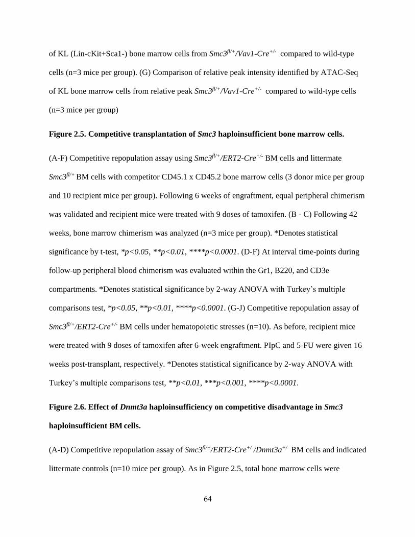
64
of KL (Lin-cKit+Sca1-) bone marrow cells from Smc3fl/+/Vav1-Cre+/- compared to wild-type
cells (n=3 mice per group). (G) Comparison of relative peak intensity identified by ATAC-Seq
of KL bone marrow cells from relative peak Smc3fl/+/Vav1-Cre+/- compared to wild-type cells
(n=3 mice per group)
Figure 2.5. Competitive transplantation of Smc3 haploinsufficient bone marrow cells.
(A-F) Competitive repopulation assay using Smc3fl/+/ERT2-Cre+/- BM cells and littermate
Smc3fl/+ BM cells with competitor CD45.1 x CD45.2 bone marrow cells (3 donor mice per group
and 10 recipient mice per group). Following 6 weeks of engraftment, equal peripheral chimerism
was validated and recipient mice were treated with 9 doses of tamoxifen. (B - C) Following 42
weeks, bone marrow chimerism was analyzed (n=3 mice per group). *Denotes statistical
significance by t-test, *p<0.05, **p<0.01, ****p<0.0001. (D-F) At interval time-points during
follow-up peripheral blood chimerism was evaluated within the Gr1, B220, and CD3e
compartments. *Denotes statistical significance by 2-way ANOVA with Turkey’s multiple
comparisons test, *p<0.05, **p<0.01, ****p<0.0001. (G-J) Competitive repopulation assay of
Smc3fl/+/ERT2-Cre+/- BM cells under hematopoietic stresses (n=10). As before, recipient mice
were treated with 9 doses of tamoxifen after 6-week engraftment. PIpC and 5-FU were given 16
weeks post-transplant, respectively. *Denotes statistical significance by 2-way ANOVA with
Turkey’s multiple comparisons test, **p<0.01, ***p<0.001, ****p<0.0001.
Figure 2.6. Effect of Dnmt3a haploinsufficiency on competitive disadvantage in Smc3
haploinsufficient BM cells.
(A-D) Competitive repopulation assay of Smc3fl/+/ERT2-Cre+/-/Dnmt3a+/- BM cells and indicated
littermate controls (n=10 mice per group). As in Figure 2.5, total bone marrow cells were

65
allowed to engraft for 6 weeks and equivalent chimerism was validated before treatment of all
cohorts with 9 doses of tamoxifen (3 mg/day). Peripheral blood chimerism was evaluated by
flow cytometry at indicated time points. (E) Bone marrow chimerism assessed by flow cytometry
26 weeks after engraftment. KL: Lin-cKit+Sca1-. (n=4 mice in each group). *Denotes statistical
significance by 2-way ANOVA with Turkey’s multiple comparisons test. *p<0.05, **p<0.01,
***p<0.001, ****p<0.0001.
Figure 2.7. Splicing analysis of exon 3 to exon 5 in wild-type and Smc3fl/+/Vav1-Cre+/- KL
cells.
Lin-cKit+Sca1- bone marrow cells were subjected to RNA-Seq (Figure 2.1C and 2.4F). Schema
indicates total number of reads spanning each splice junction from indicated mice.
Figure 2.8. Representative plot of intracellular flow cytometry data (Figure 2.1D-E).
(A) Percentages of Smc3+ cells (left) and mean fluorescence intensity (MFI) (right) of WBM
from mice used in Figure 2.1D. (B). Overlay of the two MFI plots. (C) Percentages of Smc3+
cells (left) and mean fluorescence intensity (MFI) (right) of lin-, KLS, SLAM, and KL cells from
mouse used in Figure 2.1E. The height of the peak is proportional to the number of events
collected.
Figure 2.9. Analysis of homozygous somatic Smc3 deletion.
(A) Western blot of Smc3 in total bone marrow cells following 4 doses of tamoxifen in indicated
mice. (B) Image of spleens from Smc3fl/+/ERT2-Cre+/- and Smc3fl/+ littermate controls following
4 doses of tamoxifen. (C-D) Proportion of bone marrow cells and thymocytes with indicated
immunophenotypes following 4 doses of tamoxifen in Smc3fl/+/ERT2-Cre+/-mice and littermate

66
controls (n=4 mice per group), *Denotes statistical significance by t test, *p<0.05, **p<0.01,
***p<0.001, ****p<0.0001.
Figure 2.10. Analysis of germline heterozygous Smc3 deletion.
(A) Numbers of male vs. female pups generated from Smc3fl/fl and CMV-Cre+/- intercrosses (of
note, CMV-Cre is X-linked). (B-E) Complete blood counts, total number of bone marrow cells,
percentages of HSPCs, and cells of myeloid (Gr1+ CD11b+), B cells (B220+ CD4-), and
erythroid cells (Ter119+ CD71+) in Smc3+/-/CMV-Cre+/- females and Smc3fl/+ littermate males,
(n=4 mice per group).
Figure 2.11. Immunophenotypic analysis of colonies in serial replating assay ex vivo (Figure
2.4C-E).
(A) Percentages of Gr1+ CD11b+ and cKit+ FcΕR1+ cells in the Smc3fl/+ and-Smc3fl/+/ERT2-
Cre+/- colonies week 1-4 respectively (n=4 mice per group). (B) Representative plot of the
Smc3fl/+ and-Smc3fl/+/ERT2-Cre+/- colonies week 1-4. *Denotes statistical significance by t test,
*p<0.05.
Figure 2.12. Competitive transplantation of ERT2-Cre+/- bone marrow cells.
(A) Competitive repopulation assay using ERT2-Cre+/- BM cells with competitor CD45.1 x
CD45.2 bone marrow cells (3 donor mice and 5 recipient mice). Following 6 weeks of
engraftment, equal peripheral chimerism was validated and recipient mice were treated with 9
doses of tamoxifen. (B) At interval time-points during follow-up peripheral blood chimerism was
evaluated within the Gr1, B220, and CD3e compartments.

67
Figure 2.1. Generation of Smc3 conditional deficient mice and allele validation.

68
Figure 2.2. Embryonic hematopoietic Smc3 deletion.

69
Figure 2.3. Homozygous somatic Smc3 deletion.
70
Figure 2.4. Hematopoietic Smc3 haploinsufficiency.

71
Figure 2.5. Competitive transplantation of Smc3 haploinsufficient bone marrow cells.

72
Figure 2.6. Effect of Dnmt3a haploinsufficiency on competitive disadvantage in Smc3
haploinsufficient BM cells.

73
Figure 2.7. Splicing analysis of exon 3 to exon 5 in wild-type and Smc3fl/+/Vav1-Cre+/- KL
cells.
74
Figure 2.8. Representative plot of intracellular flow cytometry data (Figure 2.1.D-E).

75
Figure 2.9. Analysis of homozygous somatic Smc3 deletion.
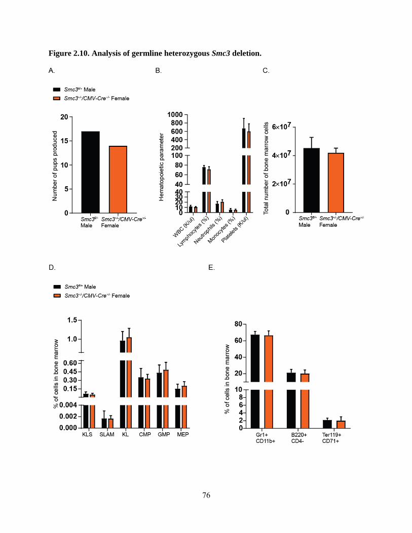
76
Figure 2.10. Analysis of germline heterozygous Smc3 deletion.

77
Figure 2.11. Immunophenotypic analysis of colonies in serial replating assay ex vivo (Figure
2.4.C-E).

78
Figure 2.12. Competitive transplantation of ERT2-Cre+/- bone marrow cells.

79
References
1. Saultz JN, Garzon R. Acute Myeloid Leukemia: A Concise Review. J Clin Med. 2016;5(3).
doi:10.3390/jcm5030033
2. Lagunas-Rangel FA, Chávez-Valencia V, Gómez-Guijosa MÁ, Cortes-Penagos C. Acute
Myeloid Leukemia—Genetic Alterations and Their Clinical Prognosis. Int J Hematol-
Oncol Stem Cell Res. 2017;11(4):328-339.
3. Cancer Genome Atlas Research Network, Ley TJ, Miller C, et al. Genomic and epigenomic
landscapes of adult de novo acute myeloid leukemia. N Engl J Med. 2013;368(22):2059-
2074. doi:10.1056/NEJMoa1301689
4. Kon A, Shih L-Y, Minamino M, et al. Recurrent mutations in multiple components of the
cohesin complex in myeloid neoplasms. Nat Genet. 2013;45(10):1232-1237.
doi:10.1038/ng.2731
5. Thota S, Viny AD, Makishima H, et al. Genetic alterations of the cohesin complex genes in
myeloid malignancies. Blood. 2014;124(11):1790-1798. doi:10.1182/blood-2014-04-
567057
6. Yoshida K, Toki T, Okuno Y, et al. The landscape of somatic mutations in Down
syndrome–related myeloid disorders. Nat Genet. 2013;45(11):1293-1299.
doi:10.1038/ng.2759
7. Barber TD, McManus K, Yuen KWY, et al. Chromatid cohesion defects may underlie
chromosome instability in human colorectal cancers. Proc Natl Acad Sci.
2008;105(9):3443-3448. doi:10.1073/pnas.0712384105
8. Gorringe KL, Ramakrishna M, Williams LH, et al. Are there any more ovarian tumor
suppressor genes? A new perspective using ultra high-resolution copy number and loss of
heterozygosity analysis. Genes Chromosomes Cancer. 2009;48(10):931-942.
doi:10.1002/gcc.20694
9. Bailey ML, O’Neil NJ, Pel DM van, Solomon DA, Waldman T, Hieter P. Glioblastoma
Cells Containing Mutations in the Cohesin Component STAG2 Are Sensitive to PARP
Inhibition. Mol Cancer Ther. 2014;13(3):724-732. doi:10.1158/1535-7163.MCT-13-0749
10. Solomon DA, Kim J-S, Bondaruk J, et al. Frequent truncating mutations of STAG2 in
bladder cancer. Nat Genet. 2013;45(12):1428-1430. doi:10.1038/ng.2800
11. Balbás-Martínez C, Sagrera A, Carrillo-de-Santa-Pau E, et al. Recurrent inactivation of
STAG2 in bladder cancer is not associated with aneuploidy. Nat Genet. 2013;45(12):1464-
1469. doi:10.1038/ng.2799

80
12. Solomon DA, Kim T, Diaz-Martinez LA, et al. Mutational Inactivation of STAG2 Causes
Aneuploidy in Human Cancer. Science. 2011;333(6045):1039-1043.
doi:10.1126/science.1203619
13. Remeseiro S, Cuadrado A, Gómez-López G, Pisano DG, Losada A. A unique role of
cohesin-SA1 in gene regulation and development. EMBO J. 2012;31(9):2090-2102.
doi:10.1038/emboj.2012.60
14. Mannini L, Cucco F, Quarantotti V, Krantz ID, Musio A. Mutation spectrum and genotype-
phenotype correlation in Cornelia de Lange syndrome. Hum Mutat. 2013;34(12):1589-
1596. doi:10.1002/humu.22430
15. Welch JS, Ley TJ, Link DC, et al. The Origin and Evolution of Mutations in Acute Myeloid
Leukemia. Cell. 2012;150(2):264-278. doi:10.1016/j.cell.2012.06.023
16. Thol F, Bollin R, Gehlhaar M, et al. Mutations in the cohesin complex in acute myeloid
leukemia: clinical and prognostic implications. Blood. 2014;123(6):914-920.
doi:10.1182/blood-2013-07-518746
17. Conese M, Liso A. Cohesin complex is a major player on the stage of leukemogenesis.
Stem Cell Investig. 2016;3:18. doi:10.21037/sci.2016.05.04
18. Xie M, Lu C, Wang J, et al. Age-related mutations associated with clonal hematopoietic
expansion and malignancies. Nat Med. 2014;20(12):1472-1478. doi:10.1038/nm.3733
19. Jaiswal S, Natarajan P, Silver AJ, et al. Clonal Hematopoiesis and Risk of Atherosclerotic
Cardiovascular Disease. N Engl J Med. 2017;377(2):111-121.
doi:10.1056/NEJMoa1701719
20. Cole CB, Russler-Germain DA, Ketkar S, et al. Haploinsufficiency for DNA
methyltransferase 3A predisposes hematopoietic cells to myeloid malignancies. J Clin
Invest. 2017;127(10):3657-3674. doi:10.1172/JCI93041
21. Ball AR, Chen Y-Y, Yokomori K. Mechanisms of cohesin-mediated gene regulation and
lessons learned from cohesinopathies. Biochim Biophys Acta BBA - Gene Regul Mech.
2014;1839(3):191-202. doi:10.1016/j.bbagrm.2013.11.002
22. Wendt KS, Yoshida K, Itoh T, et al. Cohesin mediates transcriptional insulation by
CCCTC-binding factor. Nature. 2008;451(7180):796-801. doi:10.1038/nature06634
23. Merkenschlager M, Odom DT. CTCF and cohesin: linking gene regulatory elements with
their targets. Cell. 2013;152(6):1285-1297. doi:10.1016/j.cell.2013.02.029
24. Schmidt D, Schwalie PC, Ross-Innes CS, et al. A CTCF-independent role for cohesin in
tissue-specific transcription. Genome Res. 2010;20(5):578-588. doi:10.1101/gr.100479.109
25. Genomic and Epigenomic Landscapes of Adult De Novo Acute Myeloid Leukemia. N Engl
J Med. 2013;368(22):2059-2074. doi:10.1056/NEJMoa1301689
81
26. Tusher VG, Tibshirani R, Chu G. Significance analysis of microarrays applied to the
ionizing radiation response. Proc Natl Acad Sci U S A. 2001;98(9):5116-5121.
doi:10.1073/pnas.091062498
27. Kanehisa M, Sato Y, Kawashima M, Furumichi M, Tanabe M. KEGG as a reference
resource for gene and protein annotation. Nucleic Acids Res. 2016;44(D1):D457-D462.
doi:10.1093/nar/gkv1070
28. Walter MJ, Shen D, Ding L, et al. Clonal Architecture of Secondary Acute Myeloid
Leukemia. N Engl J Med. 2012;366(12):1090-1098. doi:10.1056/NEJMoa1106968
29. Genovese G, Kähler AK, Handsaker RE, et al. Clonal Hematopoiesis and Blood-Cancer
Risk Inferred from Blood DNA Sequence. N Engl J Med. 2014;371(26):2477-2487.
doi:10.1056/NEJMoa1409405
30. Viny AD, Ott CJ, Spitzer B, et al. Dose-dependent role of the cohesin complex in normal
and malignant hematopoiesis. J Exp Med. 2015;212(11):1819-1832.
doi:10.1084/jem.20151317
31. Xu H, Balakrishnan K, Malaterre J, et al. Rad21-cohesin haploinsufficiency impedes DNA
repair and enhances gastrointestinal radiosensitivity in mice. PloS One. 2010;5(8):e12112.
doi:10.1371/journal.pone.0012112
32. Welch JS, Yuan W, Ley TJ. PML-RARA can increase hematopoietic self-renewal without
causing a myeloproliferative disease in mice. J Clin Invest. 2011;121(4):1636-1645.
doi:10.1172/JCI42953
33. Mullenders J, Aranda-Orgilles B, Lhoumaud P, et al. Cohesin loss alters adult
hematopoietic stem cell homeostasis, leading to myeloproliferative neoplasms. J Exp Med.
2015;212(11):1833-1850. doi:10.1084/jem.20151323
34. Mazumdar C, Shen Y, Xavy S, et al. Leukemia-Associated Cohesin Mutants Dominantly
Enforce Stem Cell Programs and Impair Human Hematopoietic Progenitor Differentiation.
Cell Stem Cell. 2015;17(6):675-688. doi:10.1016/j.stem.2015.09.017
35. Shirai CL, Ley JN, White BS, et al. Mutant U2AF1 Expression Alters Hematopoiesis and
Pre-mRNA Splicing In Vivo. Cancer Cell. 2015;27(5):631-643.
doi:10.1016/j.ccell.2015.04.008
36. Fei DL, Zhen T, Durham B, et al. Impaired hematopoiesis and leukemia development in
mice with a conditional knock-in allele of a mutant splicing factor gene U2af1. Proc Natl
Acad Sci U S A. 2018;115(44):E10437-E10446. doi:10.1073/pnas.1812669115
37. Kim E, Ilagan JO, Liang Y, et al. SRSF2 Mutations Contribute to Myelodysplasia by
Mutant-Specific Effects on Exon Recognition. Cancer Cell. 2015;27(5):617-630.
doi:10.1016/j.ccell.2015.04.006

82
38. Komeno Y, Huang Y-J, Qiu J, et al. SRSF2 Is Essential for Hematopoiesis, and Its
Myelodysplastic Syndrome-Related Mutations Dysregulate Alternative Pre-mRNA
Splicing. Mol Cell Biol. 2015;35(17):3071-3082. doi:10.1128/MCB.00202-15
39. Wang C, Sashida G, Saraya A, et al. Depletion of Sf3b1 impairs proliferative capacity of
hematopoietic stem cells but is not sufficient to induce myelodysplasia. Blood.
2014;123(21):3336-3343. doi:10.1182/blood-2013-12-544544
40. Matsunawa M, Yamamoto R, Sanada M, et al. Haploinsufficiency of Sf3b1 leads to
compromised stem cell function but not to myelodysplasia. Leukemia. 2014;28(9):1844-
1850. doi:10.1038/leu.2014.73
41. Visconte V, Tabarroki A, Zhang L, et al. Splicing factor 3b subunit 1 (Sf3b1)
haploinsufficient mice display features of low risk Myelodysplastic syndromes with ring
sideroblasts. J Hematol OncolJ Hematol Oncol. 2014;7:89. doi:10.1186/s13045-014-0089-
x
42. Obeng EA, Chappell RJ, Seiler M, et al. Physiologic Expression of Sf3b1(K700E) Causes
Impaired Erythropoiesis, Aberrant Splicing, and Sensitivity to Therapeutic Spliceosome
Modulation. Cancer Cell. 2016;30(3):404-417. doi:10.1016/j.ccell.2016.08.006
43. Mupo A, Seiler M, Sathiaseelan V, et al. Hemopoietic-specific Sf3b1-K700E knock-in
mice display the splicing defect seen in human MDS but develop anemia without ring
sideroblasts. Leukemia. 2017;31(3):720-727. doi:10.1038/leu.2016.251
44. Abdel-Wahab O, Gao J, Adli M, et al. Deletion of Asxl1 results in myelodysplasia and
severe developmental defects in vivo. J Exp Med. 2013;210(12):2641-2659.
doi:10.1084/jem.20131141
45. Nagase R, Inoue D, Pastore A, et al. Expression of mutant Asxl1 perturbs hematopoiesis
and promotes susceptibility to leukemic transformation. J Exp Med. 2018;215(6):1729-
1747. doi:10.1084/jem.20171151
46. ExAC Browser. http://exac.broadinstitute.org/gene/ENSG00000108055. Accessed May 18,
2018.
47. Cucco F, Musio A. Genome stability: What we have learned from cohesinopathies. Am J
Med Genet C Semin Med Genet. 2016;172(2):171-178. doi:10.1002/ajmg.c.31492
48. Ramos FJ, Puisac B, Baquero-Montoya C, et al. Clinical utility gene card for: Cornelia de
Lange syndrome. Eur J Hum Genet. 2015;23(10):1431. doi:10.1038/ejhg.2014.270
49. Kumar R, Corbett MA, Van Bon BWM, et al. Increased STAG2 dosage defines a novel
cohesinopathy with intellectual disability and behavioral problems. Hum Mol Genet.
2015;24(25):7171-7181. doi:10.1093/hmg/ddv414

83
50. Leroy C, Jacquemont M-L, Doray B, et al. Xq25 duplication: the crucial role of the STAG2
gene in this novel human cohesinopathy. Clin Genet. 2016;89(1):68-73.
doi:10.1111/cge.12567
51. Baquero-Montoya C, Gil-Rodríguez MC, Teresa-Rodrigo ME, et al. Could a patient with
SMC1A duplication be classified as a human cohesinopathy? Clin Genet. 2014;85(5):446-
451. doi:10.1111/cge.12194
52. Trapnell C, Roberts A, Goff L, et al. Differential gene and transcript expression analysis of
RNA-seq experiments with TopHat and Cufflinks. Nat Protoc. 2012;7(3):562-578.
doi:10.1038/nprot.2012.016
53. Buenrostro JD, Giresi PG, Zaba LC, Chang HY, Greenleaf WJ. Transposition of native
chromatin for fast and sensitive epigenomic profiling of open chromatin, DNA-binding
proteins and nucleosome position. Nat Methods. 2013;10(12):1213-1218.
doi:10.1038/nmeth.2688
54. Zhang Y, Liu T, Meyer CA, et al. Model-based analysis of ChIP-Seq (MACS). Genome
Biol. 2008;9(9):R137. doi:10.1186/gb-2008-9-9-r137
55. Quinlan AR, Hall IM. BEDTools: a flexible suite of utilities for comparing genomic
features. Bioinforma Oxf Engl. 2010;26(6):841-842. doi:10.1093/bioinformatics/btq033
56. Ramírez F, Dündar F, Diehl S, Grüning BA, Manke T. deepTools: a flexible platform for
exploring deep-sequencing data. Nucleic Acids Res. 2014;42(Web Server issue):W187-191.
doi:10.1093/nar/gku365
57. Love MI, Huber W, Anders S. Moderated estimation of fold change and dispersion for
RNA-seq data with DESeq2. Genome Biol. 2014;15(12):550. doi:10.1186/s13059-014-
0550-8

84
Chapter 3:
Exome analysis of treatment-related AML after APL suggests secondary evolution
85
Treatment-related acute myeloid leukemia (tAML) and treatment-related myelodysplastic
syndrome (tMDS) have been associated with many types of chemotherapy and radiation.1
Treatment-related AML or tMDS have been observed after treatment of acute promyelocytic
leukemia (APL) with combination all-trans retinoic acid (ATRA)/idarubicin/cytarabine.2, 3, 4, 5, 6
It is unknown whether tAML will emerge following exposure to all-trans retinoic acid (ATRA)
and arsenic trioxide (ATO), which should not cause DNA damage or the clonal selection of
chemotherapy-resistant clones that give rise to tAML.1, 6, 7 We describe a case of an older women
who presented initially with dysplasia. Shortly thereafter she developed APL and was treated
with ATRA/ATO. Five years later she developed what clinically appeared to be tAML. Exome
sequencing revealed a founding clone with a TET2 mutation and shared passenger mutations that
existed at all three time points. The APL sample shared these mutations, and the subsequent
“tAML” emerged as a new subclone with an NPM1 mutation, more consistent of secondary
AML rather than tAML. This case demonstrates that APL may emerge within the context of
clonal hematopoiesis, and that tAML emerging after ATRA/ATO should be evaluated for
features consistent with secondary AML.
An 81 year-old woman presented initially with cytopenias at an outside facility. A bone marrow
biopsy was performed, revealing trilineage dysplasia, 6% promyelocytes, and a normal
karyotype, 46 XX[20/20] (Table 3.1). Two months later, the cytopenias persisted and a repeat
bone marrow aspirate was hypercellular with 50% promyelocytes. Cytogenetics revealed 46 XX,
t(15;17)[18]/46 XX[2] and 89% of cells were positive for PML-RARA by FISH. She was referred
to Washington University. The bone marrow biopsy was repeated and banked with appropriate
consent for genomic analysis. The repeat biopsy was unfortunately hemodilute with 4.5%
t(15;17) by FISH [9/200], was not evaluated by cytomorphology, and RT-PCR failed (control
86
GAPDH primers did not amplify). She was treated with ATRA/ATO. Bone marrow biopsy 6
weeks later revealed 1% promyelocytes and normal karyotype by cytogenetics and FISH. Due to
her age, no further bone marrow biopsies were performed and subsequent peripheral blood RT-
PCR tests were negative. Five years later she became increasingly cytopenic and a bone marrow
aspirate revealed acute myeloid leukemia with 72% blasts, 20% promyelocytes, and a normal
karyotype. She was treated with decitabine. She deteriorated during the first cycle and
transitioned to hospice.
Exome sequencing was performed on three samples using techniques described elsewhere and
compared against a skin sample, which was used as a germline control (Figure 3.1).8 Cells
retained on a coverslip were available from the dysplastic pre-APL sample and this was used for
genomic analysis. No coverslip or other material was available from the APL sample with 89%
PML-RARA. Cryopreserved bone marrow aspirate cells were used for APL (4.5% PML-RARA)
and AML (72% blasts) analysis. The collected APL sample only generated one cryovial, which
was used for DNA synthesis. Unfortunately, no additional samples are available for RNA-Seq
analysis or sequencing of subpopulations after flow sorting.
A shared founding clone existed across all three time points that contained a TET2 mutation and
15 additional variants (Figure 3.1, black). Three additional clusters were identified: 1) variants
that were present predominantly in the dysplasia sample (SEMA4A, and ZBTB7A, orange); 2)
variants that were absent in the AML sample (TET1, SACM1L, OR7D2, SH3TC1, blue); 3)
variants that increased in the AML sample, but were present at low variant allele frequencies or
were undetected in prior samples (NPM1 and 13 additional variants, red).
Treatment-related AML has been associated with APL therapy that includes alkylating agents,2, 3,
4, 9 but has not been associated with ATRA/ATO, which are non-cytotoxic and do not damage
87
DNA. This unusual case initially appeared consistent with tAML following ATRA/ATO,
however, following exome analysis, appears to be more consistent with a pre-existing dominant
clone associated with dysplasia and a TET2 mutation, and an NPM1-associated secondary AML.
Two models are possible (Figure 3.1F). First, the APL may have emerged as a subclone of the
TET2 founding clone. Given the high TET2 variant allele frequency (VAF, consistent with 70%
and 66% tumor burden in the dysplastic and APL samples, respectively) and the high tumor
burden of PML-RARA in the bone marrow (85%), this model seems likely. Alternatively,
because the sequenced APL sample is hemodilute, it is possible that the PML-RARA clone is
independent of the TET2 clone, that the APL clone did not peripheralize, or that geographical
heterogeneity existed in this older patient. Given the absence of additional samples, it is
impossible to determine whether the APL emerged as a subclone of the TET2 founding clone
occurred independent of the TET2 clone.
In order to determine if these subclonal patterns are observed in other APL patients, we reviewed
available cytogenetic results from published APL patients. Two cases of disease progression
from MDS to APL have been described10, and cases of treatment-related APL have been
described following MDS.11 In the former cases, like this case, the timeline of evolution and
dysplastic changes in the APL morphology suggests that the APL clone likely was evolutionarily
related to the MDS clone. In the latter cases, the cytogenetics and timeline suggest the APL clone
likely emerged independent of the MDS clone.
Clonal hematopoiesis of indeterminate potential (CHIP) and MDS have been associated with
deletions involving chromosomes 5 and 712, 13, which occasionally co-occur with t(15;17).14, 15, 16
Therefore, we examined the subclonal relationship of t(15;17) with possible CHIP-related
variants among APL patients enrolled at large cancer centers. We identified 44 APL cases with

88
t(15;17) and cytogenetic abnormalities in chromosomes 5 or 7. Five cases involved chromosome
5, two of which also had abnormalities in chromosome 7. Thirty-three cases involved a
monoclonal process, suggesting that the variants in chromosomes 5 or 7 co-occurred with
t(15;17). Of the eleven cases with identified polyclones, the chromosome 5 or 7 variant was in a
subclone of t(15;17) in ten cases and in only one case was there evidence of del(7) in a clone that
was independent of the t(15;17) clone (Patient 52009 from APL 2006, 46, XY, t(15,17)(q22,
q11)[13]/46, XY, del(7)(q35)[5]/46, XY [2]) (Table 3.2). These data suggest that t(15;17) may
co-occur with additional cytogenetic deletions of chromosomes 5 and 7, but t(15;17) is almost
invariably the founding event, and it is unusual for a concurrent CHIP or MDS clone to co-exist
with t(15;17).
Other investigators have reviewed the outcomes in APL patients with cytogenetic abnormalities
that occur in addition to t(15;17).16, 17, 18 The most recurrently observed co-occurring cytogenetic
abnormalities include +8 and +21, both of which frequently can be identified as subclonal
progression events when analyzing metaphase cytogenetics or FISH, and have been observed as
progression events in MDS and AML.11 In these studies, additional cytogenetic abnormalities
have not correlated with initial clinical characteristics, or with outcomes in patients with
t(15;17).16, 17, 18
Morphologic disease switching also has been described in NPM1-mutated AML patients who
subsequently developed MDS or myelofibrosis. In each of these cases, the NPM1-mutation was
lost, and the MDS or myelofibrosis evolved from an antecedent clone with a mutation in TET2,
JAK2, ASXL1, IDH2, or a spliceosome trancript, suggesting that the two clonal diseases were
related through an ancestral clone.19

89
In summary, we present a case of what initially appeared to be tAML following non-cytotoxic
ATRA/ATO therapy for APL. Exome analysis clearly demonstrated a TET2-associated,
dominant clonal process that anteceded the APL diagnosis, persisted, and gave rise to AML
associated with evolutionary expansion of an NPM1-mutated subclone. This progression would
be more consistent with a secondary AML process, rather than a treatment-related process.
Additional characterization of this case would be interesting. Unfortunately, samples for such
analyses are unavailable. Review of additional cytogenetic abnormalities observed in APL
patients did identify one rare case with del(7) independent of the APL clone, suggesting that
APL can co-exist with CHIP or MDS clones, although it appears much more common for such
cytogenetic abnormalities to occur as subclones of the APL clone. Collectively, these results
suggest that caution must be exercised when interpreting the development of tAML following
ATRA/ATO therapy, and subclonal expansion of related or well-established clones should be
considered, especially in older patients.

90
Figure Legends
Figure 3.1. Exome analysis of patient 10DD-1029.
(A) Summary results of all somatic variants detected at any of the three time points analyzed. (B)
Cluster of variants with stable VAFs across all three samples. (C) Cluster of variants present in
the initial dysplastic sample, with reduced VAFs in subsequent samples. (D) Cluster of variants
with VAFs in the initial two samples, but which were absent in the subsequent AML sample. (E)
Cluster of variants associated with the AML progression, which were largely absent in the initial
two samples. (F) Two models for subclonal expansion.

91
Figure 3.1. Exome analysis of patient 10DD-1029.

92
Tables 3.1. Clinical data of the patient.
Dysplasia APL APL (banked) post-APL AML
Day 0 Day 49 Day 51 Day 87 Day 1897
Sequencing exome ND Exome ND exome
t(15;17) 0% 89% 4.5% 0% 0%
BM blasts 0% 1% ND 1% 72%
BM promyelocytes 6% 50% ND 1% 20%
BM myelocytes 9% 25% ND 25% 1%
BM metamyelocytes 2% 3% ND 3% 5%
BM bands 9% 2% ND 2% 0%
BM dysplasia trilineage atypia ND none none
PB WBC NA 0.9 0.9 0.9 2.2
PB % Lymphs NA 95% 95% 95% 88%
PB % Blasts NA 0% 0% 0% 6%
AML, acute myeloid leukemia; APL, acute promyelocytic leukemia; BM, bone marrow; NA, not
available; ND, not done; PB, peripheral blood; WBC, white blood cells.
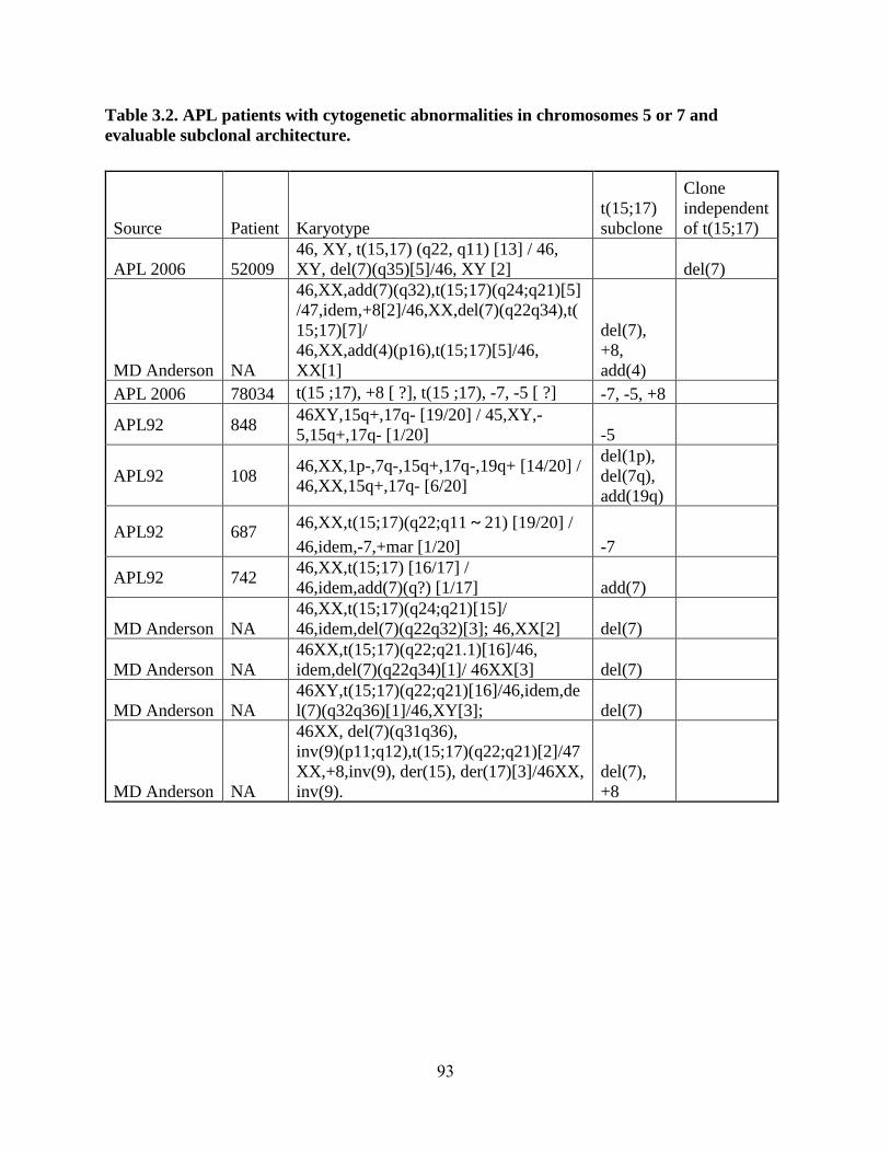
93
Table 3.2. APL patients with cytogenetic abnormalities in chromosomes 5 or 7 and
evaluable subclonal architecture.
Source Patient Karyotype
t(15;17)
subclone
Clone
independent
of t(15;17)
APL 2006 52009
46, XY, t(15,17) (q22, q11) [13] / 46,
XY, del(7)(q35)[5]/46, XY [2] del(7)
MD Anderson NA
46,XX,add(7)(q32),t(15;17)(q24;q21)[5]
/47,idem,+8[2]/46,XX,del(7)(q22q34),t(
15;17)[7]/
46,XX,add(4)(p16),t(15;17)[5]/46,
XX[1]
del(7),
+8,
add(4)
APL 2006 78034 t(15 ;17), +8 [ ?], t(15 ;17), -7, -5 [ ?] -7, -5, +8
APL92 848 46XY,15q+,17q- [19/20] / 45,XY,-
5,15q+,17q- [1/20] -5
APL92 108 46,XX,1p-,7q-,15q+,17q-,19q+ [14/20] /
46,XX,15q+,17q- [6/20]
del(1p),
del(7q),
add(19q)
APL92 687 46,XX,t(15;17)(q22;q11~21) [19/20] /
46,idem,-7,+mar [1/20] -7
APL92 742 46,XX,t(15;17) [16/17] /
46,idem,add(7)(q?) [1/17] add(7)
MD Anderson NA
46,XX,t(15;17)(q24;q21)[15]/
46,idem,del(7)(q22q32)[3]; 46,XX[2] del(7)
MD Anderson NA
46XX,t(15;17)(q22;q21.1)[16]/46,
idem,del(7)(q22q34)[1]/ 46XX[3] del(7)
MD Anderson NA
46XY,t(15;17)(q22;q21)[16]/46,idem,de
l(7)(q32q36)[1]/46,XY[3]; del(7)
MD Anderson NA
46XX, del(7)(q31q36),
inv(9)(p11;q12),t(15;17)(q22;q21)[2]/47
XX,+8,inv(9), der(15), der(17)[3]/46XX,
inv(9).
del(7),
+8
94
References
1. Wong TN, Ramsingh G, Young AL, et al. Role of TP53 mutations in the origin and
evolution of therapy-related acute myeloid leukaemia. Nature. 2015;518(7540):552-555.
doi:10.1038/nature13968
2. Park SH, Chi H-S, Cho Y-U, Jang S, Park C-J, Lee J-H. A case of therapy-related acute
myeloid leukemia with a normal karyotype after sustained molecular complete remission of
acute promyelocytic leukemia. Ann Lab Med. 2014;34(1):68-70.
doi:10.3343/alm.2014.34.1.68
3. Zompi S, Viguié F. Therapy-related acute myeloid leukemia and myelodysplasia after
successful treatment of acute promyelocytic leukemia. Leuk Lymphoma. 2002;43(2):275-
280. doi:10.1080/10428190290006044
4. Ojeda-Uribe M, Schneider A, Luquet I, et al. Therapy-related acute myeloid leukemia (t-
AML) with poor-risk cytogenetics in two patients with persistent molecular complete
remission of acute promyelocytic leukemia. Eur J Haematol. 2012;89(3):267-272.
doi:10.1111/j.1600-0609.2012.01805.x
5. Park TS, Choi JR, Yoon SH, et al. Acute promyelocytic leukemia relapsing as secondary
acute myelogenous leukemia with translocation t(3;21)(q26;q22) and RUNX1-MDS1-EVI1
fusion transcript. Cancer Genet Cytogenet. 2008;187(2):61-73.
doi:10.1016/j.cancergencyto.2008.06.015
6. de Thé H. Differentiation therapy revisited. Nat Rev Cancer. 2018;18(2):117-127.
doi:10.1038/nrc.2017.103
7. Lehmann-Che J, Bally C, Letouzé E, et al. Dual origin of relapses in retinoic-acid resistant
acute promyelocytic leukemia. Nat Commun. 2018;9(1):2047. doi:10.1038/s41467-018-
04384-5
8. Welch JS, Petti AA, Miller CA, et al. TP53 and Decitabine in Acute Myeloid Leukemia and
Myelodysplastic Syndromes. N Engl J Med. 2016;375(21):2023-2036.
doi:10.1056/NEJMoa1605949
9. Zompi S, Legrand O, Bouscary D, et al. Therapy-related acute myeloid leukaemia after
successful therapy for acute promyelocytic leukaemia with t(15;17): a report of two cases
and a review of the literature. Br J Haematol. 2000;110(3):610-613.
10. Isono S, Saigo K, Nagata K, et al. A case of acute promyelocytic leukemia with
morphologic multilineage dysplastic changes. Hematol Rep. 2013;5(1):18-20.
doi:10.4081/hr.2013.e5
11. Andersen MK, Larson RA, Mauritzson N, Schnittger S, Jhanwar SC, Pedersen-Bjergaard J.
Balanced chromosome abnormalities inv(16) and t(15;17) in therapy-related

95
myelodysplastic syndromes and acute leukemia: report from an international workshop.
Genes Chromosomes Cancer. 2002;33(4):395-400.
12. Laurie CC, Laurie CA, Rice K, et al. Detectable clonal mosaicism from birth to old age and
its relationship to cancer. Nat Genet. 2012;44(6):642-650. doi:10.1038/ng.2271
13. Zahid MF, Malik UA, Sohail M, Hassan IN, Ali S, Shaukat MHS. Cytogenetic
Abnormalities in Myelodysplastic Syndromes: An Overview. Int J Hematol-Oncol Stem
Cell Res. 2017;11(3):231-239.
14. Cervera J, Montesinos P, Hernández-Rivas JM, et al. Additional chromosome abnormalities
in patients with acute promyelocytic leukemia treated with all-trans retinoic acid and
chemotherapy. Haematologica. 2010;95(3):424-431. doi:10.3324/haematol.2009.013243
15. Ono T, Takeshita A, Iwanaga M, et al. Impact of additional chromosomal abnormalities in
patients with acute promyelocytic leukemia: 10-year results of the Japan Adult Leukemia
Study Group APL97 study. Haematologica. 2011;96(1):174-176.
doi:10.3324/haematol.2010.030205
16. De Botton S, Chevret S, Sanz M, et al. Additional chromosomal abnormalities in patients
with acute promyelocytic leukaemia (APL) do not confer poor prognosis: results of APL 93
trial. Br J Haematol. 2000;111(3):801-806.
17. Hernández JM, Martín G, Gutiérrez NC, et al. Additional cytogenetic changes do not
influence the outcome of patients with newly diagnosed acute promyelocytic leukemia
treated with an ATRA plus anthracyclin based protocol. A report of the Spanish group
PETHEMA. Haematologica. 2001;86(8):807-813.
18. Lou Y, Suo S, Tong H, et al. Characteristics and prognosis analysis of additional
chromosome abnormalities in newly diagnosed acute promyelocytic leukemia treated with
arsenic trioxide as the front-line therapy. Leuk Res. 2013;37(11):1451-1456.
doi:10.1016/j.leukres.2013.07.030
19. Morin-Zorman S, Renneville A, Adès L. Assessment of Minimal Residual Disease in
Standard-Risk AML. N Engl J Med. 2016;375(6):e9. doi:10.1056/NEJMc1603847