the role of preclinical spect in oncological and neurological research in combination with either ct...
DESCRIPTION
ARTICOLTRANSCRIPT

REVIEWARTICLE
The role of preclinical SPECT in oncological and neurologicalresearch in combination with either CT or MRI
Monique R. Bernsen & Pieter E. B. Vaissier &
Roel Van Holen & Jan Booij & Freek J. Beekman &
Marion de Jong
Received: 19 December 2013 /Accepted: 20 December 2013 /Published online: 17 April 2014# The Author(s) 2014. This article is published with open access at Springerlink.com
Abstract Preclinical imaging with SPECT combined withCT or MRI is used more and more frequently and has provento be very useful in translational research. In this article, anoverview of current preclinical research applications andtrends of SPECTcombinedwith CTorMRI, mainly in tumourimaging and neuroscience imaging, is given and the advan-tages and disadvantages of the different approaches are de-scribed. Today SPECT and CT systems are often integratedinto a single device (commonly called a SPECT/CT system),whereas at present combined SPECT and MRI is almostalways carried out with separate systems and fiducial markersto combine the separately acquired images. While preclinicalSPECT/CT is most widely applied in oncology research,SPECT combined with MRI (SPECT/MRI when integratedin one system) offers the potential for both neuroscienceapplications and oncological applications. Today CT and
MRI are still mainly used to localize radiotracer binding andto improve SPECT quantification, although both CTand MRIhave additional potential. Future technology developmentsmay include fast sequential or simultaneous acquisition of(dynamic) multimodality data, spectroscopy, fMRI along withhigh-resolution anatomic MRI, advanced CT procedures, andcombinations of more than two modalities such as combina-tions of SPECT, PET, MRI and CT all together. This will allstrongly depend on new technologies. With further advancesin biology and chemistry for imaging molecular targets and(patho)physiological processes in vivo, the introduction ofnew imaging procedures and promising new radiopharmaceu-ticals in clinical practice may be accelerated.
Introduction
Over the past decade the use of PET, SPECT, CT and MRI inpreclinical research has greatly increased due to technologicaladvances that have resulted in significant improvements inspatial and temporal resolution as well as sensitivity [1–5].These noninvasive imaging methods enable imaging of(patho)physiological and molecular processes over timein vivo, obviating the need for killing animals for each time-point being studied [6–8]. Each of these imaging modalitieshas unique qualities, in terms of their spatial and temporalresolution and their ability to measure morphology and/orfunction; the appropriate technique should be selected accord-ing to the research question. PET and SPECT allow detectionof radiopharmaceuticals at nano- to picomolar concentrationsin vivo, and have proven to be excellent tools in the transla-tional evaluation of radiotracers. CT and MRI provide a highdegree of spatial resolution that is well suited to anatomicalimaging and tissue phenotyping, including volumetry, and canprovide information regarding tissue physiology [9].
M. R. Bernsen (*) :M. de JongDepartment of Nuclear Medicine, Erasmus MC, Rotterdam,The Netherlandse-mail: [email protected]
M. R. Bernsen :M. de JongDepartment of Radiology, Erasmus MC, Rotterdam,The Netherlands
P. E. B. Vaissier : F. J. BeekmanSection Radiation Detection and Medical Imaging, Delft Universityof Technology, Delft, The Netherlands
R. Van HolenELIS Department, MEDISIP, Ghent University, iMinds,Ghent, Belgium
J. BooijDepartment of Nuclear Medicine, Academic Medical Center,University of Amsterdam, Amsterdam, The Netherlands
F. J. BeekmanMILabs B.V., Utrecht, The Netherlands
Eur J Nucl Med Mol Imaging (2014) 41 (Suppl 1):S36–S49DOI 10.1007/s00259-013-2685-3

Due to their sensitive detection capabilities, PET andSPECT both have preeminent ability to monitor and quantifydynamic processes at a molecular level in vivo. UniqueSPECT capabilities include: the ability to image ligands suchas peptides and antibodies relatively easy with 99mTc, 111In oriodine isotopes (123I, 125I), the ability to measure slow kineticprocesses due to the long half-life (compared to most PETtracers) of some of the commonly used radionuclides, and theability to probe multiple molecular pathways simultaneouslyby detecting radionuclides with different gamma energies(multiisotope imaging). Multiisotope imaging has beendemonstrated both clinically [10–13] and preclinically[14, 15]. Another advantage of SPECT over PET is thatno cyclotron and associated infrastructure and complexlogistics are required on site and that many tracers arereadily available in the form of kits.
While in clinical imaging higher spatial resolutions can beobtained with PET than with SPECT, the opposite is clearlytrue in preclinical imaging in small animals. Small imagingvolumes enable the use of high magnification apertures inSPECT imaging (Fig. 1), increasing sensitivity and resolutionrelative to their clinical counterparts [16–18]. Recently devel-oped SPECT systems can be extended to high-resolutionimaging of high-energy photons emitted by PET tracers, evensimultaneously with (multiple) SPECT tracers [14]. Sincesome SPECT systems also enable imaging of 125I-labelledtracers (<35 keV), the gap between in vitro and in vivo studiesis closed. Finally, in SPECT imaging spatial resolution andsensitivity can be adjusted by changing the size of the colli-mator apertures.
On the other hand, the drawbacks of SPECT include itslower sensitivity compared to PET, especially when high-resolution SPECT is desired. Moreover, SPECT tracer mole-cules may differ with regard to their biological properties fromtheir nonradioactive counterparts after introduction of a radio-nuclide–chelator complex, which is not the case for several
PET tracers in which endogenous atoms (such as hydrogen,carbon and oxygen) can be replaced by their radioactiveisotopes. In addition, the dynamic capabilities of SPECT,although recently greatly improved, are often limited com-pared to those of PET.
In current clinical practice combining images from differenttomographic modalities is common. Also in preclinicalresearch multimodality imaging strategies are useful, asdifferent modalities can provide highly complementaryinformation. Spatially registered images enable localiza-tion, enhanced visualization and accurate quantificationof spread and uptake of radiolabelled molecules withinthe anatomical context provided by CT or MRI. Inaddition, functional information derived from advancedCT and MRI techniques such as perfusion imaging canbe related to expression and function of specific mole-cules as measured by PET or SPECT.
In this review we discuss recent applications and techno-logical advances of preclinical SPECT in combination withCT or MRI in the fields of oncology and neuroscience. Over-views by others and Golestani et al. addressing preclinicalSPECT combined with MRI and CT in other research fields,such as cardiovascular research, regenerative medicine andinflammation, have recently been published [19–22]. Thespace constraints of this article prevented coverage of everyaspect of this exciting field, but we aimed to provide a goodappreciation of the possibilities, and also the limitations andremaining challenges.
Applications of SPECT combined with CT or MRI
Tumour imaging
Hanahan andWeinberg [23, 24] introduced the notion that thetumour microenvironment plays a crucial role in the develop-ment and behaviour of tumours, including receptiveness andsensitivity to treatment. The resulting understanding that can-cer is a complex disease with significant involvement of thetumour stroma has led to the interest in imaging tumour cellcharacteristics as well as noncancer cell components in vivo[25, 26], especially with regard to molecular diagnostics anddrug development. Since it would be impossible to coverevery aspect of this rapidly developing field, we only addresssome key aspects in tumour imaging and the roles thatSPECT, and SPECT combined with CT or MRI have beenplaying in this field.
Imaging targets and probes
Tumours and tumour cells exhibit different characteristicscompared to normal tissue and cells; this is reflected in alteredphysiology, tissue composition and expression of intra- and
Fig. 1 State-of-the-art whole-body SPECT bone images acquired for60 min with 250 MBq 99mTc-HDP and with 0.25-mm resolution colli-mators (image courtesy of Oleksandra Ivashchenko, TU-Delft/MIlabs)
Eur J Nucl Med Mol Imaging (2014) 41 (Suppl 1):S36–S49 S37
extracellular molecules [23, 24, 26–28]. All these aspects canbe used as imaging targets in relation to diagnostics, drugdevelopment and treatment response assessment. SPECTprobes (or tracers) can be classified according to theirbiodistribution and targeting characteristics, i.e. thebiodistribution of some radiopharmaceuticals is determinedby their chemical/physical properties, whereas that of othertracers is determined by their specific interaction with atarget. For details the reader is referred to a review byMüller and Schibli [29].
Tumours are known often to display an aberrant vascularnetwork and microcirculation, which in turn underlies featuressuch as interstitial hypertension, hypoxia and acidosis, char-acteristics that contribute to malignant phenotypes and resis-tance to various treatments [30]. Within this environment,tumour cells can also display altered energy metabolism, asreflected in, for example, increased glucose uptake and shiftedbalances in metabolic products. At the preclinical level, avariety of SPECT tracers are under evaluation for use asmarkers for (neo)angiogenesis [31–33], hypoxia [34–37], ac-idosis [38–40], metabolic activity [41] and proteolytic activity[42, 43]. Moreover, MRI and to a lesser extend CT offeroptions for interrogating tumour physiological characteristics,either through the use of specific probes or the use of sophis-ticatedMRI techniques, as recently reviewed by Bernsen et al.[9]. Besides metabolic tracers, much effort has been put intothe development and validation of SPECT probes specific fortumour target molecules such as antigens, receptors or othermolecules also overexpressed in tumour tissue. The use ofpeptides interacting with receptors [44], antibodies and anti-body fragments targeting their epitopes [45], vitamin-basedradiopharmaceuticals [28] and nucleoside analogues [46],significantly increases the possibilities for tumour detection,localization and staging.
Specific points of interest in translational preclinical imag-ing studies include efforts directed at improved tumour spec-ificity [47], tumour uptake/retention [48] and minimized phar-macological effects [49, 50] of imaging probes. In most pre-clinical studies involving the use of SPECT combined withCT or MRI to date, the CT or MRI components have beenmostly used to provide anatomical reference andmore recentlyalso for attenuation correction [51]. However, CT and MRIoffer more than anatomical information, and some examplesof the use of more sophisticated CT and MRI techniques arediscussed and provided in the technology sections below.
Biodistribution studies/dosimetry/response assessment
In drug development, biodistribution and pharmacokineticproperties of a candidate drug or therapeutic agent are crucialfor their therapeutic potential and safety in patients. Afterbinding of a suitable radionuclide to the molecule or particleof interest, preclinical SPECT imaging provides a valuable
noninvasive tool to study candidate drugs. Especially in de-velopment of targeted treatment strategies with radiolabelledmolecules such as peptides, antibodies and vitamin-basedanalogues, SPECT imaging combined with CT or MRI hasbeen widely used [45, 52–56]. Next to in vivo evaluation ofsuch molecules, SPECT combined with CT or MRI is alsobeing applied in the preclinical evaluation of (nano)particlesfor treatment and/or diagnosis of cancer. Various studieshave investigated the biodistribution and therapeutic poten-tial of, for example, liposomes [57–61], radiolabelledsuperparamagnetic iron oxide nanoparticles and 166Ho mi-crospheres (166HoAcAcMS), using multimodality imagingapproaches with SPECT/CT and SPECT/MRI [62, 63]. Thecombined imaging data allow accurate assessment ofbiodistribution and retention as well as dosimetry calculations.
Many of the imaging biomarkers addressed in the previoussection are also being evaluated as markers to monitorresponse to treatment. Elimination of tumour cells mightbe accompanied by loss of tracer uptake directed attumour-associated antigens or decreased metabolic activity,whereas changes in vascular properties and tissue hypoxiamay be expected after antiangiogenic therapies, allowingthese markers to be used for response assessment. Whilesuch an approach may appear fairly straightforward, somelimitations and pitfalls need to be taken into account. Lossof tumour-associated antigen expression may also be aresult of changed tumour physiology not related to tumourcell death [64]. Another process of interest as an imagingbiomarker for response is apoptosis [65, 66]. Expectationswere raised that visualization and quantification of apopto-sis, as a more specific and relevant marker of cell death,may provide better specificity for assessing actual tumourcell elimination following treatment. Apoptosis imagingusing a tracer specific for annexin could reveal earlytumour cell death after chemotherapy [65], but its valueas a robust marker for treatment response still needs to beestablished.
For the assessment of potential treatment efficacy, Bol et al.recently reported on the added value of dual modality imagingusing SPECT and MRI [67]. In a rat model of neuroendocrinepancreatic tumour, radiolabelled peptide uptake was assessedin conjunction with measurement of tumour perfusion usingDCE-MRI. A substantial correlation between tumour uptakeof 111In-DTPA-octreotide and tumour perfusion parameterswas observed (Fig. 2). It was shown that even in tumour areaswith high receptor expression no peptide uptake occurredwhen perfusion was low, indicating that combined SPECTand MRI may be useful in treatment planning and/or responseprediction in patients treated with PRRT.
Imaging of cell trafficking has also been an area of interestin which SPECT in combination with either CT or MRI hasbeen employed, an approach that has already been part ofclinical routine for several decades for identifying infection or
S38 Eur J Nucl Med Mol Imaging (2014) 41 (Suppl 1):S36–S49

inflammation sites by leucocyte scintigraphy [68]. Recently,the interest in in vivo cell tracking has received a tremendousboost from the realization that knowledge about the in vivofate of infused cells is crucial to the development of safe andeffective cell-based therapeutic strategies, including stem celltherapy [69, 70]. SPECT has largely been used to investigatethe short-term fate of transplanted cells labelled with radio-tracers such as 111In-oxine, 99mTc-hexamethylpropyleneamine oxine (HMPAO) and 111In-tropolone as intracellularlabels [71]. However, due to the lack of anatomical informa-tion and the limited life-time of the radionuclides, preventinglongitudinal follow up, other imaging techniques such asMRIhave been widely used as well [72]. Since MRI also has somespecific limitations for in vivo cell tracking such as lowsensitivity and specificity, and challenges in quantification ofthe MRI probe, alternative approaches have been sought, withspecific interest in reporter gene technology [70]. For SPECTthe sodium iodide symporter gene (NIS) and the herpes sim-plex virus type 1 thymidine kinase gene (HSV1-tk) are so farthe most commonly used reporter genes in combination withradioactive substrates [73, 74]. Reporter gene technology withthese and other reporter genes, e.g. norepinephrine transporterand the somatostatin receptor, is being used not only in in vivocell tracking applications for cell-based therapy [75, 76], butalso to monitor metastatic spread of tumour cells [77–79], aswell as gene delivery and expression of genes in targeted genetherapy approaches [80, 81].
Finally, in medical research, the successful choice of atarget molecule that is a key disease biomarker has the poten-tial to lead to the development not only of a molecular imagingprobe, but also of a therapeutic agent to inhibit the diseaseprocess. Examples include peptides [53, 82, 83], antibodies orfragments thereof [84–87], and nanoparticles [26, 88], similarcompounds or particles that can be labelled with radionuclidesfor either imaging or therapy. Receptor targeting with smallradiolabelled peptides for receptor-targeted tumour imaging(PET and SPECT) as well as for radionuclide therapy [89]provide good examples of such theranostic potential in
nuclear oncology and have paved the way for further devel-opments in this field.
Neuroscience
Preclinical SPECT studies in small laboratory animal modelsof neurodegenerative diseases
Parkinson’s disease (PD) is a neurodegenerative disease char-acterized by loss of neurons producing dopamine (DA), andconsequently loss of the DA transporter (DAT) [90–95]. Pre-clinical SPECT studies initially focused on the feasibility ofdetecting striatal DAT binding in small laboratory animals perse [96, 97]. In the past decade, pinhole SPECT studies haveshown the possibility of detecting loss of striatal DAT bindingin rodent models of PD using [123I]FP-CIT and [123I]β-CIT asradiotracers [98, 99]. Initially, single-pinhole SPECT systemswere used to image DAT [91, 100], and the SPECT imageswere coaligned with MR images (or templates) acquired onclinical MRI scanners (using dedicated coils), with or withoutthe use of external markers [99, 101, 102]. Another recentstudy, however, used a preclinical systemwith high-resolutionparallel-hole collimators (X-SPECT system) to evaluate DATloss (using [123I]altropane as a radiotracer) in a rat model ofPD, and the SPECT images were registered with CT images[103]. Another DAT ([123I]FP-CIT) SPECT study in a mousemodel of PD used a double-headed gamma camera equippedwith a multipinhole aperture. The SPECT images were notcoaligned with CT or MR images [104, 105]. Finally, MRI isan important tool in the field of neuroimaging. In thisregard, it is of interest that Lee et al. proposed an imageregistration algorithm which can be used to register indi-vidual DAT SPECT ([99mTc]TRODAT was used as aradiotracer on a NanoSPECT/CT system) and brain MRimages (acquired on a 3-T system) in rodent models ofPD without using external markers [106].
Neurodegenerative diseases like multiple system atrophy,progressive supranuclear palsy and Huntington’s disease, are
Fig. 2 Multimodality imaging of tumour uptake of targeted radiolabelledpeptide and tumour perfusion. Rats bearing a syngeneic, somatostatinreceptor overexpressing, neuroendocrine pancreatic tumour, were imagedby SPECT/CT and MRI to study tumour uptake of a 111In-labelledsomatostatin analogue ([111In-DTPA]octreotide) and tumour perfusionby DCE-MRI respectively. Left Tumour perfusion depicted by theAUC value over the first 60 s as assessed by DCE-MRI; centre
tumour uptake of radiolabelled [111In-DTPA]octreotide of the sametumour section as imaged by MRI; right colour-coded overlay of theMR image and the SPECT image with MRI values depicted in redand SPECT values depicted in green. For correct image registration,MRI data were resampled to match the lower resolution of theSPECT/CT images (image courtesy of Joost Haeck and Karin Bol,Erasmus MC)
Eur J Nucl Med Mol Imaging (2014) 41 (Suppl 1):S36–S49 S39

characterized by loss of striatal DA D2 receptors [92]. A studypublished in 2002 demonstrated the feasibility of pinholeSPECT for measuring striatal DA D2/3 receptor binding inthe mouse brain in vivo [107]. [123I]IBF was used to assessstriatal D2/3 receptor binding and SPECT images were notregistered with CT or MR images. Not long afterwards, an-other study in rats confirmed the feasibility of assessing DAD2/3 receptor binding in- vivo, using [
123I]IBZM as radiotracerand a dedicated small animal SPECT system [108]. In thatstudy, SPECT images were not registered with CT or MRimages, but a region of interest template was constructed andused to evaluate receptor binding [108].
Scherfler et al. showed the ability of single-pinhole SPECTto detect loss of striatal DA D2/3 receptors in a rat model ofHuntington’s disease [109]. In that study, the [123I]IBZMSPECT images were registered on a MRI template. Impor-tantly, in vivo [123I]IBZM binding was highly correlated withthe loss of medium-sized spiny neurons that express DA D2
receptors demonstrated ex vivo [110].Alzheimer’s disease (AD) is the most common dementia in
humans, and is characterized by the deposition of β-amyloidplaques and neurofibrillary tangles. PET tracers have beendeveloped successfully to image this neuropathology [111].The deposition of amyloid has also been evaluated in micro-PET studies in animal models of AD [112, 113]. SPECTtracers have also been developed for labelling of amyloidplaques [112]. Although [123I]IMPY shows high affinity foramyloid in vitro and amyloid plaques in post-mortem braintissue of AD patients and animal models of AD, the specific tononspecific binding ratios are too low to be of value forstudies in animal models of AD [114, 115].
Preclinical SPECT studies in small laboratory animalsrelevant to studies on psychosis or addiction
A consistent finding of imaging studies in drug addiction is lossof striatal DA D2/3 receptors. An increase in D2 receptorexpression may therefore be beneficial in its treatment [116].Interestingly, some drugs may induce an increase in D2/3 re-ceptors [117–119], which has been supported by SPECT im-aging in rats [117]. In the latter study an ultrahigh-resolutionpinhole SPECT system was used (U-SPECT-II), but SPECTimages were not registered with CT or MR images. Due to thehigh spatial as well as temporal resolution of this system,changes in DAToccupancy by cocaine over time can be studiedin the mouse in vivo [120]. Alterations in the expression of DAD2/3 receptors have been reported in schizophrenia. In a recentstudy, in which the SPECT images were registered with CTimages (X-SPECT/CT system), decreases in DA D2/3 receptoravailability in the striatum and midbrain have been shown in arat model of schizophrenia using [123I]epidepride as radiotracer[121]. DA D2/3 receptor imaging can be used to evaluate DArelease [122]. Increased DA release has been reported in
schizophrenia, whereas DA release may be reduced in cocainedependency [123, 124]. Interestingly, recent pinhole SPECTstudies in mice and rats have also shown the ability to measureDA release [125, 126]. In both studies, SPECT images were notregistered with CT or MR images.
Preclinical SPECT studies focused on brain perfusion
Brain perfusion studies may be of relevance for the study of,for example, the aetiology of stroke. Using a multipinholeSPECT system (NanoSPECT), the kinetics of the perfusiontracers [99mTc]HMPAO and [99mTc]ECD were compared di-rectly in control mice. SPECT images were registered on aMRI template [127]. It was shown that [99mTc]ECD washoutwas much faster than that of [99mTc]HMPAO. In anotherstudy, [123I]iodoamphetamine was used to assess hypoperfu-sion in infarcted brain areas in mice [128]. A single-pinholecollimator systemwas used, and CTandMRI images acquiredon other systems were used for the alignment of the SPECTimages. Finally, Ceulemans et al. performed brain perfusionSPECT studies ([99mTc]HMPAO, 1-mm pinhole collimatorpositioned on a dual-head gamma camera, coregistered onindividual CT images) to quantify the infarct size in rats [129].
Deep brain stimulation (DBS) is commonly used in thetreatment of PD, but has recently also been used in thetreatment of other neuropsychiatric disorders [130]. Interest-ingly, Wyckhuys et al. studied the effects of DBS on brainperfusion in rats [131]. In all rats, they acquired individualbrain perfusion studies with SPECT (U-SPECT-II) after DBS(stimulator on and off), micro-CT scans and, after the animalswere killed and the electrodes removed, MRI scans on aclinical MRI scanner using a dedicated rat brain oil [131].After registration of the images and analysis of each voxel,hypoperfusion induced by DBS could be located accurately insmall brain areas (Fig. 3). This approach highlights the poten-tial of multimodality imaging to evaluate and locate the effectsof interventions/treatments in small brain areas of rodents.
Preclinical SPECT studies focused on neurooncology
Micro-SPECT studies have also been performed successfullyin the field of neurooncology. For example, Yang et al. recent-ly showed the feasibility of using [99mTc]DTPA to study theintegrity of the blood–brain barrier and tumour activity inglioma-bearing rats [132]. A preclinical pinhole SPECT/CTsystem (FLEX Triumph) was used which offers the ability tocoalign the SPECT and CT images [132]. Angiogenesis isessential for tumour growth. Furthermore, malignant cells canrelease vascular endothelial growth factors (VEGFs) whichare important promoters and regulators of angiogenesis.SPECT studies showed the possibility of imaging VEGFreceptors in rats. [99mTc]HYNIC-VEGF uptake was increasedin glioma-bearing rats pretreated with a VEGR receptor
S40 Eur J Nucl Med Mol Imaging (2014) 41 (Suppl 1):S36–S49

tyrosine kinase inhibitor [64]. In that study, SPECT imageswere acquired on a dedicated multiple-pinhole SPECT system(NanoSPECT), but the SPECT images were not registeredwith CT or MR images. In addition, Huang et al. evaluated a188Re-labelled liposome as a diagnostic and therapeutic agentin glioma-bearing rats [60], using a preclinical multiple-pinhole SPECT/CT system (NanoSPECT/CT). Importantly,uptake in the brain tumour could be visualized, and specificbinding was confirmed histopathologically [121]. Anotherstudy in glioma-bearing rats evaluated new treatment strate-gies for glioma, and imaged 99mTc-labelled nanoparticlesusing a clinical SPECT system [133]. Finally, SPECT/CT(parallel hole SPECT system) studies were performed to ex-amine successfully glioblastoma xenografts that were locatedsubcutaneously in mice using, for example, 125I-labelledmonoclonal antibodies against chemokine receptor 4 [134].
Technology of SPECT combined with CT or MRI
Combined imaging approaches/systems, introduction
In order to fully benefit from multimodality imaging, accuratespatial registration of the images is crucial. Below we addressways to adequately combine SPECTwith CT or MRI.
Side-by-side systems
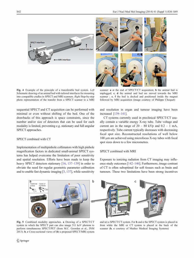
In contrast to clinical imaging of patients, small animals canbe transported – including the bed – between imaging deviceswith gentle fixation with tape preventing movement of the
animal on the bed. This requires beds that can be easily, rigidlyand reproducibly mounted on different scanners (Fig. 4). Mul-timodal fiducial markers attached to the animal (or bed) or apremeasured transformation matrix can be used for spatialcoregistration [135, 136]. Such side-by-side use of separatescanners offers flexibility in adding and/or replacing indi-vidual modalities while both systems can be used in paral-lel facilitating higher through-put. However, maintaininganaesthesia may be a challenge during transport, especiallywhen the machines are far apart.
In-line systems
A second approach to imaging with SPECT in combinationwith CTorMRI is to mount the separate modalities in line (i.e.back-to-back) on a single gantry (Fig. 5).When the bedmovesin the axial direction, images of the different modalities can beacquired shortly after each other.With this approach it is easierto continuously provide anaesthesia and no animal handlingbetween scans is required. However, simultaneous use of theseparate modalities is not possible, limiting flexibility andthrough-put. Furthermore, close proximity of the SPECT andMRI systems limits theMRI field strengths that can be appliedpotentially resulting in impractically long MRI acquisitiontimes. MRI-compliant SPECT hardware will most likely tack-le these problems in the future.
Integrated systems
Figure 6 shows an example of a system where the SPECTandCT are mounted on the same gantry. An advantage is that fast
Fig. 3 Coronal, sagittal andtransverse anatomical T1-weighted MRI scans coregisteredwith coloured subtraction SPECTdata illustrating the changes inregional cerebral blood flowinduced by deep brain stimulation(DBS). The white arrows indicatea DBS electrode artefact in thehippocampus. The correspondingsections, modified from the ratbrain atlas of Paxinos and Watson[183] are shown on the right(CA1-CA3; DG dentate gyrus,Sub subiculum, Ent entorhinalcortex). The differenthippocampal structures arecoloured and the position of theDBS electrode is indicated(courtesy Tine Wyckhuys [131])
Eur J Nucl Med Mol Imaging (2014) 41 (Suppl 1):S36–S49 S41
sequential SPECT and CT acquisition can be performed withminimal or even without shifting of the bed. One of thedrawbacks of this approach is space constraints, since thenumber and/or size of detectors that can be used for eachmodality is limited, preventing e.g. stationary and full angularSPECT approaches.
SPECT combined with CT
Implementation of multipinhole collimators with high pinholemagnification factors in dedicated small-animal SPECT sys-tems has helped overcome the limitation of poor sensitivityand spatial resolution. Efforts have been made to keep theheavy SPECT detectors stationary [16, 137–139] in order toobviate the need for regular geometric parameter calibrationand to enable fast dynamic imaging [3, 137], while sensitivity
and resolution in organ and tumour imaging have beenincreased [139–141].
CT systems currently used in preclinical SPECT/CT usu-ally contain a variable energy X-ray tube. Tube voltage andcurrent are in the range of 20 – 80 kVp and 0.2 – 1 mA,respectively. Tube current typically decreases with decreasingfocal spot size. Reconstructed resolutions of well below100 μm are achieved using microfocus X-ray tubes with focalspot sizes down to a few micrometres.
SPECT combined with MRI
Exposure to ionizing radiation from CT imaging may influ-ence study outcomes [142–146]. Furthermore, image contrastof CT is often suboptimal for soft tissues such as brain andtumours. These two limitations have been strong incentives
Fig. 4 Example of the principle of a transferable bed system. LeftSchematic drawing of an animal bed with tailored interfaces for mountinginto compatible cradles in SPECT and MRI scanners. Right Step-by-stepphoto representation of the transfer from a SPECT scanner to a MRI
scanner: a at the end of SPECT/CT acquisition; b the animal bed isunplugged; c, d the animal and bed are moved towards the MRIscanner ; e, f the bed is docked and positioned inside the magnetfollowed by MRI acquisition (image courtesy of Philippe Choquet)
Fig. 5 Combined modality approaches. a Drawing of a SPECT/CTsystem in which the SPECT part can also image 511 keV photons toperform simultaneous SPECT/PET (from M.C. Goorden et al., JNM2013). b, c Cross-sectional views of (b) a proposed SPECT/MRI system
and (c) a SPECT/CT system. For b and c the SPECT system is placed infront while the MRI or CT system is placed at the back of thescanner (b, c courtesy of Mediso Medical Imaging Systems)
S42 Eur J Nucl Med Mol Imaging (2014) 41 (Suppl 1):S36–S49
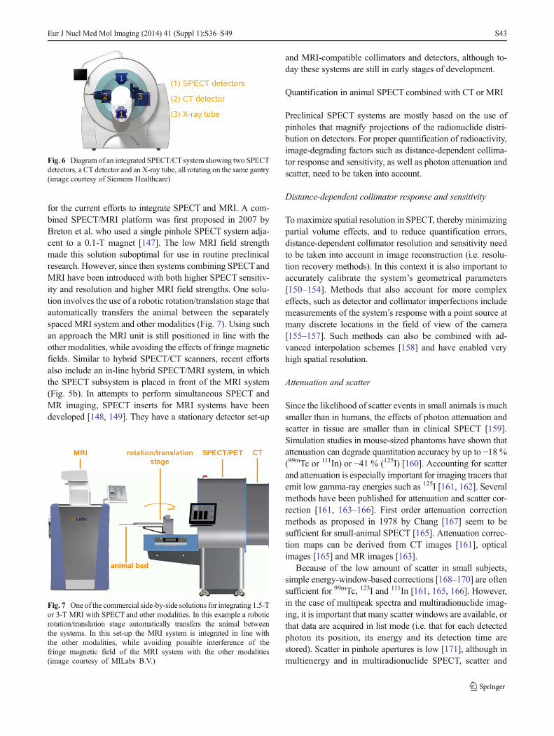
for the current efforts to integrate SPECT and MRI. A com-bined SPECT/MRI platform was first proposed in 2007 byBreton et al. who used a single pinhole SPECT system adja-cent to a 0.1-T magnet [147]. The low MRI field strengthmade this solution suboptimal for use in routine preclinicalresearch. However, since then systems combining SPECTandMRI have been introduced with both higher SPECT sensitiv-ity and resolution and higher MRI field strengths. One solu-tion involves the use of a robotic rotation/translation stage thatautomatically transfers the animal between the separatelyspaced MRI system and other modalities (Fig. 7). Using suchan approach the MRI unit is still positioned in line with theother modalities, while avoiding the effects of fringe magneticfields. Similar to hybrid SPECT/CT scanners, recent effortsalso include an in-line hybrid SPECT/MRI system, in whichthe SPECT subsystem is placed in front of the MRI system(Fig. 5b). In attempts to perform simultaneous SPECT andMR imaging, SPECT inserts for MRI systems have beendeveloped [148, 149]. They have a stationary detector set-up
and MRI-compatible collimators and detectors, although to-day these systems are still in early stages of development.
Quantification in animal SPECT combined with CT or MRI
Preclinical SPECT systems are mostly based on the use ofpinholes that magnify projections of the radionuclide distri-bution on detectors. For proper quantification of radioactivity,image-degrading factors such as distance-dependent collima-tor response and sensitivity, as well as photon attenuation andscatter, need to be taken into account.
Distance-dependent collimator response and sensitivity
Tomaximize spatial resolution in SPECT, thereby minimizingpartial volume effects, and to reduce quantification errors,distance-dependent collimator resolution and sensitivity needto be taken into account in image reconstruction (i.e. resolu-tion recovery methods). In this context it is also important toaccurately calibrate the system’s geometrical parameters[150–154]. Methods that also account for more complexeffects, such as detector and collimator imperfections includemeasurements of the system’s response with a point source atmany discrete locations in the field of view of the camera[155–157]. Such methods can also be combined with ad-vanced interpolation schemes [158] and have enabled veryhigh spatial resolution.
Attenuation and scatter
Since the likelihood of scatter events in small animals is muchsmaller than in humans, the effects of photon attenuation andscatter in tissue are smaller than in clinical SPECT [159].Simulation studies in mouse-sized phantoms have shown thatattenuation can degrade quantitation accuracy by up to −18 %(99mTc or 111In) or −41 % (125I) [160]. Accounting for scatterand attenuation is especially important for imaging tracers thatemit low gamma-ray energies such as 125I [161, 162]. Severalmethods have been published for attenuation and scatter cor-rection [161, 163–166]. First order attenuation correctionmethods as proposed in 1978 by Chang [167] seem to besufficient for small-animal SPECT [165]. Attenuation correc-tion maps can be derived from CT images [161], opticalimages [165] and MR images [163].
Because of the low amount of scatter in small subjects,simple energy-window-based corrections [168–170] are oftensufficient for 99mTc, 123I and 111In [161, 165, 166]. However,in the case of multipeak spectra and multiradionuclide imag-ing, it is important that many scatter windows are available, orthat data are acquired in list mode (i.e. that for each detectedphoton its position, its energy and its detection time arestored). Scatter in pinhole apertures is low [171], although inmultienergy and in multiradionuclide SPECT, scatter and
Fig. 6 Diagram of an integrated SPECT/CTsystem showing two SPECTdetectors, a CT detector and an X-ray tube, all rotating on the same gantry(image courtesy of Siemens Healthcare)
Fig. 7 One of the commercial side-by-side solutions for integrating 1.5-Tor 3-T MRI with SPECT and other modalities. In this example a roboticrotation/translation stage automatically transfers the animal betweenthe systems. In this set-up the MRI system is integrated in line withthe other modalities, while avoiding possible interference of thefringe magnetic field of the MRI system with the other modalities(image courtesy of MILabs B.V.)
Eur J Nucl Med Mol Imaging (2014) 41 (Suppl 1):S36–S49 S43

photon penetration in the collimator can be a significant issue,e.g. with a combination of SPECT and PET tracers used on aSPECT camera, although in such a case excellent quantitativeimages have been recently obtained using a dedicated high-energy (clustered-)pinhole collimator and window-based scat-ter corrections [14].
Concluding remarks and future perspectives
Recent advances in small-animal SPECT/CT and SPECT/MRI devices, radiochemistry, probe development, targetfinding and suitable animal models have provided moreadvanced and increased applications of these combinedimaging strategies.
In most preclinical SPECT imaging studies to date, CT orMRI merely fulfil a supportive role to provide anatomicalreference and in some cases attenuation correction. In smalllaboratory animals, acquisition of detailed anatomical infor-mation, performance of dynamic scans or functional imagingwith CT has specific challenges compared to imaging inhumans. To reach diagnostic image quality high CT radiationdoses and/or large volumes of contrast agent are necessary.These aspects are not compatible with longitudinal studies,since they may severely affect the wellbeing of animals. Newdevelopments in small-animal CT [172–177] and the use ofnew contrast agents for CT should provide better image qual-ity at lower radiation doses and/or with lower volumes ofcontrast agent.
MRI offers detailed anatomical imaging of soft tissuescompatible with longitudinal studies. Separately spacedSPECT and MRI systems can pose challenges with respectto image registration, imaging times and anaesthesia times; theeffects of these issues can potentially influence study results.On the other hand, physical integration of SPECT and MRItechnologies is hampered by various incompatibilities; thecomponents and working mechanisms of the separate modal-ities currently degrade the other’s performance.
A major benefit of higher magnetic field strengths is thatmore signal is provided which can be used to shorten acqui-sition times, but higher magnetic field strengths do not alwaysguarantee higher image quality per se. Currently, most high-field magnets are cooled with cryogenic liquids. One of themain drivers towards small MRI systems is the developmentof cryogen-free magnets or systems using helium gas insteadof liquid helium. This development could lead to significantreductions in the size, site requirements and cost of high-fieldMRI systems. Today, such helium gas-based commercialMRIsystems are limited to 3 T.
While not covered in detail in this review, an extremelyimportant aspect to consider during imaging is animal welfare.Animal handling and especially anaesthesia is demanding tothe animal and can severely affect the outcome of imaging
studies [35, 178–180]. Also issues regarding radiation doseswill have to be taken into account [142, 143, 181, 182].
Taking these issues into account, further advances in tech-nology and chemistry, for example the development of newimaging procedures and promising new radiopharmaceuticals,for imaging molecular targets as well as (patho)physiologicalprocesses in vivo, the step from bench to bedside mightbecome more successful and shorter; e.g. accelerating theintroduction of new imaging procedures and promising newradiopharmaceuticals into clinical practice.
Disclosure Freek J. Beekman is the founder, a part-time officer andshareholder of MILabs B.V.
Open Access This article is distributed under the terms of the CreativeCommons Attribution License which permits any use, distribution, andreproduction in any medium, provided the original author(s) and thesource are credited.
References
1. Deleye S, Van Holen R, Verhaeghe J, Vandenberghe S, StroobantsS, Staelens S. Performance evaluation of small-animal multipinholemuSPECT scanners for mouse imaging. Eur J Nucl Med MolImaging. 2013;40:744–58. doi:10.1007/s00259-012-2326-2.
2. Schambach SJ, Bag S, Schilling L, Groden C, Brockmann MA.Application of micro-CT in small animal imaging. Methods.2010;50:2–13. doi:10.1016/j.ymeth.2009.08.007.
3. Vaissier PE, Goorden MC, Vastenhouw B, van der Have F,Ramakers RM, Beekman FJ. Fast spiral SPECT with stationarygamma-cameras and focusing pinholes. J Nucl Med. 2012;53:1292–9. doi:10.2967/jnumed.111.101899.
4. Greco A, Fiumara G, Gargiulo S, Gramanzini M, Brunetti A,Cuocolo A. High-resolution positron emission tomography/computed tomography imaging of the mouse heart. Exp Physiol.2013;98:645–51. doi:10.1113/expphysiol.2012.068643.
5. Sauter AW, Wehrl HF, Kolb A, Judenhofer MS, Pichler BJ.Combined PET/MRI: one step further in multimodality imaging.TrendsMolMed. 2010;16:508–15. doi:10.1016/j.molmed.2010.08.003.
6. Massoud TF, Gambhir SS. Molecular imaging in living subjects:seeing fundamental biological processes in a new light. Genes Dev.2003;17:545–80.
7. Pichler BJ, Wehrl HF, JudenhoferMS. Latest advances in molecularimaging instrumentation. J Nucl Med. 2008;49 Suppl 2:5S–23.
8. Weissleder R, Mahmood U. Molecular imaging. Radiology.2001;219:316–33.
9. Bernsen MR, Ruggiero A, van Straten M, Kotek G, Haeck JC,Wielopolski PA, et al. Computed tomography and magnetic reso-nance imaging. Recent Results Cancer Res. 2013;187:3–63. doi:10.1007/978-3-642-10853-2_1.
10. Ma KH, HuangWS, Chen CH, Lin SZ, Wey SP, Ting G, et al. DualSPECT of dopamine system using [99mTc]TRODAT-1 and[123I]IBZM in normal and 6-OHDA-lesioned formosan rock mon-keys. Nucl Med Biol. 2002;29:561–7.
11. Nakazawa A, Ikeda K, Ito Y, Iwase M, Sato K, Ueda R, et al.Usefulness of dual 67Ga and 99mTc-sestamibi single-photon-emission CT scanning in the diagnosis of cardiac sarcoidosis.Chest. 2004;126:1372–6. doi:10.1378/chest.126.4.1372.
S44 Eur J Nucl Med Mol Imaging (2014) 41 (Suppl 1):S36–S49

12. Sanchez-Crespo A, Petersson J, Nyren S, Mure M, Glenny RW,Thorell JO, et al. A novel quantitative dual-isotope method forsimultaneous ventilation and perfusion lung SPET. Eur J NuclMed Mol Imaging. 2002;29:863–75. doi:10.1007/s00259-002-0803-8.
13. Weinmann P, FaraggiM,Moretti JL, Hannequin P. Clinical validationof simultaneous dual-isotope myocardial scintigraphy. Eur J NuclMedMol Imaging. 2003;30:25–31. doi:10.1007/s00259-002-0995-y.
14. Goorden MC, van der Have F, Kreuger R, Ramakers RM,Vastenhouw B, Burbach JP, et al. VECTor: a preclinical imagingsystem for simultaneous submillimeter SPECT and PET. J NuclMed. 2013;54:306–12. doi:10.2967/jnumed.112.109538.
15. Melis M, Valkema R, Krenning EP, de Jong M. Reduction of renaluptake of radiolabeled octreotate by amifostine coadministration. JNucl Med. 2012;53:749–53. doi:10.2967/jnumed.111.098665.
16. Beekman FJ, van der Have F, Vastenhouw B, van der Linden AJ,van Rijk PP, Burbach JP, et al. U-SPECT-I: a novel system forsubmillimeter-resolution tomography with radiolabeled moleculesin mice. J Nucl Med. 2005;46:1194–200.
17. Schramm NU, Ebel G, Engeland U, Schurrat T, Behe M, Behr TM.High-resolution SPECT using multipinhole collimation. IEEETrans Nucl Sci. 2003;50:315–20. doi:10.1109/tns.2003.812437.
18. Ivashchenko O, van der Have F, Villena J, Beekman FJ. Quartermillimeter resolution pre-clinical SPECTwith quarter-mm pinholes.Eur J Nucl Med Mol Imaging. 2014. In press.
19. de Kemp RA, Epstein FH, Catana C, Tsui BM, Ritman EL. Small-animal molecular imaging methods. J Nucl Med. 2010;51:18s–32.doi:10.2967/jnumed.109.068148.
20. Golestani R,WuC, Tio RA, Zeebregts CJ, PetrovAD, Beekman FJ,et al. Small-animal SPECT and SPECT/CT: application in cardio-vascular research. Eur J Nucl MedMol Imaging. 2010;37:1766–77.doi:10.1007/s00259-009-1321-8.
21. Studwell AJ, Kotton DN. A shift from cell cultures to creatures:in vivo imaging of small animals in experimental regenerativemedicine. Mol Ther. 2011;19:1933–41. doi:10.1038/mt.2011.194.
22. Kielland A, Carlsen H. Molecular imaging of transcriptional regu-lation during inflammation. J Inflamm (Lond). 2010;7:20. doi:10.1186/1476-9255-7-20.
23. Hanahan D, Weinberg RA. The hallmarks of cancer. Cell.2000;100:57–70.
24. Hanahan D, Weinberg RA. Hallmarks of cancer: the next genera-tion. Cell. 2011;144:646–74. doi:10.1016/j.cell.2011.02.013.
25. Cavallo F, De Giovanni C, Nanni P, Forni G, Lollini PL. 2011: theimmune hallmarks of cancer. Cancer Immunol Immunother.2011;60:319–26.
26. Ruoslahti E, Bhatia SN, Sailor MJ. Targeting of drugs and nano-particles to tumors. J Cell Biol. 2010;188:759–68.
27. Movahedi K, Schoonooghe S, Laoui D, Houbracken I, Waelput W,Breckpot K, et al. Nanobody-based targeting of the macrophagemannose receptor for effective in vivo imaging of tumor-associatedmacrophages. Cancer Res. 2012;72:4165–77. doi:10.1158/0008-5472.can-11-2994.
28. Muller C. Folate based radiopharmaceuticals for imaging andtherapy of cancer and inflammation. Curr Pharm Des. 2012;18:1058–83.
29. Müller C, Schibli R. Single photon emission computed tomographytracer. Recent Results Cancer Res. 2013;187:65–105.
30. Vaupel P. Pathophysiology of solid tumors. In: Molls M, Vaupel P,Nieder C, Anscher MS, editors. The impact of tumor biology oncancer treatment and multidisciplinary strategies. Heidelberg:Springer; 2009. p. 51–92.
31. Bredow S, Lewin M, Hofmann B, Marecos E, Weissleder R.Imaging of tumour neovasculature by targeting the TGF-beta bind-ing receptor endoglin. Eur J Cancer. 2000;36:675–81.
32. Tsiapa I, Loudos G, Varvarigou A, Fragogeorgi E, Psimadas D,Tsotakos T, et al. Biological evaluation of an ornithine-modified
(99m)Tc-labeled RGD peptide as an angiogenesis imaging agent.Nucl Med Biol. 2013;40:262–72. doi:10.1016/j.nucmedbio.2012.10.015.
33. Zhou Y, Chakraborty S, Liu S. Radiolabeled cyclic RGD peptides asradiotracers for imaging tumors and thrombosis by SPECT.Theranostics. 2011;1:58–82.
34. Fujii H, Yamaguchi M, Inoue K, Mutou Y, Ueda M, Saji H, et al. Invivo visualization of heterogeneous intratumoral distribution ofhypoxia-inducible factor-1alpha activity by the fusion of high-resolution SPECT and morphological imaging tests. J BiomedBiotechnol. 2012;2012:262741. doi:10.1155/2012/262741.
35. Kersemans V, Cornelissen B, Hueting R, Tredwell M, Hussien K,Allen PD, et al. Hypoxia imaging using PETand SPECT: the effectsof anesthetic and carrier gas on [Cu]-ATSM, [Tc]-HL91 and [F]-FMISO tumor hypoxia accumulation. PLoS One. 2011;6:e25911.doi:10.1371/journal.pone.0025911.
36. Kimura S, Umeda IO, Moriyama N, Fujii H. Synthesis and evalu-ation of a novel (99m)Tc-labeled bioreductive probe for tumorhypoxia imaging. Bioorg Med Chem Lett. 2011;21:7359–62. doi:10.1016/j.bmcl.2011.10.022.
37. Umeda IO, Tani K, Tsuda K, Kobayashi M, Ogata M, Kimura S,et al. High resolution SPECT imaging for visualization ofintratumoral heterogeneity using a SPECT/CT scanner dedicatedfor small animal imaging. Ann Nucl Med. 2012;26:67–76. doi:10.1007/s12149-011-0542-7.
38. Bauwens M, De Saint-Hubert M, Cleynhens J, Brams L, Devos E,Mottaghy FM, et al. Radioiodinated phenylalkyl malonic acidderivatives as pH-sensitive SPECT tracers. PLoS One. 2012;7:e38428. doi:10.1371/journal.pone.0038428.
39. Macholl S, Morrison MS, Iveson P, Arbo BE, Andreev OA,Reshetnyak YK, et al. In vivo pH imaging with (99m)Tc-pHLIP.Mol ImagingBiol. 2012;14:725–34. doi:10.1007/s11307-012-0549-z.
40. Weerakkody D, Moshnikova A, Thakur MS, Moshnikova V,Daniels J, Engelman DM, et al. Family of pH (low) insertionpeptides for tumor targeting. Proc Natl Acad Sci U S A.2013;110:5834–9. doi:10.1073/pnas.1303708110.
41. von Forstner C, Zuhayra M, Ammerpohl O, Zhao Y, Tiwari S,Jansen O, et al. Expression of L amino acid transport system 1and analysis of iodine-123-methyltyrosine tumor uptake in a pan-creatic xenotransplantation model using fused high-resolution-micro-SPECT-MRI. Hepatobiliary Pancreat Dis Int. 2011;10:30–7.
42. Kondo N, Temma T, Shimizu Y, Watanabe H, Higano K, Takagi Y,et al. Miniaturized antibodies for imaging membrane type-1 matrixmetalloproteinase in cancers. Cancer Sci. 2013;104:495–501. doi:10.1111/cas.12102.
43. LeBeau AM, Duriseti S, Murphy ST, Pepin F, Hann B, Gray JW,et al. Targeting uPAR with antagonistic recombinant human anti-bodies in aggressive breast cancer. Cancer Res. 2013;73:2070–81.doi:10.1158/0008-5472.can-12-3526.
44. SchotteliusM,Wester HJ. Molecular imaging targeting peptide recep-tors. Methods. 2009;48:161–77. doi:10.1016/j.ymeth.2009.03.012.
45. Heskamp S, van Laarhoven HW, Molkenboer-Kuenen JD,Bouwman WH, van der Graaf WT, Oyen WJ, et al. Optimizationof IGF-1R SPECT/CT imaging using (111)In-labeled F(ab′)(2) andFab fragments of the monoclonal antibody R1507. Mol Pharm.2012;9:2314–21. doi:10.1021/mp300232n.
46. Mariani G, Di Sacco S, Volterrani D, Di Luca L, Buralli S, DiStefano R, et al. Tumor targeting by intra-arterial infusion of 5-[123I]iodo-2′-deoxyuridine in patients with liver metastases fromcolorectal cancer. J Nucl Med. 1996;37:22s–5.
47. Aloj L, Aurilio M, Rinaldi V, D’Ambrosio L, Tesauro D, Peitl PK,et al. Comparison of the binding and internalization properties of 12DOTA-coupled and 111In-labelled CCK2/gastrin receptor bindingpeptides: a collaborative project under COSTAction BM0607. EurJ Nucl Med Mol Imaging. 2011;38:1417–25. doi:10.1007/s00259-011-1816-y.
Eur J Nucl Med Mol Imaging (2014) 41 (Suppl 1):S36–S49 S45

48. Forrer F, Valkema R, Bernard B, Schramm NU, Hoppin JW,Rolleman E, et al. In vivo radionuclide uptake quantification usinga multi-pinhole SPECT system to predict renal function in smallanimals. Eur J Nucl Med Mol Imaging. 2006;33:1214–7. doi:10.1007/s00259-006-0178-3.
49. Abiraj K, Mansi R, Tamma ML, Fani M, Forrer F, Nicolas G, et al.Bombesin antagonist-based radioligands for translational nuclearimaging of gastrin-releasing peptide receptor-positive tumors. JNucl Med. 2011;52:1970–8.
50. Fani M, Braun F, Waser B, Beetschen K, Cescato R, Erchegyi J,et al. Unexpected sensitivity of sst2 antagonists to N-terminalradiometal modifications. J Nucl Med. 2012;53:1481–9. doi:10.2967/jnumed.112.102764.
51. Hwang AB, Hasegawa BH. Attenuation correction for small ani-mal SPECT imaging using x-ray CT data. Med Phys. 2005;32:2799–804.
52. Williams SP. Tissue distribution studies of protein therapeuticsusing molecular probes: molecular imaging. AAPS J. 2012;14:389–99. doi:10.1208/s12248-012-9348-3.
53. van Essen M, Krenning EP, Kam BL, de Jong M, Valkema R,Kwekkeboom DJ. Peptide-receptor radionuclide therapy for endo-crine tumors. Nat Rev Endocrinol. 2009;5:382–93. doi:10.1038/nrendo.2009.105.
54. Muller C, Vlahov IR, Santhapuram HK, Leamon CP, Schibli R.Tumor targeting using 67Ga-DOTA-Bz-folate – investigations ofmethods to improve the tissue distribution of radiofolates. NuclMedBiol. 2011;38:715–23. doi:10.1016/j.nucmedbio.2010.12.013.
55. Joosten L, Laverman P, Boerman OC, Roosenburg S, Eek A, RutjesFP, et al. In vitro and in vivo characterization of three 68Ga- and111In-labeled peptides for cholecystokinin receptor imaging. MolImaging. 2012;11:401–7.
56. Nayak TK, Hathaway HJ, Ramesh C, Arterburn JB, Dai D, SklarLA, et al. Preclinical development of a neutral, estrogen receptor-targeted, tridentate 99mTc(I)-estradiol-pyridin-2-yl hydrazine deriv-ative for imaging of breast and endometrial cancers. J Nucl Med.2008;49:978–86. doi:10.2967/jnumed.107.048546.
57. Brinkhuis RP, Stojanov K, Laverman P, Eilander J, Zuhorn IS,Rutjes FP, et al. Size dependent biodistribution and SPECT imagingof 111In-labeled polymersomes. Bioconjug Chem. 2012;23:958–65. doi:10.1021/bc200578s.
58. Chang YJ, Chang CH, Yu CY, Chang TJ, Chen LC, Chen MH, et al.Therapeutic efficacy and microSPECT/CT imaging of 188Re-DXR-liposome in a C26 murine colon carcinoma solid tumor model. NuclMed Biol. 2010;37:95–104. doi:10.1016/j.nucmedbio.2009.08.006.
59. de Smet M, Langereis S, van den Bosch S, Bitter K, Hijnen NM,Heijman E, et al. SPECT/CT imaging of temperature-sensitiveliposomes for MR-image guided drug delivery with high intensityfocused ultrasound. J Control Release. 2013;169:82–90. doi:10.1016/j.jconrel.2013.04.005.
60. Huang FY, Lee TW, Kao CH, Chang CH, Zhang X, Lee WY, et al.Imaging, autoradiography, and biodistribution of 188Re-labeledPEGylated nanoliposome in orthotopic glioma bearing rat model.Cancer Biother Radiopharm. 2011;26:717–25. doi:10.1089/cbr.2011.1052.
61. Tsai CC, Chang CH, Chen LC, Chang YJ, Lan KL, Wu YH, et al.Biodistribution and pharmacokinetics of 188Re-liposomes and theircomparative therapeutic efficacy with 5-fluorouracil in C26 colonicperitoneal carcinomatosis mice. Int J Nanomedicine. 2011;6:2607–19. doi:10.2147/ijn.s23834.
62. Bult W, Kroeze SG, Elschot M, Seevinck PR, Beekman FJ, de JongHW, et al. Intratumoral administration of Holmium-166 acetylaceto-natemicrospheres: antitumor efficacy and feasibility of multimodalityimaging in renal cancer. PLoS One. 2013;8:e52178. doi:10.1371/journal.pone.0052178.
63. Madru R, Kjellman P, Olsson F, Wingårdh K, Ingvar C, Ståhlberg F,et al. 99mTc-labeled superparamagnetic iron oxide nanoparticles for
multimodality SPECT/MRI of sentinel lymph nodes. J Nucl Med.2012;53:459–63.
64. Ali MM, Janic B, Babajani-Feremi A, Varma NR, Iskander AS,Anagli J, et al. Changes in vascular permeability and expression ofdifferent angiogenic factors following anti-angiogenic treatment inrat glioma. PLoS One. 2010;5:e8727. doi:10.1371/journal.pone.0008727.
65. De Saint-Hubert M, Wang H, Devos E, Vunckx K, Zhou L,Reutelingsperger C, et al. Preclinical imaging of therapy responseusing metabolic and apoptosis molecular imaging. Mol ImagingBiol. 2011;13:995–1002. doi:10.1007/s11307-010-0412-z.
66. Vangestel C, Van de Wiele C, Mees G, Mertens K, Staelens S,Reutelingsperger C, et al. Single-photon emission computed tomo-graphic imaging of the early time course of therapy-induced celldeath using technetium 99m tricarbonyl His-annexin A5 in a colo-rectal cancer xenograft model. Mol Imaging. 2012;11:135–47.
67. Bol K, Haeck JC, Groen HC, NiessenWJ, BernsenMR, de JongM,Veenland JF. Can DCE-MRI Explain the Heterogeneity inRadiopeptide Uptake Imaged by SPECT in a PancreaticNeuroendocrine Tumor Model? PLoS One. 2013;8(10):e77076.doi:10.1371/journal.pone.0077076.
68. Bar-ShalomR, YefremovN, Guralnik L, Keidar Z, Engel A, NiteckiS, et al. SPECT/CT using 67Ga and 111In-labeled leukocyte scin-tigraphy for diagnosis of infection. J Nucl Med. 2006;47:587–94.
69. Hong H, Yang Y, Zhang Y, Cai W. Non-invasive cell tracking incancer and cancer therapy. Curr TopMed Chem. 2010;10:1237–48.
70. Ruggiero A, Thorek DL, Guenoun J, Krestin GP, Bernsen MR. Celltracking in cardiac repair: what to image and how to image. EurRadiol. 2012;22:189–204. doi:10.1007/s00330-011-2190-7.
71. Welling MM, Duijvestein M, Signore A, van der Weerd L. Invivo biodistribution of stem cells using molecular nuclear med-icine imaging. J Cell Physiol. 2011;226:1444–52. doi:10.1002/jcp.22539.
72. Bernsen MR, Moelker AD, Wielopolski PA, van Tiel ST, KrestinGP. Labelling of mammalian cells for visualisation by MRI. EurRadiol. 2010;20:255–74. doi:10.1007/s00330-009-1540-1.
73. de Vries EF, Buursma AR, Hospers GA, Mulder NH, Vaalburg W.Scintigraphic imaging of HSVtk gene therapy. Curr Pharm Des.2002;8:1435–50.
74. Penheiter AR, Russell SJ, Carlson SK. The sodium iodidesymporter (NIS) as an imaging reporter for gene, viral, and cell-based therapies. Curr Gene Ther. 2012;12:33–47.
75. Doubrovin MM, Doubrovina ES, Zanzonico P, Sadelain M, LarsonSM, O’Reilly RJ. In vivo imaging and quantitation of adoptivelytransferred human antigen-specific T cells transduced to express ahuman norepinephrine transporter gene. Cancer Res. 2007;67:11959–69. doi:10.1158/0008-5472.can-07-1250.
76. Templin C, Zweigerdt R, Schwanke K, Olmer R, Ghadri JR, EmmertMY, et al. Transplantation and tracking of human-induced pluripotentstem cells in a pig model of myocardial infarction: assessment of cellsurvival, engraftment, and distribution by hybrid single photon emis-sion computed tomography/computed tomography of sodium iodidesymporter transgene expression. Circulation. 2012;126:430–9. doi:10.1161/circulationaha.111.087684.
77. Deng WP, Wu CC, Lee CC, Yang WK, Wang HE, Liu RS, et al.Serial in vivo imaging of the lung metastases model and gene therapyusing HSV1-tk and ganciclovir. J Nucl Med. 2006;47:877–84.
78. Inubushi M, Jin YN, Murai C, Hata H, Kitagawa Y, Saga T. Single-photon emission computed tomography of spontaneous liver me-tastasis from orthotopically implanted human colon cancer cell linestably expressing human sodium/iodide symporter reporter gene.EJNMMI Res. 2012;2:46. doi:10.1186/2191-219x-2-46.
79. Marsee DK, Shen DH, MacDonald LR, Vadysirisack DD, Lin X,Hinkle G, et al. Imaging of metastatic pulmonary tumors followingNIS gene transfer using single photon emission computed tomogra-phy. Cancer Gene Ther. 2004;11:121–7. doi:10.1038/sj.cgt.7700661.
S46 Eur J Nucl Med Mol Imaging (2014) 41 (Suppl 1):S36–S49
80. Merron A, Baril P, Martin-Duque P, de la Vieja A, Tran L, Briat A,et al. Assessment of the Na/I symporter as a reporter gene tovisualize oncolytic adenovirus propagation in peritoneal tumours.Eur J Nucl Med Mol Imaging. 2010;37:1377–85. doi:10.1007/s00259-009-1379-3.
81. Moroz MA, Serganova I, Zanzonico P, Ageyeva L, Beresten T,Dyomina E, et al. Imaging hNET reporter gene expression with124I-MIBG. J Nucl Med. 2007;48:827–36. doi:10.2967/jnumed.106.037812.
82. Ambrosini V, Fani M, Fanti S, Forrer F, Maecke HR. Radiopeptideimaging and therapy in Europe. J Nucl Med. 2011;52 Suppl 2:42S–55.
83. Reubi JC. The role of peptides and their receptors as tumor markers.Endocrinol Metab Clin N Am. 1993;22:917–39.
84. Olafsen T, Wu AM. Antibody vectors for imaging. Semin NuclMed. 2010;40:167–81.
85. Devoogdt N, Xavier C, Hernot S, Vaneycken I, D’Huyvetter M, DeVos J, et al. Molecular imaging using Nanobodies: a case study.Methods Mol Biol. 2012;911:559–67.
86. Goldenberg DM, Rossi EA, Sharkey RM,McBrideWJ, Chang CH.Multifunctional antibodies by the Dock-and-Lock method for im-proved cancer imaging and therapy by pretargeting. J Nucl Med.2008;49:158–63.
87. Lofblom J, Feldwisch J, TolmachevV, Carlsson J, Stahl S, Frejd FY.Affibody molecules: engineered proteins for therapeutic, diag-nostic and biotechnological applications. FEBS Lett. 2010;584:2670–80.
88. Jokerst JV, Gambhir SS. Molecular imaging with theranostic nano-particles. Acc Chem Res. 2011;44:1050–60.
89. de Jong M, Breeman WA, Kwekkeboom DJ, Valkema R, KrenningEP. Tumor imaging and therapy using radiolabeled somatostatinanalogues. Acc Chem Res. 2009;42:873–80. doi:10.1021/ar800188e.
90. Kish SJ, Shannak K, Hornykiewicz O. Uneven pattern of dopamineloss in the striatum of patients with idiopathic Parkinson’s disease:pathophysiologic and clinical implications. N Engl J Med.1988;318:876–80. doi:10.1056/nejm198804073181402.
91. Booij J, Tissingh G, Boer GJ, Speelman JD, Stoof JC, Janssen AG,et al. [123I]FP-CIT SPECT shows a pronounced decline of striataldopamine transporter labelling in early and advanced Parkinson’sdisease. J Neurol Neurosurg Psychiatry. 1997;62:133–40.
92. Booij J, Tissingh G,Winogrodzka A, van Royen EA. Imaging of thedopaminergic neurotransmission system using single-photon emis-sion tomography and positron emission tomography in patients withparkinsonism. Eur J Nucl Med. 1999;26:171–82.
93. Catafau AM, Tolosa E. Impact of dopamine transporter SPECTusing 123I-Ioflupane on diagnosis and management of patients withclinically uncertain Parkinsonian syndromes.Mov Disord. 2004;19:1175–82. doi:10.1002/mds.20112.
94. Ponsen MM, Stoffers D, Booij J, van Eck-Smit BL, Wolters E,Berendse HW. Idiopathic hyposmia as a preclinical sign ofParkinson’s disease. Ann Neurol. 2004;56:173–81. doi:10.1002/ana.20160.
95. Ziebell M, Andersen BB, Thomsen G, Pinborg LH, Karlsborg M,Hasselbalch SG, et al. Predictive value of dopamine transporterSPECT imaging with [123I]PE2I in patients with subtle parkinso-nian symptoms. Eur J Nucl Med Mol Imaging. 2012;39:242–50.doi:10.1007/s00259-011-1976-9.
96. Acton PD, Hou C, Kung MP, Plossl K, Keeney CL, Kung HF.Occupancy of dopamine D2 receptors in the mouse brain measuredusing ultra-high-resolution single-photon emission tomography and[123]IBF. Eur J Nucl Med Mol Imaging. 2002;29:1507–15.
97. Habraken JB, de Bruin K, Shehata M, Booij J, Bennink R, van EckSmit BL, et al. Evaluation of high-resolution pinhole SPECTusing asmall rotating animal. J Nucl Med. 2001;42:1863–9.
98. Booij J, de Bruin K, Habraken JB, Voorn P. Imaging of dopaminetransporters in rats using high-resolution pinhole single-photon
emission tomography. Eur J Nucl Med Mol Imaging. 2002;29:1221–4.
99. Scherfler C, Donnemiller E, SchockeM, Dierkes K, Decristoforo C,Oberladstatter M, et al. Evaluation of striatal dopamine transporterfunction in rats by in vivo beta-[123I]CIT pinhole SPECT.Neuroimage. 2002;17:128–41.
100. Laruelle M, Baldwin RM, Malison RT, Zea-Ponce Y, Zoghbi SS,al-Tikriti MS, et al. SPECT imaging of dopamine and serotonintransporters with [123I]beta-CIT: pharmacological characteriza-tion of brain uptake in nonhuman primates. Synapse. 1993;13:295–309.
101. Booij J, de Bruin K, de Win MM, Lavini C, den Heeten GJ,Habraken JB. Imaging of striatal dopamine transporters in rat brainwith single pinhole SPECTand co-aligned MRI is highly reproduc-ible. Nucl Med Biol. 2003;30:643–9.
102. Andringa G, Drukarch B, Bol JG, de Bruin K, Sorman K, HabrakenJB, et al. Pinhole SPECT imaging of dopamine transporters corre-lates with dopamine transporter immunohistochemical analysis inthe MPTP mouse model of Parkinson’s disease. Neuroimage.2005;26:1150–8. doi:10.1016/j.neuroimage.2005.03.034.
103. Gleave JA, Farncombe TH, Saab C, Doering LC. Correlative singlephoton emission computed tomography imaging of [123I]altropanebinding in the rat model of Parkinson’s. Nucl Med Biol. 2011;38:741–9. doi:10.1016/j.nucmedbio.2010.12.006.
104. Alvarez-Fischer D, Blessmann G, Trosowski C, Behe M, SchurratT, Hartmann A, et al. Quantitative [(123)I]FP-CIT pinhole SPECTimaging predicts striatal dopamine levels, but not number ofnigral neurons in different mouse models of Parkinson’s dis-ease. Neuroimage. 2007;38:5–12. doi:10.1016/j.neuroimage.2007.05.056.
105. Depboylu C, Maurer L, Matusch A, Hermanns G, Windolph A,Behe M, et al. Effect of long-term treatment with pramipexole orlevodopa on presynaptic markers assessed by longitudinal[123I]FP-CIT SPECT and histochemistry. Neuroimage. 2013;79:191–200. doi:10.1016/j.neuroimage.2013.04.076.
106. Lee JD, Chu YH, Chen CW, Lin KJ. Multi-image registration forevaluating the 99mTc-TRODAT-1 of Parkinson’s rat model. ConfProc IEEE Eng Med Biol Soc. 2009;2009:5801–4. doi:10.1109/iembs.2009.5335192.
107. Acton PD, Choi SR, Plossl K, Kung HF. Quantification of dopa-mine transporters in the mouse brain using ultra-high resolutionsingle-photon emission tomography. Eur J Nucl Med MolImaging. 2002;29:691–8. doi:10.1007/s00259-002-0776-7.
108. Nikolaus S, Larisch R, Wirrwar A, Jamdjeu-Noune M, Antke C,Beu M, et al. [123I]Iodobenzamide binding to the rat dopamine D2receptor in competition with haloperidol and endogenous dopa-mine – an in vivo imaging study with a dedicated small animalSPECT. Eur J Nucl Med Mol Imaging. 2005;32:1305–10. doi:10.1007/s00259-005-1839-3.
109. Scherfler C, Scholz SW, Donnemiller E, Decristoforo C,Oberladstatter M, Stefanova N, et al. Evaluation of [123I]IBZMpinhole SPECT for the detection of striatal dopamine D2 receptoravailability in rats. Neuroimage. 2005;24:822–31. doi:10.1016/j.neuroimage.2004.10.005.
110. Fisher RS, Levine MS, Sibley DR, Ariano MA. D2 dopaminereceptor protein location: Golgi impregnation-gold toned and ultra-structural analysis of the rat neostriatum. J Neurosci Res. 1994;38:551–64. doi:10.1002/jnr.490380508.
111. Herholz K, Ebmeier K. Clinical amyloid imaging in Alzheimer’sdisease. Lancet Neurol. 2011;10:667–70. doi:10.1016/s1474-4422(11)70123-5.
112. Svedberg MM, Rahman O, Hall H. Preclinical studies of potentialamyloid binding PET/SPECT ligands in Alzheimer’s disease. NuclMed Biol. 2012;39:484–501. doi:10.1016/j.nucmedbio.2011.10.007.
113. Teng E, Kepe V, Frautschy SA, Liu J, Satyamurthy N, Yang F, et al.[F-18]FDDNP microPET imaging correlates with brain Abeta
Eur J Nucl Med Mol Imaging (2014) 41 (Suppl 1):S36–S49 S47

burden in a transgenic rat model of Alzheimer disease: effects ofaging, in vivo blockade, and anti-Abeta antibody treatment.Neurobiol Dis. 2011;43:565–75. doi:10.1016/j.nbd.2011.05.003.
114. Kung MP, Hou C, Zhuang ZP, Cross AJ, Maier DL, Kung HF.Characterization of IMPY as a potential imaging agent for beta-amyloid plaques in double transgenic PSAPPmice. Eur J Nucl MedMol Imaging. 2004;31:1136–45. doi:10.1007/s00259-004-1487-z.
115. Kung MP, Hou C, Zhuang ZP, Zhang B, Skovronsky D,Trojanowski JQ, et al. IMPY: an improved thioflavin-T derivativefor in vivo labeling of beta-amyloid plaques. Brain Res. 2002;956:202–10.
116. Nader MA, Morgan D, Gage HD, Nader SH, Calhoun TL,Buchheimer N, et al. PET imaging of dopamineD2 receptors duringchronic cocaine self-administration in monkeys. Nat Neurosci.2006;9:1050–6. doi:10.1038/nn1737.
117. Crunelle CL, de Wit TC, de Bruin K, Ramakers RM, van der HaveF, Beekman FJ, et al. Varenicline increases in vivo striatal dopamineD2/3 receptor binding: an ultra-high-resolution pinhole [123I]IBZMSPECTstudy in rats. Nucl Med Biol. 2012;39:640–4. doi:10.1016/j.nucmedbio.2011.11.006.
118. Crunelle CL, van de Giessen E, Schulz S, Vanderschuren LJ, deBruin K, van den Brink W, et al. Cannabinoid-1 receptor antagonistrimonabant (SR141716) increases striatal dopamine D2 receptoravailability. Addict Biol. 2013;18:908–11. doi:10.1111/j.1369-1600.2011.00369.x.
119. Crunelle CL, Miller ML, de Bruin K, van den Brink W, Booij J.Varenicline increases striatal dopamine D(2/3) receptor binding in rats.Addict Biol. 2009;14:500–2. doi:10.1111/j.1369-1600.2009.00168.x.
120. Vastenhouw B, van der Have F, van der Linden AJ, von Oerthel L,Booij J, Burbach JP, et al. Movies of dopamine transporter occu-pancy with ultra-high resolution focusing pinhole SPECT. MolPsychiatry. 2007;12:984–7. doi:10.1038/sj.mp.4002028.
121. Huang YR, Shih JM, Chang KW, Huang C, Wu YL, Chen CC.[123I]Epidepride neuroimaging of dopamine D2/D3 receptor inchronic MK-801-induced rat schizophrenia model. Nucl MedBiol. 2012;39:826–32. doi:10.1016/j.nucmedbio.2012.01.005.
122. Laruelle M, Abi-Dargham A, van Dyck CH, Rosenblatt W, Zea-Ponce Y, Zoghbi SS, et al. SPECT imaging of striatal dopaminerelease after amphetamine challenge. J Nucl Med. 1995;36:1182–90.
123. Laruelle M, Abi-Dargham A, van Dyck CH, Gil R, D’Souza CD,Erdos J, et al. Single photon emission computerized tomographyimaging of amphetamine-induced dopamine release in drug-freeschizophrenic subjects. Proc Natl Acad Sci U S A. 1996;93:9235–40.
124. Martinez D, Narendran R, Foltin RW, Slifstein M, Hwang DR,Broft A, et al. Amphetamine-induced dopamine release: markedlyblunted in cocaine dependence and predictive of the choice to self-administer cocaine. Am J Psychiatry. 2007;164:622–9. doi:10.1176/appi.ajp.164.4.622.
125. Jongen C, de Bruin K, Beekman F, Booij J. SPECT imaging of D2dopamine receptors and endogenous dopamine release in mice. EurJ Nucl Med Mol Imaging. 2008;35:1692–8. doi:10.1007/s00259-008-0795-0.
126. Nikolaus S, Antke C, Beu M, Kley K, Wirrwar A, Huston JP, et al.Binding of [123I]iodobenzamide to the rat D2 receptor after chal-lenge with various doses of methylphenidate: an in vivo imagingstudy with dedicated small animal SPECT. Eur J Nucl Med MolImaging. 2011;38:694–701. doi:10.1007/s00259-010-1668-x.
127. Apostolova I, Wunder A, Dirnagl U, Michel R, Stemmer N, LukasM, et al. Brain perfusion SPECT in the mouse: normal patternaccording to gender and age. Neuroimage. 2012;63:1807–17. doi:10.1016/j.neuroimage.2012.08.038.
128. Zeniya T, Watabe H, Hayashi T, Ose T, Myojin K, Taguchi A, et al.Three-dimensional quantitation of regional cerebral blood flow inmice using a high-resolution pinhole SPECT system and 123I-
iodoamphetamine. Nucl Med Biol. 2011;38:1157–64. doi:10.1016/j.nucmedbio.2011.04.007.
129. Ceulemans AG, Hernot S, Zgavc T, Caveliers V, Hachimi-Idrissi S,Sarre S, et al. Serial semiquantitative imaging of brain damage usingmicro-SPECT and micro-CT after endothelin-1-induced transientfocal cerebral ischemia in rats. J Nucl Med. 2011;52:1987–92.doi:10.2967/jnumed.110.085902.
130. Figee M, de Koning P, Klaassen S, Vulink N, Mantione M, van denMunckhof P, et al. Deep brain stimulation induces striatal dopaminerelease in obsessive-compulsive disorder. Biol Psychiatry. 2013.doi:10.1016/j.biopsych.2013.06.021.
131. Wyckhuys T, Staelens S, VanNieuwenhuyse B, Deleye S, Hallez H,Vonck K, et al. Hippocampal deep brain stimulation induces de-creased rCBF in the hippocampal formation of the rat. Neuroimage.2010;52:55–61. doi:10.1016/j.neuroimage.2010.04.017.
132. Yang FY, Wang HE, Lin GL, Teng MC, Lin HH, Wong TT, et al.Micro-SPECT/CT-based pharmacokinetic analysis of 99mTc-diethylenetriaminepentaacetic acid in rats with blood–brain barrierdisruption induced by focused ultrasound. J Nucl Med. 2011;52:478–84. doi:10.2967/jnumed.110.083071.
133. Han L, Ren Y, Long L, Zhong Y, Shen C, Pu P, et al. Inhibition ofC6 glioma in vivo by combination chemotherapy of implanta-tion of polymer wafer and intracarotid perfusion of transferrin-decorated nanoparticles. Oncol Rep. 2012;27:121–8. doi:10.3892/or.2011.1459.
134. Nimmagadda S, Pullambhatla M, Pomper MG. Immunoimaging ofCXCR4 expression in brain tumor xenografts using SPECT/CT. JNucl Med. 2009;50:1124–30. doi:10.2967/jnumed.108.061325.
135. Chow PL, Stout DB, Komisopoulou E, Chatziioannou AF. Amethod of image registration for small animal, multi-modalityimaging. Phys Med Biol. 2006;51:379–90. doi:10.1088/0031-9155/51/2/013.
136. Ji C, van der Have F, Gratama van Andel H, Ramakers R, BeekmanF. Accurate coregistration between ultra-high-resolution micro-SPECT and circular cone-beam micro-CT scanners. Int J BiomedImaging. 2010;2010:654506. doi:10.1155/2010/654506.
137. Furenlid LR, Wilson DW, Chen YC, Kim H, Pietraski PJ, CrawfordMJ, et al. FastSPECT II: a second-generation high-resolution dy-namic SPECT imager. IEEE Trans Nucl Sci. 2004;51:631–5. doi:10.1109/tns.2004.830975.
138. Kastis GK, Barber HB, Barrett HH, Gifford HC, Pang IW,Patton DD, et al. High resolution SPECT imager for three-dimensional imaging of small animals [abstract]. J Nucl Med.1998;39(5 Suppl):9P.
139. van der Have F, Vastenhouw B, Ramakers RM, Branderhorst W,Krah JO, Ji C, et al. U-SPECT-II: an ultra-high-resolution device formolecular small-animal imaging. J Nucl Med. 2009;50:599–605.doi:10.2967/jnumed.108.056606.
140. BranderhorstW, VastenhouwB, van der Have F, Blezer EL, BleekerWK, Beekman FJ. Targeted multi-pinhole SPECT. Eur J Nucl MedMol Imaging. 2011;38:552–61. doi:10.1007/s00259-010-1637-4.
141. Shao G, Zhou Y, Wang F, Liu S. Monitoring glioma growth andtumor necrosis with the U-SPECT-II/CT scanner by targetingintegrin alphavbeta3. Mol Imaging. 2013;12:39–48.
142. Bitar A, Lisbona A, Thedrez P, SaiMaurel C, Le Forestier D, BarbetJ, et al. A voxel-based mouse for internal dose calculations usingMonte Carlo simulations (MCNP). Phys Med Biol. 2007;52:1013–25. doi:10.1088/0031-9155/52/4/010.
143. Kersemans V, Thompson J, Cornelissen B,WoodcockM,Allen PD,Buls N, et al. Micro-CT for anatomic referencing in PET andSPECT: radiation dose, biologic damage, and image quality. JNucl Med. 2011;52:1827–33. doi:10.2967/jnumed.111.089151.
144. Stabin M. Nuclear medicine dosimetry. Phys Med Biol. 2006;51:R187–202. doi:10.1088/0031-9155/51/13/R12.
145. Willekens I, Buls N, Lahoutte T, Baeyens L, Vanhove C, CaveliersV, et al. Evaluation of the radiation dose in micro-CT with
S48 Eur J Nucl Med Mol Imaging (2014) 41 (Suppl 1):S36–S49

optimization of the scan protocol. Contrast Media Mol Imaging.2010;5:201–7. doi:10.1002/cmmi.394.
146. Xie T, Zaidi H. Age-dependent small-animal internal radiationdosimetry. Mol Imaging. 2013;12:364–75.
147. Breton E, Choquet P, Goetz C, Kintz J, Erbs P, Rooke R, et al. DualSPECT/MR imaging in small animal. Nucl Instrum Methods PhysRes A. 2007;571:446–8.
148. Meier D, Wagenaar DJ, Chen S, Xu J, Yu J, Tsui BM. A SPECTcamera for combined MRI and SPECT for small animals. NuclInstrum Methods Phys Res A. 2011;652:731–4. doi:10.1016/j.nima.2010.09.116.
149. Tan J-W, Cai L,MengL-J. A prototype of theMRI-compatible ultra-high resolution SPECT for in vivo mice brain imaging. In: Yu B,editor. 2009 I.E. Nuclear Science Symposium Conference Record.New York: IEEE; 2009. p. 2800–5.
150. Beque D, Nuyts J, Bormans G, Suetens P, Dupont P. Characterizationof pinhole SPECT acquisition geometry. IEEE Trans Med Imaging.2003;22:599–612. doi:10.1109/tmi.2003.812258.
151. Defrise M, Vanhove C, Nuyts J. Perturbative refinement of thegeometric calibration in pinhole SPECT. IEEE Trans MedImaging. 2008;27:204–14. doi:10.1109/tmi.2007.904687.
152. Beque D, Nuyts J, Suetens P, Bormans G. Optimization of geomet-rical calibration in pinhole SPECT. IEEE Trans Med Imaging.2005;24:180–90.
153. Li J, Jaszczak RJ, Wang H, Greer KL, Coleman RE. Determinationof both mechanical and electronic shifts in cone beam SPECT. PhysMed Biol. 1993;38:743–54.
154. Metzler SD, Jaszczak RJ. Simultaneous multi-head calibration forpinhole SPECT. IEEE Trans Med Imaging. 2006;53:113–20.
155. Chen YC, Furenlid LR, Wilson DW, Barrett HH. Calibration ofscintillation cameras and pinhole SPECT imaging systems. In:Kupinski MA, Barrett HH, editors. Small animal SPECT imaging.New York: Springer; 2005. p. 195–201.
156. Rowe RK, Aarsvold JN, Barrett HH, Chen JC, Klein WP, MooreBA, et al. A stationary hemispherical SPECT imager for three-dimensional brain imaging. J Nucl Med. 1993;34:474–80.
157. Liu Z, Kastis GA, Stevenson GD, Barrett HH, Furenlid LR,Kupinski MA, et al. Quantitative analysis of acute myocardialinfarct in rat hearts with ischemia-reperfusion using a high-resolution stationary SPECT system. J Nucl Med. 2002;43:933–9.
158. van der Have F, Vastenhouw B, Rentmeester M, Beekman FJ.System calibration and statistical image reconstruction for ultra-high resolution stationary pinhole SPECT. IEEE Trans MedImaging. 2008;27:960–71. doi:10.1109/tmi.2008.924644.
159. Hutton BF, Buvat I, Beekman FJ. Review and current status ofSPECT scatter correction. Phys Med Biol. 2011;56:R85–112. doi:10.1088/0031-9155/56/14/r01.
160. Chen CL, Wang Y, Lee JJ, Tsui BM. Toward quantitative smallanimal pinhole SPECT: assessment of quantitation accuracy prior toimage compensations.Mol ImagingBiol. 2009;11:195–203. doi:10.1007/s11307-008-0181-0.
161. Wu C, de Jong JR, Gratama van Andel HA, van der Have F,Vastenhouw B, Laverman P, et al. Quantitative multi-pinholesmall-animal SPECT: uniform versus non-uniform Chang attenua-tion correction. Phys Med Biol. 2011;56:n183–93. doi:10.1088/0031-9155/56/18/n01.
162. Hwang AB, Taylor CC, VanBrocklin HF, Dae MW, Hasegawa BH.Attenuation correction of small animal SPECT images acquiredwith 125I-iodorotenone. IEEE Trans Nucl Sci. 2006;53:1213–20.
163. Keereman V, Fierens Y, Vanhove C, Lahoutte T, Vandenberghe S.Magnetic resonance-based attenuation correction for micro-single-photon emission computed tomography. Mol Imaging. 2012;11:155–65.
164. Wu C, van Andel HA, Laverman P, Boerman OC, Beekman FJ.Effects of attenuation map accuracy on attenuation-corrected micro-SPECT images. EJNMMIRes. 2013;3:7. doi:10.1186/2191-219x-3-7.
165. Wu C, van der Have F, Vastenhouw B, Dierckx RA, Paans AM,Beekman FJ. Absolute quantitative total-body small-animal SPECTwith focusing pinholes. Eur J Nucl Med Mol Imaging. 2010;37:2127–35. doi:10.1007/s00259-010-1519-9.
166. Vanhove C, Defrise M, Bossuyt A, Lahoutte T. Improved quantifi-cation in single-pinhole and multiple-pinhole SPECT using micro-CT information. Eur J Nucl Med Mol Imaging. 2009;36:1049–63.doi:10.1007/s00259-009-1062-8.
167. Chang LT. A method for attenuation correction in radionu-clide computed tomography. IEEE Trans Nucl Sci. 1978;25:638–43.
168. Bowsher JE, Johnson VE, Turkington TG, Jaszczak RJ, Floyd CR,Coleman RE. Bayesian reconstruction and use of anatomical a prioriinformation for emission tomography. IEEE Trans Med Imaging.1996;15:673–86.
169. Ogawa K, Harata Y, Ichihara T, Kubo A, Hashimoto S. Apractical method for position-dependent Compton-scatter correc-tion in single photon emission CT. IEEE Trans Med Imaging.1991;10:408–12.
170. Smith MF, Jaszczak RJ. Generalized dual-energy-window scattercompensation in spatially varying media for SPECT. Phys MedBiol. 1994;39:531–46.
171. van der Have F, Beekman FJ. Photon penetration and scatter inmicro-pinhole imaging: a Monte Carlo investigation. Phys MedBiol. 2004;49:1369–86.
172. Badea CT, Guo X, Clark D, Johnston SM, Marshall CD, PiantadosiCA. Dual-energy micro-CT of the rodent lung. Am J Physiol LungCell Mol Physiol. 2012;302:L1088–97. doi:10.1152/ajplung.00359.2011.
173. De Man B, Nuyts J, Dupont P, Marchal G, Suetens P. An iterativemaximum-likelihood polychromatic algorithm for CT. IEEE TransMed Imaging. 2001;20:999–1008.
174. Nuyts J, De Man B, Fessler JA, Zbijewski W, Beekman FJ.Modelling the physics in the iterative reconstruction for transmis-sion computed tomography. Phys Med Biol. 2013;58:r63–96. doi:10.1088/0031-9155/58/12/r63.
175. Ritschl L, Sawall S, Knaup M, Hess A, Kachelriess M. Iterative 4Dcardiac micro-CT image reconstruction using an adaptive spatio-temporal sparsity prior. Phys Med Biol. 2012;57:1517–25. doi:10.1088/0031-9155/57/6/1517.
176. Schlomka JP, Roessl E, Dorscheid R, Dill S, Martens G, Istel T,et al. Experimental feasibility of multi-energy photon-counting K-edge imaging in pre-clinical computed tomography. PhysMed Biol.2008;53:4031–47. doi:10.1088/0031-9155/53/15/002.
177. Zentai G. Comparison of CMOS and a-Si flat panel imagers for X-ray imaging. Proceedings of IEEE International Workshop onImaging Systems and Techniques (IST), 2011. doi:10.1109/IST.2011.5962217.
178. Baba JS, Endres CJ, Foss CA, Nimmagadda S, JungH, Goddard JS,et al. Molecular imaging of conscious, unrestrained mice withAwakeSPECT. J Nucl Med. 2013;54:969–76. doi:10.2967/jnumed.112.109090.
179. Gargiulo S, Greco A, GramanziniM, Esposito S, Affuso A, BrunettiA, et al. Mice anesthesia, analgesia, and care, Part II: anestheticconsiderations in preclinical imaging studies. ILAR J. 2012;53:E70–81. doi:10.1093/ilar.53.1.70.
180. Hildebrandt IJ, Su H, Weber WA. Anesthesia and other consider-ations for in vivo imaging of small animals. ILAR J. 2008;49:17–26.
181. Carlson SK, Classic KL, Bender CE, Russell SJ. Small animalabsorbed radiation dose from serial micro-computed tomographyimaging. Mol Imaging Biol. 2007;9:78–82. doi:10.1007/s11307-007-0080-9.
182. Funk T, Sun M, Hasegawa BH. Radiation dose estimate in smallanimal SPECT and PET. Med Phys. 2004;31:2680–6.
183. Paxinos G, Watson C. The rat brain in stereotaxic coordinates. SanDiego: Academic; 1997.
Eur J Nucl Med Mol Imaging (2014) 41 (Suppl 1):S36–S49 S49