the relationship between cleavage, dna …dev.biologists.org/content/develop/79/1/139.full.pdfthe...
TRANSCRIPT

J. Embryol. exp. Morph. 79, 139-163 (1984)
Printed in Great Britain © The Company of Biologists Limited 1984
The relationship between cleavage, DNA replication,and gene expression in the mouse 2-cell embryo
By V. N. BOLTON1, P. J. OADES1 AND M. H. JOHNSON1
From the Department of Anatomy, University of Cambridge
SUMMARYThe 2-cell stage of mouse embryogenesis is characterized by two phases of a-amanitin-
sensitive polypeptide synthetic activity, which appear to mark the first major expression of theembryonic genome, as assessed by examination of in vitro translates of mRNA. Using popula-tions of embryos synchronized to the first cleavage division, we have established that DNAreplication takes place over the period 1 to 5-5 h after the first cleavage division; the two burstsof putative transcription take place before and immediately after DNA replication, and thetranslation products are detectable in each case within 3-4 h. In addition, we have shown thatsuppression of cytokinesis and the second round of DNA replication does not affect synthesisof the a-amanitin-sensitive polypeptides, and that neither DNA replication nor the loss ofmaternal mRNA that take place during the 2-cell stage are dependent upon synthesis of thea-amanitin-sensitive polypeptides.
INTRODUCTION
Early development in embryos of most, if not all, species appears to take placelargely or exclusively under the control of the maternal genome (Denny & Tyler,1964; Brachet, Ficq & Tencer, 1968; Woodland, Flynn & Wyllie, 1979; Braude,Pelham, Flach & Lobatto, 1979; Rosenthal, Hunt & Ruderman, 1980; Wood-land & Ballantine, 1980; Wells, Showman, Klein & Raff, 1981; Van Blerkom,1981; Pratt, Bolton & Gudgeon, 1983) with the sequential activation and utiliza-tion of components synthesized and stored in the oocyte during oogenesis(Davidson, 1976). The duration of this period of exclusive maternal controlvaries in different species (Davidson, 1976; Johnson & Pratt, 1983). In the mouseembryo, the period of exclusive maternal regulation appears to extend fromovulation to cleavage to the early 2-cell stage (i.e. a period lasting 18 to 22 h;Pratt et al. 1983; reviewed by Johnson, 1981). Although limited synthesis ofRNA on the DNA template of mouse 1-cell zygotes has been reported (Moore,1975; Young, Sweeney & Bedford, 1978; Clegg & Piko, 1982,1983), this activityis apparently not involved in proximate development. Thus, physical enuclea-tion, or treatment with the transcriptional inhibitor a-amanitin, do not appearto affect the normal sequence of morphological and molecular changes that takeplace during development to the early 2-cell stage (Braude etal. 1979; Petzoldt,
1 Authors' address: Department of Anatomy, Downing Street, Cambridge, CB2 3DY, U.K.

140 V. N. BOLTON, P. J. OADES AND M. H. JOHNSON
Hoppe & Illmensee, 1980; Schultz etal. 1981; Van Blerkom, 1981; reviewed byJohnson, 1981). Indeed it has been demonstrated that these changes are explic-able by sequential activation of subsets of mRNA (Braude et al. 1979; Cascio &Wassarman, 1982; Pratt et al. 1983) and by post translational modifications topolypeptides (Van Blerkom, 1981; Cascio & Wassarman, 1982; Pratt etal. 1983).
Several lines of investigation have indicated that the transition from maternalto embryonic control of development in the mouse embryo takes place during the2-cell stage. Genetic evidence suggests that by the late 2-cell stage, the embryon-ic genome is active transcriptionally (Sawicki, Magnuson & Epstein, 1981;reviewed by McLaren, 1979; Magnuson & Epstein, 1981). Biochemical evidenceshows that the first major incorporation of RNA precursors into heterogeneousRN A takes place during the 2-cell stage (Woodland & Graham, 1969; Knowland& Graham, 1972; Clegg & Piko, 1977,1982; Levey, Stull & Brinster, 1978; Piko& Clegg, 1982) and coincides with major qualitative changes in the profile ofpolypeptides synthesized (Van Blerkom & Brockway, 1975; Cullen, Emigholz& Monahan, 1980; Levinson, Goodfellow, Vadeboncoeur & McDevitt, 1978;Howe & Solter, 1979). Furthermore, using both one- and two-dimensionalcharacterization of polypeptide synthetic profiles, we have presented evidencepreviously that the first detectable polypeptide synthetic events that are sensitiveto a-amanitin occur in two phases, at the early and mid 2-cell stages (Flach et al.1982). The first a-amanitin-sensitive event is completed soon after the firstcleavage division, and results in the synthesis of a complex of polypeptides ofrelative molecular mass (Mr) ~ 67 x 103 within a few hours; the synthesis of thesepolypeptides has been confirmed in an independent study in which the polypep-tides were identified as heat-shock proteins (Bensaude, Babinet, Morange &Jacob, 1983). The second event occurs later, from the mid 2-cell stage onwards,and results in many changes in the polypeptide synthetic profile of 2-cell em-bryos. We have suggested that these events might represent bursts of transcrip-tional activity occurring prior to and immediately after the second round of DNAreplication (Luthardt & Donahue, 1975; Sawicki, Abramczuk & Blaton, 1978).
In our previous report, there was considerable heterogeneity of developmentalstaging among embryos of the same chronological age which arose due to varia-tion in developmental rates up to the 2-cell stage. This heterogeneity made theprecise timing of events impossible. Here we present data in which synchronizedpopulations of 2-cell embryos of different ages have been examined for changeswith time in their polypeptide synthetic profiles, DNA content, and sensitivity toa-amanitin. This approach has permitted the definition of the precise sequenceof putative transcription, translation and DNA replication in the second cellcycle of mouse embryogenesis. In addition, we have investigated the influencesof inhibitors of cell division, DNA replication and transcription on events takingplace during the 2-cell stage in an attempt to establish whether these threeparameters are related causally. We have demonstrated that the two bursts ofputative transcriptional activity form a sandwich about DNA replication, and
Cleavage, DNA replication and gene expression 141
that each burst is coupled tightly to utilization of the new transcripts. Further-more, we have shown that neither the second round of DNA replication, nor thepresumed activation of the embryonic genome and subsequent translation ofembryonic transcripts, are dependent on the first round of cytokinesis. More-over, activation of a-amanitin-sensitive polypeptide synthesis takes place in theabsence of the second round of DNA replication.
MATERIALS AND METHODS
Preparation and culture of embryos
Female HC-CFLP (Hacking & Churchill Ltd) and ^LAC (C57BL $ xCBA/Ca d \ bred in the laboratory) mice aged 3-5 weeks were superovulatedwith intraperitoneal injections of 5i.u. of PMS (Folligon), followed 48h laterwith 5 i. u. hCG (Chorulon). For in vivo fertilization, the females were caged withHC-CFLP males and a vaginal plug taken as an indication of successful mating.Late 1-cell zygotes were flushed from the oviducts with phosphate-bufferedmedium 1 -I- 0-4 % (w/v) bovine serum albumin (PB1 + BSA; Whittingham &Wales, 1969) at 26h post hCG (hphCG), collected and cultured in drops ofmedium 16 + 0-4% (w/v) BSA (M16+BSA; Whittingham, 1971). All incuba-tions were carried out under paraffin oil in Sterilin tissue culture dishes at 37 °Cin an atmosphere of 5 % CO2 in air.
For in vitro fertilization, spermatozoa were collected from the vas deferensand epididymidis of male HC-CFLP mice and incubated in 500 jul drops of Whit-tingham's medium containing 3 % (w/v) BSA (W + 30; Fraser & Drury, 1975)for l£h. Cumulus masses were recovered from the oviducts of FiLAC femalemice at 12|hphCG and placed in 1 ml drops of W + 30. Insemination was carriedout at 13^hphCG with a suitable volume of sperm suspension to give a finalsperm concentration of (1-4) x 106 ml"1. Fertilized eggs were recovered after 4 hincubation and transferred to M16 + BSA for further culture.
Assay of nuclear DNA content by micro densitometry
Embryos were incubated for 15min in Ca2+- and Mg2+-free Hank's medium(Gibco) at 37 °C, air dried onto clean microscope slides, fixed in a mixture ofethanol and acetic acid (3:1) for 5min followed by ethanol, acetic acid andformaldehyde (85:5:10) for l h , dried and stored at -12°C. Samples werestained using Schiffs basic stain for the Feulgen reaction: 5 N HC1 for 55 min at26 °C; double-distilled water for 5 min; freshly prepared and filtered Schiffs stainfor 2h in the dark; freshly prepared sulphurous acid (1 % potassium metabisul-phite + 0-1 N HC1 in a ratio of 1:1) for three washes of 10min each; tap waterrinse; dehydration in graded alcohols through to xylene. Samples were mountedin Depex and analysed within two weeks of staining using an integrating micro-densitometer (Vickers Instruments Ltd) which determines the quantity of DNA

142 V. N . BOLTON, P. J . OADES AND M. H. JOHNSON
in nuclei by measuring the light absorption at a wavelength of 560 nm. A sampleof mouse liver was fixed and stained simultaneously in all experiments to providereference values for the absorption shown by nuclei containing 2C and 4Camounts of DNA.
RNA extraction and translation in vitro
Total RNA was extracted from 2-cell embryos and translated in vitro in thepresence of [35S]methionine in a message-dependent rabbit reticulocyte lysateexactly as described by Braude & Pelham (1979), except that the concentrationof EDTA in the extraction buffer was reduced from 0-01 M to 0-001 M. 10 fAsamples of translation mixture were added to an equal volume of double-strengthlysis buffer (O'Farrell, 1975) in preparation for resolution by two-dimensionalSDS polyacrylamide gel electrophoresis.
One- or two-dimensional SDS polyacrylamide gel electrophoresis
Polypeptide synthetic profiles were analysed after one- or two-dimensionalSDS polyacrylamide gel electrophoresis. Although a more detailed analysis ispossible after two-dimensional electrophoretic separation, in experiments wheremultiple samples were to be compared, this approach was not practical. The two-dimensional electrophoretic mobility of the 67 x 103 complex of polypeptideshas already been characterized in two independent studies (Flach et al. 1982;Bensaude et al. 1983), and that of the characteristic late 2-cell polypeptides hasbeen described previously (Flach etal. 1982). Therefore, despite the inevitableloss of resolution that results from superimposition of polypeptides, the majorityof the analyses were restricted to one-dimensional SDS polyacrylamide gelelectrophoresis.
For one-dimensional separation, embryos were incubated for 1-5 or 3 h in 5 /A[35S]methionine (1000-1400 Ci/mMol, Amersham International Ltd) in 50^1 ofM16 + BSA. Equal numbers of embryos were rinsed three times with PB1 freeof BSA and placed in 5 /il double-strength SDS sample buffer (Laemmli, 1970).Samples were separated according to molecular weights on either linear gradient(7-5-15 %) or uniform (10 %) SDS polyacrylamide gels as described previously(Flach et al. 1982). For comparative analysis at | h intervals, an equal number ofembryos (usually 8-15) were harvested at each time point in any given experi-ment. Thus, differences in the relative intensities between tracks in any one gelwill reflect either genuine incorporation differences at different time points, orrandom variations between embryos or in recovery losses at the various stagesof processing of the embryonic polypeptides. Pilot experiments were carried out,in which the incorporation of label into protein in each sample was measured,and the amount of protein applied was adjusted such that for any one gel,incorporated counts applied were identical for each track. However, this did notproduce any appreciable reduction in the minor variations in intensity due torandom losses. Furthermore, since the application of labelled protein from

Cleavage, DNA replication and gene expression 143identical numbers of embryos gives additional information, this procedure wasused throughout. In some cases, densitometric traces of gels were made, and theproportions of changing polypeptides assessed quantitatively.
For two-dimensional separation, [35S]methionine-labelled polypeptidesderived from in vitro translation were separated in the first dimension accordingto isoelectric points in cylindrical 4 % acrylamide gels, and in the second dimen-sion according to molecular weights on uniform (10 %) SDS polyacrylamide gels(Braude & Pelham, 1979). After electrophoresis, gels were processed asdescribed by Bonner & Laskey (1974) and exposed to preflashed Fuji RX X-rayfilm (Laskey & Mills, 1975) for fluorography at -70°C.
Pulse-chase experiments
The stock solution of [35S]methionine (1000-1400 Ci/mMol) was diluted 1:10with double-distilled water and lyophilized in aliquots of 5 [A in order to reducethe cytotoxic effects of the labelled precursor (MacQueen, 1979; Van Blerkom,1981). To each lyophilized aliquot was added 50/̂ 1 M16 + BSA supplementedwith 0-01 ^M unlabelled methionine to further dilute the [35S]methionine. Em-bryos were incubated in this medium for 1 h and either harvested immediatelyfor one-dimensional SDS polyacrylamide gel electrophoresis as described in theprevious section, or washed three times in warm, pre-equilibrated M16 + BSAcontaining unlabelled methionine, phenylalanine and leucine, each at a con-centration of 100 jUM, to facilitate rapid efflux of incorporated labelledmethionine by exchange diffusion (Holmberg & Johnson, 1979; Kaye et al.1982). Washed embryos were then cultured for a further period in fresh drops ofthe same medium. At the appropriate time, embryos were harvested for one-dimensional SDS polyacrylamide gel electrophoresis as described above. Thisprocedure has been shown previously to result in detection of post-translationalmodifications to pre-existing polypeptides (Pratt et al. 1983).
Assay for putative transcription
Because of the low permeability of early embryos to radioactive precursors ofRNA such as [3H]uridine (Clegg & Piko, 1977), it is not possible to obtainreliable quantitative data on the earliest synthesis of mRNA. The timing ofputative transcriptional activity was, therefore, assessed indirectly, by measuringthe sensitivity of embryos to a-amanitin. While changes produced by the actionof any drugs must be interpreted with caution due to possible non-specific sideeffects (discussed at length in Flach et al. 1982), a-amanitin does show selectiveeffects on RNA polymerase II, and therefore on mRNA synthesis, for the firstfew hours of treatment in a variety of eukaryotic systems (Kedinger et al. 1970;Lindell et al. 1970; Hadjiolov, Davera & Mackedowski, 1974) as well as in themouse embryo (Levey & Brinster, 1978). However, the possibility cannot beexcluded that other, as yet undescribed, side effects of a-amanitin could explainour observations, such as effects on splicing of selected species of hnRNA, or cell

144 V. N. BOLTON, P. J. OADES AND M. H. JOHNSON
cycle variation in a-amanitin uptake or access to RNA polymerase II. Wetherefore refer throughout to events that are inhibited by a-amanitin as 'putativetranscriptional' events.
RESULTS
1. Collection of timed 2-cell embryos
Variation in the time of division of 1-cell zygotes to yield 2-cell embryos isconsiderable after in vivo fertilization followed by embryo recovery at 26 hphCGand culture in M16 + BSA (Fig. 1). This variation may be reduced by in vitrofertilization, especially if the source of the spermatozoa is limited to the caudaepididymidis (Fig. 1). This observation confirms previous reports that there isconsiderable variation in times of ovulation (in hphCG) and in the intervalbetween coitus and fertilization (Edwards & Gates , 1959; Krzanowska, 1964;Nicol & McLaren, 1974). In order to reduce asynchrony further, 1-cell zygotes(derived either by in vivo or in vitro fertilization) were cultured, and examinedat half-hour intervals. Any 2-cell embryos formed within the previous half hourwere picked off and cultured separately. Embryos at this stage of developmentproved to be particularly sensitive to low temperature or p H variation. Allmanoeuvres were therefore performed with small numbers of embryos, on aheated stage with pre-equilibrated, warm medium under oil.
In experiments using fertilization in vitro, embryonic development was timedin terms of either hours post-insemination (hpi) or hours post pick-off afterdivision to the 2-cell stage (hppo) (which occurs over the period 16-20hpi) . Forfertilization in vivo, since the times of ovulation, insemination and fertilization
IOCH
n = 981O—O n-504
n = 287
27 28 29 30 31 32 33 34 35 36 37 38 hphCGTime »•
Fig. 1. Timing of first cleavage division of mouse embryos after fertilization in vivo( • — • ) ; in vitro with sperm from both vas deferens and epididymus ( • • ) ; invitro with sperm from epididymus only (O—O). Time axis is expressed in hourspost hCG (hphCG) and hours post insemination (hpi). n = number of embryosscored.

Cleavage, DNA replication and gene expression 145
vary considerably, it is not possible to express time in hpi. Development wastherefore timed in hours after the injection of hCG (hphCG) (which inducesovulation between 11-13hphCG), or in hppo after division to the 2-cell stage(which takes place 27-35 hphCG).
2. Timing of changes in polypeptide synthesisAt timed intervals after division to the 2-cell stage, groups of in vitro fertilized
embryos were placed in [35S]methionine for 1-5 h, harvested and the labelledpolypeptides synthesized by each group of embryos were resolved by one-dimensional SDS gel electrophoresis (Figs 2 & 3). The synthesis of the relative
200
92
69
46
30
0 12 1 112 2 212 3 312 4
Fig. 2. One-dimensional SDS PAGE separation of radiolabelled polypeptides: Mrrelative molecular mass markers: 200,92, 69,46 and 30 (xlO3); embryos cultured inM16 + BSA and labelled: (0) 0-I4hppo; (4) 4-2hppo; (1) l-24hppo; (14)14-3 hppo; (2) 2-3|hppo; (24) 24-4 hppo; (3) 3-44hppo; (34) 34-5 hppo; (4)4-54 hppo (hppo - hours after 2-cells picked off having divided from 1-cell withinprevious 4h). Arrow indicates position of MT 67 x 103 complex of polypeptides.Densitometric traces of this region show an increase in synthesis of 67 x 103 polypep-tides from 0-46 % to 3-2 % of total polypeptide synthesis between tracks (4) and (4).

146 V. N. BOLTON, P. J. OADES AND M. H. JOHNSON
200
92
69
46
30
9 10 11 12 15 24
Fig. 3. One-dimensional SDS PAGE separation of radiolabelled polypeptides: A/rrelative molecular mass markers as for Fig. 2; embryos cultured in M16 + BSA andlabelled (0) 0-3hppo; (9) 9-101 hppo; (10) 10-1lihppo; (11) 11-121 hppo; (12)12-131 hppo; (15) 15-164hppo; (24) 24-27hppo. Details as for Fig. 2. Largearrowheads indicate positions of representative early 2-cell polypeptides whosesynthesis ceases during the 2-cell stage; small arrowheads indicate positions ofrepresentative late 2-cell polypeptides.
molecular mass 67 x 103 complex of polypeptides characteristic of the early 2-cellstage (Flach et al. 1982; Bensaude et al. 1983) was first detected in embryosplaced in label for l | h at 2 hppo (Fig. 2). The earliest evidence of the synthesisof new polypeptides characteristic of the mid to late 2-cell stage was observed inembryos placed in label for Hh at 11 hppo (Fig. 3), although the majority ofchanges only became distinct when embryos were placed in label over the en-suing 1-3 h. Over the same period, a number of polypeptides that weresynthesized by the 1-cell and early 2-cell embryo could no longer be detected,suggesting that their synthesis was reduced substantially.
Cleavage, DNA replication and gene expression 147
4C = 200
S 180
« 140coo
| 120 HQ
2C=100
80-70 4
n = 95
32
24
41
37
41
79
29
28
24
45
2517
18
5 6 7Time (hppo)
10 16
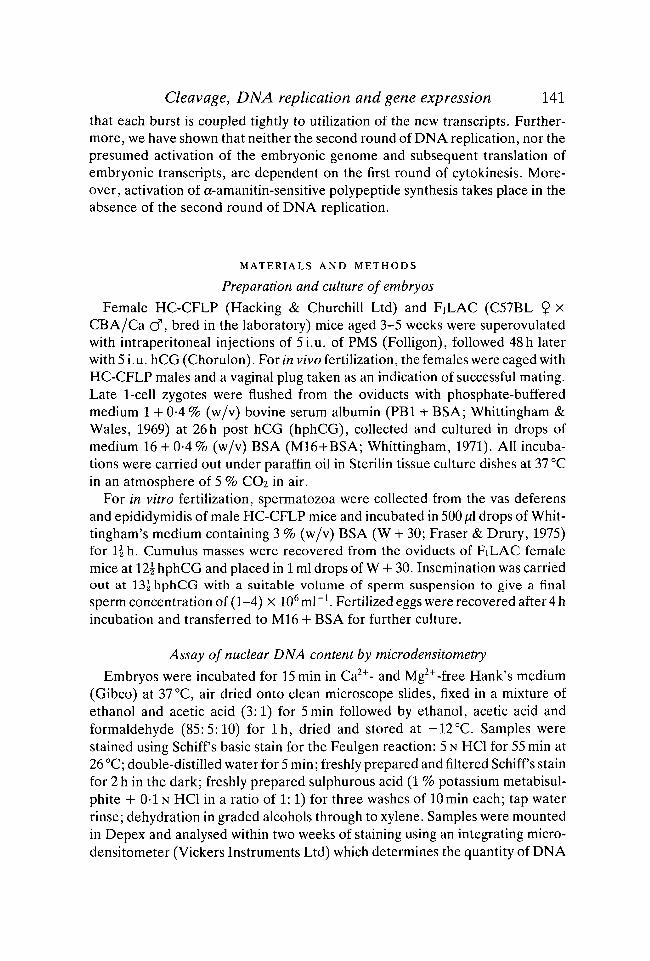
Fig. 4. Time course of DNA replication in nuclei of 2-cell mouse embryos culturedfrom late 1-cell stage in M16 + BSA (O) or M16 + BSA + 11 jig/ml a-amanitin (•) ,as judged by microdensitometric analysis at A = 560 nm after the Feulgen reaction.Each point represents the mean, and bars show the standard deviation, of severalnuclei (number of nuclei per sample indicated beside each point).
3. Timing of DNA replication
In order to establish the time course of DNA replication after the first cleavagedivision, synchronized groups of 2-cell embryos derived by fertilization in vitrowere fixed at hourly intervals after pick-off and stained with Schiff s basic stainfor the Feulgen reaction. The DNA content of individual nuclei was analysed bymicrodensitometry, mouse liver cells providing 2C and 4C reference values.Since standard values vary between experiments, the results of separate experi-ments were normalized with respect to standard values of 2C = 100, 4C = 200.The time course of DNA replication in data pooled from three experiments isshown in Fig. 4 (open circles). DNA replication commences in embryos fixed at1 hppo, and is completed in embryos fixed at 5 hppo.
4. Timing of a-amanitin sensitivity of polyp eptide synthesis
2-cell embryos derived from fertilization in vitro were taken at various timespost pick-off (hppo), placed in a-amanitin and cultured further for either 8h or27 h. For the last three hours of these culture periods, the embryos were in-cubated in [35S]methionine. Control embryos, and embryos that wereinseminated and cultured in the continuous presence of a-amanitin, were alsoincubated in [35S]methionine over the same 3 h labelling periods. In the case of

148 V. N. BOLTON, P. J. OADES AND M. H. JOHNSON
200
92
69
46
30
Mr
embryos cultured for 27 h either with or without a-amanitin, the incidence ofdivision to 4-cells at the end of the labelling period was recorded. The labelledpolypeptides in each group of embryos were resolved by SDS polyacrylamide gelelectrophoresis (Figs 5 & 6), and the appearance of the Mr 67 x 103 complex ofpolypeptides recorded for the 8h groups, and of the late 2-cell polypeptidesrecorded for the 27 h groups. Embryos placed in a-amanitin before 1 h post pick-off (1 hppo) failed to synthesize the 67 x 103 complex (Fig. 5), whereas embryosplaced in a-amanitin at later times did. Embryos placed in a-amanitin prior to8 hppo failed to synthesize late 2-cell polypeptides and synthesized only traces of
i 0 12 1 112 2 212 3 312 4 M16Fig. 5. One-dimensional SDS PAGE separation of radiolabelled polypeptides:(Mr) relative molecular mass markers as for Fig. 2; embryos cultured in M16 + BSAand transferred to M16 + BSA + 11/ig/ml a-amanitin at (i) 4hpi; (0) Ohppo; (4)4hppo; (1) lhppo; (14) 14hppo; (2) 2 hppo; (24) 24hppo; (3) 3 hppo; (34) 34hppo;(4) 4 hppo; (M16) control embryos cultured in M16 + BSA without exposure toa-amanitin. All embryos were labelled 8-11 hppo. Arrow indicates position of Mr67 x 103 complex of polypeptides. Densitometric traces of this region show an in-crease in synthesis of 67 x id3 polypeptides from 0-83 % to 5-6 % of total polypeptidesynthesis between tracks (i) and (M16).

Cleavage, DNA replication and gene expression 149early 2-cell polypeptides (Fig. 6) and failed to cleave (Fig. 7, lower panel),whereas embryos placed in a-amanitin at 9 hppo or later cleaved (Fig. 7), ceasedthe synthesis of early 2-cell polypeptides and synthesized late 2-cell polypeptides(Fig. 6). From this we infer that an early a-amanitin-sensitive (putative transcrip-tional) event has occurred by 0-5-1 hppo, and a later event by 8-9 hppo.
In order to establish whether the Mr 67 x 103 complex of polypeptides issynthesized de novo or as a result of a-amanitin-sensitive post-translationalmodification of presynthesized polypeptides after the first cleavage division, apulse-chase experiment was undertaken. Late 1-cell embryos derived from eggsfertilized in vivo were taken at 28hphCG and labelled for l h in diluted,
200
92
69
46
30
0 5 6 7 8 9 M16
Fig. 6. One-dimensional SDS PAGE separation of radiolabelled polypeptides:(Mr) relative molecular mass markers as for Fig. 2; embryos cultured in M16 + BSAand (0) labelled 0-3 hppo; labelled 24-27 hppo after transfer to M16 + BSA +lljUg/ml a-amanitin at (5) 5 hppo; (6) 6hppo; (7) 7 hppo; (8) 8 hppo; (9) 9 hppo;(M16) control embryos cultured in M16 + BSA and labelled 24-27 hppo withoutexposure to a-amanitin. Arrow indicates position of Mr 67 x 103 complex of polypep-tides; arrowheads indicate positions of representative late 2-cell polypeptides.
150 V. N. BOLTON, P. J. OADES AND M. H. JOHNSON
lyophilized [35S]methionine in M16 + BSA containing 0-01 /JM unlabelledmethionine. After the labelling period, one group of embryos was harvestedimmediately for SDS polyacrylamide gel electrophoresis, and the remainingembryos were washed and incubated for a further 8h (to 36hphCG) in M16+BSA supplemented with unlabelled methionine, leucine and phenylalanine.Under these conditions, all the embryos cleaved to 2-cells, and the prolongedpresence of [35S]methionine produced no evidence of cytotoxicity. A final groupof control mid 2-cell embryos were labelled for l h at 36hphCG in diluted,lyophilized [35S]methionine in M16+BSA containing 0-01 fM unlabelled meth-ionine. The labelled polypeptides synthesized by each group of embryos were
Mr 67 x 103 polypeptidesfirst detected
a-Amanitinblocks Mr 67 x 103
polypeptides
4C = 200
ft 180
cS
IS 160'
Late 2-cellpolypeptidesfirst detected
a-Amanitinblocks late 2-cell
polypeptides
MO-
120
2C = 100
= 80,
<N 60AS40I 20
4 6 8 10 12 14 16Time (hours post pick-off)
22 4654 33 2 11 j 22 EH]
i :
4 6 8 10 12 14Time embryos placed in a-amanitin (hppo)
16
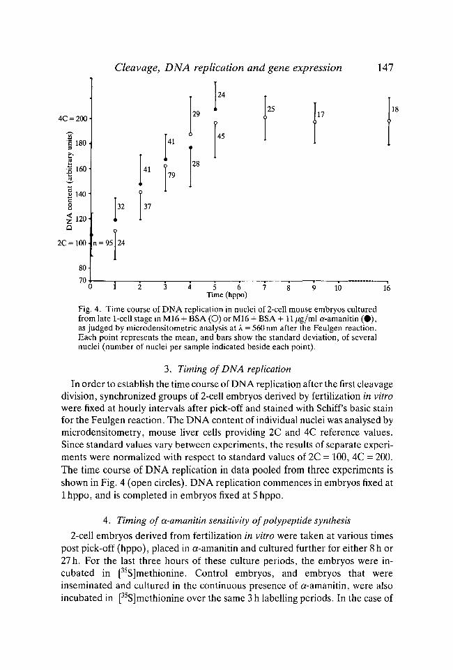
Fig. 7. Summary diagram illustrating temporal relationship between cell division(first cleavage division = time 0; second cleavage division = histogram: lower panel),DNA replication (solid dots); a-amanitin-sensitivity, and translational activity.

Cleavage, DNA replication and gene expression 151resolved by one dimensional SDS polyacrylamide gel electrophoresis (Fig. 8).Embryos that were labelled with a pulse of [35S]methionine at the late 1-cell stage(28-29 hphCG) did not synthesize the 67 x 103 complex of polypeptides, whetherthey were harvested immediately or whether the label was 'chased' for a further 8 h(tracks b and d, Fig. 8), whereas embryos that were labelled at the mid 2-cell stage(36-37 hphCG) did synthesize these polypeptides (tracks, Fig. 8). This result in-dicates that the MT 67 x 103 complex of polypeptides is not the product of post-translational modification to polypeptides synthesized at the late 1-cell stage.
200
30
Fig. 8. One-dimensional SDS PAGE separation of radiolabelled polypeptides: (a)relative molecular mass markers as for Fig. 2; embryos labelled in M16 + BSA +0-01 (AM unlabelled methionine containing diluted, lyophilized stock [35S]methio-nine: (b) late 1-cell embryos placed into label at 28hphCG and harvested at29hphCG; (c) mid 2-cell embryos placed into label at 36hphCG and harvested at37hphCG; (d) mid 2-cell embryos placed into label at the late 1-cell stage, washedat 29hphCG and cultured in M16 + BSA containing 100 jUM unlabelled methionine,phenylalanine and leucine, and harvested at 37 hphCG. Arrow indicates position ofMr 67 x 103 complex of polypeptides.

152 V. N. BOLTON, P. J. OADES AND M. H. JOHNSON
Finally, in order to demonstrate that the mRNA populations of 2-cell embryosdo indeed change between the very early and the late 2-cell stages, and that thischange is blocked in the presence of a-amanitin, mRNA was extracted fromnewly formed 2-cell and late 2-cell embryos (18 hpi and 38 hpi respectively) afterfertilization in vitro and culture in M16 + BSA with or without a-amanitin.Following translation in vitro in a rabbit reticulocyte cell-free system in thepresence of [35S]methionine, the labelled polypeptides synthesized were resol-ved by one- and two-dimensional SDS polyacrylamide gel electrophoresis. The
9A
B
Fig. 9. Two-dimensional SDS PAGE separation of [35S]methionine-labelledpolypeptides translated in vitro: (A) on mRNA extracted from control newly formed2-cell embryos at 18 hpi; (B) on mRNA extracted from control late 2-cell embryosat 38 hpi; (C) on mRNA extracted from late 2-cell embryos at 38 hpi after incubationin a-amanitin from 18 hpi; (D) by rabbit reticulocyte cell-free system independentlyof exogenous mRNA. Open arrows indicate mRNA-independent incorporation of[35S]methionine (Braude & Pelham, 1979); larger arrowheads indicate represen-tative early 2-cell polypeptides (asterisks indicate those whose synthesis is discussedin Pratt et al. 1983); small arrowheads indicate representative late 2-cell polypeptides(asterisk indicates putative Mr 67 x 103 polypeptide; see Flach etal. 1982). Isoelectricfocussing is from left (pH7-0) to right (pH4-5).

Cleavage, DNA replication and gene expression 153
results of the latter separation are shown in Fig. 9. The patterns of polypeptidestranslated in vitro from mRNA extracted from very early 2-cell and late 2-cellembryos are completely different (panels a and b, Fig. 9), and furthermore, nodetectable polypeptides at all are translated in vitro from mRNA extracted fromlate 2-cell embryos that had been incubated in the presence of a-amanitin (panelc, Fig. 9). This result shows that the transformation in polypeptide syntheticprofiles that occurs between the early and late 2-cell stages is accompanied bymajor changes in the populations of translatable mRNA, and that the gain of newtranscripts, but not the loss of old transcripts, is blocked by a-amanitin.
5. Investigation of the causal relationships between cytokinesis, DNA replicationand putative transcription
We first examined the effect of inhibition of the first round of cytokinesis onsubsequent DNA replication and transcription. Late 1-cell embryos (from in
4C = 2003 190-1 180-
I 160-£ 150| 140-§ 130o< 120-Z v
2C= 100-
90-80-70
Experimentalgroups
, n = 20,180
35
22
118
112
28
70
E2 L2FUdR FUdR
10~4MFUdR
APHID. CCD MIT.C
Fig. 10. DNA content of nuclei of embryos, determined by microdensitometricanalysis after various culture conditions. Each point represents the mean, and thebars the standard deviation, of several nuclei (number of nuclei per sample markedby each point). LI = late 1-cell embryos in M16 + BSA; E2 = early 2-cell embryosin M16 + BSA; L2 = late 2-cell embryos in M16 + BSA; 1CT6, 10"5, 10"4M-FUdR = late 2-cell embryos cultured from the late 1-cell stage in M16 + BSA + theindicated concentration of 5-fluorodeoxyuridine; APHID = late 2-cell embryos cul-tured from late 1-cell stage in M16 + BSA + 2jwg/ml aphidicolin; CCD = embryoscultured from late 1-cell stage in M16 + BSA + 0-5]Ug/ml cytochalasin D untilcontrol embryos were at late 2-cell stage; MITC = late 2-cell embryos cultured fromlate 1-cell stage in M16 -I- BSA + 25/ig/ml mitomycin C.
154 V. N. BOLTON, P. J. OADES AND M. H. JOHNSON
92
69
46
30
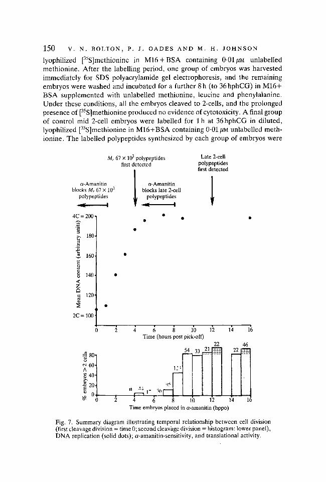
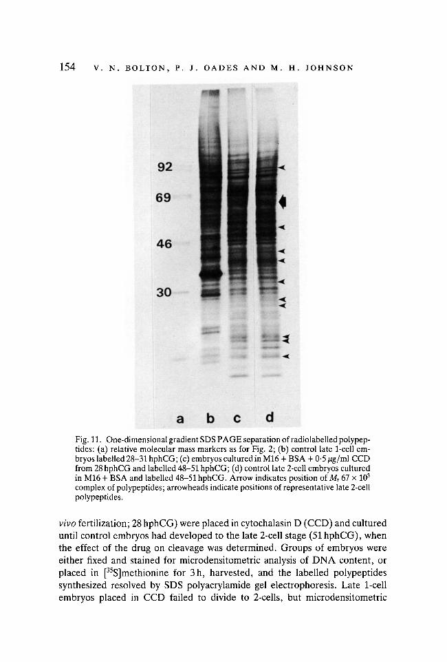
Fig. 11. One-dimensional gradient SDS PAGE separation of radiolabelled polypep-tides: (a) relative molecular mass markers as for Fig. 2; (b) control late 1-cell em-bryos labelled 28-31 hphCG; (c) embryos cultured in M16 + BSA + 0-5 /ig/ml CCDfrom 28 hphCG and labelled 48-51 hphCG; (d) control late 2-cell embryos culturedin M16 + BSA and labelled 48-51 hphCG. Arrow indicates position of Mr 67 x 103
complex of polypeptides; arrowheads indicate positions of representative late 2-cellpolypeptides.
vivo fertilization; 28 hphCG) were placed in cytochalasin D (CCD) and cultureduntil control embryos had developed to the late 2-cell stage (51 hphCG), whenthe effect of the drug on cleavage was determined. Groups of embryos wereeither fixed and stained for microdensitometric analysis of DNA content, orplaced in [35S]methionine for 3h, harvested, and the labelled polypeptidessynthesized resolved by SDS polyacrylamide gel electrophoresis. Late 1-cellembryos placed in CCD failed to divide to 2-cells, but microdensitometric

Cleavage, DNA replication and gene expression
200
155
30
Fig. 12. One-dimensional gradient SDS PAGE separation of radiolabelled polypep-tides: (a) relative molecular mass markers as for Fig. 2; (b) control early 2-cellembryos cultured in M16 + BSA and labelled 30-33 hphCG. Late 2-cell embryoslabelled 51-54 hphCG after (c) culture in M16 + BSA + 2//g/ml aphidicolin fromthe late 1-cell stage (30-33 hphCG); (d) culture in M16 + BSA + 2 jug/ml aphidicolinfrom the mid 2-cell stage (44hphCG); (e) culture in M16 + BSA without exposureto aphidicolin. Arrow indicates position of Mr 67 x 103 complex of polypeptides;arrowheads indicate positions of representative late 2-cell polypeptides.
analysis revealed that the blocked 1-cell embryos were binucleate, and eachnucleus had completed the second round of DNA replication (Fig. 10). Further-more, analysis of one-dimensional gels indicates that CCD-treated embryoswere synthesizing late 2-cell polypeptides (Fig. 11). From these results, we con-clude that the first cleavage division is not a prerequisite for DNA replication,nor for the synthesis of a-amanitin-sensitive polypeptides.
We next attempted to establish whether the early burst of putative transcrip-tional activity is required for the round of DNA replication that follows it. Late1-cell embryos (from in vitro fertilization; 18 hpi) were placed in a-amanitin andcultured until various times after cleavage to 2-cells. At hourly intervals after

156 V. N. BOLTON, P. J. OADES AND M. H. JOHNSON
pick-off, groups of embryos were fixed, stained and analysed by micro-densitometry. Despite inhibition of synthesis of the MT 67 x 103 complex ofpolypeptides, the time course of DNA replication in a-amanitin-treated em-bryos was the same as in controls (Fig. 4; closed circles).
Finally, we examined the effect of inhibition of DNA replication on thesynthesis of late 2-cell polypeptides. Three inhibitors of DNA replication wereused: mitomycin C, aphidicolin and 5-fluorodeoxyuridine (FUdR; at three con-centrations). None of the drugs produced any evidence of cytotoxicity at thedoses and incubation periods used. Each drug was used with embryos derived
200
92
69
46
30
•&>•
a b c d eFig. 13. One-dimensional gradient SDS PAGE separation of radiolabelled polypep-tides: (a) relative molecular mass markers as for Fig. 2; (b) control late 1-cell/early2-cell embryos cultured in M16 + BSA and labelled 30-33 hphCG; late 2-cell em-bryos labelled 51-54 hphCG after culture in (c) FUdR from the early 2-cell stage(30hphCG); (d) M16 + BSA; (e) FUdR from the mid 2-cell stage (44hphCG).Arrow indicates position of Mr 67 x 103 complex of polypeptides; arrowheads in-dicate positions of representative late 2-cell polypeptides.

Cleavage, DNA replication and gene expression 157
both from in vitro and from in vivo fertilization and, as the results were identicalfor each condition, they are pooled. The drugs were applied either to late 1-cellembryos (17-18 hpi; 30-33 hphCG), which then divided to yield 2-cell embryos,or to very early 2-cell embryos of the same ages in hpi or hphCG. Additionally,each drug was applied to mid 2-cell embryos after the period of DNA replicationwas completed, as judged directly in control embryos by microdensitometry(27 hpi or 44 hphCG). Embryos were analysed at the late 2-cell stage (38-41 hpi;51-54 hphCG) for evidence of inhibition of DNA replication (Fig. 10) and forpatterns of polypeptide synthesis (Figs 12 & 13).
Mitomycin C did not suppress DNA replication (Fig. 10). If added to late 1-cell or early 2-cell embryos, mitomycin reduced total incorporation ofmethionine into late 2-cell polypeptides and prevented the appearance of mostof the new polypeptides, with only traces of early 2-cell polypeptides remaining(gels not shown). Addition of mitomycin to mid 2-cell embryos did not affect thesynthesis of the late 2-cell polypeptides. Thus, mitomycin seems to interfere withthe putative activation of transcription at the mid 2-cell stage but by a mechanismthat does not depend upon inhibition of DNA replication.
Aphidicolin suppresses DNA replication (Fig. 10), but is almost without effecton the transition from an early to late 2-cell polypeptide pattern (Fig. 12).Maternally-coded early 2-cell polypeptides disappear and embryo-coded late 2-cell polypeptides appear. One or two minor differences are evident, and totalincorporation seems to be reduced slightly, but the major changes in polypeptidesynthetic pattern appear to be unaffected by the block to DNA replication.
5-fluorodeoxyuridine showed a dose-dependent effect on DNA replication(Fig. 10), but was without effect on the change in polypeptide synthetic profileat any dose (Fig. 13).
DISCUSSION
The use of highly synchronized populations of 2-cell embryos has permitted amore precise description of the events taking place during the 2-cell stage ofmouse development than was possible previously (Flach et al. 1982). The tem-poral relationships established between the first cleavage division, the round ofDNA replication that follows it, putative transcriptional activity (as assessed bya-amanitin-sensitivity) and the translation of the presumptive embryonic tran-scripts during the second cell cycle are summarized in Fig. 7. By 1 h after pick-off (1-1-5 h after cleavage) an a-amanitin-sensitive event has occurred, thatresults in the detectable synthesis of the Mr 67 x 103 complex of polypeptideswithin 3 h (3-5-4-0 h after cleavage). DNA replication commences 1 h after pick-off and is completed by 5h after pick-off (extends between 1-0 and 5-5 h aftercleavage). A second burst of a-amanitin-sensitive activity has occurred between8 and 9 h after pick-off and results in new protein synthesis by 12-5 h after pick-off (12-5-13h after cleavage). These results provide support for our earlier
EMB79

158 V. N. BOLTON, P. J. OADES AND M. H. JOHNSON
suggestion that the two bursts of transcriptional activity in the 2-cell embryomight form a sandwich about DNA replication, and that each burst is coupledtightly to utilization of the transcripts (Flach et al. 1982). They also indicate thepresence of a very short Gi phase in the second cell cycle.
Our evidence on the detailed timing of putative transcriptional activity at the2-cell stage rests on the use of a-amanitin. The assumption that a-amanitinsensitivity does indeed reflect transcriptional activity is reasonable on threegrounds: (i) the sharpness of the temporal changes in response to the drug at both1-2 hppo and 8-9 hppo suggests that non-specific toxic actions of the drug areunlikely to explain its effects; (ii) the sensitivity of the early (Mr 67 x 103)complex of polypeptides to a-amanitin is unlikely to be due to some post-translational modification that is sensitive to the drug, as shown by the pulse-chase experiment; (iii) the major change in mRNA populations that occursbetween the newly formed 2-cell and late 2-cell stages includes not only mRNAscoding for polypeptides in the MT 67 x 103 region, but also for many other newpolypeptides. Moreover, the appearance of the mRNA species coding for allthese new polypeptides is sensitive to a-amanitin. Taken together, and with thecautions outlined earlier in mind, these results suggest that our equation ofa-amanitin-sensitivity with transcriptional activity is not unreasonable.
The establishment of a precise temporal sequence of molecular events overwhat appears to be the earliest period of large-scale gene expression in the mouseembryo, provides us with a basis for the study of the mechanism(s) by which geneactivation might be controlled. We therefore attempted to establish whetherthere are any causal relationships between cytokinesis, DNA replication andgene expression. When the first cleavage division was inhibited by cytochalasinD, both the replication and putative transcription of DNA were unaffected. Thisresult might be expected in light of previous demonstrations that the first to fifthrounds of cytokinesis are expressions of the developmental programme ratherthan prerequisites for that expression (Surani, Barton & Burling, 1980; Pratt,Chakraborty & Surani, 1981; Petzoldt, Burki, Illmensee & Illmensee, 1983).Similarly inhibition of the early burst of putative transcription by a-amanitin didnot prevent DNA replication, which also might be expected, since the firstevidence of translational effects resulting from it was not detected until afterreplication had commenced. In addition, a-amanitin did not affect the declinein synthesis of 1-cell and early 2-cell polypeptides observed at the mid to late 2-cell stage. This result confirms our previous observation (Flach et al. 1982) andextends it by showing that by the mid to late 2-cell stage the mRNA species whichcode for these early proteins, and which are presumably maternal in origin, arenot only inactive in situ, but are not translatable in vitro, whether or not theembryos are cultured in the presence of a-amanitin. Thus, we can conclude thatthe whole of the first cell cycle, cleavage to the 2-cell stage, the second round ofDNA replication and the loss of maternal mRNA are all regulated at a post-transcriptional level, independently of the expression of the embryonic genome.

Cleavage, DNA replication and gene expression 159Which, if any, of the elements of this maternally-regulated programme in-
fluence activation of the embryonic genome? We examined the influence of thesecond round of DNA replication on this event and found it to have surprisinglyfew immediate effects on development. A number of studies have suggested thatthe reprogramming of chromatin assumed to occur before new genes are ex-pressed requires a round(s) of DNA replication, or that a critical number ofcyclic nuclear events are required for the activation of transcription. Of the threeinhibitors of replication examined, mitomycin C proved to be unsatisfactory.The time course of action of mitomycin C, and its critical active concentration,are rather variable, and the drug tends to produce delayed, drawn out and erraticeffects on DNA which occur at the same time as side effects on RNA synthesis(Shatkin, Reich, Franklin & Tatum, 1962). Despite the application of high dosesof the drug, DNA replication was affected only marginally and embryonicchromatin became disorganized only after extended exposure (unpublisheddata). In contrast, mitomycin suppressed totally the presumed de novo transcrip-tion from embryonic genes, although once activated the mRNAs produced werenot obviously affected by the mitomycin. Therefore, the results with this drugwere not illuminating.
In contrast, both FUdR (at 10~4M) and aphidicolin reduced DNA replicationto insignificant levels. FUdR is a thymidine analogue which inhibits DNAsynthesis by rendering thymidine incorporation rate limiting (Cozzarelli, 1977).The relatively high concentration required for effective inhibition probablyreflects the large endogenous nucleotide pool in embryos and the slow rate ofuptake (Epstein & Daentl, 1971; Quinn & Wales, 1973; Clegg & Piko, 1977).Aphidicolin inhibits DNA polymerase a which is responsible for most DNAreplication (Ikegami et al. 1978).
Despite the inhibition of DNA replication produced by either drug, expressionof the genes coding for the late 2-cell polypeptides was barely affected, thesynthetic patterns being almost indistinguishable from control late 2-cell em-bryos in which replication had occurred. A few minor, variable differences wereobserved, and total incorporation seemed slightly less in aphidicolin-treatedembryos compared with controls. It is possible that although both drugs inhibitedDNA replication, a small but critical portion of the DNA required for expressionof the late 2-cell genes might have escaped inhibition. However, there is noevidence to support a selective resistance of potentially active DNA sequencesto replication inhibitors of very different types, and indeed even partialsuppression of DNA replication in other systems has been associated with afailure of genomic reprogramming (Weintraub, 1975). It is not therefore possibleto conclude from these data that the second round of DNA replication in themouse embryo is required for the putative major gene activation that follows it.
Should this gene expression indeed prove to be independent of the precedinground of DNA replication, constraints are placed on the types of model that canexplain gene activation. Such models fall into two general categories within the

160 V. N. BOLTON, P. J. OADES AND M. H. JOHNSON
broader concept of 'quantal cell cycles' (Dienstman & Holtzer, 1975). The firstkind of quantal cycle is seen as a permissive process and the concept invokesspecific interactions between the DNA or chromatin and cytoplasmic regulatorsubstances which, after a critical number of nuclear cyclic events, allow tran-scription to proceed (Davidson & Britten, 1971). For example, the segregationand consequent dilution of cytoplasmic components during cleavage, withconsecutive cycles of DNA replication causing an exponential increase in therelative nuclear DNA content, may permit eventual expression of the genomewhen the ratio of DNA: regulator substance(s) reaches a critical level. Thedevelopment of acetylcholinesterase activity in ascidian embryos (Satoh &Ikegami, 1971a,b) and the midblastula transition in Xenopus embryos (Newport& Kirschner, 1982) may be controlled in this way. Our results suggest that themid 2-cell gene activation of the mouse embryo cannot be regulated in thismanner.
The second kind of scheme for quantal cell cycles envisages a causal role forthe cell cycle, whereby a specific, programmed change in DNA or chromatin isa requirement for gene expression. Transcriptionally active chromatin may becharacterized by a number of features, and it has been suggested that thesemodifications allow gene expression by affecting the tightness of the packing ofthe chromatin, and thereby presumably the access of RNA polymerase. Severalmodels have been proposed by which stable changes in chromatin might beproduced, many of which require a round of DNA replication for their imple-mentation. For example, chromatin modifications allowing gene expression thatinvolve changes in methylation patterns (Razin & Riggs, 1980), nucleosomalorganization (Weintraub, 1979; Allfrey, 1980) or histone structure or type(Borun, 1975) appear to require DNA replication in order to operate. Clearly,if such modifications are involved in the proposed mid 2-cell gene activation ofthe mouse embryo then either they must occur in the absence of DNA replica-tion, or modification of one DNA strand at the first round of replication must beadequate for subsequent altered expression. We are currently investigating thesealternatives.
We wish to acknowledge the assistance of Gin Tampkins, Tracy Kelly, Raith Overhill, TimCrane and Ian Edgar, the gift of aphidicolin from ICI Ltd., and the use of the microdensi-tometer of Dr D. R. Sharman and Dr Holzbauer-Sharman of the ARC Institute, Babraham.The work was supported by grants from the MRC and the Cancer Research Campaign.V.N.B. acknowledges receipt of MRC and Cambridge Philosophical Society training awardsand P.J.O. acknowledges support from the H. E. Durham Fund, King's College, Cambridge.
REFERENCES
ALLFREY, U. G. (1980). Molecular aspects of the regulation of eukaryotic transcription:Nucleosomal proteins and their postsynthetic modifications in the control of DNA con-formation and template function. In Cell Biology 3, (eds L. Goldstein & D. M. Prescott),pp. 348-438. London: Academic Press.

Cleavage, DNA replication and gene expression 161BENSAUDE, O., BABINET, C , MORANGE, M. & JACOB, F. (1983). Heat shock proteins, first
major products of zygotic gene activity in the mouse embryo. Nature (in press).BONNER, W. M. & LASKEY, R. A. (1974). A film detection method for tritium-labelled
proteins and nucleic acids in polyacrylamid gels. Eur. J. Biochem. 46, 83-88.BORUN, T. W. (1975). Histones, differentiation and the cell cycle. In Cell Cycle and Dif-
ferentiation 7, (eds J. Reinert & H. Holtzer), pp. 248-290. Berlin: Springer Verlag.BRACHET, J., FICQ, A. & TENCER, R. (1968). Amino acid incorporation into proteins of
nucleate and anucleate fragments of sea urchin eggs: effect of parthenogenetic activation.Expl Cell Res. 32, 168-170.
BRAUDE, P. R. & PELHAM, H. R. B. (1979). A microsystem for the extraction and in vitrotranslation of mouse embryo mRNA. /. Reprod. Fertil. 56, 153-158.
BRAUDE, P. R., PELHAM, H. R. B., FLACH, G. & LOBATTO, R. (1979). Post transcriptionalcontrol in the early mouse embryo. Nature 282, 102-105.
CASCIO, S. M. & WASSARMAN, P. M. (1982). Program of early development in the mammal:Post transcriptional control of a class of proteins synthesized by mouse oocytes and earlyembryos. Devi Biol. 89, 397-408.
CLEGG, K. B. & PIK6, L. (1977). Size and specific activity of the UTP pool and overall ratesof RNA synthesis in early mouse embryos. Devi Biol. 58, 76-95.
CLEGG, K. B. & PIK6, L. (1982). RNA synthesis and cytoplasmic polyadenylation in the one-cell mouse embryo. Nature 295, 342-345.
CLEGG, K. B. & PIKO, L. (1983). Poly (A) length, cytoplasmic adenylation and synthesis ofpoly (A)+ RNA in early mouse embryos. Devi Biol. 95, 331-341.
COZZARELLI, N. R. (1977). The mechanism of action of inhibitors of DNA synthesis. Ann.Rev. Biochem. 46, 641-668.
CULLEN, B., EMIGHOLZ, K. & MONAHAN, J. (1980). The transient appearance of specificproteins in one cell mouse embryos. Devi Biol. 76, 215-221.
DAVIDSON, E. H. (1976). Gene Activity in Early Development. New York: Academic Press.DAVIDSON, E. H. & BRITTEN, R. J. (1971). Note on the control of gene expression during
development. /. theor. Biol. 32, 123-130.DENNY, P. C. & TYLER, A. (1964). Activation of protein biosynthesis in non-nucleate frag-
ments of sea urchin eggs. Biochem. Biophys. Res. Comm. 14, 245-249.DIENSTMAN, S. R. & HOLTZER, H. (1975). Myogenesis: a cell lineage interpretation. In Cell
Cycle and Differentiation (eds J. Reinert & H. Holtzer), 7, pp. 1-25. Berlin: SpringerVerlag.
EDWARDS, R. G. & GATES, A. H. (1959). Timing of the stages of the maturation divisions,ovulation, fertilisation and the first cleavage of eggs of adult mice treated with gonado-trophins. /. Endocrin. 18, 292-304.
EPSTEIN, C. J. & DAENTL, D. L. (1971). Precursor pools and RNA synthesis in preimplanta-tion mouse embryos. Devi Biol. 26, 517-524.
FLACH, G., JOHNSON, M. H., BRAUDE, P. R., TAYLOR, R. A. S. & BOLTON, V. N. (1982). Thetransition from maternal to embryonic control in the 2-cell mouse embryo. The EM BOJournal 1, 681-686.
FRASER, L. R. & DRURY, L. (1975). The relationship between sperm concentration and fer-tilization in vitro of mouse eggs. Biol. Reprod. 13, 513-518.
HADJIOLOV, A. A., DAVERA, M. D. & MACKEDOWSKI, V. V. (1974). The action of a-amanitinin vivo on the synthesis and maturation of mouse liver ribonucleic acids. Biochem. J. 138,321-334.
HOLMBERG, S. R. M. & JOHNSON, M. H. (1979). Amino acid transport in the unfertilized andfertilized egg. J. Reprod. Fert. 56, 223-231.
HOWE, C. C. & SOLTER, D. (1979). Cytoplasmic and nuclear protein synthesis in preimplanta-tion mouse embryos. Devi Biol. 52, 209-225.
IKEGAMI, S., TAGUCHI, T., OHASHI, M., OGUM, M., NAGANO, H. & MANO, Y. (1978). Aphi-dicolin prevents mitotic cell division by interfering with activity of DNA polymerase a.Nature 275, 458-460.
JOHNSON, M. H. (1981). The molecular and cellular basis of preimplantation mouse develop-ment. Biol. Rev. 56, 463-498.

162 V. N. BOLTON, P. J. OADES AND M. H. JOHNSON
JOHNSON, M. H. & PRATT, H. P. M. (1983). Cytoplasmic localisation and cell interactions inthe formation of the mouse blastocyst. In Cytoplasmic Localisations and Determination ofEmbryonic Cells (ed. W. J. Jeffery). Place: Alan R. Liss (in press).
KAYE, P. L., SCHULTZ, G. A., JOHNSON, M. H., PRATT, H. P. M. & CHURCH, R. B. (1982).Amino acid transport and exchange in preimplantation mouse embryos. /. Reprod. Fert. 65,367-380.
KEDINGER, C , GNIADOWSKI, M., MANDEL, J. L., GISSINGER, F. & CHAMBON, P. (1970).a-Amanitin: a specific inhibitor of one or two DNA-dependent RNA polymerase activitiesfrom calf thymus. Biochem. Biophys. Res. Comm. 38, 166-171.
KNOWLAND, J. & GRAHAM, C. F. (1972). RNA synthesis at the two-cell stage of mousedevelopment. /. Embryol. exp. Morph. 27, 167-176.
KRZANOWSKA, H. (1964). Time interval between copulation and fertilization in inbred linesof mice and their crosses. Folia biol., Krakow 12, 231-244.
LAEMMLI, U. K. (1970). Cleavage of structural proteins during the assembly of the head ofbactriophage T4. Nature 227, 680-685.
LASKEY, R. A. & MILLS, A. D. (1975). Quantitative film detection of 3H and 14C inpolyacrylamide gels by fluorography. Eur. J. Biochem. 56, 335-341.
LEVEY, I. L. & BRINSTER, R. L. (1978). Effects of a-amanitin on RNA synthesis by mouseembryos in culture. /. exp. Zool. 203, 351-360.
LEVEY, I. L., STULL, G. B. & BRINSTER, R. L. (1978). Poly (A) and synthesis of polyadenylatedRNA in the preimplantation mouse embryo. Devi Biol. 64, 140-148.
LEVINSON, J., GOODFELLOW, P., VADEBONCOEUR, M. & MCDEVITT, H. (1978). Identificationof stage-specific polypeptides synthesized during murine preimplantation development.Proc. natn. Acad. ScL, U.S.A. 75, 3332-33336.
LINDELL, T. J., WEINBERG, F., MORRIS, P. W., ROEDER, R. G. & RUTTER, W. J. (1970).Specific inhibition of nuclear RNA polymerase II by a-amanitin. Science 170, 447-448.
LUTHARDT, F. W. & DONAHUE, R. P. (1975). DNA synthesis in developing two-cell mouseembryos. Devi Biol. 44, 210-216.
MACQUEEN, H. (1979). Lethality of radioisotopes in early mouse embryos. J. Embryol. exp.Morph. 53, 202-208.
MAGNUSON, T. & EPSTEIN, C. J. (1981). Genetic control of very early mammalian develop-ment. Biol. Rev. 56, 369-408.
MCLAREN, A. (1979). The impact of prefertilization events on post-fertilization developmentin mammals. In Maternal Effects in Development, (eds D. R. Newth & M. Balls), pp.287-320. Cambridge University Press.
MOORE, G. P. M. (1975). The RNA polymerase activity of the preimplantation mouse em-bryo. /. Embryol. exp. Morph. 34, 291-298.
NEWPORT, J. & KIRSCHNER, M. (1982). A major developmental transition in early Xenopusembryos: II Control of the onset of transcription. Cell 30, 687-696.
NICOL, A. & MCLAREN, A. (1974). An effect of the female genotype on sperm transport inmice. /. Reprod. Fert. 39, 421-424.
O'FARRELL, P. H. (1975). High resolution two-dimensional electrophoresis of proteins. J.biol. Chem. 250, 4007-4021.
PETZOLDT, U., HOPPE, P. C. & ILLMENSEE, K. (1980). Protein synthesis in enucleated fertilizedand unfertilized mouse eggs. Wilhelm Roux' Arch, devl Biol. 189, 215-219.
PETZOLDT, U., BURKI, K., ILLMENSEE, G. R. & ILLMENSEE, K. (1983). Protein synthesis inmouse embryos with experimentally produced asynchrony between chromosome replica-tion and cell division. Wilhelm Roux' Arch, devl Biol. 192, 138-144.
PIKO, L. & CLEGG, K. B. (1982). Quantitative changes in total RNA, total poly (A) andribosomes in early mouse embryos. Devl Biol. 89, 362-378.
PRATT, H. P. M., CHAKRABORTY, J. & SURANI, M. A. H. (1981). Molecular and morphologicaldifferentiation of the mouse blastocyst after manipulations of compaction usingcytochalasin D. Cell 26, 279-292.
PRATT, H. P. M., BOLTON, V. N. & GUDGEON, K. A. (1983). The legacy from the oocyte andits role in controlling early development of the mouse embryo. In The Molecular Biologyof Egg Maturation. Ciba Symposium 98, pp. 197-227. Pitman Press.

Cleavage, DNA replication and gene expression 163QUINN,P. & WALES, R. G. (1971). Adenosine triphosphate content of preimplantation mouse
embryos. /. Reprod. Fert. 25, 133-135.RAZIN, A. & RIGGS, A. D. (1980). DNA methylation and gene function. Science 210,
604-610.ROSENTHAL, E. T., HUNT, T. & RUDERMAN, J. V. (1980). Selective translation of mRNA
controls the pattern of protein synthesis during early development of the surf clam Spisulasolidissima. Cell 20, 487-494.
SATOH, N. & IKEGAMI, S. (1981a). A definite number of aphidicolin-sensitive cell-cyclic eventsare required for acetylcholinesterase development in the presumptive muscle cells of theascidian embryos. /. Embryol. exp. Morph. 64, 1-13.
SATOH, N. & IKEGAMI, S. (19816). On the "clock" mechanisms determining the time of tissue-specific enzyme development during ascidian embryogenesis II: Evidence for association ofclock with cycle of DNA replication. J. Embryol. exp. Morph. 64, 61-71.
SAWICKI, W., ABRAMCZUK, J. & BLATON, O. (1978). DNA synthesis in the second and thirdcycles of mouse preimplantation development. Expl Cell Res. 112, 199-205.
SAWICKI, J. A., MAGNUSON, T. & EPSTEIN, C. J. (1981). Evidence for expression of thepaternal genome in the two-cell mouse embryo. Nature 294, 450-451.
SCHULTZ, G. A., CLOUGH, J. R., BRAUDE, P. R., PELHAM, H. R. B. & JOHNSON, M. H. (1981).A re-examination of messenger RNA populations in the preimplantation mouse embryo.In Cellular and Molecular Aspects of Implantation, (eds S. R. Glasser & D. W. Bullock),pp. 137-154. London and New York: Plenum Press.
SHATKIN, A. J., REICH, E., FRANKLIN, R. M. & TATUM, E. L. (1962). Effect of mitomycin Con mammalian cells in culture. Bioch. Biophys. Acta 55, 277-289.
SURANI, M. A. H., BARTON, S. C. & BURLING, A. (1980). Differentiation of two-cell and eight-cell mouse embryos arrested by cytoskeletal inhibitors. Expl Cell Res. 125, 275-286.
VAN BLERKOM, J. (1981). Structural relationship and post translational modification of stage-specific proteins synthesized during early preimplantation development in the mouse. Proc.natn. Acad. ScL, U.S.A. 78, 7629-7633.
VAN BLERKOM, J. & BROCKWAY, G. O. (1975). Qualitative patterns of protein synthesis in thepreimplantation mouse embryo. Devi Biol. 44, 148-157.
WEINTRAUB, H. (1975). The organisation of red cell differentiation. In Cell Cycle and Dif-ferentiation 7, (eds J. Reinert & H. Holtzer), pp. 26-42. Berlin: Springer Verlag.
WEINTRAUB, H. (1979). Assembly of an active chromatin structure during replication. NucleicAcid Res. 7, 781-792.
WELLS, D. E., SHOWMAN, R. M., KLEIN, W. H. & RAFF, R. A. (1981). Delayed recruitmentof maternal histone H3 mRNA in sea urchin embryos. Nature 292, 477-478.
WHITTINGHAM, D. G. (1971). Culture of mouse ova. /. Reprod. Fert. Suppl. 14, 7-21.WHITTINGHAM, D. G. & WALES, R. G. (1969). Storage of two-cell mouse embryos in vitro.
Aust. J. biol. Sci. 22, 1065-1068.WOODLAND, H. R. & BALLANTINE, J. E. M. (1980). Paternal gene expression in developing
hybrid embryos of Xenopus laevis and Xenopus borealis. J. Embryol. exp. Morph. 60,359-372.
WOODLAND, H. R., FLYNN, J. M. & WYLLIE, A. J. (1979). Utilization of stored mRNA inXenopus embryos and its replacement by newly synthesized transcripts: histone HIsynthesis using interspecies hybrid. Cell 18, 165-171.
WOODLAND, H. R. & GRAHAM, C. F. (1969). RNA synthesis during early development of themouse. Nature 221, 327-332.
YOUNG, R. J., SWEENEY, K. & BEDFORD, J. M. (1978). Uridine and guanosine incorporationby the mouse one-cell embryo. /. Embryol. exp. Morph. 44, 133-148.
(Accepted 28 September 1983)
