disruption of a topoisomerase-dna cleavage complex by a dna
TRANSCRIPT
Proc. Nati. Acad. Sci. USAVol. 91, pp. 12031-12035, December 1994Biochemistry
Disruption of a topoisomerase-DNA cleavage complex by aDNA helicaseMICHAEL T. HOWARD*, SUE H. NEECEt, STEVEN W. MATSON*t, AND KENNETH N. KREUZERt*Department of Biology and tCurriculum in Genetics and Molecular Biology, University of North Carolina, Chapel Hill, NC 27599; and tDepartment ofMicrobiology, Duke University Medical Center, Durham, NC 27710
Communicated by Nicholas R. Cozzarelli, August 23, 1994
ABSTRACT The type I DNA topoisomerases are targetsfor a variety of chemotherapeutic agents, Includin the anti-bacterial quinolones and several famile of antitumor drugs.These agents stabilize an epzyme-DNA cleavage complex thatconsists of the topoisomerase covalently linked to the 5' phos-phates of a double-stranded DNA break. Although the drug-stabilized cleavage complex is readily reversible, it can result incell death by a mechanism that remains uncertain. Here wedemonstrate that the action of a DNA helicase can convert thecleavage complex into a nonreversible DNA break by displacingDNA strands from the complex. Formation of a nonreversibleDNA break, induced by a DNA helicase, could explain thecytotoxicity of these topoisomerase poisons.
Topoisomerases play a major role in cellular DNA metabo-lism by altering the topological state of DNA. Type II DNAtopoisomerases effect this change by creating a transientdouble-stranded DNA (dsDNA) break through which a sec-ond DNA segment is passed (1-3). Several classes of chemo-therapeutic drugs, including the acridines, anthracyclines,ellipticines, and epipodophyllotoxins, inhibit the type IItopoisomerases (4, 5). These drugs stabilize a protein-DNAcomplex, called the cleavage complex, in which the enzymeis covalently linked to each 5' phosphate of a staggereddsDNA break. Although both strands of the DNA helix arethought to be broken, the linear integrity of the DNA ismaintained by a noncovalent topoisomerase bridge. Thesimplest model is that the inhibitors trap the cleavage com-plex by inhibiting the ligation step of the enzymatic cycle.The type II topoisomerases are a primary cellular target for
many of the chemotherapeutic drugs described above. Stud-ies on drug-resistant mammalian cell lines revealed alter-ations in topoisomerase levels or activity, suggesting that theenzyme is an important component of the cytotoxic response(6, 7). Genetic studies in Saccharomyces cerevisiae havedemonstrated that a temperature-sensitive mutation in thetype II topoisomerase gene greatly decreased drug sensitivityat the semipermissive temperature, presumably due to areduction in topoisomerase activity (8). Perhaps the clearestevidence for a type II topoisomerase being the primary drugtarget comes from the T4 bacteriophage system. The prop-erties of the T4 topoisomerase closely resemble those of themammalian enzyme (1, 9). Most importantly, both are sen-sitive to the aminoacridine m-AMSA [4'-(9-acridinylami-no)methanesulfon-m-anisidide] (10, 11). A point mutation inone of the genes encoding the bacteriophage T4 topoisom-erase allows drug-resistant phage growth, and topoisomerasepurified from the mutant phage has drug-resistant topoisom-erase activity (12, 13).Topoisomerase inhibitors convert the enzymes that they
target into poisons by inducing formation of the cleavagecomplex, a form of DNA damage. This view was first
articulated when it was found that bacteriophage 17 could beinhibited by the quinolone nalidixic acid, even though phagegrowth was not significantly reduced by mutational inacti-vation of DNA gyrase (14). Importantly, mutational inacti-vation of DNA gyrase protected 17 from inhibition by thedrug (14). More recent results imply that the cleavage com-plex is critical for the cytotoxicity of antitumor agents thatinhibit topoisomerases. For example,. defects in DNA repairhave been shown to increase drug sensitivity (refs. 15-17;S.H.N., K. Carles-Kinch, and K.N.K., unpublished data),indicating that the topoisomerase inhibitors induce DNAdamage. In spite of the importance of the cleavage complexin cytotoxicity, the complexes are reversible both in vitro andin vivo. To explain this apparent paradox, it has beenproposed that an active mechanism, such as transcription orreplication, converts reversible cleavage complexes into non-reversible cytotoxic DNA lesions (ref. 18; also see Discus-sion).We investigated whether helicase action could convert a
type II topoisomerase cleavage complex into a nonreversibleDNA break by examining the DNA products generated whenEscherichia coli helicase II unwinds a DNA substrate con-taining an m-AMSA-induced T4 topoisomerase cleavagecomplex. These two enzymes were selected for the followingreasons: (i) The bacteriophage T4 topoisomerase closelyresembles mammalian type II topoisomerases in reactionmechanism and sensitivity to inhibitors (see above). (ii) Astrong m-AMSA-inducible T4 topoisomerase cleavage sitehas been cloned near the middle of a 703-bp DNA fragment(G. Hong and K.N.K., unpublished data), facilitating anal-ysis of cleavage complexes occurring at a single site. Thesequence of this cleavage site was based on mutationalanalyses of a T4 topoisomerase cleavage site and on exper-iments with in vitro substrates containing optimal sequencesfor DNA cleavage (ref. 19 and C. Freudenreich and K.N.K.,unpublished data). (iii) Several topoisomerase inhibitorshave been shown to block helicase activity in vitro (20-23);however, E. coli helicase II was found to be insensitive tom-AMSA (20). (iv) Unlike most other DNA helicases, E. colihelicase II does not require a single-stranded DNA (ssDNA)loading site to initiate DNA strand separation (24). Thisallows the use of fully duplex linear DNA substrates, whichis important because the T4 topoisomerase interacts stronglywith ssDNA and ssDNA-dsDNA junctions (25).
MATERIALS AND METHODSDNA Substrate. The DNA substrate was prepared from a
pBR322-derived plasmid, pGH2, containing a 28-bp insertbetween the unique BamHI and Sal I restriction sites. Theinsert was designed based on the studies ofFreudenreich andKreuzer (19) to produce a very efficient m-AMSA-inducible
Abbreviations: dsDNA, double-stranded DNA; m-AMSA, 4'-(-acri-dinylamino)methanesulfon-m-anisidide); ssDNA, single-strandedDNA; ATP[yS], adenosine 5'-[(-thio]triphosphate.
12031
The publication costs of this article were defrayed in part by page chargepayment. This article must therefore be hereby marked "advertisement"in accordance with 18 U.S.C. §1734 solely to indicate this fact.

12032 Biochemistry: Howard et al.
topoisomerase cleavage site (insert sequence is 5'-AAGC-TAAAGTTATATAACTTTATTCAAG-3'). The 703-bp frag-ment was generated by cleaving pGH2 DNA with HindIIIand Nru I, filling in the recessed 3' end with the Klenowfragment of DNA polymerase I (Boehringer Mannheim),treating with calf intestinal phosphatase and proteinase K,and purifying by electrophoresis through a nondenaturing 6%polyacrylamide gel. The DNA fragment was electroeluted,ethanol-precipitated, and stored at -700C. The substrate waslabeled at the 5' ends using T4 polynucleotide kinase and[y-32P]ATP (Amersham). Labeling was followed by a 5-minchase at 370C with 50 gM (unlabeled) ATP to providecompletely phosphorylated 5' ends. Restriction enzymes,calf intestinal phosphatase, T4 polynucleotide kinase, andproteinase K were from New England Biolabs.T4 Topoisomerase and Helicase II Reactions. Except where
indicated, topoisomerase cleavage reaction mixtures (40 1ul)contained 40 mM Tris-OAc (pH 7.8), 20 mM KOAc, 8 mMMg(OAc)2, 0.5 mM EDTA, 2.5 mM. ATP, 0.5 mM dithio-threitol, bovine serum albumin (35 ,ug/ml), 5% (vol/vol)glycerol, 12.5 ,uM m-AMSA (NSC 249992, provided by theNational Cancer Institute), -1 fmol of 5'-32P-end-labeled703-bp DNA fragment (see below), and purified T4 type IItopoisomerase (9, 13) at a molar ratio of -20 topoisomerasedimers per duplexDNA fragment. Controls lacking m-AMSAreceived 0.02% (final concentration) dimethyl sulfoxide (m-AMSA solvent) and those lacking enzyme received topo-isomerase dilution buffer (9). Incubation was for 5 min at320C.
Helicase II reactions were initiated by adding 2.8 pmol (1,ul) of purified E. coli helicase 11 (26) to the cleavage com-plexes above; helicase storage buffer was added to samplesthat received no helicase. Incubation was then continued for5 min at 32°C. After this incubation, either EDTA (50 mM,final concentration) or SDS (1%, final concentration) wasadded and incubation was continued for 20 min at 320C. Allsamples were then adjusted to contain 50 mM EDTA and 1%SDS and were treated with proteinase K (380 ug/ml) for 20min at 37°C.
Gel Electrophoresis. Reaction products were resolved byelectrophoresis through polyacrylamide gels and visualizedby autoradiography. Denaturing gels contained 7 M urea and6% polyacrylamide [acrylamide/N,N'-methylenebisacryl-amide = 20:1 (wt/wt)], with lx TBE (89 mM Tris/89 mMborate/2 mM EDTA) as running buffer; electrophoresis wasat 21-23 V/cm for -4 hr. Samples for the denaturing gelsconsisted of 20-,l aliquots that were mixed with 15 Al ofloading solution [85% (vol/vol) formamide/lx TBE/0.1%bromophenol blue/0.1% xylene cyanole] and heated for 5 minat 1000C immediately before loading. Nondenaturing gelscontained 5% polyacrylamide, 0.1% SDS, and 25% glycerol,with lx TBE containing 0.1% SDS as running buffer; elec-trophoresis was at 4.4 V/cm for =21 hr. Samples for thenondenaturing gels consisted of 20-,ul aliquots that weremixed with 15 ,ul of loading solution (40o glycerol/50 mMEDTA/0.5% SDS/0.02% bromophenol blue/0.02% xylenecyanole). DNA markers (BioVentures, Murpheesboro, TN)were labeled by incubation with T4 polynucleotide kinase and[y-32P]ATP.
RESULTSDNA Helicase II Prevents Reversal of m-AMSA-Induced T4
Topoisomerase Cleavage Complexes. Cleavage complexeswere established by incubating the T4 type II topoisomerasewith m-AMSA and the 703-bp 5'-end-labeled DNA substrate.The DNA substrate contains a strong topoisomerase IIcleavage site located 337 bp from one end. The amount ofT4topoisomerase cleavage complex was determined by stop-ping the reactions with SDS, incubating with proteinase K,
and analyzing the products on polyacrylamide gels. Labelingthe DNA substrate at the 5' ends confines analysis to thecleavage products that were not covalently linked to thetopoisomerase. An autoradiograph of a denaturing polyacryl-amide gel revealed the two major products of cleavage at thestrong topoisomerase site along with bands generated fromminor topoisomerase cleavage sites (Fig. 1A, lanes 1 and 2).To determine the impact of helicase activity on an
m-AMSA-induced cleavage complex, we used a cleavagereversal assay in which the samples were exposed to EDTAafter the enzymatic reaction but prior to treatment with SDSand proteinase K. This treatment causes reversal of cleavagecomplexes formed by eukaryotic (27, 28), T4 (29), and E. coli(30) type II topoisomerases. The mechanism of reversal isunknown; however, the simplest explanation is that chelationof magnesium prevents topoisomerase from initiating a newround of cleavage but does not prevent DNA religation.When EDTA was added to the topoisomerase reactionswithout helicase, about 80% of the cleavage complexes atboth the major and minor sites were reversed (Fig. 1A, lanes2 and 3). However, EDTA reversal was almost completelyblocked in reactions that contained helicase (Fig. 1A, lanes4 and 5). The presence of the cleavage products was depen-dent on m-AMSA (Fig. 1A, lanes 7 and 8) and topoisomerase(Fig. 1A, lanes 9 and 10). Thus, reversible m-AMSA-inducedtype II topoisomerase cleavage complexes were converted tononreversible DNA breaks by helicase II.
Aliquots of the same reactions were analyzed on a nonde-naturing polyacrylamide gel to examine further the nature ofthe cleavage products and determine the extent of helicaseunwinding. The addition of helicase to topoisomerase reac-tions resulted in the appearance of several new bands (Fig.1B, lanes 2 and 4). These bands comigrated with bandsproduced when the helicase-free topoisomerase cleavagereactions were subjected to heat denaturation prior to elec-trophoresis (Fig. 1B, lane 6; note that one of the majorssDNA cleavage products comigrates with one of the majordsDNA cleavage products). Therefore, helicase II can un-wind DNA cleavage complexes to yield ssDNA cleavageproducts. Furthermore, the ssDNA cleavage products gen-erated by topoisomerase plus helicase are not reversible byEDTA treatment (Fig. 1B, lanes 4 and 5). Helicase II alsounwound the topoisomerase-free substrate DNA to generatefull-length ssDNA molecules that migrated slower than thefull-length duplex DNA (Fig. 1B, lanes 4, 5, and 7-10). Again,the presence of cleavage products was strictly dependent onm-AMSA (Fig. 1B, lanes 7 and 8) and topoisomerase (Fig.1B, lanes 9 and 10). We conclude from this analysis that,coincident with converting reversible cleavage complexes tononreversible DNA breaks, helicase II unwinds DNA con-strained within the cleavage complex to produce ssDNAcleavage products.T4 topoisomerase can cleave ssDNA and form a covalent
protein-DNA complex that is resistant to reversal by EDTAtreatment (25). Therefore, an alternative interpretationseemed possible: perhaps the nonreversible DNA breaksanalyzed in this study were due to formation of cleavagecomplexes on the ssDNA product of the helicase II unwind-ing reaction. To test this possibility, topoisomerase-inducedcleavage products were analyzed using the 703-bp DNAsubstrate that had been heat-denatured immediately prior tothe topoisomerase reaction. Although cleavage sites weredetected on the ssDNA, the products did not comigrate withthe nonreversible topoisomerase cleavage products gener-ated from dsDNA in the presence of helicase (Fig. 1C, lanes5-7). In addition, the binding of helicase II to the heat-denatured substrate inhibited the ssDNA cleavage by the T4topoisomerase (Fig. 1C, lanes 7 and 9). Therefore, thenonreversible cleavage complexes were not formed by the
Proc. Natl. Acad. Sci. USA 91 (1994)

Biochemistry: Howard et al.
ATopo - + + + + + + +
m-AMSA + + + + + + - - +
Hell - - - + + - + + + +
EDTA - - + - + - - + - + - M
ATP ATP S;
TopoHelin+EDTA -- t
ntDenatured
1000 Substrate
700 Substrate - t - __ * a 9
J___ _~~~~~~~~1500
400
300
0^:3C. du4Um as
C}
- 200- 8 .1
1 2 3 4 5 6 7 8 9 10 11 12
BTopo - + + + + + + + ..m-AMSA + + + + + + - - + + +HelI - - - + + + + + +
EDTA - - + + + + M
DenaturedSubstrate owm inm _nSubstrate _ m_ S.
1 2 3 4 5 6 7 8 9 10 11 1 2
Heat Denatured Substrate
Topo + + + + + + + + + +Hell - - + + - - + +
EDTA + - + - - + - + nt
_* 10(
Substrate _
- ft-- ..-"I- _-*- 5C
BTopowtHel IIK35MHel 11EDTA
nt
1000
700
500
400
DenaturedSubstrate-~Substrate-A
*, ..-1(5
300
200 FIG. 2. Requirement for ATP hydrolysis and DNA unwinding byhelicase II. (A) The three stages of the reactions were the same asthose in Fig. 1 except that reaction mixtures 6-10 contained ATP[-S]instead of ATP. All reaction mixtures contained m-AMSA. Reactionproducts were analyzed on a nondenaturing 5% polyacrylamide/0.1%SDS gel as described in Fig. 1. Samples S and 10 were heat-denatured(100TC, 5 min) immediately before loading on the gel. (B) The threestages of the reactions were the same as those in Fig. 1 except thatsome samples were incubated with wild-type helicase II (wtHel II;
St lanes 4 and 5) and others with the K35M mutant helicase II (K35MHel00 II; lanes 6 and 7; same concentration as the wtHel II). All reactions
contained m-AMSA. Reaction products were analyzed on a nonde-oo naturing 5% polyacrylamide/0.1% SDS gel as described in Fig. 1.00
A- 400
i- 300
200
1 2 3 4 5 6 7 8 9 10
FIG. 1. Effect of E. coli helicase II on the T4 topoisomerasecleavage reaction. In the first stage of the reactions, the 5'-32P-end-labeled DNA substrate was incubated for 5 min at 32°C with (+) or
without (-) T4 type II topoisomerase (Topo) and m-AMSA. The703-bp substrate contains a strong m-AMSA-inducible cleavage site337 bp from one end. In the second stage of the reactions, E. colihelicase II (Hel II) was added to some reactions (+), but not others(-), and incubation was continued for 5 min at 32TC. In the third
action of topoisomerase on the ssDNA products of helicaseII unwinding.The Effect of DNA Helicase H on m-AMSA-Induced Cleav-
age Complexes Is Dependent on ATP Hydrolysis. We nextinvestigated whether the generation of a nonreversible DNA
stage, either EDTA (+) or SDS (-) was added, and incubation wascontinued for 20 min at 32TC. After the third stage, all samples were
adjusted to contain the same final concentrations ofEDTA and SDS,respectively. Samples were then treated with proteinase K andsubjected to electrophoresis on a denaturing6% polyacrylamide/7Murea gel (A) and on a nondenaturing 5% polyacrylamide gel (B). Allsamples inA and samples 6 and 11 in B were heat-denatured (100TC,5 min) immediately before loading on the gel. In C, the DNAsubstrate in samples 7-10 was heat-denatured immediately prior tothe topoisomerase reaction and samples were subjected to electro-phoresis on a denaturing gel as in A. Sample 6 in C is composed ofa mixture of samples identical to samples 5 and 7. Samples 9 and 10in C had helicase added in the first stage of the reactions andtopoisomerase added in the second stage. DNA markers (M), withsizes in nucleotides, are in lanes 12 ofA andB; the sizes and positionsof markers are indicated on the right in C.
A
Substrate -___
Proc. Natl. Acad. Sci. USA 91 (1994) 12033
t;13,0
4fts Mm=am"b ..amMOa
MII 1
1000
700
500
400
300
200
nlt
1000
700
500
400
300
200
C
uSi
Ion0>
cIV!
onpNO4w m W-
.4
ftk.. 11
14% i"I"'. "I'","O'.44mo.MOM
12034 Biochemistry: Howard et al.
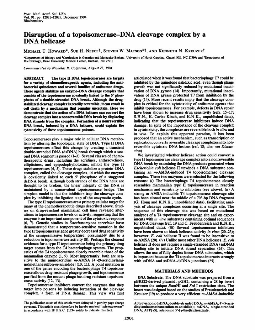
break by helicase II was dependent on the unwinding activityof the enzyme. In the first approach, topoisomerase cleavagereactions were performed in the presence of ATP or adeno-sine 5'-[thio]triphosphate (ATP[yS]), a poorly hydrolyzedanalogue of ATP. T4 topoisomerase cleavage products areformed with nearly equal efficiency in the presence of ATPor ATP[yS] (S.H.N. and K.N.K., unpublished data; Fig. 2A,compare lanes 1 and 6); however, unlike ATP, ATP[-yS]cannot support the unwinding reaction catalyzed by helicase11 (31). It should also be noted that ATP[yS] is a competitiveinhibitor ofthe ATP hydrolysis reaction catalyzed by helicaseII and should, therefore, bind at the active site of the enzyme.As indicated previously, the cleavage complexes are resistantto reversal when they are exposed to helicase II in thepresence of ATP (Fig. 2A, lanes 3 and 4). In contrast, thereactions that were exposed to helicase II in the presence ofATP[yS] were readily reversible (Fig. 2A, lanes 8 and 9),indicating that the impact of helicase II on the cleavagecomplex is dependent on ATP hydrolysis by helicase II. In asecond approach, we tested the K35M mutant helicase II,which contains a single amino acid substitution rendering theenzyme unable to hydrolyze ATP or unwind duplex DNA(32). The complexes exposed to the mutant enzyme, in thepresence ofATP, were readily reversed (Fig. 2B, lanes 6 and7). Finally, the effect of increasing concentrations of helicaseII on the reversibility of the cleavage complexes was exam-ined. This analysis revealed roughly a direct correlationbetween the total amount ofDNA unwound and the quantityof nonreversible cleavage products (data not shown). Thus,these results strongly argue that ATP hydrolysis and DNAunwinding are necessary to convert reversible cleavage prod-ucts to nonreversible DNA breaks.DNA Helicase I Can Disrupt an m-AMSA-Induced Cleavage
Complex in the Absence of Protein Denaturation. While DNAwithin the cleavage complex is thought to be cleaved, thecontinuity ofthe DNA duplex is maintained by a noncovalenttopoisomerase bridge. The DNA break is generally observedonly when the cleavage complex is treated with a proteindenaturant (e.g., SDS) that disrupts the topoisomerase bridge(2, 3). In all of the above experiments, the products weretreated with SDS and proteinase K. One could argue thatunwinding the DNA around the cleavage complex alters thecomplex without disrupting it, so that it cannot be reversed.In this model, cleaved DNA products are not released untiltreatment with SDS and proteinase K. However, if helicaseII actually disrupts the cleavage complex, then DNA cleav-age products should be detectable without protein denatur-ation. To distinguish between these alternatives, we analyzedreaction products directly on a nondenaturing gel withoutSDS or proteinase K treatment (Fig. 3, lanes 1-6). Indeed,cleavage products were observed in a reaction that wasstrictly dependent on helicase II (Fig. 3, lane 3). Thesecleavage products were further analyzed by including a mildheat treatment (65°C) prior to electrophoresis, which releaseshelicase II from the ssDNA (M.T.H. and S.W.M., unpub-lished data). Because the DNA substrate is labeled at the 5'ends, the cleavage products that are covalently linked to T4topoisomerase are not detected in this analysis. After beingreleased from helicase II, the cleavage products observed inthe absence of SDS and proteinase K (Fig. 3, lane 4)comigrated with topoisomerase cleavage products that hadbeen treated with SDS and proteinase K and then heat-denatured at 100°C (Fig. 3, lane 7). We conclude that helicaseII can displace ssDNA from the cleavage complex in areaction that is independent of protein denaturation or heattreatment. These results also provide compelling evidencefor the prevalent view that the DNA within the topoisomer-ase cleavage complex is cleaved, even without protein de-naturation, and contradict an alternative model in which
SDS. Prot K
Topc, -!Hell!65 C -- +
Origin _ _ - .." ti-s,
DenaturedSubstrate
Substrate _
in!
, ..
CD
a,
+ SDS, rProt K
M_nft
_k.* - J400
300
_
FIG. 3. Release of ssDNA fragments from topoisomerase cleav-age complexes. The first two stages of the reactions were the sameas those in Fig. 1. All reactions contained m-AMSA. After the secondstage some of the reactions were heat-treated for 20 min at 65TC (+)and others were not (-). Only the reaction products in lanes 7-11were treated with SDS and proteinase K (Prot K). Reaction productswere analyzed on a nondenaturing 5% polyacrylamide gel as de-scribed in Fig. 1, except that the gel and the running buffer lackedSDS and reaction mixtures with no SDS were chilled and loaded onthe gel in a glycerol/dye solution containing no SDS. After treatmentwith SDS and proteinase K, sample 7 was extracted with phenol andpassed through a Sepharose CL-6B spin column, and samples 6 and7 were heat-denatured (100TC, 5 min) immediately before loading onthe gel.
DNA cleavage is induced by the protein denaturation step(refs. 30 and 33; also see refs. 28 and 34).The results in Fig. 3 also provide an independent confir-
mation of the nonreversibility of DNA breaks induced byhelicase II. The 65TC treatment resulted in reversal of topo-isomerase cleavage complexes in the absence, but not in thepresence, of helicase II (Fig. 3, lanes 8-11).
DISCUSSIONTo explain the cytotoxicity of drug-induced cleavage com-plexes, it has been argued that a cellular process, such asreplication or transcription, converts the reversible cleavagecomplexes into permanent cytotoxic DNA lesions (18). Therole of replication and transcription in the cellular responseto topoisomerase inhibitors has been previously investigatedby measuring drug cytotoxicity under conditions where DNAor RNA synthesis is inhibited (35-37). Transcription inhibi-tors had little effect on the cytotoxicity of type I topoisom-erase inhibitors, but partially protected cells treated withinhibitors of type II topoisomerases (37). In addition, theDNA synthesis inhibitor aphidocolin abrogated the cytotox-icity of inhibitors targeting type I and type II topoisomerases(36, 37). Recent studies in a cell-free simian virus 40 repli-cation system provided evidence that camptothecin-inducedtype I topoisomerase cleavage complexes arrest replicationforks and lead to dsDNA breaks (38).
Helicase-driven separation of the double helix occursduringDNA replication and also during otherDNA metabolicactivities such as repair and recombination (39, 40). There-fore, we sought to determine whether helicase action onDNA containing a type II topoisomerase cleavage complex
Proc. Natl. Acad. Sci. USA 91 (1994)

Proc. Natl. Acad. Sci. USA 91 (1994) 12035
3' -- i = 3'
3 51
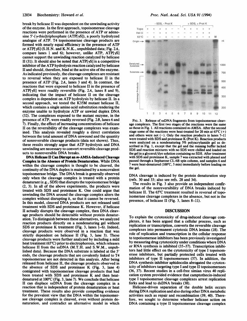
FiG. 4. Model for disruption of a topoisomerase cleavage com-plex byDNA helicase II. Steps: I, initiation ofunwinding by helicaseII on the DNA of an m-AMSA-induced topoisomerase-DNA cleav-age complex; II, encounter of helicase H with the topoisomerasecleavage complex; Ill, release ofa ssDNA cleavage product from thecomplex, with one of the possible remaining products shown.
could create a DNA lesion by displacing broken DNA fromthe complex. The generation of such a DNA lesion would bea plausible step in the pathway that generates a cytotoxicresponse to topoisomerase inhibitors.The data presented in this paper provide biochemical
evidence that DNA helicases can indeed convert drug-induced type II topoisomerase cleavage complexes to irre-versible DNA lesions. We have shown that E. coli DNAhelicase II converts a T4 topoisomerase cleavage complexinto a DNA break that cannot be reversed by either EDTAor heat treatment. Furthermore, helicase II disruption oftopoisomerase-DNA cleavage complexes releases cleavedssDNA fragments in a reaction that requires ATP hydro-lysis (Fig. 4).Although E. coli helicase II disrupts cleavage complexes in
vitro, we believe that a T4-encoded helicase is more likely toplay a physiological role in disrupting T4 topoisomerasecleavage complexes during a phage infection in vivo. None-theless, the results presented here confirm the general prem-ise that DNA helicases can disrupt topoisomerase cleavagecomplexes.DNA helicases may also play a role in various recombi-
nation and/or mutation events induced by DNA topo-isomerases. Topoisomerase inhibitors that induce the cleav-age complex have been shown to stimulate homologousrecombination, sister chromatid exchange, gross geneticrearrangements, and frameshift mutations at sites of cleavagecomplex formation (4, 18, 41). For each of these geneticevents, the helicase-mediated generation of discrete DNAbreaks from a cleavage complex would be a reasonable firststep.DNA helicases have recently been found to be sensitive to
certain topoisomerase inhibitors, and each helicase appearsto have its own unique spectrum of sensitivity (20-23). If aDNA helicase converts the topoisomerase cleavage complexinto a cytotoxic lesion in vivo, then topoisomerase inhibitors
that do not inhibit the helicase should be more cytotoxic thanthose that do.
M.T.H. and S.H.N. contributed equally to the work presented inthis paper. We thank James George for valuable discussions, JamesGeorge and Robert Brosh for purfying wild-type and K35M helicaseII, Anne Huff for purifying T4 type II topoisomerase, and GeorgeHongforpGH2 plasmid. This work was supported by grants from theBristol-Myers Squibb Company and National Cancer Institute(CA60836) to K.N.K. and by National Institutes of Health GrantGM33476 to S.W.M..
1. Wang, J. C. (1985) Annu. Rev. Biochem. 54, 665-697.2. Maxwell, A. & Gellert, M. (1986) Adv. Protein Chem. 38, 69-107.3. Hsieh, T.-S. (1990) in DNA Topology and Its Biological Effects, eds.
Cozzarelli, N. R. & Wang, J. C. (Cold Spring Harbor Lab. Press,Plainview, NY), pp. 243-255.
4. Liu, L. F. (1989) Annu. Rev. Biochem. S8, 351-375.5. Chen, A. Y. & Liu, L. F. (1994) Annu. Rev. Pharmacol. Toxicol. 34,
191-218.6. Ross, W. E., Sullivan, M. & Chow, K.-C. (1988) in Important Advances
in Oncology, eds. DeVita, V. T., Jr., Heilman, S. & Rosenberg, S.(Lippincott, Philadelphia), pp. 65-81.
7. De Isabella, P., Capranico, G. & Zunino, F. (1991) Life Sci. 48,2195-2205.
8. Nitiss, J., Liu, Y.-X. & Hsiung, Y. (1993) Cancer Res. 3, 89-93.9. Kreuzer, K. N. & Jongeneel, C. V. (1983) Methods Enzymol. 100,
144-160.10. Nelson, E. M., Tewey, K. M. & Liu, L. F. (1984) Proc. Natl. Acad. Sci.
USA 81, 1361-1365.11. Rowe, T. C., Tewey, K. M. & Liu, L. F. (1984) J. Biol. Chem. 259,
9177-9181.12. Huff, A. C., Leatherwood, J. K. & Kreuzer, K. N. (1989) Proc. Nad.
Acad. Sci. USA 86, 1307-1311.13. Huff, A. C., Ward, R. E., IV, & Kreuzer, K. N. (1990) Mol. Gen. Genet.
221, 27-32.14. Kreuzer, K. N. & Cozzarelli, N. R. (1979) J. Bacteriol. 140, 424-435.15. Nitiss, J. &Wang, J. C. (1988)Proc. Natd. Acad. Sci. USA 85, 7501-7505.16. Eng, W., Faucette, L., Johnson, R. K. & Sternganz, R. (1989) Mol.
Pharmacol. 34, 755-760.17. Caldecott, K., Banks, G. & Jeggo, P. (1990) Cancer Res. 5Y, 5778-5783.18. D'Arpa, P. & Liu, L. F. (1989) Biochim. Biophys. Acta 989, 163-177.19. Freudenreich, C. H. & Kreuzer, K. N. (1993) EMBO J. 12, 2085-2097.20. George, J. W., Ghate, S., Matson, S. W. & Besterman, J. M. (1992) J.
Biol. Chem. 267, 10683-10689.21. Bachur, N. R., Yu, F., Johnson, R., Hickey, R., Wu, Y. & Malkas, L.
(1992) Mol. Pharmacol. 41, 993-998.22. Bachur, N. R., Johnson, R., Yu, F., Hickey, R., Applegren, N. &
Malkas, L. (1993) Mol. Pharmacol. 44, 1064-1069.23. Naegeli, H., Modrich, P. & Friedberg, E. C. (1993) J. Biol. Chem. 268,
10388-10392.24. Runyon, G. T. & Lohman, T. M. (1989)J. Biol. Chem. 264,17502-17512.25. Kreuzer, K. N. (1984) J. Biol. Chem. 259, 5347-5354.26. Runyon, G. T., Wong, I. & Lohman, T. M. (1993) Biochemistry 32,
602-612.27. Sander, M. & Hsieh, T.-s. (1983) J. Biol. Chem. 258,8421-8428.28. Osheroff, N. & Zechiedrich, E. L. (1987) Biochemistry 26, 4303-4309.29. Kreuzer, K. N. & Alberts, B. M. (1984) J. Biol. Chem. 259, 5339-5346.30. Sugino, A., Peebles, C. L., Kreuzer, K. N. & Cozzareili, N. R. (1977)
Proc. Nad. Acad. Sci. USA 74, 4767-4771.31. Matson, S. W. & George, J. W. (1987) J. Biol. Chem. 262, 2066-2076.32. George, J. W., Brosh, R. & Matson, S. W. (1994) J. Mol. Biol. 235,
424-435.33. Gellert, M., Mizuuchi, K., O'Dea, M. H., Itoh, T. & Tomizawa, J.-I.
(1977) Proc. Natl. Acad. Sci. USA 74, 4772-4776.34. Anderson, A. H., Sorensen, B. S., Christiansen, K., Svejstrup, J. Q.,
Lund, K. & Westergaard, 0. (1991) J. Biol. Chem. 2X, 9203-9210.35. Holm, C., Covey, J. M., Kernigan, D. & Pommier, Y. (1989) CancerRes.
49, 6365-6368.36. Hsiang, Y. H., Lihou, M. G. & Liu, L. F. (1989) Cancer Res. 49,
5077-5082.37. D'Arpa, P., Beardmore, C. & Liu, L. F. (1990) Cancer Res. 54, 6919-
6924.38. Tsao, P.-T., Russo, A., Nyamuswa, G., Silber, R. & Liu, L. F. (1993)
Cancer Res. 53, 5908-5914.39. Matson, S. W. & Kaiser-Rogers, K. A. (1990) Annu. Rev. Biochem. 59,
289-329.40. Matson, S. W., Bean, D. W. & George, J. W. (1994) BioEssays 16,
13-22.41. Ripley, L. S., Dubins, J. S., deBoer, J. G., DeMarini, D. M., Bogerd,
A. M. & Kreuzer, K. N. (1988) J. Mol. Biol. 200, 665-680.
Biochemistry: Howard et al.